1
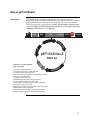
Champion™ pET Directional TOPO®
Expression Kits
Five-minute, directional TOPO® Cloning of
blunt-end PCR products into vectors for
high-level, inducible expression in E. coli
Catalog nos. K100-01, K101-01, K102-01, K15101, K200-01
Rev. Date 7 June 2010
Manual part no. 25-0400
MAN0000214
User Manual
ii
Table of Contents
TOPO® Cloning Procedure for Experienced Users .......................................................................................... v
Kit Contents and Storage .................................................................................................................................... vi
Accessory Products................................................................................................................................................x
Introduction ................................................................................................................... 1
Overview.................................................................................................................................................................1
How Directional TOPO® Cloning Works ...........................................................................................................3
T7-Regulated Expression ......................................................................................................................................4
BL21 Star™ E. coli Strains.......................................................................................................................................6
Thioredoxin ............................................................................................................................................................7
Experimental Outline ............................................................................................................................................8
Designing PCR Primers ................................................................................................ 9
Basic Requirements ...............................................................................................................................................9
Specific Requirements for Cloning into pET100/D-TOPO® and pET200/D-TOPO® ................................11
Specific Requirements for Cloning into pET101/D-TOPO® ..........................................................................12
Specific Requirements for Cloning into pET102/D-TOPO® ..........................................................................13
Specific Requirements for Cloning into pET151/D-TOPO® ..........................................................................15
Producing Blunt-End PCR Products ......................................................................... 16
TOPO® Cloning Reaction and Transformation ......................................................... 17
Setting Up the TOPO® Cloning Reaction..........................................................................................................17
Transforming One Shot® TOP10 Competent Cells..........................................................................................19
Analyzing Transformants...................................................................................................................................22
Expression and Purification....................................................................................... 24
General Guidelines for Expression....................................................................................................................24
Expressing the PCR Product ..............................................................................................................................26
Analyzing Samples..............................................................................................................................................28
Purifying the Recombinant Fusion Protein......................................................................................................31
Removing N-terminal Fusion Tags ...................................................................................................................33
Troubleshooting ...................................................................................................................................................34
Appendix...................................................................................................................... 37
Performing the Control Reactions .....................................................................................................................37
Gel Purifying PCR Products...............................................................................................................................40
Map and Features of pET100/D-TOPO® and pET200/D-TOPO® ................................................................42
iii
Map and Features of pET101/D-TOPO® ..........................................................................................................44
Map and Features of pET102/D-TOPO® ..........................................................................................................46
Map and Features of pET151/D-TOPO® ..........................................................................................................48
Map of pET100/D/lacZ and pET200/D/lacZ .................................................................................................50
Map of pET101/D/lacZ ......................................................................................................................................51
Map of pET102/D/lacZ ......................................................................................................................................52
Map of pET151/D/lacZ ......................................................................................................................................53
Recipes...................................................................................................................................................................54
Technical Service..................................................................................................................................................56
Purchaser Notification ........................................................................................................................................57
References .............................................................................................................................................................60
iv
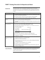
TOPO® Cloning Procedure for Experienced Users
Introduction
This quick reference sheet is provided for experienced users of the TOPO®
Cloning procedure. If you are performing the TOPO® Cloning procedure for the
first time, we recommend that you follow the detailed protocols provided in the
manual.
Step
Design PCR Primers
Amplify Your Gene of
Interest
Perform the TOPO®
Cloning Reaction
Action
•
Include the 4 base pair sequences (CACC) necessary for directional cloning
on the 5′ end of the forward primer.
•
Design the primers such that your gene of interest will be optimally
expressed and fused in frame with any epitope tags, if desired.
1.
Use a thermostable, proofreading DNA polymerase and the PCR primers
above to produce your blunt-end PCR product.
2.
Use agarose gel electrophoresis to check the integrity and yield of your PCR
product.
1.
Set up the following TOPO® Cloning reaction. For optimal results, use a
0.5:1 to 2:1 molar ratio of PCR product:TOPO® vector.
Note: If you plan to transform electrocompetent E. coli, use Dilute Salt Solution in
the TOPO® Cloning reaction.
0.5 to 4 μl
Fresh PCR product
Transform TOP10
Chemically Competent
E. coli
Control Reaction
Salt Solution
1 μl
Sterile water
add to a final volume of 5 μl
®
TOPO vector
1 μl
Total volume
6 μl
2.
Mix gently and incubate for 5 minutes at room temperature.
3.
Place on ice and proceed to transform One Shot® TOP10 chemically
competent E. coli, below.
1.
Add 3 μl of the TOPO® Cloning reaction into a vial of One Shot® TOP10
chemically competent E. coli and mix gently.
2.
Incubate on ice for 5 to 30 minutes.
3.
Heat-shock the cells for 30 seconds at 42°C without shaking. Immediately
transfer the tube to ice.
4.
Add 250 μl of room temperature S.O.C. medium.
5.
Incubate at 37°C for 1 hour with shaking.
6.
Spread 100-200 μl of bacterial culture on a prewarmed selective plate and
incubate overnight at 37°C.
We recommend using the Control PCR Template and the Control PCR Primers
included with the kit to perform the control reaction. See the protocol on
pages 37-39 for instructions.
v
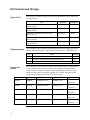
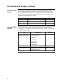
Kit Contents and Storage
Types of Kits
This manual is supplied with the following pET Directional TOPO® Expression
kits listed below.
Kit
Quantity
™
Shipping/Storage
Champion pET100 Directional TOPO
Expression Kit
20 reactions
K100-01
Champion™ pET101 Directional TOPO®
Expression Kit
20 reactions
K101-01
Champion™ pET102 Directional TOPO®
Expression Kit
20 reactions
K102-01
Champion™ pET151 Directional TOPO®
Expression Kit
20 reactions
K151-01
Champion™ pET200 Directional TOPO®
Expression Kit
20 reactions
K200-01
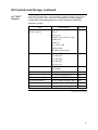
The Champion™ pET Directional TOPO® Expression Kits are shipped on dry ice.
Each kit contains three boxes. Upon receipt, store the boxes as detailed below.
Box
1
2
3
Vectors and
Primers
pET TOPO® Kit
pET100
Catalog no.
®
Item
Storage
®
pET TOPO Reagents
-20°C
®
One Shot TOP10 Chemically Competent E. coli
™
-80°C
®
BL21 Star (DE3) One Shot Chemically Competent E. coli
-80°C
Each Champion™ pET Directional TOPO® Expression Kit contains a pET-TOPO®
vector, an expression control, and primers for sequencing. The pET-TOPO®
vector, expression control, and primers differ depending on the kit. The table
below lists the vectors and primers supplied with each kit. For details on the
amount of each component provided, see the next page.
Catalog no.
K100-01
TOPO® Vector
pET100/D-TOPO®
Expression Control
pET100/D/lacZ
Primers
T7
T7 Reverse
pET101
K101-01
pET101/D-TOPO®
pET101/D/lacZ
T7
T7 Reverse
pET102
K102-01
pET102/D-TOPO®
K151-01
pET151/D-TOPO
®
pET200/D-TOPO
®
pET102/D/lacZ
TrxFus Forward
T7 Reverse
pET151
pET151/D/lacZ
T7
T7 Reverse
pET200
K200-01
pET200/D/lacZ
T7
T7 Reverse
continued on next page
vi
Kit Contents and Storage, continued
pET TOPO®
Reagents
pET TOPO® Reagents (Box 1) are listed below. Each box includes PCR reagents
and the appropriate vectors and primers. Note that the user must supply a
thermostable, proofreading polymerase and the appropriate PCR buffer.
Store Box 1 at -20°C.
Item
®
pET TOPO vector,
TOPO®-adapted
Concentration
Amount
15-20 ng/μl linearized plasmid
DNA in:
20 μl
50% glycerol
50 mM Tris-HCl, pH 7.4 (at 25°C)
1 mM EDTA
2 mM DTT
0.1% Triton X-100
100 μg/ml BSA
30 μM bromophenol blue
dNTP Mix
10 μl
12.5 mM dATP
12.5 mM dCTP
12.5 mM dGTP
12.5 mM dTTP
in water, pH 8
Salt Solution
50 μl
1.2 M NaCl
0.06 M MgCl2
Sterile Water
--
1 ml
Forward Sequencing Primer
0.1 μg/μl in TE Buffer, pH 8
20 μl
Reverse Sequencing Primer
0.1 μg/μl in TE Buffer, pH 8
20 μl
Control PCR Primers
0.1 μg/μl each in TE Buffer, pH 8
10 μl
Control PCR Template
0.1 μg/μl in TE Buffer, pH 8
10 μl
Expression Control Plasmid
0.01 μg/μl in TE buffer, pH 8
10 μl
continued on next page
vii
Kit Contents and Storage, continued
Sequences of the
Primers
Each Champion™ pET Directional TOPO® Expression Kit provides a forward and
reverse sequencing primer to facilitate sequence analysis of your expression
constructs (see the table on page vi for the specific primers included with each
kit). The sequences of the forward and reverse primers are listed below. Two
micrograms of each primer are supplied.
Primer
One Shot® TOP10
Reagents
Sequence
pMoles Supplied
T7
5´-TAATACGACTCACTATAGGG-3´
327
TrxFus Forward
5´-TTCCTCGACGCTAACCTG-3´
371
T7 Reverse
5´-TAGTTATTGCTCAGCGGTGG-3´
325
The table below lists the items included in the One Shot® TOP10 Chemically
Competent E. coli kit (Box 2). Transformation efficiency is at least 1 x 10⁹ cfu/μg
DNA. Store Box 2 at -80°C.
Item
Composition
S.O.C. Medium
2% Tryptone
(may be stored at room
temperature or +4°C)
0.5% Yeast Extract
Amount
6 ml
10 mM NaCl
2.5 mM KCl
10 mM MgCl2
10 mM MgSO4
20 mM glucose
TOP10 cells
--
21 x 50 μl
pUC19 Control DNA
10 pg/μl in 5 mM Tris-HCl,
0.5 mM EDTA, pH 8
50 μl
continued on next page
viii
Kit Contents and Storage, continued
BL21 Star™(DE3)
One Shot®
Reagents
The table below describes the items included in the BL21 Star™(DE3) One Shot®
Chemically Competent E. coli kit (Box 3). Transformation efficiency is at least
1 x 10⁸ cfu/μg DNA. Store Box 3 at -80°C.
Item
Composition
S.O.C. Medium
2% Tryptone
(may be stored at room
temperature or +4°C)
0.5% Yeast Extract
Amount
6 ml
10 mM NaCl
2.5 mM KCl
10 mM MgCl2
10 mM MgSO4
20 mM glucose
BL21 Star (DE3)
--
21 x 50 μl
pUC19 Control DNA
10 pg/μl in 5 mM Tris-HCl, 0.5
mM EDTA, pH 8
50 μl
™
Genotype of
TOP10
Use this E. coli strain for general cloning of blunt-end PCR products into the pET
TOPO® vectors.
Genotype: F- mcrA Δ(mrr-hsdRMS-mcrBC) Φ80lacZΔM15 ΔlacΧ74 recA1 araD139
Δ(ara-leu)7697 galU galK rpsL (StrR) endA1 nupG
Genotype of BL21
Star™(DE3)
Use this E. coli strain for expression only. Do not use these cells to propagate
or maintain your construct.
Genotype: F- ompT hsdSB (rB-mB-) gal dcm rne131 (DE3)
The DE3 designation means this strain contains the lambda DE3 lysogen which
carries the gene for T7 RNA polymerase under the control of the lacUV5
promoter. IPTG is required to induce expression of the T7 RNA polymerase.
The strain is an E. coli B/r strain and does not contain the lon protease. It also has
a mutation in the outer membrane protease, OmpT. The lack of these two key
proteases reduces degradation of heterologous proteins expressed in the strain.
The strain carries a mutated rne gene (rne131) which encodes a truncated RNase
E enzyme that lacks the ability to degrade mRNA, resulting in an increase in
mRNA stability (see page 6).
ix
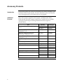
Accessory Products
Introduction
The products listed in this section are intended for use with the Champion™ pET
Directional TOPO® Expression Kits. For more information, refer to our Web site
(www.invitrogen.com) or call Technical Service (see page 56).
Additional
Products
Many of the reagents supplied in the Champion™ pET Directional TOPO®
Expression Kits and other reagents suitable for use with the kits are available
separately from Invitrogen. Ordering information for these reagents is
provided below.
Item
Quantity
Catalog no.
10 x 50 μl
C4040-10
20 x 50 μl
C4040-03
BL21 Star™(DE3) One Shot® Chemically
Competent E. coli
20 x 50 μl
C6010-03
BL21 Star™(DE3)pLysS One Shot®
Chemically Competent E. coli
20 x 50 μl
C6020-03
BL21-AI™ One Shot® Chemically
Competent E. coli
20 x 50 μl
C6070-03
PureLink™ HQ Mini Plasmid Purification
Kit
100 reactions
K2100-01
PureLink™ Quick Gel Extraction Kit
50 reactions
K2100-12
Ampicillin
200 mg
11593-027
Kanamycin Sulfate
5g
11815-024
25 g
11815-032
Carbenicillin
5g
10177-012
Isopropylthio-β-galactoside (IPTG)
1g
15529-019
250 units
E180-01
1000 units
E180-02
1000 units
12575-015
®
One Shot TOP10 Chemically Competent
E. coli
™
EKMax
AcTEV Protease
continued on next page
x
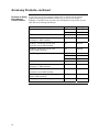
Accessory Products, continued
Products to Detect Expression of your recombinant fusion protein can be detected using an
antibody to the appropriate epitope. The table below describes the products
Recombinant
available from Invitrogen for detection of fusion proteins expressed using the
Proteins
appropriate pET TOPO® vector (see pages 11-15 for details about the N- and/or
C-terminal tags present on each pET TOPO® vector).
The amount of antibody supplied is sufficient for 25 western blots.
Product
™
Anti-Xpress Antibody
™
Anti-Xpress -HRP Antibody
Mechanism of Detection
™
Detects 8 amino acid Xpress
epitope:
Catalog no.
R910-25
R911-25
DLYDDDDK
Anti-HisG Antibody
Anti-HisG-HRP Antibody
Anti-HisG-AP Antibody
Anti-V5 Antibody
Anti-V5-HRP Antibody
Anti-V5-AP Antibody
Detects the N-terminal
polyhistidine (6xHis) tag
followed by glycine:
R940-25
R941-25
R942-25
HHHHHHG
Detects 14 amino acid epitope
derived from the P and V
proteins of the paramyxovirus,
SV5 (Southern et al., 1991):
R960-25
R961-25
R962-25
GKPIPNPLLGLDST
Anti-His (C-term) Antibody
Anti-His(C-term)-HRP
Antibody
Anti-His(C-term)-AP
Antibody
Anti-Thio™ Antibody
Detects the C-terminal
polyhistidine (6xHis) tag
(requires the free carboxyl group
for detection (Lindner et al.,
1997):
R930-25
R931-25
R932-25
HHHHHH-COOH
Detects His-Patch thioredoxin
fusion proteins
R920-25
Note: The exact epitope detected by
this antibody has not been mapped
continued on next page
xi
Accessory Products, continued
Products to Purify
Recombinant
Fusion Proteins
If you clone your gene of interest in frame with a C-terminal or N-terminal
peptide containing a polyhistidine (6xHis) tag, you may use Invitrogen’s
ProBond™ or Ni-NTA resins to purify your recombinant fusion protein. See the
table below for ordering information.
Product
ProBond™ Nickel-Chelating Resin
Quantity
Catalog no.
50 ml
R801-01
150 ml
R801-15
6 purifications
K850-01
1 kit
K851-01
1 kit
K853-01
ProBond™ Purification System with
Anti-V5-HRP Antibody
1 kit
K854-01
Ni-NTA Agarose
10 ml
R901-01
25 ml
R901-15
100 ml
R901-10
Ni-NTA Purification System
6 purifications
K950-01
Ni-NTA Purification System with
Anti-Xpress™-HRP Antibody
1 kit
K951-01
Ni-NTA Purification System with
1 kit
K953-01
Ni-NTA Purification System with
Anti-V5-HRP Antibody
1 kit
K954-01
Polypropylene Columns (empty)
50
R640-50
™
ProBond Purification System
™
ProBond Purification System with
™
Anti-Xpress -HRP Antibody
ProBond™ Purification System with
Anti-His(C-term)-HRP Antibody
Anti-His(C-term)-HRP Antibody
xii
Introduction
Overview
The Champion™ pET Directional TOPO® Expression Kits utilize a highly efficient,
5-minute cloning strategy ("TOPO® Cloning") to directionally clone a blunt-end
PCR product into a vector for high-level, T7-regulated expression in E. coli. Bluntend PCR products clone directionally at greater than 90% efficiency, with no
ligase, post-PCR procedures, or restriction enzymes required.
Introduction
Depending on the vector chosen, the pET TOPO® vectors are available with:
•
N-terminal or C-terminal peptide tags for production of recombinant
fusion proteins that may be easily detected or purified
•
Protease recognition site for removal of the N-terminal peptide tag from
your recombinant fusion protein
•
Antibiotic resistance marker for selection of transformants
See the table below for a list of the available pET TOPO® vectors and the fusion
tag, cleavage site, and selection marker for each vector.
pET TOPO® vector
Fusion Peptide
pET100/D-TOPO®
N-terminal
Fusion Tag
Xpress™, 6xHis
Cleavage Site
EK
pET200/D-TOPO®
Selection Marker
Ampicillin
Kanamycin
pET101/D-TOPO
®
C-terminal
V5, 6xHis
--
Ampicillin
pET102/D-TOPO
®
N-terminal
His-Patch thioredoxin
EK
Ampicillin
C-terminal
V5, 6xHis
N-terminal
V5, 6xHis
TEV protease
Ampicillin
pET151/D-TOPO
®
EK = enterokinase; TEV = tobacco etch virus
The Champion™
pET Expression
System
The Champion™ pET Expression System is based on expression vectors
originally developed by Studier and colleagues, and takes advantage of the high
activity and specificity of the bacteriophage T7 RNA polymerase to allow
regulated expression of heterologous genes in E. coli from the T7 promoter
(Rosenberg et al., 1987; Studier & Moffatt, 1986; Studier et al., 1990). For more
information about the Champion™ pET Expression System, see page 4.
continued on next page
1
Overview, continued
Features of the
Champion™ pET
Directional TOPO®
Vectors
2
The pET TOPO® vectors are designed to facilitate rapid, directional TOPO®
Cloning of blunt-end PCR products for regulated expression in E. coli. Features of
the vectors include:
•
T7lac promoter for high-level, IPTG-inducible expression of the gene of
interest in E. coli (Dubendorff & Studier, 1991; Studier et al., 1990)
•
Directional TOPO® Cloning site for rapid and efficient directional cloning of
blunt-end PCR products (see page 3 for more information)
•
N- or C-terminal fusion tags for detection and purification of recombinant
fusion proteins (choice of tag depends on the particular vector; see the
previous page)
•
Protease recognition site for cleavage of the fusion tag from the recombinant
protein of interest (present on N-terminal fusion vectors)
•
N-terminal His-Patch thioredoxin for increased translation efficiency and
solubility of heterologous proteins (pET102/D-TOPO® only)
•
lacI gene encoding the lac repressor to reduce basal transcription from the
T7lac promoter in the pET TOPO® vector and from the lacUV5 promoter in the
E. coli host chromosome (see page 4 for more information)
•
Antibiotic resistance marker for selection in E. coli
•
pBR322 origin for low-copy replication and maintenance in E. coli
How Directional TOPO® Cloning Works
How
Topoisomerase I
Works
Topoisomerase I from Vaccinia virus binds to duplex DNA at specific sites and
cleaves the phosphodiester backbone after 5′-CCCTT in one strand (Shuman,
1991). The energy from the broken phosphodiester backbone is conserved by
formation of a covalent bond between the 3′ phosphate of the cleaved strand and
a tyrosyl residue (Tyr-274) of topoisomerase I. The phospho-tyrosyl bond
between the DNA and enzyme can subsequently be attacked by the 5′ hydroxyl
of the original cleaved strand, reversing the reaction and releasing
topoisomerase (Shuman, 1994). TOPO® Cloning exploits this reaction to
efficiently clone PCR products.
Directional TOPO®
Cloning
Directional joining of double-strand DNA using TOPO®-charged oligonucleotides
occurs by adding a 3′ single-stranded end (overhang) to the incoming DNA
(Cheng & Shuman, 2000). This single-stranded overhang is identical to the 5′ end
of the TOPO®-charged DNA fragment. At Invitrogen, this idea has been modified
by adding a 4 nucleotide overhang sequence to the TOPO®-charged DNA and
adapting it to a ‘whole vector’ format.
In this system, PCR products are directionally cloned by adding four bases to the
forward primer (CACC). The overhang in the cloning vector (GTGG) invades the
5′ end of the PCR product, anneals to the added bases, and stabilizes the PCR
product in the correct orientation. Inserts can be cloned in the correct orientation
with efficiencies equal to or greater than 90%.
Topoisomerase
Tyr-274
P
O
----CCCTT
----GGGAAGTGG
Overhang
CACC ATG NNN --- --- --- NNN
GTGG TAC NNN --- --- --- NNN
PCR product
Overhang invades double-stranded
DNA, displacing the bottom strand.
Tyr-274
AAG GG---TTC CC----
O
P
Topoisomerase
GT
GG
----CCCTTCACC ATG NNN --- --- --- NNN AAG GG- ------GGGAAGTGG TAC NNN --- --- --- NNN TTC CC- ---
3
T7-Regulated Expression
The Basis of T7Regulated
Expression
The Champion™ pET Expression System uses elements from bacteriophage T7 to
control expression of heterologous genes in E. coli. In the pET TOPO® vectors,
expression of the gene of interest is controlled by a strong bacteriophage T7
promoter that has been modified to contain a lac operator sequence (see below). In
bacteriophage T7, the T7 promoter drives expression of gene 10 (φ10). T7 RNA
polymerase specifically recognizes this promoter. To express the gene of interest, it
is necessary to deliver T7 RNA polymerase to the cells by inducing expression of
the polymerase or infecting the cell with phage expressing the polymerase. In the
Champion™ pET Directional TOPO® Expression System, T7 RNA polymerase is
supplied by the BL21 Star™(DE3) host E. coli strain in a regulated manner (see
below). When sufficient T7 RNA polymerase is produced, it binds to the T7
promoter and transcribes the gene of interest.
Regulating
Expression of T7
RNA Polymerase
The BL21 Star™(DE3) E. coli strain is specifically included in each Champion™ pET
Directional TOPO® Expression kit for expression of T7-regulated genes. This
strain carries the DE3 bacteriophage lambda lysogen. This λDE3 lysogen contains
a lac construct consisting of the following elements:
•
the lacI gene encoding the lac repressor
•
the T7 RNA polymerase gene under control of the lacUV5 promoter
•
a small portion of the lacZ gene.
This lac construct is inserted into the int gene such that it inactivates the int gene.
Disruption of the int gene prevents excision of the phage (i.e. lysis) in the absence
of helper phage. The lac repressor (encoded by lacI) represses expression of T7
RNA polymerase. Addition of the gratuitous inducer, isopropyl β-D-thiogalactoside (IPTG) allows expression of T7 RNA polymerase from the lacUV5 promoter.
The BL21 Star™(DE3) strain also contains other features which facilitate high-level
expression of heterologous genes. For more information, see page 6.
T7lac Promoter
Studies have shown that there is always some basal expression of T7 RNA
polymerase from the lacUV5 promoter in λDE3 lysogens even in the absence of
inducer (Studier & Moffatt, 1986). In general, this is not a problem, but if the
gene of interest is toxic to the E. coli host, basal expression of the gene of interest
may lead to plasmid instability and/or cell death.
To address this problem, the pET TOPO® vectors have been designed to contain
a T7lac promoter to drive expression of the gene of interest. The T7lac promoter
consists of a lac operator sequence placed downstream of the T7 promoter. The
lac operator serves as a binding site for the lac repressor (encoded by the lacI
gene) and functions to further repress T7 RNA polymerase-induced basal
transcription of the gene of interest in BL21 Star™(DE3) cells.
continued on next page
4
T7-Regulated Expression, continued
Expressing Toxic
Genes
In some cases, the gene of interest is so toxic to BL21 Star™(DE3) cells that other
E. coli host strains may be required for expression. For a discussion of other
alternative strains that may be used, see page 6.
Using TOP10 Cells One Shot® TOP10 competent E. coli, which do not contain T7 RNA polymerase,
are included in each Champion™ pET Directional TOPO® Expression kit to
provide a host for stable propagation and maintenance of recombinant plasmids.
As mentioned on the previous page, the presence of T7 RNA polymerase, even at
basal levels, can lead to expression of the desired gene even in the absence of
inducer. If the gene of interest is toxic to the E. coli host, plasmid instability
and/or cell death may result. We recommend that you transform your TOPO®
Cloning reaction into TOP10 cells for characterization of the construct,
propagation, and maintenance. When you are ready to perform an expression
experiment, transform your construct into BL21 Star™(DE3) E. coli.
5
BL21 Star™ E. coli Strains
BL21 Star™
Strains
The BL21 Star™(DE3) E. coli strain is included in each Champion™ pET
Directional TOPO® Expression Kit for use as a host for expression. Other BL21
Star™ strains are also available from Invitrogen (see below). In addition to the
λDE3 lysogen which allows high-level expression of T7-regulated genes (see
page 3), the BL21 Star™ strains also contain the rne131 mutation. This particular
mutation further enhances the expression capabilities of BL21 Star™.
rne131 Mutation
The rne gene encodes the RNase E enzyme, an essential, 1061 amino acid E. coli
endonuclease which is involved in rRNA maturation and mRNA degradation as
a component of a protein complex known as a “degradosome” (GrunbergManago, 1999; Lopez et al., 1999). Various studies have shown that the
N-terminal portion of RNase E (approximately 584 amino acids) is required for
rRNA processing and cell growth while the C-terminal portion of the enzyme
(approximately 477 amino acids) is required for mRNA degradation (Kido et al.,
1996; Lopez et al., 1999). The rne131 mutation (present in the BL21 Star™ strains)
encodes a truncated RNase E which lacks the C-terminal 477 amino acids of the
enzyme required for mRNA degradation (Kido et al., 1996; Lopez et al., 1999).
Thus, mRNAs expressed in the RNase E-defective BL21 Star™ strains exhibit
increased stability when compared to other BL21 strains. When heterologous
genes are expressed in the BL21 Star™ strains from T7-based expression vectors,
the yields of recombinant proteins generally increase.
BL21
Star™(DE3)pLysS
Strain
If you discover that your gene is toxic to BL21 Star™(DE3) cells, you may want to
perform your expression experiments in the BL21 Star™(DE3)pLysS strain (see
page x for ordering information). The BL21 Star™(DE3)pLysS strain contains the
pLysS plasmid, which produces T7 lysozyme. T7 lysozyme binds to T7 RNA
polymerase and inhibits transcription. This activity results in reduced basal
levels of T7 RNA polymerase, leading to reduced basal expression of T7-driven
heterologous genes. For more information about BL21 Star™(DE3)pLysS, refer to
our Web site (www.invitrogen.com) or call Technical Service (see page 56).
Note that while BL21 Star™(DE3)pLysS reduces basal expression from the gene
of interest when compared to BL21 Star™(DE3), it also generally reduces the
overall induced level of expression of recombinant protein.
6
Thioredoxin
Introduction
The pET102/D-TOPO® vector allows you to clone your gene of interest as a
fusion to a mutated thioredoxin protein (His-Patch thioredoxin). For more
information about thioredoxin and His-Patch thioredoxin, see below.
Thioredoxin
The 11.7 kDa thioredoxin protein is found in yeast, plants, and mammals, as
well as in bacteria. It was originally isolated from E. coli as a hydrogen donor for
ribonuclease reductase (see Holmgren, 1985 for a review). The gene has been
completely sequenced (Wallace & Kushner, 1984). The protein has been
crystallized and its three-dimensional structure determined (Katti et al., 1990).
When overexpressed in E. coli, thioredoxin is able to accumulate to
approximately 40% of the total cellular protein and still remain soluble. When
used as a fusion partner, thioredoxin can increase translation efficiency, and in
some cases, solubility, of eukaryotic proteins expressed in E. coli.
Examples of eukaryotic proteins that have been produced as soluble C-terminal
fusions to the thioredoxin protein in E. coli (LaVallie et al., 1993) include:
His-Patch
Thioredoxin
•
Murine interleukins 2, 4, and 5 and human interleukin-3
•
Human macrophage colony stimulating factor
•
Murine steel factor
•
Murine leukemia inhibitory factor
•
Human bone morphogenetic protein-2
The thioredoxin protein in pET102/D-TOPO® has been mutated to contain a
metal binding domain, and is termed “His-Patch thioredoxin”. To create a metal
binding domain in the thioredoxin protein, the glutamate residues at position 32
and the glutamine residue at position 64 were mutated to histidine residues.
When His-Patch thioredoxin folds, the histidines at positions 32 and 64 interact
with a native histidine at position 8 to form a “patch”. This histidine patch has
been shown to have high affinity for divalent cations (Lu et al., 1996). His-Patch
thioredoxin (HP-thioredoxin) proteins can therefore be purified on metal
chelating resins (e.g. ProBond™ or Ni-NTA).
7
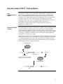
Experimental Outline
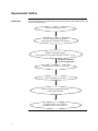
Flow Chart
The flow chart below describes the general steps required to clone and express your
blunt-end PCR product.
Determine strategy for PCR
Produce blunt-end PCR product
using properly designed PCR primers
TOPO® Cloning Reaction:
Mix together PCR product and pET-TOPO® vector
Incubate 5 minutes
at room temperature
Transform into TOP10 E. coli cells
Select and analyze colonies
Choose a positive transformant and
isolate plasmid DNA
Transform BL21 StarTM(DE3)
and induce expression with IPTG
8
Designing PCR Primers
Basic Requirements
Designing Your
PCR Primers
General
Requirements for
the Forward
Primer
The design of the PCR primers to amplify your gene of interest is critical for
expression. Depending on the pET TOPO® vector you are using, consider the
following when designing your PCR primers:
•
Sequences required to facilitate directional cloning (see below)
•
Whether or not you wish to clone your PCR product in frame with the
appropriate N-terminal and/or C-terminal peptide tag (see pages 11-15 for
information about each pET TOPO® vector)
To enable directional cloning, the forward PCR primer must contain the
sequence, CACC, at the 5′ end of the primer. The 4 nucleotides, CACC, base pair
with the overhang sequence, GTGG, in each pET TOPO® vector.
For example, below is the DNA sequence of the N-terminus of a theoretical
protein and the proposed sequence for your forward PCR primer:
DNA sequence:
5′-ATG GGA TCT GAT AAA
Proposed Forward PCR primer: 5′-C ACC ATG GGA TCT GAT AAA
See pages 11-15 for other factors to consider when designing the forward primer.
General
Requirements for
the Reverse
Primer
In general, design the reverse PCR primer to allow you to clone your PCR product
in frame with any C-terminal tag, if desired. To ensure that your PCR product
clones directionally with high efficiency, the reverse PCR primer MUST NOT be
complementary to the overhang sequence GTGG at the 5′ end. A one base pair
mismatch can reduce the directional cloning efficiency from 90% to 75%, and
may increase the chances of your ORF cloning in the opposite orientation. We
have not observed evidence of PCR products cloning in the opposite orientation
from a two base pair mismatch, but this has not been tested thoroughly.
Example: Below is the sequence of the C-terminus of a theoretical protein. You
want to clone in frame with the C-terminal tag. The stop codon is underlined.
DNA sequence: AAG TCG GAG CAC TCG ACG ACG GTG TAG-3′
One solution is to design the reverse PCR primer to start with the codon just upstream of the stop codon, but the last two codons contain GTGG (underlined
below), which is identical to the overhang sequence. As a result, the reverse primer
will be complementary to the overhang sequence, increasing the probability that
the PCR product will clone in the opposite orientation. You want to avoid this
situation.
DNA sequence:
AAG TCG GAG CAC TCG ACG ACG GTG TAG-3′
Proposed Reverse PCR primer sequence:
TG AGC TGC TGC CAC-5′
Another solution is to design the reverse primer so that it hybridizes just downstream of the stop codon, but still includes the C-terminus of the ORF. Note that
you will need to replace the stop codon with a codon for an innocuous amino acid
such as glycine or alanine.
continued on next page
9
Basic Requirements, continued
•
Remember that the pET TOPO® vectors accept blunt-end PCR products.
Refer to pages 11-15 for a discussion of specific factors to consider when
designing PCR primers for cloning into each pET TOPO® vector.
•
Do not add 5´ phosphates to your primers for PCR. This will prevent
ligation into the pET TOPO® vectors.
•
We recommend gel-purifying your oligonucleotides, especially if they are
long (> 30 nucleotides).
Important
Example of Primer
Design
The example below uses a theoretical protein and is for illustration purposes
only. In this case, PCR primers are designed to allow cloning of the PCR product
into pET101/D-TOPO®. In this example, the N-terminus of the protein is
encoded by:
5′-ATGGCCCCCCCGACCGATGTCAGCCTGGGGGACGAA…
1.
Design the forward PCR primer to be:
5′-CACCATGGCCCCCCCGACCGAT-3′
2.
For the reverse primer, analyze the C-terminus of the protein. The stop
codon is underlined (see the top strand below).
…GCG GTT AAG TCG GAG CAC TCG ACG ACT GCA TAG-3′
…CGC CAA TTC AGC CTC GTG AGC TGC TGA CGT ATC-5′
3.
To fuse the ORF in frame with the V5 epitope and 6xHis tag, remove the stop
codon by starting with nucleotides homologous to the last codon (TGC) and
continue upstream (underlined sequence in the bottom strand above). The
reverse primer will be:
5′-TGC AGT CGT CGA GTG CTC CGA CTT-3′
4.
This will amplify the C-terminus without the stop codon and allow you to
clone the ORF in frame with the V5 epitope and 6xHis tag.
If you don’t want the V5 epitope and 6xHis tag, simply begin with the stop
codon:
5′-CTA TGC AGT CGT CGA GTG CTC CGA CTT-3′
10
Specific Requirements for Cloning into pET100/D-TOPO® and
pET200/D-TOPO®
pET100/D-TOPO® and pET200/D-TOPO® allow expression of recombinant
protein with an N-terminal tag containing the Xpress™ epitope and a 6xHis tag.
The N-terminal tag also includes an enterokinase (EK) recognition site to enable
removal of the tag after protein purification using enterokinase (e.g. EKMax™).
Introduction
Additional Cloning In addition to the guidelines on page 9, consider the following when designing
PCR primers to clone your DNA into pET100/D-TOPO® or pET200/D-TOPO®.
Considerations
Be sure to include a stop codon in the reverse primer or design the reverse
primer to hybridize downstream of the native stop codon.
If you wish to...
Then...
™
include the Xpress
epitope and 6xHis tag
design the forward PCR primer to place the gene of interest in frame with the
N-terminal tag. Note that:
express your protein with
a native N-terminus, i.e.
without the N-terminal
peptide
•
a ribosome binding site (RBS) is included upstream of the initiation ATG
in the N-terminal tag to ensure optimal spacing for proper translation
•
at least five nonnative amino acids will be present between the EK
cleavage site and the start of your gene
design the forward PCR primer to include the following:
a stop codon to terminate the N-terminal peptide
•
a second ribosome binding site (AGGAGG) 9-10 base pairs 5′ of the
initiation ATG codon of your protein
•
Note: The first three base pairs of the PCR product following the 5′ CACC overhang will constitute a complete codon.
TOPO® Cloning
Site of pET100/DTOPO® and
pET200/D-TOPO®
121
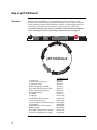
Use the diagram below to help you design suitable PCR primers to clone your PCR
product into pET100/D-TOPO® or pET200/D-TOPO®. Restriction sites are labeled
to indicate the actual cleavage site. The sequence of each vector is available for
downloading from our Web site or from Technical Service (see page 56). For
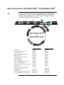
more information about pET100/D-TOPO® or pET200/D-TOPO®, see pages 42-43.
ATAGGCGCCA GCAACCGCAC CTGTGGCGCC GGTGATGCCG GCCACGATGC GTCCGGCGTA GAGGATCGAG ATCTCGATCC
T7 promoter/priming site
lac operator
T7 promoter
201
CGCGAAATTA ATACGACTCA CTATAGGGGA ATTGTGAGCG GATAACAATT CCCCTCTAGA AATAATTTTG TTTAACTTTA
281
AGAAGGAGAT ATACAT ATG CGG GGT TCT CAT CAT CAT CAT CAT CAT GGT ATG GCT AGC ATG ACT GGT GGA
Met Arg Gly Ser His His His His His His Gly Met Ala Ser Met Thr Gly Gly
RBS
Polyhistidine region
Nde I
Nhe I
XpressTM epitope
CAG CAA ATG GGT CGG GAT CTG TAC GAC GAT GAC GAT AAG GAT CAT CCC TT C ACC
GGG AAG TGG
Gln Gln Met Gly Arg Asp Leu Tyr Asp Asp Asp Asp Lys Asp His Pro Phe Thr
... ... AAGGGC
... ...
EK recognition site
411
G
TG
G
351
EK cleavage site
T7 reverse priming site
GAGCTCAACG ATCCGGCTGC TAACAAAGCC CGAAAGGAAG CTGAGTTGGC TGCTGCCACC GCTGAGCAAT AACTAGCATA
11
Specific Requirements for Cloning into pET101/D-TOPO®
Additional Cloning pET101/D-TOPO® allows expression of recombinant protein with a native
N-terminus and a C-terminal fusion tag. In addition to the guidelines on page 9,
Considerations
consider the following when designing PCR primers to clone your DNA into
pET101/D-TOPO®.
For maximal expression of native protein, the forward PCR primer should be
designed to place the initial ATG codon of the desired protein approximately 9 to
10 base pairs from the ribosome binding site (Gold, 1988; Miller, 1992). This will
ensure the optimal spacing for proper translation.
If you wish to...
Then...
express your protein with a native design the forward PCR primer such that
N-terminus using the vector
the initial ATG codon of your protein
encoded ribosome binding site
directly follows the 5´ CACC overhang.
include the C-terminal V5 epitope
and 6xHis tag
design the reverse PCR primer to remove
the native stop codon in the gene of
interest and preserve the reading frame
through the C-terminal tag.
not include the C-terminal V5
epitope and 6xHis tag
include the native sequence containing the
stop codon in the reverse primer or make
sure the stop codon is upstream from the
reverse PCR primer binding site.
Note: The first three base pairs of the PCR product following the 5′ CACC overhang will
constitute a complete codon.
TOPO® Cloning Site Use the diagram below to help you design suitable PCR primers to clone your
of pET101/D-TOPO® PCR product into pET101/D-TOPO®. Restriction sites are labeled to indicate the
actual cleavage site. The sequence of the vector is available for downloading
from our Web site or from Technical Service (see page 56). For more
information about pET101/D-TOPO®, see pages 44-45.
T7 promoter/priming site
T7 promoter
151
GGTGATGCCG GCCACGATGC GTCCGGCGTA GAGGATCGAG ATCTCGATCC CGCGAAATTA ATACGACTCA CTATAGGGGA
231
ATTGTGAGCG GATAACAATT CCCCTCTAGA AATAATTTTG TTTAACTTTA AGAAGGAATT CAGGAGCCCT T C ACC ATG ...
GGGA AG TGG TAC ...
306
AAG GGC GAG CTC AAT TCG AAG CTT GAA GGT AAG CCT ATC CCT AAC CCT CTC CTC GGT CTC GAT TCT
Lys Gly Glu Leu Asn Ser Lys Leu Glu Gly Lys Pro Ile Pro Asn Pro Leu Leu Gly Leu Asp Ser
372
ACG CGT ACC GGT CAT CAT CAC CAT CAC CAT TGA GTTTGA TCCGGCTGCT AACAAAGCCC GAAAGGAAGC
Thr Arg Thr Gly His His His His His His ***
441
TGAGTTGGCT GCTGCCACCG CTGAGCAATA ACTAGCATAA CCCCTTGGGG CCTCTAAACG
lac operator
RBS
G
TG
G
RBS
Sac I
Age I
BstB I
V5 epitope
Polyhistidine region
T7 reverse priming site
12
Specific Requirements for Cloning into pET102/D-TOPO®
Specific Features
for Expression in
pET102/D-TOPO®
pET102/D-TOPO® is designed with some specific features to facilitate expression.
They are:
•
The initiation ATG is correctly spaced from the optimized ribosome binding
site (RBS) to ensure optimal translation
•
HP-thioredoxin acts to increase translation efficiency and in some cases,
solubility
•
HP-thioredoxin can be removed after protein purification using enterokinase
(e.g. EKMax™, Catalog no. E180-01).
Additional Cloning In addition to the guidelines on page 9, you should consider the following when
designing PCR primers to clone your DNA into pET102/D-TOPO®. Use the
Considerations
diagram on the next page to help you design your PCR primers.
If you wish to...
Then...
clone in frame with thioredoxin
design the forward PCR primer to ensure
that your protein is in frame with the
N-terminal leader peptide.
include the C-terminal V5
epitope and 6xHis tag
design the reverse PCR primer to remove
the native stop codon in the gene of interest
and preserve the reading frame through the
C-terminal tag.
not include the C-terminal V5
epitope and 6xHis tag
include the native sequence containing the
stop codon in the reverse primer or make
sure the stop codon is upstream from the
reverse PCR primer binding site.
Note: The first three base pairs of the PCR product following the 5′ CACC overhang will
constitute a complete codon.
continued on next page
13
Specific Requirements for Cloning into pET102/D-TOPO®,
continued
TOPO® Cloning Site Use the diagram below to help you design appropriate PCR primers to clone
of pET102/D-TOPO® your PCR product into pET102/D-TOPO®. Restriction sites are labeled to
indicate the actual cleavage site. The complete sequence of the vector is
available for downloading from our Web site or from Technical Service (see
page 56). For more information about pET102/D-TOPO®, refer to the
Appendix, pages 46-47.
T7 promoter/priming site
T7 promoter
151
GGTGATGCCG GCCACGATGC GTCCGGCGTA GAGGATCGAG ATCTCGATCC CGCGAAATTA ATACGACTCA CTATAGGGGA
231
ATTGTGAGCG GATAACAATT CCCCTCTAGA AATAATTTTG TTTAACTTTA AGAAGGAGAT ATACATA ATG GGA TCT GAT
Met Gly Ser Asp
310
AAA ATT ATT CAT CTG ACT GAT GAT TCT TTT GAT ACT GAT GTA CTT AAG GCA GAT GGT GCA ATC CTG
Lys Ile Ile His Leu Thr Asp Asp Ser Phe Asp Thr Asp Val Leu Lys Ala Asp Gly Ala Ile Leu
376
GTT GAT TTC TGG GCA CAC TGG TGC GGT CCG TGC AAA ATG ATC GCT CCG ATT CTG GAT GAA ATC GCT
Val Asp Phe Trp Ala His Trp Cys Gly Pro Cys Lys Met Ile Ala Pro Ile Leu Asp Glu Ile Ala
442
GAC GAA TAT CAG GGC AAA CTG ACC GTT GCA AAA CTG AAC ATC GAT CAC AAC CCG GGC ACT GCG CCG
Asp Glu Tyr Gln Gly Lys Leu Thr Val Ala Lys Leu Asn Ile Asp His Asn Pro Gly Thr Ala Pro
508
AAA TAT GGC ATC CGT GGT ATC CCG ACT CTG CTG CTG TTC AAA AAC GGT GAA GTG GCG GCA ACC AAA
Lys Tyr Gly Ile Arg Gly Ile Pro Thr Leu Leu Leu Phe Lys Asn Gly Glu Val Ala Ala Thr Lys
lac operator
RBS
His-patch (HP) thioredoxin
TrxFus forward priming site
574
GTG GGT GCA CTG TCT AAA GGT CAG TTG AAA GAG TTC CTC GAC GCT AAC CTG GCC GGC TCT GGA TCC
Val Gly Ala Leu Ser Lys Gly Gln Leu Lys Glu Phe Leu Asp Ala Asn Leu Ala Gly Ser Gly Ser
Enterokinase (EK) recognition site
EK cleavage site
Sac I
Hind III
GGT GAT GAC GAT GAC AAG CTG GGA ATT GAT CCC TTC ACC
GGG AAG TGG
Gly Asp Asp Asp Asp Lys Leu Gly Ile Asp Pro Phe Thr
700
GGT AAG CCT ATC CCT AAC CCT CTC CTC GGT CTC GAT TCT ACG CGT ACC GGT CAT CAT CAC CAT CAC
Gly Lys Pro Ile Pro Asn Pro Leu Leu Gly Leu Asp Ser Thr Arg Thr Gly His His His His His
766
CAT TGA GTTTGATCC GGCTGCTAAC AAAGCCCGAA AGGAAGCTGA GTTGGCTGCT GCCACCGCTG AGCAATAACT AGCA
His ***
... ... AAG GGC GAG CTC AAG CTT GAA
... ... TTC CCG CTC
Lys Gly Glu Leu Lys Leu Glu
G
TG
G
640
V5 epitope
Age I
Polyhistidine (6xHis) region
T7 reverse priming site
14
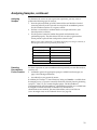
Specific Requirements for Cloning into pET151/D-TOPO®
pET151/D-TOPO® allows expression of recombinant protein with an N-terminal
tag containing the V5 epitope and a 6xHis tag. The N-terminal tag also includes a
TEV protease cleavage site to enable removal of the tag after protein purification
using TEV protease.
Introduction
Additional Cloning In addition to the guidelines on page 9, consider the following when designing
PCR primers to clone your DNA into pET151/D-TOPO®.
Considerations
Be sure to include a stop codon in the reverse primer or design the reverse
primer to hybridize downstream of the native stop codon.
If you wish to...
Then...
include the V5 epitope and 6xHis
tag
design the forward PCR primer to place the gene of interest in
frame with the N-terminal tag. Note that:
a ribosome binding site (RBS) is included upstream of the
initiation ATG in the N-terminal tag to ensure optimal
spacing for proper translation
at least six nonnative amino acids will be present between the
TEV cleavage site and the start of your gene
express your protein with a native design the forward PCR primer to include the following:
N-terminus, i.e. without the
a stop codon to terminate the N-terminal peptide
N-terminal peptide
a second ribosome binding site (AGGAGG) 9-10 base pairs 5′
of the initiation ATG codon of your protein
Note: The first three base pairs of the PCR product following the 5′ CACC overhang will constitute a
complete codon.
TOPO® Cloning
Site of
pET151/D-TOPO®
121
Use the diagram below to help you design suitable PCR primers to clone your PCR
product into pET151/D-TOPO®. Restriction sites are labeled to indicate the actual
cleavage site. The sequence of pET151/D-TOPO® is available for downloading
from our Web site or from Technical Service (see page 56). For more information
about pET151/D-TOPO®, see pages 48-49.
ATAGGCGCCA GCAACCGCAC CTGTGGCGCC GGTGATGCCG GCCACGATGC GTCCGGCGTA GAGGATCGAG ATCTCGATCC
T7 promoter/priming site
lac operator
T7 promoter
201
CGCGAAATTA ATACGACTCA CTATAGGGGA ATTGTGAGCG GATAACAATT CCCCTCTAGA AATAATTTTG TTTAACTTTA
281
AGAAGGAGAT ATACAT ATG CAT CAT CAC CAT CAC CAT GGT AAG CCT ATC CCT AAC CCT CTC CTC GGT CTC
Met His His His His His His Gly Lys Pro Ile Pro Asn Pro Leu Leu Gly Leu
351
GAT TCT ACG GAA AAC CTG TAT TTT CAG GGA ATT GAT CCC TT C ACC
GGG AAG TGG
Asp Ser Thr Glu Asn Leu Tyr Phe Gln Gly Ile Asp Pro Phe Thr
RBS
V5 epitope
Polyhistidine region
... ... AAGGG CGAGCTCAGA
... ...
TEV cleavage site
411
G
TG
G
TEV recognition site
T7 reverse priming site
TCCGGCTGCT AACAAAGCCC GAAAGGAAGC TGAGTTGGCT GCTGCCACCG CTGAGCAATA ACTAGCATAA
15
Producing Blunt-End PCR Products
Introduction
Once you have decided on a PCR strategy and have synthesized the primers, you
are ready to produce your blunt-end PCR product using any thermostable,
proofreading polymerase. Follow the guidelines below to produce your blunt-end
PCR product.
Materials Needed
You should have the following materials on hand before beginning.
Note: dNTPs (adjusted to pH 8) are provided in the kit.
Producing PCR
Products
Checking the PCR
Product
16
•
Thermocycler and thermostable, proofreading polymerase
•
10X PCR buffer appropriate for your polymerase
•
DNA template and primers for PCR product
Set up a 25 μl or 50 μl PCR reaction using the guidelines below:
•
Follow the instructions and recommendations provided by the manufacturer
of your thermostable, proofreading polymerase to produce blunt-end PCR
products.
•
Use the cycling parameters suitable for your primers and template. Make sure
to optimize PCR conditions to produce a single, discrete PCR product.
•
Use a 7 to 30 minute final extension to ensure that all PCR products are
completely extended.
•
After cycling, place the tube on ice or store at -20ºC for up to 2 weeks. Proceed
to Checking the PCR Product, below.
After you have produced your blunt-end PCR product, use agarose gel
electrophoresis to verify the quality and quantity of your PCR product. Check
for the following outcomes below.
•
Be sure you have a single, discrete band of the correct size. If you do not
have a single, discrete band, follow the manufacturer’s recommendations for
optimizing your PCR with the polymerase of your choice. Alternatively, you
may gel-purify the desired product (see page 40s).
•
Estimate the concentration of your PCR product. You will use this
information when setting up your TOPO® Cloning reaction (see Amount of
PCR Product to Use in the TOPO® Cloning Reaction, next page for details).
TOPO® Cloning Reaction and Transformation
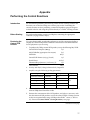
Setting Up the TOPO® Cloning Reaction
Introduction
Once you have produced the desired PCR product, you are ready to TOPO®
Clone it into the pET TOPO® vector and transform the recombinant vector into
One Shot® TOP10 E. coli. You should have everything you need set up and ready
to use to ensure that you obtain the best possible results. We suggest that you
read the this section and the section entitled Transforming Competent Cells
before beginning. If this is the first time you have TOPO® Cloned, perform the
control reactions on pages 37-39 in parallel with your samples.
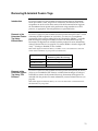
Amount of PCR
Product to Use in
the TOPO®
Cloning Reaction
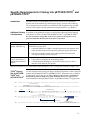
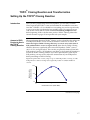
When performing directional TOPO® Cloning, we have found that the molar ratio
of PCR product:TOPO® vector used in the reaction is critical to its success. To
obtain the highest TOPO® Cloning efficiency, use a 0.5:1 to 2:1 molar ratio of
PCR product:TOPO® vector (see figure below). Note that the TOPO® Cloning
efficiency decreases significantly if the ratio of PCR product: TOPO® vector is
<0.1:1 or >5:1 (see figure below). These results are generally obtained if too little
PCR product is used (i.e. PCR product is too dilute) or if too much PCR product is
used in the TOPO® Cloning reaction. If you have quantitated the yield of your
PCR product, you may need to adjust the concentration of your PCR product
before proceeding to TOPO® Cloning.
Tip: For the pET TOPO® vectors, using 1-5 ng of a 1 kb PCR product or 5-10 ng of a 2 kb
PCR product in a TOPO® Cloning reaction generally results in a suitable number of
colonies.
Relative Activity
(colonies/reaction)
100%
50%
0%
0.1
1
10
PCR Product:Vector (Molar Ratio)
continued on next page
17
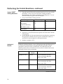
Setting Up the TOPO® Cloning Reaction, continued
Using Salt
Solution in the
TOPO® Cloning
Reaction
Performing the
TOPO® Cloning
Reaction
You will perform TOPO® Cloning in a reaction buffer containing salt (i.e. using the
stock salt solution provided in the kit). Note that the amount of salt added to the
TOPO® Cloning reaction varies depending on whether you plan to transform
chemically competent cells (provided) or electrocompetent cells (see page x for
ordering information).
•
If you are transforming chemically competent E. coli, use the stock Salt
Solution as supplied and set up the TOPO® Cloning reaction as directed below.
•
If you are transforming electrocompetent E. coli, the amount of salt in the
TOPO® Cloning reaction must be reduced to 50 mM NaCl, 2.5 mM MgCl2 to
prevent arcing during electroporation. Dilute the stock Salt Solution 4-fold
with water to prepare a 300 mM NaCl, 15 mM MgCl2 Dilute Salt Solution. Use
the Dilute Salt Solution to set up the TOPO® Cloning reaction as directed
below.
Use the procedure below to perform the TOPO® Cloning reaction. Set up the
TOPO® Cloning reaction depending on whether you plan to transform chemically
competent E. coli or electrocompetent E. coli. Reminder: For optimal results, be
sure to use a 0.5:1 to 2:1 molar ratio of PCR product:TOPO® vector in your TOPO®
Cloning reaction.
Note: The blue color of the TOPO® vector solution is normal and is used to visualize the
solution.
Reagents*
Chemically Competent E. coli
Electrocompetent E. coli
Fresh PCR product
0.5 to 4 μl
0.5 to 4 μl
Salt Solution
1 μl
--
Dilute Salt Solution (1:4) --
1 μl
add to a final volume of 5 μl
add to a final volume of 5 μl
®
TOPO vector
1 μl
1 μl
Total Volume
6 μl
6 μl
Sterile Water
*Store all reagents at -20°C when finished. Salt solution and water can be stored at room temperature or +4°C.
1.
Mix reaction gently and incubate for 5 minutes at room temperature (22-23°C).
Note: For most applications, 5 minutes will yield plenty of colonies for analysis.
Depending on your needs, the length of the TOPO® Cloning reaction can be varied from
30 seconds to 30 minutes. For routine subcloning of PCR products, 30 seconds may be
sufficient. For large PCR products (> 1 kb) or if you are TOPO® Cloning a pool of PCR
products, increasing the reaction time may yield more colonies.
2.
Place the reaction on ice and proceed to Transforming One Shot® TOP10
Competent Cells, next page.
Note: You may store the TOPO® Cloning reaction at -20°C overnight.
18
Transforming One Shot® TOP10 Competent Cells
MEND
ION
AT
RECOM
Introduction
Once you have performed the TOPO® Cloning reaction, you will transform your
pET TOPO® construct into competent E. coli. One Shot® TOP10 Chemically
Competent E. coli (Box 2) are included with the kit to facilitate transformation,
however, you may also transform electrocompetent cells. Protocols to transform
chemically competent or electrocompetent E. coli are provided in this section.
To maintain the stability of your construct, we recommend that you transform
your TOPO® Cloning reaction into TOP10 cells and characterize transformants
in TOP10 before proceeding to expression studies using BL21 Star™(DE3).
Expression of T7 RNA polymerase in BL21 Star™(DE3) may be leaky and may
lead to rearrangement or loss of your plasmid.
Materials Supplied In addition to general microbiological supplies (i.e. plates, spreaders), you will
need the following reagents and equipment.
by the User
•
42°C water bath (or electroporator with cuvettes, optional)
•
LB plates containing the appropriate antibiotic for selection (two for each
transformation)
•
37°C shaking and non-shaking incubator
There is no blue-white screening for the presence of inserts. Most
transformants will contain recombinant plasmids with the PCR product of
interest cloned in the correct orientation, reducing the number of colonies to be
analyzed. Sequencing primers are included in the kit to sequence across an
insert in the multiple cloning site to confirm orientation and reading frame.
Preparing for
Transformation
For each transformation, you will need one vial of competent cells and two
selective plates.
•
Equilibrate a water bath to 42°C (for chemical transformation) or set up your
electroporator if you are using electrocompetent E. coli.
•
Warm the vial of S.O.C. medium from Box 2 to room temperature.
•
Warm LB plates containing the appropriate antibiotic (i.e. 50-100 μg/ml
ampicillin or 50-100 μg/ml kanamycin, as appropriate) at 37°C for 30 minutes.
•
Thaw on ice 1 vial of One Shot® TOP10 cells from Box 2 for each
transformation.
continued on next page
19
Transforming One Shot® TOP10 Competent Cells, continued
Important
The number of colonies obtained after transforming the pET TOPO® vectors into
One Shot® TOP10 cells is generally lower when compared to the number of
colonies obtained after transforming other prokaryotic TOPO® vectors (e.g.
pCR®T7 TOPO®, pBAD/Thio-TOPO®). This is due to the following:
•
Directional TOPO® Cloning generally yields 2 to 5-fold fewer colonies than
traditional bidirectional TOPO TA Cloning®
•
Transforming low-copy number TOPO® plasmids generally yields 2 to 5fold fewer colonies than transforming high-copy number TOPO® plasmids
If you have TOPO® Cloned previously, note that we have slightly modified the
One Shot® TOP10 transformation protocols (see below and the next page) to
address this issue. Briefly, we recommend the following:
•
Increase the amount of TOPO® Cloning reaction that you transform into
TOP10 cells (use 3 μl) and
•
Increase the amount of transformed cells that you plate (use 100-200 μl for
chemically competent cells and 50-100 μl for electrocompetent cells)
Example: When directionally TOPO® Cloning a 750 bp test insert into any of the pET
TOPO® vectors, we generally obtain 500-1500 total colonies. Although fewer total colonies
are obtained, greater than 90% of the colonies will contain plasmid with your PCR insert
in the correct orientation.
One Shot® TOP10
Chemical
Transformation
Protocol
1.
Add 3 μl of the TOPO® Cloning reaction from Performing the TOPO®
Cloning Reaction, Step 2, page 18 into a vial of One Shot® TOP10 Chemically
Competent E. coli and mix gently. Do not mix by pipetting up and down.
2.
Incubate on ice for 5 to 30 minutes.
Note: Longer incubations on ice seem to have a minimal effect on transformation
efficiency. The length of the incubation is at the user’s discretion.
3.
Heat-shock the cells for 30 seconds at 42°C without shaking.
4.
Immediately transfer the tubes to ice.
5.
Add 250 l of room temperature S.O.C. medium.
6.
Cap the tube tightly and shake the tube horizontally (200 rpm) at 37°C for
1 hour.
7.
Spread 100-200 l from each transformation on a prewarmed selective plate
and incubate overnight at 37°C. We recommend plating two different
volumes to ensure that at least one plate will have well-spaced colonies.
8.
An efficient TOPO® Cloning reaction may produce several hundred colonies.
Pick ~5 colonies for analysis (see Analyzing Positive Clones, page 22).
Note: If you see few transformants, refer to the Troubleshooting section, page 34 for
tips to optimize your TOPO® Cloning and transformation reactions.
continued on next page
20
Transforming Competent Cells, continued
Transformation by
Electroporation
Use ONLY electrocompetent cells for electroporation to avoid arcing. Do not
use the One Shot® TOP10 chemically competent cells for electroporation.
1.
Add 3 μl of the TOPO® Cloning reaction from Performing the TOPO®
Cloning Reaction, Step 2, page 18 into 50 μl of electrocompetent E. coli and
mix gently. Do not mix by pipetting up and down. Avoid formation of
bubbles. Transfer the electrocompetent cells to a 0.1 cm cuvette.
2.
Electroporate your samples using your own protocol and an electroporator.
3.
Immediately add 250 μl of room temperature S.O.C. medium.
4.
Transfer the solution to a 15 ml snap-cap tube (i.e. Falcon) and shake for at
least 1 hour at 37°C to allow expression of the antibiotic resistance marker.
5.
Spread 50-100 μl from each transformation on a prewarmed selective plate
and incubate overnight at 37°C. We recommend plating two different
volumes to ensure that at least one plate will have well-spaced colonies.
6.
An efficient TOPO® Cloning reaction may produce several hundred colonies.
Pick ~5 colonies for analysis (see Analyzing Positive Clones, page 22).
Note: If you have problems with arcing, see below.
MEND
ION
AT
RECOM
Note: If you see few transformants, refer to the Troubleshooting section, page 34 for
tips to optimize your TOPO® Cloning and transformation reactions.
To prevent arcing of your samples during electroporation, the volume of cells
should be between 50 and 80 μl (0.1 cm cuvettes) or 100 to 200 μl (0.2 cm
cuvettes).
If you experience arcing during transformation, try one of the following
suggestions:
•
Reduce the voltage normally used to charge your electroporator by 10%
•
Reduce the pulse length by reducing the load resistance to 100 ohms
•
Ethanol precipitate the TOPO® Cloning reaction and resuspend in water prior
to electroporation
21
Analyzing Transformants
Analyzing Positive 1. Pick 5 colonies and culture them overnight in LB or S.O.B. medium
containing the appropriate antibiotic.
Clones
2.
Isolate plasmid DNA using your method of choice. We recommend using the
PureLink™ HQ Mini Plasmid Purification Kit (Catalog no. K2100-01).
Note: Since the pET TOPO® vectors are low-copy number plasmids, you may need to
increase the amount of bacterial culture to obtain enough plasmid DNA for sequencing
or analysis purposes. Use extra care during purification to obtain plasmid DNA of
sufficiently pure quality for sequencing (see below).
3.
Sequencing
Analyze the plasmids by restriction analysis to confirm the presence and
correct orientation of the insert. Use a restriction enzyme or a combination of
enzymes that cut once in the vector and once in the insert.
We recommend sequencing your construct to confirm that your gene is in frame
with the appropriate N-terminal or C-terminal fusion tag, if desired. The table
below lists the primers included in each kit to help you sequence your insert.
Vector
Important
Forward Primer
Reverse Primer
pET100/D-TOPO
®
T7
T7 Reverse
pET101/D-TOPO
®
T7
T7 Reverse
pET102/D-TOPO
®
TrxFus Forward
T7 Reverse
pET151/D-TOPO
®
T7
T7 Reverse
pET200/D-TOPO
®
T7
T7 Reverse
If you download the sequence from our Web site, note that the overhang sequence
(GTGG) will be shown already hybridized to CACC. No DNA sequence analysis
program allows us to show the overhang without the complementary sequence.
continued on next page
22
Analyzing Transformants, continued
Analyzing
Transformants by
PCR
You may analyze positive transformants using PCR. For PCR primers, use a
combination of the Forward sequencing primer or the Reverse sequencing primer
and a primer that hybridizes within your insert. You will have to determine the
amplification conditions. If you are using this technique for the first time, we
recommend performing restriction analysis in parallel. Artifacts may be obtained
because of mispriming or contaminating template. The protocol below is provided
for your convenience. Other protocols are suitable.
Materials Needed
PCR SuperMix High Fidelity (Invitrogen, Catalog no. 10790-020)
Appropriate forward and reverse PCR primers (20 μM each)
Procedure
Important
Long-Term
Storage
1.
For each sample, aliquot 48 μl of PCR SuperMix High Fidelity into a 0.5 ml
microcentrifuge tube. Add 1 μl each of the forward and reverse PCR primer.
2.
Pick 5 colonies and resuspend them individually in 50 μl of the PCR cocktail
from Step 1, above.
3.
Incubate reaction for 10 minutes at 94°C to lyse cells and inactivate nucleases.
4.
Amplify for 20 to 30 cycles.
5.
For the final extension, incubate at 72°C for 10 minutes. Store at +4°C.
6.
Visualize by agarose gel electrophoresis.
If you have problems obtaining transformants or the correct insert, perform the
control reactions described on page 37-39. These reactions will help you
troubleshoot your experiment. Refer to the Troubleshooting section, page 34 for
additional tips.
Once you have identified the correct clone, be sure to purify the colony and
make a glycerol stock for long-term storage. We recommend that you store a
stock of plasmid DNA at -20°C.
1.
Streak the original colony out for single colony on LB plates containing the
appropriate antibiotic.
2.
Isolate a single colony and inoculate into 1-2 ml of LB containing the
appropriate antibiotic.
3.
Grow until culture reaches stationary phase.
4.
Mix 0.85 ml of culture with 0.15 ml of sterile glycerol and transfer to a
cryovial.
5.
Store at -80°C.
23
Expression and Purification
General Guidelines for Expression
Introduction
BL21 Star™(DE3) One Shot® E. coli (Box 3) are included with each Champion™ pET
Directional TOPO® Expression Kit for use as the host for expression. You will need
pure plasmid DNA of your pET TOPO® construct to transform into BL21
Star™(DE3) for expression studies. Since each recombinant protein has different
characteristics that may affect optimal expression, we recommend performing a
time course of expression to determine the best conditions for expression of your
protein. Each Champion™ pET Directional TOPO® Expression Kit also includes the
appropriate pET TOPO® vector containing the lacZ gene for use as a positive
expression control (see below).
BL21 Star™
Strains
The BL21 Star™(DE3) E. coli strain is specifically designed for expression of genes
regulated by the T7 promoter. Each time you perform an expression experiment,
you will transform your plasmid into BL21 Star™(DE3). Do not use this strain for
propagation and maintenance of your plasmid. Use TOP10 instead. Basal level
expression of T7 polymerase, particularly in BL21 Star™(DE3) cells, may lead to
plasmid instability if your gene of interest is toxic to E. coli.
Note: If you are expressing a highly toxic gene, the BL21 Star™(DE3)pLysS strain is also
available from Invitrogen for expression purposes. The BL21 Star™(DE3)pLysS strain
contains the pLysS plasmid to further reduce basal level expression of the gene of interest.
For more information, see page 6.
Positive Controls
Each Champion™ pET Directional TOPO® Expression Kit includes a positive
control vector for use as an expression control (see the table below). In each case,
the gene encoding β-galactosidase is directionally TOPO® Cloned into the
appropriate pET TOPO® vector (see pages 50-53 for details). Transform 10 ng of
each plasmid into BL21 Star™(DE3) cells using the procedure on page 26.
Kit
Positive Control
Champion™ pET100 Directional TOPO® Expression Kit
pET100/D/lacZ
™
®
pET101/D/lacZ
™
®
pET102/D/lacZ
™
®
pET151/D/lacZ
™
®
pET200/D/lacZ
Champion pET101 Directional TOPO Expression Kit
Champion pET102 Directional TOPO Expression Kit
Champion pET151 Directional TOPO Expression Kit
Champion pET200 Directional TOPO Expression Kit
continued on next page
24
General Guidelines for Expression, continued
Basic Strategy
The basic steps needed to induce expression of your gene in BL21 Star™(DE3)
E. coli are outlined below.
1.
Isolate plasmid DNA using standard procedures and transform your
construct and the positive control separately into BL21 Star™(DE3) One Shot®
cells.
2.
Grow the transformants and induce expression with IPTG over several hours.
Take several time points to determine the optimal time of expression.
3.
Optimize expression to maximize the yield of protein.
Plasmid
Preparation
You may prepare plasmid DNA using your method of choice. We recommend
using the PureLink™ HQ Mini Plasmid Purification Kit (Catalog no. K2100-01)
for isolation of pure plasmid DNA. Note that since you are purifying a low-copy
number plasmid, you may need to increase the amount of bacterial culture that
you use to prepare your plasmid construct.
Ampicillin
Selection
For pET TOPO® vectors containing the ampicillin resistance gene, ampicillin
generally works well for selection of transformants and expression experiments.
However, if you find that your expression levels are low, you may want to use
carbenicillin instead. The resistance gene for ampicillin encodes the protein,
β-lactamase. β-lactamase is secreted into the medium where it hydrolyzes
ampicillin, inactivating the antibiotic. Since β-lactamase is catalytic, ampicillin is
rapidly removed from the medium, resulting in non-selective conditions. If your
plasmid is unstable, this may result in the loss of plasmid and low expression
levels.
Using
Carbenicillin
Carbenicillin is generally more stable than ampicillin, and studies have shown
that using carbenicillin in place of ampicillin may help to increase expression
levels by preventing loss of the pET TOPO® plasmid. If you wish to use
carbenicillin, perform your transformation and expression experiments in LB
containing 50 μg/ml carbenicillin.
Note: If your gene of interest is highly toxic, increasing the concentration of carbenicillin
used from 50 μg/ml to 200 μg/ml may help to increase expression levels.
Note that cyclic AMP-mediated derepression of the lacUV5 promoter in λDE3
lysogens can result in an increase in basal expression of T7 RNA polymerase. If
you are expressing an extremely toxic gene, the pET construct may be unstable
in BL21 Star™(DE3) cells. Adding 1% glucose to the bacterial culture medium
may help to repress basal expression of T7 RNA polymerase and stabilize your
pET construct.
25
Expressing the PCR Product
Materials to Have
on Hand
Transforming
BL21 Star™(DE3)
One Shot® Cells
Be sure to have the following solutions and equipment on hand before starting
your expression experiment:
•
Your pET TOPO® expression construct (>10 μg/ml)
•
The appropriate pET/D/lacZ positive control plasmid
•
BL21 Star™(DE3) One Shot® cells (Box 3 supplied with the kit)
•
S.O.B. or LB containing the appropriate antibiotic for selection (plus
1% glucose, if desired)
•
37°C incubator (shaking and nonshaking)
•
42°C water bath
•
1 M isopropyl β-D-thiogalactoside (IPTG; Invitrogen, Catalog no. 15529-019)
•
Liquid nitrogen
To transform your construct or the positive control (10 ng each) into BL21
Star™(DE3) One Shot® cells, follow the instructions below. You will need one vial
of cells per transformation.
Note: You will not plate the transformation reaction, but inoculate it into medium for
growth and subsequent expression.
1.
Thaw on ice, one vial of BL21 Star™(DE3) One Shot® cells per transformation.
2.
Add 5-10 ng plasmid DNA in a 1 to 5 μl volume into each vial of BL21
Star™(DE3) One Shot® cells and mix by stirring gently with the pipette tip. Do
not mix by pipetting up and down.
3.
Incubate on ice for 30 minutes.
4.
Heat-shock the cells for 30 seconds at 42°C without shaking.
5.
Immediately transfer the tubes to ice.
6.
Add 250 μl of room temperature S.O.C. medium.
7.
Cap the tube tightly, tape the tube on its side (for better aeration), and
incubate at 37°C for 30 minutes with shaking (200 rpm).
8.
Add the entire transformation reaction to 10 ml of LB containing the
appropriate antibiotic (and 1% glucose, if desired).
9.
Grow overnight at 37°C with shaking. Proceed to Pilot Expression, next page.
continued on next page
26
Expressing the PCR Product, continued
Pilot Expression
1.
Inoculate 10 ml of LB containing the appropriate antibiotic (and 1% glucose,
if desired) with 500 μl of the overnight culture from Step 8, previous page.
2.
Grow two hours at 37°C with shaking. OD600 should be about 0.5-0.8 (midlog).
3.
Split the culture into two 5 ml cultures. Add IPTG to a final concentration of
0.5-1 mM to one of the cultures. You will now have two cultures: one
induced, one uninduced.
4.
Remove a 500 μl aliquot from each culture, centrifuge at maximum speed in
a microcentrifuge for 30 seconds, and aspirate the supernatant.
5.
Freeze the cell pellets at -20°C. These are the zero time point samples.
6.
Continue to incubate the cultures at 37°C with shaking. Take time points for
each culture every hour for 4 to 6 hours.
7.
For each time point, remove 500 μl from the induced and uninduced cultures
and process as described in Steps 4 and 5. Proceed to Analyzing Samples,
next page.
27
Analyzing Samples
Materials to Have
on Hand
Preparing
Samples
Have the following materials on hand before starting:
•
Lysis Buffer (see page 55 for recipe)
•
1X and 2X SDS-PAGE sample buffer (see page 55 for recipes)
•
Reagents and apparatus to perform SDS-PAGE electrophoresis
•
Boiling water bath
Once you have finished your pilot expression, you are ready to analyze the
samples you have collected. Before starting, prepare SDS-PAGE gels or use one of
the pre-cast polyacrylamide gels available from Invitrogen (see below).
Note: If you wish to analyze your samples for soluble protein, see below.
Preparing
Samples for
Soluble/Insoluble
Protein
1.
Thaw the samples (from Pilot Expression, Steps 5 and 7, previous page) and
resuspend each cell pellet in 80 μl of 1X SDS-PAGE sample buffer.
2.
Boil 5 minutes and centrifuge briefly.
3.
Load 5-10 μl of each sample on an SDS-PAGE gel and electrophorese. Save
your samples by storing them at -20°C.
1.
Thaw and resuspend each cell pellet in 500 μl of Lysis Buffer (see Recipes,
page 55).
2.
Freeze sample in dry ice or liquid nitrogen and then thaw at 42°C. Repeat 2
to 3 times.
Note: To facilitate lysis, you may need to add lysozyme or sonicate the cells.
Polyacrylamide
Gel
Electrophoresis
3.
Centrifuge samples at maximum speed in a microcentrifuge for 1 minute at
+4°C to pellet insoluble proteins. Transfer supernatant to a fresh tube and
store on ice.
4.
Mix together equivalent amounts of supernatant and 2X SDS-PAGE sample
buffer and boil for 5 minutes.
5.
Add 500 μl of 1X SDS-PAGE sample buffer to the pellets from Step 3 and boil
5 minutes.
6.
Load 10 μl of the supernatant sample and 5 μl of the pellet sample onto an
SDS-PAGE gel and electrophorese.
To facilitate separation and visualization of your recombinant fusion protein by
polyacrylamide gel electrophoresis, a wide range of pre-cast NuPAGE® and
Novex® Tris-Glycine polyacrylamide gels and electrophoresis apparatus are
available from Invitrogen. In addition, Invitrogen also carries a large selection of
molecular weight protein standards and staining kits. For more information about
the appropriate gels, standards, and stains to use to visualize your recombinant
protein, refer to our Web site (www.invitrogen.com) or call Technical Service (see
page 56).
continued on next page
28
Analyzing Samples, continued
Analyzing
Samples
To determine the success of your expression experiment, you may want to
perform the following types of analyses:
1.
Stain the polyacrylamide gel with Coomassie blue and look for a band of
increasing intensity in the expected size range for the recombinant protein.
Use the uninduced culture as a negative control.
2.
Perform a western blot to confirm that the overexpressed band is your
desired protein (see below).
3.
Use the positive control to confirm that growth and induction were
performed properly. The table below lists the size of the β-galactosidase
fusion protein expressed from each positive control vector.
Note: β-galactosidase Antiserum is available from Invitrogen (Catalog no. R901-25) to
detect β-galactosidase fusion proteins by western blot.
Vector
Detecting
Recombinant
Fusion Proteins
Size of the Control Protein
pET100/D/lacZ
121 kDa
pET101/D/lacZ
120 kDa
pET102/D/lacZ
133 kDa
pET151/D/lacZ
120 kDa
pET200/D/lacZ
121 kDa
To detect expression of your recombinant fusion protein by western blot analysis,
you may use:
•
Antibodies against the appropriate epitope available from Invitrogen (see
page xi for ordering information)
•
An antibody to your protein of interest
In addition, the Positope™ Control Protein (Catalog no. R900-50) is available from
Invitrogen for use as a positive control for detection of fusion proteins containing
an Xpress™, HisG, V5, or C-terminal 6xHis epitope. The WesternBreeze®
Chromogenic Kits and WesternBreeze® Chemiluminescent Kits are available from
Invitrogen to facilitate detection of antibodies by colorimetric or
chemiluminescence methods. For more information, refer to our Web site
(www.invitrogen.com) or call Technical Service (see page 56).
continued on next page
29
Analyzing Samples, continued
Expression of your protein with the N- and/or C-terminal tags will increase the
size of your recombinant protein. The table below lists the increase in the
molecular weight of your recombinant fusion protein that you should expect from
the particular N- or C-terminal fusion tag in each pET TOPO® vector. Be sure to
account for any additional amino acids between the fusion tag and the start of
your protein.
Vector
The Next Step
Fusion Tag
Expected Size Increase
(kDa)
pET100/D-TOPO®
N-terminal
3 kDa
pET101/D-TOPO
®
C-terminal
3 kDa
pET102/D-TOPO
®
N-terminal
13 kDa
C-terminal
3 kDa
pET151/D-TOPO
®
N-terminal
4 kDa
pET200/D-TOPO
®
N-terminal
3 kDa
If you are satisfied with expression of your gene of interest, proceed to Purifying
the Recombinant Fusion Protein, page 31.
If you have trouble expressing your protein or wish to optimize expression, refer
to the Troubleshooting section, page 37.
30
Purifying the Recombinant Fusion Protein
Introduction
The presence of the N-terminal or C-terminal polyhistidine (6xHis) tag in all
pET TOPO® vectors allows purification of your recombinant fusion protein
with a metal-chelating resin such as ProBond™ or Ni-NTA.
ProBond™ and
Ni-NTA
ProBond™ and Ni-NTA are nickel-charged agarose resins that can be used for
affinity purification of fusion proteins containing the 6xHis tag and/or the HPthioredoxin protein. Proteins bound to the resin may be eluted with either low
pH buffer or competition with imidazole or histidine.
Important
Scaling-up
Expression for
Purification
•
To scale up your pilot expression for purification, see below.
•
To purify your fusion protein using ProBond™ or Ni-NTA, refer to the
manual included with each product. You may download the manuals from
our Web site (www.invitrogen.com).
•
To purify your fusion protein using another metal-chelating resin, refer to
the manufacturer’s instructions.
Note that under denaturing conditions, the Ni2+ binding site encoded by the
histidine “patch” in pET102/D-TOPO® will be destroyed because the HPthioredoxin protein will be denatured. The binding of nickel ion to the 6xHis tag
is not affected by denaturing conditions.
We generally scale-up expression to a 50 ml bacterial culture for purification
using a 2 ml ProBond™ or Ni-NTA column. Depending on the expression level of
your recombinant fusion protein, you may need to adjust the culture volume to
bind the maximum amount of recombinant fusion protein to your column.
To grow and induce a 50 ml bacterial culture:
1.
Inoculate 10 ml of S.O.B. or LB containing the appropriate antibiotic with
500 μl of the culture of the transformation reaction (see page 26, step 9).
2.
Grow overnight at 37°C with shaking (225-250 rpm) to OD600 = 1-2.
3.
The next day, inoculate 50 ml of S.O.B. or LB containing the appropriate
antibiotic with 1 ml of the overnight culture.
Note: You can scale up further and inoculate all of the 10 ml overnight culture into
500 ml of medium, but you will need to adjust the bed volume of your ProBond™ or
Ni-NTA column accordingly.
4.
Grow the culture at 37°C with shaking (225-250 rpm) to an OD600 = ~0.5
(2-3 hours). The cells should be in mid-log phase.
5.
Add 0.5-1 mM IPTG to induce expression.
6.
Grow at 37°C with shaking until the optimal time point determined by the
pilot expression is reached. Harvest the cells by centrifugation (3000 x g for
10 minutes at +4°C).
7.
Proceed to purification or store the cells at -80°C for future use.
continued on next page
31
Purifying the Recombinant Fusion Protein, continued
Additional
Purification Steps
32
There may be cases when your specific fusion protein may not be completely
purified by metal affinity chromatography. Other protein purification
techniques may be utilized in conjunction with ProBond™ or Ni-NTA to purify
the fusion protein (see Deutscher, 1990 for more information).
Removing N-terminal Fusion Tags
Introduction
If you have expressed your recombinant fusion protein from any N-terminal
fusion pET TOPO® vector, the presence of either the enterokinase (EK) or TEV
recognition site in the vector allows removal of the N-terminal fusion tag from
the recombinant fusion protein after purification using enterokinase or TEV
protease, as appropriate. General guidelines are provided below.
Removal of the
N-terminal Fusion
Tag Using
Enterokinase
If you have expressed your recombinant fusion protein from pET TOPO® vectors
containing the EK recognition site (see below), you may use a recombinant
preparation of the catalytic subunit of bovine enterokinase (EKMax™) available
from Invitrogen (Catalog no. E180-01) to remove the N-terminal fusion tag.
Instructions for digestion are included with the product. For more information,
contact Technical Service (see page 56). To remove EKMax™ from the digest, EKAway™ (Catalog no. R180-01) is also available.
Note: After digestion with enterokinase, a number of vector-encoded amino acids will
remain at the N-terminus of your protein (see table below).
Vector
pET100/D-TOPO®
5
®
7
pET200/D-TOPO®
5
pET102/D-TOPO
Removal of the
N-terminal Fusion
Tag Using TEV
Protease
Number of Amino Acids
Remaining After EK Cleavage
If you have expressed your recombinant fusion protein from pET151/D-TOPO®,
you may use recombinant TEV Protease available from Invitrogen (Catalog no.
12575-015) to remove the N-terminal fusion tag. Instructions for digestion are
included with the product. For more information, contact Technical Service (see
page 56).
Note: After digestion with enterokinase, six vector-encoded amino acids will remain at
the N-terminus of your protein.
33
Troubleshooting
TOPO® Cloning
Reaction and
Transformation
The table below lists some potential problems and possible solutions that may
help you troubleshoot the TOPO® Cloning and transformation reactions. To help
evaluate your results, we recommend that you perform the control reactions (see
pages 37-39) in parallel with your samples.
Problem
Few or no colonies obtained
from sample reaction and
the transformation control
gave colonies
Reason
Solution
Suboptimal ratio of PCR
product:TOPO® vector used in
the TOPO® Cloning reaction
Use a 0.5:1 to 2:1 molar ratio of PCR
product:TOPO® vector.
Too much PCR product used in
the TOPO® Cloning reaction
Dilute the PCR product.
PCR product too dilute
• Concentrate the PCR product.
Use a 0.5:1 to 2:1 molar ratio of PCR
product:TOPO® vector.
• Use a 0.5:1 to 2:1 molar ratio of PCR
product:TOPO® vector.
PCR primers contain
5′ phosphates
Do not add 5′ phosphates to your PCR
primers.
Incorrect PCR primer design
Make sure that the forward PCR
primer contains the sequence,
CACC, at the 5′ end.
Make sure that the reverse PCR
primer does not contain the
sequence, CACC, at the 5′ end.
Used Taq polymerase or a
Taq/proofreading polymerase
mixture for PCR
Use a proofreading polymerase for
PCR.
Large PCR product
Increase the incubation time of the
TOPO® reaction from 5 minutes to
30 minutes.
Gel-purify the PCR product to remove
primer-dimers and other artifacts.
PCR reaction contains artifacts
(i.e. does not run as a single,
discrete band on an agarose gel)
• Optimize your PCR using the
proofreading polymerase of your
choice.
• Gel-purify your PCR product.
Cloning large pool of PCR
products or a toxic gene
• Increase the incubation time of the
TOPO® reaction from 5 minutes to
30 minutes.
• Use a 0.5:1 to 2:1 molar ratio of PCR
product:TOPO® vector.
continued on next page
34
Troubleshooting, continued
TOPO® Cloning Reaction and Transformation, continued
Problem
Large percentage of inserts
cloned in the incorrect
orientation
Large number of incorrect
inserts cloned
Reason
Solution
Incorrect PCR primer design
Make sure that the forward PCR
primer contains the sequence, CACC,
at the 5′ end.
Reverse PCR primer is
complementary to the GTGG
overhang at the 5′ end
Make sure that the reverse PCR
primer does not contain the sequence,
CACC, at the 5′ end.
PCR cloning artifacts
Gel-purify your PCR product to
remove primer-dimers and smaller
PCR products.
Optimize your PCR.
Include a final extension step of 7 to
30 minutes during PCR. Longer
PCR products will need a longer
extension time.
Incorrect PCR primer design
Few or no colonies obtained One Shot® competent E. coli
from sample reaction and
stored incorrectly
the transformation control
gave no colonies
Make sure that the forward and
reverse PCR primers are designed
correctly.
Store One Shot® competent E. coli at
-80°C.
If you are using another E. coli strain,
follow the manufacturer’s
instructions.
One Shot® transformation
protocol not followed correctly
Follow the One Shot® transformation
protocol provided on page 20.
Insufficient amount of E. coli
plated
Increase the amount of E. coli plated.
Transformants plated on
selective plates containing the
wrong antibiotic
Use the appropriate antibiotic for
selection.
continued on next page
35
Troubleshooting, continued
Expression
The table below lists some potential problems and possible solutions that may
help you troubleshoot your expression experiment. To help evaluate your
results, we recommend including the expression control supplied with kit in
your experiment.
Problem
No expression of
recombinant protein
Low expression
Reason
Solution
Gene of interest not in frame
with the epitope tag
Sequence your construct to verify if
the insert is in frame with the epitope
tag. If not in frame, redesign your
PCR primers.
Incorrect antibody used for
detection
Use an antibody to your protein or
one of the antibodies listed on page xi,
as appropriate
Plasmid instability observed
when using ampicillin for
selection
Substitute carbenicillin for ampicillin
in your transformation and expression
experiments (see page 25).
Toxic gene
• Add 1% glucose to the bacterial
culture medium during
transformation and expression.
Note: Evidence of toxicity includes
loss of plasmid or slow growth
relative to the control.
• Transform BL21 Star™(DE3) cells
using the protocol on page 26, then
perform the expression by growing
cells at room temperature rather
than 37°C for 24-48 hours.
• Transform your expression
construct into a pLysS-containing
strain (e.g. BL21 Star™(DE3)pLysS).
• Transform your expression
construct into an E. coli strain in
which expression of T7 RNA
polymerase is tightly regulated (e.g.
BL21-AI™ available from
Invitrogen; see our Web site for
more information).
• Infect TOP10F′ (or other suitable
F′ episome-containing host strain)
with M13 or lambda phage
expressing T7 RNA polymerase.
36
Appendix
Performing the Control Reactions
Introduction
We recommend performing the following control TOPO® Cloning reactions the
first time you use the kit to help you evaluate your results. Performing the
control reactions involves producing a control PCR product using the reagents
included in the kit and using this product directly in a TOPO® Cloning reaction.
Before Starting
For each transformation, prepare two LB plates containing the appropriate
antibiotic (see page 54 for a recipe).
Producing the
Control PCR
Product
Use your thermostable, proofreading polymerase and the appropriate buffer to
amplify the control PCR product. Follow the manufacturer’s recommendations
for the polymerase you are using.
1.
To produce the 750 bp control PCR product, set up the following 50 μl PCR:
Control DNA Template (100 ng)
1 μl
10X PCR Buffer (appropriate for enzyme)
5 μl
0.5 μl
dNTP Mix
Control PCR Primers (0.1 μg/μl each)
1 μl
41.5 μl
Sterile Water
1 μl
Thermostable polymerase (1-2.5 units/μl)
50 μl
Total Volume
2.
Overlay with 70 μl (1 drop) of mineral oil, if required.
3.
Amplify using the following cycling parameters:
Step
Time
Temperature
Initial Denaturation
2 minutes
94°C
Denaturation
1 minute
94°C
Annealing
1 minute
55°C
Extension
1 minute
72°C
Final Extension
7 minutes
72°C
Cycles
1X
25X
1X
4.
Remove 10 μl from the reaction and analyze by agarose gel electrophoresis. A
discrete 750 bp band should be visible.
5.
Estimate the concentration of the PCR product, and adjust as necessary such
that the amount of PCR produce used in the control TOPO® Cloning reaction
results in an optimal molar ratio of PCR product:TOPO® vector (i.e. 0.5:1 to
2:1). Proceed to Control TOPO® Cloning Reactions, next page.
continued on next page
37
Performing the Control Reactions, continued
Control TOPO®
Cloning Reactions
1.
Using the control PCR product produced on the previous page and the
pET TOPO® vector, set up two 6 μl TOPO® Cloning reactions as
described below. If you plan to transform electrocompetent E. coli, use
Dilute Salt Solution in place of the Salt Solution.
2.
Set up control TOPO® Cloning reactions:
Reagent
"Vector Only"
"Vector + PCR Insert"
Sterile Water
4 μl
3 μl
Salt Solution
1 μl
1 μl
Control PCR Product
--
1 μl
1 μl
1 μl
®
pET TOPO vector
6 μl
6 μl
Incubate at room temperature for 5 minutes and place on ice.
Final volume
3.
Analysis of
Results
4.
Transform 3 l of each reaction into separate vials of One Shot® TOP10
cells (page 20).
5.
Spread 100-200 l of each transformation mix onto LB plates containing
the appropriate antibiotic. Be sure to plate two different volumes to
ensure that at least one plate has well-spaced colonies.
6.
Incubate overnight at 37°C.
Hundreds of colonies from the vector + PCR insert reaction should be produced.
To analyze the transformations, isolate plasmid DNA and digest with the
appropriate restriction enzyme as listed below. The table below lists the
digestion patterns that you should see for inserts that are cloned in the correct
orientation or in the reverse orientation.
Vector
Restriction
Enzyme
pET100/D-TOPO®
Hind III
Expected Digestion Patterns (bp)
Correct orientation: 838, 5674
Reverse orientation: 500, 6012
Empty vector: 5764
pET101/D-TOPO
®
Cla I
Correct orientation: 599, 5896
Reverse orientation: 929, 5566
Empty vector: 5753
pET102/D-TOPO
®
Pst I
Correct orientation: 1990, 5065
Reverse orientation: 1484, 5571
Empty vector: 6315
continued on next page
38
Performing the Control Reactions, continued
Analysis of Results, continued
Vector
pET151/D-TOPO®
Restriction
Enzyme
EcoR I and Sst I
Expected Digestion Patterns (bp)
Correct orientation: 653, 5861
Reverse orientation: 101, 6413
Empty vector: 5760
pET200/D-TOPO®
Hind III
Correct orientation: 845, 5646
Reverse orientation: 511, 5980
Empty vector: 5741
Greater than 90% of the colonies should contain the 750 bp insert in the correct
orientation.
Relatively few colonies should be produced in the vector-only reaction.
Transformation
Control
pUC19 plasmid is included to check the transformation efficiency of the One
Shot® TOP10 competent cells. Transform one vial of One Shot® TOP10 cells with
10 pg of pUC19 using the protocol on page 20. Plate 10 μl of the transformation
mixture plus 20 μl of S.O.C. on LB plates containing 100 μg/ml ampicillin.
Transformation efficiency should be ~1 x 10⁹ cfu/μg DNA.
39
Gel Purifying PCR Products
Introduction
Smearing, multiple banding, primer-dimer artifacts, or large PCR products
(>3 kb) may necessitate gel purification. If you intend to purify your PCR
product, be extremely careful to remove all sources of nuclease contamination.
There are many protocols to isolate DNA fragments or remove oligonucleotides.
Refer to Current Protocols in Molecular Biology, Unit 2.6 (Ausubel et al., 1994) for
the most common protocols. Two simple protocols are provided below.
Using the
PureLink™ Quick
Gel Extraction Kit
The PureLink™ Quick Gel Extraction Kit (page x) allows you to rapidly purify
PCR products from regular agarose gels.
1.
Equilibrate a water bath or heat block to 50°C.
2.
Cut the area of the gel containing the desired DNA fragment using a clean,
sharp blade. Minimize the amount of surrounding agarose excised with the
fragment.
3.
Weigh the gel slice.
4.
Add Gel Solubilization Buffer (GS1) supplied in the kit as follows:
•
For <2% agarose gels, place up to 400 mg gel into a sterile, 1.5-ml
polypropylene tube. Divide gel slices exceeding 400 mg among
additional tubes. Add 30 μl Gel Solubilization Buffer (GS1) for every
10 mg of gel.
•
For >2% agarose gels, use sterile 5-ml polypropylene tubes and add 60 μl
Gel Solubilization Buffer (GS1) for every 10 mg of gel.
5.
Incubate the tube at 50°C for 15 minutes. Mix every 3 minutes to ensure gel
dissolution. After gel slice appears dissolved, incubate for an additional
5 minutes.
6.
Preheat an aliquot of TE Buffer (TE) to 65-70°C
7.
Place a Quick Gel Extraction Column into a Wash Tube. Pipette the mixture
from Step 5,above onto the column. Use 1 column per 400 mg agarose.
8.
Centrifuge at >12,000 x g for 1 minute. Discard the flow-through. Place the
column back into the Wash Tube.
9.
Optional: Add 500 μl Gel Solubilization Buffer (GS1) to the column. Incubate
at room temperature for 1 minute. Centrifuge at >12,000 x g for 1 minute.
Discard the flow-through. Place the column back into the Wash Tube.
10. Add 700 μl Wash Buffer (W9) with ethanol (add 96–100% ethanol to the Wash
Buffer according to instructions on the label of the bottle) to the column and
incubate at room temperature for 5 minutes. Centrifuge at >12,000 x g for
1 minute. Discard flow-through.
11. Centrifuge the column at >12,000 x g for 1 minute to remove any residual
buffer. Place the column into a 1.5 ml Recovery Tube.
12. Add 50 μl warm (65-70°C) TE Buffer (TE) to the center of the cartridge.
Incubate at room temperature for 1 minute.
13. Centrifuge at >12,000 x g for 2 minutes. The Recovery Tube contains the purified
DNA. Store DNA at –20°C. Discard the column.
14. Use 4 μl of the purified DNA for the TOPO® Cloning reaction.
continued on next page
40
Gel Purifying PCR Products, continued
Low-Melt Agarose
Method
If you prefer to use low-melt agarose, use the procedure below. Note that gel
purification will result in dilution of your PCR product and a potential loss of
cloning efficiency. Use only chemically competent cells for transformation.
1.
Electrophorese as much as possible of your PCR reaction on a low-melt
agarose gel (0.8 to 1.2%) in TAE buffer.
2.
Visualize the band of interest and excise the band.
3.
Place the gel slice in a microcentrifuge tube and incubate the tube at 65°C
until the gel slice melts.
4.
Place the tube at 37°C to keep the agarose melted.
5.
Add 4 μl of the melted agarose containing your PCR product to the TOPO®
Cloning reaction as described on page 18.
6.
Incubate the TOPO® Cloning reaction at 37°C for 5 to 10 minutes. This is to
keep the agarose melted.
7.
Transform 2 to 4 μl directly into One Shot® TOP10 cells using the method on
page 20.
The cloning efficiency may decrease with purification of the PCR product. You
may wish to optimize your PCR to produce a single band.
41
Map and Features of pET100/D-TOPO® and pET200/D-TOPO®
T7
lacO
RBS
ATG
6xHis
TOPO
Xpress
epitope
TT
AAG GGC
EK CCC
GGG AAG TGG TTC CCG
T7 term
TOPO
lacI
in
yc
m
llin
ci
Am
pi
Ka
na
Sac I
Nde I
Nhe I
The figure below shows the features of the pET100/D-TOPO® (5764 bp) and
pET200/D-TOPO® (5741 bp) vectors. The complete sequence of each vector is
available for downloading from our Web site (www.invitrogen.com) or by
contacting Technical Service (see page 56).
Map
pET100/D-TOPO®
&
pET200/D-TOPO®
ro
p
p BR
T7 promoter
T7 promoter/priming site
lac operator (lacO)
Ribosome binding site (RBS)
Initiation ATG
Polyhistidine (6xHis) region
Xpress™ epitope
EK recognition site
TOPO® Cloning site (directional)
T7 reverse priming site
T7 transcription termination region
bla promoter
Ampicillin (bla) resistance gene
Kanamycin resistance gene
pBR322 origin
ROP ORF (complementary strand)
lacI ORF (complementary strand)
o
32 2
pET100/D-TOPO®
209-225
209-228
228-252
282-288
297-299
309-326
366-389
375-389
396-409
466-485
427-555
856-954
955-1815
--2022-2757
3001-3192
4507-5595
ri
pET200/D-TOPO®
209-225
209-228
228-252
282-288
297-299
309-326
366-389
375-389
396-409
466-485
427-555
856-954
--955-1749
1969-2610
2978-3169
4481-5572
continued on next page
42
Map and Features of pET100/D-TOPO® and pET200/D-TOPO®,
continued
Features of
pET100/D-TOPO®
and pET200/DTOPO®
The pET100/D-TOPO® (5764 bp) and pET200/D-TOPO® (5741 bp) vectors
contain the following elements. All features have been functionally tested.
Feature
Benefit
T7 promoter
Permits high-level, IPTG-inducible expression of
your recombinant protein in E. coli strains
expressing the T7 RNA polymerase.
T7 forward priming site
Allows sequencing in the sense orientation.
lac operator (lacO)
Binding site for lac repressor that serves to reduce
basal expression of your recombinant protein.
Ribosome binding site
Optimally spaced from the TOPO® Cloning site for
efficient translation of PCR product.
N-terminal 6xHis tag
Permits purification of recombinant fusion protein
on metal-chelating resin (i.e. ProBond™ or Ni-NTA).
In addition, allows detection of recombinant protein
with the Anti-HisG Antibodies.
Xpress™ epitope
(Asp-Leu-Tyr-Asp-Asp-Asp-Asp-Lys)
Enterokinase (EK) recognition site
(Asp-Asp-Asp-Asp-Lys)
Allows detection of the fusion protein by the AntiXpress™ Antibodies.
Allows removal of the N-terminal tag from your
recombinant protein using an enterokinase such as
EKMax™.
TOPO® Cloning site (directional)
Permits rapid cloning of your PCR product for
expression in E. coli.
T7 Reverse priming site
Allows sequencing of the insert.
T7 transcription termination region
Sequence from bacteriophage T7 which permits
efficient transcription termination.
bla promoter
Allows expression of the ampicillin resistance gene.
Ampicillin resistance gene (β-lactamase)
Allows selection of the plasmid in E. coli.
®
(pET100/D-TOPO only)
Kanamycin resistance gene
Allows selection of the plasmid in E. coli.
®
(pET200/D-TOPO only)
pBR322 origin of replication (ori)
Permits replication and maintenance in E. coli.
ROP ORF
Interacts with the pBR322 origin to facilitate lowcopy replication in E. coli.
lacI ORF
Encodes lac repressor which binds to the T7lac
promoter to block basal transcription of the gene of
interest and to the lacUV5 promoter in the host
chromosome to repress transcription of T7 RNA
polymerase.
43
Map and Features of pET101/D-TOPO®
pET101/D-TOPO®
Map
The figure below shows the features of the pET101/D-TOPO® (5753 bp) vector.
The complete sequence of the vector is available for downloading from our
Web site (www.invitrogen.com) or by contacting Technical Service (see
page 56).
lacO
TT
AAG GGC
RBS CCC
GGG AAG TGG TTC CCG
Sac I
BstB I
T7
Xba I
TOPO
V5 epitope
6xHis Stop T7 term
TOPO
lacI
llin
ci
Am
pi
pET101/D-TOPO®
5753 bp
p
ro
Comments for pET101/D-TOPO®
5753 nucleotides
T7 promoter: bases 209-225
T7 promoter priming site: bases 209-228
lac operator (lacO): bases 228-252
Ribosome binding site (RBS): bases 282-288, 292-296
TOPO® cloning site (directional): bases 297-310
V5 epitope: bases 333-374
Polyhistidine (6xHis) region: bases 384-401
T7 reverse priming site: bases 455-474
T7 transcription termination region: bases 416-544
bla promoter: bases 845-943
Ampicillin (bla) resistance gene (ORF): bases 944-1804
pBR322 origin: bases 1949-2622
ROP ORF: bases 2990-3181 (complementary strand)
lacI ORF: bases 4493-5584 (complementary strand)
p
2
B R3 2
or
i
continued on next page
44
Map and Features of pET101/D-TOPO®, continued
Features of
pET101/D-TOPO®
The pET101/D-TOPO® (5753 bp) vector contains the following elements. All
features have been functionally tested.
Feature
Benefit
T7 promoter
Permits high-level, IPTG-inducible expression of
your recombinant protein in E. coli strains
expressing the T7 RNA polymerase.
T7 forward priming site
Allows sequencing in the sense orientation.
lac operator (lacO)
Binding site for lac repressor that serves to reduce
basal expression of your recombinant protein.
Ribosome binding site
Optimally spaced from the TOPO® Cloning site for
efficient translation of PCR product.
TOPO® Cloning site (directional)
Permits rapid cloning of your PCR product for
expression in E. coli.
C-terminal V5 epitope tag
Allows detection of the fusion protein by the AntiV5 Antibodies (Southern et al., 1991).
(Gly-Lys-Pro-Ile-Pro-Asn-Pro-Leu-LeuGly-Leu-Asp-Ser-Thr)
C-terminal 6xHis tag
Permits purification of recombinant fusion protein
on metal-chelating resins (i.e. ProBond™ or Ni-NTA).
In addition, allows detection of recombinant protein
with the Anti-His(C-term) Antibodies (Lindner et al.,
1997).
T7 Reverse priming site
Allows sequencing of the insert.
T7 transcription termination region
Sequence from T7 bacteriophage which permits
efficient transcription termination.
bla promoter
Allows expression of the ampicillin resistance gene.
Ampicillin resistance gene (β-lactamase)
Allows selection of the plasmid in E. coli.
pBR322 origin of replication (ori)
Permits replication and maintenance in E. coli.
ROP ORF
Interacts with the pBR322 origin to facilitate lowcopy replication in E. coli.
lacI ORF
Encodes lac repressor which binds to the T7lac
promoter to block basal transcription of the gene of
interest and to the lacUV5 promoter in the host
chromosome to repress transcription of T7 RNA
polymerase.
45
Map and Features of pET102/D-TOPO®
The figure below shows the features of the pET102/D-TOPO® (6315 bp) vector.
The complete sequence of the vector is available for downloading from our
Web site (www.invitrogen.com) or by contacting Technical Service (see
page 56).
TOPO
T7
lacO RBS
HP thioredoxin
TT
AAG GGC
EK CCC
GGG AAG TGG TTC CCG
Sac I
Hind III
pET102/D-TOPO®
Map
TOPO
in
yc
m
lacI
6xHis Stop T7 term
Ka
na
llin
ci
Am
pi
V5
epitope
pET102/D-TOPO®
ro
p
22
p B R3
T7 promoter
T7 promoter/priming site
lac operator (lacO)
Ribosome binding site (RBS)
His-patch (HP) thioredoxin ORF
TrxFus forward priming site
EK recognition site
TOPO® Cloning site (directional)
V5 epitope
Polyhistidine (6xHis) region
T7 reverse priming site
T7 transcription termination region
bla promoter
Ampicillin (bla) resistance gene
pBR322 origin
ROP ORF (complementary strand)
lacI ORF (complementary strand)
or
i
pET102/D-TOPO®
209-225
209-228
228-252
282-288
298-627
607-624
643-657
670-683
700-741
751-768
822-841
783-911
1407-1505
1506-2366
2511-3184
3552-3743
5055-6146
continued on next page
46
Map and Features of pET102/D-TOPO®, continued
Features of
pET102/D-TOPO®
The pET102/D-TOPO® (6315 bp) vector contains the following elements. All
features have been functionally tested.
Feature
Benefit
T7 promoter
Permits high-level, IPTG-inducible expression of
your protein in E. coli strains expressing the T7 RNA
polymerase.
T7 forward priming site
Allows sequencing in the sense orientation.
lac operator (lacO)
Binding site for lac repressor that serves to reduce
basal expression of your recombinant protein.
Ribosome binding site
Optimally spaced from the TOPO® Cloning site for
efficient translation of PCR product.
His-Patch (HP)-thioredoxin
Provides an efficient fusion partner for translation of
the fusion protein.
TrxFus forward priming site
Permits sequencing of the insert in the sense
orientation.
Enterokinase (EK) recognition site
(Asp-Asp-Asp-Asp-Lys)
Allows removal of the N-terminal tag from your
protein using an enterokinase such as EKMax™.
TOPO® Cloning site (directional)
Permits rapid cloning of your PCR product.
C-terminal V5 epitope tag
Allows detection of the fusion protein by the AntiV5 Antibodies (Southern et al., 1991).
C-terminal 6xHis tag
Permits purification of recombinant fusion protein
on metal-chelating resins (i.e. ProBond™ or Ni-NTA).
In addition, allows detection of the recombinant
protein with the Anti-His(C-term) Antibodies
(Lindner et al., 1997).
T7 Reverse priming site
Allows sequencing of the insert.
T7 transcription termination region
Sequence from T7 bacteriophage which permits
efficient transcription termination.
bla promoter
Allows expression of the ampicillin resistance gene.
Ampicillin resistance gene (β-lactamase)
Allows selection of the plasmid in E. coli.
®
(pET102/D-TOPO only)
pBR322 origin of replication (ori)
Permits replication and maintenance in E. coli.
ROP ORF
Interacts with the pBR322 origin to facilitate lowcopy replication in E. coli.
lacI ORF
Encodes lac repressor which binds to the T7lac
promoter to block basal transcription of the gene of
interest and to the lacUV5 promoter in the host
chromosome to repress transcription of T7 RNA
polymerase.
47
Map and Features of pET151/D-TOPO®
Map of
pET151/D-TOPO®
The figure below shows the features of the pET151/D-TOPO® (5760 bp) vector.
The complete sequence of the vector is available for downloading from our
Web site (www.invitrogen.com) or by contacting Technical Service (see
page 56).
T7
lacO
RBS
ATG
6xHis
V5
epitope
TT
AAG GGC
TEV CCC
GGG AAG TGG TTC CCG
Sac I
TOPO
T7 term
TOPO
lacI
llin
ci
Am
pi
pET151/D-TOPO®
5760 bp
p
ro
Comments for pET151/D-TOPO®
5760 nucleotides
T7 promoter: bases 209-225
T7 promoter priming site: bases 209-228
lac operator (lacO): bases 228-252
Ribosome binding site (RBS): bases 282-289
Initiation ATG: bases 297-299
Polyhistidine (6xHis) region: bases 300-317
V5 epitope: bases 318-359
TEV recognition site: bases 360-380
TOPO® cloning site (directional): bases 387-400
T7 reverse priming site: bases 455-474
T7 transcription termination region: bases 416-544
bla promoter: bases 849-947
Ampicillin (bla) resistance gene: bases 948-1808
pBR322 origin: bases 1953-2626
ROP ORF: bases 2997-3188 (complementary strand)
lacI ORF: bases 4500-5612 (complementary strand)
pB R
o
32 2
ri
continued on next page
48
Map and Features of pET151/D-TOPO®, continued
Features of
pET151/D-TOPO®
The pET151/D-TOPO® (5760 bp) vector contains the following elements. All
features have been functionally tested.
Feature
Benefit
T7 promoter
Permits high-level, IPTG-inducible expression of
your recombinant protein in E. coli strains
expressing the T7 RNA polymerase.
T7 forward priming site
Allows sequencing in the sense orientation.
lac operator (lacO)
Binding site for lac repressor that serves to reduce
basal expression of your recombinant protein.
Ribosome binding site
Optimally spaced from the TOPO® Cloning site for
efficient translation of PCR product.
N-terminal 6xHis tag
Permits purification of recombinant fusion protein
on metal-chelating resin (i.e. ProBond™ or Ni-NTA).
In addition, allows detection of recombinant protein
with the Anti-HisG Antibodies.
V5 epitope
Allows detection of the fusion protein by the AntiV5 Antibodies (Southern et al., 1991).
TEV recognition site
Allows removal of the N-terminal tag from your
recombinant protein using TEV protease (Carrington
& Dougherty, 1988; Dougherty et al., 1988).
TOPO® Cloning site (directional)
Permits rapid cloning of your PCR product for
expression in E. coli.
T7 Reverse priming site
Allows sequencing of the insert.
T7 transcription termination region
Sequence from bacteriophage T7 which permits
efficient transcription termination.
bla promoter
Allows expression of the ampicillin resistance gene.
Ampicillin resistance gene (β-lactamase)
Allows selection of the plasmid in E. coli.
pBR322 origin of replication (ori)
Permits replication and maintenance in E. coli.
ROP ORF
Interacts with the pBR322 origin to facilitate lowcopy replication in E. coli.
lacI ORF
Encodes lac repressor which binds to the T7lac
promoter to block basal transcription of the gene of
interest and to the lacUV5 promoter in the host
chromosome to repress transcription of T7 RNA
polymerase.
49
Map of pET100/D/lacZ and pET200/D/lacZ
Nhe I
pET100/D/lacZ (8836 bp) and pET200/D/lacZ (8813 bp) are vectors containing a
lacZ gene that has been directionally TOPO® Cloned into pET100/D-TOPO® and
pET200/D-TOPO®, respectively. In each vector, the lacZ gene is cloned in frame
with the N-terminal peptide containing the Xpress™ epitope and the 6xHis tag.
The size of the β-galactosidase fusion protein is approximately 121 kDa. The
nucleotide sequence of each vector is available for downloading from Web site
(www.invitrogen.com) or by contacting Technical Service (see page 56).
Nde I
Description
T7
lacO
RBS
ATG
6xHis
Xpress
epitope
EK
in
yc
m
lacI
T7 term
Ka
na
llin
ci
Am
pi
lacZ
pET100/D/lacZ
&
pET200/D/lacZ
ro
p
p
T7 promoter
T7 promoter/priming site
lac operator (lacO)
Ribosome binding site (RBS)
Initiation ATG
Polyhistidine (6xHis) region
Xpress™ epitope
EK recognition site
lacZ ORF
T7 reverse priming site
T7 transcription termination region
bla promoter
Ampicillin (bla) resistance gene
Kanamycin resistance gene
pBR322 origin
ROP ORF (complementary strand)
lacI ORF (complementary strand)
50
2
B R3 2
pET100/D/lacZ
209-225
209-228
228-252
282-288
297-299
309-326
366-389
375-389
405-3476
3538-3557
3499-3627
3928-4026
4027-4887
--5032-5705
6073-6264
7576-8667
or
i
pET200/D/lacZ
209-225
209-228
228-252
282-288
297-299
309-326
366-389
375-389
405-3476
3538-3557
3499-3627
3928-4026
--4027-4821
5009-5682
6050-6241
7553-8644
Map of pET101/D/lacZ
The pET101/D/lacZ (8825 bp) vector contains a lacZ gene that has been
directionally TOPO® Cloned into pET101/D-TOPO® in frame with the C-terminal
peptide containing the V5 epitope and the 6xHis tag. The size of the β-galactosidase fusion protein is approximately 120 kDa. The nucleotide sequence of the
vector is available for downloading from Web site (www.invitrogen.com) or by
contacting Technical Service (see page 56).
T7
lacO
Xba I
Description
RBS
lacZ
V5 epitope
6xHis Stop T7 term
lacI
llin
ci
Am
pi
pET101/D/lacZ
8825 bp
p
ro
Comments for pET101/D/lacZ
8825 nucleotides
T7 promoter: bases 209-225
T7 promoter priming site: bases 209-228
lac operator (lacO): bases 228-252
Ribosome binding site (RBS): bases 282-288, 292-296
lacZ ORF: bases 306-3362
V5 epitope: bases 3405-3446
Polyhistidine (6xHis) region: bases 3456-3473
T7 reverse priming site: bases 3527-3546
T7 transcription termination region: bases 3488-3616
bla promoter: bases 3917-4015
Ampicillin (bla) resistance gene (ORF): bases 4016-4876
pBR322 origin: bases 5021-5694
ROP ORF: bases 6062-6253 (complementary strand)
lacI ORF: bases 7565-8656 (complementary strand)
p
2
B R3 2
or
i
51
Map of pET102/D/lacZ
Description
pET102/D/lacZ (9384 bp) is a vector containing a lacZ gene that has been
directionally TOPO® Cloned into pET102/D-TOPO®. The lacZ gene is cloned in
frame with the N-terminal HP thioredoxin and the C-terminal peptide tag. The
size of the β-galactosidase fusion protein is approximately 133 kDa. The
nucleotide sequence of the vector is available for downloading from Web site
(www.invitrogen.com) or by contacting Technical Service (see page 56).
T7
lacO RBS
HP thioredoxin
EK
lacZ
V5
epitope
lacI
llin
ci
Am
pi
pET102/D/lacZ
ro
p
p BR
T7 promoter
T7 promoter/priming site
lac operator (lacO)
Ribosome binding site (RBS)
His-patch (HP) thioredoxin ORF
TrxFus forward priming site
EK recognition site
lacZ ORF
V5 epitope
Polyhistidine (6xHis) region
T7 reverse priming site
T7 transcription termination region
bla promoter
Ampicillin (bla) resistance gene
pBR322 origin
ROP ORF (complementary strand)
lacI ORF (complementary strand)
52
6xHis Stop T7 term
o
32 2
ri
pET102/D/lacZ
209-225
209-228
228-252
282-288
298-627
607-624
643-657
679-3732
3769-3810
3820-3837
3891-3910
3852-3980
4476-4574
4575-5435
5580-6253
6621-6812
8124-9215
Map of pET151/D/lacZ
Description
The pET151/D/lacZ (8832 bp) vector contains a lacZ gene that has been
directionally TOPO® Cloned into pET151/D-TOPO® in frame with the N-terminal
peptide containing the V5 epitope and the 6xHis tag. The size of the β-galactosidase fusion protein is approximately 120 kDa. The nucleotide sequence of the
vector is available for downloading from Web site (www.invitrogen.com) or by
contacting Technical Service (see page 56).
T7
lacO
RBS
ATG
6xHis
V5
epitope
TEV
lacZ
T7 term
lacI
llin
ci
Am
pi
pET151/D/lacZ
8832 bp
Comments for pET151/D/lacZ
8832 nucleotides
ro
p
T7 promoter: bases 209-225
T7 promoter priming site: bases 209-228
lac operator (lacO): bases 228-252
Ribosome binding site (RBS): bases 282-289
Initiation ATG: bases 297-299
Polyhistidine (6xHis) region: bases 300-317
V5 epitope: bases 318-359
TEV recognition site: bases 360-380
lacZ ORF: bases 396-3467
T7 reverse priming site: bases 3527-3546
T7 transcription termination region: bases 3488-3616
bla promoter: bases 3921-4019
Ampicillin (bla) resistance gene: bases 4020-4880
pBR322 origin: bases 5025-5698
ROP ORF: bases 6069-6260 (complementary strand)
lacI ORF: bases 7572-8684 (complementary strand)
p
2
B R32
or
i
53
Recipes
LB (Luria-Bertani)
Medium and
Plates
1.0% Tryptone
0.5% Yeast Extract
1.0% NaCl
pH 7.0
1.
For 1 liter, dissolve 10 g tryptone, 5 g yeast extract, and 10 g NaCl in 950 ml
deionized water.
2.
Adjust the pH of the solution to 7.0 with NaOH and bring the volume up to
1 liter.
3.
Autoclave on liquid cycle for 20 minutes. Allow solution to cool to ~55°C
and add antibiotic if needed.
4.
Store at room temperature or at +4°C.
LB agar plates
S.O.B. Medium
(with Antibiotic)
1.
Prepare LB medium as above, but add 15 g/L agar before autoclaving.
2.
Autoclave on liquid cycle for 20 minutes.
3.
After autoclaving, cool to ~55°C, add antibiotic and pour into 10 cm plates.
4.
Let harden, then invert and store at +4°C, in the dark.
2% Tryptone
0.5% Yeast Extract
0.05% NaCl
2.5 mM KCl
10 mM MgCl2
1.
Dissolve 20 g tryptone, 5 g yeast extract, and 0.5 g NaCl in 950 ml deionized
water.
2.
Make a 250 mM KCl solution by dissolving 1.86 g of KCl in 100 ml of
deionized water. Add 10 ml of this stock KCl solution to the solution in
Step 1.
3.
Adjust pH to 7.5 with 5 M NaOH and add deionized water to 1 liter.
4.
Autoclave this solution, cool to ~55°C, and add 10 ml of sterile 1 M MgCl2.
You may also add antibiotic, if needed.
5.
Store at +4°C. Medium is stable for only 1-2 weeks.
continued on next page
54
Recipes, continued
Lysis Buffer
50 mM potassium phosphate, pH 7.8
400 mM NaCl
100 mM KCl
10% glycerol
0.5% Triton X-100
10 mM imidazole
1.
Prepare 1 M stock solutions of KH2PO4 and K2HPO4.
2.
For 100 ml, dissolve the following reagents in 90 ml of deionized water:
0.3 ml KH2PO4
4.7 ml K2HPO4
2.3 g NaCl
0.75 g KCl
10 ml glycerol
0.5 ml Triton X-100
68 mg imidazole
2X SDS-PAGE
Sample Buffer
1X SDS-PAGE
Sample Buffer
3.
Mix thoroughly and adjust pH to 7.8 with HCl. Bring the volume to 100 ml.
4.
Store at +4°C.
1.
Combine the following reagents:
0.5 M Tris-HCl, pH 6.8
Glycerol (100%)
β-mercaptoethanol
Bromophenol Blue
SDS
2.5 ml
2.0 ml
0.4 ml
0.02 g
0.4 g
2.
Bring the volume to 10 ml with sterile water.
3.
Aliquot and freeze at -20°C until needed.
1.
Combine the following reagents:
0.5 M Tris-HCl, pH 6.8
Glycerol (100%)
β-mercaptoethanol
Bromophenol Blue
SDS
1.25 ml
1.0 ml
0.2 ml
0.01 g
0.2 g
2.
Bring the volume to 10 ml with sterile water.
3.
Aliquot and freeze at -20°C until needed.
55
Technical Service
Web Resources
Visit the Invitrogen Web site at www.invitrogen.com for:
•
Contact Us
•
•
Technical resources, including manuals, vector maps and sequences, application
notes, SDSs, FAQs, formulations, citations, handbooks, etc.
Complete technical service contact information
Access to the Invitrogen Online Catalog
•
Additional product information and special offers
For more information or technical assistance, call, write, fax, or email. Additional
international offices are listed on our Web page (www.invitrogen.com).
Corporate Headquarters:
Invitrogen Corporation
5791 Van Allen Way
Carlsbad, CA 92008 USA
Tel: 1 760 603 7200
Tel (Toll Free): 1 800 955 6288
Fax: 1 760 602 6500
E-mail:
[email protected]
Japanese Headquarters:
Invitrogen Japan
LOOP-X Bldg. 6F
3-9-15, Kaigan
Minato-ku, Tokyo 108-0022
Tel: 81 3 5730 6509
Fax: 81 3 5730 6519
E-mail:
[email protected]
European Headquarters:
Invitrogen Ltd
Inchinnan Business Park
3 Fountain Drive
Paisley PA4 9RF, UK
Tel: +44 (0) 141 814 6100
Tech Fax: +44 (0) 141 814 6117
E-mail:
[email protected]
SDS
SDSs (Material Safety Data Sheets) are available on our web site at
www.invitrogen.com/sds.
Certificate of
Analysis
The Certificate of Analysis (CofA) provides detailed quality control information for each
product. The CofA is available on our website at www.invitrogen.com/cofa, and is
searchable by product lot number, which is printed on each box.
Limited Warranty
Invitrogen is committed to providing our customers with high-quality goods and services.
Our goal is to ensure that every customer is 100% satisfied with our products and our
service. If you should have any questions or concerns about an Invitrogen product or
service, contact our Technical Service Representatives.
Invitrogen warrants that all of its products will perform according to specifications stated
on the certificate of analysis. The company will replace, free of charge, any product that
does not meet those specifications. This warranty limits Invitrogen Corporation’s liability
only to the cost of the product. No warranty is granted for products beyond their listed
expiration date. No warranty is applicable unless all product components are stored in
accordance with instructions. Invitrogen reserves the right to select the method(s) used to
analyze a product unless Invitrogen agrees to a specified method in writing prior to
acceptance of the order.
Invitrogen makes every effort to ensure the accuracy of its publications, but realizes that
the occasional typographical or other error is inevitable. Therefore Invitrogen makes no
warranty of any kind regarding the contents of any publications or documentation. If you
discover an error in any of our publications, please report it to our Technical Service
Representatives.
Invitrogen assumes no responsibility or liability for any special, incidental, indirect or
consequential loss or damage whatsoever. The above limited warranty is sole and
exclusive. No other warranty is made, whether expressed or implied, including any
warranty of merchantability or fitness for a particular purpose.
56
Purchaser Notification
Introduction
Use of the Champion™ pET Directional TOPO® Expression Kit is covered under a
number of different licenses including those detailed below.
Information for
European
Customers
The BL21 Star™(DE3) strain is genetically modified and carries the bacteriophage λ DE3
lysogen containing the T7 RNA polymerase gene. As a condition of sale, this product
must be in accordance with all applicable local legislation and guidelines including EC
Directive 90/219/EEC on the contained use of genetically modified organisms.
Limited Use Label
License No. 5:
Invitrogen
Technology
The purchase of this product conveys to the buyer the non-transferable right to use the
purchased amount of the product and components of the product in research conducted
by the buyer (whether the buyer is an academic or for-profit entity). The buyer cannot
sell or otherwise transfer (a) this product (b) its components or (c) materials made using
this product or its components to a third party or otherwise use this product or its
components or materials made using this product or its components for Commercial
Purposes. The buyer may transfer information or materials made through the use of this
product to a scientific collaborator, provided that such transfer is not for any
Commercial Purpose, and that such collaborator agrees in writing (a) not to transfer
such materials to any third party, and (b) to use such transferred materials and/or
information solely for research and not for Commercial Purposes. Commercial Purposes
means any activity by a party for consideration and may include, but is not limited to:
(1) use of the product or its components in manufacturing; (2) use of the product or its
components to provide a service, information, or data; (3) use of the product or its
components for therapeutic, diagnostic or prophylactic purposes; or (4) resale of the
product or its components, whether or not such product or its components are resold for
use in research. For products that are subject to multiple limited use label licenses, the
terms of the most restrictive limited use label license shall control. Life Technologies
Corporation will not assert a claim against the buyer of infringement of patents owned
or controlled by Life Technologies Corporation which cover this product based upon the
manufacture, use or sale of a therapeutic, clinical diagnostic, vaccine or prophylactic
product developed in research by the buyer in which this product or its components was
employed, provided that neither this product nor any of its components was used in the
manufacture of such product. If the purchaser is not willing to accept the limitations of
this limited use statement, Life Technologies is willing to accept return of the product
with a full refund. For information about purchasing a license to use this product or the
technology embedded in it for any use other than for research use please contact Out
Licensing, Life Technologies, 5791 Van Allen Way, Carlsbad, California 92008; Phone
(760) 603-7200 or e-mail: [email protected] (760) 603-7200. Fax (760) 602-6500.
Email: [email protected].
Limited Use Label
License No. 22:
Vectors and
Clones Encoding
Histidine Hexamer
This product is licensed under U.S. Patent Nos. 5,284,933 and 5,310,663 and foreign
equivalents from Hoffmann-LaRoche, Inc., Nutley, NJ and/or Hoffmann-LaRoche Ltd.,
Basel, Switzerland and is provided only for use in research. Information about licenses for
commercial use is available from QIAGEN GmbH, Max-Volmer-Str. 4, D-40724 Hilden,
Germany.
continued on next page
57
Purchaser Notification, continued
Limited Use Label
License No. 30: T7
Expression
System
The composition and/or use of this product may be claimed in U.S. Patent No. 5,693,489
licensed to Life Technologies Corporation by Brookhaven Science Associates, LLC. The T7
expression system is based on technology developed at Brookhaven National Laboratory
under contract with the U.S. Department of Energy, and is the subject of patents and patent
applications assigned to Brookhaven Science Associates, LLC (BSA,). By provisions of the
Distribution License Agreement granted to Invitrogen covering said patents and patent
applications, Invitrogen grants you a non-exclusive sub-license under patents assigned to
BSA for the use of this technology, including the enclosed materials, based upon the
following conditions: 1 – these materials are to be used for non-commercial research
purposes only. A separate license under patents owned by BSA is required for any
commercial use, including the use of these materials for research purposes or production
purposes by any commercial entity. Information about commercial license may be obtained
from The Office of Technology Transfer, Brookhaven National Laboratory, Bldg. 475D, P.O.
Box 5000, Upton, New York 11973-5000. Phone (516) 344-7134. 2 - No materials that contain
the cloned copy of the T7 gene 1, the gene for T7 RNA polymerase, may be distributed
further to third parties outside of your laboratory, unless the recipient receives a copy of
this sub-license and agrees to be bound by its terms. This limitation applies to strains
BL21(DE3), BL21(DE3)pLysS and BL21(DE3)pLysE, CE6, BL21-SI Competent Cells and any
derivatives that are made of them. You may refuse this sub-license by returning this
product unused in which case Invitrogen accept return of the product with a full refund. By
keeping or using this product, you agree to be bound by the terms of this license.
continued on next page
58
Purchaser Notification, continued
Limited Use Label
License No. 50:
rne131 Cells
Invitrogen Corporation (“Invitrogen”) has an exclusive license to sell the rne131 genotype
to scientists for research or commercial evaluation purposes only, under the terms
described below. Use of the rne131 genotype by commercial entities for Commercial
Purposes, as defined below, beyond evaluation requires the user to obtain a commercial
license as detailed below. Before using the BL21 Star™ strain, please read the following
license agreement. If you do not agree to be bound by its terms, contact Invitrogen within
10 days for authorization to return the unused BL21 Star™ product and to receive a full
credit. The rne131 genotype is covered by one or more French patents or patent
applications and corresponding foreign patents or patent applications owned by CNRS.
Information about commercial licenses may be obtained from: Centre National de la
Recherche Scientifique (CNRS) , 3, rue Michel Ange, 75794 PARIS Cedex 16, France, REF:
L00084, Contact: [email protected]. Invitrogen grants you a non-exclusive license to use
the enclosed BL21 Star™ strain for research or for commercial evaluation purposes only.
The strain is being transferred to you in furtherance of, and reliance on, such license. You
may not use the strain, or the materials contained therein, for any Commercial Purpose, as
defined below, beyond a one-year evaluation without a license for such purpose from
CNRS. If you are a commercial entity, each of your laboratories is allowed a one-year
evaluation (not free use) period, after which time this right automatically terminates. To
use any portion of BL21 Star™ strain for a Commercial Purpose as defined below,
commercial entities must obtain a commercial license from CNRS for each of their
laboratories. Contact information for commercial entities purchasing a BL21 Star™ strain
will be provided to CNRS, who may contact them during the evaluation period regarding
their desire for a commercial license. Commercial Purposes include: Any use of rne131 in a
Commercial Product; Any use of rne131 in the manufacture of a Commercial Product; Any
sale of rne131 or products having used in a commercial process the rne131 genotype. Access
to the BL21 Star™ strain must be limited solely to those officers, employees and students of
your institution who need access to perform the above-described research or evaluation.
You must inform each such officer, employee and student of the provisions of this license
agreement and require them to agree to be bound by the provisions of this license
agreement. You may not distribute the BL21 Star™ strain to others, even to those within
your own institution. You may only transfer modified, altered, or original material from the
cell line to a third party following written notification of, and written approval from,
Invitrogen so that the recipient can be licensed. You may not assign, sub-license, rent, lease
or otherwise transfer this license agreement or any of the rights or obligation there under,
except as expressly permitted by Invitrogen and CNRS. This license agreement is effective
until terminated. You may terminate it at any time by destroying all BL21 Star™ products in
your control. It will also terminate automatically if you fail to comply with the terms and
conditions of the license agreement. You shall, upon termination of the license agreement,
destroy all BL21 Star™ strains in your control, and so notify Invitrogen in writing.
59
References
Ausubel, F. M., Brent, R., Kingston, R. E., Moore, D. D., Seidman, J. G., Smith, J. A., and Struhl, K. (1994)
Current Protocols in Molecular Biology, Greene Publishing Associates and Wiley-Interscience, New
York
Carrington, J. C., and Dougherty, W. G. (1988) A Viral Cleavage Site Cassette: Identification of Amino
Acid Sequences Required for Tobacco Etch Virus Polyprotein Processing. Proc. Natl. Acad. Sci.
USA 85, 3391-3395
Cheng, C., and Shuman, S. (2000) Recombinogenic Flap Ligation Pathway for Intrinsic Repair of
Topoisomerase IB-Induced Double-Strand Breaks. Mol. Cell. Biol. 20, 8059-8068
Deutscher, M. P. (ed)) (1990) Guide to Protein Purification Vol. 182. Methods in Enzymology. Edited by
Abelson, J. N., and Simon, M. I., Academic Press, San Diego, CA.
Dougherty, W. G., Carrington, J. C., Cary, S. M., and Parks, T. D. (1988) Biochemical and Mutational
Analysis of a Plant Virus Polyprotein Cleavage Site. EMBO J. 7, 1281-1287
Dubendorff, J. W., and Studier, F. W. (1991) Controlling Basal Expression in an Inducible T7 Expression
System by Blocking the Target T7 Promoter with lac Repressor. J. Mol. Biol. 219, 45-59
Gold, L. (1988) Posttranscriptional Regulatory Mechanisms in Escherichia coli. Ann. Rev. Biochem. 57,
199-233
Grunberg-Manago, M. (1999) Messenger RNA Stability and its Role in Control of Gene Expression in
Bacteria and Phages. Annu. Rev. Genet. 33, 193-227
Holmgren, A. (1985) Thioredoxin. Ann. Rev. Biochem. 54, 237-271
Katti, S. K., LeMaster, D. M., and Eklund, H. (1990) Crystal Structure of Thioredoxin from E. coli at 1.68
Angstroms Resolution. J. Mol. Biol. 212, 167-184
Kido, M., Yamanaka, K., Mitani, T., Niki, H., Ogura, T., and Hiraga, S. (1996) RNase E Polypeptides
Lacking a Carboxyl-terminal Half Suppress a mukB mutation in Escherichia coli. J. Bacteriol. 178,
3917-3925
LaVallie, E. R., DiBlasio, E. A., Kovacic, S., Grant, K. L., Schendel, P. F., and McCoy, J. M. (1993) A
Thioredoxin Gene Fusion Expression System That Circumvents Inclusion Body Formation in the
E. coli Cytoplasm. Bio/Technology 11, 187-193
Lindner, P., Bauer, K., Krebber, A., Nieba, L., Kremmer, E., Krebber, C., Honegger, A., Klinger, B.,
Mocikat, R., and Pluckthun, A. (1997) Specific Detection of His-tagged Proteins With
Recombinant Anti-His Tag scFv-Phosphatase or scFv-Phage Fusions. BioTechniques 22, 140-149
Lopez, P. J., Marchand, I., Joyce, S. A., and Dreyfus, M. (1999) The C-terminal Half of RNase E, Which
Organizes the Escherichia coli Degradosome, Participates in mRNA Degradation but not rRNA
Processing in vivo. Mol. Microbiol. 33, 188-199
Lu, Z., DiBlasio-Smith, E. A., Grant, K. L., Warne, N. W., LaVallie, E. R., Collins-Racie, L. A., Follettie, M.
T., Williamson, M. J., and McCoy, J. M. (1996) Histidine Patch Thioredoxins. J. Biol. Chem. 271,
5059-5065
Miller, J. H. (1992) A Short Course in Bacterial Genetics: A Laboratory Manual and Handbook for Escherichia coli
and Related Bacteria, Cold Spring Harbor Laboratory Press, Plainview, New York
Rosenberg, A. H., Lade, B. N., Chui, D.-S., Lin, S.-W., Dunn, J. J., and Studier, F. W. (1987) Vectors for
Selective Expression of Cloned DNAs by T7 RNA Polymerase. Gene 56, 125-135
Shuman, S. (1991) Recombination Mediated by Vaccinia Virus DNA Topoisomerase I in Escherichia coli is
Sequence Specific. Proc. Natl. Acad. Sci. USA 88, 10104-10108
Shuman, S. (1994) Novel Approach to Molecular Cloning and Polynucleotide Synthesis Using Vaccinia
DNA Topoisomerase. J. Biol. Chem. 269, 32678-32684
continued on next page
60
References, continued
Southern, J. A., Young, D. F., Heaney, F., Baumgartner, W., and Randall, R. E. (1991) Identification of an
Epitope on the P and V Proteins of Simian Virus 5 That Distinguishes Between Two Isolates with
Different Biological Characteristics. J. Gen. Virol. 72, 1551-1557
Studier, F. W., and Moffatt, B. A. (1986) Use of Bacteriophage T7 RNA Polymerase to Direct Selective
High-Level Expression of Cloned Genes. J. Mol. Biol. 189, 113-130
Studier, F. W., Rosenberg, A. H., Dunn, J. J., and Dubendorff, J. W. (1990) Use of T7 RNA Polymerase to
Direct Expression of Cloned Genes. Meth. Enzymol. 185, 60-89
Wallace, B. J., and Kushner, S. R. (1984) Genetic and Physical Analysis of the Thioredoxin (trxA) Gene of
Escherichia coli K-12. Gene 32, 399-408
©2010 Life Technologies Corporation. All rights reserved.
For research use only. Not intended for any animal or human therapeutic or diagnostic use.
The trademarks mentioned herein are the property of Life Technologies Corporation or their respective
owners.
61
Corporate Headquarters
5791 Van Allen Way
Carlsbad, CA 92008
T: 1 760 603 7200
F: 1 760 602 6500
E: [email protected]
For country-specific contact information, visit our web site at www.invitrogen.com