1
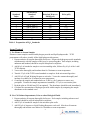
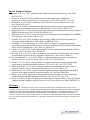
Product Manual OxiSelect™ Hydrogen Peroxide Assay Kit (Colorimetric), Trial Size Catalog Number STA-343-T 50 assays FOR RESEARCH USE ONLY Not for use in diagnostic procedures Introduction Oxidative stress is a physiological condition where there is an imbalance between concentrations of reactive oxygen species (ROS) and antioxidants. Research has shown that excessive ROS accumulation will lead to cellular injury, such as damage to DNA, proteins, and lipid membranes. Peroxides, such as hydrogen peroxide (H2O2), is one of the most well documented ROS produced under oxidative stress conditions. Hydrogen peroxide is an ROS that is a toxic product of normal aerobic metabolism and pathogenic ROS production involving oxidase and superoxide dismutase reactions. Hydrogen peroxide is poisonous to eukaryotic cells and in high doses can initiate oxidation of DNA, lipids, and proteins, which can lead to mutagenesis and cell death. The cellular damage caused by peroxides have been implicated in the development of many pathological conditions, such as ageing, asthma, arthritis, diabetes, cardiovascular disease, atherosclerosis, Down’s Syndrome, and neurodegenerative diseases. Cell Biolabs’ OxiSelect™ Hydrogen Peroxide Assay Kit is a quantitative assay for measuring hydrogen peroxides from both aqueous and lipid solutions. The kit employs a simple HTScompatible assay for measuring hydrogen peroxide concentrations in biological samples without any need for pretreatment. It can measure aqueous hydrogen peroxides and lipid hydrogen peroxides, and no extraction step is needed for lipid assay format. Absorbance values are proportional to the hydrogen peroxide levels within the samples. The Trial Size Hydrogen Peroxide Assay Kit has a detection sensitivity limit of 1 μM. Each kit provides sufficient reagents to perform up to 50 assays, including standard curve and unknown samples. Assay Principle The OxiSelect™ Hydrogen Peroxide Assay Kit is a quantitative assay for measuring hydrogen peroxide in aqueous and lipid samples. For aqueous samples, sorbitol first converts peroxide to a peroxyl radical, which oxidizes Fe+2 into Fe+3. For lipid samples, peroxide converts Fe+2 into Fe+3 directly. Then Fe+3 reacts with an equal molar amount of xylenol orange in the presence of acid to create a purple product that absorbs maximally between 540-600 nm. The antioxidant BHT is provided to prevent further undesirable chain peroxidation. The peroxide content in unknown samples is determined by comparison with the predetermined H2O2 standard curve. Related Products 1. STA-320: OxiSelect™ Oxidative DNA Damage ELISA Kit (8-OHdG Quantitation) 2. STA-330: OxiSelect™ TBARS Assay Kit (MDA Quantitation) 3. STA-341: OxiSelect™ Catalase Activity Assay Kit 4. STA-342: OxiSelect™ Intracellular ROS Assay Kit (Green Fluorescence) 5. STA-344: OxiSelect™ Hydrogen Peroxide/Peroxidase Assay Kit 6. STA-345: OxiSelect™ ORAC Activity Assay Kit 7. STA-347: OxiSelect™ In Vitro ROS/RNS Assay Kit (Green Fluorescence) 8. STA-832: OxiSelect™ MDA Adduct Competitive ELISA Kit 9. STA-838: OxiSelect™ HNE Adduct Competitive ELISA Kit 2 Kit Components 1. Xylenol Orange Dye (Part No. 234301-T): One 150 µL tube of a 12.5 mM solution 2. AFS Reagent (Part No. 234302-T): One 150 µL tube of AFS in H2SO4 3. Sorbitol Solution (Part No. 234303): One 1.5 mL tube of a 4 M solution 4. Hydrogen Peroxide (Part No. 234102-T): One 20 μL amber tube of an 8.8 M solution 5. 1000X BHT Solution (Part No. 234305-T): One 100 μL tube of 5% Butylated hydroxytoluene (BHT) in methanol 6. TCEP Solution (Part No. 234304): One 1.5 mL tube of a 1 mM Tris(2-carboxyethyl) phosphine solution in methanol Materials Not Supplied 1. Standard 96-well microtiter plates for use in microplate reader 2. Distilled or deionized water 3. 1X PBS for sample dilutions 4. Methanol for preparing lipid-based assays 5. H2SO4 6. 10 μL to 1000 μL adjustable single channel micropipettes with disposable tips 7. 50 μL to 300 μL adjustable multichannel micropipette with disposable tips 8. 96-well microtiter plate 9. Multichannel micropipette reservoir 10. Microplate reader capable of reading 560 nm Storage Store all kit components at 4ºC. Preparation of Reagents Aqueous Working Reagent: Prepare the Working Reagent for Aqueous assays by diluting the Xylenol Orange 1:100, the Sorbitol 1:40, and the AFS Reagent 1:100 together with deionized water (eg. For a 6.5 mL Working Reagent solution: add 0.065 mL of Xylenol Orange, 0.163 mL Sorbitol, and 0.065 mL AFS Reagent together and QS with deionized water to 6.5 mL. This is enough for 25 assays. Stir or vortex to homogeneity. The Working Reagent is stable for 1 day. Prepare only enough for immediate applications. Note: AFS Reagent contains acid. Use caution when handling. Lipid Working Reagent: Prepare the Working Reagent for Lipid assays by diluting the Xylenol Orange 1:100, AFS Reagent 1:100 and BHT 1:1000 together with methanol (90% minimum)(eg. For a 6.5 mL Working Reagent solution: add 0.065 mL of Xylenol Orange, 0.065 mL AFS Reagent and 6.5 μL BHT together and QS with methanol to 6.5 mL). Stir or vortex to 3 homogeneity. The Working Reagent is stable for 1 day. Prepare only enough for immediate applications. Preparations of Samples All samples should be assayed immediately or store at -80°C for up to 1-2 months. The assay can be used on cell culture supernatants, serum, plasma, urine, as well as other biological fluids. High levels of interfering substances may cause variations in results. Run proper controls as necessary. Always run a standard curve with samples. Media containing ferrous salts should be avoided, as they will interfere with the assay. Use PBS for dilution and preparation of samples. Cell Culture Supernatants: To remove insoluble particles, spin at 10,000 g for 5 min. The supernatant can be assayed directly or stored at -80°C as necessary. Cell supernatants can be assayed undiluted. Serum, Plasma or Urine: To remove insoluble particles, spin at 10,000 g for 5 min. The supernatant can be assayed directly or stored at -80°C as necessary. Notes: High Hydrogen peroxide (>200 µM) can bleach color from the dye, resulting in a lower absorbance value. The working range for this assay is 200 µM to 1 µM. If sample concentrations are unknown, perform several 1:10 serial dilutions of the sample in order to achieve a value within the assay’s working range. If absorbance values of diluted samples are the same or higher than the original values, this indicates excessive hydrogen peroxide. Repeat dilutions until the sample values are within range. If endogenous iron or other transition metals are suspected to be present in the sample, they may cause an increase in the signal. Compensate for the high signal by preparing a Working Reagent Blank (Working Reagent without AFS Reagent) as outlined in Preparation of Reagents by replacing AFS Reagent with 2.5 M H2SO4. The result will then be obtained by subtracting the sample value obtained with the Working Reagent from the sample value obtained with the Working Reagent Blank. Several chemicals are known to interfere and should be avoided in sample preparation. These include fructose, sorbitol, sucrose, glucose, formic acid and detergents such as SDS, Tween-20, NP-40 and Triton X-100. Cell and tissue lysates are not compatible with this assay. For best results we recommend the OxiSelect™ Hydrogen Peroxide/Peroxidase Assay Kit (Fluorometric), Cat. # STA-344. Preparation of Standard Curve 1. To prepare the Hydrogen Peroxide standards, first perform a 1:1000 dilution of the stock Hydrogen Peroxide in deionized water (aqueous assay) or methanol (lipid assay). Use only enough for immediate applications (eg. Add 5 μL of Hydrogen Peroxide to 4.995 mL deionized water). This solution has a concentration of 8.8 mM. 2. Use the 8.8 mM H2O2 solution to prepare standards in the concentration range of 0 µM – 100 µM by further diluting in water (aqueous assay) or methanol (lipid assay) (see Table 1). H2O2 diluted solutions and standards should be prepared fresh. Use the table below as a reference only. The volumes and concentrations of the standard may be adjusted by the user. 4 Standard Tubes 1 2 3 4 5 6 7 8 9 10 8.8 mM H2O2 Standard (µL) 23 500 of Tube #1 500 of Tube #2 500 of Tube #3 500 of Tube #4 500 of Tube #5 500 of Tube #6 500 of Tube #7 500 of Tube #8 0 Deionized Water or MeOH (µL) 977 500 500 500 500 500 500 500 500 500 H2O2 (µM) 200 100 50 25 12.5 6.25 3.125 1.56 0.78 0 Table 1. Preparation of H2O2 Standards Assay Protocol I. For Plasma or Serum Samples Plasma or serum samples contain both hydrogen peroxide and lipid hydroperoxide. TCEP pretreatment will reduce virtually all the lipid hydroperoxide present. 1. Prepare and mix all reagents thoroughly before use. Prepare the hydrogen peroxide standards simultaneously with the samples so they may be assayed together. Each sample, including unknown and standard, should be assayed in duplicate or triplicate. 2. Add 90 µL of standard or sample to a microcentrifuge tube, followed by 10 µL of the 1 mM TCEP solution. 3. Vortex tubes thoroughly and incubate them for 30 minutes at room temperature. 4. Transfer 25 µL of the TCEP treated standard or sample to fresh microcentrifuge tubes. 5. Add 250 µL of Lipid Working Reagent to each tube. Vortex the contents thoroughly and incubate on a shaker for 30 minutes at room temperature. 6. Centrifuge the sample and standard tubes at 12,000 x g for 5 minutes to remove any precipitate. Transfer the sample supernatant or standard (~275 µL) to a microtiter plate. 7. Read the plate at 540-600 nm (595 nm optimal). The absorbance should be read immediately. 8. Calculate the concentration of hydrogen peroxide within samples by comparing the sample absorbance to the standard curve. II. For Cell Culture Supernatants, Urine or other Biological Fluids 1. Prepare and mix all reagents thoroughly before use. Each sample, including unknown and standard, should be assayed in duplicate or triplicate. 2. Add 25 µL of standard or sample to the microtiter plate wells. 3. Add 250 µL of Aqueous or Lipid Working Reagent to each well. Mix the well contents thoroughly and incubate on a shaker for 30 minutes at room temperature. 5 4. Read the plate at 540-600 nm (595 nm optimal). The absorbance should be read immediately. 5. Calculate the concentration of hydrogen peroxide within samples by comparing the sample absorbance to the standard curve. Example of Results The following figures demonstrate typical Peroxide Assay results. One should use the data below for reference only. This data should not be used to interpret actual results. Aqueous Aqueous 1.8 0.4 1.6 0.35 0.3 OD 540nm OD 540nm 1.4 1.2 1 0.8 0.6 0.25 0.2 0.15 0.1 0.4 0.2 0.05 0 0 0 50 100 150 200 0 250 5 15 [H2O2] (µM) [H2O2] (µM) Lipid Lipid 1.2 0.3 1 0.25 0.8 OD 540nm OD 540nm 10 0.6 0.4 0.2 0.2 0.15 0.1 0.05 0 0 0 50 100 150 200 250 0 5 10 [H2O2] (µM) 15 20 25 [H2O2] (µM) Figure 1. Aqueous and Lipid based H2O2 Standard Curves. References 1. Jiang, Z.Y., Hunt, J.V. and Wolff, S.P. Anal. Biochem. (1992) 202: 384-389. 2. Jiang, Z.Y., Woolard, A.C.S. and Wolff, S.P. FEBS. (1990) 268(1): 69-71. 3. Jiang, Z.Y., Woolard, A.C.S. and Wolff, S.P. Lipids. (1991) 26: 853-856. 4. Nourooz-Zadeh, J. Tajaddini-Sarmadi, J. and Wolff, S.P. Anal. Biochem. (1994) 220: 403-409. 6 30 Recent Product Citations 1. Koman, V. B. et al. (2015). Multiscattering-enhanced absorption spectroscopy. Anal Chem. 87:1536-1543. 2. Otręba, M. et al. (2015). Effect of thioridazine on antioxidant status of HEMn-DP melanocytes. Naunyn Schmiedebergs Arch Pharmacol. doi:10.1007/s00210-015-1144-z. 3. Koňariková, K. et al. (2015). Anticancer effect of black tea extract in human cancer cell lines. SpringerPlus. 4:127. 4. Otręba, M. et al. (2015). Melanogenesis and antioxidant defense system in normal human melanocytes cultured in the presence of chlorpromazine. Toxicol In Vitro. 29:221-227. 5. Delijewski, M. et al. (2014). Nicotine impact on melanogenesis and antioxidant defense system in HEMn-DP melanocytes. Mol Cell Biochem. 395:109-116. 6. Delijewski, M. et al. (2014). Effect of nicotine on melanogenesis and antioxidant status in HEMnLP melanocytes. Environ Res. 134:309-314. 7. Auciello, F. R. et al. (2014). Oxidative stress activates AMPK in cultured cells primarily by increasing cellular AMP and/or ADP. FEBS Lett. 588:3361-3366. 8. Abudabos, A. M. & Al-Mufarrej, S. I. (2014). Effects of organic acid supplementation on antioxidant capacity and immune responses of broilers challenged orally with Salmonella enterica subsp. enterica Typhimurium. S Afr J Anim Sci. 44:360-370. 9. Chang, C. H. et al. (2014). Curcumin-protected PC12 cells against glutamate-induced oxidative toxicity. Food Technol Biotechnol. 52:468-478. 10. De Long, N. E. et al. (2014). Fluoxetine-induced pancreatic beta cell dysfunction: New insight into the benefits of folic acid in the treatment of depression. J Affect Disord. 166:6-13. 11. Di Cesare Mannelli, L. et al. (2014). PPAR- γ impairment alters peroxisome functionality in primary astrocyte cell cultures. Biomed Res Int. doi: 10.1155/2014/546453. 12. Gomez, I. G. et al. (2014). Anti-microRNA-21 oligonucleotides prevent Alport nephropathy progression by stimulating metabolic pathways. J Clin Invest. doi: 10.1172/JCI75852. 13. Medina-Leendertz, S. et al. (2014). Longterm melatonin administration alleviates paraquat mediated oxidative stress in Drosophila melanogaster. Invest Clin. 55:352-364. 14. Mahdavian, E. et al. (2014). Caspase-dependent signaling underlies glioblastoma cell death in response to the fungal metabolite, fusarochromanone. Int J Mol Med. 34:880-885. 15. Kang, J. et al. (2014). Suppression of photosynthetic gene expression in roots is required for sustained root growth under phosphate deficiency. Plant Physiol. 27:1156-1170. 16. Ke, K. et al. (2014). Reactive oxygen species induce the association of SHP-1 with c-Src and the oxidation of both to enhance osteoclast survival. Am J Physiol Endocrinol Metab. 307:E61-E70. Warranty These products are warranted to perform as described in their labeling and in Cell Biolabs literature when used in accordance with their instructions. THERE ARE NO WARRANTIES THAT EXTEND BEYOND THIS EXPRESSED WARRANTY AND CELL BIOLABS DISCLAIMS ANY IMPLIED WARRANTY OF MERCHANTABILITY OR WARRANTY OF FITNESS FOR PARTICULAR PURPOSE. CELL BIOLABS’ sole obligation and purchaser’s exclusive remedy for breach of this warranty shall be, at the option of CELL BIOLABS, to repair or replace the products. In no event shall CELL BIOLABS be liable for any proximate, incidental or consequential damages in connection with the products. 7 Contact Information Cell Biolabs, Inc. 7758 Arjons Drive San Diego, CA 92126 Worldwide: +1 858-271-6500 USA Toll-Free: 1-888-CBL-0505 E-mail: [email protected] www.cellbiolabs.com 2013-2015: Cell Biolabs, Inc. - All rights reserved. No part of these works may be reproduced in any form without permissions in writing. 8