1
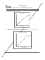
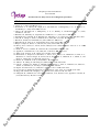
t.
uc
od
ur
pr
Phospho-p53 Ser392 ELISA Kit
User’s Manual
For Research Use Only, Not for use in diagnostic procedures
ith
yo
ELISA Kit for Measuring Phosphorylated Human p53 Ser392
ew
CycLex Phospho-p53 Ser392 ELISA Kit
ca
at
th
al
an
u
rm
se
fe
rt
o
th
eu
Intended Use................................................ 1
Storage......................................................... 1
Introduction.................................................. 2
Principle of the Assay.................................. 2-3
Materials Provided....................................... 4
Materials Required but not Provided........... 4
Precautions and Recommendations............. 5
Detailed Protocol......................................... 6-9
Calculations.................................…............ 9
Measurement Range.................................... 9
Troubleshooting........................................... 9-10
Reagent Stability......................................... 10
Assay Characteristics.................................. 10
Example of Test Results............................. 11
References................................................... 12
Related Products......................................... 13
m
Cat# CY-7051
re
Intended Use
Pl
ea
se
The CycLex Research Product CycLex Phospho-p53 Ser392 ELISA Kit is designed to detect and
quantify the level of human p53 protein that is phosphorylated at serine 392. Since the amino acid
sequence surrounding serine 392 is not conserved in mouse and rat p53, this ELISA kit can not be used
for mouse and rat cells. This assay is intended for the detection of phosphorylated human p53 at serine
392 from cell lysate.
nl
y!
This assay kit is for research use only and not for use in diagnostic or therapeutic procedures.
Storage
er
en
ce
Pu
rp
os
eO
• Upon receipt store all components at 4°C.
• Don’t expose reagents to excessive light.
Fo
rR
ef
Cat#: CY-7051
1
Version#: 140318
t.
uc
od
yo
ur
pr
Phospho-p53 Ser392 ELISA Kit
User’s Manual
For Research Use Only, Not for use in diagnostic procedures
ith
Introduction
rm
an
u
al
th
at
ca
m
ew
p53 is a short-lived, non-abundant protein that regulates the response of cells to DNA damage, in part
through transcriptional activation of genes involved in cell cycle control, DNA repair, and apoptosis (1).
p53 is a tumor suppressor protein; consequently, mice in which the p53 gene has been disrupted develop
tumors with high frequency (2, 3), and deletions or mutations in the p53 gene are prevalent in a majority
of human cancers (4, 5). The protein level and activity of p53 are regulated, and this is accomplished by
MDM2 (murine double minute 2) (6–10). The MDM2 gene is induced by p53, and MDM2 prevents
apoptosis by inhibiting p53 activity and promoting its degradation (6, 9, 11).
In response to DNA damage, the p53 protein is phosphorylated on each of the seven serines and one
threonine the in the first 50 amino acids of its N-terminal transactivation domain as well as at several
sites in its C-terminal tetramerization/regulatory domain (12, 13). As a transcription factor, p53 induces
or represses several genes that regulate cell cycle arrest, DNA repair or apoptosis, including p21WAF1,
MDM2, GADD45, p53R2, and p53AIP1. Recent studies suggest that specific p53 phosphorylation
events are important for the activation or repression of specific promoters (14-17).
Phosphorylation of human p53 Ser-392 C-terminal regulatory domain occurs following UV but not
ganmma irradiation (18, 19) and results in enhancement of sequence-specific binding activity in vitro
(20), possibly by promoting stable tetramer formation (21).
eu
se
Principle of the Assay
er
en
ce
Pu
rp
os
eO
nl
y!
Pl
ea
se
re
fe
rt
o
th
The CycLex Research CycLex Phospho-p53 Ser392 ELISA Kit is a solid phase sandwich ELISA.
An antibody specific for human p53 has been coated onto the wells of the microtiter strips provided.
Samples, including a standard containing phosphorylated form of p53 at Serine 392, control specimens,
and unknowns, are pipetted into these wells. During the first incubation, p53 protein binds to the capture
antibody on the well. After washing, mouse monoclonal antibody, specific for phosphorylated p53
Ser392 as a detection antibody, is added to the wells. During the second incubation, this antibody serves
as a detector by binding to the immobilized phosphorylated p53 protein at Ser392 captured during the
first incubation. After removal of excess detection antibody, followed by binding with horseradish
peroxidase conjugated anti-mouse IgG, which then catalyzes the conversion of the chromogenic
substrate tetra-methylbenzidine (TMB) from a colorless solution to a blue solution (or yellow after the
addition of stopping reagent). The color is quantitated by spectrophotometry and reflects the relative
amount of acetylated form of p53 at serine 392 present in the original specimen.
Fo
rR
ef
Cat#: CY-7051
2
Version#: 140318
t.
uc
od
yo
ur
pr
Phospho-p53 Ser392 ELISA Kit
User’s Manual
For Research Use Only, Not for use in diagnostic procedures
ith
Summary of Procedure
ew
Culture cells in culture flask or dish at 50-70 % confluency
Incubate O/N at 37°C in CO2 incubator
ca
Incubate appropriate time at 37°C in CO2 incubator
m
Add appropriate amount of the drug for induction of p53 Ser392 phosphorylation
th
at
Harvest the cells by scraping and centrifugation
Add 100 µL of diluted cell lysate to the wells
rm
Incubate for 1 hour at room temp.
an
u
al
Make cell lysate by adding extraction buffer and centrifugation
se
Wash the wells
eu
Add 100 µL of Primary Antibody Solution (anti-Phospho-p53 Ser392 monoclonal antibody)
th
Incubate for 1hour at room temp.
rt
o
Wash the wells
fe
Add 100 µL of Secondary Antibody Solution (HRP conjugated anti-mouse IgG antibody)
se
Wash the wells
re
Incubate for 1hour at room temp.
Pl
ea
Add 100 µL of Substrate Reagent
nl
y!
Add 100 µL of Stop Solution
er
en
ce
Pu
rp
os
eO
Measure absorbance at 450 nm
Fo
rR
ef
Cat#: CY-7051
3
Version#: 140318
t.
uc
od
yo
ur
pr
Phospho-p53 Ser392 ELISA Kit
User’s Manual
For Research Use Only, Not for use in diagnostic procedures
ith
Materials Provided
ew
All samples and standards should be assayed in duplicate. The following components are supplied and
are sufficient for the one 96-well microplate kit.
at
10X Wash Buffer: One 100 mL bottle of 10X buffer containing 2%Tween®-20
ca
m
Microplate: One microplate supplied ready to use, with 96 wells (12 strips of 8-wells) in a foil, zip-lock
bag with a desiccant pack. Wells are coated with anti-human p53 antibody as a capture antibody.
al
th
Cell Extraction Buffer: One bottle containing 20 mL of 1X buffer. For long term storage (over 3
months), store at -20°C.
an
u
Dilution Buffer: One bottle containing 50 mL each of 1X buffer; use for sample dilution. Ready to use.
rm
Phospho-p53 Ser392 Standard: One vial containing 240 units of lyophilized phosphorylated p53
Ser392.
eu
se
Primary Antibody Solution (Anti-Phospho-p53 Ser392 Monoclonal Antibody FPS392): One vial
containing 12 mL of Anti-Phospho-p53 Ser392 monoclonal antibody. Ready to use.
th
Secondary Antibody Solution (HRP conjugated Anti-Mouse IgG): One vial containing 12 mL of
HRP (horseradish peroxidase) conjugated anti-mouse IgG antibody. Ready to use.
rt
o
Substrate Reagent: One bottle containing 20 mL of the chromogenic substrate, tetra methylbenzidine
(TMB). Ready to use.
re
fe
Stop Solution: One bottle supplied ready to use, containing 20 mL of 1 N H2SO4.
se
Materials Required but not Provided
er
en
ce
Pu
rp
os
eO
nl
y!
Pl
ea
• Protease inhibitor cocktail: ex. Sigma Cat. # P-2714 (reconstituted according to manufacturer’s
guideline). Add 250 µL per 5 mL Cell Extraction Buffer.
• Orbital microplate shaker
• Pipettors: 2-20 µL, 20-200 µL and 200-1000 µL precision pipettors with disposable tips.
• Precision repeating pipettor
• Microcentrifuge and tubes for sample preparation.
• Vortex mixer
• Microplate washer: optional (Manual washing is possible but not preferable)
• Plate reader capable of measuring absorbance in 96-well plates at dual wavelengths of 450 nm/540
nm. Dual wavelengths of 450/550 or 450/595 nm can also be used. The plate can also be read at a
single wavelength of 450 nm, which will give a somewhat higher reading.
• Software package facilitating data generation and analysis :optional
• 500 or 1000 mL graduated cylinder
• Reagent reservoirs
• Deionized water of the highest quality
• Disposable paper towels
Fo
rR
ef
Cat#: CY-7051
4
Version#: 140318
t.
uc
od
yo
ur
pr
Phospho-p53 Ser392 ELISA Kit
User’s Manual
For Research Use Only, Not for use in diagnostic procedures
ith
Precautions and Recommendations
ew
• Allow all the components to come to room temperature before use.
m
• All microplate strips that are not immediately required should be returned to the zip-lock pouch, which
must be carefully resealed to avoid moisture absorption.
ca
• Do not use kit components beyond the indicated kit expiration date.
th
at
• Use only the microtiter wells provided with the kit.
al
• Rinse all detergent residue from glassware.
an
u
• Use deionized water of the highest quality.
rm
• Do not mix reagents from different kits.
eu
• Do not mouth pipette or ingest any of the reagents.
se
• The buffers and reagents in this kit may contain preservatives or other chemicals. Care should be taken
to avoid direct contact with these reagents.
o
th
• Do not smoke, eat, or drink when performing the assay or in areas where samples or reagents are
handled.
rt
• Dispose of tetra-methylbenzidine (TMB) containing solutions in compliance with local regulations.
re
fe
• Avoid contact with the acidic Stop Solution and Substrate Solution, which contains hydrogen peroxide.
ea
se
• Wear gloves and eye protection when handling immunodiagnostic materials and samples of human
origin, and these reagents. In case of contact with the Stop Solution and the Substrate Solution, wash
skin thoroughly with water and seek medical attention, when necessary.
nl
y!
Pl
• Biological samples may be contaminated with infectious agents. Do not ingest, expose to open
wounds or breathe aerosols. Wear protective gloves and dispose of biological samples properly.
er
en
ce
Pu
rp
os
eO
• CAUTION: Sulfuric Acid is a strong acid. Wear disposable gloves and eye protection when
handling Stop Solution.
Fo
rR
ef
Cat#: CY-7051
5
Version#: 140318
t.
uc
od
yo
ur
pr
Phospho-p53 Ser392 ELISA Kit
User’s Manual
For Research Use Only, Not for use in diagnostic procedures
ith
Detailed Protocol
ca
m
ew
The CycLex Research Product CycLex Phospho-p53 Ser392 ELISA Kit is provided with removable
strips of wells so the assay can be carried out on separate occasions using only the number of strips
required for the particular determination. Since experimental conditions may vary, an aliquot of the
Phospho-p53 Standard within the kit, should be included in each assay as a calibrator. Disposable pipette
tips and reagent troughs should be used for all liquid transfers to avoid cross-contamination of reagents
or samples.
at
Preparation of Working Solutions
an
u
al
th
All reagents need to be brought to room temperature prior to the assay. Assay reagents are supplied
ready-to-use, with the exception of 10X Wash Buffer, Cell Extraction Buffer and Phospho-p53
Ser392 Standard.
rm
1. Prepare a working solution of Wash Buffer by adding 100 mL of the 10X Wash Buffer to 900 mL of
deionized (distilled) water (ddH2O). Mix well.
se
2. Prepare a working solution of Cell Extraction Buffer by adding 250 µL of Protease inhibitor cocktail
(Sigma Cat. # P-2714) to 5 mL of Cell Extraction Buffer. Mix well.
th
eu
3. Reconstitute Phospho-p53 Ser392 Standard with 1.2 mL of ddH2O. The concentration of the
phospho-p53 Ser392 in vial should be 200 units/mL, which is referred as a Master Standard of
phospho-p53 Ser392.
re
fe
rt
o
Prepare Standard Solutions as follows:
Use the Master Standard to produce a dilution series (below). Mix each tube thoroughly before
the next transfer. The 100 units/mL standard (Std.1) serves as the highest standard. The Dilution
Buffer serves as the zero standard (Blank).
eO
nl
y!
Pl
ea
se
Volume of Standard
300 µL of Master Standard
300 µL of Std. 1 (100 units/mL)
300 µL of Std. 2 (50 units/mL)
300 µL of Std. 3 (25 units/mL)
300 µL of Std. 4 (12.5 units/mL)
300 µL of Std. 5 (6.25 units/mL)
300 µL of Std. 6 (3.13 units/mL)
-
Dilution Buffer
300 µL
300 µL
300 µL
300 µL
300 µL
300 µL
300 µL
300 µL
Concentration
100 units/mL
50 units/mL
25 units/mL
12.5 units/mL
6.25 units/mL
3.13 units/mL
1.56 units/mL
0 units/mL
os
Std.1
Std.2
Std.3
Std.4
Std.5
Std.6
Std.7
Blank
er
en
ce
Pu
rp
Note: Do not use a Repeating pipette. Change tips for every dilution. Wet tip with Dilution Buffer
before dispensing. Unused portions of Standards should be aliquoted and stored at below -70°C
immediately. Avoid multiple freeze and thaw cycles.
Fo
rR
ef
Cat#: CY-7051
6
Version#: 140318
t.
uc
od
yo
ur
pr
Phospho-p53 Ser392 ELISA Kit
User’s Manual
For Research Use Only, Not for use in diagnostic procedures
ith
Assay Procedure
ew
A. Treatment of Cells with compounds
1. Plate adherent cells or non-adherent cells in culture flasks at 50-70 % confluency.
ca
m
2. Incubate the culture flasks at 37°C over night in CO2 incubator.
at
3. Add appropriate amount of test compounds to each flask.
th
4. Incubate the culture flasks at 37°C for appropriate time.
an
u
al
B. Cell Extraction
rm
Note: This protocol has been successfully applied to several cell lines. Users should optimize the cell
extraction procedure for their own applications.
se
1. Collect cells in PBS by centrifugation (non-adherent cells) or scraping from culture flasks (adherent
cells).
eu
2. Wash cells twice with cold PBS.
o
th
3. Remove and discard the supernatant and collect the cell pellet.
At this point the cell pellet can be frozen at below -70°C and lyse at a later date.
fe
rt
4. Lyse the cell pellet in 0.5 mL* of Cell Extraction Buffer for 30 minutes, on ice, with vortexing at
10-minute intervals.
ea
se
re
* To get a rough idea you could adjust the cell concentration to around 2 x 10⁷ cells/mL.
Resulting protein concentration of the cell lysate should be 2-4 mg/mL using this Cell
Extraction Buffer.
nl
y!
Pl
* The volume of Cell Extraction Buffer depends on the cell number in cell pellet and
phosphorylation level of p53. For example, 1.2 x 10⁷ MCF-7 cells (in 10 cm dish) can be
extracted in 0.5 mL of Cell Extraction Buffer. Under these conditions, the protein concentration
should be 2-4 mg/mL, and use of 5-10 µL of the clarified cell extract diluted to a volume of 100
µl/well in Dilution Buffer is sufficient for the detection of phospho-p53 Ser392.
eO
5. Transfer the lysates to microcentrifuge tubes and centrifuge at 13,000 rpm for 10 minutes at 4°C.
Pu
rp
os
6. Aliquot the clear lysate to clean microfuge tubes. These samples are ready for assay. The lysates can
be stored at below -70°C. Avoid multiple freeze/thaw cycles.
er
en
ce
NOTE: THE ABOVE PROCEDURES ARE INTENDED ONLY AS A GUIDELINE. THE
OPTIMAL EXPERIMENTAL CONDITIONS WILL VARY DEPENDING ON THE
PARAMETERS BEING INVESTIGATED, AND MUST BE DETERMINED BY THE
INDIVIDUAL USER. NO WARRANTY OR GUARANTEE OF PERFORMANCE
USING THESE PROCEDURES IS MADE OR IMPLIED.
Fo
rR
ef
Cat#: CY-7051
7
Version#: 140318
t.
uc
od
ur
pr
Phospho-p53 Ser392 ELISA Kit
User’s Manual
For Research Use Only, Not for use in diagnostic procedures
yo
C. ELISA
ew
ith
1. Remove the appropriate number of microtiter wells from the foil pouch and place them into the well
holder. Return any unused wells to the foil pouch, refold, seal with tape and store at 4°C.
m
2. Dilute the lysate 1:10* with Dilution Buffer (e.g. 30 µL sample + 270 µL Dilution Buffer).
at
ca
* Lysates prepared in Cell Extraction Buffer must be diluted 1:10 or greater in Dilution Buffer. While a
1:10 lysate dilution has been found to be satisfactory, higher dilutions such as 1:20 or 1:40 may be
optimal. The dilution chosen should be optimized for each experimental system.
al
th
3. Pipette 100 µL of Standard Solutions (Std1-Std7, Blank) and diluted lysates in duplicates, into the
appropriate wells.
an
u
4. Incubate the plate at room temperature (ca. 25°C) for 1 hour, shaking at ca. 300 rpm on an orbital
microplate shaker.
se
rm
5. Wash 4-times by filling each well with Wash Buffer (350 µL) using a squirt bottle, multi-channel
pipette, manifold dispenser or microplate washer*.
eu
6. Add 100 µL of Primary Antibody Solution into each well.
th
7. Incubate the plate at room temperature (ca. 25°C) for 1 hour, shaking at ca. 300 rpm on an orbital
microplate shaker.
fe
rt
o
8. Wash 4-times by filling each well with Wash Buffer (350 µL) using a squirt bottle, multi-channel
pipette, manifold dispenser or microplate washer.
re
9. Add 100 µL of Secondary Antibody Solution into each well.
ea
se
10. Incubate the plate at room temperature (ca. 25°C) for 1 hour, shaking at ca. 300 rpm on an orbital
microplate shaker.
Pl
11. Wash 4-times by filling each well with Wash Buffer (350 µL) using a squirt bottle, multi-channel
pipette, manifold dispenser or microplate washer.
eO
nl
y!
12. Add 100 µL of Substrate Reagent. (Avoid exposing the microtiter plate to direct sunlight Covering
the plate with e.g. aluminum foil is recommended). Return Substrate A to 4°C immediately after the
necessary volume is removed
Pu
rp
os
13. Incubate the plate at room temperature (ca. 25°C) for 10-20 minutes, shaking at ca. 300 rpm on an
orbital microplate shaker. (The incubation time may be extended up to 30 minutes if the reaction
temperature is below than 20°C).
14. Add 100 µL of Stop Solution to each well in the same order as the previously added Substrate
Reagent.
er
en
ce
15. Measure absorbance in each well using a spectrophotometric microplate reader at dual wavelengths
of 450/540 nm. Dual wavelengths of 450/550 or 450/595 nm can also be used. Read the microplate
at 450 nm if only a single wavelength can be used. Wells must be read within 30 minutes of adding
the Stop Solution.
Fo
rR
ef
Cat#: CY-7051
8
Version#: 140318
t.
uc
od
yo
ur
pr
Phospho-p53 Ser392 ELISA Kit
User’s Manual
For Research Use Only, Not for use in diagnostic procedures
th
at
ca
m
ew
ith
Note-1: Complete removal of liquid at each step is essential to good performance. After the last
wash, remove any remaining Wash Buffer by aspirating or decanting. Invert the plate and
blot it against clean paper towels.
Note-2: Reliable standard curves are obtained when either O.D. values do not exceed 0.2 units for
the blank (zero concentration), or 2.5 units for the highest standard concentration. The plate
should be monitored at 5-minute intervals for approximately 30 minutes.
Note-3: If the microplate reader is not capable of reading absorbance greater than the absorbance of
the highest standard, perform a second reading at 405 nm. A new standard curve,
constructed using the values measured at 405 nm, is used to determine Phospho-p53 Ser392
concentration of off-scale samples. The readings at 405 nm should not replace the on-scale
readings at 450 nm.
an
u
al
Calculations
th
eu
se
rm
Average the duplicate readings for each standard, control, and sample and subtract the average zero
standard optical density. Plot the optical density for the standards versus the concentration of the
standards and draw the best curve. The data can be linearized by using log/log paper and regression
analysis may be applied to the log transformation. To determine the Phospho-p53 Ser392 concentration
of each sample, first find the absorbance value on the y-axis and extend a horizontal line to the standard
curve. At the point of intersection, extend a vertical line to the x-axis and read the corresponding
Phospho-p53 Ser392 concentration. If the samples have been diluted, the concentration read from the
standard curve must be multiplied by the dilution factor.
re
fe
rt
o
A. The dose-response curve of this assay fits best to a sigmoidal 5-parameter logistic equation. The
results of unknown samples can be calculated with any computer program having a 5-parameter
logistic function. It is important to make an appropriate mathematical adjustment to accommodate for
the dilution factor.
Pl
ea
se
B. Most microtiter plate readers perform automatic calculations of analyte concentration. The calibration
curve is constructed by plotting the absorbance (Y) of calibrators versus log of the known
concentration (X) of calibrators, using the four-parameter function. Alternatively, the logit log
function can be used to linearize the calibration curve (i.e. logit of absorbance (Y) is plotted versus log
of the known concentration (X) of calibrators).
nl
y!
Measurement Range
er
en
ce
Pu
rp
os
eO
The measurement range is 1.56 units/mL to 100 units/mL. Any sample reading higher than the highest
standard should be diluted with Dilution Buffer in higher dilution and re-assayed. Dilution factors need
to be taken into consideration in calculating the phospho-p53 Ser392 concentration.
Fo
rR
ef
Cat#: CY-7051
9
Version#: 140318
t.
uc
od
yo
ur
pr
Phospho-p53 Ser392 ELISA Kit
User’s Manual
For Research Use Only, Not for use in diagnostic procedures
ith
Troubleshooting
ew
1. The Human Phospho-p53 Ser392 Standard should be run in duplicate, using the protocol described in
the Detailed Protocol. Incubation times or temperatures significantly different from those specified
may give erroneous results.
at
ca
m
2. Poor duplicates, accompanied by elevated values for wells containing no sample, indicate insufficient
washing. If all instructions in the Detailed Protocol were followed accurately, such results indicate a
need for washer maintenance.
al
th
3. Overall low signal may indicate that desiccation of the plate has occurred between the final wash and
addition of Substrate Reagent. Do not allow the plate to dry out. Add Substrate Reagent immediately
after wash.
an
u
Reagent Stability
eu
se
rm
All of the reagents included in the CycLex Research Product CycLex Phospho-p53 Ser392 ELISA
Kit have been tested for stability. Reagents should not be used beyond the stated expiration date. Upon
receipt, kit reagents should be stored at 4°C, except the reconstituted Human Phospho-p53 Ser392
Standard must be stored at below -70°C. Coated assay plates should be stored in the original foil bag
sealed by the zip lock and containing a desiccant pack.
th
For research use only, not for use in diagnostic or therapeutic procedures
rt
o
Assay Characteristics
se
re
fe
1. Sensitivity
The limit of detection (defined as such a concentration of Phospho-p53 Ser392 giving absorbance
higher than mean absorbance of blank* plus three standard deviations of the absorbance of blank: A
blank + 3*SD blank) is better than 1.413 units/ml of sample.
ea
* Dilution Buffer is pipetted into blank wells.
nl
y!
Pl
Eighty assays were evaluated and the minimum detectable dose (MDD) of Phospho-p53 Ser392
ranged from 0.723 – 1.624 units/mL. The mean MDD was 1.413 units /mL. The MDD was determined
by adding three standard deviations to the mean optical density value of twenty zero standard replicates
and calculating the corresponding concentration.
os
eO
2. Specificity
The antibodies in the CycLex Phospho-p53 Ser392 ELISA Kit are highly specific of human
Phospho-p53 Ser392, with no detectable cross-reactivity to mouse and rat Phospho-p53.
er
en
ce
Pu
rp
3. Linearity
To assess the linearity of the assay, samples containing and/or spiked with high concentrations of
Phospho-p53 Ser392 were serially diluted with the Dilution Buffer to produce samples with values
within the dynamic range of the assay.
Fo
rR
ef
Cat#: CY-7051
10
Version#: 140318
t.
uc
od
yo
ur
pr
Phospho-p53 Ser392 ELISA Kit
User’s Manual
For Research Use Only, Not for use in diagnostic procedures
ith
Example of Test Results
ew
Fig.1 Typical standard of phospho-p53 Ser392
Standard curve
m
3.0
ca
2.5
at
th
1.5
al
A450
2.0
an
u
1.0
rm
0.5
0
20
se
0.0
40
60
80
100
eu
Phospho-p53 S392 (U/ml)
rt
o
th
Fig.2 Phospho-p53 Ser392 ELISA using breast cancer cell line MCF-7 that expresses wild type p53,
which have been treated with 10 µM SN-38 for indicated time
fe
Phospho-p53 S392 ELISA: M CF-7 treated with
10 uM SN-38 for indicated time
re
3.5
se
3.0
Pl
2.0
er
en
ce
Pu
rp
os
eO
nl
y!
A450
ea
2.5
Fo
rR
ef
Cat#: CY-7051
1.5
1.0
0.5
0.0
0
1
2
3
4
5
6
7
SN-38 treatment time(hr.)
11
Version#: 140318
t.
uc
od
yo
ur
pr
Phospho-p53 Ser392 ELISA Kit
User’s Manual
For Research Use Only, Not for use in diagnostic procedures
ith
References
er
en
ce
Pu
rp
os
eO
nl
y!
Pl
ea
se
re
fe
rt
o
th
eu
se
rm
an
u
al
th
at
ca
m
ew
1. Levine, A. J. (1997) Cell 88, 323–331
2. Donehower, L. A., Harvey, M., Slagle, B. L., McArthur, M. J., Montgomery, C. A., Jr., Butel, J. S.,
and Bradley, A. (1992) Nature 356, 215–221
3. Harvey, M., McArthur, M. J., Montgomery, C. A., Jr., Bradley, A., and Donehower, L. A. (1993)
FASEB J. 7, 938–943
4. Hollstein, M., Sidransky, D., Vogelstein, B., and Harris, C. C. (1991) Science 253, 49–53
5. Greenblatt, M. S., Bennett, W. P., Hollstein, M., and Harris, C. C. (1994) Cancer Res. 54, 4855–4878
6. Chen, H. E., Chang, S., Trub, T., and Neel, B. G. (1996) Mol. Cell. Biol. 16, 3685–3697
7. Honda, R., Tanaka, H., and Yasuda, H. (1997) FEBS Lett. 420, 25–27
8. Kubbutat, M. H., Jones, S. N., and Vousden, K. H. (1997) Nature 387, 299–303
9. Haupt, Y., Maya, R., Kazaz, A., and Oren, M. (1997) Nature 387, 296–299
10. McCoy, M. A., Gesell, J. J., Senior, M. M., and Wyss, D. F. (2003) Proc. Natl. Acad. Sci. U. S. A. 100,
1645–1648
11. Barak, Y., Juven, T., Haffner, R., and Oren, M. (1993) EMBO J. 12, 461–468
12. Appella, E., and Anderson, C. W. (2001) Eur. J. Biochem. 268, 2764-2772
13. Wahl, G. M., and Carr, A. M. (2001) Nat. Cell Biol. 3, E277-E286
14. Buschmann, T., Potapova, O., Bar-Shira, A., Ivanov, V. N., Fuchs, S. Y., Henderson, S., Fried, V. A.,
Minamoto, T., Alarcon-Vargas, D., Pincus, M. R., Gaarde, W. A., Holbrook, N. J., Shiloh, Y., and
Ronai, Z. (2001) Mol. Cell. Biol. 21, 2743-2754
15. Dumaz, N., and Meek, D. W. (1999) EMBO J. 18, 7002-7010
16. Jabbur, J. R., Huang, P., and Zhang, W. (2000) Oncogene 19, 6203-6208
17. Oda, K., Arakawa, H., Tanaka, T., Matsuda, K., Tanikawa, C., Mori, T., Nishimori, H., Tamai, K.,
Tokino, T., Nakamura, Y., and Taya, Y. (2000) Cell 102, 849-862
18. Kapoor, M. and Lozano, G., (1998) Proc. Natl. Acad. Sci. USA 95, 2834–2837.
19. Lu, H., Taya, Y., Ikeda, M. and Levine, A.J. (1998) Proc. Natl. Acad. Sci. USA 95, 6399–6402.
20. Hupp, T.R., Meek, D.W., Midgley, C.A. and Lane, D.P. (1992) Cell 71, 875–886.
21. Sakaguchi, K., Sakamoto, H., Lewis, M.S., Anderson, C.W., Erickson, J.W., Appella, E. and Xie, D.
(1997) Biochemistry 36, 10117–10124.
Fo
rR
ef
Cat#: CY-7051
12
Version#: 140318
t.
uc
od
yo
ur
pr
Phospho-p53 Ser392 ELISA Kit
User’s Manual
For Research Use Only, Not for use in diagnostic procedures
ew
eu
se
rm
an
u
al
th
at
ca
m
* CycLex Total p53 ELISA Kit: Cat# CY-7049
* CycLex Phospho-p53 Ser46 ELISA Kit: Cat# CY-7050
* CycLex Phospho-p53 Ser392 ELISA Kit: Cat# CY-7051
* Anti-Phospho-p53 Ser46 (TK-4D4) monoclonal antibody: Cat# CY-M1022
ith
Related Products
o
rt
fe
re
se
CycLex Co., Ltd.
1063-103 Terasawaoka
Ina, Nagano 396-0002
Japan
Fax: +81-265-76-7618
e-mail: [email protected]
URL: http://www.cyclex.co.jp
th
PRODUCED BY
er
en
ce
Pu
rp
os
eO
nl
y!
Pl
ea
CycLex/CircuLex products are supplied for research use only. CycLex/CircuLex products and
components thereof may not be resold, modified for resale, or used to manufacture commercial
products without prior written approval from CycLex Co., Ltd.. To inquire about licensing for
such commercial use, please contact us via email.
Fo
rR
ef
Cat#: CY-7051
13
Version#: 140318


![LS5105 Document No 2 [PDF 1MB] - Australian Electoral Commission](http://vs1.manualzilla.com/store/data/005655823_1-2458abda02bbd8390d0ac9ba8bd86ac6-150x150.png)