1
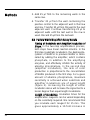
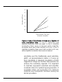
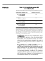
User Manual
ELISA Amplification System
Cat. No. 19589-019
Table of Contents
1. Notices to Customer .................................... 1
1.1 Important Information ........................... 1
1.2 Precautions .......................................... 1
2. Overview ..................................................... 2
3. Methods ...................................................... 4
3.1 Components......................................... 4
3.2 Storage ................................................ 4
3.3 Advance Preparations.......................... 5
3.4 Recommendations for Optimal
Results ............................................ 6
3.5 ELISA Amplification System
Protocol .......................................... 7
3.6 Color Measurement Assays ................. 8
3.7 Use of the Positive Control .................. 9
3.8 Factors Which May Affect
Assay Results .............................. 10
3.9 Performance Standards .................... 13
4. Troubleshooting Guide .............................. 14
5. References ................................................ 15
i
Table of
Contents
Figures:
1. Schematic of Alkaline Phosphatase
Signal Amplification .............................. 3
2. Rate of Absorbance Increase as a
Function of Total Incubation Time ...... 11
Table:
1. ELISA Amplification Versus pNPP
in an Indirect ELISA ............................. 12
ii
1
Notices to Customer
1.1
Important Information
This product is authorized for laboratory research
use only. The product has not been qualified or
found safe and effective for any human or animal
diagnostic or therapeutic application. Uses for
other than the labeled intended use may be a violation of applicable law.
1.2
Precautions
PRECAUTIONS FOR USE. Warning: This product
contains hazardous reagents. It is the end-user’s
responsibility to consult the applicable MSDS(s)
before using this product. Disposal of waste organics, acids, bases, and radioactive materials must
comply with all appropriate federal, state, and local
regulations. If you have any questions concerning
the hazards associated with this product, please
call the Invitrogen Environmental Health and
Safety Chemical Emergency hotline at (301) 4318585.
1
2
Overview
The ELISA Amplification System is designed to
amplify the amount of color generated by a given
quantity of immobilized alkaline phosphatase in a
soluble substrate enzyme-linked immunosorbent
assay (ELISA). In conventional detection systems,
bound enzyme acts directly on the substrate to
produce a colored end product. The signal generated by this type of system is limited by the linear
nature of the reaction kinetics. In the ELISA
Amplification System, the bound enzyme acts on a
substrate whose product initiates a secondary
cyclic enzyme reaction, resulting in a colored product. Each molecule of product from the first reaction takes part in many cycles of the second reaction; thus, the signal generated by the first
enzyme reaction is amplified by participation of its
end product in the cycles of the second reaction.
The substrate in this system is the reduced form of
nicotinamide adenine dinucleotide phosphate
(NADPH), which is dephosphorylated by bound
alkaline phosphatase to reduced nicotinamide adenine dinucleotide (NADH). NADH activates a secondary enzyme system which comprises a redox
cycle driven by diaphorase and alcohol dehydrogenase. In this cycle, NADH, in the presence of
diaphorase, reduces a tetrazolium salt (iodonitrotetrazolium violet or INT-violet) to form an intensely
2
colored formazan dye and is itself oxidized to form
NAD+. NAD+ is then reduced to NADH and ethanol
is oxidized to acetaldehyde in the presence of
alcohol dehydrogenase. The rate of reduction of
the tetrazolium salt is directly proportional to the
concentration of NADH origin-ally formed by the
enzyme in the bound conjugate. A schematic of
the reaction is shown in figure 1.
NAD+
Formazan*
Ethanol
Alcohol Dehydrogenase
Diaphorase
INT-Violet
Acetaldehyde
NADH
Alkaline Phosphatase
Pi
NADPH
*Colored end product
Figure 1. Schematic of alkaline phosphatase signal
amplification.
3
3
Methods
3.1 Components
The components of the ELISA Amplification
System are as follows. Each vial of substrate, substrate diluent, amplifier, and amplifier diluent
contains enough reagent to perform two 96-well
microtiter plate assays with the volumes suggested
in the instructions. Each system provides reagents
sufficient to perform 10 microtiter plate assays.
Component
Substrate (lyophilized NADPH)
Substrate diluent (1X buffer for substrate)
Amplifier [lyophilized amplifying enzymes
(alcohol dehydrogenase and diaphorase)
Amplifier diluent (1X buffer for amplifier)
Positive control (streptavidin alkaline
phosphatase conjugate (1 mg/ml)
Amount
5 vials
60 ml
(5 × 12 ml)
5 vials
60 ml
(5 × 12 ml)
100 µl
3.2 Storage
Upon receipt, the reagents in the ELISA
Amplification System should be stored at 4°C. The
substrate and amplifier are supplied lyophilized.
After reconstitution, the amplifier is stable for
1 week at -20°C, and the substrate is stable for
8 days at 2-8°C. A slight pink color in the amplifier
solution may develop during storage. This will not
4
affect the interpretation of results.
3.3 Advance Preparations
The following reagents are required, but they are
not provided with the system. Prepare them as follows:
Final washing buffer/positive control
diluent: Tris-buffered saline (TBS); 0.05 M TrisHCl (pH 7.5) and 0.15 M NaCl. This amount of
buffer is sufficient for two 96-well plate assays.
To prepare, add 1.51 g of Tris base (MW
121.14) and 2.19 g of sodium chloride (MW
58.5) to 230 ml of deionized water. Stir until
dissolved. Adjust the pH to 7.5 with 2 M HCl.
Adjust the volume to 250 ml with deionized
water.
Stop Solution: 0.3 M H2SO4. To 59 ml of deionized water, slowly add 1 ml of concentrated sulfuric acid (MW 98, specific gravity
1.84, purity 96% to 98%). Caution: When using
sulfuric acid, exercise caution and follow
manufacturer’s safety recommendations. When
making dilutions, always add acid to water.
1. Bring all reagents, except the positive control, to
room temperature before use.
2. Reconstitute the substrate at room temperature
10 min before use by adding 12 ml of substrate
diluent directly to the substrate vial. Gently mix
until completely dissolved, using clean pipets to
avoid contamination. Do not leave uncovered.
Reconstituted substrate is stable for 8 days at
2-8°C
5
Methods
3. Reconstitute the amplifier 10 min before use by
adding 12 ml of amplifier diluent. Gently mix
until completely dissolved, using clean pipets to
avoid contamination. Do not leave uncovered.
Reconsituted amplifier is stable for 1 week at
-20°C
4. Dilute the positive control before use. Please
read Section 3.7, Use of the Positive Control,
for instructions.
5. Select a color measurement assay as described
in the Section 3.6, Color Measurement Assays.
3.4 Recommendations for Optimal Results
1. Keep pipets and laboratory vessels used to
dispense alkaline phosphatase conjugate and
the positive control away from those used for
other components, especially the substrate.
2. Minimize any nonspecific binding of antibody or
conjugate by carefully washing the microtiter
plate wells. Amplification reagents will amplify
any nonspecific signal.
3. Avoid using phosphate in any wash buffer. It
strongly inhibits the substrate reaction.
4. The substrate and amplifier reactions occur
quite rapidly and require precise timing. To
ensure equal incubation times in each well, add
the substrate, amplifier, and stop solution in the
same well-to-well sequence.
6
3.5 ELISA Amplification System Protocol
N o t e : Please read the
protocol thoroughly before
beginning. Some of the
steps in this procedure need
to be performed simultaneously.
1. Perform a standard ELISA using alkaline
phosphatase. Leave the last row of the
microtiter plate empty at this stage for addition
of the positive control.
2. Remove excess alkaline phosphatase by
washing each well four times with 0.25 ml of
TBS buffer (see Section 3.3, Advance
Preparations). Wet the positive control wells
with this buffer. Remove the fourth wash from
all wells just before you proceed with the next
step.
3. Add the positive control dilutions (see Section
3.7, Use of the Positive Control) to the last row
of the microtiter plate.
4. Substrate: Add 50 µl of reconstituted substrate
(prepared in advance) to each well, including
the wells you have reserved for the positive
control. Incubate for 15 min at 25°C. To
increase sensitivity, extend the length of incubation, as described in Section 3.8, Factors Which
May Affect Results.
Amplifier: Add 50 µl of reconstituted amplifier
(prepared in advance) to each well. Incubate for
at least 15 min at 25°C, being careful not to
cross-contaminate wells.
Add amplifier and substrate in same sequence.
5. Measure the color development, using the
assay method you have selected in advance.
E n d p o i n t o r s t o p p e d a s s a y : Stop color
development after 15 min with 50 µl of
0.3 M H2SO4 per well. Add the stop reagent in
7
Methods
the same time sequence as the amplifier.
Record the absorbance at 495 nm.
Kinetic assay: Read the absorbance at 495 nm
of each well every 3 min for 15 min.
6. To maintain the effectiveness of reconstituted
substrate and amplifier for future use, store
them immediately at 4°C.
3.6 Color Measurement Assays
The following descriptions apply to measurement
of color obtained in microtiter plate-based assays.
However, the general principle is applicable to
many systems employing a variety of reaction vessels.
End Point or Stopped Assays. In this type of
assay, color development is allowed to proceed
for a predetermined time so that sufficient color
develops in the test wells. Stopping the
reaction, in effect halting any further cycling
activity, is achieved by adding dilute acid. The
stop solution must be added to the wells in a
timed sequence identical to that used for the
addition of substrate and amplifier. Assuming a
reaction volume of 50 µl of substrate plus 50 µl
of amplifier, 50 µl of acid will stop the reaction.
Once all the wells have been stopped, the color
can be quantified by reading with a suitable
colorimeter at 495 nm.
Kinetic Assays. In a kinetic assay, color development is not stopped, but read repeatedly over
a period of time and the rate of color development calculated. Since cycling reactions show
linear kinetics, rates of reaction can be
8
determined by plotting a regression line through
the series of optical density measurements.
The rates on the unknowns are then compared
with the rates of standard samples. This method
of reading also reduces problems which can
occur with some multioptic readers from
“channel-to-channel variation” in optical density.
3.7 Use of the Positive Control
The positive control included with this system
ensures that the components provided are
performing properly. The positive control is alkaline
phosphatase chemically coupled to streptavidin, a
reagent used in many ELISA systems. When
dilutions of the alkaline phosphatase conjugate are
mixed with the amplifications reagents, color
development occurs. The expected absorbance
from each dilution will vary for each detector;
however, the optical densities should decrease
linearly as the control is diluted.
The approximate concentration of alkaline phosphatase in the conjugate (before dilution) is
1 mg/ml. Therefore, final dilution in each well
ranges from 1:4,000 (250 ng/ml) to 1:512,000
(~2 ng/ml) in two-fold increments per well. We
recommend you reserve the last row of each
assay plate for the positive control reaction, using
25 µl per well of serially diluted positive control.
1. Dilute the Positive Control 1:4,000 with TBS
(see Section 3.3, Advance Preparations).
2. Transfer 50 µl of the diluted positive control to
the first well of the last row in the microtiter
plate.
9
Methods
3. Add 25 µl TBS to the remaining wells in the
same row.
4. Transfer 25 µl from the well containing the
positive control to the adjacent well in that row
and mix. Transfer 25 µl from this well to the next
adjacent well. Continue transferring 25 µl to
adjacent wells until the last well in the row is
used. Discard 25 µl from the last well.
3.8 Factors Which May Affect Assay Results
Timing of Substrate and Amplifier Incubation of
Steps. In the two-step amplifications process,
both steps have linear reaction kinetics. In the
first step (substrate incubation), the dephosphorylation of NADPH to produce NADH is terminated by adding the amplifier, which contains
phosphate, in addition to the amplifying
enzymes, and effectively inhibits the activity of
alkaline phosphatase. In the second step
(amplifier incubation), the rate of color
production is proportional to the concentration
of NADH produced in the first step. For a given
amount of alkaline phosphatase, maximum
sensitivity is achieved when substrate and
amplifier incubations are of equal length
(figure 2). Increasing the length of either
incubation alone will increase the signal but to a
lesser degree than equal length incubations.
Length of Incubation. Incubation times for the
amplification reagents can be varied depending
on the sensitivity required. We recommend that
you incubate each reagent for 15 min. This
gives approximately a 10-fold increase in
10
1.4
Absorbance (495 nm)
1.2
1.0
1
0.8
2
0.6
0.4
3
0.2
10
20
30
40
50
60
70
80
90
100
Total Incubation Time (min)
(Substrate + Amplifier)
Figure 2. Rate of absorbance increase as a function of
t o t a l i n c u b a t i o n t i m e . In line 1, substrate and
amplifier incubation times are equal. In line 2, substrate
incubation times were increased, while amplifier
incubation times were held constant at 15 min. In line 3,
substrate incubation times were held constant at 15 min,
while amplifier incubation times were increased.
sensitivity over the traditionally used substrate,
pNPP, as demonstrated in table 1. If more (or
less) sensitivity is required, incubation of both
reagents for longer (or shorter) time periods can
achieve the sensitivity required. It is important
to maintain equivalent substrate and amplifier
incubation times for optimal signal generation. If
incubation times are increased, it is critical to
minimize any nonspecific reactions during the
ELISA procedure.
11
Methods
Table 1. ELISA amplification versus pNPP
in an indirect ELISA.
Total Incubation Time of
Amplification Reagents (min)
Approximate Fold Increase in
Sensitivity over pNPP
30 (15 for substrate
15 for amplifier)
10
45 (22.5 for substrate,
22.5 for amplifier)
15
60 (30 for substrate,
30 for amplifier)
20
90 (45 for substrate,
45 for amplifier)
30
Fold increase was determined by comparing the quantity of antigen that
gave equivalent absorbances within the linear range of the detector. Antigen
(purified rabbit IgG) concentrations ranged from 50 ng to 0.1 pg/well. All
wells were incubated with optimal dilutions of Biotinylated Goat Anti-rabbit
IgG, followed by Streptavidin-Alkaline Phosphatase Conjugate. The
substrate, pNPP, was used at 1 mg/ml in 10% diethanolamine (pH 9.8),
0.5 mM MgCl2, and incubated for 60 min.
Temperature. Because the amplification steps
are enzymatic reactions, they are temperature
dependent. For consistent results, we recommend that the amplification reagents be used at
a constant temperature between 20°C and
25°C.
Reagent Volumes and Concentrations. The signal may be strengthened by increasing the final
assay volumes of the substrate and amplifier
and, to some extent, by using a more
concentrated amplifier. However, the reagents
supplied with this system are paired, and we do
not ensure the stability or performance of
nonstandard volumes or concentrations.
12
3.9 Performance Standards
The strength of signal and sensitivity of the ELISA
Amplification System are routinely analyzed to
ensure performance. We require the ELISA
Amplification System to give maximal absorbance
readings versus 10 ng of purified antigen within
30 min during the substrate and amplifier incubations. In addition, the system must be able to
detect as little as 100 pg of purified antigen within
the same timeframe. We measure these parameters in an indirect ELISA using optimal dilutions of
biotinylated antibodies and Streptavidin-Alkaline
Phosphatase Conjugate.
13
4
Troubleshooting Guide
Problem
Probable Causes
High background
Substrate not fresh.
Substrate/amplifier incubated too long.
Stop Solution omitted.
Insufficient washing.
Wash buffer too dilute.
Contamination of substrate with phosphatase.
Reagent too old or improperly stored.
High level of nonspecific binding.
Random high-absorbing wells
Positive control splashed into other wells.
Pipet tips contaminated with positive control.
Insufficient washing.
Completely red plate
Weak color development in
positive control
Substrate/amplifier not properly dissolved or thoroughly mixed.
Substrate mixed in a container which previously contained
alkaline phosphatase.
Incubation temperature of the assay too high or low.
Substrate/amplifier incubation time too short.
Incorrect preparation of substrate/amplifier.
Substrate/amplifier not fully dissolved.
Unexpectedly high, low, or
variable absorbance readings
Gross variation in signal
Positive control omitted.
Plate reader at wrong wavelength.
Debris on bottom of wells.
Bubbles in wells caused by inaccurate pipetting or
incorrect preparation of substrate/amplifier.
Inefficient removal of wash buffer.
Inaccurate pipetting.
Contaminated pipet tips.
Incorrect preparation of substrate/amplifier.
14
5
References
1. Self, C.H. (1985) J. Immunol. Method. 76, 389.
2. Stanley, C.J., Johannsson, A., and Self, C.H.
(1985) J. Immunol. Method. 83, 89.
15
Notes
16
Notes
17
Notes
18
United States Headquarters:
Invitrogen Corporation
5791 Van Allen Way
Carlsbad, California 92008
Tel: 1 760 603 7200
Fax: 1 760 602 6500
Email: [email protected]
For country-specific contact information,
visit our website at
www.invitrogen.com
Part No. 19589
Rev. Date: 14 July 2008