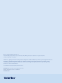
1
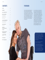
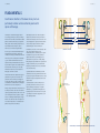
Care at home Guide to Spinal Cord Injury Paraplegia and Urology 2 contents Care at home 3 CONTENTS FOREWORD Foreword 3 Fundamentals 4 Aims of urological treatment 6 Neurourological diagnosis 7 8 Imaging procedures 9 Functional diagnosis 10 Kidney function tests 11 Laboratory tests Treatment of bladder dysfunction Reducing bladder pressure Emptying the bladder 12 13 14 Urinary tract infections 18 18 Clinical signs It is not uncommon for people living with spinal cord injury to have bladder dysfunction. Depending on severity, this can represent a serious threat to kidney function and/or greatly restrict quality of life. For a long time kidney function disorders were the most common cause of death among paraplegics. In the last 30 years, advances in neurological diagnosis and therapy have drastically reduced the risk of kidney damage, so that today life expectancy for the majority of paraplegics has vastly improved. However, urological issues are life-long concerns for those affected by them. After completion of the first rehabilitation phase, problems with bladder function are the most common reason for a paraplegic to visit a doctor. An understanding of the principles of bladder health and knowledge of the possibilities and limitations of urological treatment are consequently of extreme importance. This guide is intended to address the most common issues encountered during management of bladder function disorders and to supply basic information with the aim of achieving, in conjunction with urologists, independent control of bladder health while maintaining the best possible quality of life. Prof. Dr. med. Jürgen Pannek, Head Physician Neurourology Swiss Paraplegic Centre, Nottwil Alternatives to intermittent catheterization 21 Incontinence 24 New trends in the treatment of bladder dysfunction 25 Sexuality and paraplegia 26 Fertility28 Urine measurement 29 Homecare urology products 30 Information in this brochure is for your general knowledge and is not intended to be a substitute for the advice of a qualified medical professional. The information should not be considered complete and should not be used in place of a visit, call, consultation or advice of your physician or other healthcare provider. Teleflex does not practice medicine nor provide medical services or medical advice. You should seek prompt medical advice for any specific health issues and consult a qualified medical professional before purchasing any product(s). You should not disregard professional medical advice or delay in seeking it because of something you have read in this brochure. Do not consult this brochure in the event of a medical emergency. 4 fundamentals Care at home 5 FUNDAMENTALS Coordination disorders of the lower urinary tract are particularly common and are extremely severe after spinal cord damage. Renal pelvis Ureter Our urinary tract is divided into an upper and lower section. The upper urinary tract consists of the kidneys and ureters. The lower urinary tract includes the bladder, sphincter, urethra and, in men, the prostate gland. The kidneys produce urine that is transported by the ureters into the bladder. The lower urinary tract has two functions: to store urine and to empty the bladder. To do these jobs, not only must the individual organs be intact but their interaction must be controlled and coordinated. The nervous system fulfills these functions. The nervous system receives impulses from the bladder (e.g., the sensation felt when the bladder needs to be emptied) and conveys this information via the nerves in the pelvis minor to the spinal cord, and then from there to the brain stem. Here the various pieces of incoming information are interconnected to be controlled and managed by centers in the cerebrum. The instructions from brain to bladder take the opposite path (brain stem – spinal cord – nerves of the minor pelvis) back to the lower urinary tract and trigger the actions required. Thus, the centers for voluntary control (cerebrum) and for coordination of the incoming signals (brain stem) are located above the spinal cord. Damage to the spine means that the connections between the urinary tract and the control centers in the skull are completely or partially disconnected. There are two reflex centers in the spinal cord that are capable of activating, but not of coordinating, the bladder impulses: an upper and lower center. If the reflex centers in the spinal cord located below the injury take over control, bladder and sphincter activity becomes uncoordinated. With an injury above the lower center, both organs are fully disconnected from the nervous system and do not function at all. If the injury occurs above the lower center, the relaxing impulses are assigned to the bladder muscle; the reason why the spinal cord sends uncoordinated stimulation impulses to the lower urinary tract. The bladder becomes active even if it contains only a little urine. Often, the sphincter muscle becomes simultaneously tensed, i.e., the bladder goes into ‘spasm.’ The bladder tries to squeeze the urine out while the sphincter muscle bars the way. The high pressure to which the urine is exposed causes secondary damage. Kidney damage develops when the urine is prevented from flowing out of the kidneys or is squeezed back into the kidneys. The permanently overactive state of the bladder leads to muscular damage. Bladder Neck of the bladder Urethra Urethral opening URINARY TRACT IN WOMEN Prostate External sphincter Penis URINARY TRACT IN MEN A potential visible result is the involuntary loss of urine (incontinence) because at any given moment, which cannot be predicted, the pressure in the bladder becomes so strong that the urine is squeezed out against the resistance of the sphincter muscle. Coordination disorders of the lower urinary tract are particularly common and are extremely severe after spinal cord damage (caused by injury or congenital abnormalities, e.g., spina bifida or meningomyelocele). In principle, however, all disorders of the nervous system (e.g., multiple sclerosis, diabetes, slipped disks, etc.) can lead to bladder dysfunction. Path of healthy nerve impulses in the spinal cord Spinal cord damage Urinary bladder Healthy nervous system (left) and disconnected nervous system (right) 6 aims of urological treatment | neurourological diagnosis Care at home 7 AIMS OF UROLOGICAL TREATMENT NEUROUROLOGICAL DIAGNOSIS All urological therapy aims to protect kidney function and to maintain the highest possible quality of life. Many factors influence the type of bladder dysfunction that develops in a person with paraplegia. Protecting kidney function includes voluntary emptying of the bladder and continence (no involuntary loss of urine). Quality of life concerns include sexual dysfunction and fertility disorders and are addressed as part of urological treatment. The type of bladder dysfunction that develops in a person with paraplegia depends on many factors: complete or incomplete paralysis, the degree of injury, the duration of the damage and/or other diseases (e.g., diabetes, injuries to the pelvis minor, craniocerebral trauma). Obviously, factors that we do not completely understand also play a role, for it is impossible to predict on the basis of the factors listed above which form of bladder dysfunction will develop. In addition, the dysfunctional bladder is subject to dynamic changes. Over time the disorder can change. Urology is actually a surgical specialty. The treatment of neurogenic bladder dysfunction, however, does not primarily involve surgery on a diseased organ. The consequences of dysfunction are treated in organs that are themselves healthy. This requires another qualification. Urologists who have specialized in the treatment of bladder dysfunction, sexual dysfunction and fertility disorders caused by damage to the nervous system are known as Neurourologists. Since less than one-third of those affected are made aware of a change by symptoms (e.g., incontinence, urinary tract infections, spasticity and problems emptying the bladder), regular neurourological assessments are necessary. In the early phase after the injury, the initial examination should take place after approximately 6 weeks with an assessment after 3 months. Further appointments depend on findings and individual risk profile. As a rule, an assessment should be carried out every 1 to 2 years. If the patient’s condition is stable, this interval can be extended. EXAMINATIONS A typical initial examination consists of a detailed discussion (medical history), a physical examination, urinalysis, an ultrasound scan of the kidneys and bladder and video urodynamic testing. Whether further examinations are necessary will be decided by the results of this “basic diagnosis.” In addition, kidney function should be determined using blood and urine tests or by kidney function testing (the renal clearance test used in nuclear medicine). What do these specialist terms actually mean? MEDICAL HISTORY A discussion takes place between neurourologists and the persons affected to ascertain exact details of the technique used to empty the bladder, how often the bladder is emptied, whether and how those affected sense that the bladder is full, whether problems with bladder function (e.g., incontinence, urinary tract infections, problems with catheterization) have occurred, which medicines are taken and how happy the person is with the current treatment. The effects of bladder problems on quality of life can be measured using questionnaires. The type of bowel evacuation and sexual function, such as whether the person concerned wishes to have children, should also be discussed. As many of the points discussed may change between two assessments, it is extremely important that these discussions take place regularly at every assessment. Because there are generally several months or even years between assessments it is useful to keep a record at home about the frequency of urinary tract infections and to write down possible questions. 8 neurourological diagnosis Care at home 9 IMAGING PROCEDURES FUNCTIONAL DIAGNOSIS The position and appearance of kidneys and bladder can be assessed with ultrasound and contrast radiography. Bladder examination may include video urodynamic testing and cystoscopy. ULTRASOUND (SONOGRAPHY) An ultrasound machine sends out high-frequency sound waves, which reflect off body structures. A computer receives these reflected waves and uses them to create a picture. Unlike with an x-ray or CT scan, there is no ionizing radiation exposure with this test. Ultrasound can be used to assess the position and appearance of kidneys and bladder without exposure to radiation. This technique can detect stones in the urinary tract, bladder outflow obstruction (which prevents urine drainage) and scar tissue on the kidney or renal tumors. Stones or tumors can be found when the bladder is full. In addition, sonography is a quick and easy way to measure the urine that remains after the bladder has been emptied (residual urine). Special probes that are inserted into the rectum can also be used if there are particular problems and to determine the size and appearance of the prostate by ultrasound. VIDEO URODYNAMIC TESTING (RADIOGRAPHIC OR CYSTOMANOMETRY) Video Urodynamic testing (measuring bladder pressure), also called radiographic or cystomanometry, is used to test bladder function and at the same time to find out whether urine is flowing back to the kidneys (reflux). A pressure-measuring catheter is inserted via the urethra into the bladder and the bladder is slowly filled with sterile contrast. Ultrasound image of the bladder with residual urine Images supplied by: SPC Nottwil Ultrasound can assess the appearance but not the function of the kidneys. This means that another way of measuring kidney function besides ultrasound must be used. CONTRAST RADIOGRAPHY OF THE URETHRA (URETHROGRAPHY) By inserting contrast medium (a dye) into the male urethra it is possible to see urethral narrowing, scars or injuries. In women, this examination is necessary only in very rare exceptional cases. To prevent false pressure readings caused by pressure variations in the abdomen, a soft catheter is used simultaneously to record the pressure in the colon. Moreover, adhesive electrodes record muscle activity in the sphincter muscle. Exposure to radiation is very low with modern radiography equipment. Contrast radiography of the urethra Continual measurement of pressure in the bladder during filling and emptying, in combination with testing for reflux, enables a precise classification of the bladder dysfunction and a risk assessment of kidney function to be achieved in a single procedure. Measurement of bladder pressure does not have to be combined with radiography at every assessment. An examination without radiography is known as urodynamic testing or cystomanometry. BLADDER EXAMINATION (CYSTOSCOPY) Cystoscopy is used for direct inspection of the inside of the urethra and urinary bladder. A thin optical device is pushed through the urethra into the bladder. This makes the inner walls of the bladder and urethra visible. Scars, stones, tumors, foci of infection and other pathological changes are identified directly. Changes that cannot be identified by other imaging procedures can be diagnosed early by means of this visual inspection. Today’s technology offers flexible instruments, which cause no further discomfort in bladder and urethra than that caused by a thin catheter, even in patients who retain sensation. Analysis protocol for video urodynamic testing 10 neurourological diagnosis Care at home 11 KIDNEY FUNCTION TESTS LABORATORY TESTS As the muscles are often affected to a very different degree after paraplegia, the results are often inaccurate. Bacteria are accurately classified in the laboratory and the appropriate antibiotic is tested. BLOOD TESTS Blood tests to assess kidney function are very unreliable in persons with paraplegia because kidney function is calculated on the basis of the muscle mass of a non-paralyzed person. As the muscles are often affected to a very different degree after paraplegia, the results are often inaccurate. BLOOD TESTING COMBINED WITH URINALYSIS The accuracy of blood testing can be greatly improved by combined testing of excretory products in blood and urine. To perform this test, however, it is necessary to collect all urine excreted over 12 hours, which is often technically very difficult to carry out in outpatient assessments. RENAL SCINTIGRAPHY This examination is the most accurate procedure used to measure kidney function. A radioactive agent is injected into a vein and the distribution of the radioactivity in the kidneys is subsequently measured. The quantity of radioactivity or radiation administered is extremely low. Ideally blood tests, urinalysis and scintigraphy are carried out in rotation so that an examination involving radioactive substances is necessary every 2–5 years at the most. URINALYSIS Urinalysis can be performed using a test strip or under the microscope. Test strips are more suited to preliminary testing. A detailed examination requires white and red blood cells to be counted under the microscope and a test of whether or not bacteria are present in the urine. If bacteria are detected, a urine culture is set up. Bacteria are accurately classified in the laboratory and the appropriate antibiotic is tested. Test strips for uRinalysis 12 treatment of bladder dysfunction Care at home 13 TREATment of BLADDER DYSFUNCTION REDUCING BLADDER PRESSURE Bladder dysfunctions are assessed through neurological examination and divided into risk groups. Drug treatment with drugs known as anticholinergics, also called antimuscarinics, can suppress this bladder activity. A complete neurological examination is essential in assessing bladder dysfunction. Dysfunctions can fundamentally be differentiated into “flaccid” and “spastic” bladder. In the case of flaccid bladder, bladder muscle and sphincter have lost their function; in the spastic form both muscles are overactive and work against each other in an uncontrolled manner. DRUG TREATMENT When the bladder musculature is overactive, even if bladder volume is low, involuntary uncontrollable contractions occur, also known as bladder spasticity or spasm. Drug treatment with drugs known as anticholinergics, also called antimuscarinics, can suppress this bladder activity by blocking the nerve endings directly at the bladder musculature. Various medications are currently available. The kidneys are always at risk when high internal pressure is present in the bladder even when the bladder is filling. “Spastic” bladder therefore presents a higher risk for kidney function than “flaccid” paralysis. Further risk factors for kidney function are as follows: backward flow of urine to the kidneys (reflux), obstruction of the flow of urine to the bladder (when pressure in the bladder is higher than in the kidneys the urine cannot flow out and accumulates in the kidneys) and loss of bladder elasticity. High blood pressure and the headaches this causes (“autonomic dysregulation”) can also be a warning signal from the urinary tract. In addition, an overactive bladder can also cause incontinence. These medications work according to the same principle, but are differently constructed in chemical terms. Because these medicines block nerve endings not only in the bladder but also in other organs, they can cause adverse effects (e.g., dry mouth, constipation). As people react differently to medications, it is important to carefully select the product that is the safest and most effective for every person affected. The ideal way to prevent the complications mentioned would be complete restoration of nervous system control of the bladder. Unfortunately, this has proved impossible to date. Therefore, urological treatment is limited to protecting the kidneys by sufficiently reducing pressure in the bladder. Bladder Sphincter muscle Spastic Flaccid/areflexic (absence of reflexes) BOTULINUM TOXIN A If the medications are not sufficiently effective or are not well tolerated, there is the option of injecting botulinum toxin A (e.g., Botox ®) into the muscle of the bladder. This medicine is injected directly into the bladder musculature using cystoscopy and acts almost exclusively in the bladder. Side effects are very rare. The drug is renowned to be very effective over and above other medications. The treatment is only effective for a certain period and so needs to be repeated every 6–9 months. As treatment is given via cystoscopy, local numbing or anesthesia may be necessary. In January of 2013, the FDA expanded the use of Botox to Injection of botulinum toxin Image supplied by: SPC Nottwil treat adults with overactive bladder who did not respond well to anticholinergics. It was previously approved to treat urinary incontinence in people with neurologic conditions such as SCI and MS who have overactive bladder. SURGICAL THERAPY If the bladder muscles are already severely scarred or have lost their elasticity, it is no longer possible to achieve a reduction in bladder pressure by means of the measures named above. In these cases, the bladder has to be enlarged using parts of the small intestine. In this procedure, part of the damaged bladder is removed via an incision in the abdomen and a piece of small intestine is attached to the remaining part of the bladder. The small intestine is very elastic so the result is the enlargement and also improved elasticity of the bladder. 14 treatment of bladder dysfunction Care at home 15 EMPTYING THE BLADDER This kind of treatment is unnecessary in patients who had flaccid paralysis of the bladder from the outset. If one of the procedures for reducing bladder pressure has been successful in suppressing bladder activity, i.e., making a “spastic” bladder into a “flaccid” bladder, the kidneys are protected. This kind of treatment is unnecessary in patients who had flaccid paralysis of the bladder from the outset. However, a flaccid bladder cannot empty by itself. External intervention is therefore required to empty the bladder. INDWELLING CATHETER At first, draining the bladder using an indwelling catheter via the urethra seems to be a very practical idea because it does not require any further measures. The use of a urethral indwelling catheter over a longer period is the very last resort. Independently of the amount of liquid a person has drunk and of how well that person looks after the catheter, indwelling catheters result in colonization of the urine with bacteria within a few weeks. The risk of infection of the bladder, kidneys, prostate and testicles is greatly increased. Small crystals develop because of the chronic irritation, which block the catheter or cause bladder stones. When the catheter is used for a long period, contracted bladder or even malignant bladder tumors may develop. An indwelling urethral catheter is useful in exceptional cases only for a short period such as after urological surgery or during a flight. INTERMITTENT CATHETERIZATION (IC) “Intermittent” means “occurring occasionally or at regular intervals.” During intermittent catheterization (IC), the bladder is emptied at certain times using a single-use catheter. The frequency of voiding is roughly equivalent to the frequency with which a non-paraplegic person would empty their bladder, 4–5 times a day depending on the amount the person has drunk. Patients who sense the desire to urinate are guided by the sensation of urgency. People who do not experience the desire to urinate are guided by the time that has passed since the last bladder emptying. IC is performed by the person himself or herself (intermittent self-catheterization: ISC) or by another person, e.g., a relative, caregiver (intermittent catheterization by another). The bladder can be emptied without pressure and without leaving residual urine using this procedure. This protects kidney function while also reducing the frequency of inflammation of the bladder. Many patients become continent by using IC. A further benefit is that the procedure is not associated with irreversible changes. If the situation alters, the procedure can be stopped at any time without causing damage. CONDITIONS The IC technique is not equally well suited to everyone. Certain conditions in relation to both bladder function and the situation of the affected person as a whole must be met. Understanding and motivation to want to perform the technique are essential requirements. The person concerned should be capable of understanding the fundamentals of the technique and the consequences of performing IC irregularly. Hand function must be preserved to the extent that the person is able to access the urethra independently. If this is not possible in a sitting position, then it needs to be ascertained that the person is perfectly able to reposition independently, into a position that permits the catheterization to take place. To reiterate, hand function needs to be effective enough for ISC to be carried out independently (with or without aids). Indwelling catheter Sufficient space must be available to lay out the materials and to perform IC. Anatomical changes or injuries to the urethra can make IC impossible. Bladder spasticity must be sufficiently well suppressed. Bladder capacity should be 400 – 500 mL. Suprapubic fistula catheter SUPRAPUBIC FISTULA CATHETER (SC) This is an indwelling catheter that is inserted between the pubic bone and the navel by means of a puncture through the abdominal wall. Compared to an indwelling catheter inserted into the urethra, the SC, also known as suprapubic catheter, is less likely to cause complications. Inflammation of the bladder, bladder stones and blocking of the catheter can also occur, however, with the SC. If long-term use of an indwelling catheter cannot be avoided the SC is the better option. Intermittent catheterization Image Labeling Text 16 treatment of bladder dysfunction Different types of catheter tips: Care at home 17 Nelaton Tip Tiemann Tip CATHETER MATERIAL Today, a variety of different catheter materials are available. The basic difference is between coated, lubricated (hydrophilic) catheters and catheters that are inserted using a sterile lubrication gel. Today, almost all manufacturers offer hydrophilic catheters because they have better lubrication properties and thus appear to be associated with less trauma to the urethra. In addition, they are more convenient as no additional lubricating gel is required. However, there are certain situations in which the use of lubricating gel can have advantages. Magnified catheter eye. The example shown here is of an eye that is rounded off internally and externally. COMPLICATIONS Potential complications include injuries to the urethra and urinary tract infections caused by introducing bacteria during catheterization. However, these may be reduced by using appropriate techniques. It is extremely important to strictly follow certain basic guidelines during catheterization: The hands and urethral opening are cleaned (washed) and disinfected with a disinfection agent. A sterile catheter should be used for every catheterization. When the catheter is introduced, the part of the catheter that is inserted into the urethra and bladder should never be handled or come into contact with the environment (the non-touch technique). This can also be achieved by utilizing the catheter packaging sleeve (with certain catheters). The catheter is left in the sleeve and held firmly while it is advanced. If in doubt, it is better to discard a catheter and to repeat the process with a new catheter! I f possible, ISC should always be the preferred technique, as fewer injuries to the urethra and fewer urinary tract infections occur with self-catheterization than via alternative means of catheterization (e.g., long-term indwelling catheter also called a Foley catheter). In addition, indwelling catheter changes invariably depend on other people to make the changes and this often leads to further inconvenience. Normal bladder capacity (400 – 500 mL) should not be exceeded. If catheterization is not frequent enough or the bladder is overstretched the rate of infection increases. T he diameter of the catheter chosen should not be too big, so as to avoid injury to the urethra. In addition, if the catheter shaft diameter is too small, it will take too long for the urine to flow out of the bladder. The diameter of catheters is measured by French size (Fr.) otherwise known as Charrière in Europe after the inventor. In adults, catheters of size 12 – 14 Fr. (3 Fr. = 1 mm) have proved the best. LEARNING THE TECHNIQUE Clinical studies show that patients who have been trained effectively are far less likely to cause any trauma to the urethra and have fewer infections than patients who are not familiar with the technique. Careful training is therefore decisive for a low complication rate and long-term satisfaction with the procedure. Specialist health care professionals are qualified to teach the ISC technique. Various aids are available, e.g., a leg position holder in severe spasticity of the thigh, cystoscopy systems, aids to remove and put on trousers, catheterization aids if hand function is restricted, etc., which can be introduced during detailed training. In some cases, they can make catheterization substantially easier. During training, it is important to go into the individual needs of those concerned. Besides conveying the technique, this also involves banishing fears and uncertainties. Some people only discover during practical exercises that pain-free catheterization, even when residual sensitivity is maintained, is generally possible. Specially packaged catheters can be carried discreetly, even several at a time, in a handbag, trouser or jacket pocket. Catheters with integrated collection bags are available to facilitate catheterization in the workplace or on holiday, for example. The important thing is to remember to take enough catheters with you. A further fundamental difference is catheter length. Short catheters are available for women and longer catheters for men. Various “ultrashort” catheters have been developed recently for women that can be carried very discreetly because of their short length. However, these products are not suitable for all women. If movement of the legs is restricted, this kind of catheter may be too short to empty the bladder completely. If there is too much residual urine left, it is simply not a suitable catheter. Because of their substantially longer urethra, men need a longer catheter. There are straight catheter tips and bent tips known as Tiemann or Coudé tips. The latter are more suitable for men with prostate enlargement to overcome the curvatures of the male urethra. IC technique is used by those affected several times daily for a long indefinite period. Because of this, the catheters must fulfill certain quality requirements to guarantee safety even during long-term use. Packaging should be ready to use and easy to open even in cases where hand function is limited. The openings in the catheter through which the urine drains, known as catheter eyes, should be rounded off and smooth so as not to cause injuries. The coating should not lose its lubrication during use – it should be possible to remove the catheter, even if voiding takes somewhat longer, as easily as it was to insert it. There is a huge variety of catheter types in the market place today. This is very positive as such a situation offers choice. One catheter is perfect for one but may be not so perfect for another person. Every person should have the opportunity to test different types. This is the only way to determine absolute suitability of the product for any given individual. The main considerations are ease of access via packaging, long-term comfort, duration to drain and the coating. 18 urinary tract infections Care at home 19 URINARY TRACT INFECTIONS When bacteria multiply in the urinary tract (bladder, urethra, kidney and prostate), this is described as colonization of germs. When bacteria (or other microorganisms) multiply in the urinary tract (bladder, urethra, kidney and prostate), this is described as colonization of germs. As these microorganisms attack the mucosa they induce a defensive reaction by the body. On account of this, white and possibly red blood cells penetrate the urine. As soon as colonization by germs causes clinical symptoms it is described as inflammation of the urinary tract. Inflammation of the renal pelvis is particularly dangerous as inflammation of the kidneys generates serious symptoms with chills and fever and can also leave scarring on the kidney tissue. Urinary tract infections are more likely to occur in people with paraplegia. Residual urine, inadequately treated spastic bladder and catheterization represent risk factors for infections. The risk of infection with indwelling catheters is much higher than with IC. CLINICAL SIGNS Not every germ colonization of the bladder has to be treated. It is not necessary to test the urine regularly e.g., with test strips if you do not have any symptoms. Clinical signs of urinary tract infection can be fever without other source, recently occurring involuntary loss of urine, sudden reduction in bladder capacity, pain in the abdomen and the urethra, increased spasticity, generally feeling unwell or loss of capacity. Fever indicates severe inflammation, which can progress in extreme cases to blood poisoning or kidney damage, and rapid advanced diagnosis is a matter of urgency. A change in the odor of the urine or cloudy urine can be initial signs of urinary tract infection, but does not require treatment when it is the only symptom as long as there is no particularly negative impact on the person concerned. DIAGNOSTICS If urinary tract infection is suspected, the urine should be tested using test strips or better still by examination under the microscope (urinary sediment). If bacteria and white blood cells (the body’s defense cells) are present a urine culture is set up to determine the type of bacteria and the antibiotics to which these pathogens are sensitive. Increased white blood cells in the urine show that the body is fighting the bacteria. If there is evidence of bacteria without white blood cells, “peaceful coexistence” can be assumed. ADVANCED DIAGNOSTICS In the case of severe, febrile urinary tract infections or repeatedly occurring (recurrent) infections a physical examination and ultrasound scan of the kidneys, bladder and the prostate in men if necessary should be carried out to exclude organ involvement. TREATMENT Treatment of acute urinary tract infection depends on how severe it is. Infections without fever and with only a few symptoms can be treated by drinking more water (> 1.5 liters/day approximately 6 eight ounce glasses). Cranberry juice or tablets are a therapy option. Homeopathic medicines are also an alternative. In this case, it is important to seek the advice of a highly trained homeopath rather than to use self-medication. The standard treatment is therapy with antibiotics. An antibiotic is a medication that destroys the bacteria in the body or so strongly inhibits their growth and multiplication that they can no longer spread. Not all bacteria are sensitive to all antibiotics. Therefore, germ testing should be performed before every treatment to find out which antibiotic should be administered. Treatment should be given for 5–7 days for non-febrile infections and for 10-14 days for febrile infections. (In urgent cases, treatment can be initiated before the results of antibiotic sensitivity testing have arrived. If it becomes evident from the results that the bacteria are not destroyed by the antibiotic, the drug should be changed if necessary). Antibiotics are not able to differentiate between pathogens that have penetrated from outside and the body’s own bacteria. This means that antibiotics can cause side effects such as diarrhea (because the protective intestinal bacteria die away) or fungal infestation of the vagina, penis, oral cavity or bowel (also because of the destruction of the local flora). Bacteria can learn to protect themselves against certain antibiotics. Moreover, it is possible to develop allergies to antibiotics. As allergies can be threatening in the worst case, if you have known allergies, it is advisable to obtain an allergy alert card to show to the doctor treating you. There can be no doubt that antibiotics are extremely important medications that enable human beings to survive infectious diseases. Without them, many diseases would not be treatable. Overuse of these medications is dangerous, however. Antibiotics administered unnecessarily provoke the development of non-sensitive (resistant) pathogens, which are not only more difficult to treat but can also have side effects. Antibiotics should therefore be used specifically and sparingly. REPEATEDLY OCCURRING/RECURRENT URINARY TRACT INFECTIONS If you have four urinary tract infections or more per year, this is called recurrent infection. As these infections are burdensome and distressing subjectively, prevention (prophylaxis) should be discussed when the infection rate becomes this high. PROPHYLAXIS Before prophylaxis is initiated, all sources that promote frequent urinary tract infections should first be clarified: poorly controlled spastic bladder, stones or foreign bodies in the urinary tract, chronic inflammation of the prostate and imperfect catheter technique. In patients who do not use catheterization to empty the bladder, residual urine is a source of infection. If these factors are excluded, the following means of prophylaxis are available. HERBAL MEDICINES Cranberry products may reduce the frequency of urinary tract infections, but they appear to act on certain species of bacteria only (e.g., E. coli). The same applies to a sugar (D-mannose) that binds bacteria in the urine and inactivates them. The effects of certain teas known as “bladder and kidney teas” are not proven. caution Consult your doctor or health care professional before you start, stop, or change any prescribed part of your health care plan or treatment and to determine what course of therapy is right for you. 20 urinary tract infections | alternatives to intermittent catheterization Care at home 21 ALTERNATIVES TO INTERMITTENT CATHETERIZATION The effect of this acidification is restricted as many bacteria are capable of life independently of the degree of acidity of the urine. ACIDIFYING URINE The acidity of the urine can be reduced by L-methionine. The effect of this acidification is restricted as many bacteria are capable of life independently of the degree of acidity of the urine. A non-medicinal alternative to acidification is cider vinegar. BLADDER RINSING Regular rinsing of the bladder with disinfectant solutions or water is not suitable for preventing infection if you use IC to empty your bladder. In people with indwelling catheters, rinsing can flush out deposits and small stones and thus the frequency of symptomatic infections can be limited. VACCINATION There exists an oral vaccination against E. coli bacteria, very common causative pathogens of urinary tract infections, which can reduce the number of infections. HOMEOPATHIC AGENTS Self-medication (from a ”homeopathic medicine chest”) is not advisable as prophylaxis of urinary tract infections. In general, it is true that homeopathic medicines belong in the hands of well-trained experts (homeopaths or doctors who are trained in homeopathy) if they are intended to be used against a chronic disease (such as preventing urinary tract infections). No one should simply order an antibiotic from the internet without specialist knowledge and the same applies to homeopathic medicines. ANTIBIOTICS After acute treatment, continuing to give a low dosage of antibiotics (to treat the current bacterium) as a single dose in the evening, over 6 weeks to 3 months, is effective, but also has side effects. One alternative is a type of switch therapy in which two antibiotics are selected and administered alternately once a week (e.g., medicine A every Wednesday in week 1, 3 and 5 etc., medicine B every Wednesday in week 2, 4 and 6). Resistance may be effectively avoided with this treatment and the burden caused by the medicines is lower than when they are taken daily. It is often necessary to reduce sphincter spasticity by making small incisions in the internal sphincter (sphincterotomy) to ensure pressure-free outflow of urine. EMPTYING THE BLADDER THROUGH REFLEX EMPTYING Men tetraplegics, who cannot perform ISC because their hands do not function, often develop a spastic bladder. If it is possible to immobilize the sphincter to such a degree that it can no longer close the bladder, then a small degree of bladder activity is enough to evacuate the urine. One advantage of this procedure is that even tetraplegics can empty the bladder independently. In this case, the pressure that is required to empty the bladder should be so low that no kidney damage occurs even in the longterm. Evacuation of the bladder is caused by stimulating the reflex zones, generally by percussion (“triggering”) of the lower abdomen. It is often necessary to reduce sphincter spasticity by making small incisions in the internal sphincter (sphincterotomy) to ensure pressure-free outflow of urine. During cystoscopy, an incision is made in the ring-shaped sphincter muscle. As a result the muscle can no longer contract effectively so that the urine can flow out without pressure. Most patients who use this method of emptying the bladder need a urinary condom because the outflow of urine cannot be controlled. For this reason, the procedure is performed exclusively in men. MANUAL BLADDER MANIPULATION In cases of flaccid paralysis of bladder and sphincter, many people succeed by using their hands to manually manipulate the bladder (Credé maneuver). This method may damage the bladder because unnaturally high pressure is exerted on the bladder and sphincter, damaging the organs. In addition, urine may be squeezed back into the kidneys. Consult your health care provider before attempting this maneuver. SURGICAL PROCEDURES ON THE SYSTEM REGULATING THE LOWER URINARY TRACT All treatments touched upon so far are performed on the bladder. As the source of the actual disorder is not the bladder but the nerves, it is logical to turn to treatment of the regulatory system. Devices known as “bladder pacemakers” have been developed for this. This term is often used to describe two completely different surgical procedures that are very different from each other: sacral neuromodulation and sacral deafferentation with implantation of an anterior root stimulator. SACRAL NEUROMODULATION “Neuromodulation” is used to describe an effect on intact nerves that do not function normally. A condition for successful neuromodulation is the retention of nerve function between bladder and brain. Therefore, the technique cannot be used in people with complete paraplegia. 22 alternatives to intermittent catheterization Care at home 23 The goal of sacral neuromodulation is to influence the connection between brain and bladder by exerting specific permanent impulses on the nerve pathways. Sacral neuromodulation functions similarly to a heart pacemaker which regulates the heart rate. In spastic bladder, the impulses that stimulate the bladder are suppressed and the over-activity is reduced. In flaccid bladder, the sphincter muscle is relaxed and the bladder activated to make evacuation via the urethra possible. In this minimally invasive procedure, electrodes are applied at the sacral bone to the sacral nerves leading to the bladder. These nerves are regulated by an emitter with an integrated battery. Because this device works by permanent stimulation, the battery has to be changed after about 5 years on average. It can be changed easily in a minor procedure. DEAFFERENTATION AND ANTERIOR ROOT STIMULATION (“BRINDLEY PACEMAKER,” SDAF/SARS) After complete paraplegia in the chest or neck region, reflex activity (spasticity) of the bladder develops which causes uncontrolled evacuations. In the operation, the sacral sensory nerves (which conduct sensations) and the reflex arc, which is responsible for the uncontrolled bladder activity, are severed. This means that the bladder center in the spinal cord no longer retains any activating impulses. As a result, bladder contractions no longer occur. At the same time, the autonomic reflexes, such as sweating, goose bumps, an increase in blood pressure and headaches, are suppressed when the bladder is full. Severance of the nerve fibers that cause spastic bladder is the most important step in this operation because this is what calms the bladder down. The sacral motor nerves (which cause activity) remain intact and can be stimulated by electrodes. These are connected with a small receiver that is implanted in the subcutaneous tissue in the abdominal region. The bladder can be activated and emptied by means of a transmitting antenna that is attached above the receiver. Thus the ‘calmed’ bladder can be emptied in a controlled fashion “at the touch of a button.” Therefore, the bladder retains its original tone and can be emptied voluntarily again by stimulation when a suitable time and place are found. To be able to separate the motor and sensory nerves safely it is necessary to sever them within the spinal cord. This procedure is performed exclusively in cases of complete paraplegia. The procedure is unsuitable for cases of incomplete and congenital paraplegia. In contrast to neuromodulation, in which permanent output of electricity has long-term effects on nerve function, this procedure results in the nerves being activated to voiding only by short-term electrical output. The energy for this stimulation comes from the patient’s controller; an implanted battery is not required for this procedure. The operation was developed in Great Britain by Professor Giles Brindley. The success rate with regard to bladder function is approximately 90%. The technical life expectancy of the implant is at least 25 years. If the implant fails, it can be changed. By an appropriate change in stimulation of the various nerves, the rectum can generally be evacuated as well. By implantation of an anterior root stimulator, voluntary evacuation of bladder and bowel can be achieved and continence attained at the same time. Moreover, the risk of kidney damage is substantially decreased and the frequency of urinary tract infections markedly reduced, which prolongs the life expectancy of people with paraplegia. For those with a high degree of paralysis, implantation of the anterior root stimulator means independence from helpers and significant improvement of quality of life. As nerves are severed and thus permanently destroyed during the operation, the procedure is irreversible. If possible, the operation should be performed only if non-surgical treatment options have been exhausted Urinary sheath and urine collection bag without success. The earliest time for surgery is one year after the injury. Male External Catheter In some people affected, the sphincter muscle is weak after paraplegia, which means that even in cases of mild physical stress, e.g., coughing, changing position or resting in a wheelchair, urine flows out of the urethra. This can be a desired effect, e.g., after sphincterotomy, or may occur as a result of paralysis of the sphincter muscle. In men, treatment with a device known as a Male External Catheter (MEC) or Condom Catheter is possible in this case. After sphincterotomy, treatment with a urinary sheath means that the bladder can be emptied by "triggers" without permanent pads or incontinence pads being necessary. long-term use. When selecting a suitable MEC the following points must be taken into account: An MEC looks like a normal condom but has a hole and an attachment for a urine collection bag at the end. The MEC is pulled on over the penis and attached. Both self-adhesive models and MECs which are attached with adhesive or an adhesive strip can be used. Problems with MECs can arise when there is no reflex erection (problems with attaching the MEC), chronic skin damage, reflex pulling back of the penis (MEC is pulled off) or by a reduction in the adhesive area (“contracted penis“). An MEC is generally a long-term treatment. It is extremely important that it causes no skin damage even during T he MEC should not cause skin allergies (e.g., no latex-containing products). It should be attached easily, safely and in a skin-friendly manner. It does not matter whether self-adhesive MEC or with additional adhesive are used. It is important to test as many different variants as possible. MEC size should be carefully measured and adjusted. One that is too large does not adhere well and one too small may cause skin damage and circulatory disorders and result in serious damage to the penis. This procedure cannot be used for women for anatomical reasons. To date, there are no alternatives for women that make a similarly safe form of incontinence treatment possible. 24 incontinence | new trends in the treatment of bladder dysfunction Care at home 25 Urinary incontinence, an involuntary and uncontrollable loss of urine, is both a medical and a social problem. Urinary incontinence, an involuntary and uncontrollable loss of urine, is both a medical and a social problem. Incontinence can lead to skin changes (fungal infestation, inflammation) and cause pressure sores. In addition, the odor nuisance, the need to use incontinence aids and the resulting lack of confidence have an extremely negative effect on quality of life. Many of those affected withdraw from social life because of urinary incontinence. Incontinence may occur as the result of an overactive bladder that squeezes the urine through an intact sphincter. This type of incontinence is called urge incontinence or reflex incontinence in people with paraplegia. It can be treated by calming the spastic bladder. Stress incontinence is another type. This is caused by too weak a sphincter. Even when the bladder is completely at rest, loss of urine may occur when coughing, sneezing, moving or playing sports, because the flaccid sphincter cannot control the additional strain. Stress incontinence can often only be treated surgically, e.g., by inserting an artificial sphincter or through inserting a tension-free tape under the urethra. If an artificial sphincter is used, a plastic cuff is placed around the exit to the bladder, which is connected to a balloon and a pump. All parts are inserted into the body. NEW TRENDS IN THE TREATMENT OF BLADDER DYSFUNCTION Research has focused on restoring bladder function. The pump is placed in the scrotum or in the labia majora so that they can be operated from outside. The cuff, which is filled with liquid, ensures that no urine loss occurs. When the bladder is voided the cuff is emptied using the pump. After a few minutes, the cuff automatically refills with liquid and the bladder is “tight” again. In recent years, there have been many attempts to overcome paraplegia. Despite all efforts, neither stem cell therapy nor nerve transplantation (e.g., from the nasal mucosa) nor drug therapies (e.g., anti-NOGO A) have proved so successful that they present an effective clinical treatment. The risks associated with the operation are the typical complications connected with every procedure in which foreign bodies are inserted: a material defect and/or infection of the device implanted. Approximately 30% of patients need to be operated on for a second time within 5 years. Research has therefore focused on restoring bladder function. For a time, one procedure caused a sensation. It involved suturing nerves from the chest onto the bladder nerves to “bypass” the damaged nerves. The very good results obtained by the person who invented this technique did not stand up to critical testing, which meant that the technique was not translated into practice. The tension-free tapes are inserted through a small skin incision around the lower part of the urethra and support the function of the sphincter. The operation is much less serious but the procedure is less likely to be successful than the artificial sphincter. In addition, the procedure has been in existence for only a short time. Very little experience is available in paraplegics. Reservoir Bladder Sphincter cuff Balloon pump The Scott artificial urinary sphincter Early electric stimulation by the implantation of electrodes in the sacral region as used in sacral neuromodulation was able to prevent spastic bladder from occurring in a very small group of affected persons. To date, however, these results have not been confirmed by any other research group. Image supplied by: SPC Nottwil INCONTINENce As both types of incontinence may also occur together, an essential prerequisite of successful treatment is initial testing to ascertain which type of incontinence is present. The wrong therapy is not just frustrating; it also involves medical risks that may include damage to kidney function. Radiograph of an artificial sphincter 26 sexuality and paraplegia Care at home 27 SEXUALITY AND PARAPLEGIA Sexual dysfunction includes sensory dysfunction, orgasmic incapacity and infertility. The nerve supply to the genitals uses the same nerves as the supply to the bladder. If paralysis of the bladder is present, sexual dysfunction will also have to be confronted. Sexual dysfunction includes sensory dysfunction, orgasmic incapacity and infertility. In women, vaginal moistening and in men erection and ejaculation may not function normally. ORGASMIC CAPACITY In cases of incomplete paralysis in sensory terms, the sensation of orgasm may be equivalent to the earlier sensation. People with paraplegia feel the physical sensation in the genital and pelvic region but not as intensely as they did before the paralysis occurred. Orgasm is either experienced in a different way, through a feeling of well-being or warmth in the pelvis, not experienced at all or experienced as unpleasant, for example as spasticity in the legs or abdomen or as autonomic dysreflexia (the abrupt onset of excessively high blood pressure also referred to as hyperreflexia). The rhythmic, involuntary muscle contractions that occur during orgasm may last longer and be experienced as unpleasant by the person concerned after the onset of paraplegia. In the case of paraplegia above Th12, orgasm is often preceded by spastic reactions in the legs. Moreover, it takes longer to reach orgasm than before paralysis. Women talk in terms of an orgasm that they describe as ”para-orgasm.” This means that the orgasm they experience differs from genital orgasm and has a completely different quality. It can consist of a combination of physical sensation, emotional reaction, memories, fantasies and visual and/or auditive stimulation and thus more closely resemble a holistic physical experience. SENSATION To date, no established procedure exists for restoring the sensation of touch in the paralyzed region. Many of those affected report that they have discovered, over time and through mutual experimentation, areas in the non-paralyzed part of their body that are experienced as erogenous zones. Courage and the willingness to experiment are vital here. MOISTENING OF THE VAGINA In women with complete high paraplegia, the vagina can be moistened by direct stimulation. This procedure is known as reflex lubrication. Women with complete low paraplegia have no reflex lubrication but can moisten their vagina psychogenically to some extent. If the vagina cannot be moistened, lubricants (oil or water-based lubricants) can be used. ERECTION In principle, psychogenic erection can occur in men with paraplegia when the paraplegia is below T11 to L2, even if the sacral roots or sacral spine is destroyed. How strong the psychogenic components are and how strong a direct stimulation is required differs from one individual to another and depends on age. Reflex erection can occur when the sacral roots and sacral spine are intact. In reflex erection, stimulation of the sacral spine is produced by direct genital stimulation. Reflex erection is possible only in cases of paraplegia above T11. Because the signal from the brain is missing, stimulation must be constant to maintain the erection. It is often inadequate in men and does not last long enough for sexual intercourse. When erection is inadequate, most men choose drug therapy as the preferred treatment because it is easier to deal with. Phosphodiesterase-5 inhibitors (PDE5 inhibitors) are medications that help erection. They work in about twothirds of men affected. Although the medications come from the same class of drug, their effect is similar but not the same. These medications should be tested before another type of therapy is chosen. It is important that these medications should not be taken together with certain blood-pressure lowering medications (nitrocontaining drugs), because a life-threatening decrease in blood supply to the heart may occur. Further potential side effects of all medications are headaches, nausea and transient changes in color vision. If these medications are not sufficiently effective or are contraindicated, other medicines can be injected directly into the penis using a syringe (self-injection into the erectile tissue) or by application in tablet form into the urethra. The first method is generally much more successful. As these medications are very reliable, a prosthetic penis is usually not necessary. In addition, such prostheses have been associated with a risk of perforation in patients with paraplegia because of reduced sensory function. It is important that all methods named affect only erection, not sensation or the desire for sexual intercourse. EJACULATION After paraplegia, premature ejaculation may occur, fail to occur or the semen may go backwards into the bladder. In the case of premature ejaculation, improvement can be achieved by applying a mild anesthetic cream to the glans or by using medications. In the case of ejaculation into the bladder, an attempt at drug treatment is also possible. If this is unsuccessful, sperm can be collected from the urine, e.g., for fertilization. If no sperm are ejaculated, electrostimulation can be applied to obtain sperm. This can be achieved by a kind of vibrostimulation. If this is unsuccessful, electric stimulation can be attempted using a probe inserted into the colon. Both procedures can only be conducted independently of sexual intercourse and are used to obtain sperm for fertilization only. The last-named procedure in particular can be associated with substantial side effects (hypertensive crisis, massive spasticity, pain) and should be done under medical supervision only. 28 sexuality and paraplegia Care at home 29 FERTILITY FERTILITY IN WOMEN After spinal injury, menstrual bleeding may initially fail to occur for several months to return at a later time of its own accord. Pregnancy is still possible, just as with non-paralyzed women. Because of this, contraception must be used if the woman does not wish to have children. The pill or contraceptive coil (“IUD”) can be considered as contraceptives. The diaphragm (a cap placed over the cervix) is less reliable. Some medications used to suppress spastic bladder are not approved for use by pregnant women. Every pregnant woman with bladder dysfunction which requires drug treatment should contact a urologist as soon as possible. Towards the end of pregnancy, labor pains can go unnoticed because of reduced sensitivity. Caesarian section is no more frequently required than in non-paralyzed women. In cases of high paralysis, extremely high blood pressure increases can occur during birth. URINE MEASUREMENT FERTILITY IN MEN In men with paraplegia, sperm quality is somewhat inhibited for reasons that have yet to be determined. However, sperm quality seems to remain consistent for many years, which means that there is no point in freezing sperm directly after paralysis. NAME: Time DATE: Amount drunk Because no spontaneous ejaculation occurs in most men, sperm are obtained by stimulation, or in extremely rare cases, taken directly from the testicles in a surgical procedure, it is not unusual for methods of assisted fertilization to be used. Inserting the sperm obtained into the uterus using a syringe, for example, is not usually successful. In most cases, therefore, the techniques of in-vitro fertilization (specific insertion of the sperm into the egg cell in the laboratory) are used. The success rates are approximately 25%. TOTAL Duplicate master copy as needed. Amount of urine Wet pad Dry pad Remarks 30 homecare urology products Care at home 31 HOMECARE UROLOGY PRODUCTS Your Life, Your Health, Your Choice flocath quick™ Hydrophilic Intermittent Catheter The Rüsch FloCath Quick Hydrophilic Intermittent Catheter is an “All-In-One” intermittent catheter providing the ultimate convenience. Rüsch® MMG H2O® Hydrophilic Intermittent Catheter Closed System Balancing your personal and professional life can be challenging. It’s time for a product that fits into your life, without compromise. It’s time for the Rüsch MMG H 2O Intermittent Catheter Closed System. The Rüsch MMG H 2O Hydrophilic Intermittent Catheter Closed System maximizes patient comfort and helps reduce the risk of urethral trauma. The unique hydrophilic coating, activated by the integrated sterile 0.9% saline pouch, allows the MMG H 2O Catheter to glide easily through the urethra. The soft silicone introducer protects the catheter from bacteria residing in the first few centimeters of the urethra, helping to reduce the risk of urinary tract infection. Rüsch® MMG™ Intermittent Catheter Closed System The Rüsch MMG Intermittent Catheter Closed System is the only clinically-proven closed system catheter to reduce the risk of urinary tract infections.1 For more than two decades, this product has protected individuals from recurrent UTIs. Simply remove the protective cap, load the pre-lubricated catheter into the Guardian Tip™ Introducer, insert introducer into the urethra and pass catheter into the bladder. 1. Bennett J, Carol, Young N, Mary, Razi S, Salman, Adkins, Rodney, Diaz, Frances and McCrary, Annie. The Effect Of Urethral Introducer Tip Catheters On The Incidence Of Urinary Tract Infection Outcomes In Spinal Cord Injured Patients. Journal Of Urology. Vol. 158, No. 2, August 1997. The integrated package contains a FloCath Hydrophilic Catheter with our patented handling sheath and a packet of sterile 0.9% saline. Before opening, break the sterile saline pouch to hydrate the catheter. Sterile saline is much closer to the body’s natural fluids than sterile water, giving the catheter an extremely lubricious coating for a much more comfortable insertion. The handling sheath provides an easy grip, allowing for touchless insertion. easycath™ Intermittent Catheter The EasyCath Intermittent Catheter is uncoated and features smooth, polished Soft-Eye Technology and a gently tapered tip for a more comfortable, safer insertion. The EasyCath Catheter is not made with natural rubber latex. A full line of Straight Tip, Coudé Tip, and Female lengths are available. Also available in kits with insertion supplies. Rüsch® belly bag ® Collection Device The Belly Bag Collection Device is a urine bag designed to be worn at the waist. It is designed for either males or females who have an indwelling catheter/Foley and not to be used for Suprapubic drainage or with a male external catheter. The bag fastens around the waist with a woven belt and a quick release buckle. It has a 1,000 mL capacity and is not manufactured from materials containing natural rubber latex. Botox is a registered trademark of Allergan, Inc. Teleflex, Belly Bag, EasyCath, FloCath Quick, Guardian Tip, MMG, MMG H²O and Rusch are trademarks or registered trademarks of Teleflex Incorporated or its affiliates. Teleflex is a global provider of medical products designed to enable healthcare providers to protect against infections and improve patient and provider safety. The company specializes in products and services for vascular access, respiratory, general and regional anesthesia, cardiac care, urology and surgery. Teleflex also provides specialty products for device manufacturers. © 2014 Teleflex Incorporated. All rights reserved. 2013-1700 Teleflex PO Box 12600 Research Triangle Park, NC 27709 Toll Free: 866.246.6990 Phone: +1.919.544.8000 Teleflex.com