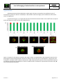
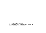
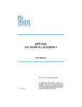
1
Created :
Live Cell imaging of mitochondria on micropatterns
2012/11/14
MO-EXT-18
Material and reagents :
Name
Hela cells
MEM+GlutaMax
Supplier
ATCC
Invitrogen
Reference
CCL-2
41090
FBS
Penicillin/Streptomycin
DPBS -/0,05% Trypsine EDTA 1X
CYTOOchamber
CYTOOchips Starter’s
Mitotracker Green FM
DMSO
Hepes
Flasks (T75)
Sigma
Gibco
Invitrogen
Gibco
CYTOO
CYTOO
Invitrogen
Sigma
Gibco
Falcon
2442
15140
14190
25300-054
30-010
10-900
M22425
D2438
15630
353136
Comments / quantities for one experiment
supplemented with 10% FBS and 0.5%
penicillin/streptomycin
See PRO-EXT-10 CYTOOchamber User Manual
20 nM, 15 min treatment
25 mM
0.2 µm Vented Blue Plug Seal Cap
Recommended protocol:
-
Warm up 1 bottle of media and trypsin.
Rinse the flask with 10 mL of PBS -/Trypsinize 70-80% confluent HeLa cells by adding 2 ml of Trypsine in T75 flasks. Incubate 5 min at 37°C
(Check under microscope for efficient dissociation).
Add 8 ml of 10% FBS supplemented media to inactivate the trypsin. To ensure the dissociation of last cell
aggregates, thoroughly pipet up and down the cell solution.
Spin 4 min at 300g.
Remove the supernatant and add 10 ml of fresh media, thoroughly pipet up and down the cell solution.
Count cells and prepare a cell solution at 40,000 cells/ml in growth media
Dispense 500 µl (20 000 cells) per CYTOOchamber, containing the appropriate CYTOOchips (See appendix 1,
for CYTOOchamber use).
Incubate for 2h at 37°C then add 2.5 ml of culture media was added.
Note: This step is required for timelapse experiments starting immediately after seeding. It allows HeLa cells to
sediment, to attach to the micropatterns and to spread entirely, which is necessary to set up your experiment.
-
Incubate for 23h45 at 37°C
Add 6 µl of Mitotracker Green FM solution to the cells for 15 minutes (20nM final, See appendix 2 for
preparation).
Replace the media containing the Mitotracker Green FM, with 3 ml of fresh media.
Place the CYTOOchamber in a controlled environment imaging system and acquire living cells (humidified
atmosphere, 7% CO2, 37°C). A protocol to test mitochondria network shape on CYTOO micropatterns in
fixed conditions is currently in progress.
-
Version 0
Page 1 sur 4
Created :
Live Cell imaging of mitochondria on micropatterns
2012/11/14
MO-EXT-18
Typical Results:
Following the protocol described above, 20-40 single cells were analyzed for the 12 different types of
micropatterns present on Starter’s CYTOOchips. Mitochondria networks integrity was classified into two classes:
intact or fragmented.
As presented in figure 1, we observed that the mitochondrial network of HeLa cells is 100% intact except for
Small and Large Discs with less than 10% fragmentation.
A
B
Figure 1: Integrity of micropattern networks after 24Hrs culture in CYTOOchamber. (A) The graphic represents the
percentage of intact and fragmented networks according to the size and type of microaptterns. Integrity was also
evaluated in standard culture conditions (Fibronectin, right). Representative pictures of the mitochondrial network
on different type of large microaptterns as well as standard culture conditions are presented at the lower side. (B)
Class of mitochondrial networks
Version 0
Page 2 sur 4
Created :
Live Cell imaging of mitochondria on micropatterns
2012/11/14
MO-EXT-18
Troubleshooting:
Problems
Mitochondrial network is abnormal in
more than 10% of the cells nicely
spread on micropatterns
Mitochondrial network seems to be
condensed
Solution proposed
- If you use Mitotracker Green FM, it can be toxic for high
concentrations and/or long incubations. Do not exceed
concentration of 20 nM during 15 minutes incubation. See
appendix 3 for more details.
- Addition of Hepes (25mM, pH = 7.4) for long time videomicroscopy
is often required to buffer pH variation. Hepes does not affect
mitochondrial integrity on micropattern.
- If you are not using Mitotracker Green FM, check for potential
toxicity due to the marker (concentration, incubation time, etc…)
- Wash carefully the CYTOOchamber before use as residual ethanol
or precipitated salts might have a toxic effect (Please refer to
Appendix1)
-
The micropattern might be too small for your cell line
Appendix 1: CYTOOchamber use
See PRO-EXT-10 CYTOOchamber User Manual
Appendix 2: Preparation of Mitotracker Green FM solution
One vial (50 µg) of Mitotracker Green FM is dissolved in 74.4 µl of DMSO to make a 1 mM stock solution.
Then it is diluted to 1% in PBS -/-, and 6 µl (20 nM) of the last solution is provided per CYTOOchamber containing 3
ml of culture media. Incubate 15 minutes before removing the media and add 3 ml of fresh one.
Appendix 3: Mitotracker effects on mitochondrial network shape
In order to prevent some discrepancies due to the use of mitotracker on cells cultured on micropatterns, we
realized a dose-response of Mitotracker Green FM concentration and time of incubation on 20-30 single cells using
the Starter’s CYTOOchips. Mitochondria networks integrity was classified into two classes: intact or fragmented.
As presented in figure 2, the use of Mitotracker Green FM treatment on live HeLa cells seeded on
micropatterns induces few mitochondria network fragmentation. It indicates that CYTOO micropatterns and CYTOO
chambers are not toxic for cells. Moreover, the use of 20 nM Mitotracker Green FM treatment during 15 min do not
induces stress to the cells. Increased Mitotracker concentration and/or time exposure highly favor a global toxicity to
the cells.
Version 0
Page 3 sur 4
Created :
Live Cell imaging of mitochondria on micropatterns
2012/11/14
MO-EXT-18
Figure 2: Micropattern network integrity using increasing amount of mitotracker concentration and time of
incubation. Cells were cultured 24 hours on 1600 µm2 crossbow micropatterns (A) The histogram represents the
percentage of intact and fragmented networks according to the concentrationand incubation time of mitotracker
treatment. (B and C) Representative pictures of 20 and 100 nM Mitotracker Green FM 15 minutes incubation
treatment are presented on panels B and C respectively. Mitotracker Green FM is in green and micropatterns are
stained using Alexa 546 fluorochrome and are presented as red.
Version 0
Page 4 sur 4