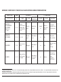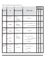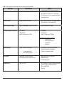
1
Prins Leopold Instituut voor Tropische Geneeskunde Institut de Médecine Tropicale Prince Léopold Prince Leopold Institute of Tropical Medicine Instituto de Medicina Tropical Principe Leopoldo Nationalestraat, 155 B – 2000 Antwerpen Stichting van Openbaar Nut 0410.057.701 POSTGRADUATE IN TROPICAL MEDICINE AND INTERNATIONAL HEALTH MODULE 2 CLINICAL & BIOMEDICAL SCIENCES OF TROPICAL DISEASES Practical notes __________________________ TROPICAL HAEMATOLOGY JANUARY 2009 Philippe Gillet – Luc Boel - Jan Jacobs [email protected] , [email protected] , [email protected] Table of Contents GENERAL HAEMATOLOGY BLOOD COLLECTION _____________________________________________________ 5 CAPILLARY BLOOD _________________________________________________________________ 5 VENOUS BLOOD____________________________________________________________________ 6 HAEMOGLOBIN DETERMINATION _________________________________________ 6 INTRODUCTION ___________________________________________________________________________________________ 6 REFERENCE VALUES ______________________________________________________________________________________ 6 HCS METHOD (HAEMOGLOBIN COLOUR SCALE) ____________________________________________________________ 6 PRINCIPLE : ________________________________________________________________________ EQUIPMENT AND SUPPLIES :________________________________________________________ BLOOD COLLECTION : ______________________________________________________________ METHOD : __________________________________________________________________________ MAINTENANCE : ____________________________________________________________________ 6 6 6 6 6 LOVIBOND® METHOD (Harrison’s method) ____________________________________________________________________ 6 PRINCIPLE : ________________________________________________________________________ MATERIAL : ________________________________________________________________________ BLOOD COLLECTION : ______________________________________________________________ METHOD : __________________________________________________________________________ SMALL PROBLEMS AND SOLUTIONS :________________________________________________ CONVERSION TABLE % LOVIBOND TO g / 100 ml : ____________________________________ 6 6 6 6 6 6 SAHLI METHOD ___________________________________________________________________________________________ 6 PRINCIPLE : ________________________________________________________________________ MATERIAL : ________________________________________________________________________ REAGENTS : _______________________________________________________________________ BLOOD COLLECTION : ______________________________________________________________ METHOD : __________________________________________________________________________ 6 6 6 6 6 DRABKIN METHOD________________________________________________________________________________________ 6 PRINCIPLE : ________________________________________________________________________ BLOOD COLLECTION : ______________________________________________________________ MATERIAL : ________________________________________________________________________ REAGENTS : _______________________________________________________________________ METHOD : __________________________________________________________________________ 090122-pg-tropical haematology and blood transfusion 6 6 6 6 6 2 / 77 HEMOCUE® B _____________________________________________________________________________________________ 6 PRINCIPLE : ________________________________________________________________________ MATERIAL : ________________________________________________________________________ BLOOD COLLECTION : ______________________________________________________________ METHOD : __________________________________________________________________________ 6 6 6 6 DHT HAEMOGLOBINOMETER (Developing Health Technology) (Ammonia technique) _____ 6 PRINCIPLE : ________________________________________________________________________ MATERIAL : ________________________________________________________________________ BLOOD COLLECTION : ______________________________________________________________ REAGENTS : _______________________________________________________________________ METHOD : __________________________________________________________________________ 6 6 6 6 6 PACKED CELL VOLUME BY CENTRIFUGATION or HEMATOCRIT ____________ 6 PRINCIPLE : ________________________________________________________________________ EQUIPMENT AND SUPPLIES :________________________________________________________ BLOOD COLLECTION : ______________________________________________________________ METHOD : __________________________________________________________________________ REMARKS :_________________________________________________________________________ REFERENCE VALUES : _____________________________________________________________ INTERPRETATION OF PCV:__________________________________________________________ 6 6 6 6 6 6 6 RED BLOOD CELL INDICE (MCHC)_________________________________________ 6 SOURCES OF ERRORS :____________________________________________________________ 6 QUALITY CONTROL :________________________________________________________________ 6 CELL NUMBER CONCENTRATION _________________________________________ 6 GENERAL PRINCIPLE : ______________________________________________________________ 6 GENERAL MATERIALS :_____________________________________________________________ 6 COUNTING CHAMBERS : ____________________________________________________________ 6 WHITE CELL COUNT IN BLOOD ___________________________________________ 6 REAGENT : _________________________________________________________________________ BLOOD COLLECTION : ______________________________________________________________ METHOD : __________________________________________________________________________ REFERENCE RANGES: ______________________________________________________________ QUALITY ASSURANCE : _____________________________________________________________ SOURCES OF ERRORS : ____________________________________________________________ 6 6 6 6 6 6 RED CELL COUNT IN BLOOD : ____________________________________________ 6 RED CELL INDICES _______________________________________________________ 6 MCV _______________________________________________________________________________ 6 MCH _______________________________________________________________________________ 6 ADDENDUM 1 : TECHNIQUES FOR ASSESSING ANAEMIA (PRICE LIST) __________________________________________ 6 ADDENDUM 2 : SOME HAEMATOLOGICAL REFERENCE RANGES (GUIDELINE FIGURES) __________________________ 6 ADDENDUM 3 : MORPHOLOGY OF BLOOD CELLS IN A MAY-GRÜNWALD-GIEMSA STAINED BLOOD FILM__________ 6 090122-pg-tropical haematology and blood transfusion 3 / 77 BLOOD TRANSFUSION Short summary of basics genetics ______________________________________________ 6 Short summary of basic immunology ___________________________________________ 6 BLOOD GROUPING ________________________________________________________________________________________ 6 A The ABO blood group system_________________________________________________________ 6 B The Rhesus system _________________________________________________________________ 6 TRANSFUSIONAL RULES ___________________________________________________________________________________ 6 COMPATIBLITY TESTS_____________________________________________________________________________________ 6 Rapid test (rapid cross match)____________________________________________________________ 6 Complete Major compatibility test in saline medium __________________________________________ 6 followed by an indirect Coombs test in LISS-Albumin medium. _________________________________ 6 Auto test on the receptor ________________________________________________________________ 6 (In case of positive compatibility tests in saline medium) ______________________________________ 6 Test of minor compatibility______________________________________________________________ 6 Table 5 : Comparison of different (major) compatibility tests useful on district laboratory level. ___________________________ 6 Table 6 : Use of compatibility tests on district laboratory level described in the notes. ____________________________________ 6 Table 7 : Blood transfusion on district hospital level: from minimum until extra possibilities.______________________________ 6 ANNEX 1 Wash of the red blood cells___________________________________________________________________________ 6 ANNEX 2 Screening of dangerous O donors _____________________________________________________________________ 6 ANNEX 3 Screening for infectious diseases ______________________________________________________________________ 6 ANNEX 4 Compatibility test on slide, Polybrene method ___________________________________________________________ 6 ANNEX 5 Indicative price list (Diamed, blood grouping) http://www.diamed.ch/ _______________________________________ 6 ANNEX 6 HIV rapid test example______________________________________________________________________________ 6 ANNEX 7 HBsAg rapid test example ___________________________________________________________________________ 6 ANNEX 8 HCV rapid test example _____________________________________________________________________________ 6 ANNEX 9 RPR test example __________________________________________________________________________________ 6 ANNEX 10 Relation rotation speed of a centrifuge and the centrifugal power: _________________________________________ 6 ANNEX 11 Useful internet sites________________________________________________________________________________ 6 GLOSSARY ________________________________________________________________________________________________ 6 090122-pg-tropical haematology and blood transfusion 4 / 77 BLOOD COLLECTION CAPILLARY BLOOD Capillary blood is the cheapest and the easiest method for blood collection. Capillary blood is mainly used when the volume of required blood is small (up to 100 μl). Disadvantages in using capillary blood for blood test include: • • • • Great possibility of sampling errors, particularly when the blood is not free-flowing (dilution of the sample with tissue fluid). Difficulty in obtaining sufficient blood (for more than one test). Rapid clotting of blood. Test cannot be repeated or further tests cannot be performed when results are unexpected. Capillary blood is obtained by puncturing the skin with a lancet. In adults or children, the best place rd th will be on the 3 or 4 finger of the left hand, at the side of the finger, which is less sensitive than the tip. In infants, the best place will be the side of the heel or the big toe (The puncture should not be too deep because of the risk of osteomyelitis!). Never collect blood from an infected finger of foot. Never collect blood from an arm in which an intravenous infusion is being given (haemodilution). The puncture should be deep enough to result in free bleeding. A free flow of blood is essential and only the gentlest squeezing is permissible (risk of dilution with tissue fluid resulting in unreliable values). 1. Prepare all supplies in advance : sterile lancet, 2 pieces of cotton wool, one dry, the other soaked with alcohol 70 % and blood collection material (Sahli pipette, slide, capillary tube,…). 2. If possible, ask the patient to clean his hands with soap and hot water (vasodilatation), next dry his hands thoroughly. 3. Slightly massage the place where blood will be taken. Make sure the puncture area is warm enough to allow the blood to flow freely. If necessary, soak the hand or foot of an infant in warm water prior to collecting a sample. 4. Cleanse the puncture area with a cotton swab dipped in 70 % alcohol, let alcohol react for at least 30 seconds, then with a dry cotton to remove any remaining ethanol. 5. Using a sterile lancet, make a rapid puncture, sufficiently deep to allow the free flow of blood. Discard immediately the lancet in a safety container. 6. Execute a slight pressure on the finger to realise a better blood flow. st 7. Wipe away the 1 drop of blood with a dry piece of cotton wool since it may contain tissue fluid or disinfectant. (Except for microfilaria detection, since the first drop contains more microfilaria). 8. Press the finger (not too hard) to produce in one time the required amount of blood. 9. When sufficient blood has been collected, press a piece of cotton dipped in 70 % alcohol over the puncture area until bleeding stops. For glycaemia determination: The use of ether or any disinfectant should be avoided as it may interfere with the reagent strip (glucose oxidase peroxidase reaction). To decrease risk of infection and food contamination (fruit juice, ... ) it is therefore important to clean the hands with soap, before blood collection. 090122-pg-tropical haematology and blood transfusion 5 / 77 VENOUS BLOOD Venous blood is used when more than 100 μl of whole blood is required or when serum from a clotted blood sample is needed. If whole blood is needed, an anticoagulant should be used to prevent clotting and/or morphological blood cell changes. An anticoagulant acts by removing calcium (example : EDTA, trisodium citrate) or by interference with coagulation factors (example : Heparin) For most haematological tests (Haemoglobin, PVC, WBC count, blood group determination, …), dipotassium EDTA is recommended. Preparation of dipotassium EDTA tubes : Bring 40 μl of a dipotassium EDTA solution (10 g / 100 ml distilled water) in 3 ml tube. Leave the open tubes to dry at room temperature (protect from dust). Close when dry. The correct amount of blood must be added to avoid blood cell changes (2,5 ml). Excess EDTA causes shrinkage and degenerative changes, lack of EDTA will not prevent the coagulation. For measuring the ESR, trisodium citrate is used to anticoagulate the blood. 4 volumes of venous blood with 1 volume of trisodium citrate 32 g/l. DIFFERENCE BETWEEN SEDIMENTATION AND COAGULATION Without anticoagulant Coagulation (Solidification) With anticoagulant Sedimentation (or after centrifugation) Serum Plasma WBC (Buffy coat) WBC+RBC trapped in a fibrin structure Prothrombin RBC 2+ Thrombin + Ca Fibrinogen Stable fibrin SERUM = PLASMA WITHOUT COAGULATION PROTEINS 090122-pg-tropical haematology and blood transfusion 6 / 77 HAEMOGLOBIN DETERMINATION INTRODUCTION Anaemia is defined as having an amount of haemoglobin below reference values. Anaemia is present when haemoglobin concentration falls below 11 g/100 ml (hematocrit below 33 %). Anaemia is described as severe when the haemoglobin is below 7 g/100 ml (hematocrit below 21 %). Some authors (and maybe the WHO in the future) use 8 g/100 ml as cut-off value for severe anaemia (hematocrit below 24 %). V o lu m e p la s m a tiq u e V o lu m e g lo b u laire N o rm a l A n ém ie v ra ie F a u sse a n ém ie (h é m o d ilu tio n ) Fig.1 : True and “false” anaemia due to haemodilution. In some circumstances, the reduction of hemogram values is related to a haemodilution caused by plasma excess resulting in a “false anemia”: pregnancy, splenomegaly, heart insufficiency, monoclonal immunoglobulines, especially IgM…. Haemoglobin is the most important part of the red blood cell. It is the red pigment that gives the colour to the red blood cells. It carries oxygen and carbon dioxide. Each molecule of haemoglobin contains four linked polypeptide (globin) chains and four haem groups. (Fig. 2). Haem is an iron-containing porphyrin pigment, which is the oxygen carrying part of the haemoglobin molecule. Oxygen binds 2+ reversibly with ferrous ions (Fe ), contained in each haem group. More than 97 % of normal adult (Į2ȕ2). Up to 3,5 % is HbA2, haemoglobin is Hb A1, having 2 alpha chains and 2 beta chains consisting of 2 alpha chains and 2 delta chains (Į2į 2). Less than 1 % is HbF (foetal) composed of 2 alpha chains and 2 gamma chains (Į2Ȗ2). HbF is the predominant haemoglobin in foetus; untill 3-6 months of life. Fig 2 : Haemoglobin structure (Haemoglobin A) globin α β Fe Fe 141 aa 146 aa haem (2-3 DPG) β α Fe Fe 146 aa 141 aa 2-3 DPG = 2-3 diphosphoglycerate The measurement of haemoglobin is important for the diagnosis of the severity of anaemia. Different techniques, more or less reliable, are available for haemoglobin determination. They are based on different principles, which can be classified in 3 families: 090122-pg-tropical haematology and blood transfusion 7 / 77 1. Techniques based on the red colour of blood without dilution nor haemolysis (Talquist, HCS, Lovibond, …) 2. Techniques based on the red colour of blood after haemolysis of the red blood cells (DHT,…). 3. Techniques based on the transformation of the haemoglobin (Sahli, Hemocue, Drabkin, …). The PCV (Packed cell volume) or hematocrit may also be used to screen for anaemia. The choice between these techniques will be based on reliability, repeatability, precision, accuracy, price, equipment needed, level of technical difficulty, staff training level, … REFERENCE VALUES1 The reference ranges for haemoglobin vary by age and sex as shown in the table below. Age Sex Units Haemoglobin g/100 ml 3 months – 12 months Men and Women 10,0 – 14,0 1 year – 12 years Men and Women 10,5 – 15,0 12 years – 100 years Men (Europe) 13,2 – 17,3 12 years – 100 years Women (Europe) 11,7 – 15,5 1 Reference ranges vary in different population and in different laboratories (different techniques). District laboratories should check the figures above for the technique in use with their nearest hematology reference laboratory. 090122-pg-tropical haematology and blood transfusion 8 / 77 HCS METHOD (HAEMOGLOBIN COLOUR SCALE) PRINCIPLE : The intensity of the red colour of blood corresponds with the amount of haemoglobin. The degree of anaemia can be visually assessed by matching the colour of a drop of blood on special filter paper against a standardized colour chart. The colour chart is developed to represent the colour range of normal to anaemic blood on filter paper : 14, 12, 10, 8, 6 and 4 g /100 ml. EQUIPMENT AND SUPPLIES : Blood collection equipment and supplies + Kit HCS (booklet of 6 shades of red, instructions for use, dispenser of 200 special absorbent test strips in handy box). Use only the special test-strips that are provided by Copack, since others may give inaccurate results. Keep these test-strips dry, clean and protected from direct sunlight at any time. 70 % Alcohol. BLOOD COLLECTION : Capillary or venous blood. For venous blood, dry anticoagulant should be used (to avoid dilution). EDTA di-potassium salt or heparin are recommended. METHOD : Find a suitable place: a room which is well-lit by daylight and/or artificial light. Avoid direct sunlight and marked shade. Do not read the scale in your own shadow. 1. Place a drop of blood on one end of the test-strip so that it forms a spot, which is large enough to spread beyond the area of an aperture in the scale (about 1 cm in diameter). Correct amount of blood : sufficient to spread beyond the area of an aperture in the scale. Too little blood : The area of an aperture in the scale will not be covered. Too much blood : The spread will be too thick and the blood will take too long to dry. 2. Wait about 30 seconds after applying the blood; then read immediately. Any delay in reading the test will cause an error as the blood stain will change colour, becoming lighter and unreliable. Starting from the lightest shade or darkest shade, slide the blood stain up and down behind the apertures in the scale until you find the best colour match. When reading, keep the test-strip close to the back of the scale to prevent any stray of light from entering. • • • If the blood stain matches one of the shades of red exactly, record the haemoglobin value. If the colour lies between two shades on the scale, record the mid-value. If there is any doubt between two shades, record the lower value. Example: 14 g/100 ml :Too light 12 g/100 ml: Correct. 10 g/100 ml: Too dark. 8 g/100 ml : Too dark. 6 g/100 ml : Too dark. 4 g/100 ml : Too dark. 090122-pg-tropical haematology and blood transfusion 9 / 77 MAINTENANCE : To clean the scale, wipe the back side with a humid tissue (alcohol 70 %), then with a dry tissue. The scale should be cleaned at the end of each session and during the session if the surface becomes soiled during use. The scale can be used for thousands of tests, but to avoid deterioration of the colours, always keep the booklet closed after use and never leave it exposed to direct sunlight. It should be replaced periodically. LOVIBOND® METHOD (Harrison’s method) PRINCIPLE : The Harrison’s method is a simplified version of a visual comparative technique for estimating haemoglobin. The red colour of blood corresponds with the amount of haemoglobin. Blood is inserted directly into a special cell (thickness of 0,004”) without any preliminary manipulation. The colour is compared with a series of reference glasses in a Lovibond® comparator. MATERIAL : Blood collection equipment and supplies + Lovibond 2000 comparator, standard discs Lovibond 5/8 A and 5/8 B, Lovibond cell 0,004”, soft paper, Beaker, chlorine solution 1 %, 70 % alcohol. Lovibond® 2000 comparator Lovibond® 0,004”2 Disc Metal clip Cover-plate Stud base-plate The blood cell consists of a base-plate and a cover-plate constructed from plain white glass. Fused on the cover-plate are three small studs of glass which create, when in position a cell of 0,004” thickness. This cell is filled from the side by capillarity. BLOOD COLLECTION : Capillary blood. [For venous blood, dry anticoagulant should be used (to avoid dilution). EDTA dipotassium salt or heparin are recommended]. METHOD : 1. Clean and decontaminate the base-plate and the cover-plate, first with water, then with 70 % alcohol. 2. Place the two plates in position and join them with the clip. 2 0,004 inch = 1.016 mm 090122-pg-tropical haematology and blood transfusion 10 / 77 3. Check if the discs are clean. Wipe with a cloth if they become dirty. Select the appropriate disc (evaluation of conjunctival pallor) and insert it into the comparator with the values towards the front of the instrument. Rotate the disc until the lowest or the highest value. CHECK THE CELL BEFORE USING IT : Decontamination with hydrochloric solution, next with 70 % alcohol? Is it dry and dust free, and clean, no lines nor finger prints? Does the cover plate cover properly the base plate ? Are the figures 004 well readable? 4. Fill the cell by capillarity with capillary blood. If there are air bubbles in the cell, restart from point 1. 5. Clean the blood excess around the cell with a soft paper. 6. Remove the clip. 7. Place the cell, containing the prepared sample in the right compartment. Using a diffuse light (facing southern daylight in the southern hemisphere), rotate the disc until the closest colour matches with the sample. (Be quick to avoid desiccation or coagulation). The Lovibond value will be shown in the window in the bottom right corner of the comparator. Find a suitable place for the colour comparison : • • • During the day : Facing a white surface (southern daylight in the southern hemisphere). During the night : Facing a white surface illuminated by a white lamp (not fluorescent light). Direct sunlight or direct artificial light gives incorrect results. 8. Convert the Lovibond value in g/100 ml (cf. conversion table). 9. Decontaminate the Lovibond cell (contact with 1 % chlorine solution during 30 minutes). 10. To avoid deterioration of the colours, store immediately the disks in a box. 10. Clean and decontaminate the base-plate and the cover-plate, first with water, then with 70 % alcohol. 11. For practical reasons it can be advised to mount the plates in advance so that no time will be lost in case of emergency. SMALL PROBLEMS AND SOLUTIONS : ALWAYS CHECK THE CONCORDANCE BETWEEN THE HAEMOGLOBIN VALUE AND THE COLOUR OF THE CONJONCTIVES. 1. No value appears in the inner opening of the comparator. The disk is placed back to front. Take out the disk and turn it round. 2. No colour corresponds with the colour of the blood of the patient. The chosen disk does not correspond with the expected haemoglobin value. Use the other disk. Thee patient is icteric (jaundice). His upper conjunctives are yellow. Find the closest colour and note the presence of jaundice in the report. The blood colour is inferior to the minimum of the comparator. Give as result < than 20 % or < than 3,3 g/100 ml. 3. The measured haemoglobin value seems to be too low. Beware that the cover plate is placed in the correct way (figures 004 readable). If this is not the case, restart the measurement with another plate. 090122-pg-tropical haematology and blood transfusion 11 / 77 Beware there are no air bubbles between base and cover plate. If this is not the case, restart the measurement with another plate. Check the cleanliness of the comparison disk and, if necessary, clean it with a soft tissue. Check the cleanliness of the translucent plate of the comparator and, if necessary clean it with a hydrochloric solution of 1 %, next with filtered water. If non of these solutions are helping and if the values are systematically too low, make a comparison between the new and the old disks (the colour of the disks is degrading with light). 4. Too high haemoglobin values. The upper side of the cover-plate and/or the under side of the base-plate are soiled with blood. Take of the blood on both sides with a soft paper. The disinfectant used for the blood taking is stained (e.g.Betadine). Restart the measurement using Ethanol à 70 %. Check the cleanliness of the translucent plate of the comparator and, if necessary clean it with a hydrochloric solution of 1 % , next with filtered water. Thee patient is icteric (jaundice). His upper conjunctives are yellow. Find the closest colour and note the presence of jaundice in the report. The patient is dehydrated (provoking a haemoconcentration). answer. No solution, note the dehydration in the CONVERSION TABLE % LOVIBOND TO g / 100 ml : IT IS HELPFUL TO TAKE THE COLOR MATCH UNTIL SLIGHTLY BELOW, THEN SLIGHTLY ABOVE, IN ORDER TO FIND THE BEST COLOR MATCH. DISKS Disc N° 5/8 A (light colour) For low Haemoglobin values Disc N° 5/8 B (dark colour) For high Haemoglobin values 090122-pg-tropical haematology and blood transfusion PERCENTAGE g / 100 ml 20 3.3 24 28 32 36 4.0 4.7 5.3 6.0 40 46 52 58 6.7 7.3 8.7 9.7 64 10.7 70 76 84 92 11.7 12.7 14.0 15.3 100 110 120 130 16.7 18.3 20.0 21.7 12 / 77 SAHLI METHOD PRINCIPLE : The Sahli method is based on converting haemoglobin to acid haematin (brown colour) and then visually matching its colour against a solid glass standard. Diluted hydrochloric acid is mixed into a graduated cylinder with an accurate volume of blood sample and distilled water is added until the colour of the diluted blood sample matches the glass standard. The dilution will be determined by the Haemoglobin level of the blood sample. The Sahli method, still used in a lot of places, is not recommended. The Sahli method is not an accurate way of estimating haemoglobin: Not all the forms of haemoglobin are changed into acid haematin, the colour changes when viewed visually are not very great and the brown colour of the glass standard is not a true match for an acid haematin solution. MATERIAL : Blood collection equipment and supplies + Sahli haemoglobinometer, small glass rod, Sahli pipette, safety device for pipetting, Sahli tube and dropping pipette. The Sahli haemoglobinometer is equipped with two glass colour standards. The Sahli tube graduated until 16 g/100 ml and/or in “percentage” (16g/100 ml = 100 %), is placed inbetween. REAGENTS : Hydrochloric acid 0,1 N : Hydrochloric acid, concentrated1 Distilled water : : 9,5 ml up to 1 litre 1 Caution, hydrochloric acid is irritant and corrosive. Handle with care in well ventilated area (or in a fume cupboard). Fill until half a 1.000 ml volumetric flask with distilled water. Add slowly 9.5 ml concentrated hydrochloric acid. After cooling, fill until the 1,000 mark with distilled water and mix well. This reagent is stable for at least 1 year at room temperature. Distilled water (or filtered water). BLOOD COLLECTION : Capillary or venous blood. For venous blood, dry anticoagulant should be used (to avoid dilution). EDTA di-potassium salt or heparin are recommended. METHOD : 1. Fill the graduated measuring tube up to the bottom graduation line with 0.1 N hydrochloric acid. (The mark level should be equal with the bottom of the meniscus formed by the liquid). 2. Check the tip of the Sahli pipette. Discard if broken (volume error). Check if the pipette is dry. 3. Draw the blood a little bit further than the 20 μl mark of the Sahli pipette. Do not allow air bubbles to enter. Wipe the outside of the pipette with absorbent paper and adjust the blood on the 20 μl mark. 4. Blow the blood from the pipette into the graduated tube of the acid solution. 090122-pg-tropical haematology and blood transfusion 13 / 77 5. Rinse the pipette by drawing in and blowing out the acid solution. 6. Allow to stand for 1 minute. The mixture will become dark brown and clear. 7. Place the graduated tube in the haemoglobinometer, compare the colour in diffused day light. Add water drop by drop and mix with the glass stirrer until the colour of the solution matches the colour of the reference tube. 8. When equal colours are reached, take the glass stirrer out of the graduated tube and read the level of the base of the menisci of the liquid. Hold the instrument about 50 cm away from your eyes, on the same height under diffuse light. 9. Note the reached mark that corresponds with the level on the tube. 090122-pg-tropical haematology and blood transfusion 14 / 77 DRABKIN METHOD PRINCIPLE : The haemiglobin cyanide method is the most accurate method of measuring haemoglobin and is considered as the reference standard (« gold standard”). Whole blood is precisely diluted 1 on 201 in a Drabkin solution. The red cells are haemolysed and the haemoglobin is oxidized by the ferricyanide to methaemoglobin. This is converted by the cyanide to stable haemoglobin cyanide. Hb + K3Fe(CN)6 Î MHb MHb + KCN Î HiCN [Hb = haemoglobin, MHb = methaemoglobin, HiCN = haemiglobincyanide] The chemical reaction takes place at a pH stabilized by a monopotassium phosphate buffer in order to obtain a complete reaction in a reasonable time. Addition of a detergent facilitates haemolysis and prevents turbidity caused by plasmatic proteins. The optical density is in proportion with the haemoglobin quantity that is present in the blood. Absorbance of the HiCN is read in a spectrophotometer at the wavelength 540 nm or in a colorimeter using a yellow-green filter. The absorbance obtained is compared with that of a reference HiCN standard solution. Haemoglobin values are obtained in the calibration graph. BLOOD COLLECTION : Capillary or venous blood. For venous blood, dry anticoagulant should be used (to avoid dilution). EDTA di-potassium salt or heparin are recommended. After thoroughly mixing with anticoagulant, the blood can be frozen for as long as 2 years (and used as control). MATERIAL : Blood collection equipment and supplies + spectrophotometer (or colorimeter) that transmits light at 540 nm, Sahli pipette (or 20 μl automatic pipette), 5 ml graduated pipette (or 5,0 ml dispenser), safety device for pipetting, test tubes, cuvettes, different volumetric flasks and different calibrated pipettes (for calibrator’s preparation), graph paper. REAGENTS : Drabkin’s neutral diluting fluid (pH 7,0 – 7,4) (also “ready to use” available example Sigma D 5941) : Potassium hexacyano ferrate (K3Fe(CN)6) Potassium Cyanide (KCN)1 Potassium dihydrogen phosphate (KH2PO4) Tween 20 Distilled water : : : : : 0,20 g 0,05 g 0,14 g 1 ml to 1 litre (Tween20 can be replaced by 0.5 ml Brij-35 at 30 %) 1 CAUTION : Potassium cyanide is highly poisonous. It is a pale yellow clear fluid which may no longer be used if its colour is lost or if it becomes turbid. The prepared Drabkin solution is stable for at least 6 months at room temperature protected from light (amber bottle). Haemiglobin cyanide (cyanmethaemoglobin) standard for calibration: HiCN reference standard solutions are stable for long periods and are commercially available as: Haemoglobin standard (SIGMA), HiCN BS 3985 (Merck/BDH), … Example with Haemoglobin standard from SIGMA : 090122-pg-tropical haematology and blood transfusion 15 / 77 - Reconstitute one vial of the standard by adding 50,0 ml of the Drabkin solution. Mix well. Wait at least 30 minutes before use. The standard must be stored at 2- 8°C in the dark and is stable for at least for 6 months. Prepare a calibration graph from this HiCN reference standard. Plot the absorbance readings at 540 nm of different standard dilutions against their known concentrations of haemoglobin. The curve is linear, passing through the origin. Dilution’s example: Tube Volume in ml N° Drabkin solution 1 4 2 3.5 3 3 4 2 5 1 6 0 Volume in ml diluted standard 0 0.5 1 2 3 4 final concentration in g % 0 2.25 4.5 9 13.5 18 METHOD : 1. 2. 3. 4. 5. 6. Set the spectrophotometer wavelength at 540 nm. Set up a series of labelled test tubes for blank and tests. Add 4.0 ml of the Drabkin solution to all tubes. Add 20 μl of whole blood sample to each labelled test tube. Rinse the pipette 3 times with the reagent. Mix well and allow to stand for at least 5 minutes at room temperature. (Attention, the reaction can take until 30 minutes for a sample containing an increased proportion of carboxy haemoglobin). 7. Read and record absorbance of each test (or control) versus the blank as the reference at 540 nm in the same instrument used for preparing the calibration curve. The colour is stable for several hours. Determine the haemoglobin concentration of each test directly from the calibration curve. O.D. [Hb] in g/100 ml Or Calculated: O.D. test ---------------------- x calibrator’s concentration = sample’s Hb concentration O.D. Calibrator 090122-pg-tropical haematology and blood transfusion 16 / 77 HEMOCUE® B PRINCIPLE : The HemoCue® is an example of a robust, portable and accurate haemoglobinometer readily available for use. Although not affordable by most district laboratories, it may be used for survey. The HemoCue uses calibrated disposable cuvettes that are treated with chemicals (sodium desoxycholate, sodium nitrite and sodium azide) which rupture the red cell wall and combine with the haemoglobin to form a compound (azidemethaemoglobin) which can be measured photometrically (modified Vanzetti reaction : Sodium nitrite converts the haemoglobin iron from the ferrous to the ferric state to form methaemoglobin. The methaemoglobin then combines with azide to form azidemethaemoglobin and is measured photometrically at two wavelengths, 570nm and 880nm). The result in g/100 ml is displayed in digital form on the face of the instrument. Web site : http://www.hemocue.co.uk/ MATERIAL : Blood collection equipments and supplies + HemoCue® B disposable cuvettes, HemoCue® B instrument, HemoCue® B standard control cuvette, batteries. BLOOD COLLECTION : Capillary or venous blood. For venous blood, dry anticoagulant should be used (to avoid dilution). EDTA di-potassium salt or heparin are recommended. METHOD : 1. Check the stability of the calibration : Place the control cuvette into the cuvette older and push it into the measuring position. The displayed value should not deviate more from the assigned value on the control cuvette card than + /- 0.3 g/100 ml (for our control cuvette N° 0149-003-037 the value lies between 11.0 and 11.6 g/100 ml). 2. Fill the disposable cuvette with the blood drop by touching the capillary tip of the cuvette with the blood drop. 3. Be sure that the cuvette is entirely filled with blood. If air bubbles are present, discard the cuvette and fill a new disposable cuvette. 4. Wipe off the excess of blood on the outside of the cuvette tip. Make sure that no blood is drawn out of the cuvette in this procedure. 5. Place the filled cuvette into the cuvette holder immediately and push it into the measuring position. 6. After approximately 30 to 50 seconds the result is displayed (in g/100 ml (or g/l or mmol/l). The filled cuvette should be analysed at least 10 minutes after it has been filled (evaporation). 090122-pg-tropical haematology and blood transfusion 17 / 77 N.B. : 3 different models are now commercialized : HemoCue® B et HemoCue® 201+ for « normal working conditions » (temperature < 30°C and low humidity) and The Hb 301 system is optimized for use in primary care and designed for high temperatures and humidity. If the 2 first model (Band and 201) use the principle explained above, the model 301 use a different analytical method : The measurement takes place in the analyzer, which measures the absorbance of whole blood at a Hb/HbO2 isobestic point. The analyzer measures at two wavelengths (506 and 880 nm) in order to compensate for turbidity. In fact, it’s an automatised version of the Lovibond technic.3 Hemocue® B Hemocue® 201+ Hemocue® 301+ Microcuvettes B and 201 are to be stored at room temperature (15-30°C). The reagents contained within the HemoCue® microcuvettes B and 201 are moisture sensitive: Recap vial immediately after removing cuvettes and do not remove desiccant from the vial. The microcuvettes 301 are to be stored at 10–40 °C (50–104 °F). Once the seal of the vial is broken, the microcuvettes are stable for 3 months. An unopened vial of microcuvettes can be stored for a shorter period of time (6 weeks) between -18–50 °C. HemoCue Hb 301 Analyzer is only to be used with correspondent HemoCue Hb microcuvettes. HemoCue® B-HB Photometer Hemoglobin Controls HYC84665 3x3 ml (1 low, 1 normal, 1 high) : HemoCue® B-HB Photometer Hemoglobin Controls. Features a 2-year expiration from date of manufacture at 2 – 8°C, 60-day open vial stability at 2 – 8°C, and a 30-day open vial stability at room temperature. Utilizes stabilized whole human red cells which process like a fresh patient sample. 3 Evaluation of the utility of the Hemocue 301 haemoglobinometer for blood donor screening. L. D. Morris, A. Ossei-Bimpong, D. McKeown, D. Roper and S.M. Lewis. Vox sanguinis (2007) 93, 64-69. 090122-pg-tropical haematology and blood transfusion 18 / 77 DHT HAEMOGLOBINOMETER (Developing Health Technology) (Ammonia technique) PRINCIPLE : Whole blood is diluted 1 in 101 in a weak ammonia solution. The red cells are haemolysed and the amount of haemoglobin is measured over a narrow spectral band. The measurement of the optical density is carried out at a wavelength of 523 nm, the crossing point of absorption curves of various haemoglobin forms, which are thus detected with equal sensitivity. This optical density is automatically converted to read-out directly as haemoglobin concentration in g/l on a liquid crystal display. No zero adjustment or calibration is required and no calibrating solutions are needed. Web site : www.dht-online.co.uk Cuvette Glass window Interference filter Photodiode LED (Diode emitting a green light) MATERIAL : Blood collection equipment and supplies + DHT haemoglobinometer, test tubes, Sahli pipette (or 20 μl automatic pipette), 2 ml graduated pipette (or 2,0 ml dispenser), safety device for pipetting, 1.000 ml volumetric flask, 10 mm light path cuvettes. BLOOD COLLECTION : Capillary or venous blood. For venous blood, dry anticoagulant should be used (to avoid dilution). EDTA di-potassium salt or heparin are recommended. REAGENTS : Ammonia solution 0,04 % (v /v): Preparation with ammonia 28 % concentrated: Ammonia 28 % concentrated Distilled water 1,4 ml up to 1.000,0 ml (volumetric flask) CAUTION : Ammonia solution is a corrosive chemical with an irritating vapour. Handle with care in well-ventilated area (or in a fume cupboard). Keep the stock bottle well stoppered. This solution is stable when kept in a tightly stoppered bottle. Renew every 4 weeks Any concentration of any ammonia solution can be made by taking a volume of concentrated solution equivalent to the required % and making this up with distilled or deionised water to a volume equivalent to the % of concentration : C1 x V1 = C2 x V2 C1 = Concentration of the concentrated ammonia solution V1 = Volume of the concentrated ammonia solution 090122-pg-tropical haematology and blood transfusion 19 / 77 C2 = Needed ammonia concentration V2 = Needed volume of the weak ammonia solution Example : C1 = 28 % V1 = ? C2 = 0,04 % V2 = 1.000 ml 28 % x V1= 0,04 % x 1000 ml Î V1 = 1,429 ml METHOD : It’s vital to understand the factors that influence accuracy of measurement and dilution. The accuracy of haemoglobin measurement depends on accuracy of proportion of blood to diluent. The quality of the used water and ammonia as diluent is also quite important. INITIAL CHECKS OF OPERATION AND FACTORY CALIBRATION : (Caution : Used values are only valid for the haemoglobinometer N° 0931) that is used at the ITM. Do not touch the clear working side areas of the cuvette. Avoid contamination of these, handling the cuvette only by the top of the etched surfaces at front and back (non optical sides). Always ensure the outer surface of the cuvette is completely dry. When filling cuvette, ensure no air bubbles are present to affect measurement. A. CHECK BLANK : Install a CLEAN and DRY cuvette into the cuvette aperture. (Use the same kind of cuvette which is used for haemoglobin determination). The value on the display must correspond with the blank value BR1 (+/- 5) [14-24 g/l for our haemoglobinometer 0931]. If this is not the case check for the following cause: PROBABLE CAUSE Cuvette not well installed : Scratched cuvette : Dirty or wet cuvette surface : Value of scale factor M incorrect : Zero level of the device incorrect Other type of cuvette used for the calibration : Dirty or wet photocell windows : REQUIRED ACTION Insert the cuvette with the mat etched surface in front of you. Check again the reading for BR1. Use new cuvette. Check again the reading for BR1. Clean and dry the cuvette. Check again the reading for BR1. Press and hold down the « R » button located at the back of the device. The symbol “HHH”, and after some seconds the scale factor M will appear. Compare this value with the figure for M shown in “certification” (162 for our device). If there is a difference between these values, then adjust the displayed value by pressing the “L” button to lower the value and the “R” button to raise the value. Check again the reading for BR1. Place a clean cuvette filled with distilled water into the cuvette window. Press and hold down the “L” button. At first the display will show the last reading, followed after several seconds by a “beep”. After this beep the LCD turns off and the new zero level is stored in the device. Check again the reading for BR1. Place a clean cuvette filled with distilled water into the cuvette window. Press and hold down the “L” button. At first the display will show the last reading, followed after several seconds by a “beep”. After this beep the LCD turns off and the new zero level is stored in the device. Check again the reading for BR1. Clean the glass of the photocell windows with alcohol on cotton swap stick. Check again the reading for BR1. B. CHECK ZERO : Place a cuvette containing 1-2 ml of the weak ammonia solution (0,04 %) into the cuvette aperture. (Use the same kind of cuvette which is used for haemoglobin determination). The value on the display should be zero. If this is not the case remove the cuvette and within 2 seconds press and hold the “L” button until the beep. Zero will automatically be reset. Check again the reading for zero. 090122-pg-tropical haematology and blood transfusion 20 / 77 C. CHECK CALIBRATION : The permanent calibration control standard (checker), supplied with each instrument, is numbered and matched uniquely to one instrument. Place the checker into the cuvette aperture. The value of the displayed reading must correspond to the value CR1 (+/- 5) [133143 g/l for our haemoglobinometer 0931]. If this is not the case check for the following causes: PROBABLE CAUSE Cuvette not well installed : Scratched control cuvette : Dirty or wet control cuvette surface : Value of scale factor M incorrect : Zero level of the device incorrect Other type of cuvette used for the calibration : Dirty or wet photocell windows : REQUIRED ACTION Insert the cuvette with the plastic surface in front of you. Check again the reading for CR1. Buy a new cuvette. Check again the reading for CR1. Clean and dry the cuvette. Check again the reading for CR1. Press and hold down the « R » button located at the back of the device. The symbol “HHH”, and after some seconds the scale factor M will appear. Compare this value with the figure for M shown in “certification” (162 for our device). If there is a difference between these values, then adjust the displayed value by pressing the “L” button to lower the value and the “R” button to raise the value. Check again the reading for BR1 Place a clean cuvette filled with distilled water into the cuvette window. Press and hold down the “L” button. At first the display will show the last reading, followed after several seconds by a “beep”. After this beep the LCD turns off and the new zero level is stored in the device. Check again the reading for BR1. Place a clean cuvette filled with distilled water into the cuvette window. Press and hold down the “L” button. At first the display will show the last reading, followed after several seconds by a “beep”. After this beep the LCD turns off and the new zero level is stored in the device. Check again the reading for BR1. Clean the glass of the photocell windows with alcohol on cotton swap stick. Check again the reading for BR1. TEST METHOD : Attention: The precision of the measurement depends for a great part on the skill and proficiency of the technician in preparing the right concentration and volume of ammonia and blood and of the quality of the distilled water. 1. 2. 3. 4. 5. 6. 7. 8. 9. Bring 2,0 ml of the ammonia reagent in a test tube for each sample to be measured Check the pipette on its cleanliness, if it’s dry and if the point is not broken. Measure 20 μl of capillary blood or well-mixed venous blood Aspirate the blood a bit higher than the 20 μl line of the pipette. Clean the blood on the outside of the pipette and adjust the volume with a clean absorbent paper. Dispense this volume into the 2 ml of the ammonia diluting fluid by rinsing the pipette 3 times (aspirate and blow out the pipette). Stop the tube and mix. The solution can be read immediately. The colour is stable for 6-8 hours. Transfer the patient’s sample to a clean 10 mm light-path cuvette. Place the cuvette into the cuvette holder, wait for the audible signal and read the displayed haemoglobin value. Return the sample to its tube and allow the cuvette to drain, e.g. invert it on a paper towel. Caution, if the audible signal, accompanying the photometry process, ends before the cuvette is fully seated into the device, the result may be wrong. Wait a few seconds for the next measurement cycle to complete. 090122-pg-tropical haematology and blood transfusion 21 / 77 PACKED CELL VOLUME BY CENTRIFUGATION or HEMATOCRIT PRINCIPLE : The hematocrit level, or packed cell volume, is a measure of the ratio of red cells to the total volume of whole blood (plasma, white blood cells and red blood cells) and is expressed as a percentage. Volume of red blood cells PCV = -------------------------------------- x 100 = x % Volume of whole blood In the new units, the PCV is expressed as a ratio (litre/litre), the same formula but without a multiplication by 100. The blood is placed in a standard size capillary tube and centrifuged at high speed. After centrifugation, the volume occupied by the red cells is measured. Because of a uniform bore of the capillary, the length is directly proportional to the volume. Length of red cell column (mm) PCV = ------------------------------------------ x 100 = x % Length of total column (mm) EQUIPMENT AND SUPPLIES : Blood collection equipments and supplies + micro hematocrit centrifuge (radius greater than 8 cm, able to achieve maximum speed within 30 seconds and to maintain a centrifugal force of at least 10.000g for 5 minutes without exceeding a temperature of 45°C) , Disposable heparinised capillary tubes (length 75 mm diameter 1.5 mm), spirit lamp (or clay sealant or plasticine), reference chart. BLOOD COLLECTION : Capillary or venous blood. For venous blood, dry anticoagulant should be used (to avoid dilution). EDTA di-potassium salt or heparin is recommended. In this case, plain capillary tubes should be used. Since the hematocrit increases in function of the conservation time, the examination must be executed within 6 hours. METHOD : 1. Fill about three quarters of either: A plain capillary with well mixed EDTA anticoagulated blood or A heparinised capillary with capillary blood. 2. Seal by heath the unfilled end using a small flame from a spirit lamp (or seal the unfilled end using a sealant material). 3. Carefully locate the filled capillary in one of the numbered slots of the micro hematocrit rotor with the sealed end against the rim gasket. Write the number of the slot on the patient’s form. 4. Balance the diametrically opposite slot with a capillary and centrifuge at high speed for 5 minutes. 090122-pg-tropical haematology and blood transfusion 22 / 77 5. After centrifugation, the capillary tube will show 3 layers: ¾ At the top, a column of plasma (P). ¾ In the middle, a very thin white layer (Buffy coat) of white cells and platelets (GB). ¾ At the bottom, a column of red cells (GR). Using a reference chart : Line up the bottom of the red cells at the zero mark. Slide the capillary tube along the scale until the top level of plasma reaches the 100 mark. The line passing through the top of the red cell column will indicate the packed cell volume. Do not include the buffy coat as part of the red cell level in the reading. After reading discard safety the capillary tube. REMARKS : Other information from the PCV : Plasma from normal blood appears straw-coloured. In iron deficiency it appears colourless. When it contains an increased amount of bilirubin, it will appear abnormally yellow. If the plasma is pink-red, this indicates a haemolysed sample (a new blood sample should be tested). In thalassaemia major, the red cells column appears dark-red. When white cell numbers are significantly increased (> 20.000/ mm³), this will be reflected in an increase in the volume of the buffy coat layer. The microscopical examination of the dividing line between the blood cells and the plasma, is used for microfilaria or trypanosome detection (Woo technique). 4 REFERENCE VALUES : In a similar way to haemoglobin levels, PCV values vary according to age, gender and altitude. PCV % Age Children at birth Children (3 months) Children (1 year) 5 years Adult women Adult men (international units) 50 to 58 35 to 40 31 to 36 33 to 37 36 to 45 42 to 49 (0.50 to 0.58) (0.35 to 0.40) (0.31 to 0.36) (0.33 to 0.37) (0.36 to 0.45) (0.42 to 0.49) 4 Reference ranges vary in different population and in different laboratories (different techniques). District laboratories should check the above figures for the technique in use with their nearest hematology reference laboratory. 090122-pg-tropical haematology and blood transfusion 23 / 77 INTERPRETATION OF PCV: PCV is an easy, simple but indirect technique to detect anaemia. It is of diagnostic value in patients suffering from anaemia, dehydration, shock or burns. The number of red blood cells, the size of red blood cells and plasma volume influence the PCV. If the number of red blood cell stays in the normal range, 1% PCV represents more or less 110.000 red blood cells/mm³ of blood. As the ratio between the haemoglobin concentration in the red blood cells and the red blood cell volume is quite stable, there is normally a linear relationship between PCV and haemoglobin concentration. The formula [PCV (in %) x 0,3] + 2 gives roughly an idea of the haemoglobin concentration. This is only true in case of normocytic or normochromic anaemia. This formula will not substitute the haemoglobin determination. Example : the estimation of the haemoglobin concentration for a PCV of 33 % is 10.1 g/100 ml [(33 x 0.3) + 0.2]. In case of a normocytic or normochromic anaemia, the formula stays equal, since hematocrit and haemoglobin are equally reduced. Example: For a hematocrit of 27 %, the haemoglobin is estimated on: (27 x 0.3) + 0.2 or 8.4 g/100 ml. In case of a macrocytic or hypochromic or megaloblastic anaemia, microcytic anaemia, this estimation does not work. but also in case of a V o lu m e p la s m a tiq u e V o lu m e g lo b u la ir e N orm al A n é m ie v r a ie F a u s s e a n é m ie (h é m o d ilu tio n ) Fig.3 : True and “false” anaemia due to haemodilution. PCV is decreased in anaemia. PCV values are increased in case of loss of plasma (dehydration, cholera, diarrhoea, severe burns, …), in dengue haemorrhagic fever, and (rarely) in all forms of polycythaemia. 090122-pg-tropical haematology and blood transfusion 24 / 77 RED BLOOD CELL INDICE (MCHC) Red cell indices are frequently used in the investigation of anaemia. If a laboratory is able to measure a PCV and to perform an accurate haemoglobin determination, an MCHC (Mean Corpuscular Haemoglobin Concentration) can be calculated. The MCHC gives the concentration of haemoglobin in red blood cells. MCHC (g/100 ml) = Haemoglobin concentration (g/100 ml) / PCV (%) x 100 Example : A patient with a haemoglobin of 16 g/100 ml and PCV de 45 %. MCHC = 16/45 x 100 = 36 g %. Fig. 4 : MCHC Determination. A guideline reference range for MCHC in health is 30 - 36 g%. These figures should be checked locally. The MCHC is used in the classification of anaemia : MCHC < 30 g% Hypochromic anaemia. MCHC between 30 and 36 g% Normochromic anaemia. Hyperchromic anaemia does not really exist : A red blood cell cannot contain more haemoglobin than the maximal continence (with one exception for the megaloblastic anaemia). Hypochrome anaemia < 30 normochrome anaemia 090122-pg-tropical haematology and blood transfusion > 36 “ hyperchrome anaemia ” 25 / 77 SOURCES OF ERRORS : Erroneously high PCV may be due to : • • • • • Patient’s position during the blood collection (10 % of difference between lying or standing). Storing the specimen beyond 6-8 hours before performing the test. Delay of reading after centrifugation (plasma evaporation). Prolonged stasis caused by constriction with a tourniquet for more than 1 minute (haemoconcentration). … Erroneously low PCV may be due to : • • • • • • • • • • • Leakage from the tube during centrifugation due to insufficient sealing of the capillary tubes. Heparin degradation in hot climate (Tubes should be stored in a cool place). Dilution by interstitial fluid, especially where there has been difficulty in venous puncture or failure to obtain free flow of capillary blood Blood coagulation if the blood is not immediately mixed with anticoagulant. EDTA in excess of 2 mg/ml (diminution of the red cells volume). Secondary haemolysis to forcible passage through a fine bore needle. Haemolysis caused by heating of the blood, during the sealing of the capillary. Inadequate centrifugation (too short or with a too low centrifugal force. Blood not properly oxygenated: the blood must be sufficiently, but gently mixed before performing the test. Haemolysis of the sample during centrifugation by an overheated centrifuge. … Erroneously high or low PCV may be due to : • • • • • Using an inappropriate anticoagulant. Reading error (parallax error). Poor quality tubes which are not uniform bored. Inadequate mixing of the blood prior to filling the micro hematocrit tube. … QUALITY CONTROL : Specimens run in duplicate must agree within 3 %. The complete packing of the red blood cells should be verified. After reading the PCV, re-centrifuge the tube for 2, 3 and 5 minutes more. No decreasing of the PCV should be found. In case of PCV value decreasing, choose the centrifugation time which give a constant PCV value. 090122-pg-tropical haematology and blood transfusion 26 / 77 CELL NUMBER CONCENTRATION GENERAL PRINCIPLE : The count of cells per volume unit is quite useful as diagnostic tool. Electronic counter systems are often not available (or affordable) in district laboratories. The only realistic alternative will be to use a counting chamber in which the cells are counted under the microscope. Depending on the kind of liquid to be analysed (expected cell number), a dilution and/or destruction of undesirable cells must be done. A simple calculation taking in account the volume in which the cells are counted and the dilution, will give the number of cells per mm³ or per μl, in the initial biological liquid. GENERAL MATERIALS : Collection material + Sahli pipette (or automatic pipette), 1 ml graduated pipette, safety device for pipetting, test tubes, absorbent paper, diluting liquid, pencil, counting chamber, counting chamber cover glasses, plastic Pasteur pipette, hand tally counter, lens paper, microscope (objective 10 X and 40 x), chlorine solution. COUNTING CHAMBERS : C C B A B C Neubauer counting chamber, sectional view B A B C Neubauer counting chamber B A B Neubauer counting chamber, sectional view The upperside of the counting chamber is divided in 5 parts by moats or wells, (C, B, A, B and C). The central part A is also divided in 2 by a transversal channel. Each central part of the chamber contains a specially grid area with dimensions as shown in the figures (depending on the type of counting chamber). Counting chambers are so constructed that the distance between the underside of the cover glass and the surface of the chamber is constant (= depth of the chamber). The area and the depth of the counting will define a precise volume. 090122-pg-tropical haematology and blood transfusion 27 / 77 Characteristics of the most common counting chambers : Small volume counting chamber : Thoma counting chamber: Area : 1 mm x 1 mm : 1 mm². Depth : 0,1 mm. Total volume: 0,1 mm³ or μl. Medium volume counting chambers : Neubauer counting chamber : Area : 3 mm x 3 mm = 9 mm². Depth : 0,1 mm. Total volume: 0,9 mm³ or μl. Neubauer (double improved) counting chamber: Area : 3 mm x 3 mm = 9 mm². Depth : 0,1 mm. Total volume: 0,9 mm³ or μl. 090122-pg-tropical haematology and blood transfusion 28 / 77 Malassez counting chamber : Area : 2 mm x 2,5 mm = 5 mm². Depth : 0,2 mm. Total volume : 1 mm³ or μl. Big volume counting chambers : Fuchs-Rosenthal counting chamber : Area : 4 mm x 4 mm = 16 mm². Depth : 0.2 mm. Total volume : 3,2 mm³ or μl. Nageotte counting chamber: Area : 10 mm x 10 mm = 100 mm². Depth : 0,25 mm ; 0,50 mm ; or 1mm. Total volume : 25 mm³ or μl ; 50 mm³ or μl ; 100 mm³ or μl. The used type of counting chamber is a matter of availability. Small volume counting chambers are better for liquids with a lot of cells (red blood cells count in blood for example). Big volume counting chambers are better for liquids with few cells (white blood cells in CSF for example). Medium volume counting chambers may be used for all kind of liquids. The Neubauer double improved counting chamber is the most common type. 090122-pg-tropical haematology and blood transfusion 29 / 77 WHITE CELL COUNT IN BLOOD REAGENT : Turck solution : Fill a bottle with 96 ml of distilled water Add 3 ml concentrated (glacial) acetic acid (CH3COOH) and mix Add 1 ml gentian violet 1 % (w/v). 1 1 Caution: acetic acid is a corrosive chemical with an irritating vapour. Handle with care in well ventilated area (or in a fume cupboard). Never pour water in pure acetic acid. Addition of a small quantity of water in acid produces enough heath to cause an explosion of the bottle. This reagent is stable for at least 3 months in a fridge. BLOOD COLLECTION : Capillary or venous blood. For venous blood, dry anticoagulant should be used (to avoid dilution). EDTA di-potassium salt or heparin are recommended. METHOD : 1. Check the tip of the Sahli pipette. Discard if broken (volume error). Check if the pipette is clean and dry. 2. Pipette 0,38 ml of Turck solution into a test tube, using the 1 ml graduated pipette. 3. Label the tube with the patient’s name and/or number. Draw venous or capillary blood at the 0.020 mark of Sahli pipette. Do not allow air bubbles to enter. 4. Wipe the outside of the pipette with absorbent paper. Check that the blood is still on the 0.02 ml mark. 5. Expel the blood into the test tube. Rinse the pipette by drawing in and discharging the fluid from the test tube 3 times. The dilution of blood is 1 on 20. 6. Mix the diluted blood well and wait 3 minutes before filling the counting chamber (red blood cells lysis). 7. Assemble the counting chamber : Prior moistening of the chamber surface on each side of the grid areas (« b »), is necessary for the cover glass to adhere to the chamber. Slide the cover glass into position over the grid areas and press down on each side until rainbow colour (Newton’s rings) are seen. C B A B Neubauer counting chamber C 8. Mix the diluted blood well. Use a Pasteur pipette (or a Sahli pipette) to fill the counting chamber completely. Take care not to overfill beyond the ruled area. Caution : if the liquid overflows into the channel between the two chambers, you must start again : remove and clean the coverslip, clean the counting chamber and refill with another drop. 9. Leave the counting chamber on the bench for 3 minutes to allow the cells to settle. 090122-pg-tropical haematology and blood transfusion 30 / 77 10. Place the counting chamber on the microscope stage. 11. Using the 10 x objective with the condenser iris sufficiently closed to give good contrast, focus the rulings of the chamber. Then, using the 40 x objective, count the leukocytes in the four large corner squares of the chamber which have a surface of 1 mm². Leukocytes appear as small, transparent cells, with light blue nucleus. Do not take dust or unlysed red cells for leukocytes. Include the cells, lying on the lines of two sides of each square in the count (use all the time the same lines) and exclude the cells on the two other sides. 12. Calculate the number of leukocytes in 1 mm³ of blood by multiplying the number of leukocytes counted in the four large squares by 50. Explanation of calculation : • • Dilution factor : 20 μl of blood + 380 μl Turck solution, gives a 20 x blood dilution [(20+380)/20]. Volume factor : 4 large squares counted, or 4 x 0.1 mm³. Thus division by 4 and multiplication by 10 will give the number of leukocytes in 1 mm³ of diluted blood [10/4 = 2,5]. Global factor 50 [20 x 2,5]. Example : Large square n° 1 Large square n° 2 Large square n° 3 Large square n° 4 Total for 4 larges squares : 25 leukocytes 26 leukocytes 24 leukocytes 26 leukocytes 101 leukocytes Number of leukocytes par mm3 (or μl) of blood = 101 x 50 = 5.050 REFERENCE RANGES: 5 Normal leukocyte number concentration by age group (per mm3 of blood) AGE 1 day 15 days 2 months 6 months 2 years 6 years 12 years 9.000 6.000 5.500 6.000 6.000 5.000 5.000 Number to to to to to to to 30.000 20.000 18.800 17.500 17.000 14.500 13.500 Adults 4.000 to 11.000 QUALITY ASSURANCE : For patient samples done in duplicate, the difference between the two counts should not be more than 20 %. For statistical reasons, the precision of the measurement will decrease with the number of counted cells. To decrease the errors for low counts, it may be good to repeat the count, using a lower dilution or counting the cells in more than 4 large squares. (Caution, in these cases, the calculation should be adapted) It can be good to compare the distribution of the cells in the 4 large squares from the Neubauer chamber. The number of cells, counted in each of the 4 squares should not differ by more than 10 %. 5 Cf. also addendum 2. References ranges vary in different population and in different laboratories (different techniques). District laboratories should check the above figures for the technique in use with their nearest hematology reference laboratory. 090122-pg-tropical haematology and blood transfusion 31 / 77 SOURCES OF ERRORS : • • • • • • • • • • • • • • • • • • • • • • • • Position of the patient during the blood collection [haemoconcentration]. Nucleated red cells may cause an erroneously high count of WBC. This can be corrected by determining the proportion of nucleated red cells to white cells on a blood film. Prolonged stasis caused by constriction with a tourniquet for more than 1 minute [haemoconcentration]. Cells counting from a dehydrated patient [heamoconcentration]. Secondary haemolysis to forcible passage through a fine bore needle. Storing the specimen beyond 6-8 hours before performing the test (Disintegration of leukocytes when specimen stands°. Blood collection from an arm in which an intravenous infusion is being given [haemodilution]. Using an inappropriate anticoagulant. Sample coagulation. When using anticoagulated blood, not mixing the blood sufficiently or not checking the sample for clots. Counting chamber or cover glass dirty. Incorrect measurement of blood or dilution fluid due to poor technique or using a wet or chipped pipette. Dilution fluid contaminated with dust particles. Inadequate mixing of blood with diluting fluid. Insufficient lyses of RBC may create problems in identification of leucocytes. Air bubbles in the counting chamber or in the pipette [volume error]. Inappropriate covering of the counting chamber [volume error]. Over-filling a counting chamber or counting cells when sample contains air-bubbles. Counting chamber is not sufficiently filled. Not allowing sufficient time for the cells to settle in the chamber. Use of a too intense light source or not reducing the iris diaphragm to give good contrast. Calculation error. Administrative error. … RED CELL COUNT IN BLOOD : The same kind of technique can be used for the red blood cell count. Unfortunately, the precision is poor and it is not recommended for clinical practice. To calculate the MCV and the MCM indices, an accurate red blood cells count is needed. This needs an electronic cell analyser. Most district laboratories will not therefore be able to calculate these indices. However, examining a well–stained blood film can help to detect macrocytosis or microcytosis. 090122-pg-tropical haematology and blood transfusion 32 / 77 RED CELL INDICES MCV Mean Corpuscular Volume MCV = PCV (%) -15 -------------------------------------------------------------- x 10 x 10 l Red blood cell count per μl (in millions) Reference range : 82 to 92 fl (for adults. Age related variation for children) [a femtolitre (fl) is 10-15 of a litre] FOR ADULTS microcytic anaemia < 82 normocytic anaemia > 92 macrocytic anaemia MCH Mean Corpuscular Haemoglobin MCH = Haemoglobin (in g/l) -------------------------------------------------------Red blood cell count per μl (in millions) Reference range: 28 to 32 pg (for adults ; Age related variation for children) FOR ADULTS > 32 “hyperchromic anaemia” 090122-pg-tropical haematology and blood transfusion 33 / 77 ADDENDUM 1 : TECHNIQUES FOR ASSESSING ANAEMIA (PRICE LIST) HCS: • • • • • Starter kit containing: 1 Cover-box with 1 dispenser and 200 test-strips, 1 Booklet with Colour scale, 1 instruction manual, 4 refill dispenser with each 200 tests strips Price: 21.80 € (Feb. 2004, Copack). Refill kit containing: 10 dispenser boxes with each 200 test-strips (total 2000 tests) Price 32.75 € (Feb. 2004, Copack). MICROHAEMATOCRIT CENTRIFUGE: • • • • Centrifuge DHT 590: 832.25 € (Feb. 2004, DHT) Centrifuge Transfer : 1500 € (Feb. 2004, Transfer) Capillary tubes heparinised 75 mm (200 tubes): 4.80 € (Feb 2004, DHT) Sealing paste for capillary tubes (6): 4€ (Feb 2004, DHT) LOVIBOND HAEMOGLOBINOMETER: • • • • Comparator Lovibond 2000 : 456.22 € (Jan 2004, Transfer) Special capillary chamber for Lovibond : 53.33 € (Jan 2004, Transfer) Colour standard disc 5/8 A lower haemoglobin : 90.05 € (Jan 2004, Transfer) Colour standard disc 5/8 B higher haemoglobin : 161.98 € (Jan 2004, Transfer) DRABKIN colorimetric determination: • • • • • • • • • Colorimeter WPA C0700D, wavelength range 400-700 nm: 734 € (Feb 2004, DHT) Total Haemoglobin kit sigma 525-A 1000 determination, with haemoglobin standard: 80.74 € (Dec 2003) TRANSPORT AND STORAGE 2-8°C. Sahli pipette: 1.34 € (Feb 2004, DHT) Graduated glass pipette 5 ml: 1.93 € (Feb 2004,VWR) or dispenser 4 ml Ceramus 217 € (Feb 2004, VWR). Devices for pipetting : 2.92 € (Feb 2004, VWR) Optical cuvettes 10 mm light path (100 cuvettes): 4.58 € (Feb 2004,DHT) + Bottles + Test tubes + Test tubes rack HEMOCUE: • • • • • • • Hemocue® B photometer with control cuvette: 555 € (Feb 2004, Hemocue) Disposable micro cuvettes Hemocue ® B (50 cuvettes): 60 € (Feb 2004, Hemocue) Hemocue® 201 photometer with control cuvette: 327,80 € (July 2006, MSF Supply) Disposable micro cuvettes Hemocue ® 201 (200 cuvettes): 84,66 € (March 2007, MSF Supply) Hemocue® 301 photometer with control cuvette: 350,00 € (July 2007, Hemocue) Disposable micro cuvettes Hemocue ® 301 (200 cuvettes): 75 € (July 2007, Hemocue) Batteries 4 (Type N alkaline cells) : 6.35 € (Feb 2004, DHT) DHT HAEMOGLOBIN METER: • • • • • • • • • • Haemoglobin meter DHT HB 523: 543 € (Feb 2004, DHT) Ammonia 30 % 1 litre: 5.35 € (Feb 2004, VWR) ! Corrosive and irritant ! Sahli pipette: 1.34 € (Feb 2004, DHT) Graduated glass pipette 2 ml: 1.93 € (Feb 2004,VWR) or dispenser 2 ml Ceramus 217 € (Feb 2004, VWR). Devices for pipetting 2.92 € (Feb 2004, VWR) Optical cuvettes 10 mm light path (100 cuvettes): 4.58 € (Feb 2004,DHT) Batteries 4 (Type N alkaline cells) : 6.35 € (Feb 2004, DHT) + Bottles + Test tubes + Test tubes rack SAHLI: • • Sahli kit : 57.35 € (Feb 2004, VWR) Hydrochloric acid 37 % : 9.35 € (Feb 2004, VWR) ! Corrosive and irritant ! GENERAL : • • • • • Blood lancet Disinfectant (Sodium hypochlorite or chlorine-releasing disinfectants) Cotton Alcohol 70 % … 090122-pg-tropical haematology and blood transfusion 34 / 77 ADDENDUM 2 : SOME HAEMATOLOGICAL REFERENCE RANGES (GUIDELINE FIGURES)6 Sex Age Haemoglobin RBC PCV MCHC MCV WBC g/100 ml per μl x 10 6 % g/100 ml fl per μl x 10 ³ Units 3 – 12 months Male 10,0 – 14,0 3,0 – 5,6 26 – 41 28,4 – 40,0 70 - 105 5,5 – 17,5 3 – 12 months Female 10,0 – 14,0 3,0 – 5,6 26 – 41 28,4 – 40,0 70 – 105 5,5 – 17,5 1 – 12 years Male 10,5 – 15,0 3,8 – 5,5 32 – 42 32,0 – 37,0 72 - 94 5,0 – 14,0 1 – 12 years Female 10,5 – 15,0 3,8 – 5,5 32 – 42 32,0 – 37,0 72 – 94 5,0 – 14,0 32,0 – 36,0 81 - 100 4,0 – 11,0 31,5 – 36,0 81 – 100 4,0 – 11,0 Male 13,2 – 17,3 4,3 – 5,7 39 – 49 (Europe) Female 12 – 100 years 11,7 – 15,5 3,8 – 5,1 35 – 45 (Europe) N.B. : Man and women (Asia) : 4.000 – 10.000 leukocytes /μl Man and women (Africa) : 2.600 – 8.300 leucocytes /μl 12 – 100 years Differential WBC reference range : CELL TYPE Percentage (absolute number) EUROPE + ASIA AFRICA Basophils 0 – 1 % (0 - 200) 0 – 1 % (0 - 200) Eosinophils 0 – 4 % (0 - 400) 0 – 5 % (0 - 500) Neutrophils (non segmented) 0 – 5 % (0 - 700) 0 – 5 % (0 - 700) 50 – 75 % (1.800 - 7.000) 30 – 40 % (900 - 4.000) 30 – 40 % (1.000 - 4.000) 40 – 60 % (1.200 - 6.000) 0 – 8 % (0 - 800) 0 – 8 % (0 - 800) Polymorphonuclear Neutrophils (segmented) Monomorphonuclear Lymphocytes Monocytes Reticulocyte count : Percentage : Adults and children : Infants at birth : 2 - 15 / 1.000 RBC 20 - 60 / 1.000 RBC Absolute number : Adults and children : Infants at birth : 25.000 - 160.000 / μl up to 150.000 /μl Platelet count : 150.000 - 400.000 / μl 6 References ranges vary in different population and in different laboratories (different techniques). District laboratories should check the above figures for the technique in use with their nearest haematology reference laboratory. 090122-pg-tropical haematology and blood transfusion 35 / 77 ADDENDUM 3 : MORPHOLOGY OF BLOOD CELLS IN A MAY-GRÜNWALD-GIEMSA STAINED BLOOD FILM CELL TYPE SIZE GRANULOCYTES μm NUCLEUS FORM COLOR CYTOPLASM CHROMATIN STRUCTURE QUANTITY COLOR GRANULES LEUKOCYTES : POLYMORPHONUCLEAR GRANULOCYTES IMMATURE NON SEGMENTED NEUTROPHILS “Band forms” or “S” shaped SEGMENTED NEUTROPHILS EOSINOPHILS BASOPHILS 12 – 15 Horseshoe, Central curvature is maximum a third part of the width of the lobes 7 Clear blue purple Strands of fine chromatin 12 – 15 2 to 5 lobes 8 Deep blue purple Rather thick and coarse 12– 15 Usually a bi-lobed nucleus Blue purple Rather thick and coarse 11 – 13 Hardly visible lobes, not well separated (polymorph) Blue purple Rather thick and coarse, covered by granules abundant +++ +++ +++ +++ Dusty rose (=very small granules) Rose Rose Light rose small granules, light purple or violet Not always present Small granules, Pink or pink mauve Many large, uniform granules, red orange Very large, well separated, variable granules Deep purple Small in number 7 Left deviation of the Arneth formula: an increase over 16 % of non segmented neutrophils, yet immature forms, occurring in inflammations, but also in stress conditions,… 2 to 5 segmented neutrophils are the major fraction of the neutrophils in a normal leukocyte type. 8 Right deviation of the Arneth formula: in contrast with the left deviation, where segmented cells are rarely seen, this image shows hyper segmented neutrophils, with 5 or more lobes. A hyper segmentation is characteristic for megaloblastic anaemia. In the early phase, more than one neutrophil with 6 lobes per 100 granulocytes is found. 090122-pg-tropical haematology and blood transfusion 36 / 77 CELL TYPE AGRANULOCYTES SIZE μm NUCLEUS FORM COLOR CYTOPLASM CHROMATIN STRUCTURE QUANTITY COLOR GRANULES LEUKOCYTES : MONOMORPHONUCLEAR AGRANULOCYTES SMALL LYMPHOCYTES LARGE LYMPHOCYTES 7 -10 10 – 15 Round or oval Or slightly indented Round or oval Or slightly indented Round, oval, indented or bean form Deep purple Red, purple Blue to slightly violet Big clumps of intensely stained chromatin Clumps of deep stained chromatin and other clumps which are less intensely stained Fine, spongy like (-) or + ++ Grey or blue grey rose 15 – 25 ERYTHROCYTES 6,7 – 7,7 Biconcave discus shape TROMBOCYTES 1,5 – 2 (5) Slightly blue 37 / 77 Sky blue +++ Vacuoles often demonstrable. MONOCYTES 090122-pg-tropical haematology and blood transfusion Sky blue (Often absent) Absent or a few granules azurophils (rose violet) Very fine granules (dusty like), azurophils (rose violet) none Reddish Prins Leopold Instituut voor Tropische Geneeskunde Institut de Medecine Tropicale Prince Leopold Prince Leopold Institute of Tropical Medicine Instituto de Medicina Tropical Principe Leopoldo Nationalestraat, 155 B – 2000 Antwerpen Stichting van Openbaar Nut 0410.057.701 POSTGRADUATE IN TROPICAL MEDICINE AND INTERNATIONAL HEALTH MODULE 1 BLOOD TRANSFUSION (In remote areas) _____________________ Practical notes Philippe Gillet, Luc Boel, Jan Jacobs November 2007 Short summary of basics genetics The genetic information required for cell life is stored in the DNA. Human cells contain a total of 23 pairs of chromosomes (diploid number). A chromosome is the concentration form of the chromatin (DNA + histones) which becomes visible during cell division (mitosis or meiosis). During the development of male and female reproductive cells, a special type of cell division occurs: the meiosis. This reduces the number of chromosomes in the spermatozoid or the ovum to half the number (haploid number) found in normal body cells. So, when the ovum is fertilized by one spermatozoid, the zygote which results contains the full diploid number of chromosomes (46, 23 from the father and 23 from the mother). The chromosomes from a same pair are homologous. Genes are the small units strung along the length of chromosomes. A gene is the factor at a particular point or locus on the chromosome which represents a hereditary characteristic. Alternative or slightly contrasting forms of the gene are known as alleles. Alleles are generally represented by a character, in capital for the dominant allele and in small character for the recessive allele). When the locus on a pair of homologous chromosomes is occupied by the same allele, the person is homozygous for the particular gene characteristic (homozygote ZZ or zz). If however, the alleles are different, the person is said to be heterozygous for the particular gene characteristic (heterozygote for instance Zz). The alleles inherited for any particular characteristic can be dominant, co-dominant or recessive. A dominant allele will always show itself if it is present, whereas a recessive allele will only show itself if there is no dominant allele. If both alleles are expressed together, they are co-dominant. A recessive allele is manifested only in the homozygote. The genetic composition for a particular inherited characteristic is called the genotype (gene composition) and its manifestation, or biological effect, is called the phenotype (gene expression). Short summary of basic immunology Immune reactions, used or involved, in the blood group determinations, in post transfusion reactions or compatibility tests are mostly humoral immune responses. Principal characteristics of the humoral immune response: 1. 2. 3. 4. 5. 6. Essential contact with an antigen. Immune tolerance. The capacity of the organism to make it unresponsive to "foreign" or "self" antigens. Specific response of the antibody against the antigen. Immunogenecity: The capacity to induce the formation of antibodies is different for different antigens (Antigenic power). A step in the antibody response. Production of different antibody classes. Memory, difference in primary and secondary response. Fig. 1 : Immune response evolution Antibody level IgG IgM IgG IgM Latency 8 to 15 days Primary immunisatio Latency 2 to 3 days Days Secondary immunisation (boost) PRIMARY RESPONSE 090122-pg-tropical haematology and blood transfusion SECONDARY REPOSNSE 39 / 77 BLOOD GROUPING A blood group is a group of individuals who has a (allotypical) character in common which distinguishes them from other groups. This characteristic, which is carried by the blood elements, has an antigenic activity. However, by language restriction, the expression « blood groups » is only used for the red blood cell groups, but there exist also blood groups for platelets, for polynuclear WBC, lymphocytes and proteins. The antigens of the blood groups are located on the membrane of the red blood cells (either exclusively, either also on other types of cells). They can be proteins, but most of the time they consists of glucides (sugar) complexes: Glycoproteins, glycolipids etc. They are essentially known by their antigenic capacity; their biological functions are often hardly known: Transporters and membranous channels (proteins assuring the molecule transport through the membrane), of enzymes, of structural proteins of the membrane (« skeleton » of the cell), of adhesion molecules, or of membranous receptors (proteins capable to link with a signal or informative molecule, …). The immunology of blood groups is essentially circulating (immunity with antibody and complement interaction). Their study must thus consider the corresponding antigens and antibodies. The classification is made on two levels: 1. The first level consists of all the antigens of blood groups. Up to now over 650 groups are described. (Examples: Ag A, Ag B, Ag D, Ag Fya, …). 2. All these antigens are grouped in a second level in systems. A blood group system is the total of antigens developed by the alleles of the same genetic mono factorial unit. A system is composed of the total of antigenic variants of membranous components. Antigens are thus immunologically defined, while the systems have a genetic definition. 29 blood systems are described at this moment. [International Society of Blood transfusion]. (Examples ABO, Rhesus, Duffy, Kell, Lewis, P, Diego, Lutheran, Chido/Rodgers, …) In the current minimal transfusion practice, only the two most important systems are taken into account: • The ABO system that represents the major obstacle in all transfusions by the presence of natural antibodies (it is also a system of tissular histocompatibility antigens HLA). • And the Rhesus system, since the D antigen is the most immunogenic of all the blood group antigens. 9 The determination of blood groups is following the “4 x 2 rule ”: ¾ Two technicians. ¾ Two series of different reagents from different producers, using different techniques (in tubes, on slides, in gel, …). ¾ Two different techniques. (test and counter test, or forward and reverse blood grouping) ¾ Two samples taken at different moments. This rule of "4 x 2" permits to give a definite card of blood group. In the practice of a small laboratory these "4 x 2" rule is not applicable. Without giving a blood group card, it is possible to realise transfusions relatively sure, based upon one determination, by one person on one sample. But then the compatibility test or at least the « rapid cross match » must be absolutely performed. (Verification of the ABO compatibility) A The ABO blood group system 9 République Française, Ministère de la santé, de la famille et des personnes handicapées : CIRCULAIRE DGS/DHOS/AFSSAPS N° 03/ 582 of 15 december 2003 relative à la réalisation de l’acte transfusionnel et CIRCULAIRE DGS/SQ 3 N° 99/14 du 12 janvier 1999 relative au respect de la réglementation en vigueur pour la détermination des groups sanguins ABO. 090122-pg-tropical haematology and blood transfusion 40 / 77 The ABO blood groups system was the first to be discovered. This was attributed to Karl Landsteiner In 1901. For this observation, which he described as the ABO (0) blood group system, Landsteiner was awarded the Nobel Prize for medicine (1930). The ABO system presents an important characteristic that is at the same time at the origin of techniques of blood grouping and that explains also its crucial role in blood transfusion : the constant presence of antibodies anti-A and anti-B corresponding with the absent antigens on red blood cells of the subject. The ABO antigens are terminal sugars of complex membrane macromolecules. The addition of these sugars is coded by a gene. This gene bears 3 alleles: 1. 2. 3. O : inactive enzyme (no added sugar). A : N-Acetyl galactosamine transferase enzyme. B : Galactose transferase enzyme. The transmission of the ABO groups follows the laws of Mendel : A and B are dominant over O A and B are co dominant O is relatively recessive to A and B The ABO Antibodies are principally natural antibodies, complete, cold (optimally at 4°C, but they still agglutinate at 37°C), of IgM type. They appear spontaneously during young childhood by cross antigenic stimulation with surface antigens of saprophytic bacteria of the intestinal flora. They appear usually rd th between the 3 and the 6 month of life and their concentration reaches a maximum at the age of 10 years. They are present on every individual who does not possess the corresponding antigen on his own red blood cells. Summary ABO blood group system [ ] Iso agglutinins always present in the plasma [ ] [ ] [ ] Erythrocytes with antigens on their surface Blood groups Table 1 : Estimation of the frequency and function of the skin colour: ABO blood group AB A Frequency of occurrence of ABO groups (related to skin colour) White Yellow Black 4% 13 % 4% 44 % 28 % 27 % B O 9% 23 % 21 % 43 % 36 % 48 % 10 N.B. 1 : Subgroups A1 and A2 : Antigen A exists as strongly reacting antigen A1 , and a weakly reacting antigen A2. Most people who are group A or AB posses A1 antigen, but up to 20 % belong to the subgroup A2 or A2B Very soon a first complexity level has been reported concerning the phenotype A : The phenotype A1, which is found in 80 % of subjects A and the phenotype A2 in 20 %. The phenotype A1 is characterised by the presence of the A1 antigen while the phenotype A2 does not possess the antigen A1 The number of antigenic sites in phenotype A1 subjects is much higher (1.000.000) than in subjects of phenotype A2 (about 200.000 per red blood cell). This is resulting in weaker intensity reactions for phenotypes A2 than for phenotypes A1 (this explains the importance of the quality of the used reagents in the blood grouping). The practical distinction between these two phenotypes is of no importance in the transfusion context. One can although observe sometimes anti-A1 or irregular natural anti-H, but this concerns most often of cold antibodies of a low titter, without any consequences in the transfusion aspect. Other rare phenotypes have also been described (phenotypes cis-AB, phenotype B(A) and A(B), acquired phenotype A, …). Other variants of group A (A3, Aerd, Abantu, Ax , etc.) and more exceptionally of group B weakly reacting have been reported. N.B. 2 : Occasionally, IgG hyper immune anti-A or Anti-B can be found in the serum of group O persons in response to stimulation by A and/or B like substances present in the environment, following pregnancy immunization or following the injection of some vaccines (immune irregular antibodies). Serious haemolytic reactions can occur when Group O whole blood containing anti-A and/or anti-B haemolysins is used to transfuse non group O persons (cf. dangerous O page 6). As IgG anti-A and/or anti-B can cross the placenta, there are also involved in some haemolytic disease of newborn. Technique for ABO grouping on card (Beth-Vincent method or forward ABO grouping) 10 Approximate average percentages. Marked differences can occur between ethnic groups. 090122-pg-tropical haematology and blood transfusion 41 / 77 The determination of the ABO group reposes on demonstration of red blood cell surface antigens. Therefore known sera, directed against these antigens, are used. These serums agglutinate the red blood cells possessing antigens against which they are directed. There also exists the inverse determination (reverse blood grouping), which demonstrates antibodies that are present in the serum: known erythrocytes are then used. The serum assay is used as a confirmation of the red blood cell assay. Example: Serum Anti-A + Red blood cells A = Agglutination Samples: Receptor blood obtained by capillary sampling (or venous blood on EDTA). Reagents: Anti-A, anti-B, [anti-AB] humans Diamed (Diaclon) for slide method. Blood group cart (or on a glass slide or on an opaline plate), timer. 11 Technique: The manufacture’s instructions must be followed . 1. Put on a card 2 [or 3] drops of receptor blood. 2. Near to each blood drop, depose a drop of each anti serum (anti-A, anti-B, [anti-AB]). 3. Mix the blood with the anti serum with the bottom of a tube. 4. Tilt the slide during 1 minute. 5. Read and note the result of the agglutinations immediately. Anti-A +++ +++ Anti-B +++ +++ Anti-AB +++ +++ +++ Group ABO A B O AB +++ = agglutination (mostly very strong), - = absence of agglutination. Agglutination indicates the presence of the corresponding surface antigen on the red blood cells. Possible problems: False negative reactions: Immature antigens A and B (newborns). Genetic variants: Weak groups A or B, ... Antiserum of bad quality (antibody titre too weak). … False positive reactions: Coagulation of the blood to be determined Presence of cold agglutinins in the tested blood. Bacterial contamination of the test reagents Chronical infection (rouleaux formation by increased plasmatic proteins). Infection of trypanosomiasis (presence of auto agglutinins and rouleaux formation) Antigenic modifications during malign pathologies .… 11 This technique is only applicable for the human Diacon antisera (Diamed). The technique may vary in function of the used reagents. Always follow the particular instructions indicated in the note of the company. Verify if the antisera may be used for the determination on slide. 090122-pg-tropical haematology and blood transfusion 42 / 77 B The Rhesus system The discovery of the Rhesus system, as well as numerous other systems of red blood cell groups has been the result of the exploration of transfusion incidents and of haemolytic diseases of neonates. The attribution of the name « Rhesus » to this system has its origin in a historical confusion with another system: In fact in 1939, Levine and Stetson concluded that the serum of a woman who delivered a baby attacked by a neo-natal haemolytic disease, agglutinating not only the erythrocytes of the child and the father, but also these of 85 % of the tested subjects. Landsteiner and Wiener in 1940 found that the diluted serum of a rabbit immunised with erythrocytes of Macacus rhesus agglutinated the erythrocytes in the same subjects. In fact, taking into account that the non diluted serum of the rabbit recognised 100% des subjects, it seemed that these hetero antibodies recognised a different antigen than the D antigen, being present on the majority of human erythrocytes, but from which the antigenic density is more important in subjects bearing the D antigen than in subjects which are deprived of it. The involved antigen in this confusion was named LW (Landsteiner and Wiener) and the term Rhesus has been maintained to design the initially concerned system. The Rhesus antigens are membrane proteins. The addition of these proteins is coded by two genes. One gene coded for the D, one gene coding for the Cc and Ee. It is a very polymorph system: Nowadays 52 antigens are described (Within these, the DCcEe antigens are the most immunogenic). Its nomenclature, somewhat careless, reposes on 3 genetic hypotheses, the base of 3 theories, of which each is used according the circumstances: It is described as DCE (conception of Fisher and Race), it is named « Rh » (concept of Wiener), and it is exposed in figures (concept of Rosenfield). For the current minimal transfusion practice, only the D antigen is important. It is own to human and to red blood cells. It is the most immunogenic of the blood group systems. A subject possessing the D antigen on the surface of his red blood cells is called Rhesus positive (D + or Rh +) A subject who does not possess the D antigen is Rhesus negative (D - or Rh – or d) This system does not possess natural antibodies: So a « normal » person does not possess any anti Rhesus antibodies in his plasma. They appear by immunisation as a consequence of blood transfusions or by pregnancy. The Rhesus antibodies are immune antibodies, warm, of the IgG type, incomplete (non agglutinating in saline solution). Table 2 Geographical distribution of the D antigen : Percentages of Rhesus positive persons 12 12 South-East Asia and the Pacific 98 to 100 % Equator and Chilli 91 to 97 % Brazil and Argentina 82 to 94 % Africa (Bantus, Ethiopians) 94 to 97 % Africa (others) 82 to 94 % Western Europe and North America 80 to 85 % Approximate average percentages. Marked differences can occur between ethnic groups. 090122-pg-tropical haematology and blood transfusion 43 / 77 Technique for the Rhesus grouping on slide (restricted to antigen D) The determination of the Rhesus group rests on the detection of surface antigens of red blood cells. Therefore known serum is used, directed against these D antigens. This antiserum agglutinates red blood cells which possess antigen D. For the slide technique the D antiserum must be of the IgM type. Samples: Receptor blood obtained by capillary sampling (or venous blood on EDTA). Reagents: Monoclonal Anti-D Diaclon of Diamed (for slide method). Blood group cart (or on a glass slide or on an opaline plate), timer, (spirit lamp). 13 Technique : 1. [Preheat a glass slide to 37°C.] The quality of the actual antisera is such that heating is only necessary if the test is negative. 2. Put on the glass slide (or on an opaline plate) 1 drop of blood to be tested. 3. Put a drop of anti-D near to the drop of blood. 4. Mix the blood and the anti-serum with the bottom of a tube. 5. Tilt the slide during 1 minute. 6. Read and note the result of the agglutinations immediately. A positive reaction (presence of agglutination) means Rhesus positive. indicates a Rhesus negative result. Absence of agglutination In case of doubt, observe the slide under the microscope (magnification 100x) to distinguish better the agglutinations. In order to make lecture easier, incline slightly the slide before lecture under the microscope to see the red blood cells while they are moving. Microphotography of a suspension of red blood cells in serum, presenting a weak proportion of rouleaux (red blood cells on a pile of plates). Magnification 100x. [N.B. : Rouleaux formation is related to plasmatic proteins concentration]. Microphotography of a suspension of red blood cells in serum showing a weak agglutination (red blood cells in small clusters). Magnification 100x. 13 This technique is only applicable for monoclonal Diacon (Diamed anti-D antiserum). The technique may vary in function of the used reagent. The manufacture’s instructions must always be followed. Check if the antiserum may be used for a reaction on slide and if it contains IgM. 090122-pg-tropical haematology and blood transfusion 44 / 77 Possible problems in the Rhesus grouping: False negative reactions: Weak D (weak expression of the antigen) u Partial D (D ). Quality of anti-D. (antibody titre to low, or IgG and not IgM … False positive reactions Coagulation of the blood to be determined Presence of cold agglutinins in the blood to be determined. Bacterial contamination of the test reagents Chronical infection (rouleaux phenomena by increased plasmatic proteins). Antigenic modifications during malign pathology Infection of trypanosomiasis (presence of auto agglutinins and rouleaux formation) … 090122-pg-tropical haematology and blood transfusion 45 / 77 TRANSFUSIONAL RULES 1. Avoid Antigen – Antibody conflicts Two types of antigen - antibody conflicts can be distinguished: The major and the minor conflicts. The important difference between these two is based upon the quantity and concentration of the involved antibodies. Major conflict: The blood of the receptor may not possess antibodies directed to the antigens of the red blood cells of the donor (Principle defined by Ottemberg in 1911). This compatibility in the ABO system is an essential condition for transfusions. Based on this principle, the blood compatibility can be resumed by the next scheme (for blood): A O AB B Moreover the blood grouping, major compatibility test (and the rapid cross match) permits to detect the presence of this type of antibodies. Minor conflict: It is also important to avoid to transfuse blood (especially when it is complete blood) containing antibodies directed against red blood cells of the receptor. This problem is greatly avoided by transfusing in iso-groups. To determine the presence of other antibodies, a test of minor compatibility can also be executed. 2. Avoid as much as possible the production of antibodies Another important remark in blood transfusion is to avoid introduction of an antigen (especially when it is very immunogenic) which the receptor does not possess (Principe non nocere). . In fact, this introduction will bring along the production of antibodies which may have dramatical consequences for future transfusions (or for future pregnancies). Summary for transfusions on district hospital level (For whole blood, with ABO and Rhesus grouping) Æ Execute as much as possible transfusions in iso groups ABO and if impossible, follow table 3 on next page. Æ A Rhesus negative person must be transfused with Rhesus negative blood. 090122-pg-tropical haematology and blood transfusion 46 / 77 Table 3 Order of choice in the selection of a blood donor (for whole blood, based upon the ABO group and upon the determination of the D antigen of the Rhesus system): Blood group of the receptor Preferred group of the donor Order of choice in case that no iso group nor isoRhesus blood can be found A Rh + A Rh + A Rhesus –, O Rhesus +, O Rhesus – A Rh – A Rh – O Rhesus – B Rhesus + B Rhesus + B Rhesus –, O Rhesus +, O Rhesus – B Rhesus – B Rhesus – O Rhesus – AB Rhesus + AB Rhesus + AB Rhesus –, A Rhesus +, A Rhesus –, B Rhesus +, B Rhesus –, O Rhesus +, O Rhesus – AB Rhesus – AB Rhesus – A Rhesus –, B Rhesus –, O Rhesus – O Rhesus + O Rhesus + O Rhesus – O Rhesus – O Rhesus – / N.B. In transfusion situations of complete blood not in iso-groups, the present IgM anti-A or anti B cause mostly only small problems because they are in insufficient quantity for provoking important haemolysis during a standard transfusion. (In all cases of complete blood transfusion, it is as well recommended not to mix the blood pocket to reduce the quantity of transfused plasma by sedimentation). Nevertheless a situation of haemolytic accident risk exists when blood is transfused from a donor presenting IgG anti-A or, more rarely anti-B. This type of rare haemolysin occurs mainly in donors of group O and appears by commutation from IgM anti-A or anti-B (cf. N.B.2 of ABO group). The haemolytic potential of an IgG is much more important than these of IgM which explains that these accidents happen with very low quantities of transfused haemolysin during a standard transfusion (this is even true for concentred cells !). Dangerous universal donors are individuals of group O presenting a haemolysin of the ABO system. Their blood must be reserved for iso-groups transfusions (thus for an O receptor). Since a small laboratory doesn’t have the possibility to detect the dangerous O donors, it is very important to privilege as much as possible transfusions in iso-groups. A basic technique permitting to detect a part of dangerous O is although described on page 23. This difficult operational technique is rarely used on district level, since dangerous O donors are rare and the preferred iso-group policy. COMPATIBLITY TESTS Moreover the ABO antibodies (mostly natural and regular), one can find other antibodies directed against non ABO erythrocyte antigens. Generally they are irregular immune antibodies which take particular techniques to be demonstrated. Their presence in the blood of an individual is mostly due to an immunisation against one or more antigens during a preceding blood transfusion or in women during pregnancy. The risks depend on the immunogenecity of antigens: by range of importance in the Rhesus system D, E, c,e,C ; the K of the Kell system; the Fya of the Duffy system; the Jka of the Kidd system,… To do well, all these antigens must be taken into account (or at least the most immunogenic antigens) before executing a transfusion. This is obviously impossible, even for a well equipped laboratory. In isolated situations, where only the ABO groups and the D of the Rhesus antigen are determined, the approach of the haemolysin (compatibility tests) is therefore most important. The aim of the compatibility tests is to prevent an immunological transfusion reaction by demonstrating the incompatibility between donor and receptor. It permits thus to assure to the receptor the benefit of a transfusion with reduced immunological risk. In case of a positive compatibility test (presence of haemolysin), the search for compatible blood will not be easy : Without knowing neither the antigen or the concerning antigens, neither the principal donor’s blood group, finding compatible blood will just depend on the perseverance in the search of a donor and in a great part of luck. In this notes only the « rapid cross match» and the major compatibility in saline medium, associated with the indirect Coombs in LISS albumin medium, will be practiced. The majority of the considered techniques in a district laboratory are resumed in table 5 (page 19) and 6 (page 20). In this table, the most important advantages, but also the most important inconveniences are considered for each technique. 090122-pg-tropical haematology and blood transfusion 47 / 77 Rapid test (rapid cross match) To avoid confusion errors of the patient, it is recommended to do an ultimate bed side control of the ill person in order to detect the ABO errors. In basic laboratory conditions, this minimal compatibility test can be performed in the laboratory as a “compatibility test”. Nevertheless it cannot replace the major compatibility test (detection of IgM and IgG). It is only useful for transfusions in iso-groups (presence of antibodies anti-A and anti-B in the blood of a person of group O). This test has thus only a restricted capacity, since it will only detect, almost exclusively, an ABO grouping error (restricted detection of IgM) in the context of an iso-group transfusion. Reagents: Alcohol 70°. Material: Lancets, slides, needles. Technique: 1. Verify the identity of the patient to be transfused. If he is able to, confirm this information by the patient, loudly spoken. 2. Verify if the blood group of the patient matches the blood group of the pocket. 3. Prepare an object slide. 4. Disinfect with alcohol 70° the tubing of the blood pocket and the arm of the patient. 5. Prick by means of a sterile lancet the finger of the receptor 6. Put a drop of blood of the receptor on the slide. 7. Prick by means of a needle the tubing of the blood pocked (!!! In order to avoid contamination of the blood pocket, there must be pricked between two knots or between two weldings). 8. Put a blood drop of the tubing on the slide. 9. Mix both blood drops with the corner of another slide. 10. Move and tilt during at least one minute. 11. Watch if any agglutination appears. If agglutination is observed: ABO incompatibility or presence of antibodies directed against red blood cells of IgM type. DO NOT TRANSFUSE THE BLOOD BAG. Check the blood group of the donor and receptor. If absence of agglutination: The blood bag can be transfused. In case of doubt, observe the slide under the microscope (magnification 100x) to distinguish better the agglutinations. In order to make lecture easier, incline slightly the slide before lecture under the microscope to see the red blood cells while they are moving. (cf. microphotography page 8 for interpretation). Possible problems in the « rapid cross match » False negative reactions: Too weak red blood cell concentration, resulting in a difficult lecture (p.e. a very anaemic patient, with not much red blood cells). Too short reaction time. … False positive reactions: Coagulation of the blood to be determined. Presence of cold agglutinins in the blood to be determined. Chronical infection in the donor or the receptor (rouleaux phenomena by increased plasmatic proteins). Infection of trypanosomiasis (presence of auto agglutinins and rouleaux formation). … 090122-pg-tropical haematology and blood transfusion 48 / 77 More complete tests Complete Major compatibility test in saline medium followed by an indirect Coombs test14 in LISS-Albumin medium. Antibody examination of the receptor versus erythrocytes of the donor. (IgM in Saline medium and IgG in LISS-Albumin). Sample: Serum of the receptor. Red blood cells of the donor (blood taken on anticoagulant). Reagents: Polyvalent Coombs Serum directed against human IgG and the fractions C3 of the complement. Diaclon Coombs serum (Diamed). LISS Diamed medium (DiaLISS - albumin). [LISS = Low Ionic Strength Solution] Physiological water 0.9 % (p/v) in NaCl (= saline or saline solution). Sodium chlorite (NaCl)..................................................... Distilled water................................................................... 9g 1000 ml CONSERVATION : a few months. CONDITIONS : Brown or white flask of 1000 ml. Label : physiological water (or saline solution) and note the date of preparation. Material: Haemolysis tubes in plastic, 10mm x 75mm, Plastic Pasteur pipettes, Bulb pipettes for physiological water, Haematological centrifuge, Vacuum pump, Water bath 37°C, Microscope mirror [slides, microscope]. Technique 15 : 1. Take a sample of red blood cells of the donor in a haemolysis tube. 2. Wash the red blood cells 3 times with physiological water. 3. Dilute the red blood cells to 5 % in physiological water (50 μl of the pellet of the washed red blood cells + 950 μl physiological water). Homogenise the tube well. 4. Take 2 haemolysis tubes: 14 The principle of the reaction is explained on page 6. This technique is only applicable for Diaclon Coombs antiserum (Diamed), associated with DiaLISS-albumin medium (Diamed). The technique may vary in function of the used reagents. Always follow the particular instructions which are indicated in the user manual of the producer. 15 090122-pg-tropical haematology and blood transfusion 49 / 77 TUBE 1 Saline medium 2 drops of red blood cells of the donor 3 times washed and diluted at 5 % in physiological water. 2 drops of serum of the receptor. Incubate 5 minutes at room T° (22°C). Centrifuge 1 minute at 1.000 RPM (100g). Read and evaluate the result. [a test is positive if there is presence of agglutination or haemolysis (total or partial) of red blood cells] Demonstration of antibodies (complete of IgM type) : ABO Error Cold allo antibodies Cold auto antibodies ACTIONS (CF. MORE DETAILED EXPLANATIONS ON PAGE 6) EXCLUDE THE ABO ERROR BEFORE TRANSFUSION: Verify ABO group of donor Verify ABO group of receptor Verify if false positive (Perform an auto-test with the receptor) TUBE 2 Coombs indirect LISS albumin Medium 2 drops of red blood cells of the donor 3 times washed and diluted at 5 % in physiological water 2 drops of serum of the receptor. 4 drops of LISS (DiaLISS-Albumin). Incubate 5 minutes at 37°C. (sensibilisation step) Wash three times in physiological water; pour off well the supernatant after the last wash. Add 2 drops of Coombs polyvalent serum. (demonstration step) Centrifuge 1 minute at 1.000 RPM (100g). Read and evaluate the result. [a test is positive if there is presence of agglutination or haemolysis (total or partial) of red blood cells] Demonstration of antibodies (incomplete of IgG type) : Warm allo antibodies Warm auto antibodies False positives ACTIONS Verify if false positive This always concerns dangerous antibodies DO NOT TRANSFUSE, FIND ANOTHER DONOR 090122-pg-tropical haematology and blood transfusion 50 / 77 PROBLEMS AND INTERPRETATION OF THE MAJOR COMPATIBILITY TESTS False negative reactions (in saline medium and/or in LISS albumin medium) : Incorrect cell concentration involving a difficult lecture. Incorrect wash of the red blood cells resulting in an inhibition of the Coombs serum. Unclean material bringing along an inhibition of the Coombs serum. Quality of the Coombs serum (expired, specificity, activity, titre, …). Incorrect concentration of the reagents (Coombs or LISS-albumin). Temperature and/or incubation time incorrect. … POSITIVE REACTION IN SALINE MEDIUM : False positives Presence of fibrin or bacterial contamination of the serum … 1 Haemolysis : 2 Rouleaux><agglutination : check agglutination microphotograpy page 8 for interpretation): Concentration of NaCl incorrect of physiological water Centrifugation too fast, … under a microscope (see Chronical infections (rouleaux formation caused by plasmatic proteins increase). Trypanosomiasis infection (presence of auto agglutinins and rouleaux formation). … ABO error : Do not transfuse, verify the blood group of the donor and receptor and find a compatible donor Execute an auto test (see page 17) [Not so useful in a district laboratory]. Exclusion of ABO error and exclusion of false positives, with positive auto-test : presence of cold agglutinins (cold agglutinins are cold auto antibodies which are active between 4 and 22°C). Practically, blood may be transfused at 37°C (except if the indirect Coombs is positive). Exclusion of ABO error and exclusion of false positives, with negative auto-test : Presence of cold allo antibodies (these cold allo antibodies are not dangerous from the point of view of transfusion on condition that they are not active at 37°C. Practically, blood may be transfused at 37°C (except if the indirect Coombs in LISS-albumin medium is positive). POSITIVE REACTION IN INDIRECT COOMBS IN LISS ALBUMIN MEDIUM : False positives : Presence of fibrin or bacterial contamination of the serum Quality of the Coombs serum (adsorption of antibodies against human red blood cells). Insufficient washed red blood cells or contaminated solution by quartz 1. Haemolysis : Concentration of NaCl incorrect of physiological water Centrifugation too fast, … 2. Rouleaux><agglutination : check agglutination under microphotograpy page 8 for interpretation): a microscope (see Chronical infections (rouleaux formation caused by plasmatic proteins increase). Trypanosomiasis infection (presence of auto agglutinins and rouleaux formation). … If a false positive can be excluded, it always concerns a dangerous antibody from the point of view of transfusion. DO NOT TRANSFUSE, FIND ANOTHER DONOR. 090122-pg-tropical haematology and blood transfusion 51 / 77 PRINCIPLE OF THE MAJOR COMPATIBILITY, INDIRECT COOMBS TEST 1. Antigens present on the surface of the red blood cells of the donor. (in blue for instance the antigens Fya). 2. The serum of the receptor is added. The antibodies anti Fya, present in the serum will bind on the corresponding antigen. All the other present antibodies stay free in the serum. Sensibilisation step. 3. After triple wash, all the antibodies which are unfixed on the red blood cells, are eliminated. 4. After adding Coombs serum, the anti- human antibodies are binding with the anti-Fya antibody. Revelation step. 5. The fixation of the anti-human antibodies on two different anti-Fya antibodies are forming a “bridge” between the red blood cells resulting in an agglutination 090122-pg-tropical haematology and blood transfusion 6. This agglutination is macroscopically visible in the tube as a clot. Auto-test on the receptor 52 / 77 Auto test on the receptor (In case of positive compatibility tests in saline medium) The autotest can be executed in case the of a positive compatibility test in saline medium, in order to make the distinction between cold agglutinins and cold allo antibodies. As these two types of antibodies are not very dangerous in the context of transfusion and in practice one may transfuse the blood in both situations (but at 37°C), this test is not very useful on district laboratory level. Sample: Serum of the receptor. Red blood cells of receptor (blood taken on EDTA anticoagulant). Reagents: Physiological water [or saline solution] (cf. page 6). Material: Plastic haemolysis tubes of 10mm x 75mm, plastic Pasteur pipettes, bulb pipette for physiological water, haematological centrifuge, vacuum pump, fridge, microscope mirror [slides, cover slips 22mm x 22mm, microscope]. Technique: 1. Take a blood tube of the receptor (on EDTA). 2. Wash the red blood cells 3 times with physiological water. 3. Dilute the red blood cells at 5 % in physiological water. 4. Take 1 haemolysis tube. 5. Bring 2 drops of red blood cells of the receptor, washed 3 times and diluted to 5 %, in physiological water (point 3). 6. Add 2 drops of serum of the receptor. 7. Incubate 5 minutes at 22°C, centrifuge 1 minute at 1.000 RPM, read and evaluate the results. 8. Incubate 20 minutes at 4°C, centrifuge 1 minute at 1.000 RPM, read and evaluate the results. A test is positive if there are agglutinations. Table 4 Interpretations of an auto-test. 4°C 22°C + + + - - - - + Interpretations Presence of cold agglutinins (if major compatibility is also positive in saline medium). Presence of cold agglutinins (if major compatibility is also positive in saline medium). Presence of cold allo antibodies (if major compatibility is also positive in saline medium). Incorrect test + = agglutination, - = absence of agglutination 090122-pg-tropical haematology and blood transfusion 53 / 77 Test of minor compatibility Examination of antibodies of the donor versus red blood cells of the receptor. This test makes only sense for transfusions of blood in iso-group (presence of antibodies anti-A and anti-B in the serum of a person of group O). This test is only rarely executed in routine in a small laboratory. Taken into account that the minor test detects the antibodies of the donor and that these are much diluted in the circulation of the receptor, this test is of restricted interest. Of course, in case of plasma transfusion, the minor compatibility test is the most important. The executing of the test is similar to the major compatibility, but by inversing donor and receptor. Washed red blood cells of the receptor are brought in contact with the serum of the donor. 090122-pg-tropical haematology and blood transfusion 54 / 77 Table 5 : Comparison of different (major) compatibility tests useful on district laboratory level. Type of compatibility test Principle « Rapid Cross match » Mix on a slide a drop of whole blood of the donor and a drop of whole blood of the receptor. Observe eventual agglutination of the red blood cells. « Improved 16 match » Mix on a slide or in a tube, a drop of whole blood of the donor + a drop of serum of receptor. Observe eventual agglutination of red blood cells. Mix in a tube 2 drops of washed red blood cells of donor + 2 drops of serum of the receptor. Incubate 5 minutes at room T° (22°C), next centrifuge 1 minute at 1.000 rpm (100 g). Observe eventual agglutination of the red blood cells. Cross Major Compatibility (in saline medium) Polybrene Method on 17 slide (cf. article in annex 4 page 6) On a slide, 1 drop of washed and diluted at 20 % blood of the donor + 2 drops of serum of the receptor + 3 drops of a medium with weak ionic strength. Mix for 1 minute next add 1 drop of Polybrene, mix and observe eventual agglutination of red blood cells. Major compatibility in saline medium + Coombs indirect in albumin medium) Rather complex, see protocol compatibility in LISS- albumin medium page 6, but when using albumin instead of LISS-albumin, the incubation time has to be increased. Major compatibility in saline medium + indirect Coombs in LISS-albumin medium) Rather complex, see protocol page 6. 16 17 Advantages Inconveniences Detection of ABO errors (and irregular antibodies of IgM type). Fast (+/- 2 minutes). Easy. Feasible on the bed side of the patient. Neither equipment nor electricity needed. Low costs. … Detection of ABO errors (and irregular antibodies of IgM type). Also applicable for transfusions of non iso-groups. Moderate fast (+/10 minutes). Moderate easy. … Detection of ABO errors (and of irregular antibodies of IgM type). Also applicable for transfusions of non iso-groups. … Only applicable for iso-group transfusions. Detects only IgM, so almost exclusively ABO errors. … Detects only IgM, so almost exclusively ABO errors. Necessitates an electric centrifuge. Necessitates a big enough quantity of receptor blood (serum) Æ Small anaemic children? … Detects only IgM, so almost exclusively ABO errors. Necessitates a big enough quantity of receptor blood (serum) Æ Small anaemic children? Necessitates an electric centrifuge. Slow (+/30 minutes). Rather complex. … Detection of ABO errors (and irregular antibodies of IgM type and of certain IgG). Also applicable for transfusions of non iso-groups. Low costs Rather fast (+/- 30 minutes). … Detection of ABO errors and irregular antibodies of IgM and IgG type. Also applicable for transfusions of non iso-groups. … Detection of ABO errors, of irregular antibodies of IgM and IgG type. Also applicable for transfusions of non iso-groups. Rather fast (+/- 30 minutes). High sensitivity. High specificity. … Une autre amélioration supplémentaire est d’utiliser des globules rouges lavés du donor, ceci complique et rallonge le test pour un résultat équivalent. Marie Lin. Compatibility testing without a centrifuge : the slide polybrene method. Transfusion 2004; 44: 410-413. Bad detection of certain irregular antibodies of IgG type (antibody anti-Kell for instance, so feasible in Asia, but not often in Africa or in Europe) Necessitates an electric centrifuge. Necessitates a big enough quantity of receptor blood (serum) Æ Small anaemic children? Reagents rather difficult to prepare locally. Rather complex. … Sensitivity and specificity not as good as in LISS-albumin medium. Necessitates a big enough quantity of receptor blood (serum) Æ Small anaemic children? Necessitates a centrifuge and a water bath (electricity). Moderate expensive. Rather complex. Slow (+/- 60 minutes). … Necessitates a big enough quantity of receptor blood (serum) Æ Small anaemic children? Necessitates a centrifuge and a water bath (electricity). Rather expensive Rather complex. … Table 6 : Use of compatibility tests on district laboratory level described in the notes. Necessary Equipment(s) Rapid Cross Match. Transfusion of whole blood or of concentrated red blood cells: IgM in the receptor against the red blood cells of the donor : In Iso-group. Major compatibility test in saline medium. Transfusion of whole blood or of concentrated red blood cells: In iso-group. In non iso-group. ABO Error Cold Auto antibodies Cold Allo antibodies IgM in the receptor against the red blood cells of the donor : ¿ Transfusion of whole blood or of concentrated red blood cells: IgG in the receptor against the red blood cells of the donor : In iso-group. In non iso-group. Warm Allo antibodies Minor compatibility test in saline medium. Transfusion of whole blood or of concentrated red blood cells: IgM in the donor against the red blood cells of the receptor : X X x x x x x x Error of ABO grouping? Do not transfuse if + ¿ Error of ABO grouping? ¿ Does the receptor have antibodies (IgM) against red blood cells of donor? ABO Error Cold Auto antibodies Cold Allo antibodies Major compatibility test in indirect Coombs in LISSalbumin medium. In iso-group. Questions / Actions Water bath Demonstrated antibodies Centrifuge Use Electricity Type of test If + and ABO error excluded, transfuse at 37°C ¿ Does the receptor have antibodies (IgG) against red blood cells of donor? Do not transfuse if + ¿ ABO Error Cold Auto antibodies Cold Allo antibodies x Does the receptor have antibodies (IgM) against red blood cells of donor? Whole blood or concentrated red blood cells : If + and ABO error excluded, transfuse at 37°C Transfusion of plasma : Plasma or concentrated red blood cells : In iso-group. In non iso-group. Minor compatibility test in indirect Coombs in LISSalbumin medium. Transfusion of whole blood or of concentrated red blood cells: Do not transfuse if + IgG in the donor against the red blood cells of the receptor : In iso-group. ¿ Does the receptor have antibodies (IgG) against red blood cells of donor? Whole blood or concentrated red blood cells : Warm Allo antibodies Do not transfuse if + Transfusion of plasma : Plasma : In iso-group. In non iso-group. 090122-pg-tropical haematology and blood transfusion Do not transfuse if + 56 / 77 x Table 7 : Blood transfusion on district hospital level: from minimum until extra possibilities. Activities The minimum More… (in function of possibilities, listed for each activity in order of importance) Blood donor • None remunerated familial blood donors. Selection of donors • Questionnaire and clinical selection. Type of serological screening • Rapid tests after/during the taking of the blood unit. • • • Living blood bank. Mini blood bank for emergencies (1 to 2 blood bags). Blood bank based on none remunerated and regular voluntary blood donors. … Delay of reflexion between the first screening test and the first blood donation (for the voluntary donors). … Rapid tests before the blood taking. … • • • HIV (antibodies). HBsAg (antigens). Syphilis (non treponemal test - RPR). • • • HIV (antigens). HCV (antibodies). Syphilis (Test treponemal - TPPA type). Serological screening on the donors Type de transfusion • • • • • • And in function of the region a screening for : o Chagas Disease ? o African Trypanosomiasis ? o Leishmaniasis ? o Microfilaria ? • … • «Cold» transfusion of whole blood (blood bank). • Transfusion of packed cells (blood bank). • [Transfusion of plasma (blood bank)]. • … « Warm » transfusion of whole blood o o Iso-group (general rule). Non iso-group (exception). Blood grouping • • ABO grouping: Forward blood grouping Rhesus grouping: Limited to antigen D on slide. Compatibility test • Rapid Cross Match. • • • • • • • • 090122-pg-tropical haematology and blood transfusion 57 / 77 ABO grouping: Reverse blood grouping. ABO grouping in tube. Rhesus grouping in tube. … Major compatibility test in saline medium associated with an indirect Coombs in LISS-albumin medium. [Minor compatibility test]. [Auto-tests]. … ANNEX 1 Wash of the red blood cells The purpose of the wash of the red blood cells is to eliminate all the plasmatic antibodies which are free or non specifically fixed on the surface of the red blood cells. Sample: Blood taken on anticoagulant (EDTA). Reagents: Physiological water (or saline solution cf. preparation page 6). Material: Plastic haemolysis tubes 10mm x 75mm, Plastic pipettes Pasteur, Bulb pipette for physiological water, Haematological centrifuge, [Vacuum pump]. Technique : 1. Centrifuge sample 5 minutes at 3000 RPM. 2. Aspire the plasma with a vacuum pump or take off with a Pasteur pipette. 3. Bring 1 volume of the cell clot in the haemolysis tube. 4. Add at least 10 volumes of physiological water to the red blood cells clot with a bulb pipette (use the pressure of the pipette to bring the red blood cells in suspension). 5. Centrifuge the tube 5 minutes at 3000 RPM. 6. Aspire the plasma with a vacuum pump or take off with a Pasteur pipette. 7. Repeat two times steps 4 to 6. 090122-pg-tropical haematology and blood transfusion 58 / 77 ANNEX 2 Screening of dangerous O donors It can happen, in case of emergency, that it is necessary to transfuse O blood to a receptor A, B or AB. The plasma of certain O persons may contain an important quantity of antibodies anti A or more rarely anti B which may cause a reaction with the red blood cells A, B or AB of the receptor. These persons, dangerous O donors, must be detected and may not be considered as “universal donors”. Blood of « dangerous O donors » may only be transfused to receptors of group O. The screening of dangerous O donors is only possible in the context of a blood bank and should be systematically performed at the moment of the arrival of the pocket of the O blood in the fridge (duration of the test). Nevertheless, for reason of rareness of dangerous O donors and the maximal use of iso-group transfusion, this test is little useful. The fresh serum of an O donor is incubated with small quantities of red blood cells A1 and B. If there are too many antibodies anti A or anti B these red blood cells will be haemolysed and the serum will be pink stained. Sample: Serum of the donor. Known red blood cells A1 and B, diluted at 5 % in physiological water. Reagents: Physiological water (or saline solution, cf. preparation page 6). Material: Plastic haemolysis tubes of 10mm x 75mm plastic Pasteur pipettes bulb pipette for physiological water haematological centrifuge vacuum pump fridge microscope mirror [slides, cover slips 22mm x 22mm, microscope]. Technique : 1. Take the serum of the donor O to be tested. The serum must be used within 6 hours. 2. Wash the known red blood cells A1 and B 3 times with physiological water. 3. Dilute these red blood cells at 5 % in physiological water. 4. Take 2 haemolysis tubes. 5. Bring in a tube 1 drop of red blood cells A1 diluted to 5 % and in the other 1 drop of red blood cells B diluted to 5 %. 6. Add to each tube 9 drops of serum of the donor O to be tested. 7. Incubate 2 hours at 37°C. 8. Bring the red blood cells back in suspension by slightly tapping the haemolysis tubes. 9. Centrifuge 1 minute at 1.000 RPM. 10. Control the colour of the supernatant. If the serum is yellow, with sediment of red blood cells, this donor O can be considered as universal. If the serum is pink, with limited sediment of red blood cells, this donor O must be considered as « dangerous donor ». His blood may only be transfused to a receptor of group O. 090122-pg-tropical haematology and blood transfusion 59 / 77 ANNEX 3 Screening for infectious diseases Blood transfusion is known to be an efficient way for transmitting infectious diseases. It is therefore important to screen blood before its potential use in order to discard any blood unit capable of infecting a recipient. Viral and related diseases : • Hepatitis B, C, (A)? D,E G, (VHA), VHD, VHE, VHG/VGB-C • HIV 1 / 2 (Human Immunodeficiency Virus). • HTLV 1 / 2 (Human T-Lymphocytotrophic Virus). • CMV (Cytomegalovirus). • EBV (Epstein Barr virus). • TTV (TT virus) • HHV-6, HHV-8 (human herpes virus type 6 and 8) • SEN-V (SEN virus • HPB19 (human parvovirus) • Creutzfeld-Jacob disease (and other prions). • [Hemorrhagic fevers]. • … Parasitical disease : • Malaria. • Leishmaniasis. • Toxoplasmosis. • Chagas disease. • African trypanosomiasis. • Babesiosis. • (Microfilaria). • … Bacterial diseases : • • • • Syphilis Borreliosis. Brucellosis. … Bacterial contamination of blood products. This is another often observed risk disorder directly associated with blood transfusion. Most commonly associated with contamination during blood collection or during handling of blood products, and on occasion, associated with bacterial infection of the donor, it is sometimes recognizable by obvious changes in the appearance of the blood product. When grossly contaminated, blood appears haemolysed and dark in colour. Sometimes, infectious agents can be detected directly in blood (for example HBs antigen detection reflects directly an HBV infection). More often blood will be analyzed in order to detect specific antibodies. For some infectious diseases, the presence of antibodies may reflect a past infection and does not mean that the blood is infectious (hepatitis for example); in other cases, on contrary, antibodies may reflect a current transmissible infection (for example anti-HIV antibodies). The latency, characterizing some infections, has also to be taken into account for two main reasons : first, the latent phase is often infectious, secondly, tests detecting viral antigens before antibodies become detectable are not always available (for example HCV). In many developing countries, the prevalence of infectious diseases in the general population is high. For that reason, high rates of infected blood donors can be expected, and proportionally, a high rate of co-infections. Laboratory testing of blood donors for infectious diseases is therefore an essential phase in assessing blood safety. On the other hand, this high prevalence will also increase the risk of missing some detection because of the latency (windows period and/or sensitivity limitation)… This problem can be partially solved by a good donor’s selection. 090122-pg-tropical haematology and blood transfusion 60 / 77 LEVELS OF SCREENING STRATEGY Laboratory testing should be considered at least at three levels: 1. Screening tests applied to blood units: To be useful in the improvement of blood safety, screening tests have to be applied systematically on all blood units in order to identify any potentially dangerous blood. In this context, a positive test result is by itself a sufficient reason to discard the blood unit from therapeutic use. Therefore, for the purpose of blood safety, the most sensitive test should be recommended for the screening of blood units. 2. Laboratory testing may acquire a role of diagnosis when blood donors ask for the results obtained by analyzing their blood for infectious agents. In this context, the results of screening tests have to be confirmed by confirmatory methods with high specificity. 3. The results performed in a blood bank can also be used as indicators of effectiveness of the selection criteria applied to blood donors. Indeed, the rate of sample found to be “reactive” with the screening test will give information about the prevalence in the selected population and may help to revise and / or reorient the criteria used in order to recruit and select “safer” blood donors. PARAMETERS INFLUENCING THE SCREENING STRATEGY Before dealing with technical considerations, one should keep in mind that environmental parameters as well as some intrinsic characteristics of the infectious agents themselves are likely to have an effect on the prevention of transmissible diseases in developing countries. • • • • • • Ideally, any blood for transfusion purposes should be tested for the presence of all those agents which are prevalent in a given population, and if transmitted, can cause serious disease for the recipient. Epidemiological data (if available) in the local population have to be taken into account. In endemic areas, the probability for an adult recipient, being infected prior to transfusion, and to have achieved immunity, depends on the prevalence of the disease in the population…This is not true for young children… Some infectious agents are only present in cells and are not transmitted by cell free blood components such as plasma (malaria for example) [this aspect is to be considered only if separation of blood components is feasible]. Other agents are present and infectious in cells and cell free components. Some infectious agents are killed or at least their virulence is weakened after blood storage for 72 hours at 4 to 7 °C (syphilis, trypanosomes). This could be kept in mind if storage is feasible and safe. Information given to blood donors in order to teach them about at-risk behaviour and to encourage them to “self-deferral” is less expensive, less dangerous and probably as useful as testing to discard dangerous blood units (more relevant for sexually transmitted diseases). When financial support is limited, local priority should be given to various screening tests according to the prevalence of the carrier state in the general population, the consequences of infection for the recipient’s health and the age of recipient. 090122-pg-tropical haematology and blood transfusion 61 / 77 WHICH TESTS CAN BE USED IN A REMOTE AREA ? TECHNICAL CONSIDERATION • Number of blood transfusions ? Blood bank type ? Cold blood ? Test before /during / after blood unit’s collection ? • • • • • Test availability ? Test complexity ? Time ? Human resources ? … HIV : In 2005 the WHO estimated that 5 % of HIV infections in Africa might be caused by transfusion. Therefore HIV screening is mandatory. A single positive screening test result is sufficient to decide to discard the blood unit. If the donor is to be informed, all precautions should be taken: the positive result should lead to performance of alternative tests according the adapted strategy of the prevalence. Sensitive, specific and rather cheep rapid tests are available (detection of antibodies and/or antibodies and antigens). HBV : Hepatitis B is an important transfusion hazard since it is established that blood infected with hepatitis B virus is infectious in almost 100 % of cases. In developing countries, the rate of people infected with HBV is most of the time very high and may reach 90 % in adults. It is essential that every blood unit should be screened for HBsAg for the following reasons: a great number of transfusion indications refers to paediatric patients who have not been immunized; besides, the consequences of transfusing blood infected with hepatitis B, to immunized individuals are not known. HCV : Few epidemiological data are available for developing countries. Screening for HCV antibodies is 2 times more expensive than for HIV. HCV is responsible for more than 90 % of post transfusion hepatitis, if HBV has been excluded (European data). Estimates are that 80 % of the persons receiving a transfusion with blood infected with HCV will seroconvert, and probably more than 50 % of the persons who seroconvert will develop chronic liver disease with possible serious complications 10 to 20 years after infection (liver cirrhosis, hepatic cellular carcinoma). HBA : Hepatitis A has rarely been associated with transfusion, and the infection is clinically mild; screening whole blood donors is not anticipated. CMV : The prevalence of the CMV antibody ranges from 50% to 80% of the population. Blood contaminated with CMV can cause problems in neonates or immune compromised patients. Potential problems in selected patient populations can be prevented by transfusing CMV negative blood. Donor blood is not routine tested for CMV. Tests are expensive and complex. HTLV 1 / 2 : No systematic screening recommended except in areas where the disease is frequent (epidemiological data are incomplete, but there are three known high prevalence areas : Central and South America and the Caribbean, southern Japan and sub-Saharan Africa). Risk of transmission in the United States at this time is said to be 1 in 641,000. The risk of developing HTLV-1 disease, adult T-cell leukaemia/lymphoma or tropical spastic paraparesis, is estimated to 1 or 2 per 1.000 HTLV-1 positive cases per year after an incubation period averaging 20 years. The actual estimates are that about 60 % of the persons receiving blood containing HTLV-1 will seroconvert. The test (EIA and particle agglutination assay) gives many false positive results. Screening and confirmation tests are expensive and complex. Malaria : The best method for the diagnosis of malaria is to examine a thick blood film for parasites. However, since this method requires microscopic examination of each sample, it is not suitable on a large scale. Even in endemic areas, the absence of parasites in a thick blood film, will not say that the blood is not infected (sensitivity limitation). Antibody detection is not applicable in endemic countries. In endemic areas, a medical history, seeking evidence of recent fever and illness is essential. The use of therapeutic (or prophylactic) antimalarial drug for transfusion recipients has to be considered. 090122-pg-tropical haematology and blood transfusion 62 / 77 Chagas disease : As far as blood transmission of Chagas disease by blood is concerned, the problem is most serious in South America. However, migration of people from endemic to non endemic areas has resulted in the presence of infectious individuals in previously non endemic areas (America). No systematic screening is recommended except in areas where the disease is frequent. Laboratory testing in the early phase of infection is by examination of tick blood film in order to detect the protozoa. In the acute phase, the parasite can be cultured from blood samples. None of these two methods is applicable to the screening of blood donors. Several serological tests are available for the detection of antibodies that are produced in 50 % of acute phase patients, and in 95 % with chronic infection, but their sensitivity and specificity remain questionable. In some T. cruzi endemic areas, gentian violet is added to donor blood (125 mg / 500 ml blood) followed by storage at 2-8 °C for 24 hours to kill the parasite. Guidelines regarding the most appropriate test to use in a particular area should be obtained from the nearest Chagas disease reference laboratory. African trypanosomiasis : African trypanosomiasis can be transmitted when donor blood contains T.b. gambiense (or less probably T. b. rhodesiense). It can occur in areas of high prevalence but very few instances have been reported (Quality of the data ?). The CATT test may be useful in endemic areas. If donor is to be informed, all precautions should be taken: the positive result should lead to perform of confirmation tests. Leishmaniasis : Cases of transfusion-associated leishmaniasis are growing each year world wide. This is increasingly associated with patients who are positive for HIV. Transfusion-associated leishmaniasis requires that the parasites be present in the peripheral blood of the donor, survive processing and storage in the blood bank, and infect the recipient. In endemic areas for visceral leishmaniasis, where the population of potentially infected individuals may be much higher, a serological screening process should be used. These tests are expensive, they take a long time and are a little bit complex. Microfilaria can be transmitted in blood and may cause allergic reactions but the larvae are unable to develop further in the recipient and therefore filariasis cannot occur. Wet blood examination may be used to detect infected blood. Syphilis : Testing for syphilis (RPR or VDRL) is recommended. However, a positive result does not always mean that the blood unit is infectious. Besides, retention of blood for 3 days at 4°C inactivates the infecting agent. Although the risk of post-transfusion is quite low, the screening of infected donors may be used as a marker of individual risk of STD infections (HIV, …) on account of their sexual behaviour. If the donor is to be informed, all precautions should be taken : the positive result should lead to performance of a confirmation test. Borreliosis : In areas with endemic recurrent fever, a good donor selection is the best manner to exclude the risk. The best method for diagnosis of recurrent fever is to examine a thick blood film for bacteria. However, since this method requires microscopic examination of each sample, it is not suitable for a large scale. Even in endemic areas, the absence of bacteria in a thick blood film doesn’t mean that the blood is not infectious (sensitivity limitation). … Distribution of some infectious markers (example): RWANDA 1991 (n = 500) Marker POSitives % HIV 18 3,5 HBsAg 30 6 Syphilis 15 3,0 HCV Ab 15 3,0 total 78 CONGO 2005 (n= 2500) POSITIVES % 199 8 175 7 50 2 18 (113) ? 4,5 (on 250) 537 Co-infections : Rwanda 18. As much as 12 % of blood units collected must be discarded and not transfused. Congo 134. As much as 16 % of blood units collected must be discarded and not transfused. 18 N = 250 090122-pg-tropical haematology and blood transfusion 63 / 77 ANNEX 4 Compatibility test on slide, Polybrene method 090122-pg-tropical haematology and blood transfusion 64 / 77 ANNEX 5 Indicative price list (Diamed, blood grouping) http://www.diamed.ch/ Pkg size Reference (Diamed) Price (€) 09/2005 Number of 19 tests 10 ml 107140 29,0 100 LISS modified for red cell suspension Dialiss (Liss-albumin) 10 ml 106510 11,2 50 Anti-A or anti-B, blood grouping monoclonal IgM Diaclon for slide and tube test 10 ml 8,5 200 8,5 200 17,7 200 Package Price (€) 08/2006 Number of feasible tests 1 675 S.O Package Price (€) 03/2006 Number of feasible 20 tests Determine HIV 1 / 2 100 80 100 Determine HBsAg 100 90 100 HCV SPOT 100 325 100 CATT 250 150 250 RPR card antigen suspension Becton Dickinson 500 150 500 Items Coombs-serum, polyvalent anti-IgG (rabbit), anti-C3d (monoclonal),Diaclon, green Anti-AB, blood grouping monoclonal IgM Diaclon for slide and tube test Anti-D, blood grouping monoclonal IgG and IgM antibodies, D(VI-) Diaclon for slide and tube test Items Hematological centrifuge for blood bank : Immufuge II (Baxter©) 100810 100910 10 ml 101070 10 ml Indicative price list (serological screening): Items 19 20 Without taking into account the controls and the losses. Without taking into account the controls and the losses. ANNEX 6 HIV rapid test example 090122-pg-tropical haematology and blood transfusion 66 /77 ANNEX 7 HBsAg rapid test example 090122-pg-tropical haematology and blood transfusion 67 /77 ANNEX 8 HCV rapid test example 090122-pg-tropical haematology and blood transfusion 68 /77 ANNEX 9 RPR test example BD Macro-Vue. RPR Card Tests ver: 26/09/2005 18 mm Circle Qualitative and Quantitative Brewer Diagnostic Kit for the Serologic Detection of Syphilis INTENDED USE The Macro-Vue. RPR (Rapid Plasma Reagin) 18 mm Circle Card Test is a nontreponemal testing procedure for the serologic detection of syphilis.1,2 SUMMARY AND EXPLANATION The Macro-Vue RPR Teardrop Card Test (using finger puncture blood) was the original Card Test and was developed for field use where testing could be performed without laboratory equipment.3,4 By incorporating machine rotation, ringed test surfaces, and certain other technical changes, the RPR Circle Card Test was developed for use in large scale testing in public health and clinical laboratories. The RPR 18 mm Circle Card Test is recommended when venous blood collection is employed and a large volume of serum is available, such as generally prevails in public health and clinical laboratories.5-12 When a specimen contains antibody, flocculation occurs with a coagglutination of the carbon particles of the RPR Card antigen, which appear as black clumps against the white background of the plastic-coated card. By contrast, nonreactive specimens appear to have an even light-gray color. In special situations when nontreponemal test results are needed rapidly and the specimen is collected as EDTA plasma, the RPR 18 mm Circle Card Test can be used if the test is performed within 24 h.13,14 PRINCIPLES OF THE PROCEDURE RPR Card antigen suspension is a carbon particle cardiolipin antigen1 which detects “reagin", an antibody-like substance present in serum or plasma from syphilitic persons, and occasionally in serum or plasma of persons with other acute or chronic conditions. The reagin binds to the test antigen, which consists of cardiolipin-lecithin-coated cholesterol particles, causing macroscopic flocculation. REAGENT The ingredients* of the RPR Card antigen suspension are1: 0.003% cardiolipin, 0.020-0.022% lecithin, 0.09% cholesterol, 0.0125 M EDTA, 0.01 M Na2HPO4, 0.01 M KH2PO4, 0.1% thimerosal (preservative), 0.02% charcoal (specially prepared, BD), 10% choline chloride, w/v, and deionized/distilled water. *Adjusted and/or supplemented as required to meet performance criteria. Warnings and Precautions: For in vitro Diagnostic Use. Pathogenic microorganisms, including hepatitus viruses and Human Immunodeficiency Virus, may be present in clinical specimens. Standard Precautions.15-18 and institutional guidelines should be followed in handling all items contaminated with blood and other body fluids. Antigen: Refrigeration is recommended for the RPR Card antigen suspension only. Storage in bright sunlight or temperatures above 30°C should be avoided; such conditions may cause a rough appearance of the antigen when used with nonreactive sera. If the ampule of antigen is frozen during shipment, it can be reconstituted once by warming to room temperature; avoid repeated freezing and thawing. Immediate use of a refrigerated antigen may result in decreased sensitivity of the test. Therefore, upon removal from the refrigerator, allow the antigen to warm to room temperature (23 to 29°C) before use. Do not use antigen beyond the expiration date. Diagnostic Test Cards: Specially prepared, plastic-coated cards designed for use with the RPR Card antigen. In handling, take care not to fingermark the card test areas, as this may result in an oily deposit and improper test results. When spreading specimen within confines of test areas, avoid scratching the card with the Dispenstirs “ device or stirrer” If the specimen does not spread to the outer perimeter of test area, use another test area of card. Dispenstirs. and Capillaries: In performing the Card Tests, a Dispenstirs device (18 mm Circle qualitative test only) or capillary may be used to transfer the specimen to the card surface. A new Dispenstirs device or capillary must be used for each test specimen. When transferring from the collecting tube, the specimen must not be drawn up into the rubber bulb attached to the capillary, as this will cause incorrect readings on subsequent tests. Needles: To maintain clear passage for accurate drop delivery, upon completion of the tests, remove the needle from the dispensing bottle and rinse the needle with deionized/distilled water. Do not wipe the needle since this will remove the silicone coating and may affect the accuracy of the drop of antigen being dispensed. Reading of Card Test Results: Read immediately following rotation in the “wet” state under a high intensity incandescent lamp or strong daylight. Rotation: The recommended speed for mechanical rotation is 100 ± 2 rpm. The rotator should circumscribe a circle approximately two centimeters in diameter in the horizontal plane. A moistened humidifying cover should be used to prevent drying of test specimens during rotation. Storage of Antigen: Refrigerate at 2 to 8°C. All other components of the kit should be stored in a dry place at room temperature in the original packaging. See “Warnings and Precautions” for additional information. Once placed in the dispensing bottle (provided in each kit) and refrigerated (2 to 8°C), the antigen reactivity remains satisfactory for approximately three months, or until the expiration date, if it occurs sooner. Label the dispensing bottle with the antigen lot number, expiration date, and date antigen was placed in the bottle. SPECIMEN COLLECTION AND PREPARATION No special preparation of the patient is required prior to specimen collection. To Test Unheated Serum: Collect blood by venipuncture into a clean, dry tube without anticoagulant and allow to clot. Centrifuge the specimen at a force sufficient to sediment cellular elements. Keep the serum in the original collecting tube or transfer the serum into a clean, dry test tube if testing is to be delayed. Serum, removed from the clot, may be refrigerated at 2 to 8°C, for up to 5 days or frozen at -20°C or below in a Pyrex (or equivalent) vial or capped test tube.1 Avoid repeated freeze.thawing of specimens. 090122-pg-tropical haematology and blood transfusion 69 /77 To Test Heated Serum: After collection and centrifugation, as for unheated serum, transfer to a clean dry tube and place in 56°C water bath, or a heat block for 30 min. To Test Unheated Plasma: Collect blood by venipuncture into a tube containing anticoagulant such as EDTA, heparin, potassium oxalate, potassium sequestrene or sodium fluoride. EDTA and heparin have the advantage of not being critical with respect to concentration; as little as 1 mL of blood in a tube normally used to collect 7 mL of blood produces satisfactory results. With the other anticoagulants, it is advisable to collect no less than one half a tube of blood. Centrifuge as above. Keep plasma in the original collecting tube, and if stored, store the specimen at 2 to 8°C. Test specimen within 24 h of blood collection. PROCEDURES AND RESULTS Materials Provided: Various RPR Card Test kits are available (see .Availability.) which contain sufficient card antigen suspension to perform the specified number of daily control card and card tests, and the required dispensing bottle, dispensing needle, cards and either capillaries, stirrers, or Dispenstirs devices. Materials Required But Not Provided: 1. Controls with established patterns of graded reactivity should be included in each day’s testing to confirm optimal reactivity of the antigen. See “Availability” for Macro-Vue RPR 18 mm Circle Card Test Control Cards. 2. A rotator, 100 ± 2 rpm, circumscribing a circle 2 cm in diameter, with automatic timer, friction drive, and a cover containing a moistened sponge or blotter. 3. Saline (0.9%) for use in quantitative testing. Prepare by adding 900 mg dry sodium chloride, ACS to 100 mL deionized/distilled water. 4. Serum Nonreactive to syphilis in 0.9% saline; required for diluting test specimens giving a Reactive result at the 1:16 dilution. Also required is the necessary equipment and labware used in preparation, storage and handling of serologic specimens. Preliminary Preparations: Review “Warnings and Precautions” and “Specimen Collection and Preparation” prior to performance of card tests. When tests are to be performed, the antigen suspension should be checked with controls of graded reactivity using the particular test procedure. Only those antigens which give the prescribed reactions should be used. Controls, RPR Card antigen suspension and test specimens should be at room temperature when used. Before use, vigorously shake the ampule for 10 to 15 s to resuspend the antigen and disperse any carbon particles lodged in the neck of the ampule. If any carbon should remain in the neck of the ampule after this shaking, no additional effort should be made to dislodge it as this will only tend to produce a coarse antigen. Check delivery of the needle by placing the needle firmly on a 1 mL pipet or syringe; fill the pipet or syringe with antigen suspension, and holding the pipet or syringe in a vertical position, count the number of drops delivered in 0.5 mL. The correct number of drops is given in the table opposite: Attach the needle to the tapered fitting on the dispensing bottle. Be sure the antigen is below the breakline; snap the ampule neck and withdraw all of the antigen into the dispensing bottle by collapsing the bottle and using it as a suction device. Label the dispensing bottle with the antigen lot number, expiration date, and date antigen was placed in the bottle. Shake the antigen dispensing bottle gently before each series of antigen droppings. The needle and dispensing bottle should be discarded when the kit is used up. It is imperative techniques as described herein be followed in detail. 18 mm Qualitative Card Test Using Dispenstirs. Devices : 1. Hold a Dispenstirs device between thumb and forefinger near the stirring or sealed end. Squeeze and do not release pressure until open end is below surface of specimen, holding the specimen tube vertically to minimize stirring up of cellular elements when using original blood tube. Release finger pressure to draw up the sample. 2. Holding in a vertical position directly over the card test area to which the specimen is to be delivered (not touching card surface), squeeze Dispenstirs device allowing one drop to fall onto card (approx. 0.05 mL; each Dispenstirs device is designed to expel slightly in excess of 0.05 mL to compensate for small amount of specimen retained by stirring end). 3. Invert Dispenstirs device and with sealed stirring end, spread the specimen filling entire surface of circle. (If desired, sample remaining may be discharged into specimen tube from which it was drawn.) Discard Dispenstirs device. Repeat procedure for number of specimens to be tested. 4. Gently shake antigen dispensing bottle before use. Holding in a vertical position, dispense several drops in dispensing bottle cap to make sure the needle passage is clear. Place one “free-falling” drop (20 G, yellow hub needle) onto each test area. Do not restir; mixing of antigen and specimen is accomplished during rotation. Pick up the pre-dropped antigen from bottle cap. 5. Rotate for 8 min (± 30 s) under humidifying cover, on mechanical rotator at 100 ± 2 rpm. Following rotation, to help differentiate Nonreactive from Minimally Reactive results, a brief rotating and tilting of the card by hand (3 or 4 to-and-fro motions) must be made. Immediately read macroscopically in the “wet” state under a high intensity incandescent lamp or strong daylight. Report as: Reactive : Showing characteristic clumping ranging from slight but definite (minimal-to-moderate) to marked and intense. Nonreactive : Showing no clumping. See the Reading Guide. Note: There are only two possible final reports with the Card Test : “Reactive or Nonreactive” regardless of the degree of reactivity. Reactivity minimal-to-moderate (showing slight, but definite clumping) is always reported as Reactive. Slightly granular or “rough” reactions should be repeated using an alternative procedure. For donor screening, these tests may bereported as “indeterminant” pending further evaluation. See “Limitations of the Procedure”. All reactive syphilis tests should be repeated using an alternative procedure. 18 mm Qualitative Card Test Using Capillaries: 1. Using a new capillary, attach rubber bulb to capillary and remove 0.05 mL of specimen from blood collecting tube by allowing specimen to rise to measuring line on capillary, taking care not to transfer cellular elements. (If desired, a serologic pipette may be used, but do not pipette by mouth.) 2. Place measured specimen onto circle of diagnostic test card, by compressing rubber bulb, while holding one finger over the hole in the bulb. 3. Using a new stirrer (broad end) for each specimen, spread to fill entire circle. Discard stirrer. Repeat procedure for number of specimens to be tested. 4. Gently shake antigen dispensing bottle before use. Holding in vertical position, dispense several drops in dispensing bottle cap to make sure the needle passage is clear. Place one “free-falling” drop (20 G, yellow hub needle) onto each test area. Do not restir; mixing of antigen and specimen is accomplished during rotation. Pick up the pre-dropped antigen from bottle cap. 5. Rotate for 8 min (± 30 s) under humidifying cover, on mechanical rotator at 100 ± 2 rpm. Following rotation, to help differentiate Nonreactive from Minimally Reactive results, a brief rotation and tilting of the card by hand (3 or 4 to-and-fro motions) must be made. Immediately read macroscopically in the “wet” state under a high intensity 090122-pg-tropical haematology and blood transfusion 70 /77 incandescent lamp or strong daylight. Report as: Reactive : Showing characteristic clumping ranging from slight but definite (minimum-to-moderate) to marked and intense. Nonreactive : Showing no clumping. See the Reading Guide. Note: There are only two possible final reports with the Card Test : Reactive or Nonreactive, regardless of the degree of reactivity.Reactive minimal-to-moderate (showing slight, but definite clumping) is always reported as Reactive. Slightly granular or “rough” reactions should be repeated using an alternative procedure. For donor screening, these tests may be reported as “indeterminant” pending further evaluation. See Limitations of the Procedure. All reactive syphilis tests should be repeated using an alternative procedure. 18 mm Circle Quantitative Card Test: 1. For each specimen to be tested, place 0.05 mL of 0.9% saline onto circles, numbered 2 to 5. A capillary (red line), or serological pipette, 1 mL or less, may be used. DO NOT SPREAD SALINE! 2. Using a capillary (red line graduated at 0.05 mL, to the tip) with rubber bulb attached, place 0.05 mL of specimen onto circle 1. 3. Refill capillary to red line with test specimen, and holding in a vertical position, prepare serial two-fold dilutions by drawing saline and test specimen mixture up-and-down capillary 5 to 6 times. Avoid formation of bubbles. Transfer 0.05 mL from circle 2, to 3, to 4, to 5, mixing after each transfer. Discard 0.05 mL after mixing contents in circle 5. 4. Using a new stirrer (broad end) for each specimen, start at highest dilution of serum (circle 5) and spread serum, filling the entire surface of circle. Proceed to circles 4, 3, 2 and 1 and accomplish similar spreading. 5. Gently shake antigen dispensing bottle before use. Holding in vertical position, dispense several drops in dispensing bottle cap to make sure needle passage is clear. Place one “free-falling” drop (20 G, yellow hub needle) onto each test area. Do not restir; mixing of antigen and specimen is accomplished during rotation. Pick up the pre-dropped antigen from bottle cap. 6. Rotate for 8 min (± 30 s) under humidifying cover, on mechanical rotator at 100 ± 2 rpm. Following rotation, to help differentiate Nonreactive from Reactive minimal-to- moderate (RM) results, a brief rotating and tilting of the card by hand (3 or 4 to-and-fro motions) must be made. Immediately read macroscopically in the wet state under a high intensity incandescent lamp or strong daylight. Report in terms of the highest dilution giving a Reactive including minimal-to-moderate reaction. Examples: (Prozone reaction : see “Limitations of the Procedure”) R = Reactive N = Nonreactive RM = Reactive minimal-to-moderate Unheated or Heated Serum: If the highest tested (1:16) is Reactive, proceed as follows. 1. Prepare a 1:50 dilution of Nonreactive serum in 0.9% saline. (This is to be used for making 1:32 and higher dilutions of specimens to be quantitated) 2. Prepare a 1:16 dilution of the test specimen by adding 0.1 mL of serum to 1.5 mL of 0.9% saline. Mix thoroughly. 3. Place 0.05 mL of 1:50 Nonreactive serum in circles 2, 3, 4, and 5. 4. Using capillary, place 0.05 mL of 1:16 dilution of test specimen in circle 1. 5. Refill capillary to red line, make serial two-fold dilutions and complete tests as described under steps 3 to 6. (See ”18 mm Circle Quantitative Card Test”) Higher dilutions are prepared if necessary in 1:50 Nonreactive serum. Plasma: If a baseline is to be established from which changes in titer can be determined, the test should be repeated on unheated serum (see section “Unheated Serum”). Reading and Reporting the Macro-Vue. RPR Card Tests: Individual reactions should be evaluated in the “wet” state, under a high intensity incandescent lamp or strong daylight. Immediately following rotation read and record as Reactive or Nonreactive. Quality Control Quality control requirements must be performed in accordance with applicable local, state and/or federal regulations or accreditation requirements and your laboratory’s standard Quality Control procedures. It is recommended that the user refer to pertinent NCCLS guidance and CLIA regulations for appropriate Quality Control practices. LIMITATIONS OF THE PROCEDURE The diagnosis of syphilis should not be made on a single reactive result without the support of a positive history or clinical evidence. Therefore, as with any serological testing procedure, Reactive card test specimens should be subjected to further serologic study. Serum specimens which are Reactive in qualitative testing should be quantitated to establish a baseline from which changes in titer can be determined, particularly for evaluating treatment.1 The use of plasma specimens to establish a baseline from which changes in titer can be determined has not been evaluated. False-negative results can occur because of failure to recognize prozone reactions. Prozone reactions occur in 1% to 2% of patients with secondary syphilis. These specimens may exhibit a nonreactive pattern that is slightly granular or .rough. Upon dilution, the reactivity will increase and then decrease as the endpoint titer is approached. All tests with a rough appearance should be further evaluated. False- 090122-pg-tropical haematology and blood transfusion 71 /77 negative nontreponemal test results are also seen in incubating primary and late syphilis.1 It is not necessary to perfom the quantitative procedure on reactive donor samples. The RPR Card Tests cannot be used for testing spinal fluids. The ideal specimen for neonatal testing is the infant’s serum as obtained by heel stick procedure. However, cord blood may be used for baseline screening when no other specimen is available.1 With cardiolipin type antigens, biological false positive reactions have been reported in diseases such as infectious mononucleosis, leprosy, malaria, lupus erythematosus, vaccines and virus pneumonia. In leprosy, Portnoy3 reported no false positives; Achimastos19 reported 14 of 50 leprosy cases were Reactive and Scotti20 reported 1 out of 208 cases was reactive with RPR Card which were nonreactive with the FTA-ABS and TPI tests. Dorwart 21 studied the incidence of chronic BFP reactions in various connective tissue disorders. Six out of 41 cases of systemic lupus erythematosus were reactive in the Card Test, whereas only 5 were reactive in the VDRL slide test. Only 1 out of 23 cases of rheumatoid arthritis was reactive with both RPR Card and VDRL slide tests. In pregnancy, several reports indicated the occurrence of false positive reactions.11, 22 Narcotic addiction and autoimmune diseases also may give false positive reactions.23 Pinta, yaws, bejel and other treponemal diseases produce positive reactions in this test.1 Lipemia will not interfere with the card tests, however, if the degree of lipemia is so severe as to obscure the state of the antigen particles, the specimen should be considered unsatisfactory for testing. Do not test specimens that are grossly hemolyzed, contaminated or extremely turbid; report as “Specimen unsatisfactory for testing”.1 EXPECTED VALUES AND PERFORMANCE CHARACTERISTICS RPR Card antigen suspension is tested for the established pattern of reactivity against reference antigen suspensions and meets the U.S. Centers for Disease Control and Prevention (CDC) product specifications for performing the RPR 18 mm Circle Card Tests. These performance characteristics were established from a large number of papers which have appeared in the scientific literature, from routine daily test performances in syphilis serology testing laboratories and are in conformity with CDC specifications. Reported studies show the RPR Card Tests have adequate sensitivity and specificity in relation to clinical diagnosis and a reactivity level similar to that of the VDRL slide test.6,10, 24, 25 Heating of serum specimens at 56°C for 30 min has been shown to have no effect on reactivity.20 A qualitative comparison of 1104 simultaneously collected serum and EDTA plasma specimens was conducted using the Macro-Vue RPR 18 mm Circle Card Test. There was complete agreement in test results which included 134 reactive and 970 nonreactive pairs. In other studies comparable results were found between plasma and serum pairs (306 specimens) with RPR Card Tests both in qualitative and quantitative procedures.14, 26 AVAILABILITY Cat. No. Description Macro-Vue. RPR Card Tests: 274449 275005 275239 275539 275110 275692 276709 272905 Kit No. 104: (300 qualitative tests), contains: two 3 mL amps. antigen, 20 G needle, dispensing bottle, 350 stirrers, 30 cards with ten 18 mm Circle spots ea. and 300-0.05 mL capillaries. Kit No. 110: (500 qualitative tests), contains: three 3 mL amps. antigen, 20 G needle, dispensing bottle, 50 cards with ten 18 mm Circle spots ea. and 5000.05 mL Dispenstirs. devices. Kit No. 112: (150 quantitative tests), contains: five 3 mL amps. antigen, 20 G needle, dispensing bottle, 200 stirrers, 50 cards with fifteen 18 mm Circle spots ea. and 150-0.05 mL capillaries. Kit No. 115: (150 qualitative tests), contains: one 3 mL amp. antigen, 20 G needle, dispensing bottle, 15 cards with ten 18 mm Circle spots ea. and 150-0.05 mL Dispenstirs. devices. Bulk Kit No. 510: (5,000 qualitative tests). Bulk Kit No. 532: (10,000 qualitative tests). Macro-Vue. RPR Card Test Control Cards containing graded reactivity specimens, (R, RM and N 18 mm circles). Box of 10. Dispenstirs. (single use, plastic pipettes), 0.05 mL, Box of 500. REFERENCES 1. Larsen, S.A., V. Pope, R.E. Johnson and E.J. Kennedy (ed.). 1998. A manual of tests for syphilis, 9th ed. American Public Health Association, Washington, D.C. 2. Larsen, S.A., V. Pope, and T.J. Quan. 1992. Immunologic methods for the diagnosis of spirochetal diseases, p. 467-481. In N.R. Rose, E.C. de Macario, J.L. Fahey, H. Friedman, and G.M. Penn (ed.), Manual of clinical laboratory immunology, 4th ed. American Society for Microbiology, Washington, D.C. 3. Portnoy, J., J.H. Brewer, and A. Harris. 1962. Public Health Rep., 77:645-652. 4. Portnoy, J. 1963. Military Med. 128:414-417. 5. Portnoy, J. 1965. Public Health lab. 23:43. 6. Reed, E.L. 1965. Public Health Lab. 23:96-103. 7. Reed, E.L. 1966. Public Health Lab. 24:203-206. 8. Reed, E.L. 1968. Public Health Lab. 26:123-133. 9. Reed, E.L. October 22, 1968. Presented at FTA-ABS Test Seminar, co-sponsored by Md. State Dept. of Public Health, Bureau of Laboratories, and the NCDC, VDRL. 10. Reed, E.L. 1969. J. Conf. Public Health Lab. Dir. 27:8-14. 11. Walker, A.N. 1971. Br. J. Vener. Dis. 47:259-262. 12. Croix, J.C. 1975. Feuillets de Biologie. 16:61-65. 13. Larsen, S.A., D.E. Pettit, M.W. Perryman, E.A. Hambie, R. Mullally, and W. Whittington. 1983. J. Clin. Microbiol. 17:341-345. 14. Warner, G.S., et al. 1980. Data on file. Becton Dickinson Microbiology Systems. 15. National Committee for Clinical Laboratory Standards. 2001. Approved Guideline M29-A2. Protection of laboratory workers from occupationally acquired infections, 2nd ed. NCCLS, Wayne, Pa. 16. Garner, J.S. 1996. Hospital Infection Control Practices Advisory COmmittee, U.S. Department of Health and Human Services, Centers for Disease Control and Prevention. Guideline for isolation precautions in hospitals. Infect. Control Hospital Epidemiol. 17:53-80. 17. U.S. Department of Health and Human Services. 1999. Biosafety in microbiological and biomedical laboratories, HHS Publication (CDC), 4th ed. U.S. Government Printing Office, Washington, D.C. 18. Directive 2000/54/EC of the European Parliament and of the Council of 18 September 2000 on the protection of workers from risks related to exposure to biological agents at work (seventh individual directive within the meaning of Article 16(1) of Directive 89/391/EEC). Official Journal L262, 17/10/2000, p. 0021-0045. 19. Achimastos, A., G. Tolis, G. Papadopoulos, and K. Kousoutzakoglou. 1970. Public Health Rep. 85:66-68. 20. Scotti, A.T., D.M. Mackey, and J.R. Trautman. 1970. Arch. Dermatol. 101:328-330. 21. Dorwart, B.B., and A.R. Myers. 1974. Br. J. Vener. Dis. 50:435-436. 22. Garner, M.F., and J.L. Backhouse. 1973. Med. J. Aust. 1:737-739. 23. Kaufman, R.E., S. Weiss, J.D. Moore, V. Falcone, and P.J. Weisner. 1974. Br. J. Vener. Dis. 50:350-353. 24. Falcone, V.H., G.W. Stout, and M.B. Moore, Jr. 1964. Public Health Rep. 79:491-495. 25. Portnoy, J. 1963. Am. J. Clin. Pathol. 40:473-479. 26. Larsen, S.A., B.T. Craig, M.E. Shepherd, and B. McLaurin. 1988. Data on file. Treponema Research Branch, CDC. 090122-pg-tropical haematology and blood transfusion 72 /77 ANNEX 10 Relation rotation speed of a centrifuge and the centrifugal power: Rotation speed of the centrifuge (in rpm) Radius of the centrifuge r (in cm) Force centrifuge (en g) Relation between centrifugal (g) and the number of rotations per minute (rpm) in function of the radius of the rotor of the centrifuge (r). Combining the two values r and g with a line, an intersection is obtained with the line of the rotation speed of the centrifuge, the value in rpm to use for this centrifuge and this rotor. The relation formulae between g and rpm is : g = 0.00001118 x r x rpm². (N.B. : the radius of the centrifuge is expressed in cm) 090122-pg-tropical haematology and blood transfusion 73 /77 ANNEX 11 Useful internet sites World Health Organization in French, English and Spanish (documents can be downloaded from blood transfusion): http://www.who.int/topics/blood_transfusion/fr/ International Society of blood transfusion in French and English http://www.isbt-web.org/ Public Health Agency (Canada) in French and English : http://www.phac-aspc.gc.ca/hcai-iamss/tti-it/risks_f.html#tab2 Site of the Canadian Society of blood in French and English : http://www.medecinetransfusionnelle.ca/ Site de l’hémovigilance en français : http://www.hemovigilance.org/ Site français de l’Institut National de transfusion sanguine (INTS) en français : www.ints.fr Site du service du sang de la croix rouge de Belgique en français : http://www.transfusion.be/ 090122-pg-tropical haematology and blood transfusion 74 /77 GLOSSARY Agglutinins : antibodies which provoke agglutination of the red blood cells. It always concerns IgM. Allo antibodies : (= iso antibodies) antibodies form against « foreign » antigens. Antibody : specialized proteins (immunoglobulins) which bind specifically and react with the antigen. . As a rule, antibodies to an antigen are only formed if the corresponding antigen is missing in the antibody forming organism. One Immunoglobulin will react only with a specific antigen (specificity). There are 5 classes : IgM, IgG, IgA, IgE, IgD. Antibodies (warm-): (IgG) antibodies with a maximal activity at 37 °C. Antibodies (cold-): (IgM) antibodies with a maximal activity at 4°C. Antigen : A substance which is recognized as foreign by a living organism and as a result induces a specific immune response. Every molecule recognized by the immune system is described as an antigen. This predominantly concerns proteins which occur either in the pure form or combined with other substances. Auto antibodies : Antibody form against « own » antigens. Blood donor (familial -): family member who is voluntary giving his blood. Blood donor (voluntary-): altruistic blood donor, who is not compensated for it in any way. Blood donor (regular, volontary-): regular altruistic blood donor, who is not compensated for it in any way. Blood donor (Remunerated-): a person who is selling his blood. Blood donor (Replacement-): familial blood donor in the context of a blood bank. If a blood bank doesn’t have enough voluntary blood donors, they can ask the family of a transfused patient to replace one or more transfused blood units. In this case, it is not the “familial” blood that is given to the patient, but a bag of stored blood. This type op donor permits to “reconstitute” the blood stock in the context of a non self sufficient blood bank with voluntary, non remunerated donations. Centrifuge for blood bank : Rotor, tubes and speed adapted for wash of red blood cells and for centrifugation for reading of agglutinations. Examples : model Immufuge-II de Baxter©, model Diacent-12 de Diamed©. There also exist automatic red blood cell washers of the type Diacent-CW de Diamed©. These washers permit to make the work easier and to reduce duration compatibility test, at the price of a moderate fragile instrument.. 090122-pg-tropical haematology and blood transfusion 75 /77 Diacent -cellwash Diacent -12 Immufuge II Cold agglutinins : antibodies found in some persons. These antibodies agglutinate the own red blood cells of the subject, but only at low temperature (maximal agglutination at 4° C, weaker agglutination at 22°C). These antibodies are not active at 37°C. It always concerns about IgM. They are meanly directed against l and i antigens. Complete antibodies : (also called agglutinins) Some antibodies, of the IgM class, can agglutinate erythrocytes directly in a saline medium They are cold antibodies with a maximal activity at 4 °C. IgM IgG IgM Coombs serum : anti-human globulin antibodies. Obtained by immunisation of animals (rabbit or goat). The Coombs serum can be polyvalent (directed against all human immunoglobulins : IgM, IgG, IgA,…) or mono specific (directed against just one specific human antibody). Direct Coombs Test (or : direct anti-globulin test) : Test using the Coombs serum, permitting to demonstrate non agglutinating antibodies (IgG) in vivo fixed on the red blood cells. This test permits for instance determination of maternal allo antibodies fixed on the red blood cells of the newborn or of the fœtus (p.i. in case rhesus incompatibility). Agglutinations Globulins: plasmatic proteins containing the antibodies or the immunoglobulins. Haemolysin : substance that is destroying the red blood cells. Generally it concerns antibodies, but this expression is also used for other substances (p.i. the continence of the venin of certain serpents). HLA : Human leukocyte antigens. The HLA system is a human major histocompatibility system. A part located on the human genome from which the genes code especially for the major histocompatibility antigens which intervene in the control of the immune response and in the phenomena of transplantation rejection. Humoral immunity: Mechanism of defence of an organism, involving antibodies. 090122-pg-tropical haematology and blood transfusion 76 /77 Indirect Coombs Test (or : indirect anti-globulin test) : Test using the Coombs serum, permitting to demonstrate the presence of non agglutinating antibodies in serum (IgG). It is used p.i. for the major compatibility test. Incomplete antibodies (IgG) : their fixation on the membrane of the red blood cells is not sufficient to provoke agglutination. These are warm antibodies, with a maximal activity at 37°C. Their detection is based upon the use of artificial techniques which permit to bring red blood cells together. This is provided for example by: bovine albumin photolytic enzymes Coombs serum. IgG Irregular antibodies: appearing after immunisation, in certain subjects, when the corresponding antigen does not exist on the surface of their red blood cells. Iso antibodies: antibodies developed by an organism, as an answer to an antigen from another individual of the same species. (synonym for Allo antibody) LISS albumin medium: solution with Low Ionic Strength, enhancing the fixation of antibodies on the erythrocytes, associated with macromolecules (albumins) which increase the dielectric constant of the medium (decreasing so the rejection of the red blood cells). This medium increases thus the possibilities of the reactions between red blood cells and antibodies. It is associated with the Coombs test and permits the demonstration of irregular, incomplete antibodies of type IgG. Living blood bank: registered group of people living nearby the hospital, with a known blood group, which accept to donate freely their blood in case of need. Monoclonal antibodies : Very specific antibody products from one single cell or identical precursor of this cell, directed against a specific epitope of an antigen. (As the opposite of polyclonal antibodies) Natural antibodies : Antibodies found in the serum without apparent pre immunisation from the corresponding antigen. They appear apparently spontaneously. If this is not the case, they are called immune antibodies. Packed cells: blood bag, containing mainly red blood cells, obtained after elimination of the plasma (and the white blood cells). Regular antibodies : antibodies present in all subjects in absence of the corresponding antigen. (As the opposite of irregular antibodies). Irregular antibodies occur also naturally, but not in all subjects. Saline medium : physiological medium that contains 9 g natrium chloride (NaCl) per litre of distilled water. This is an isotonic solution that permits to preserve the cell volume. Transfusion of full blood: transfusion of a blood bag, containing red blood cells, white blood cells and plasma. As an opposite of fractioned blood, permitting to transfuse packed cells (erythrocytes), plasma, etc. Transfusion (Warm): transfusion of a blood bag immediately after the taking, without storing in a refrigerator. This system is meanly applied for familial blood donors. Transfusion (Cold): Transfusion of a blood bag that has been stored in the fridge. This system is applied with voluntary blood donors and blood bank. 090122-pg-tropical haematology and blood transfusion 77 /77