1
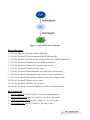
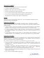
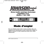
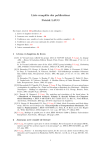
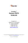
Product Manual OxiSelect™ Total Antioxidant Capacity (TAC) Assay Kit, Trial Size Catalog Number STA-360-T 20 assays FOR RESEARCH USE ONLY Not for use in diagnostic procedures Introduction Oxidative stress is a physiological condition where there is an imbalance between concentrations of reactive oxygen species (ROS) and antioxidants. However, excessive ROS accumulation will lead to cellular injury, such as damage to DNA, proteins, and lipid membranes. The cellular damage caused by ROS has been implicated in the development of many disease states, such as cancer, diabetes, cardiovascular disease, atherosclerosis, and neurodegenerative diseases. Under normal physiological conditions, cellular ROS generation is counterbalanced by the action of cellular antioxidant enzymes, macro or micro molecules, as well as other redox molecules. Antioxidants also include both hydrophilic and lipophilic molecules for metabolizing ROS. These may be localized transiently within different tissues or cells. Because of their potential harmful effects, excessive ROS must be promptly eliminated from the cells by this variety of antioxidant defense mechanisms. Although the products of ROS-induced oxidative stress are extensively used to monitor their biological effects, it is also important to evaluate the antioxidant capacity of biological fluids, cells, and extracts. Antioxidants commonly neutralize radicals via a hydrogen atom transfer (HAT) or single electron transfer (SET) mechanism. SET assays quantify the capability of an antioxidant to transfer one electron to reduce any compound, such as free radicals, carbonyls, and metals. Cell Biolabs’ OxiSelect™ Total Antioxidant Capacity (TAC) Assay Kit measures the total antioxidant capacity of biomolecules from a variety of samples via a SET mechanism. The TAC Assay is based on the reduction of copper (II) to copper (I) by antioxidants such as uric acid. Upon reduction, the copper (I) ion further reacts with a coupling chromogenic reagent that produces a color with a maximum absorbance at 490 nm. The net absorbance values of antioxidants are compared with a known uric acid standard curve. Absorbance values are proportional to the sample’s total reductive capacity. Results are expressed as “μM Copper Reducing Equivalents” or “mM Uric Acid Equivalents”. Copper is advantageous over iron-based antioxidant assays because all classes of antioxidants, including thiols, are detected with marginal radical interference. In addition, the kinetics of the copper-based reaction is also faster than the iron-based reaction, which makes the TAC Assay an ideal tool for estimating reductive or antioxidant capacity efficiently and accurately. Cell Biolabs’ OxiSelect™ TAC Assay Kit is a fast and reliable kit for the direct measurement of total antioxidant capacity from cell lysate, plasma, serum, urine, tissue homogenates, and food extracts. Each Trial Size TAC Assay Kit provides sufficient reagents to perform up to 20 assays, including blanks, antioxidant standards and unknown samples. Hydrophilic and lipophilic samples are compatible with the assay. Assay Principle Cell Biolabs’ OxiSelect™ TAC Assay Kit measures the total antioxidant capacity within a sample. Samples are compared to a known concentration of uric acid standard within a 96-well microtiter plate format. Samples and standards are diluted with a reaction reagent and, upon the addition of copper, the reaction proceeds for a few minutes. The reaction is stopped and read with a standard 96-well spectrophotometric microplate reader at 490 nm (Figure 1). Antioxidant capacity is determined by comparison with the uric acid standards. 2 Figure 1. TAC Activity Assay Principle Related Products 1. STA-310: OxiSelect™ Protein Carbonyl ELISA Kit 2. STA-312: OxiSelect™ Total Glutathione (GSSG/GSH) Assay Kit 3. STA-320: OxiSelect™ Oxidative DNA Damage ELISA Kit (8-OHdG Quantitation) 4. STA-330: OxiSelect™ TBARS Assay Kit (MDA Quantitation) 5. STA-340: OxiSelect™ Superoxide Dismutase Activity Assay 6. STA-341: OxiSelect™ Catalase Activity Assay 7. STA-342: OxiSelect™ Intracellular ROS Assay Kit (Green Fluorescence) 8. STA-343: OxiSelect™ Hydrogen Peroxide Activity Assay (Colorimetric) 9. STA-344: OxiSelect™ Hydrogen Peroxide/Peroxidase Assay (Fluorometric) 10. STA-345: OxiSelect™ ORAC Activity Assay 11. STA-346: OxiSelect™ HORAC Activity Assay 12. STA-347: OxiSelect™ In Vitro ROS/RNS Assay Kit (Green Fluorescence) Kit Components 1. Uric Acid Standard (Part No. 236001): One 100 mg tube of powder. 2. Reaction Buffer (100X) (Part No. 236002-T): One 40 μL amber tube. 3. Copper Ion Reagent (100X) (Part No. 236003-T): One 100 μL tube. 4. Stop Solution (10X) (Part No. 236004-T): One 200 μL tube. 3 Materials Not Supplied 1. Standard 96-well microtiter plates for use in microplate reader 2. 1N NaOH, 1X PBS and deionized water 3. Methanol or other organic solvent for lipid-based samples 4. 5. 6. 7. Sonicator or homogenizer for sample preparations 10 µL to 1000 µL adjustable single channel micropipettes with disposable tips 50 µL to 300 µL adjustable multichannel micropipette with disposable tips Spectrophotometric microplate reader capable of reading 490 nm Storage Upon receipt store the Reaction Buffer (100X) at 4ºC. Store all remaining kit components at room temperature until their expiration dates. Preparation of Reagents Reagents may be prepared for either hydrophilic or lipophilic samples. Although many lipophilic samples are soluble upon dilution with 1X PBS, the kit reagents may be prepared in methanol to ensure complete solubility. • 1X Reaction Buffer: Dilute the Reaction Buffer 1:100 with 1X PBS (hydrophilic) or with methanol (lipophilic). Mix to homogeneity. Store the 1X Reaction Buffer at 4ºC up to three months. • 1X Copper Ion Reagent: Dilute the Copper Ion Reagent 1:100 with deionized water (hydrophilic) or with methanol (lipophilic). Mix to homogeneity. Store the 1X Copper Ion Reagent at 4ºC up to three months. • 1X Stop Solution: Dilute the Stop Solution 1:10 with deionized water (hydrophilic) or with methanol (lipophilic). Mix to homogeneity. Store the 1X Stop Solution at 4ºC up to three months. Preparation of Samples Samples should be stored at -80ºC prior to performing the assay. Samples should be prepared at the discretion of the user. The following recommendations are only guidelines and may be altered to optimize or complement the user’s experimental design. Note: EDTA can interfere with the TAC assay and should not be present in any sample. • Tissue Lysate: Sonicate or homogenize tissue sample on cold PBS and centrifuge at 10,000 x g for 10 minutes at 4ºC. Aliquot the supernatant for storage at -80ºC, protein determination and subsequent TAC assay. • Cell Culture: Wash cells 3 times with cold PBS prior to lysis. Lyse cells with sonication or homogenation in cold PBS and centrifuge at 10,000 x g for 10 minutes at 4ºC. Aliquot the supernatant for storage at -80ºC, protein determination and subsequent TAC assay. • Plasma: Collect blood with heparin and centrifuge at 4ºC for 10 minutes. Remove the plasma and aliquot samples for testing. • Urine: Test neat or diluted with PBS if appropriate. 4 • Lipophilic Fractions: Dissolve lipophilic samples in 100% methanol or acetone and then dilute in 50% methanol or acetone. Incubate the mixture for 1 hour at room temperature with mixing. Further dilute samples as necessary prior to testing. • Food Samples: Results may vary depending on sample source and purification. Dilution and preparation of these samples is at the discretion of the user, but use the following guidelines: o Solid or High Protein Samples: Weigh solid sample and then homogenize after adding deionized water (1:2, w/v). Centrifuge the homogenate at 10,000 x g for 10 minutes at 4ºC. Recover the supernatant which is the water-soluble fraction. The insoluble fraction (pulp) is further extracted by adding pure acetone (1:4, w(solid pulp)/v) and mixing at room temperature for 30-60 minutes. Centrifuge the extract/solid at 10,000 x g for 10 minutes at 4ºC. Recover the acetone extract and dilute with PBS or water as necessary prior to running the assay. The TAC value is calculated by combining the results from the water-soluble fraction and the acetone extract from the pulp fraction. o Aqueous Samples: Centrifuge the sample at 10,000 x g for 10 minutes at 4ºC to remove any particulates. Dilute the supernatant in PBS as necessary prior to running the assay. Certain liquids such as juice extracts may be tested without dilution. Preparation of Uric Acid Standard Curve 1. Prepare fresh Uric Acid standards by weighing out the Uric Acid powder for a 10 mg/mL solution in 1N NaOH. This 10 mg/mL is equivalent to a concentration of 60 mM. Use the 60 mM Uric Acid solution to prepare a 2 mM solution of Uric Acid (eg. add 100 µL of the 60 mM Uric Acid standard to 2.900 mL of deionized water). 2. Prepare a series of the remaining Uric Acid standards according to Table 1 below. Tubes 1 2 3 4 5 6 7 8 9 10 2 mM Uric Acid Antioxidant Standard (µL) 500 500 of tube #1 500 of tube #2 500 of tube #3 500 of tube #4 500 of tube #5 500 of tube #6 500 of tube #7 500 of tube #8 0 Deionized Water (µL) 500 500 500 500 500 500 500 500 500 500 Resulting Uric Acid Concentration (mM) 1 0.5 0.25 0.125 0.0625 0.03125 0.0156 0.0078 0.0039 0.0 Table 1. Preparation of Uric Acid Standards. Note: The 60 mM stock Uric Acid standard solution may be aliquotted and stored at -70ºC for up to one week. Do not store diluted Uric Acid Standard solutions. 5 Assay Protocol Each Uric Acid Standard and sample should be assayed in duplicate or triplicate. A freshly prepared standard curve should be used each time the assay is performed. 1. Add 20 µL of the diluted Uric Acid Standards or samples to the 96-well microtiter plate. 2. Add 180 µL of the 1X Reaction Buffer to each well using either a multichannel pipette or a plate reader liquid handling system. Mix thoroughly. 3. Obtain an initial absorbance by reading the plate at 490 nm. 4. To initiate the reaction, add 50 µL of the 1X Copper Ion Reagent into each well. Incubate 5 minutes on an orbital shaker. 5. Add 50 µL of 1X Stop Solution to each well to terminate the reaction. 6. Read the plate again at 490 nm. Example of Results The following figures demonstrate typical OxiSelect™ TAC Assay results (hydrophilic). One should use the data below for reference only. This data should not be used to interpret or calculate actual sample results. 6 Figure 2: TAC Assay Standard Curve. 7 Figure 3: TAC Assay of various antioxidants. 8 Calculation of Results 1. Calculate the net absorbance by subtracting the initial absorbance readings for samples and standards (Step 3) from the final readings taken for each (Step 6). 2. Plot the net absorbance against the uric acid concentration for the uric acid standard curve. 3. Calculate the antioxidant capacity of unknown samples by comparing the net OD 490 nm values of samples to the uric acid standard curve. To determine the “mM uric acid equivalents” (UAE) for samples, extrapolate the uric acid concentration from the sample’s analogous uric acid OD 490 nm value (For example, based on the sample standard curve above, an OD 490 nm value of 1.0 corresponds to 0.75 mM uric acid equivalents (UAE)). To determine “μM Copper Reducing Equivalents” (CRE) for samples, multiply the uric acid equivalence (UAE) concentration by 2189 μM Cu++/ mM uric acid. 1 mM of uric acid = 2189 μM Copper Reducing Equivalents. (For example, 0.75 mM UAE x 2189 = 1642 Copper Reducing Equivalents (CRE)). CRE sample values are proportional to the sample’s Total Antioxidant Capacity or Total Antioxidant Power. References 1. Allard, J.P., et al. (1998) Am. J. Clin. Nutr. 67: 143-147. 2. Cerutti, P. and Trump, B. (1991) Proc. Cancer Cell 3: 1-7. 3. Frei, B., et al. (1992) “Molecular Biology of Free Radical Scavenging System” 23-45. 4. Trachootham, D., et al. (2008) Antioxid. Redox Signal. 10: 1343-1374. 5. Van-Zoeren-Grobben, et al. (1997) Acta Pediatrics. 86: 1356-1362. Recent Product Citations 1. Liu, M. et al. (2015). Shen-Kang protects 5/6 nephrectomized rats against renal injury by reducing oxidative stress through the MAPK signaling pathways. Int J Mol Med. 36:975-984. 2. Scholten, S. D. et al. (2015). Effects of vitamin D and quercetin, alone and in combination, on cardiorespiratory fitness and muscle function in physically active male adults. Open Access J Sports Med. 6:229-239. 3. Pérez, E. et al. (2015). Improved antitumor effect of paclitaxel administered in vivo as pH and glutathione-sensitive nanohydrogels. Int J Pharm. 492:10-19. 4. Antonopoulos, A. S. et al. (2015). Adiponectin as a link between type 2 diabetes and vascular NADPH oxidase activity in the human arterial wall: The regulatory role of perivascular adipose tissue. Diabetes. 64:2207-2219. 5. Turkmenoglu, F. P. et al. (2015). Characterization of volatile compounds of eleven achillea species from turkey and biological activities of essential oil and methanol extract of A. hamzaoglui arabacı & budak. Molecules. 20:11432-11458. 6. Sun, M. & Johnson, M. A. (2015). Measurement of total antioxidant capacity in sub-μL blood samples using craft paper-based analytical devices. RSC Adv. 5:55633-55639. 7. Mikail, M. A. et al. (2015). Baccaurea angulata fruit inhibits lipid peroxidation and induces the increase in antioxidant enzyme activities. Eur J Nutr. doi:10.1007/s00394-015-0961-7. 9 8. Ruiz, L. M. et al. (2015). Quercetin affects erythropoiesis and heart mitochondrial function in mice. Oxid Med Cell Longev. doi:10.1155/2015/836301 9. Park, S. Y. et al. (2015). Study on the health benefits of brown algae (Sargassum muticum) in volunteers. J Food Nutr Res. 3:126-130. 10. Beretta, G. L. et al. (2015). Unravelling “off-target” effects of redox-active polymers and polymer multilayered capsules in prostate cancer cells. Nanoscale. doi: 10.1039/C4NR07240E. 11. Youn, P. et al. (2015). Cytoprotection against beta-amyloid (aβ) peptide-mediated oxidative damage and autophagy by Keap1 RNAi in human glioma U87mg cells. Neurosci Res. doi:10.1016/j.neures.2014.12.015. 12. Sashindran, R., et al. (2015). Evaluation of neuroprotective effect of quercetin and coenzyme Q10 in ethanol induced neurotoxicity in mice. Int J Appl Biol Pharm. 6:67-71. 13. Sutkowy, P. et al. (2014). Physical exercise combined with whole-body cryotherapy in evaluating the level of lipid peroxidation products and other oxidant stress indicators in kayakers. 14. Oxid Med Cell Longev. doi: 10.1155/2014/402631. 15. Mercado, N. et al. (2014). Activation of transcription factor Nrf2 signalling by the sphingosine kinase inhibitor SKI-II is mediated by the formation of Keap1 dimers. PLoS One. 9: e88168. 16. Chien, J. W. et al. (2014). Urinary 8-hydroxy-2'-deoxyguanosine (8-oxodG) level can predict acute renal damage in young children with urinary tract infection. Biomarkers. 19:326-331. 17. Narayanan, S. et al. (2014). Impact of high night-time and high daytime temperature stress on winter wheat. Journal of Agronomy and Crop Science. doi: 10.1111/jac.12101. 18. Tran, H. et al. (2014). Effects of Spray-dried porcine plasma on growth performance, immune response, total antioxidant capacity, and gut morphology of nursery pigs. J Anim Sci. 92:4494-4504. 19. Ravikumar, P. et al. (2014). α-Klotho protects against oxidative damage in pulmonary epithelia. Am J Physiol Lung Cell Mol Physiol. 307:L566-575. 20. MacDonald, G. et al. (2014). Memo is a copper-dependent redox protein with an essential role in migration and metastasis. Sci Signal. 7:ra56. Warranty These products are warranted to perform as described in their labeling and in Cell Biolabs literature when used in accordance with their instructions. THERE ARE NO WARRANTIES THAT EXTEND BEYOND THIS EXPRESSED WARRANTY AND CELL BIOLABS DISCLAIMS ANY IMPLIED WARRANTY OF MERCHANTABILITY OR WARRANTY OF FITNESS FOR PARTICULAR PURPOSE. CELL BIOLABS’s sole obligation and purchaser’s exclusive remedy for breach of this warranty shall be, at the option of CELL BIOLABS, to repair or replace the products. In no event shall CELL BIOLABS be liable for any proximate, incidental or consequential damages in connection with the products. Contact Information Cell Biolabs, Inc. 7758 Arjons Drive San Diego, CA 92126 Worldwide: +1 858 271-6500 USA Toll-Free: 1-888-CBL-0505 E-mail: [email protected] www.cellbiolabs.com 2013-2015: Cell Biolabs, Inc. - All rights reserved. No part of these works may be reproduced in any form without permissions in writing. 10