1
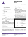
CD14 MicroBeads
human
Order no. 130-050-201
Contents
1.3 Applications
1. Description
●
Isolation of CD14+ monocytes for in vitro generation of
dendritic cells¹ or macrophages²,³.
●
Isolation of CD14+ monocytes for studies on cytotoxicity⁴ and
migration⁵.
1.1 Principle of the MACS® Separation
1.2 Background information
1.3 Applications
1.4 Reagent and instrument requirements
2. Protocol
2.1 Sample preparation
2.2 Magnetic labeling
2.3 Magnetic separation
3. Example of a separation using the CD14 MicroBeads
4. References
1. Description
1.4 Reagent and instrument requirements
●
Buffer: Prepare a solution containing phosphate-buffered
saline (PBS), pH 7.2, 0.5% bovine serum albumin (BSA), and
2 mM EDTA by diluting MACS BSA Stock Solution (# 130091-376) 1:20 with autoMACS™ Rinsing Solution (# 130-091222). Keep buffer cold (2−8 °C). Degas buffer before use, as air
bubbles could block the column.
▲ Note: EDTA can be replaced by other supplements such as anticoagulant citrate
dextrose formula-A (ACD-A) or citrate phosphate dextrose (CPD). BSA can be
replaced by other proteins such as human serum albumin, human serum, or fetal
bovine serum. Buffers or media containing Ca 2+ or Mg2+ are not recommended for
use.
●
MACS Columns and MACS Separators: CD14+ cells can be
enriched by using MS, LS, or XS Columns or depleted with
the use of LD, CS, or D Columns. Cells which strongly express
the CD14 antigen can also be depleted using MS, LS, or XS
Columns. Positive selection or depletion can also be performed
by using the autoMACS or the autoMACS Pro Separator.
Components
2 mL CD14 MicroBeads, human:
MicroBeads conjugated to monoclonal antihuman CD14 antibodies (isotype: mouse
IgG2a).
Capacity
For 10⁹ total cells, up to 100 separations.
Product format
CD14 MicroBeads are supplied in buffer
containing stabilizer and 0.05% sodium azide.
Column Max. number
of labeled cells
Storage
Store protected from light at 2−8 °C. Do not
freeze. The expiration date is indicated on the
vial label.
Positive selection
1.1 Principle of the MACS® Separation
First, the CD14+ cells are magnetically labeled with CD14
MicroBeads. Then, the cell suspension is loaded onto a MACS®
Column which is placed in the magnetic field of a MACS Separator.
The magnetically labeled CD14+ cells are retained within the column.
The unlabeled cells run through; this cell fraction is thus depleted
of CD14+ cells. After removing the column from the magnetic field,
the magnetically retained CD14+ cells can be eluted as the positively
selected cell fraction.
2 ×10⁸
MS
10⁷
MiniMACS, OctoMACS, VarioMACS, SuperMACS
LS
10⁸
2 ×10⁹
MidiMACS, QuadroMACS,
VarioMACS, SuperMACS
XS
10⁹
2 ×10¹⁰
SuperMACS
Depletion
5 ×10⁸
LD
10⁸
CS
140-000-067.06
Miltenyi Biotec GmbH
Friedrich-Ebert-Straße 68, 51429 Bergisch Gladbach, Germany
Phone +49 2204 8306-0, Fax +49 2204 85197
[email protected]
www.miltenyibiotec.com
2×10⁸
D
10⁹
MidiMACS, QuadroMACS,
VarioMACS, SuperMACS
VarioMACS, SuperMACS
SuperMACS
Positive selection or depletion
1.2 Background information
CD14 MicroBeads are used for the positive selection or depletion of
human monocytes and macrophages from cord blood or PBMCs, as
well as pleural, peritoneal, or synovial fluids or from various tissues,
such as spleen and lymph node. The CD14 antigen belongs to the
LPS receptor complex. Binding of antibody to CD14 does not trigger
signal transduction since CD14 lacks a cytoplasmatic domain. CD14
is strongly expressed on most monocytes and macrophages and
weakly on neutrophils and some myeloid dendritic cells.
Max. number Separator
of total cells
autoMACS
2×10⁸
4 ×10⁹
autoMACS, autoMACS Pro
▲ Note: Column adapters are required to insert certain columns into the
VarioMACS™ or SuperMACS™ Separators. For details see the respective MACS
Separator data sheet.
●
(Optional) Fluorochrome-conjugated CD14 antibody for
flow cytometric analysis, e.g., CD14-FITC (# 130-080-701),
CD14-PE (# 130-091-242), or CD14-APC (# 130-091-243). For
more information about other fluorochrome-conjugates see
www.miltenyibiotec.com.
Miltenyi Biotec Inc.
2303 Lindbergh Street, Auburn, CA 95602, USA
Phone 800 FOR MACS, +1 530 888 8871, Fax +1 530 888 8925
[email protected]
page 1/4
Order no. 130-050-201
●
(Optional) Propidium iodide (PI) or 7-AAD for flow cytometric
exclusion of dead cells.
●
(Optional) Dead Cell Removal Kit (# 130-090-101) for the
depletion of dead cells.
●
(Optional) Pre-Separation Filters (# 130-041-407) to remove
cell clumps.
2. Protocol
2.1 Sample preparation
When working with anticoagulated peripheral blood or buffy coat,
peripheral blood mononuclear cells (PBMCs) should be isolated by
density gradient centrifugation, for example, using Ficoll-Paque™.
For details see the General Protocols section of the respective
separator user manual. The General Protocols are also available at
www.miltenyibiotec.com/protocols.
▲ Note: To remove platelets after density gradient separation, resuspend cell
pellet in buffer and centrifuge at 200×g for 10−15 minutes at 20 °C. Carefully
aspirate supernatant. Repeat washing step.
When working with tissues or lysed blood, prepare a single-cell
suspension using standard methods. For details see the General
Protocols section of the respective separator user manual. The
General Protocols are also available at www.miltenyibiotec.com/
protocols.
▲ Dead cells may bind non-specifically to MACS MicroBeads. To
remove dead cells, we recommend using density gradient centrifugation
or the Dead Cell Removal Kit (# 130-090-101).
6. (Optional) Add staining antibodies, e.g., 10 µL of CD14-FITC
(# 130-080-701), and incubate for 5 minutes in the dark in the
refrigerator (2−8 °C).
7.
Wash cells by adding 1−2 mL of buffer per 10⁷ cells and
centrifuge at 300×g for 10 minutes. Aspirate supernatant
completely.
8. Resuspend up to 10⁸ cells in 500 µL of buffer.
▲ Note: For higher cell numbers, scale up buffer volume accordingly.
▲ Note: For depletion with LD Columns, resuspend up to 1.25×10⁸ cells in
500 µL of buffer.
9. Proceed to magnetic separation (2.3).
2.3 Magnetic separation
▲ Choose an appropriate MACS Column and MACS Separator
according to the number of total cells and the number of CD14+ cells.
For details see table in section 1.4.
Magnetic separation with MS or LS Columns
1.
Place column in the magnetic field of a suitable MACS
Separator. For details see the respective MACS Column data
sheet.
2. Prepare column by rinsing with the appropriate amount of
buffer:
MS: 500 µL
LS: 3 mL
3. Apply cell suspension onto the column.
2.2 Magnetic labeling
▲ Work fast, keep cells cold, and use pre-cooled solutions. This will
prevent capping of antibodies on the cell surface and non-specific
cell labeling.
▲ Volumes for magnetic labeling given below are for up to
10⁷ total cells. When working with fewer than 10⁷ cells, use the same
volumes as indicated. When working with higher cell numbers,
scale up all reagent volumes and total volumes accordingly (e.g.
for 2×10⁷ total cells, use twice the volume of all indicated reagent
volumes and total volumes).
▲ For optimal performance it is important to obtain a single‑cell
suspension before magnetic separation. Pass cells through 30 µm
nylon mesh (Pre-Separation Filters, # 130-041-407) to remove cell
clumps which may clog the column. Wet filter with buffer before
use.
▲ Working on ice may require increased incubation times. Higher
temperatures and/or longer incubation times may lead to nonspecific cell labeling.
1.
Determine cell number.
2. Centrifuge cell suspension at 300×g for 10 minutes. Aspirate
supernatant completely.
3. Resuspend cell pellet in 80 µL of buffer per 10⁷ total cells.
4. Collect unlabeled cells that pass through and wash column
with the appropriate amount of buffer. Collect total effluent;
this is the unlabeled cell fraction. Perform washing steps by
adding buffer three times. Only add new buffer when the
column reservoir is empty.
MS: 3×500 µL
LS: 3×3 mL
5. Remove column from the separator and place it on a suitable
collection tube.
6. Pipette the appropriate amount of buffer onto the column.
Immediately flush out the magnetically labeled cells by firmly
pushing the plunger into the column.
MS: 1 mL
LS: 5 mL
7.
(Optional) To increase the purity of CD14+ cells, the eluted
fraction can be enriched over a second MS or LS Column.
Repeat the magnetic separation procedure as described in
steps 1 to 6 by using a new column.
Magnetic separation with XS Columns
For instructions on the column assembly and the separation refer to
the XS Column data sheet.
Depletion with LD Columns
1.
Place LD Column in the magnetic field of a suitable MACS
Separator. For details see LD Column data sheet.
140-000-067.06
4. Add 20 µL of CD14 MicroBeads per 10⁷ total cells.
2. Prepare column by rinsing with 2 mL of buffer.
5. Mix well and incubate for 15 minutes in the refrigerator
(2−8 °C).
3. Apply cell suspension onto the column.
Unless otherwise specifically indicated, Miltenyi Biotec
products and services are for research use only and not for
diagnostic or therapeutic use.
page 2/4
Order no. 130-050-201
Depletion with CS Columns
1.
Assemble CS Column and place it in the magnetic field of a
suitable MACS Separator. For details see CS Column data
sheet.
2. Prepare column by filling and rinsing with 60 mL of buffer.
Attach a 22G flow resistor to the 3-way stopcock of the
assembled column. For details see CS Column data sheet.
3. Apply cell suspension onto the column.
4. Collect unlabeled cells that pass through and wash column
with 30 mL buffer from the top. Collect total effluent; this is
the unlabeled cell fraction.
3. For a standard separation choose one of the following
programs:
Positive selection: “Possel”
Collect positive fraction in row C of the tube rack.
Depletion: “Depletes”
Collect negative fraction in row B of the tube rack.
3. Example of a separation using the
CD14 MicroBeads
CD14+ monocytes were isolated from human PBMCs using CD14
MicroBeads, an MS Column, and a MiniMACS™ Separator. Cells
are fluorescently stained with CD14-FITC (# 130-080-701). Cell
debris and dead cells are excluded from the analysis based on scatter
signals and PI fluorescence.
PBMCs before separation
Relative cell number
4. Collect unlabeled cells that pass through and wash
column with 2×1 mL of buffer. Collect total effluent;
this is the unlabeled cell fraction. Perform washing
steps by adding buffer two times. Only add new buffer when
the column reservoir is empty.
Depletion with D Columns
For instructions on column assembly and separation refer to the
D Column data sheet.
Magnetic separation with the autoMACS™ Separator or the
autoMACS™ Pro Separator
▲ Refer to the respective user manual for instructions on how to
use the autoMACS™ Separator or the autoMACS Pro Separator.
▲ Buffers used for operating the autoMACS Separator or the
autoMACS Pro Separator should have a temperature of ≥ 10 °C.
CD14-FITC
Magnetic separation with the autoMACS™ Separator
1. Prepare and prime the instrument.
2. Apply tube containing the sample and provide tubes for
collecting the labeled and unlabeled cell fractions. Place
sample tube at the uptake port and the fraction collection
tubes at port neg1 and port pos1.
CD14-FITC
3. For a standard separation choose one of the following
programs:
Positive selection: “Possel”
Collect positive fraction from outlet port pos1.
Depletion: “Depletes”
Collect negative fraction from outlet port neg1.
Magnetic separation with the autoMACS™ Pro Separator
1. Prepare and prime the instrument.
2. Apply tube containing the sample and provide tubes
for collecting the labeled and unlabeled cell fractions.
Place sample tube in row A of the tube rack and fraction
collection tubes in rows B and C.
CD14 + cells
Relative cell number
Relative cell number
▲ Program choice depends on the isolation strategy, the strength
of magnetic labeling, and the frequency of magnetically labeled
cells. For details refer to the section describing the cell separation
programs in the respective user manual. Program recommendations
below refer to separation of human PBMCs.
CD14 – cells
CD14-FITC
140-000-067.06
Unless otherwise specifically indicated, Miltenyi Biotec
products and services are for research use only and not for
diagnostic or therapeutic use.
page 3/4
Order no. 130-050-201
4. References
1.
Pickl, W. F. et al. (1996) Molecular and Functional Characteristics of Dendritic
Cells Generated from Highly Purified CD14+ Peripheral Blood Monocytes. J.
Immunol. 157: 3850–3859. [274]
2.
Hanley, P. J. et al. (2004) Extracellular ATP induces oscillations of intracellular
Ca2+ and membrane potential and promotes transcription of IL-6 in
macrophages. Proc. Natl. Acad. Sci. U S A. 101: 9479–9484. [4206]
3.
Verreck, F. A. et al. (2004) Human IL-23-producing type 1 macrophages promote
but IL-10-producing type 2 macrophages subvert immunity to (myco)bacteria.
Proc. Natl. Acad. Sci. U S A. 101: 4560–4565. [4198]
4.
Ryan, E. J. et al. (2002) Dendritic cell-associated lectin-1: a novel dendritic
cell-associated, C-type lectin-like molecule enhances T cell secretion of IL-4. J.
Immunol. 169: 5638–5648. [2436]
5.
Vitale, S. et al. (2004) Soluble fractalkine prevents monocyte chemoattractant
protein-1-induced monocyte migration via inhibition of stress-activated protein
kinase 2/p38 and matrix metalloproteinase activities. J. Immunol. 172: 585–592.
[3657]
6.
de Baey, A. and Lanzavecchia, A. (2000) The Role of Aquaporins in Dendritic
Cell Macropinocytosis. J. Exp. Med. 191: 743–747. [841]
7.
Salio, M. et al. (2000) Dendritic cell maturation is induced by mycoplasma
infection but not by necrotic cells. Eur. J. Immunol. 30: 705–708. [973]
8.
Ebner, S. et al. (2002) A novel role for IL-3: human monocytes cultured in the
presence of IL-3 and IL-4 differentiate into dendritic cells that produce less IL12 and shift Th cell responses toward a Th2 cytokine pattern. J. Immunol. 168:
6199–6207. [2200]
9.
Jefford, M. et al. (2003) Functional comparison of DCs generated in vivo with
Flt3 ligand or in vitro from blood monocytes: differential regulation of function
by specific classes of physiologic stimuli. Blood 102: 1753–1763. [2874]
10.
Matsumoto, M. et al. (2003) Subcellular localization of Toll-like receptor 3 in
human dendritic cells. J. Immunol. 171: 3154–3162. [4228] Erratum (color print
of figure 5A) in: J. Immunol. 171: 4934.
All protocols and data sheets are available at www.miltenyibiotec.com.
Warnings
Reagents contain sodium azide. Under acidic conditions sodium azide yields
hydrazoic acid, which is extremely toxic. Azide compounds should be diluted with
running water before discarding. These precautions are recommended to avoid
deposits in plumbing where explosive conditions may develop.
Warranty
The products sold hereunder are warranted only to be free from defects in workmanship
and material at the time of delivery to the customer. Miltenyi Biotec GmbH
makes no warranty or representation, either expressed or implied, with respect to
the fitness of a product for a particular purpose. There are no warranties, expressed
or implied, which extend beyond the technical specifications of the products.
Miltenyi Biotec GmbH’s liability is limited to either replacement of the products or
refund of the purchase price. Miltenyi Biotec GmbH is not liable for any property
damage, personal injury or economic loss caused by the product.
MACS is a registered trademark and autoMACS, MidiMACS, MiniMACS, OctoMACS,
QuadroMACS, SuperMACS, and VarioMACS are trademarks of Miltenyi Biotec
GmbH.
Ficoll-Paque is a trademark of GE Healthcare companies.
© 2007 Miltenyi Biotec GmbH.
140-000-067.06
Unless otherwise specifically indicated, Miltenyi Biotec
products and services are for research use only and not for
diagnostic or therapeutic use.
page 4/4