1
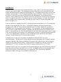
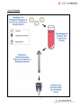
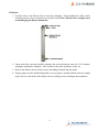
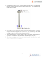
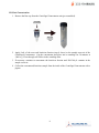
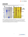
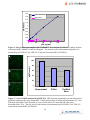
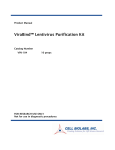
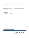
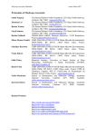
Product Manual ViraBind™ PLUS Lentivirus Concentration and Purification Mega Kit Catalog Number VPK-096 2 preps VPK-096-5 10 preps FOR RESEARCH USE ONLY Not for use in diagnostic procedures Introduction Lentivirus vectors based on the human immunodeficiency virus-1 (HIV-1) have become a promising vector for gene transfer studies. The advantageous feature of lentivirus vector is the ability for gene transfer and integration into dividing and non-dividing cells. The pseudotyped envelope with vesicular stomatitis virus envelope G (VSV-G) protein broadens the target cell range. Lentiviral vectors have been shown to deliver genes to neurons, lymphocytes and macrophages, cell types that previous retrovirus vectors could not be used. Lentiviral vectors have also proven to be effective in transducing brain, liver, muscle, and retina in vivo without toxicity or immune responses. Recently, the lentivirus system is widely used to integrate siRNA efficiently in a wide variety of cell lines and primary cells both in vitro and in vivo. Lentivirus particles are produced from 293T cells through transient transfection of 3 or 4 plasmids that encodes for the components of the virion. Viral medium containing viral particles produced by packaging cells within 48-72 hr can be harvested and frozen. To obtain a higher titer, pseudovirus supernatant can be concentrated by ultracentrifuging. As a consequence, the ultracentrifugation step also concentrates cellular debris, membrane fragments, and denatured proteins derived from culture media of virus-producing cells. This unwanted material in the crude vector preparation is toxic to target cells, especially primary cells, and may cause immunogenic reactions in experimental animal models by in vivo vector administration. Therefore, to reduce undesirable effects and increase gene transfer efficiency, the purification of virus vector becomes essential. ViraBind™ PLUS Lentivirus Concentration and Purification Mega Kit does not involve ultracentrifugation. The lentiviruses are first pelleted from viral supernatant with ViraBind™ reagents (patented technology), then further purified and concentrated with a dialysis device and concentrator, respectively (see Assay Principle below). Each preparation can handle up to 500 mL of lentiviral supernatant (106-7 TU/mL) resulting in 200-300 µL of highly purified lentivirus (109 TU/mL). ViraBind™ PLUS Lentivirus Concentration and Purification Mega Kit provides an efficient system for quick lentiviral purification with high recovery (>60%). The highly purified and concentrated viruses can be used in primary cell infections and in vivo applications. The system may be adapted to purification of other viral types, such as MMLV based retrovirus. 2 Assay Principle 3 Related Products 1. VPK-107: QuickTiter™ Lentivirus Titer Kit (Lentivirus-Associated HIV p24) 2. VPK-108-HIV-p24: QuickTiter™ Lentivirus Quantitation Kit (HIV p24 ELISA) 3. VPK-108: QuickTiter™ Lentivirus Quantitation Kit 4. LTV-200: ViraDuctin™ Lentivirus Transduction Kit 5. LTV-100: 293LTV Cell Line 6. VPK-130: ViraBind™ Retrovirus Concentration and Purification Kit 7. VPK-100: ViraBind™ Adenovirus Purification Kit Kit Components 1. ViraBind™ Lentivirus Reagent A (100X) (Part No. 309601): One sterile bottle – 10 mL. 2. ViraBind™ Lentivirus Reagent B (100X) (Part No. 309602): One sterile bottle – 10 mL. 3. Dialysis Devices (Part No. 309603): Two units with 2 floatation rings and disposable recovery pipettes. 4. Centrifugal Concentrators (Part No. 309604): Two units with 2 centrifuge tubes. 5. Purification Buffer (Part No. 309504): One bottle – 25.0 mL. Materials Not Supplied 1. Lentivirus packaging plasmid mix and expression construct 2. Transfection Reagent 3. HEK 293T cells and cell culture growth medium 4. Dialysis Buffer (25 mM Tris, 1 M NaCl, pH 7.5) 5. PBS 6. Centrifuge (capable of 10,000 x g) Storage Store all kit components at 4ºC until their expiration dates. Preparation of Reagents • Dialysis Buffer: Prepare sufficient dialysis buffer (25 mM Tris, 1 M NaCl, pH 7.5) for each purification. 4 Safety Considerations Remember that you will be working with samples containing infectious virus. Follow the recommended NIH guidelines for all materials containing BSL-2 organisms. Pseudovirus Production The following procedure is suggested for a 10cm dish and may be optimized to suit individual needs. Please refer to the user manual when the lentivirus expression systems from Invitrogen or System Biosciences are used. 1. Use HEK 293T cells that have been passaged 2-3 times prior to transfection. Culture these cells until the monolayer is 70-80% confluent. 2. Replace the cell culture media with new growth media, 10 mL per 10 cm dish. 3. Transfect cells with the packaging plasmid mix and your expression construct. When using Lipofectamine™, please refer to Invitrogen’s Lipofectamine™ reagent manual. 4. After 36-48 hrs, harvest all 10 mL medium in a 15 mL conical tube and centrifuge for 5 min at 3000 rpm to pellet the cell debris. Filter the supernatant through a 0.45 µm low protein binding filter. 5. The viral supernatant can be stored at -80ºC or immediately purified (see purification instructions below). Protocol I. Purification and Concentration The following procedure is written for purification and concentration of 500 mL of lentiviral supernatant. For lentiviral samples that are less than 500 mL, the amount of ViraBind™ Lentivirus Reagent A, B (step 1) and Purification Buffer (step 5) needed should be calculated proportionally. 1. Add 5 mL of ViraBind™ Lentivirus Reagent A (100X) to 500 mL of viral supernatant, mix by inverting. Immediately add 5 mL of ViraBind™ Lentivirus Reagent B (100X) and mix by inverting. 2. Incubate for 60 minutes at 37ºC. 3. Centrifuge the complexed lentivirus for 15 minutes at 10,000 x g. A pellet should be visible. 4. Carefully aspirate the media and collect the pellet. 5. Resuspend and dissolve the complexed lentivirus pellet in 8 mL of Purification Buffer. Vortex the solution to dissolve the pellet. Note: The solution may appear hazy. 6. Centrifuge the solution for 5 minutes at 10,000 x g to remove any insoluble material. Carefully recover the supernatant. 5 II. Dialysis 1. Carefully remove the Dialysis Device from the packaging. Firmly holding the collar, slowly twist the protective sleeve and pull away from the collar (Note: Pull the sleeve straight out to avoid damaging the dialysis membrane). 2. Unscrew the blue cap and carefully submerge the unit in deionized water for 15-30 minutes, wetting the membrane completely. Once wetted, do not allow membrane to dry out. 3. Remove the dialysis device from the water, discarding all liquid from the inside. 4. Using a pipette, not the included disposable recovery pipette, carefully add the lentivirus sample (step 6 above) to the inside of the dialysis device (making sure not to damage the membrane). 6 5. Screw the blue cap onto the device. Carefully, push the base of the dialysis device through the hole of the floatation ring (in the direction of the blue arrows below). Push the floatation ring up until it meets the collar. 6. Float the dialysis device vertically in a container with a stir bar. Dialyze against 4 L of Dialysis Buffer (see Preparation of Reagents) for 4-6 hours at 4ºC, with gentle stirring. Note: Avoid creating a strong vortex which can tip or submerge the device. 7. Transfer the dialysis device to another container of PBS or desired buffer. Dialyze against 4 L for 4 hours to overnight at 4ºC, with gentle stirring. Repeat this dialysis once more. 8. After dialysis is complete, open the screw-on cap and recover the dialyzed lentivirus sample with the included disposable recovery pipette. 7 III. Final Concentration 1. Remove the blue cap from the Centrifugal Concentrator unit (pre-assembled). 2. Apply 8 mL of the recovered lentivirus fraction (step 8 above) to the sample reservoir of the Centrifugal Concentrator. Cap the concentrator and place into a centrifuge for 30 minutes at 5,000 x g. Flow-through will collect in the centrifuge tube. 3. If necessary, continue to concentrate the lentivirus fraction until 200-300 µL remains in the sample reservoir. 4. Collect the concentrated lentivirus sample from the inside of the Centrifugal Concentrator with a pipette. 8 Example of Results The following figures demonstrate typical purification results. One should use the data below for reference only. This data should not be used to interpret actual results. Figure 1: Electrophoretic Profile of Purified GFP lentivirus. 50 mL of GFP lentiviral supernatant was concentrated and purified according to the described Assay Instructions. Lentiviral Supernatant (A), Virus Pellet (B) and Purified Virus Fraction (C) were analyzed on SDS-PAGE. Proteins were visualized by Commassie blue stain (left) and silver stain (right). The VSV-G envelope protein and p24 coat protein (the most abundant proteins in the vector) are indicated. 9 1.8 p24 in supernatant 1.6 p24 in pellet OD 450nm 1.4 1.2 1 0.8 0.6 0.4 0.2 0 0 25 50 75 100 p24 (ng/mL) Figure 2: Free p24 does not complex with ViraBind™. Recombinant p24 diluted in culture medium was treated with ViraBind™ Lentivirus Reagents. The amount of p24 in supernatant and pellet was measured by p24 ELISA (Cat# VPK-107, Lentivirus Associated HIV p24 ELISA). p24 Titer (%) 100 80 60 40 20 0 Supernatant Pellet Purified Virus Figure 3: Vector Yield Determined by p24 ELISA. GFP lentiviral supernatant was concentrated and purified according to the Assay Instructions. Purification fractions of Lentiviral Supernatant (A), Virus Pellet (B) and Purified Virus Fraction (C) were used to infect 293 cells with GFP expression determined after 72 hr. The p24 titer of each fraction was determined by p24 ELISA (Cat.# VPK-107, Lentivirus Associated HIV p24 ELISA). 10 References 1. Naldini, L., U. Blomer, P. Gallay, D. Ory, R. Mulligan, F. H. Gage, I. M. Verma, and D. Trono (1996) Science 272, 263-267. 2. Verma, I. M., and N. Somia (1997) Nature 389, 239-242 3. Kafri, T., U. Blomer, D. A. Peterson, F. H. Gage, and I. M. Verma (1997) Nat. Genet. 17, 314-317. 4. Beyer, W. R., M. Westphal, W. Ostertag, and D. von Laer (2002) J. Virol. 76, 1488-1495. Notice to Purchaser This product is sold for research and development purposes only and is not to be incorporated into products for resale without written permission from Cell Biolabs. The patented technology is covered by an exclusive license. By the use of this product you accept the terms and conditions of all applicable Limited Use Label Licenses. You may contact our Business Development department at [email protected] for information on sublicensing this technology. Warranty These products are warranted to perform as described in their labeling and in Cell Biolabs literature when used in accordance with their instructions. THERE ARE NO WARRANTIES THAT EXTEND BEYOND THIS EXPRESSED WARRANTY AND CELL BIOLABS DISCLAIMS ANY IMPLIED WARRANTY OF MERCHANTABILITY OR WARRANTY OF FITNESS FOR PARTICULAR PURPOSE. CELL BIOLABS’ sole obligation and purchaser’s exclusive remedy for breach of this warranty shall be, at the option of CELL BIOLABS, to repair or replace the products. In no event shall CELL BIOLABS be liable for any proximate, incidental or consequential damages in connection with the products. Contact Information Cell Biolabs, Inc. 7758 Arjons Drive San Diego, CA 92126 Worldwide: +1 858-271-6500 USA Toll-Free: 1-888-CBL-0505 E-mail: [email protected] www.cellbiolabs.com 2008: Cell Biolabs, Inc. - All rights reserved. No part of these works may be reproduced in any form without permissions in writing. 11