1
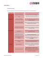
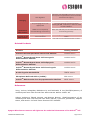
 User Manual
OriCellTM Wistar Rat Mesenchymal
Stem Cells(MSCs)
Cat. No. RAWMX-01001
Table of Contents
Contents and Storage ……………………………………………………………………………………… 3
Product Introduction……………………………………………………………………………………… 3
Cell Characteristics and Identity ……………………………………………………………………… 3
Product Application ………………………………………………………………………………………… 4
General Handling Principles …………………………………………………………………………… 4
Culturing OriCellTM Wistar Rat MSCs
Thawing and Establishing OriCellTM Wistar Rat MSCs …………..……………………………… 4
Passaging Cyagen OriCellTM Wistar Rat MSCs ……..……………………………………………… 6
Differentiation of OriCellTM Wistar Rat MSCs ………………..……………………………………… 8
Cryopreservation of OriCellTM Wistar Rat MSCs ……………..…………………………………… 12
Appendix …………………………………………………………………….…………… 13
Troubleshooting …………………………………………………………………………………………… 13
Related Products ………………………………………………………………………………………… 14
References ……………………………………………………………………………………………… 14
Technical Support ……………………………..……………………………………… 15
CONTENTS AND STORAGE
Product Name
Wistar Rat Mesenchymal Stem Cells
Catalog No.
RAWMX‐01001
Amount per Vial
1×10⁶ Cells
Cryopreserved At
Second Passage
Storage Condition
Liquid Nitrogen
CAUTION: Please handle this product as a potentially biohazardous material. This
product contains dimethyl sulfoxide (DMSO), a hazardous material, in the freezing
medium.
PRODUCT INTRODUCTION
Mesenchymal stem cells (MSCs) are multipotent stem cells that can differentiate into a
variety of cell types including osteocytes, adipocytes, and chondrocytes. MSCs
proliferate quickly and are capable of generating a local immunosuppressive
microenvironment, thus contributing to their wide application potentials in tissue
engineering, cell therapy, and gene therapy.
OriCellTM Wistar Rat Mesenchymal Stem Cells are derived from the bone marrow of
Wistar Rats. They have a strong capacity for self-renewal while maintaining their
multipotency.
In addition, these cells have been tested for:
•
Exogenous Factors: bacterial/fungal contamination, mycoplasma contamination,
and endotoxin contamination.
•
Characteristics: post-thaw viability, cell cycle, verification of undifferentiated
state, and differentiation potential.
This product is intended for laboratory research use only. It is not intended for
diagnostic, therapeutic, clinical, household, or any other applications.
CELL CHARACTERISTICS AND IDENTITY
•
Strong capacity to expand. Can be passaged at least 5 times.
•
Multipotent differentiation ability along the osteogenic, chondrogenic, and
adipogenic lineages.
IMPI0057A2 RAWMX‐01001 Page 3 of 15 •
Posittive for CD2
29, CD44, a
and CD90 (>
( 70%), and
a
negativ
ve for CD34
4 and
CD45
5 and CD11
1b (< 5%) in flow cytometry ass
says.
PRODUC
CT APPL
LICATIO
ONS
W
Wistar Rat MSCs
M
have become
b
ap
popular research targe
et due to th
heir potentiial use in
re
egenerative
e medicine and
a
tissue engineering (in areas such as ca
ardiovascula
ar, neural,
an
nd orthoped
dic disease).
star Rat MS
SCs can be used as cell models to
o evaluate the immun
noreactions,,
OriCellTM Wis
n of MSCs both
b
in vivo
o and in vittro.
prroliferation,, immigration, and diffferentiation
GENERA
AL HAND
DLING PRINCIP
P
PLES
1.. Aseptic handling
h
of the producct is necess
sary througho
out.
2.. Once the
e cells have been estab
blished, alw
ways freeze
e several vi als of OriCe
ellTM Wistarr
Rat MSCs
s as a back
kup.
Note: The O
OriCellTM Wis
star Rat MS
SCs can be frozen/tha
awed at lea
ast one time
e.
N
ecommende
ed to use ce
ells that are
e at, or und
der, an
3.. For all studies, it is strongly re
p
number of 10
0.
original passage
4.. For general mainten
nance of ce
ells, we reco
ommend th
he seeding density to be 2.02
c
.
3.0×10⁴cells/cm
5.. For general mainten
nance of ce
ells, we reco
ommend th
hat the med
dium is cha
anged if it
e pH indicattor in the medium
m
app
pears yellow
w). In general,
becomes acidic (the
very three days.
d
change the growth medium ev
6.. Do not le
et OriCellTM Wistar Ratt MSCs overrgrow as it will result iin contact inhibition.
i
When the
e cells are 80-90%
8
co
onfluent, subculturing the cells is strongly
recomme
ended.
Note: We sttrongly reco
ommend th
he use of Or
riCellTM cult
ture media and other related
N
re
eagents for optimal re
esults.
THAWIN
NG AND
D ESTABLISHIN G OriCe
ellTM WIS
STAR RA
AT MSCs
s
M
Materials Required
R
•
OriCellTM Mesenchym
mal Stem C
Cell Growth Medium (C
Cat. No. GU
UXMX-9001
11)
Th
hawing and
a
Estab
blishing W
Wistar Ratt MSCs
IMPI0057A2 RAWMX‐01001
1 Page 4 of 1
15 1.
Pre-warm
m the fully supplemen
nted (compllete) OriCellTM MSC Grrowth Medium to 37°C
C.
2.
Add 9 mL
L of OriCellTM MSC Gro
owth Mediu
um to a 15 mL conical tube.
3.
Remove the cryovia
al of OriCelllTM Wistar Rat
R MSCs frrom liquid n
nitrogen.
4.
t
the via
al in a 37°C
C water batth until the last ice cry
ystal disapp
pears.
Quickly thaw
For optim
mal results, be sure to
o finish the thawing prrocedure wiithin 3 minutes. Be
careful not to subm
merge the en
ntire vial. Maximum cell
c viability
y is depend
dent on
d and complete thawin
ng of frozen
n cells.
the rapid
ess than op
ptimal if the
e cells are thawed
t
forr more than
n 3 minutes
s.
Note: Resultts will be le
N
5.
As soon as
a the cells
s are complletely thawed, disinfec
ct the outsiide of the cryovial
c
with 70%
% v/v ethan
nol.
6.
TM
Use a pip
pette to transfer the ccells to the 15 mL coniical tube co
ontaining OriCell
O
MSC Growth Medium
m inside a biosafety cabinet.
c
Be
e careful no
ot to introdu
uce any
d
the transfer prrocess.
bubbles during
7.
Rinse the
e vial with 1 mL of the
e medium to
t reduce cell loss. Su
ubsequently
y transfer
this 1 mL
L of cell sus
spension in
nto the conical tube.
8.
Gently mix
m the cell suspension
n by slowly pipetting up
u and dow
wn. Be care
eful not to
introduce
e any bubbles.
9.
Centrifug
ge the cell suspension
s
at 250 x g for 5 minu
utes.
0. Carefully
y aspirate off as much of the supernatant as
s possible a
and add 2-3
3 mL of
10
fresh OriCellTM MSC Growth Me
edium (pre-warmed to
o 37°C).
11
1. Gently re
esuspend th
he cells in O
OriCellTM MS
SC Growth Medium.
12
2. Seed the
e cells into a T25 flask
k and add a sufficient amount
a
of OriCellTM MSC
M
Growth
h
Medium. Gently roc
ck the cultu
ure flask to
o evenly dis
stribute the
e cells.
13
3. Incubate
e the flask at
a 37°C ins ide a 5% CO
C 2 humidiffied incubattor.
14
4. The nextt day, chang
ge the med
dium with fresh growth medium (pre-warmed to 37°C
C).
15
5. Change the
t
growth medium ev
very three days thereafter.
16
6. When the
e cells are approximattely 80-90%
% confluent, they can
n be dissociated with
Trypsin-E
EDTA and passaged.
p
Note: Chang
ging Mediium
N
1.. Warm an
n appropriatte amount of medium to 37°C in a sterile co
ontainer. Replace
R
the spentt medium with
w
the pre
e-warmed, fresh medium. Once completed
d,
return th
he flask to the
t
incubattor.
2.. Avoid rep
peated war
rming and c
cooling of the medium
m. If the en
ntire conten
nt is not
needed fo
or a single procedure,, transfer only
o
the req
quired volu me to a ste
erile
secondarry container.
IMPI0057A2 RAWMX‐01001
1 Page 5 of 1
15 Fig. 1. OriCellTM Wistar Rat Mesenchymal Stem Cells are established.
PASSAGING OriCellTM WISTAR RAT MSCs
Materials Required
•
0.25%Trypsin-0.04%EDTA (Cat. No. TEDTA-10001)
•
Phosphate-Buffered Saline (1×PBS) (Cat. No. PBS-10001)
•
OriCellTM Wistar Rat Mesenchymal Stem Cells (Cat. No. RAWMX-01001)
•
OriCellTM Mesenchymal Stem Cell Growth Medium (Cat. No. GUXMX-90011)
Passaging OriCellTM Wistar Rat MSCs
1. Pre-warm the OriCellTM MSC Growth Medium, 1×PBS, and Trypsin-EDTA solution to
37°C.
2. Carefully aspirate the spent medium from the 80-90% confluent monolayer of MSCs.
3. Add 1×PBS (6 mL for T75 flask, 3 mL for T25 flask). Be careful not to disturb the
monolayer. Gently rock the flask back and forth to rinse the monolayer.
4. Aspirate 1×PBS off and discard.
5. Repeat steps 3-4 two or three times.
6. Add 0.25%Trypsin-0.04%EDTA solution (2-3 mL for T75 flask, 1 mL for T25 flask).
Gently rock the flask back and forth to ensure that the entire monolayer is covered
with the Trypsin-EDTA solution. Allow trypsinization to continue until the majority
of the cells (approximately 80%) are rounded up. At this point, gently tap the side
of the flask to release the majority of cells from the culture flask surface.
Important: Avoid leaving cells exposed to the trypsin longer than necessary (no
more than two minutes if using Cyagen’s trypsin-EDTA solution). Care should also
be taken that the cells are not forced to detach prematurely as this may result in
clumping.
7. After the cells are visibly detached, immediately add the pre-warmed OriCellTM MSC
Growth Medium (6 mL for T75 flask, 3 mL for T25 flask) to neutralize the
IMPI0057A2 RAWMX‐01001 Page 6 of 15 trypsiniza
ation.
8.. Gently piipette the medium
m
ove
er the cells to dislodge
e and resusspend the cells.
c
Repeat 5-6 times un
ntil all the ccells are dis
ssociated frrom the fla
ask and eve
enly
d into a single cell susspension.
dispersed
9.. Transfer the dissocia
ated cells in
nto a 15 mL
m conical tu
ube.
10
0. Centrifug
ge at 250 x g for 5 min
nutes.
11
1. Carefully aspirate off as much of the supe
ernatant as
s possible.
T
MSC Gro
12
2. Add 2 mL
L of OriCellTM
owth Mediu
um to the co
onical tube
e and gently
y resuspend
d
the cells thoroughly
y.
13
3. Plate the cells into appropriate
a
e flasks. OriCellTM Wis
star Rat MS
SCs can be split at 1:2
2
e ratios.
or other appropriate
14
4. Add an appropriate amount off medium to
o the cells. Incubate tthe cells att 37°C
5 CO2 hu
umidified in cubator.
inside a 5%
N
Note: Care should be taken
t
to av
void introdu
ucing bubbles during pipetting.
Additional Tips
Tiime to Cha
ange Mediium
It is recommended to change the culture me
edium if the
ere are too many dead
d cells
aftter passaging.
It is recommended to change the culture me
edium when
never the m
medium bec
comes
i the cells do not reacch 80-90%
% confluency
y. The pH iindicator in
n the
acidic, even if
um will app
pear yellow
w when acid
dic.
culture mediu
bculture
Tiime to Sub
W
When OriCelllTM Wistar Rat
R MSCs a
are 80-90%
% confluentt, it is reco
ommended that
th
he cells be subcultured
d. Do not let the cells
s overgrow as it will re
esult in con
ntact
inhibition.
Passsage 3 at 40x
TM
Fig.2 Image
es of OriCell
Passa
age 3 at 100x
Wistar Rat Mesenchymal
M
Stem
S
Cells at passage 3.
IMPI0057A2 RAWMX‐01001
1 Page 7 of 1
15 OriCellTTM WISTA
AR RAT MSC DI
IFFEREN
NTIATIO
ON USIN
NG OriCe
ellTM
DIFFER
RENTIAT
TION ME
EDIA
star Rat MS
SCs can diffferentiate in
nto a varietty of cell ty
ypes including
OriCellTM Wis
steocytes, adipocytes,
a
, and chond
drocytes.
os
O
Osteogenic Differen
ntiation
Materials Required
R
M
senchymal Stem Cell O
c Differentia
ation Mediu
um (Cat. No
o.
Osteogenic
OriCellTM Mes
UXMX-9002
21)
GU
Osteogenes
sis Protoco
ol
Note: The protocol listed below iss for 6-welll tissue cultture platess.
N
M
Wistar Ra
at MSCs in OriCellTM Mesenchyma
al Stem Cell Growth
1.. Culture the OriCellTM
a 37°C in a 5% CO2 humidified incubator.
Medium at
2.. When cellls are apprroximately 80-90% co
onfluent, they can be d
d with
dissociated
0.25%Try
ypsin-0.04%
%EDTA (Ca
at. No. TED
DTA-10001).
2
in
3.. Reseed the MSCs in
n the growtth medium at 3×10⁴ cells/cm
c
n a 6-well tissue
p
pre-co
oated with 0
0.1% gelatin solution..
culture plate
4.. Incubate
e the cells at
a 37°C insiide a 5% CO2 humidiffied incubattor.
5.. When cellls are apprroximately 60-70% co
onfluent, ca
arefully asp
pirate off the growth
medium from each well and ad
dd 2 mL of OriCellTM Mesenchyma
M
ell
al Stem Ce
nic Differentiation Med
dium.
Osteogen
6.. Feed cells every thrree days forr 2-4 weeks by completely replaccing the medium with
h
CellTM Mese
enchymal S
Stem Cell Osteogenic
O
Differentiat
D
tion Medium
m (prefresh OriC
warmed to
t 37°C).
7.. After 2-4
4 weeks of differentiat
d
ion, cells ca
an be fixed and staine
ed with aliz
zarin red S.
event osteoblasts from
m detaching
g, it is recommended tto change half
h
of the
No
ote: To pre
me
edium everry two days
s before ana
alysis.
d S Stainin
ng Analysiis
Allizarin Red
1.
After the
e cells have differentia
ated, remov
ve the osteo
ogenic diffe
erentiation medium
from the wells and rinse with 1x phospha
ate-buffered saline (PB
BS). Fix ce
ells with 2
mL of 4%
% formaldehyde solutiion for 30 minutes.
m
2.
Rinse we
ells twice with 1x PBS.. Stain the cells with 1 mL alizarrin red S working
solution for
f 3-5 min
nutes.
3.
Rinse we
ells 2-3 time
es with 1x PBS.
4.
Cells can
n now be vis
sualized an
nd analyzed
d under a microscope.
m
IMPI0057A2 RAWMX‐01001
1 Page 8 of 1
15 T
F
Fig. 3 OriCellTM
ed with alizarrin red S.
Wistar Rat MSCs are diffferentiated intto osteocytes and are staine
Adipogenic Differen
ntiation
Materials Required
R
M
senchymal Stem Cell A
Adipogenic
c Differentia
ation Mediu
um (Cat. No
o.
OriCellTM Mes
UXMX-9003
31)
GU
Ad
dipogenes
sis Protoco
ol
Note: The protocol listed below iss for 6-welll tissue cultture platess.
N
1.. Culture the
t
OriCellTM Wistar Ra
at MSCs in the OriCellTM Mesench
hymal Stem
m Cell
Growth Medium
M
at 37°C
3
in a 5
5% CO2 humidified inc
cubator.
2.. When cells are apprroximately 80-90% co
onfluent, th
hey can be dissociated
d with
EDTA (Cat. No. TEDTA
A-1000).
Trypsin-E
4
3.. Reseed the MSCs in
n growth m edium at 2x10
2
cells/cm2 in a 6--well tissue
e culture
m volume o
of 2 mL perr well.
plate with a medium
4.. Incubate
e the cells at
a 37°C in a 5% CO2 humidified
h
incubator.
5.. Feed the cells every
y three day
ys until they
y are 100%
% confluent or post-confluent.
Induction
n of adipoge
enic differe
entiation at post-confluency is strrongly reco
ommended..
6.. When the
e cells are 100% conffluent or po
ost-confluen
nt, carefully
y aspirate off
o the
spent gro
owth mediu
um from the
e wells and
d add 2 mL of OriCellTMM Mesenchy
ymal Stem
Cell Adipogenic Differentiation medium A (induction medium) per well.
7.. Three days later, ch
hange the m
medium to OriCellTM Mesenchyma
M
al Stem Cell
nic Differentiation med
dium B (ma
aintenance medium) b
by complete
ely
Adipogen
replacing
g the spent medium A .
8.. 24 hours later, chan
nge the me
edium back to MSC Ad
dipogenic D
Differentiatio
on medium
m
A.
9.. To optimally differentiate MSC
Cs into adipo
ogenic cells
s, repeat th
he cycle of induction
ntenance att least three
e times.
and main
M
10
0. After thre
ee to five cycles of ind
duction and
d maintenance, culture
e the cells in OriCellTM
Mesenchy
ymal Stem Cell Adipog
genic Differentiation medium
m
B ffor an addittional 4-7
IMPI0057A2 RAWMX‐01001
1 Page 9 of 1
15 days until the lipid droplets are big, round enough. During these days period,
change the medium every three days.
Oil Red O Stain Analysis
1. After the cells have differentiated, remove the MSC maintenance medium from the
wells and rinse with 1x phosphate-buffered saline (PBS). Fix cells with 2 mL of 4%
formaldehyde solution for 30 minutes.
2. Rinse wells twice with 1x PBS and stain cells with 1 mL of oil red O working solution
(3:2 dilution with distilled water and filter with filter paper) for 30 minutes.
3. Rinse wells 2-3 times with 1x PBS.
4. Cells can now be visualized and analyzed under a microscope.
TM
Fig.4 OriCell
Wistar Rat MSCs are differentiated into adipocytes and are stained with oil red O.
Chondrogenic Differentiation
Materials Required
OriCellTM Mesenchymal Stem Cell Chondrogenic Differentiation Medium
(Cat. No. GUXMX-90041)
Chondrogenesis Protocol
1. Calculate the total number of MSC pellet cultures required for your experiment
(2.5×10⁵ MSCs are needed to form each chondrogenic pellet). Transfer this
amount of cells into an appropriate culture tube.
2. Wash the MSCs with Incomplete Chondrogenic Medium. Centrifuge the cells at 150
x g for 5 minutes at room temperature and then aspirate off the supernatant.
Resuspend the cells in 1 mL of Incomplete Chondrogenic Medium per 7.5×10⁵ cells.
Centrifuge again at 150 x g for 5 minutes and then aspirate off the medium.
3. Resuspend the MSCs in Complete Chondrogenic medium to a concentration of
5.0×10⁵ cells/mL.
4. Aliquot 0.5 mL (2.5×10⁵ cells) of the cell suspension into 15 mL polypropylene
IMPI0057A2 RAWMX‐01001 Page 10 of 15 culture tu
ubes. Centtrifuge the cells at 150
0 x g for 5 minutes att room temperature.
DO NOT aspirate the supernattant or resu
uspend the pellet.
5.. Loosen the caps of the tubes o
one half turrn in order to allow ga
as exchange, and
ed atmosph
here of 5% CO2. Do no
ot disturb
incubate the tubes at 37°C in a humidifie
the pellets for 24 ho
ours.
6.. Feed the
e cell pelletts every 2--3 days by
y completely replacing
g the medium in each
h
tube (to avoid aspirrating the p
pellets when aspirating
g the medi um, attach a sterile 1t the end
d of the as
spirating pipette). A
Add 0.5 mL
L of freshly
y
200μL pipette tip to
d Complete Chondroge
enic Medium
m to each tube.
t
prepared
7.. After rep
placing the medium, fl ick the bottom of the tube to en sure that the pellet is
free floatting. Loose
en the capss and return
n the tubes
s to the 37°°C incubato
or.
8.. Chondrog
genic pelletts should b e harvested
d after 14-28 days in culture. Pe
ellets may
be forma
alin-fixed an
nd paraffin--embedded
d for alcian blue stain analysis.
e
Allcian Blue Staining Procedure
1.. The tissu
ue sample should
s
be fo
ormalin-fixed and paraffin-embe
edded already.
2.. Staining procedure::
a) Deparaffinize slides and hy
ydrate to distilled wate
er.
n for 30 minutes.
b) Stain in alcian blue solution
c) Wash in running
g tap water for 2 minu
utes.
e in distilled
d water.
d) Rinse
e) Visualize under a light micrroscope and capture images for analysis. Blue
B
es synthesiss of proteog
glycans by chondrocy
ytes.
staining indicate
TM
Fig.5 OriCell
O
Wista
ar Rat MSCs are
a differentiatted into cartila
ages
and are stained with
w
alcian blue.
IMPI0057A2 R
RAWMX‐01001 Page 11 of 1
15 CRYOPR
RESERVA
ATION OF
O CELL
LS USIN
NG OriCe
ellTM
CRYOPR
RESERVA
ATION MEDIA
M
R Protein-F
Free Cryoprreservation Medium (C
Cat. No. NC
CPF-10001) is a
OriCellTM NCR
use freezing
g medium. Its chemically-define
ed and prottein-free
prrotein-free,, ready-to-u
fo
ormulation has
h been optimized to
o stem cells
s and prima
ary cells, th
hus greatly enhancing
th
he viability and integrity of these cells by prrotecting th
hem from da
amage durring the
on
ne-step free
eze-thaw procedure.
p
Unlike other conventiional freezi ng media, which
re
equire a slow programmed freeze
e, this prod
duct allows the cells to
o be directly
y frozen at
-8
80°C.
Cryopreservation
ge the cultu
ure medium
m with fresh
h growth medium
m
24 h
hours before freezing
Note: Chang
g.
N
1.
Collect ce
ells that are
e in the log
garithmic growth phas
se. Perform
m a cell count to
determin
ne the viable cell dens ity.
2.
Centrifug
ge the cells for 3-5 mi nutes at 25
50 x g and 20°C. Rem
move and discard the
supernattant using a pipette.
3.
Resuspen
nd the cell pellet in th e OriCellTM NCR Protein-Free Cry
yopreservation Medium
m
at a cell density
d
of 10
1 5-106 cellls/mL.
4.
Dispense
e aliquots of the cell su
uspension into cryogenic storage
e vials that are
properly labeled.
5.
Place the
e vials direc
ctly in a -80
0°C freezerr. After 24 hours, tran
nsfer the fro
ozen vials
to liquid nitrogen fo
or long-term
m preservattion.
IMPI0057A2 R
RAWMX‐01001 Page 12 of 1
15 APPENDIX
Troubleshooting
The table below lists some potential problems and solutions for culturing MSCs.
Problem
Low cell recovery
rate
Cause
Solution
The storage condition does not meet the requirements Purchase a replacement and store in liquid nitrogen for long‐term preservation. Thawing of the cells takes too long Thaw cells for no more than 3 minutes. Cells are incompletely recovered after thawing After aspirating off medium, wash the tube with culture medium twice and transfer all of the cells to the dish. Cells are handled roughly Care should be taken to avoid introducing bubbles during pipetting. Also avoid vortexing and high‐speed centrifugation Medium is not pre‐warmed Warm medium to 37°C before recovery. Mycoplasma contamination Discard the cells in question and disinfect the laboratory environment before recovering the next batch of cells. Slow cell growth
Over digestion Wash the cells with PBS 2‐3 times to remove serum prior to trypsinization (serum will inhibit the function of trypsin). Control the digestion time.
Cell aging
Plating density is too low Increase the plating density. Inappropriate serum and medium Use Cyagen tailor‐made culture media. If
other serum and media products are used, please perform validation to ensure compatibility. Dead cells are not removed promptly Change the medium next day after recovery to ensure removal of all dead cells. Cell Contamination
Discard the cells in question and disinfect the laboratory environment before recovering the next batch of cells. Plating density is too low Some stem cells can secrete factors to support cell growth. Therefore, a certain degree of plating density must be maintained; otherwise, it will lead to cell proliferation slow down and cell aging. IMPI0057A2 RAWMX‐01001 Page 13 of 15 Over digestion Cell aging
Control the digestion time. The passaging time is not appropriate The cells should be subcultured when reaching 80‐90% confluency in order to avoid contact inhibition. DMSO is not completely removed during cell recovery
Wash the cells with pre‐warmed medium 2‐3 times during recovery.
Differentiation reagents need to be optimized Cell passage is too high
Use Cyagen tailor‐made differentiation media. Use cells at a low original passage number.
Wash the cells with PBS 2‐3 times to remove serum prior to trypsinization (serum will inhibit the function of trypsin).
Cells show
spontaneous
differentiation
Ineffective
induction of cell
differentiation
Related Products
Product
Catalog Number
OriCellTM Mesenchymal Stem Cell Growth Medium
GUXMX-90011
OriCellTM Mesenchymal Stem Cell Osteogenic
Differentiation Medium
GUXMX-90021
OriCellTM Mesenchymal Stem Cell Adipogenic
Differentiation Medium
GUXMX-90031
OriCellTM Mesenchymal Stem Cell Chondrogenic
Differentiation Medium
GUXMX-90041
0.25%Trypsin-0.04%EDTA
TEDTA-10001
Phosphate-Buffered Saline (1xPBS)
PBS-10001
OriCellTM NCR Protein-Free Cryopreservation Medium
NCPF-10001
References
Jiang, Yuehua, Jahagirdar, Balkrishna N, and Reinhardt, R Lee.(2002)Pluripotency of
mesenchymal stem cells derived from adult marrow. Nature 418:41-49.
Hideya Yoshimura, Takeshi Muneta, and Akimoto Nimura. (2006)Comparison of rat
mesenchymal stem cells derived from bone marrow, synovium, periosteum, adipose
tissue, and muscle. Cell and Tissue Research 327:449-462.
Cyagen Biosciences reserves all rights on the technical documents of its OriCellTM cell
IMPI0057A2 RAWMX‐01001 Page 14 of 15 culture products. No part of this document may be reproduced or adapted for other
purposes without written permission from Cyagen Biosciences.
IMPI0057A2 RAWMX‐01001 Page 15 of 15