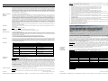
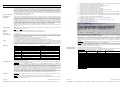
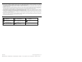
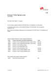
1
Note: Use diluted EnVision FLEX Target Retrieval Solution, Low pH (50x) (Code K8005) for HIER. Program: Use the protocol given below. Reagents must be appropriately diluted before use. All steps should be performed at room temperature (20-25 °C). 1. Incubate tissue section with 200 µL EnVision™ FLEX Peroxidase-Blocking Reagent (Code DM821) for 5 (±1) minutes 2. Rinse in EnVision™ FLEX Wash Buffer (Code K8007) for 1-5 minutes 3. Incubate tissue section with 200 µL Primary Antibody (Code IS082) for 20 (±1) minutes 4. Rinse in EnVision™ FLEX Wash Buffer (Code K8007) for 1-5 minutes 5. EnVision™ FLEX+, Mouse (LINKER), 200/300 µL, 15 minutes 6. Rinse in EnVision™ FLEX Wash Buffer (Code K8007) for 1-5 minutes 7. Incubate tissue section with 200 µL EnVision™ FLEX/HRP (Code DM822) for 20 (±1) minutes 8. Rinse in EnVision™ FLEX Wash Buffer (Code K8007) for 1-5 minutes 9. Incubate tissue section with 200 µL EnVision™ FLEX Wash Buffer (Code K8007) for 5 (±1) minutes (Auxiliary step) 10. Rinse in EnVision™ FLEX Wash Buffer (Code K8007) for 1-5 minutes 11. Incubate tissue section with 200 µL EnVision™ FLEX Substrate Working Solution (Codes DM823 and DM827) for 5 (±1) minutes 12. Incubate tissue section with 200 µL EnVision™ FLEX Substrate Working Solution (Codes DM823 and DM827) for 5 (±1) minutes 13. Rinse in EnVision™ FLEX Wash Buffer (Code K8007) for 1-5 minutes 14. Counterstain slide with EnVision™ FLEX Hematoxylin (Code K8018) for 5 (±1) minutes 15. Rinse in deionized water for 1-5 minutes 16. Incubate tissue section with 200 µL EnVision™ FLEX Wash Buffer (Code K8007) for 5 (±1) minutes 17. Rinse in deionized water for 1-5 minutes 18. Mount slides with permanent mounting medium. Programming grid for recommended assay: FLEX Monoclonal Mouse Anti-Human CD23 Clone DAK-CD23 Ready-to-Use (Dako Autostainer/Autostainer Plus) Code IS781 ENGLISH Intended use For in vitro diagnostic use. FLEX Monoclonal Mouse Anti-Human CD23, Clone DAK-CD23, Ready-to-Use (Dako Autostainer/Autostainer Plus), is intended for use in immunohistochemistry together with Dako Autostainer/Autostainer Plus instruments. Antibodies to CD23 are useful for the identification of both normal B lymphocytes and malignant lymphomas. Together with a panel of antibodies, anti-CD23 is particularly useful for differentiation between B-cell chronic lymphocytic leukemia/small lymphocytic lymphoma and mantle cell lymphoma (1). The clinical interpretation of any staining or its absence should be complemented by morphological studies using proper controls and should be evaluated within the context of the patient’s clinical history and other diagnostic tests by a qualified pathologist. Synonym for antigen FcεRII, BLAST-2, low affinity IgE receptor (2, 3). Summary and explanation CD23 is a 45 kDa type II membrane glycoprotein belonging to the C-type lectin family, and is identical to the low affinity IgE receptor (FcΣRII) found on B cells (4, 5). CD23 is primarily expressed on B cells and monocytes including a strong expression on EBV-transformed B lymphoblasts (2, 3). Interleukin-4 and Interleukin-13 stimulation increase CD23 expression on B cells (4). On B cells, CD23 is selectively expressed on sIgM/sIgD double-bearing cells, and is lost upon differentiation into Ig-secreting cells (2). CD23 is also present on a large variety of other cells such as T cells, eosinophils, platelets, Langerhans´ cells and a subset of thymic epithelial cells (2). However, CD23 is absent in a large number of normal non-lymphoid human tissues, such as bone, cartilage, central nervous system, connective tissue, endocrine glands, gastrointestinal tract, mesothelium, muscles, placenta, peripheral nerves, respiratory tissue, skin, synovium, umbilical cord, urinary and female/male reproductive system, and vessels (6). CD23 is typically detected in neoplastic cells from B-cell chronic lymphocytic leukemia/lymphoma (B-CLL) and absent in mantle cell lymphoma (1). Refer to Dako’s General Instructions for Immunohistochemical Staining or the detection system for instructions of IHC procedures. Reagent provided Ready-to-use monoclonal rabbit antibody provided in liquid form in a buffer containing stabilizing protein and 0.015 mol/L sodium azide. Clone: DAK-CD23. Isotype: IgG1, kappa. Immunogen Recombinant protein encoding amino acids 48-248 of human CD23. Specificity In Western blotting the antibody labels a 45 kDa band corresponding to CD23. Precautions 1. For professional users. 2. This product contains sodium azide (NaN3), a chemical highly toxic in pure form. At product concentrations, though not classified as hazardous, sodium azide may react with lead and copper plumbing to form highly explosive build-ups of metal azides. Upon disposal, flush with large volumes of water to prevent metal azide build-up in plumbing. 3. As with any product derived from biological sources, proper handling procedures should be used. 4. Wear appropriate Personal Protective Equipment to avoid contact with eyes and skin. 5. Unused solution should be disposed of according to local, State and Federal regulations. Storage Quick guide* Store at 2-8 °C. Do not use after expiration date st amped on vial. If reagents are stored under any conditions other than those specified, the conditions must be verified by the user. There are no obvious signs to indicate instability of this product. Therefore, positive and negative controls should be run simultaneously with patient specimens. If unexpected staining is observed which cannot be explained by variations in laboratory procedures and a problem with the antibody is suspected, contact Dako Technical Support. Step Fixation Pre-treatment Dilution Dilution Buffer Negative Control Visualization Counterstain Control Tissue Slides/Mounting For details, please refer to the Operator’s Manual for the dedicated instrument. The Auxiliary step should be set to “rinse buffer” in staining runs with ≤10 slides. For staining runs with >10 slides the Auxiliary step should be set to “none”. This ascertains comparable wash times. Optimal conditions may vary depending on specimen and preparation methods, and should be determined by each individual laboratory. If the evaluating pathologist should desire a different staining intensity, a Dako Application Specialist/Technical Service Specialist can be contacted for information on re-programming of the protocol. Verify that the performance of the adjusted protocol is still valid by evaluating that the staining pattern is identical to the staining pattern described in “Performance characteristics”. Counterstaining: Counterstaining in hematoxylin is recommended using EnVision FLEX Hematoxylin (Dako Autostainer/Autostainer Plus) (Code K8018). Non-aqueous, permanent mounting medium is recommended. Controls: Positive and negative controls should be run simultaneously using the same protocol as the patient specimens. The positive control tissue should include tonsil and the cells/structures should display reaction patterns as described for this tissue in “Performance characteristics” in all positive specimens. The recommended negative control reagent is FLEX Negative Control, Mouse (Dako Autostainer/Autostainer Plus) (Code IS750). Staining interpretation Cells labeled by the antibody display membrane staining. Performance characteristics Normal tissues (7): In tonsil, B cells in the mantle zone show a weak to moderate staining reaction and the follicular dendritic reticulum cells in the germinal centers show a moderate to strong staining reaction. Comments Formalin EnVision™ FLEX Low pH (Code K8005) Ready-to-use Pre-diluted FLEX Negative Control, Mouse (Dako Autostainer/Autostainer Plus) (Code IS750) EnVision™ FLEX+ (Dako Autostainer/ Autostainer Plus) (Code K8012) EnVision™ FLEX Hematoxylin (Dako Autostainer/Autostainer Plus) (Code K8018) Tonsil FLEX IHC Microscope Slides (Code K8020) 20 min HIER, 3-in-1 20 min incubation Do not dilute the antibody 20 min incubation 20 min incubation, 15 min Mouse (LINKER) incubation, 2x5 min DAB+ incubation 5 min incubation Membrane staining Recommended for greater adherence. Permanent mounting required Instrumentation Dako Autostainer/Austostainer Plus Use instrument-specific vials (Code S3425) *The user must always read the package insert for detailed instructions of the staining procedure and handling of the product. Specimen preparation Staining procedure (122095-001) Paraffin sections: The antibody can be used for labeling formalin-fixed, paraffin-embedded tissue sections. Tissue specimens should be cut into sections of approximately 4 µm. Pre-treatment: Pre-treatment of formalin-fixed, paraffin-embedded tissue sections with heat-induced epitope retrieval (HIER) is required. Optimal results are obtained by pretreating tissues with HIER using diluted EnVision™ FLEX Target Retrieval Solution, Low pH (50x) (Code K8005). Deparaffinization, rehydration and epitope retrieval can be performed in Dako PT Link (Code PT100/PT101). For details, please refer to PT Link User Guide. The following parameters should be used for PT Link: Pre-heat temperature: 65 °C; epitope retrieval tempe rature and time: 97 °C for 20 (±1) minutes; cool down to 6 5 °C. Remove slide rack from PT tank and immediately d ip slides in jar/tank (e.g., PT Link Rinse Station, Code PT109) containing diluted room temperature EnVision™ FLEX Wash Buffer (20x) (Code K8007). Leave slides in Wash Buffer for 1-5 minutes. The tissue sections should not dry out during the treatment or during the following immunohistochemical staining procedure. For greater adherence of tissue sections to glass slides, the use of FLEX IHC Microscope Slides (Code K8020) is recommended. After staining the slides should be mounted using permanent mounting medium. Tissue Type (# tested) Adrenal (3) Bone marrow (3) Breast (3) Cerebellum (3) Cerebrum (3) Cervix (3) Colon (3) Esophagus (3) Positive Tissue Elements 0/3 0/3 0/3 0/3 0/3 0/3 0/3 0/3 Kidney (3) Liver (3) Lung (3) Mesothelial cells (2) Muscle, cardiac (3) Muscle, skeletal (3) Nerve, peripheral (3) 0/3 0/3 3/3 Macrophages 0/2 0/3 0/3 0/3 Tissue Type (# tested) Ovary (3) Pancreas (3) Parathyroid (3) Pituitary (3) Prostate (3) Salivary gland (3) Skin (3) Small intestine (2) Spleen (3) Stomach (2) Testis (3) Thymus (3) Thyroid (3) Tonsil (3) Uterus (3) Positive Tissue Elements 0/3 0/3 0/3 0/3 0/3 1/3 Ductal epithelial cells 0/3 2/2 Lymphoid tissue (10%), membrane 1/3 Lymphoid tissue (15%), membrane 2/3 Lymphoid tissue (50%), membrane 0/2 0/3 3/3 Scattered cells, medulla, cytoplasmic 1/3 Follicular epithelial cells 3/3 0/3 Abnormal tissues (7): The antibody labeled 8/9 cases of B-CLL and 3/3 follicular lymphomas.. No labelling was observed in 3 mantle cell lymphomas. Visualization: The recommended visualization system is EnVision FLEX+, Mouse, High pH (Dako Autostainer/Autostainer Plus) (Code K8012). P01741EFG_002_IS781/2011.10 p. 1/7 Dako Denmark A/S | Produktionsvej 42 | DK-2600 Glostrup | Denmark | Tel. +45 44 85 95 00 | Fax +45 44 85 95 95 | CVR No. 33 21 13 17 (122095-001) P01741EFG_002_IS781/2011.10 p. 2/7 Dako Denmark A/S | Produktionsvej 42 | DK-2600 Glostrup | Denmark | Tel. +45 44 85 95 00 | Fax +45 44 85 95 95 | CVR No. 33 21 13 17 Programme : utiliser le protocole indiqué ci-après. Les réactifs doivent être dilués de la manière appropriée avant leur utilisation. Toutes les étapes doivent être réalisées à température ambiante (20 à 25 °C). 1. Incuber la coupe de tissu avec 200 µL de réactif EnVision™ FLEX Peroxidase-Blocking Reagent (réf. DM821) pendant 5 minutes (±1 minute) 2. Rincer dans le tampon de lavage EnVision™ FLEX Wash Buffer (réf. K8007) pendant 1 à 5 minutes 3. Incuber la coupe de tissu avec 200 µL d’anticorps primaire (réf. IS082) pendant 20 minutes (±1 minute) 4. Rincer dans le tampon de lavage EnVision™ FLEX Wash Buffer (réf. K8007) pendant 1 à 5 minutes 5. EnVision™ FLEX+, Mouse (LINKER), 200/300 µL, 15 minutes 6. Rincer dans le tampon de lavage EnVision™ FLEX Wash Buffer (réf. K8007) pendant 1 à 5 minutes 7. Incuber la coupe de tissu avec 200 µL de produit EnVision™ FLEX/HRP (réf. DM822) pendant 20 minutes (±1 minute) 8. Rincer dans le tampon de lavage EnVision™ FLEX Wash Buffer (réf. K8007) pendant 1 à 5 minutes 9. Incuber la coupe de tissu avec 200 µL de tampon de lavage EnVision™ FLEX Wash Buffer (réf. K8007) pendant 5 minutes (±1 minute) (étape dite « Auxiliary ») 10. Rincer dans le tampon de lavage EnVision™ FLEX Wash Buffer (réf. K8007) pendant 1 à 5 minutes 11. Incuber la coupe de tissu avec 200 µL de solution EnVision™ FLEX Substrate Working Solution (réf. DM823 et DM827) pendant 5 minutes (±1 minute) 12. Incuber la coupe de tissu avec 200 µL de solution EnVision™ FLEX Substrate Working Solution (réf. DM823 et DM827) pendant 5 minutes (±1 minute) 13. Rincer dans le tampon de lavage EnVision™ FLEX Wash Buffer (réf. K8007) pendant 1 à 5 minutes 14. Procéder à une contre-coloration de la lame avec le produit EnVision™ FLEX Hematoxylin (réf. K8018) pendant 5 minutes (±1 minute) 15. Rincer dans de l’eau déionisé pendant 1 à 5 minutes 16. Incuber la coupe de tissu avec 200 µL de tampon de lavage EnVision™ FLEX Wash Buffer (réf. K8007) pendant 5 minutes (±1 minute) 17. Rincer dans de l’eau déionisé pendant 1 à 5 minutes 18. Monter les lames avec un milieu de montage permanent. Grille de programmation pour le dosage recommandé : FRANÇAIS Intérêt Pour diagnostic in vitro. L’anticorps FLEX Monoclonal Mouse Anti-Human CD23, Clone DAK-CD23, Ready-to-Use (Dako Autostainer/Autostainer Plus), est destiné à être utilisé en immunohistochimie avec les appareils Dako Autostainer/Autostainer Plus. Les anticorps dirigés contre le CD23 sont utiles pour l’identification des lymphocytes B normaux et des lymphomes malins. Couplé avec un panel d’anticorps, l’anticorps anti-CD23 est particulièrement utile pour permettre la différenciation entre la leucémie lymphocytaire chronique à cellules B/le lymphome lymphocytaire à petits lymphocytes et le lymphome du manteau (1). L’interprétation clinique de toute coloration ou son absence doit être complétée par des études morphologiques en utilisant des contrôles appropriés et doit être évaluée en fonction des antécédents cliniques du patient et d’autres tests diagnostiques par un pathologiste qualifié. Synonyme de l’antigène FcεRII, BLAST-2, récepteur de faible affinité des IgE (2, 3). Résumé et explication Le CD23 est une glycoprotéine membranaire de type II, de 45 kDa, appartenant à la famille des lectines de type C, identique au récepteur de faible affinité des IgE (FcΣRII) retrouvé sur les lymphocytes B (4, 5). Le CD23 est exprimé principalement sur les lymphocytes B et les monocytes ; son expression est également forte sur les lymphoblastes B transformés par l’EBV (2, 3). La stimulation par les interleukines 4 et 13 augmente l’expression du CD23 sur les lymphocytes B (4). Sur les lymphocytes B, le CD23 est exprimé de manière sélective sur les cellules porteuses à la fois d’IgM sécrétoires et d’IgD sécrétoires et il disparaît lors de leur différenciation en cellules sécrétrices d’Ig (2). Le CD23 est également présent dans une large gamme d’autres cellules comme les lymphocytes T, les éosinophiles, les plaquettes, les cellules de Langerhans et un sous-ensemble de cellules épithéliales thymiques (2). Toutefois, le CD23 est absent dans un grand nombre de tissus humains sains non lymphoïdes, comme l’os, le cartilage, le système nerveux central, le tissu conjonctif, les glandes endocrines, le tractus gastro-intestinal, le mésothélium, les muscles, le placenta, les nerfs périphériques, le tissu respiratoire, la peau, la synoviale, le cordon ombilical, le système urinaire et les systèmes reproducteurs masculin et féminin, ainsi que les vaisseaux (6). Le CD23 est généralement détecté dans les cellules néoplasiques du lymphome/de la leucémie lymphocytaire chronique à cellules B (LLC-B) et est absent dans le lymphome du manteau (1). Se reporter aux Instructions générales de coloration immunohistochimique de Dako ou aux instructions du système de détection relatives aux procédures IHC pour plus d’informations. Réactif fourni Anticorps monoclonal de souris prêt à l’emploi fourni sous forme liquide dans un tampon contenant une protéine stabilisante et 0,015 mol/L d’azide de sodium. Clone : DAK-CD23. Isotype : IgG1, kappa. Immunogène Protéine recombinante codant les acides aminés 48 à 248 du CD23 humain. Spécificité Lors d’un Western blot, l’anticorps marque une bande de 45 kDa correspondant au CD23. Précautions d’emploi 1. Pour utilisateurs professionnels. 2. Ce produit contient de l’azide de sodium (NaN3), produit chimique hautement toxique dans sa forme pure. Aux concentrations du produit, bien que non classé comme dangereux, l’azide de sodium peut réagir avec le cuivre et le plomb des canalisations pour former des accumulations d’azides métalliques hautement explosifs. Lors de l’élimination, rincer abondamment à l’eau pour éviter toute accumulation d’azide métallique dans les canalisations. 3. Comme avec tout produit d’origine biologique, respecter les procédures de manipulation appropriées. 4. Porter un équipement de protection individuelle approprié pour éviter tout contact avec les yeux et la peau. 5. Les solutions non utilisées doivent être éliminées conformément aux réglementations locales et nationales. Conservation Guide rapide* Pour plus de détails, se reporter au Manuel de l’opérateur spécifique à l’appareil. L’étape dite « Auxiliary » doit être réglée sur « rinse buffer » lors des cycles de coloration de 10 lames ou moins. Pour les cycles de coloration de plus de 10 lames, l’étape dite « Auxiliary » doit être réglée sur « none ». Cela détermine des temps de lavage comparables. Les conditions optimales peuvent varier en fonction du prélèvement et des méthodes de préparation, et doivent être déterminées par chaque laboratoire individuellement. Si le pathologiste qui réalise l’évaluation désire une intensité de coloration différente, un spécialiste d’application/spécialiste de l’assistance technique de Dako peut être contacté pour obtenir des informations sur la reprogrammation du protocole. Vérifier que l’exécution du protocole modifié est toujours valide en vérifiant que le schéma de coloration est identique au schéma de coloration décrit à la section « Caractéristiques de performance ». Conserver entre 2 et 8 °C. Ne pas utiliser après la d ate de péremption indiquée sur le flacon. Si les réactifs sont conservés dans des conditions autres que celles indiquées, celles-ci doivent être validées par l’utilisateur. Il n’existe pas de signe particulier pour indiquer l’instabilité de ce produit. Par conséquent, des contrôles positifs et négatifs doivent être testés en même temps que les échantillons de patients. Si une coloration inattendue est observée, qui ne peut être expliquée par un changement des procédures du laboratoire, et en cas de suspicion d’un problème lié à l’anticorps, contacter l’assistance technique de Dako. Étape Fixation Prétraitement Dilution Tampon de dilution Contrôle négatif Visualisation Contre-coloration Tissu de contrôle Lames/Montage Contre-coloration : il est recommandé d’effectuer une contre-coloration à l’aide du produit EnVision FLEX Hematoxylin (Dako Autostainer/Autostainer Plus) (réf. K8018). L’utilisation d’un milieu de montage permanent non aqueux est recommandée. Contrôles : des contrôles positifs et négatifs doivent être testés en même temps et en suivant le même protocole que les échantillons de patients. Le tissu de contrôle positif doit comprendre l’amygdale et les cellules/structures doivent présenter des schémas de réaction semblables à ceux décrits pour ce tissu à la section « Caractéristiques de performance » pour tous les échantillons positifs. Le contrôle négatif recommandé est le produit FLEX Negative Control, Mouse, (Dako Autostainer/Autostainer Plus) (réf. IS750). Commentaires Formol EnVision™ FLEX Low pH (réf. K8005) Prêt à l’emploi Prédilué FLEX Negative Control, Mouse (Dako Autostainer/ Autostainer Plus) (réf. IS750) EnVision™ FLEX+ (Dako Autostainer/Autostainer Plus) (réf. K8012) EnVision™ FLEX Hematoxylin (Dako Autostainer/ Autostainer Plus) (réf. K8018) Amygdale FLEX IHC Microscope Slides (réf. K8020) HIER de 20 minutes, 3 en 1 Incubation de 20 minutes Ne pas diluer l’anticorps Incubation de 20 minutes Incubation de 20 minutes, incubation de 15 minutes avec le produit Mouse (LINKER), 2 incubations de 5 minutes avec le DAB+ Incubation de 5 minutes Interprétation de la coloration Les cellules marquées par l’anticorps présentent une coloration membranaire. Caractéristiques de performance Tissus sains (7) : dans l’amygdale, les lymphocytes B de la zone du manteau présentent une coloration faible à modérée et les cellules réticulaires dendritiques folliculaires des centres germinatifs présentent une coloration modérée à forte. Type de tissu (nb. testés) Surrénale (3) Moelle osseuse (3) Sein (3) Cervelet (3) Cerveau (3) Col de l’utérus (3) Côlon (3) Œsophage (3) Coloration membranaire Recommandées pour une meilleure adhérence. Milieu de montage permanent requis. Appareillage Dako Autostainer/Autostainer Plus Utiliser les flacons spécifiques à l’appareil (réf. S3425) *L’utilisateur doit toujours lire la notice pour obtenir des instructions détaillées sur la procédure de coloration et sur la manipulation du produit. Préparation des échantillons Procédure de coloration Coupes en paraffine : l’anticorps peut être utilisé pour le marquage des coupes de tissu fixées au formol et incluses en paraffine. L’épaisseur des coupes d’échantillons de tissu doit être d’environ 4 µm. Prétraitement : le prétraitement des coupes de tissu fixées au formol et incluses en paraffine par démasquage des épitopes induit par la chaleur (HIER) est nécessaire. Des résultats optimaux sont obtenus en prétraitant les tissus par la méthode HIER, à l’aide de la solution de démasquage des épitopes EnVision™ FLEX Target Retrieval Solution, Low pH (50x) (réf. K8005) diluée. Le déparaffinage, la réhydratation et le démasquage des épitopes peuvent être réalisés dans l’appareil PT Link de Dako (réf. PT100/PT101). Pour plus de détails, se reporter au Guide d’utilisation du PT Link. Les paramètres suivants doivent être utilisés pour le PT Link : température de préchauffage : 65 °C ; température et durée du démasquage des épitopes : 97 °C pendant 20 minutes (±1 minute) ; laisser refroidir jusqu’à 65 °C. Retirer le portoir de lames de la cuve PT et plonger immédiatement les lames dans une jarre/cuve (par ex. station de rinçage du PT Link, réf. PT109) contenant du tampon de lavage EnVision™ FLEX Wash Buffer (20x) dilué à température ambiante (réf. K8007). Laisser les lames dans le tampon de lavage pendant 1 à 5 minutes. Les coupes de tissu ne doivent pas sécher lors du traitement ni lors de la procédure de coloration immunohistochimique qui suit. Pour une meilleure adhérence des coupes de tissu sur les lames de verre, il est recommandé d’utiliser les lames FLEX IHC Microscope Slides (réf. K8020). Après coloration, un montage permanent des lames est recommandé. Éléments tissulaires positifs 0/3 0/3 0/3 0/3 0/3 0/3 0/3 0/3 Type de tissu (nb. testés) Ovaire (3) Pancréas (3) Parathyroïde (3) Pituitaire (3) Prostate (3) Glande salivaire (3) Peau (3) Intestin grêle (2) Rate (3) Rein (3) 0/3 Foie (3) Poumon (3) Cellules mésothéliales (2) 0/3 3/3 macrophages 0/2 Estomac (2) Testicule (3) Thymus (3) Muscle, cardiaque (3) Muscle, squelettique (3) Nerf périphérique (3) 0/3 0/3 0/3 Thyroïde (3) Amygdale (3) Utérus (3) Éléments tissulaires positifs 0/3 0/3 0/3 0/3 0/3 1/3 cellules épithéliales canalaires 0/3 2/2 : tissu lymphoïde (10 %), membranaire 1/3 : tissu lymphoïde (15 %), membranaire 2/3 : tissu lymphoïde (50 %), membranaire 0/2 0/3 3/3 : cellules disséminées, médulla, cytoplasmique 1/3 cellules épithéliales folliculaires 3/3 0/3 Tissus tumoraux (7) : l’anticorps a marqué 8 cas sur 9 de LLC-B et 3 cas sur 3 de lymphomes folliculaires. Aucun marquage n’a été observé dans 3 lymphomes du manteau. Visualisation : le système de visualisation recommandé est le système EnVision FLEX+, Mouse, High pH (Dako Autostainer/Autostainer Plus) (réf. K8012). Remarque : utiliser la solution EnVision FLEX Target Retrieval Solution, Low pH (50x) (réf. K8005) diluée pour la méthode HIER. (122095-001) P01741EFG_002_IS781/2011.10 p. 3/7 Dako Denmark A/S | Produktionsvej 42 | DK-2600 Glostrup | Denmark | Tel. +45 44 85 95 00 | Fax +45 44 85 95 95 | CVR No. 33 21 13 17 (122095-001) P01741EFG_002_IS781/2011.10 p. 4/7 Dako Denmark A/S | Produktionsvej 42 | DK-2600 Glostrup | Denmark | Tel. +45 44 85 95 00 | Fax +45 44 85 95 95 | CVR No. 33 21 13 17 1. 2. 3. 4. 5. 6. 7. 8. 9. 10. 11. 12. 13. 14. 15. 16. 17. 18. DEUTSCH Zweckbestimmung Zur Verwendung für In-vitro-Untersuchungen. FLEX Monoclonal Mouse Anti-Human CD23, Clone DAK-CD23, Ready-to-Use, (Dako Autostainer/Autostainer Plus) ist zur Verwendung in der Immunhistochemie in Verbindung mit Dako Autostainer/Autostainer Plus-Geräten bestimmt. Antikörper gegen CD23 dienen zum Nachweis von normalen B-Lymphozyten und malignen Lymphomen. Zusammen mit einem Antikörper-Panel ist Anti-CD23 besonders nützlich für die Differenzierung zwischen der chronisch lymphatischen B-Zell-Leukämie/dem kleinzelligen lymphozytischen B-Zelllymphom und dem Mantelzelllymphom (1). Die klinische Auswertung einer eventuell eintretenden Färbung sollte durch morphologische Studien mit ordnungsgemäßen Kontrollen ergänzt werden und von einem qualifizierten Pathologen unter Berücksichtigung der Krankengeschichte und anderer Diagnostiktests des Patienten vorgenommen werden. Synonym für das Antigen FcεRII, BLAST-2, niedrig-affiner IgE-Rezeptor (2, 3). Zusammenfassung und Erklärung CD23 ist ein Typ-II-Membranglykoprotein mit 45 kDa, welches der C-Typ-Lektinfamilie angehört und mit dem niedrig-affinen IgE-Rezeptor (FcΣRII), welcher sich auf B-Zellen findet, identisch ist (4, 5). CD23 wird vorwiegend auf B-Zellen und Monozyten exprimiert; die Expression ist besonders stark auf EBV-transformierten B-Lymphoblasten (2, 3). Interleukin-4- und Interleukin-13-Stimulierung steigert die CD23Expression auf B-Zellen (4). Bei B-Zellen wird CD23 selektiv auf sIgM/sIgD-positiven Zellen exprimiert und geht nach der Differenzierung zu Ig-sekretierenden Zellen verloren (2). Ferner liegt CD23 auf zahlreichen weiteren Zellen vor, so z. B. auf T-Zellen, Eosinophilen, Thrombozyten, Langerhans’schen Zellen und einer Untergruppe der Thymusepithelzellen (2). Jedoch fehlt CD23 bei einer Großzahl normaler, nicht-lymphoider menschlicher Gewebe, z. B. Knochen, Knorpel, Zentralnervensystem, Bindegewebe, endokrine Drüsen, MagenDarm-Trakt, Mesothel, Muskel, Plazenta, periphere Nerven, respiratorisches Gewebe, Haut, Synovium, Nabelschnur, Harn- und weibliches/männliches Reproduktionssystem sowie Gefäße (6). CD23 wird normalerweise auf neoplastischen Zellen bei chronisch lymphatischen B-Zell-Leukämien/Lymphomen (B-CLL) nachgewiesen und ist bei Mantelzelllymphomen nicht zu finden (1). Anweisungen zu den IHC-Verfahren finden Sie in den Allgemeinen Richtlinien zur immunhistochemischen Färbung von Dako oder dem Detektionssystem. Geliefertes Reagenz Gebrauchsfertiger, monoklonaler Maus-Antikörper in flüssiger Form in einem Puffer, der stabilisierendes Protein und 0,015 mol/L Natriumazid enthält. Klon: DAK-CD23. Isotyp: IgG1, Kappa. Immunogen Rekombinantes Protein, das die Aminosäuren 48-248 von menschlichem CD23 umfasst. Spezifität Beim Western Blottingmarkiert der Antikörper eine Bande von 45 kDa, welche CD23 entspricht. Hinweise und Vorsichtsmaßnahmen 1. Für Fachpersonal. 2. Dieses Produkt enthält Natriumazid (NaN3), eine in reiner Form äußerst giftige Chemikalie. Bei den in diesem Produkt verwendeten Konzentrationen kann Natriumazid, obwohl nicht als gefährlich klassifiziert, mit in Wasserleitungen vorhandenem Blei oder Kupfer reagieren und zur Bildung von hochexplosiven Metallazidansammlungen führen. Nach der Entsorgung stets mit viel Wasser nachspülen, um Metallazidansammlungen in den Leitungen vorzubeugen. 3. Wie alle Produkte biologischen Ursprungs müssen auch diese entsprechend gehandhabt werden. 4. Geeignete Schutzkleidung tragen, um Augen- und Hautkontakt zu vermeiden. 5. Nicht verwendete Lösung ist entsprechend örtlichen, bundesstaatlichen und staatlichen Richtlinien zu entsorgen. Lagerung Bei 2–8 °C aufbewahren. Nach Ablauf des auf dem Fläs chchen aufgedruckten Verfalldatums nicht mehr verwenden. Werden die Reagenzien nicht entsprechend den angegebenen Bedingungen aufbewahrt, müssen die Bedingungen vom Anwender geprüft werden. Es gibt keine offensichtlichen Anhaltspunkte für die mögliche Instabilität dieses Produkts. Es sollten daher die Positiv- und Negativkontrollen gleichzeitig mit den Patientenproben mitgeführt werden. Falls eine unerwartete Färbung auftritt, die sich nicht durch Unterschiede bei Laborverfahren erklären lässt und auf ein Problem mit dem Antikörper hindeutet, ist der technische Kundendienst von Dako zu verständigen. Kurzanleitung* Schritt Fixierung Vorbehandlung Verdünnung Dilution Buffer Negative Control Visualisierung Gegenfärbung Kontrollgewebe Objektträger/ Fixierung Geräte Programmierraster für empfohlenes Assay: Nähere Einzelheiten bitte dem Benutzerhandbuch für das jeweilige Gerät entnehmen. Bei Färbedurchläufen mit 10 oder weniger Objektträgern sollte der Zusatz-Schritt auf „rinse buffer“ eingestellt werden. Für Färbedurchläufe mit mehr als 10 Objektträgern den ZusatzSchritt auf „none“ einstellen. Dies gewährleistet vergleichbare Waschzeiten. Optimale Bedingungen können je nach Probe und Präparationsverfahren unterschiedlich sein und sollten vom jeweiligen Labor selbst ermittelt werden. Falls der beurteilende Pathologe eine andere Färbungsintensität wünscht, kann ein Anwendungsspezialist oder Kundendiensttechniker von Dako bei der Neuprogrammierung des Protokolls helfen. Die Leistung des angepassten Protokolls muss verifiziert werden, indem gewährleistet wird, dass das Färbemuster mit dem unter „Leistungsmerkmale“ beschriebenen Färbemuster identisch ist. Gegenfärbung: Die Gegenfärbung in Hämatoxylin sollte mit EnVision FLEX Hematoxylin (Dako Autostainer/Autostainer Plus) (Code-Nr. K8018) ausgeführt werden. Empfohlen wird ein nichtwässriges, permanentes Eindeckmedium. Kontrollen: Positiv- und Negativkontrollen sollten zur gleichen Zeit und mit demselben Protokoll wie die Patientenproben getestet werden. Das positive Kontrollgewebe sollte Mandelgewebe enthalten, und die Zellen/Strukturen müssen in allen positiven Proben die unter „Leistungsmerkmale“ für dieses Gewebe beschriebenen Reaktionsmuster aufweisen. Das empfohlene Negativ-Kontrollreagenz ist FLEX Negative Control, Mouse, (Dako Autostainer/Autostainer Plus) (Code-Nr. IS750). Anmerkungen Formalin EnVision™ FLEX, Low pH (Code-Nr. K8005) Gebrauchsfertig Vorverdünnt FLEX Negative Control, Mouse (Dako Autostainer/ Autostainer Plus) Code-Nr. IS750) EnVision™ FLEX+ (Dako Autostainer/Autostainer Plus) (Code-Nr. K8012) EnVision™ FLEX Hematoxylin (Dako Autostainer/ Autostainer Plus) (Code-Nr. K8018) Mandeln FLEX IHC Microscope Slides (Code-Nr. K8020) Dako Autostainer/Autostainer Plus 20 Min. HIER, 3-in-1 20 Min. In ubation Den Antikörper nicht verdünnen. 20 Min. Inkubation Auswertung der Färbung Mit diesem Antikörper markierte Zellen weisen eine Membranfärbung auf. Leistungseigenschaften Gesunde Gewebe (7): Die B-Zellen in der Mantelzone von Mandelgewebe zeigen eine schwache bis mäßige Färbereaktion, während die follikulären dendritischen Retikulumzellen in Keimzentren eine mäßige bis starke Färbereaktion aufweisen. 20 Min. Inkubation, 15 Min. Mouse (LINKER) Inkubation, 2 x 5 Min. DAB+ Inkubation 5 Min. Inkubation Membranfärbung Wird zur besseren Haftung empfohlen. Permanente Fixieru g erforderlich Gerätespezifische Fläschchen verwenden (Code-Nr. S3425) *Der Anwender muss stets die Packungsbeilage lesen, um sich über die detaillierten Anweisungen für das Färbeverfahren und die Handhabung des Produkts zu informieren. Probenvorbereitung Färbeverfahren Gewebeschnitte mit 200 µL EnVision™ FLEX Peroxidase-Blocking Reagent (Code-Nr. DM821) für 5 (±1) Minuten inkubieren. 1–5 Minuten in EnVision™ FLEX Wash Buffer (Code-Nr. K8007) spülen. Gewebeschnitt 20 (±1) Minuten mit 200 µL Primärantikörper (Code-Nr. IS082) inkubieren. 1–5 Minuten in EnVision™ FLEX Wash Buffer (Code-Nr. K8007) spülen. EnVision™ FLEX+, Mouse (LINKER),200/300 µL, 15 Minuten 1–5 Minuten in EnVision™ FLEX Wash Buffer (Code-Nr. K8007) spülen. Gewebeschnitte 20 (±1) Minuten mit 200 µL EnVision™ FLEX/HRP (Code-Nr. DM822) inkubieren. 1–5 Minuten in EnVision™ FLEX Wash Buffer (Code-Nr. K8007) spülen. Gewebeschnitte 5 (±1) Minuten („Zusatz“-Schritt) mit 200 µL EnVision™ FLEX Wash Buffer (Code-Nr. K8007) inkubieren. 1–5 Minuten in EnVision™ FLEX Wash Buffer (Code-Nr. K8007) spülen. Gewebeschnitte 5 (±1) Minuten mit 200 µL EnVision™ FLEX Substrate Working Solution (Code-Nr. DM823 und DM827) inkubieren. Gewebeschnitte 5 (±1) Minuten mit 200 µL EnVision™ FLEX Substrate Working Solution (Code-Nr. DM823 und DM827) inkubieren. 1–5 Minuten in EnVision™ FLEX Wash Buffer (Code-Nr. K8007) spülen. Objektträger 5 (±1) Minuten mit EnVision™ FLEX Hematoxylin (Code-Nr. K8018) gegenfärben. 1–5 Minuten mit entionisiertem Wasser spülen Gewebeschnitte 5 (±1) Minuten mit 200 µL EnVision™ FLEX Wash Buffer (Code-Nr. K8007) inkubieren. 1–5 Minuten mit entionisiertem Wasser spülen Objektträger mit einem permanenten Fixiermittel fixieren. Paraffinschnitte: Der Antikörper eignet sich zur Markierung von formalinfixierten und paraffineingebetteten Gewebeschnitten. Gewebeproben sollten in Schnitte von ca. 4 µm Stärke geschnitten werden. Vorbehandlung: Es ist eine Vorbehandlung der formalinfixierten und paraffineingebetteten Gewebeschnitte durch hitzeinduzierte Epitopdemaskierung (HIER) erforderlich. Optimale Ergebnisse können durch HIER-Vorbehandlung der Gewebe mit EnVision™ FLEX Target Retrieval Solution, Low pH (50x) (Code-Nr. K8005) erzielt werden. Die Entparaffinierung, Rehydrierung und Epitopdemaskierung können im Dako PT Link (Code-Nr. PT100/PT101) durchgeführt werden. Weitere Informationen hierzu siehe PT Link-Benutzerhandbuch. Für PT Link sollten die folgenden Parameter verwendet werden: Vorwärmtemperatur: 65 °C; Temperatur und Zeit für Epi topdemaskierung: 97 °C für 20 (±1) Minuten; auf 65 °C abkühlen. Das Autostainer Objektträgergestell mit den Objektträgern aus dem Behälter herausnehmen und die Objektträger sofort in einen Behälter (z. B. PT Link Rinse Station, Code-Nr. PT109) mit verdünntem, auf Zimmertemperatur gebrachtem EnVision™ FLEX Wash Buffer (20X) (Code-Nr. K8007) eintauchen. Die Objektträger für 1–5 Minuten im Wash Buffer belassen. Die Gewebeschnitte dürfen während der Behandlung oder des anschließenden immunhistochemischen Färbeverfahrens nicht austrocknen. Für eine bessere Haftung der Gewebeschnitte an den Glas-Objektträgern werden FLEX IHC Microscope Slides (Code-Nr. K8020) empfohlen. Nach dem Färben sollten die Schnitte mit permanentem Eindeckmedium auf die Objektträger aufgebracht werden. Gewebetyp (Anz. getestet) Nebenniere (3) Knochenmark (3) Brust (3) Zerebellum (3) Zerebrum (3) Zervix (3) Kolon (3) Speiseröhre (3) Positive Gewebe-Elemente Gewebetyp (Anz. getestet) Eierstock (3) Pankreas (3) Parathyroida (3) Hypophyse (3) Prostata (3) Speicheldrüse (3) Haut (3) Dünndarm (2) Milz (3) 0/3 0/3 0/3 0/3 0/3 0/3 0/3 0/3 Niere (3) Leber (3) Lunge (3) Mesothelzellen (2) 0/3 0/3 3/3 Makrophagen 0/2 Magen (2) Hoden (3) Thymus (3) Herzmuskel (3) Skelettmuskulatur (3) Nerv, peripher (3) 0/3 0/3 0/3 Thyroida (3) Mandel (3) Uterus (3) Positive Gewebe-Elemente 0/3 0/3 0/3 0/3 0/3 1/3 Duktale Epithelzellen 0/3 2 von 2 lymphoiden Geweben (10%), Membran 1 von 3 lymphoiden Geweben (15%), Membran 2 von 3 lymphoiden Geweben (50%), Membran 0/2 0/3 3 von 3 vereinzelten Zellen, Medulla, zytoplasmatisch 1/3 Follikelepithelzellen 3/3 0/3 Pathologische Gewebe (7): Der Antikörper markierte 8 von 9 Fällen von B-CLL und 3 von 3 follikulären Lymphomen. In 3 Mantelzelllymphomen wurde keine Markierung beobachtet. Visualisierung: Das empfohlene Visualisierungssystem ist EnVision FLEX+, Mouse, High pH (Dako Autostainer/Autostainer Plus) (CodeNr. K8012). Hinweis: Verdünnte EnVision FLEX Target Retrieval Solution, Low pH (50x) (Code-Nr. K8005) für HIER verwenden. Programm: Das unten aufgeführte Protokoll verwenden. Reagenzien müssen vor dem Einsatz entsprechend verdünnt werden. Alle Schritte bei Raumtemperatur (20–25 °C) durchführen. (122095-001) P01741EFG_002_IS781/2011.10 p. 5/7 Dako Denmark A/S | Produktionsvej 42 | DK-2600 Glostrup | Denmark | Tel. +45 44 85 95 00 | Fax +45 44 85 95 95 | CVR No. 33 21 13 17 (122095-001) P01741EFG_002_IS781/2011.10 p. 6/7 Dako Denmark A/S | Produktionsvej 42 | DK-2600 Glostrup | Denmark | Tel. +45 44 85 95 00 | Fax +45 44 85 95 95 | CVR No. 33 21 13 17 References/ Références/ Literatur 1. 2. 3. 4. 5. 6. 7. Rossi S, Laurino L, Furlanetto A, Chinellato S, Orvieto E, Canal F, et al. Rabbit monoclonal antibodies: a comparative study between a novel category of immunoreagents and the corresponding mouse monoclonal antibodies. Am J Clin Pathol 2005;124:295-302. Sarfati M. BC7. CD23 Workshop Panel report. In: Kishimoto T, Kikutani H, von dem Borne AEG, Goyert SM, Mason DY, Miyasaka M, et al., editors. Leucocyte typing VI. White cell differentiation antigens. Proceedings of the 6th International Workshop and Conference; 1996 Nov 10-14; Kobe, Japan. New York, London: Garland PublishingInc.; 1997. p.144-7. Kikutani H, Suemura M, Owaki H, Yamasaki K, Barsumian EL, Nakamura H, et al. B3.3. CD23 is a low-affinity Fce receptor (FceR): regulation of FceR expression. In: McMichael AJ, Beverley PCL, Cobbold S, Crumpton MJ, Gilks W, Gotch FM, et al., editors. Leucocyte typing III. White cell differentiation antigens. Proceedings of the 3rd International Workshop and Conference; 1986 Sep 21-26; Oxford, England. Oxford, New York, Tokyo: Oxford University Press; 1987. p. 419-22. Aubry JP, Bonnefoy JY, De Vries JE, Banchereau J. B3.2. The CD23 antigen is the human lymphocyte receptor for IgE. In: McMichael AJ, Beverley PCL, Cobbold S, Crumpton MJ, Gilks W, Gotch FM, et al., editors. Leucocyte typing III. White cell differentiation antigens. Proceedings of the 3rd International Workshop and Conference; 1986 Sep 21-26; Oxford, England. Oxford, New York, Tokyo: Oxford University Press; 1987. p. 417-9. Sarfati M, Ishihara H, Delespesse G. B7. CD23 Workshop Panel report. In: Schlossman SF, Boumsell L, Gilks W, Harlan JM, Kishimoto T, Morimoto C, et al., editors. Leucocyte typing V. White cell differentiation antigens. Proceedings of the 5th International Workshop and Conference; 1993 Nov 3-7; Boston, USA. Oxford, New York, Tokyo: Oxford University Press; 1995. p. 530-3. Pallesen G. B2.5. The distribution of CD23 in normal human tissues and in malignant lymphomas. In McMichael AJ, Beverley PCL, Cobbold S, Crumpton MJ, Gilks W, Gotch FM, et al., editors. Leucocyte typing III. White cell differentiation antigens. Proceedings of the 3rd International Workshop and Conference; 1986 Sep 21-26; Oxford, England. Oxford, New York, Tokyo: Oxford University Press; 1987. p. 383-6 M7312. Document D04962. Report on file. Explanation of symbols/ Explication des symboles/ Erläuterung der Symbole (122095-001) Catalogue number Référence catalogue Bestellnummer Temperature limitation Limites de température Use by Utiliser avant Zulässiger Temperaturbereich Verwendbar bis In vitro diagnostic medical device Dispositif médical de diagnostic in vitro In-vitro-Diagnostikum Contains sufficient for <n> tests Contenu suffisant pour <n> tests Manufacturer Fabricant Inhalt ausreichend für <n> Tests Hersteller Consult instructions for use Voir les instructions d’utilisation Gebrauchsanweisung beachten Batch code Numéro de lot Chargenbezeichnung P01741EFG_002_IS781/2011.10 p. 7/7 Dako Denmark A/S | Produktionsvej 42 | DK-2600 Glostrup | Denmark | Tel. +45 44 85 95 00 | Fax +45 44 85 95 95 | CVR No. 33 21 13 17