1
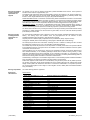
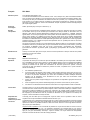
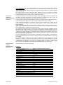
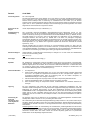
FLEX Monoclonal Rabbit Anti-Human AMACR Clone 13H4 Ready-to-Use (Dako Autostainer/Autostainer Plus) Code IS060 English Intended use For in vitro diagnostic use. FLEX Monoclonal Rabbit Anti-Human AMACR, Clone 13H4, Ready-to-Use, (Dako Autostainer/Autostainer Plus), is intended for use in immunohistochemistry together with Dako Autostainer/Autostainer Plus instruments. This antibody is useful for the identification of prostate adenocarcinomas (1-9). The clinical interpretation of any staining or its absence should be complemented by morphological studies using proper controls and should be evaluated within the context of the patient's clinical history and other diagnostic tests by a qualified pathologist. Synonyms for antigen P504S, alpha methylacyl coenzyme A racemase (1, 2). Summary and explanation Monoclonal rabbit anti-AMACR (alpha methylacyl coenzyme A racemase) recognizes a 382 amino acid protein that was identified by cDNA library subtraction in conjunction with high throughput microarray screening of prostate adenocarcinoma (1). AMACR, also known as P504S, is an enzyme that is involved in bile acid biosynthesis and β-oxidation of branched-chain fatty acids (10). AMACR is expressed in cells of premalignant high grade prostatic intraepithelial neoplasia (HGPIN) and prostate adenocarcinoma, but is present at low or undetectable levels in glandular epithelial cells of normal prostate and benign prostatic hyperplasia (1-9). AMACR is expressed in normal non-prostatic epithelium and in carcinomas from outside the prostate (2, 11-23). Refer to Dako’s General Instructions for Immunohistochemical Staining or the detection system instructions of IHC procedures for: 1) Principle of Procedure, 2) Materials Required, Not Supplied, 3) Storage, 4) Specimen Preparation, 5) Staining Procedure, 6) Quality Control, 7) Troubleshooting, 8) Interpretation of Staining, 9) General Limitations. Reagent provided Ready-to-use monoclonal rabbit antibody provided in liquid form in a buffer containing stabilizing protein and 0.015 mol/L NaN3. Clone: 13H4 (1-3). Immunogen Full length recombinant AMACR (1). Specificity The specificity of monoclonal rabbit anti-AMACR was evaluated by immunocytochemistry and Western blot analysis. Anti-AMACR positively bound formalin-fixed HEK293 cells which overexpressed AMACR, but was unreactive with cells transfected with an empty plasmid. In Western blots of lysates from primary prostate carcinoma samples, the antibody labels a 54 kDa protein corresponding to the expected molecular weight of AMACR (3). Precautions 1. For professional users. 2. This product contains sodium azide (NaN3), a chemical highly toxic in pure form. At product concentrations, though not classified as hazardous, sodium azide may react with lead and copper plumbing to form highly explosive build-ups of metal azides. Upon disposal, flush with large volumes of water to prevent metal azide build-up in plumbing. 3. As with any product derived from biological sources, proper handling procedures should be used. 4. Wear appropriate Personal Protective Equipment to avoid contact with eyes and skin. 5. Unused solution should be disposed of according to local, State and Federal regulations. Storage (117449-002) Store at 2–8 °C. Do not use after expiration date s tamped on vial. If reagents are stored under any conditions other than those specified, the conditions must be verified by the user. There are no obvious signs to indicate instability of this product. Therefore, positive and negative controls should be run simultaneously with patient specimens. If unexpected staining is observed which cannot be explained by variations in laboratory procedures and a problem with the antibody is suspected, contact Dako Technical Support. 307136EFG_002 p. 1/10 Specimen preparation including materials required but not supplied The antibody can be used for labeling formalin-fixed, paraffin-embedded tissue sections. Tissue specimens should be cut into sections of approximately 4 µm. Pre-treatment with heat-induced epitope retrieval (HIER) is required using Dako PT Link (Code PT100/PT101). For details, please refer to the PT Link User Guide. Optimal results are obtained by pretreating tissues using EnVision FLEX Target Retrieval Solution, High pH (10X) (Code K8010/K8014). Paraffin-embedded sections: Pre-treatment of formalin-fixed, paraffin-embedded tissue sections is recommended using the 3-in-1 specimen preparation procedure for Dako PT Link. Follow the pre-treatment procedure outlined in the package insert for EnVision FLEX Target Retrieval Solution, High pH (10x) (Code K8010/K8014). Note: After staining the sections must be dehydrated, cleared and mounted using permanent mounting medium. Deparaffinized sections: Pre-treatment of deparaffinized formalin-fixed, paraffin-embedded tissue sections is recommended using Dako PT Link and following the same procedure as described for paraffin-embedded sections. After staining the slides should be mounted using aqueous or permanent mounting medium. The tissue sections should not dry out during the treatment or during the following immunohistochemical staining procedure. For greater adherence of tissue sections to glass slides, the use of FLEX IHC Microscope Slides (Code K8020) is recommended. Staining procedure including materials required but not supplied The recommended visualization system is EnVision FLEX, High pH (Dako Autostainer/Autostainer Plus) (Code K8010). The staining steps and incubation times are pre-programmed into the software of Dako Autostainer/Autostainer Plus instruments, using the following protocols: Template protocol: FLEXRTU2 (200 µL dispense volume) or FLEXRTU3 (300 µL dispense volume) Autoprogram: AMACR (without counterstaining) or Autoprogram: AMACRH (with counterstaining) The Auxiliary step should be set to “rinse buffer” in staining runs with ≤10 slides. For staining runs with >10 slides the Auxiliary step should be set to “none.” This ascertains comparable wash times. All incubation steps should be performed at room temperature. For details, please refer to the Operator’s Manual for the dedicated instrument. If the protocols are not available on the used Dako Autostainer instrument, please contact Dako Technical Services. Optimal conditions may vary depending on specimen and preparation methods, and should be determined by each individual laboratory. If the evaluating pathologist should desire a different staining intensity, a Dako Application Specialist/Technical Service Specialist can be contacted for information on re-programming of the protocol. Verify that the performance of the adjusted protocol is still valid by evaluating that the staining pattern is identical to the staining pattern described in “Performance characteristics.” Counterstaining in hematoxylin is recommended using EnVision FLEX Hematoxylin (Dako Autostainer/Autostainer Plus) (Code K8018). Non-aqueous, permanent mounting medium is recommended. Positive and negative controls should be run simultaneously using the same protocol as the patient specimens. The positive control tissue should include epithelial cells of the prostate glands and the cells/structures should display reaction patterns as described for this tissue in “Performance characteristics” in all positive specimens. The recommended negative control reagent is FLEX Negative Control, Rabbit (Dako Autostainer/Autostainer Plus) (Code IS600). Staining interpretation The cellular staining pattern is cytoplasmic. Performance characteristics Normal tissues: Tissue Type (# Tested) (2-5, 11-14) Adrenal (12)2 Bladder (7)2 Brain (5)2 Breast (12)2 Colon (24)11,12 Colon, Hyperplastic polyp (28)12 Endometrium (17)2,11 Gallbladder (10)2 Heart (5)2 Kidney (12)2 Liver (31)2,13 Lung (18)2,11 Lymph Node (7)2 Ovary (14)2,11 Pancreas (15)2 Prostate (34)2,11 Prostate, benign (156)3,4,5 Salivary gland (7)2 Skin (10)2,11 Small Intestine (18)2 Spleen (12)2 Stomach (13)2 Stomach, non-neoplastic epithelium (44)14 Testes (5)2 Thyroid (5)2 (117449-002) Positively Staining Tissue Elements 0/12 0/7 0/5 0/12 20/24 colonic surface epithelium, focal and luminal 1/28 0/17 10/10 Epithelial cells 0/5 12/12 Tubular epithelial cells 18/31 Hepatocytes 12/18 Bronchial epithelial cells 0/7 0/14 0/15 0/34 11/156 0/7 0/10 0/18 0/12 0/13 2/44 0/5 0/5 307136EFG_002 p. 2/10 Abnormal tissues: Anti-AMACR is immunoreactive with the majority of prostate carcinomas tested (2-8, 11). Tissue Type (# Tested) (2, 11-23) Adrenal cortical tumor (20)2 Basal cell carcinoma of skin (20)2 Bile duct cholangiocarcinoma (14)2 Breast adenocarcinoma (315)2,11,15 Carcinoid tumors; lung and GI (10)2 Colon Carcinomas (233)11,12,16,17 Colon (35)11,16 Well differentiated (58)12 Moderately differentiated (88)12 Poorly differentiated (30)12 Metastatic (22)17 Endometrioid carcinoma (10)2 Germ cell tumors (14)2 Hepatocellular carcinoma (72)2,13 Lung carcinoma (300)2,11,18 Carcinoma (28)11 Adenocarcinoma (151)2,18 Squamous cell carcinoma (121)18 Melanoma (41)2,11 Neuroendocrine carcinoma; GI,lung and liver (167)2,18 Ovary adenocarcinoma (76)2,11,17 Pancreas adenocarcinoma (13)2 Pleural mesothelioma (16)2 Prostate adenocarcinoma (640)2-8,11 Renal tumors (105)19,20 Clear cell carcinoma (77)19,20 Chromophobe (24)19,20 Oncocytoma (29)19,20 Bellini/urothelial (5)19 Mucinous and spindle cell carcinoma (5)19 Papillary (96)19,20 Sarcomatoid (15)20 Salivary gland tumors (28)2 Small cell carcinoma; lung and skin (15)2 Soft tissue epitheliod sarcoma (12)2 Soft tissue synovial sarcoma (6)2 Squamous cell carcinoma; skin and mucosa (25)2 Stomach adenocarcinoma (413)2,14,21,22 Thymoma (8)2 Thyroid tumors (54)2 Undifferentiated carcinoma (27)2 Urothelial carcinoma (50)2,20,23 Urothelial carcinoma (17)20 Transitional cell carcinoma, bladder (29)2 Clear cell adenocarcinoma (4)23 (117449-002) Positively Staining Tumors 1/20 0/20 2/14 54/315 0/10 16/35 45/58 66/88 11/30 7/22 0/10 0/14 59/72 4/28 75/151 27/121 2/41 95/167 2/76 1/13 0/16 587/640 17/77 1/24 4/29 0/5 5/5 96/96 0/15 1/28 0/15 1/12 0/6 0/25 262/413 0/8 0/54 0/27 2/17 9/29 4/4 307136EFG_002 p. 3/10 Français Réf. IS060 Utilisation prévue Pour utilisation diagnostique in vitro. Le FLEX Monoclonal Rabbit Anti-Human AMACR, Clone 13H4, Ready-to-Use, (Dako Autostainer/Autostainer Plus), est destiné à une utilisation en immunohistochimie avec les intruments Dako Autostainer/Autostainer Plus. Cet anticorps est utile pour l’identification des adénocarcinomes de la prostate (1-9). L’interprétation clinique de toute coloration ou son absence doit être complétée par des études morphologiques en utilisant des contrôles appropriés et doit être évaluée en fonction des antécédents cliniques du patient et d’autres tests diagnostiques par un pathologiste qualifié. Synonyme de l’antigène P504S, alpha méthylacyl coenzyme A racémase (1, 2). Résumé et explication L’anticorps monoclonal de lapin anti-AMACR (alpha méthylacyl coenzyme A racémase) reconnaît une protéine de 382 acides aminés qui a été identifiée par soustraction d’une banque d’ADNc en association avec une détection à haut débit par analyse différentielle de l’adénocarcinome de la prostate (1). L'AMACR, également appelé P504S, est une enzyme impliquée dans la biosynthèse des acides biliaires et la β–oxydation des acides gras à chaîne ramifiée (10). L'AMACR est exprimé dans des cellules issues d’une néoplasie intraépithéliale prostatique de haut grade (HGPIN) prémaligne et d’un adénocarcinome de la prostate, mais est présente à des taux faibles ou indétectables dans les cellules épithéliales glandulaires de la prostate saine et dans l’hyperplasie prostatique bénigne (1-9). L'AMACR est exprimé dans l'épithélium sain non prostatique et dans les carcinomes extérieurs à la prostate (2, 11-23). Se reporter aux General Instructions for Immunohistochemical Staining de Dako ou aux instructions du système de détection relatives aux procédures IHC pour plus d’informations concernant les points suivants: (1) Principe de procédure, (2) Matériels requis mais non fournis, (3) Conservation, (4) Préparation des échantillons, (5) Procédure de coloration, (6) Contrôle qualité, (7) Dépannage, (8) Interprétation de la coloration, (9) Limites générales. Réactifs fournis Anticorps monoclonal de lapin prêt à l'emploi, fourni sous forme liquide dans un tampon contenant une protéine stabilisante et 0,015 mol/L de NaN3. Clone : 13H4 (1-3). Immunogène AMACR recombinant total (1). Spécificité La spécificité de l'anticorps monoclonal de lapin anti-AMACR a été évaluée par immunocytochimie et Western blot. L'anti-AMACR s’est lié de façon positive aux cellules HEK293 fixées au formol qui surexprimaient l'AMACR mais n’a pas interagi avec les cellules ayant subi un transfert génétique avec un plasmide vide. Dans les analyses par Western blot de lysats issus d’échantillons de carcinome primitif de la prostate, l’anticorps monoclonal de lapin anti-AMACR a identifié une protéine de 54 kDa, compatible avec le poids moléculaire attendu de l'AMACR (3). Précautions 1. Pour utilisateurs professionnels. 2. Ce produit contient de l’azide de sodium (NaN3), produit chimique hautement toxique dans sa forme pure. Aux concentrations du produit, bien que non classé comme dangereux, l’azide de sodium peut réagir avec le cuivre et le plomb des canalisations pour former des accumulations d’azides métalliques hautement explosifs. Lors de l’élimination, rincer abondamment à l’eau pour éviter toute accumulation d’azide métallique dans les canalisations. 3. Comme avec tout produit d’origine biologique, des procédures de manipulation appropriées doivent être respectées. 4. Porter un équipement de protection individuel approprié pour éviter le contact avec les yeux et la peau. 5. Les solutions non utilisées doivent être éliminées conformément aux réglementations locales et nationales. Conservation Conserver entre 2 et 8 °C. Ne pas utiliser après la date de péremption indiquée sur le flacon. Si les réactifs sont conservés dans des conditions autres que celles indiquées, celles-ci doivent être validées par l’utilisateur. Aucun signe évident n'indique l'instabilité de ce produit. Par conséquent, les contrôles positif et négatif doivent être analysés en même temps que les échantillons de patient. Si une coloration inattendue est observée, qui ne peut être expliquée par des différences dans les procédures du laboratoire et qu’un problème lié à l’anticorps est suspecté, contacter l’assistance technique de Dako. Préparation des échantillons et matériaux requis mais non fournis L’anticorps peut être utilisé pour le marquage des coupes de tissus inclus en paraffine et fixés au formol. L'épaisseur des coupes d'échantillons tissulaires doit être d’environ 4 µm. Le prétraitement avec un démasquage d’épitope induit par la chaleur (HIER) est nécessaire à l'aide de l'appareil Dako PT Link (réf. PT100/PT101). Pour plus de détails, se référer au Guide d’utilisation du PT Link. Des résultats optimaux sont obtenus en prétraitant les tissus à l’aide de la solution EnVision FLEX Target Retrieval Solution, High pH (10x) (réf. K8010/K8014). Coupes incluses en paraffine: il est recommandé de prétraiter les coupes de tissus fixés au formol et inclus en paraffine à l'aide de la procédure 3 en 1 de préparation d'échantillon pour l'appareil Dako PT Link. Suivre la procédure de prétraitement indiquée dans la notice pour la solution EnVision FLEX Target Retrieval Solution, High pH (10x) (réf. K8010/K8014). Remarque : après la coloration, les coupes doivent être déshydratées, éclaircies et montées à l’aide d’un milieu de montage permanent. (117449-002) 307136EFG_002 p. 4/10 Coupes déparaffinées: il est recommandé de prétraiter les coupes de tissus fixés au formol et inclus en paraffine qui ont été déparaffinées à l’aide de l’appareil Dako PT Link en suivant la même procédure, décrite pour les coupes incluses en paraffine. Après la coloration, les coupes doivent être montées sur un milieu de montage aqueux ou permanent. Les coupes de tissus ne doivent pas sécher lors du traitement ni lors de la procédure de coloration immunohistochimique qui suit. Pour une meilleure adhérence des coupes de tissus sur les lames de verre, il est recommandé d’utiliser des lames FLEX IHC Microscope Slides (réf. K8020). Procédure de coloration et matériaux requis mais non fournis Le système de visualisation recommandé est le EnVision FLEX, High pH (Dako Autostainer/Autostainer Plus) (réf. K8010). Les étapes de coloration et les temps d’incubation sont préprogrammés dans le logiciel des instruments Dako Autostainer/Autostainer Plus, et utilisent les protocoles suivants : Protocole modèle : FLEXRTU2 (volume de distribution de 200 µL) ou FLEXRTU3 (volume de distribution de 300 µL) Programme automatique : AMACR (sans contre-coloration) ou AMACRH (avec contre-coloration) L’étape Auxiliary doit être réglée sur « rinse buffer » lors des cycles de coloration avec ≤10 lames. Pour les cycles de coloration de >10 lames, l’étape Auxiliary doit être réglée sur « none ». Cela assure des temps de lavage comparables. Toutes les étapes d’incubation doivent être effectuées à température ambiante. Pour plus de détails, se référer au Manuel de l’opérateur spécifique à l'instrument. Si les protocoles ne sont pas disponibles sur la plate-forme Dako Autostainer utilisée, contacter le service technique de Dako. Les conditions optimales peuvent varier en fonction de l'échantillon et de la méthode de préparation et doivent être déterminées par chaque laboratoire à titre individuel. Si le pathologiste qui réalise l’évaluation désire une intensité de coloration différente, un spécialiste d’application/spécialiste du service technique de Dako peut être contacté pour obtenir des informations sur la reprogrammation du protocole. Vérifier que l'exécution du protocole modifié est toujours valide en vérifiant que le schéma de coloration est identique au schéma de coloration décrit dans les « Caractéristiques de performance ». Il est recommandé d’effectuer une contre-coloration à l'hématoxyline à l’aide de EnVision FLEX Hematoxylin (Dako Autostainer/Autostainer Plus) (réf. K8018). L’utilisation d’un milieu de montage permanent non aqueux est recommandée. Des contrôles positifs et négatifs doivent être testés en parallèle selon le même protocole que pour les échantillons de patients. Le contrôle de tissu positif doit comprendre les cellules épithéliales de la glande prostatique et les cellules/structures doivent présenter les schémas de réaction décrits pour ces tissus dans les « Caractéristiques de performance » pour tous les échantillons positifs. Le réactif de contrôle négatif recommandé est le FLEX Negative Control, Rabbit (Dako Autostainer/Autostainer Plus) (réf. IS600). Interprétation de la coloration Le profil de coloration cellulaire est cytoplasmique. Caractéristiques de performance Tissus sains : Type de tissu (nombre testé) (2-5, 11-14) Glande surrénale (12)2 Vessie (7)2 Encéphale (5)2 Sein (12)2 Côlon (24)11,12 Côlon, polypes hyperplasiques (28)12 Endomètre (17)2,11 Vésicule biliaire (10)2 Cœur (5)2 Rein (12)2 Foie (31)2,13 Poumon (18)2,11 Ganglion lymphatique (7)2 Ovaire (14)2,11 Pancréas (15)2 Prostate, normale (34)2,11 Prostate, bénigne (156)3,4,5 Glande salivaire (7)2 Peau (10)2,11 Intestin grêle (18)2 Rate (12)2 Estomac (13)2 Estomac, épithélium non néoplasique (44)14 Testicules (5)2 Thyroïde (5)2 (117449-002) Éléments tissulaires à coloration positive 0/12 0/7 0/5 0/12 20/24 épithélium de surface du côlon, focal et du lumen 1/28 0/17 10/10 cellules épithéliales 0/5 12/12 cellules épithéliales tubulaires 18/31 hépatocytes 12/18 cellules épithéliales bronchiques 0/7 0/14 0/15 0/34 11/156 0/7 0/10 0/18 0/12 0/13 2/44 0/5 0/5 307136EFG_002 p. 5/10 Tissus tumoraux : l'anticorps anti-AMACR est immunoréactif avec la majorité des carcinomes prostatiques testés (2-8, 11). Type de tissu (nombre testé) (2, 11-23) Tumeur cortico-surrénale (20)2 Carcinome basocellulaire de la peau (20)2 Cholangiocarcinome du canal biliaire (14)2 Adénocarcinome du sein (315)2,11,15 Tumeurs carcinoïdes ; pulmonaires et gastro-intestinales (10)2 Carcinomes du côlon (233)11,12,16,17 Côlon (35)11,16 Bien differenciés (58)12 Moderément differenciés (88)12 Peu differenciés (30)12 Métastatiques (22)17 Adénocarcinome endométrioïde (10)2 Tumeurs des cellues germinales (14)2 Carcinome hépatocellulaire (72)2,13 Carcinome du poumon (300)2,11,18 Carcinome (28)11 Adénocarcinome (151)2,18 Carcinome épidermoïde (121)18 Mélanome (41)2,11 Carcinome neuroendocrine ; gastro-interstinal, pulmonaire et hépatique (167)2,18 Adénocarcinome de l'ovaire (76)2,11,17 Adénocarcinome du pancréas (13)2 Mésothéliome pleural (16)2 Adénocarcinome de la prostate (640)2-8,11 Tumeurs rénales (105)19,20 Carcinome à cellules claires (77)19,20 Chromophobe (24)19,20 Oncocytome (29)19,20 Bellini/urothéliale (5)19 Carcinomes mucineux et à cellules fusiformes (5)19 Papillaire (96)19,20 Sarcomatoïde (15)20 Tumeurs des glandes salivaires (28)2 Carcinome à petites cellules ; pulmonaire et cutané (15)2 Sarcome épithélioïde des tissus mous (12)2 Sarcome synovial des tissus mous (6)2 Carcinome épidermoïde ; cutané et muqueux (25)2 Adénocarcinome de l'estomac (413)2,14,21,22 Thymome (8)2 Tumeurs de la thyroïde (54)2 Carcinome non différencié (27)2 Carcinome urothélial (50)2,20,23 Carcinome urothélial (17)20 Carcinome à cellules transitionnelles, vessie (29)2 Adénocarcinome à cellules claires (4)23 (117449-002) Tumeurs à coloration positive 1/20 0/20 2/14 54/315 0/10 16/35 45/58 66/88 11/30 7/22 0/10 0/14 59/72 4/28 75/151 27/121 2/41 95/167 2/76 1/13 0/16 587/640 17/77 1/24 4/29 0/5 5/5 96/96 0/15 1/28 0/15 1/12 0/6 0/25 262/413 0/8 0/54 0/27 2/17 9/29 4/4 307136EFG_002 p. 6/10 Deutsch Verwendungszweck Code IS060 Zur In-vitro-Diagnostik. FLEX Monoclonal Rabbit Anti-Human AMACR, Clone 13H4, Ready-to-Use (Dako Autostainer/Autostainer Plus), ist zur Verwendung in der Immunhistochemie in Verbindung mit Dako Autostainer/Autostainer Plus-Geräten bestimmt. Dieser Antikörper dient zur Erkennung von Adenokarzinomen der Prostata (1-9). Die klinische Auswertung einer eventuell eintretenden Färbung sollte durch morphologische Studien mit geeigneten Kontrollen ergänzt und von einem qualifizierten Pathologen unter Berücksichtigung der Krankengeschichte und anderer diagnostischer Tests des Patienten vorgenommen werden. Synonyme für das Antigen P504S, Alpha-Methylacyl-Coenzym-A-Racemase (1, 2). Zusammenfassung und Erklärung Das monoklonale Kaninchen-Anti-AMACR (Alpha-Methylacyl-Coenzym-A-Racemase) erkennt ein 382Aminosäuren-Protein, das bei der subtraktiven Hybridisierung der cDNA im Zusammenhang mit einer hohen Anzahl von Microarray-Screenings von Adenokarzinomen der Prostata identifiziert wurde (1). AMACR, das auch als P504S bekannt ist, ist ein Enzym, das bei der Biosynthese von Gallensäuren und der β-Oxidation von verzweigten Fettsäuren eine Rolle spielt (10). AMACR wird in den Zellen von prämaligner hochgradiger intraepithelialer Neoplasie der Prostata (HGPIN) und Adenokarzinomen der Prostata exprimiert, liegt aber auch bei gesunder Prostata und benigner Prostatahyperplasie in geringen bis nicht nachweisbaren Mengen in den Drüsenepithelzellen vor (1-9). AMACR wird in gesundem Epithel sowie Karzinomen außerhalb der Prostata exprimiert (2, 11-23). Folgende Angaben bitte den General Instructions for Immunohistochemical Staining von Dako oder den Anweisungen des Detektionssystems für IHC-Verfahren entnehmen: 1) Verfahrensprinzip, 2) Erforderliche, aber nicht mitgelieferte Materialien, 3) Aufbewahrung, 4) Vorbereitung der Probe, 5) Färbeverfahren, 6) Qualitätskontrolle, 7) Fehlersuche und -behebung, 8) Auswertung der Färbung, 9) Allgemeine Beschränkungen. Geliefertes Reagenz Gebrauchsfertiger, monoklonaler Kaninchen-Antikörper in flüssiger Form in einem Puffer, der stabilisierendes Protein und 0,015 mol/L NaN3 enthält. Klon: 13H4 (1-3). Immunogen Rekombinante AMACR mit voller Länge (1). Spezifität Die Spezifität des monoklonalen Kaninchen-Anti-AMACR wurde durch Immunzytochemie und Western-BlotAnalyse überprüft. Anti-AMACR war positiv bindend für formalinfixierte HEK293-Zellen mit überexprimiertem AMACR, reagierte jedoch nicht mit Zellen, die mit einem leeren Plasmid transfiziert waren. In Western-Blot-Tests von Lysaten aus Proben eines primären Prostatakarzinoms markierte der Antikörper ein 54-kDa-Protein, was dem erwarteten Molekulargewicht von AMACR entspricht (3). Vorsichtsmaßnahmen 1. Für Fachpersonal. 2. Dieses Produkt enthält Natriumazid (NaN3), eine in reiner Form äußerst giftige Chemikalie. Natriumazid kann auch in als ungefährlich eingestuften Konzentrationen mit Blei- und Kupferrohren reagieren und hochexplosive Metallazide bilden. Nach der Entsorgung stets mit viel Wasser nachspülen, um Metallazidansammlungen in den Leitungen vorzubeugen. 3. Wie alle Produkte biologischen Ursprungs müssen auch diese entsprechend gehandhabt werden. 4. Geeignete Schutzkleidung tragen, um Kontakt mit Augen und Haut zu vermeiden. 5. Nicht verwendete Lösung ist entsprechend örtlichen, bundesstaatlichen und staatlichen Richtlinien zu entsorgen. Lagerung Bei 2–8 °C aufbewahren. Nach Ablauf des auf dem Flä schchen aufgedruckten Verfallsdatums nicht mehr verwenden. Werden die Reagenzien nicht entsprechend den angegebenen Bedingungen aufbewahrt, müssen die Bedingungen vom Anwender geprüft werden. Es gibt keine offensichtlichen Anzeichen für eine eventuelle Produktinstabilität. Positiv- und Negativkontrollen sollten daher zur gleichen Zeit wie die Patientenproben getestet werden. Falls es zu einer unerwarteten Färbung kommt, die sich nicht durch Unterschiede bei Laborverfahren erklären lässt und auf ein Problem mit dem Antikörper hindeutet, ist der technische Kundendienst von Dako zu verständigen. Vorbereitung der Probe und erforderliche, aber nicht mitgelieferte Materialien Der Antikörper eignet sich zur Markierung von formalinfixierten und paraffineingebetteten Gewebeschnitten. Gewebeproben sollten in Schnitte von ca. 4 µm Stärke geschnitten werden. Die Vorbehandlung durch hitzeinduzierte Epitopdemaskierung (HIER) mit Dako PT Link (Code PT100/PT101) ist erforderlich. Weitere Informationen hierzu siehe PT Link-Benutzerhandbuch. Optimale Ergebnisse können durch Vorbehandlung der Gewebe mit EnVision FLEX Target Retrieval Solution, High pH (10X) (Code K8010/K8014) erzielt werden. Paraffineingebettete Schnitte: Die Vorbehandlung der formalinfixierten, paraffineingebetteten Schnitte mit dem 3-in-1-Probenvorbereitungsverfahren für Dako PT Link wird empfohlen. Vorbehandlung gemäß der Beschreibung in der Packungsbeilage für EnVision FLEX Target Retrieval Solution, High pH (10x) (Code K8010/K8014) durchführen. Hinweis: Nach dem Färben müssen die Schnitte dehydriert, geklärt und mit permanentem Einbettmedium auf den Objektträger aufgebracht werden. (117449-002) 307136EFG_002 p. 7/10 Entparaffinierte Schnitte: Eine Vorbehandlung der entparaffinierten, formalinfixierten, paraffineingebetteten Gewebeschnitte mit Dako PT Link und nach demselben Verfahren, wie für die paraffineingebetteten Schnitte beschrieben, wird empfohlen. Nach dem Färben sollten die Schnitte mit wässrigem oder permanentem Einbettmedium auf die Objektträger aufgebracht werden. Die Gewebeschnitte dürfen während der Behandlung oder des anschließenden immunhistochemischen Färbeverfahrens nicht austrocknen. Zur besseren Haftung der Gewebeschnitte an den Glasobjektträgern wird die Verwendung von FLEX IHC Microscope Slides (Code K8020) empfohlen. Färbeverfahren und erforderliche, aber nicht mitgelieferte Materialien Das empfohlene Visualisierungssystem ist EnVision FLEX, High pH (Dako Autostainer/Autostainer Plus) (Code K8010). Die Färbeschritte und Inkubationszeiten sind in der Software der Dako Autostainer/Autostainer PlusGeräte mit den folgenden Protokollen vorprogrammiert: Matrix-Protokoll: FLEXRTU2 (200 µL Abgabevolumen) oder FLEXRTU3 (300 µL Abgabevolumen) Autoprogram: AMACR (ohne Gegenfärbung) oder Autoprogram: AMACRH (mit Gegenfärbung) Bei Färbedurchläufen mit höchstens 10 Objektträgern sollte der Zusatz-Schritt auf „Pufferspülgang“ eingestellt werden. Für Färbedurchläufe mit mehr als 10 Objektträgern den Zusatz-Schritt auf „Keine“ einstellen. Dies gewährleistet vergleichbare Waschzeiten. Alle Inkubationsschritte sollten bei Raumtemperatur durchgeführt werden. Nähere Einzelheiten bitte dem Benutzerhandbuch für das jeweilige Gerät entnehmen. Wenn die Färbeprotokolle auf dem verwendeten Dako Autostainer-Gerät nicht verfügbar sind, bitte den Technischen Kundendienst von Dako verständigen. Optimale Bedingungen können je nach Probe und Präparationsverfahren unterschiedlich sein und sollten vom jeweiligen Labor selbst ermittelt werden. Falls der beurteilende Pathologe eine andere Färbungsintensität wünscht, kann ein Anwendungsspezialist oder Kundendiensttechniker von Dako bei der Neuprogrammierung des Protokolls helfen. Die Leistung des angepassten Protokolls muss verifiziert werden, indem gewährleistet wird, dass das Färbemuster mit dem unter „Leistungsmerkmale“ beschriebenen Färbemuster identisch ist. Die Gegenfärbung in Hämatoxylin sollte mit EnVision FLEX Hematoxylin (Dako Autostainer/Autostainer Plus) (Code-Nr. K8018) ausgeführt werden. Empfohlen wird ein nichtwässriges, permanentes Einbettmedium. Positiv- und Negativkontrollen sollten zur gleichen Zeit und mit demselben Protokoll wie die Patientenproben getestet werden. Das positive Kontrollgewebe sollte Epithelzellen der Prostata enthalten und die Zellen/Strukturen müssen in allen positiven Proben die für dieses Gewebe unter „Leistungsmerkmale“ beschriebenen Reaktionsmuster aufweisen. Das empfohlene Negativ-Kontrollreagenz ist FLEX Negative Control, Rabbit (Dako Autostainer/Autostainer Plus) (Code-Nr. IS600). Auswertung der Färbung Das zelluläre Färbemuster ist zytoplasmatisch. Leistungsmerkmale Gesunde Gewebe: Gewebetyp (Anz. getestet) (2-5, 11-14) Nebenniere (12)2 Harnblase (7)2 Gehirn (5)2 Brust (12)2 Kolon (24)11,12 Kolon, hyperplastischer Polyp (28)12 Endometrium (17)2,11 Gallenblase (10)2 Herz (5)2 Niere (12)2 Leber (31)2,13 Lunge (18)2,11 Lymphknoten (7)2 Ovar (14)2,11 Pankreas (15)2 Prostata (34)2,11 Prostata, benigne (156) 3,4,5 Speicheldrüsen (7)2 Haut (10)2,11 Dünndarm (18)2 Milz (12)2 Magen (13)2 Magen, nichtneoplastisches Epithel (44)14 Hoden (5)2 Schilddrüse (5)2 (117449-002) Gewebeelemente mit positiver Färbung 0/12 0/7 0/5 0/12 20/24 Kolonoberflächenepithel, fokal und luminal 1/28 0/17 10/10 Epithelzellen 0/5 12/12 Tubulusepithelzellen 18/31 Hepatozyten 12/18 bronchiale Epithelzellen 0/7 0/14 0/15 0/34 11/156 0/7 0/10 0/18 0/12 0/13 2/44 0/5 0/5 307136EFG_002 p. 8/10 Pathologische Gewebe: Anti-AMACR ist immunreaktiv mit den meisten getesteten Prostatakarzinomen (2-8, 11). Gewebetyp (Anz. getestet) (2, 11-23) Nebennierenrindentumor (20)2 Basalzellkarzinom der Haut (20)2 Gallengang, Cholangiokarzinom (14)2 Adenokarzinom der Brust (315)2,11,15 Karzinoide, Lunge und GI (10)2 Kolonkarzinome (233)11,12,16,17 Kolon (35)11,16 gut differenziert (58)12 mäßig differenziert (88)12 schlecht differenziert (30)12 metastasierend (22)17 Endometriumkarzinom (10)2 Keimzelltumoren (14)2 Hepatozelluläres Karzinom (72)2,13 Lungenkarzinom 300)2,11,18 Karzinom (28)11 Adenokarzinom (151)2,18 Plattenepithelkarzinom (121)18 Melanom (41)2,11 Neuroendokrines Karzinom, GI, Lunge und Leber (167)2,18 Adenokarzinom des Ovars (76)2,11,17 Adenokarzinom des Pankreas (13)2 Pleuramesotheliom (16)2 Adenokarzinom der Prostata (640)2-8,11 Nierentumoren (105)19,20 klarzelliges Karzinom (77)19,20 chromophob (24)19,20 Onkozytom (29)19,20 Bellini-Gang/Urothel (5)19 muzinöse und Spindelzellkarzinome (5)19 papillär (96)19,20 sarkomatoid (15)20 Speicheldrüsentumoren (28)2 Kleinzelliges Karzinom, Lunge und Haut (15)2 Weichteilsarkome, Epitheloidsarkom (12)2 Weichteilsarkome, Synovialsarkom (6)2 Plattenepithelkarzinom, Haut und Schleimhäute (25)2 Adenokarzinom des Magens (413)2,14,21,22 Thymom (8)2 Schilddrüsentumoren (54)2 Undifferenziertes Karzinom (27)2 Urothelkarzinom (50)2,20,23 Urothelkarzinom (17)20 Übergangszellkarzinom der Harnblase (29)2 klarzelliges Adenokarzinom (4)23 (117449-002) Tumoren mit positiver Färbung 1/20 0/20 2/14 54/315 0/10 16/35 45/58 66/88 11/30 7/22 0/10 0/14 59/72 4/28 75/151 27/121 2/41 95/167 2/76 1/13 0/16 587/640 17/77 1/24 4/29 0/5 5/5 96/96 0/15 1/28 0/15 1/12 0/6 0/25 262/413 0/8 0/54 0/27 2/17 9/29 4/4 307136EFG_002 p. 9/10 References Bibliographie Literaturhinweise 1. 2. 3. 4. 5. 6. 7. 8. 9. 10. 11. 12. 13. 14. 15. 16. 17. 18. 19. 20. 21. 22. 23. Xu J, Stolk JA, Zhang X, Silva SJ, Houghton RL, Matsumura M, Vedvick TS, Leslie KB, Badaro R, Reed SG. Identification of differentially expressed genes in human prostate cancer using subtraction and microarray. Canc Res. 2000; 60:1677 Jiang Z, Fanger GR, Woda BA, Banner BF, Algate P, Dresser K, Xu J, Chu PG. Expression of α-methylacylCoA racemase (P504S) in various malignant neoplasms and normal tissues: A study of 761 cases. Human Pathology. 2003; 34(8):792 Jiang Z, Woda BA, Rock KL, Xu Y, Savas L, Khan A, Pihan G, Cai F, Babcook JS, Rathanaswami P, Reed SG, Xu J, Fanger GR. P504S: a new molecular marker for the detection of prostate carcinoma. Am J Surg Pathol. 2001; 25(11):1397 Jiang Z, Wu C, Woda BA, Dresser K, Xu J, Fanger GR, Yang XJ. P504S/α-methylacyl-CoA racemase: a useful marker for diagnosis of small foci of prostatic carcinoma on needle biopsy. Am J Surg Path. 2002; 26(9):1169 Yang XJ, Wu C, Woda BA, Dresser K, Tretiakova M, Fanger GR, Jiang Z. Expression of α-methylacyl-CoA racemase (P504S) in atypical adenomatous hyperplasia of the prostate. Am J Surg Path. 2002; 26(7):921 Zhou M, Jiang Z, Epstein JI. Expression and diagnostic utility of alpha-methylacyl-CoA-racemase (P504S) in foamy gland and pseudohyperplastic prostate cancer. Am J Surg Path. 2003; 27(6):772 Beach R, Gown AM, de Peralta-Venturina MN, Folpe AL, Yaziji H, Salles PG, Grignon DJ, Fanger GR, Amin MG. P504S immunohistochemical detection in 405 prostatic specimens including 376 18-gauge needle biopsies. Am J Surg Pathol. 2002; 26(12):158 Wu C-L, Yang XJ, Tretiakova M, Patton KT, Halpern EF, Woda BA, Young RH, Jiang Z. Analysis of αmethylacyl-coA racemase (P504S) expression in high-grade prostatic intraepithelial neoplasia. Hum Pathol 2004;35(8):1008-13 Jiang Z, Woda B, Wu C-L, Yang X. Discovery and clinical application of a novel prostate cancer marker. Am J Clin Pathol 2004;122:275-89 Ferdinandusse S, Denis S, Ijst L, Dacremont G, Waterham HR, Wanders JA. Subcellular localization and physiological role of α-methylacyl-CoA racemase. Journal of Lipid Research. 2000; 41:1890 Nassar A, Amin MB, Sexton DG, Cohen C. Utility of α-methylacyl coenzyme A racemase (P504S) antibody) as a diagnostic immunohistochemical marker for cancer. Appl Immunohistochem Mol Morphol 2005;13(3):252-5 Jiang Z, Fanger GR, Banner BF, Woda BA, Algate P, Dresser K, Xu J, Reed SG, Rock KL, Chu PG. A dietary enzyme: alpha-methylacyl-CoA racemase/P504S is overexpressed in colon carcinoma. Cancer Detect Prev. 2003;27(6):422 Li W, Cagle PT, Botero RC, Liang JJ, Zhang Z, Tan D. Significance of overexpression of alpha methylacylcoenzyme A racemase in hepatocellular carcinoma. J Exp Clin Cancer Res 2008;27:21 Lee WA. α-methylacyl-CoA-racemase expression in adenocarcinoma, dysplasia and non-neoplastic epithelium of the stomach. Oncology 2006;71:246-50 Witkiewicz AK, Varambally S, Shen R, Mehra R, Sabel MS, Ghosh D, Chinnaiyan AM, Rubin MA, Kleer CG. α-methylacyl CoA racemase protein expression is associated with the degree of differentiation in breast cancer using quantitative image analysis. Canc Epidemiol Biomarkers Prev 2005;14(6):1418-23 Osunkoya AO, Netto GJ, Epstein JI. Colorectal adenocarcinoma involving the prostate: report of 9 cases. Human Pathol 2007;38:1836-41 Logani S, Oliva E, Arnell PM, Amin MB, Young RH. Use of novel immunohistochemical markers expressed in colonic adenocarcinoma to distinguish primary ovarian tumors from metastatic colorectal carcinoma. Mod Pathol 2005;18:19-25 Shilo K, Dracheva T, Mani H, Fukuoka J, Sesterhenn IA, Chu W-S, Shih JH, Travis WD, Franks TJ. αmethylacyl CoA racemase in pulmonary adenocarcinoma, squamous cell carcinoma, and neuroendocrine tumors. Arch Pathol Lab Med 2007;131:1555-60 Molinié V, Balaton A, Rotman S, Mansouri D, de Pinieux I, Homsi T, Guillou L. Alpha-methyl CoA racemase expression in renal cell carcinomas. Human Pathol 2006;37:698-703 Tretiakova MS, Sahoo S, Takahashi M, Turkyilmaz M, Vogelzang NJ, Lin F, Krausz T, Teh BT, Yang XJ. Expression of alpha-methylacyl-coA racemase in papillary renal cell carcinoma. Am J Surg Pathol 2004;28(1):69-76 Truong CD, Li W, Feng W, Cagle P, Khoury T, Sadir A, Xie K, Yao J, Tan D. Alpha-methylacyl-CoA racemase expression is upregulated in gastric adenocarcinoma: a study of 249 cases. Int J Clin Exp Path 2008;1:518-23 Cho EY, Kim K-M, Park CK, Kim JJ, Sohn TS, Kim DW. AMACR is highly expressed in gastric adenomas and intestinal-type carcinomas. APMIS 2007;115:713-8 Sun K, Huan Y, Unger PD. Clear cell adenocarcinoma of urinary bladder and urethra: another urinary tract lesion immunoreactive for P504S. Arch Pathol Lab Med 2008;132:1417-22 Edition 12/08 (117449-002) 307136EFG_002 p. 10/10