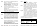
1
Counterstaining in hematoxylin is recommended using EnVision FLEX Hematoxylin, (Dako Autostainer/Autostainer Plus) (Code K8018). Non-aqueous, permanent mounting medium is recommended. Positive and negative controls should be run simultaneously using the same protocol as the patient specimens. The positive control tissue should include tonsil and the cells/structures should display reaction patterns as described for this tissue in “Performance characteristics” in all positive specimens. The recommended negative control reagent is FLEX Negative Control, Mouse, (Dako Autostainer/Autostainer Plus) (Code IS750). FLEX Monoclonal Mouse Anti-Human BCL6 Protein Clone PG-B6p Ready-to-Use (Dako Autostainer/Autostainer Plus) Staining interpretation Cells labeled by the antibody show a diffuse/microgranular staining confined to the nucleus. Intense cytoplasmic staining unassociated with metaphase chromosomes is noted only in mitotic figures (1). Performance characteristics Normal tissues: In tonsil, the antibody strongly labels the nuclei of centroblasts in the dark zone and centrocytes in the basal and apical light zones of germinal centres. In addition the antibody labels approximately 10% of germinal centre T cells. No labeling was observed in plasma cells, macrophages, follicular dendritic cells, and IgD+/IgM+ follicular mantle lymphocytes. In spleen, the staining pattern was similar to that of tonsil, but in addition the antibody labeled a few scattered lymphoid-like cells of undefined phenotype. In extra-lymphoid tissues, the antibody faintly labeled squamous epithelia in the tonsil, thymus, and skin. No labeling was observed in liver, thyroid, striated muscle, or kidney (1). In tonsil, germinal center B cells show a moderate to strong staining reaction, whereas squamous epithelial cells show a weak to moderate staining reaction. Abnormal tissues: 173 cases of human lymphoid neoplasms were tested with the antibody. Of B-cell lymphomas or leukemias, 24/24 follicle centre lymphomas, 29/30 diffuse large cell lymphomas, and 13/13 Burkitt’s lymphomas, were positive. No labeling was observed in 7/7 pre-B acute lymphoblastic leukemias, 16/16 small lymphocytic/B-CLL, 6/6 hairy cell leukemias, 22/22 mantle cell leukemias, and 14/14 marginal zone lymphomas. Of T-cell lymphomas or leukemias, 4/8 anaplastic large cell lymphomas were positive, whereas no labeling was observed in 10/10 acute lymphoblastic lymphomas, 3/3 mycosis fungoides, 6/6 peripheral T-cell lymphomas, and 2/2 peripheral T-cell (AILD-like) lymphomas. In Hodgkin’s disease, 5/5 nodular, lymphocyte predominance, 1/4 nodular sclerosis, and 1/3 mixed cellularity types were positive (1). Code IS625 ENGLISH For in vitro diagnostic use. FLEX Monoclonal Mouse Anti-Human BCL6 Protein, Clone PG-B6p, Ready-to-Use, (Dako Autostainer/Autostainer Plus), is intended for use in immunohistochemistry together with Dako Autostainer/Autostainer Plus instruments. This antibody is useful for classification of diffuse large B-cell lymphomas, follicular lymphomas and Burkitt’s lymphomas (1). The clinical interpretation of any staining or its absence should be complemented by morphological studies using proper controls and should be evaluated within the context of the patient's clinical history and other diagnostic tests by a qualified pathologist. Intended use The proto-oncogene BCL6 encodes a 92-98 kDa POZ/zinc finger protein. Through its six C-terminal zinc-finger motifs, the BCL6 protein binds to specific DNA sequences of target genes, where it acts as a transcriptional repressor (2, 3). BCL6 expression may be ubiquitous, as low levels are detected in many tissues. However, high levels of BCL6 expression are limited to cells of the B-lymphoid linage and skeletal muscle. In the B-lymphoid cell linage, BCL6 expression seems to be tightly regulated during differentiation, and strong expression is observed preferentially in germinal centre (GC) B cells, but not in plasma cells, and precursor, mantle zone, and memory B cells. Neoplastic counterparts of GC B cells, including diffuse large B-cell lymphomas, follicular and Burkitt’s lymphomas, also display high levels of BCL6 expression (3). BCL6 may be implicated in chromosomal translocations where the regulatory region of the BCL6 gene is replaced by a heterologous reciprocal partner such as the immunoglobulin genes. Promotor substitution leads to deregulation of the BCL6 expression, which may be associated with lymphomagenesis (3). BCL6 rearrangement is one of the most common genetic abnormalities in B-cell non-Hodgkin’s lymphoma, with an especially high frequency (28-40%) in diffuse large cell lymphoma (2). Refer to Dako’s General Instructions for Immunohistochemical Staining or the detection system instructions of IHC procedures for: 1) Principle of Procedure, 2) Materials Required, Not Supplied, 3) Storage, 4) Specimen Preparation, 5) Staining Procedure, 6) Quality Control, 7) Troubleshooting, 8) Interpretation of Staining, 9) General Limitations. Summary and explanation Reagent provided Ready-to-use monoclonal mouse antibody provided in liquid form in a buffer containing stabilizing protein and 0.015 mol/L sodium azide. Clone: PG-B6p (1). Isotype: IgG1, kappa. Immunogen Recombinant glutathione S-transferase (GST)-BCL6 protein corresponding to amino acids 3-484 of the human BCL6 protein (1). Specificity In Western blotting of total lysates of Bjab cells and BCL6 transfected EB3 cells, or control transfected EB3 cells, the antibody did not label any bands, suggesting lack of reactivity with denatured BCL6 protein (1). SDS-PAGE analysis of immunoprecipitates formed between total lysates of Bjab cells (BCL6+) and BCL6 transfected EB3 cells and the antibody, shows reaction with a ~95 kDa fragment corresponding to BCL6. No reaction is observed with Rd cells (BCL6-) and EB3 cells transfected with a control plasmid (1). Precautions 1. For professional users. 2. This product contains sodium azide (NaN3), a chemical highly toxic in pure form. At product concentrations, though not classified as hazardous, sodium azide may react with lead and copper plumbing to form highly explosive build-ups of metal azides. Upon disposal, flush with large volumes of water to prevent metal azide build-up in plumbing. 3. As with any product derived from biological sources, proper handling procedures should be used. 4. Wear appropriate Personal Protective Equipment to avoid contact with eyes and skin. 5. Unused solution should be disposed of according to local, State and Federal regulations. Store at 2-8 °C. Do not use after expiration date sta mped on vial. If reagents are stored under any conditions other than those specified, the conditions must be verified by the user. There are no obvious signs to indicate instability of this product. Therefore, positive and negative controls should be run simultaneously with patient specimens. If unexpected staining is observed which cannot be explained by variations in laboratory procedures and a problem with the antibody is suspected, contact Dako Technical Support. Storage Specimen preparation including materials required but not supplied FRANÇAIS Utilisation prévue Résumé et explication Le proto-oncogène BCL6 code pour une protéine POZ/doigt de zinc de 92–98 kDa. Par l’intermédiaire de six motifs doigt de zinc C-terminaux, la protéine BCL6 se lie à des séquences spécifiques de l’ADN des gènes cibles, où elle agit comme un répresseur de transcription (2, 3). L’expression de la BCL6 peut être générale, étant donné que des niveaux faibles sont détectés dans de nombreux tissus. Cependant, les taux d'expression élevés de BCL6 se limitent aux cellules de la lignée lymphoïde de type B et aux cellules du muscle squelettique. Dans la lignée lymphoïde de type B, l’expression de la BCL6 semble être fermement régulée au cours de la différenciation, et une forte expression est observée de préférence dans les lymphocytes B du centre germinatif (CG), mais pas dans les cellules plasmatiques ni dans les lymphocytes B précurseurs, de la zone du manteau et à mémoire. Les équivalents néoplasiques des lymphocytes B du CG, y compris les lymphomes diffus à grands lymphocytes B, les lymphomes folliculaires et les lymphomes de Burkitt, présentent également des taux d’expression élevés de la BCL6 (3). La BCL6 pourrait être impliquée dans les translocations chromosomiques où la région du gène BCL6 est remplacée par un partenaire réciproque hétérologue comme les gènes d’immunoglobuline. La substitution du promoteur entraîne une dérégulation de l’expression de la BCL6, ce qui peut être associé à la lymphomagenèse (3). La réorganisation de la BCL6 est l’une des anomalies génétiques les plus courantes dans le lymphome non hodgkinien à lymphocytes B, avec une fréquence particulièrement élevée (28–40 %) dans les cas de lymphomes diffus à grandes cellules (2). Se référer aux Instructions générales de coloration immunohistochimique de Dako ou aux instructions du système de détection relatives aux procédures IHC pour plus d’informations concernant les points suivants : 1) Principe de procédure, 2) Matériels requis mais non fournis, 3) Conservation, 4) Préparation des échantillons, 5) Procédure de coloration, 6) Contrôle qualité, 7) Dépannage, 8) Interprétation de la coloration, 9) Limites générales. Réactifs fournis Anticorps monoclonal de souris prêt à l’emploi fourni sous forme liquide dans un tampon contenant une protéine stabilisante et 0,015 mol/L d’azide de sodium. Clone : PG-B6p (1). Isotype : IgG1, kappa. Immunogène Protéine BCL6 recombinante glutathione-S-transférase (GST) correspondant aux acides aminés 3-484 de la protéine BCL6 humaine (1). Spécificité Dans les analyses par Western blot des lysats totaux de cellules Bjab et de cellules EB3 transfectées par la BCL6, ou de cellules EB3 de contrôle transfectées, l’anticorps n’a marqué aucune bande, ce qui laisse penser à un manque de réactivité à une protéine BCL6 dénaturée (1). L’analyse par immunoblot PAGE en présence de SDS d’immunoprécipités formés entre les lysats totaux de cellules Bjab (BCL6+) et les cellules EB3 transfectées par la BCL6 et l’anticorps a révélé une réaction à un fragment d’environ 95 kDa correspondant à la BCL6. Aucune réaction n’est observée avec les cellules Rd (BCL6-) et les cellules EB3 transfectées avec un plasmide de contrôle (1). Précautions 1. Pour utilisateurs professionnels. 2. Ce produit contient de l’azide de sodium (NaN3), produit chimique hautement toxique dans sa forme pure. Aux concentrations du produit, bien que non classé comme dangereux, l’azide de sodium peut réagir avec le cuivre et le plomb des canalisations et former des accumulations d’azides métalliques hautement explosifs. Lors de l’élimination, rincer abondamment à l’eau pour éviter toute accumulation d’azide métallique dans les canalisations. 3. Comme avec tout produit d’origine biologique, des procédures de manipulation appropriées doivent être respectées. 4. Porter un vêtement de protection approprié pour éviter le contact avec les yeux et la peau. 5. Les solutions non utilisées doivent être éliminées conformément aux réglementations locales et nationales. Conservation Conserver entre 2 et 8 °C. Ne pas utiliser après la date de péremption indiquée sur le flacon. Si les réactifs sont conservés dans des conditions autres que celles indiquées, celles-ci doivent être validées par l’utilisateur. Il n’y a aucun signe évident indiquant l’instabilité de ce produit. Par conséquent, des contrôles positifs et négatifs doivent être testés en même temps que les échantillons de patient. Si une coloration inattendue est observée, qui ne peut être expliquée par un changement des procédures du laboratoire, et en cas de suspicion d’un problème lié à l’anticorps, contacter l’assistance technique de Dako. Préparation des échantillons y compris le matériel requis mais non fourni L’anticorps peut être utilisé pour le marquage des coupes de tissus inclus en paraffine et fixés au formol. L'épaisseur des coupes d'échantillons de tissu doit être d’environ 4 µm. Le prétraitement avec un démasquage d’épitope induit par la chaleur (HIER) est nécessaire. Des résultats optimaux sont obtenus en prétraitant les tissus à l’aide de la EnVision( FLEX Target Retrieval Solution, High pH (10x), (Dako Autostainer/Autostainer Plus) (Réf. K8012/K8014). Coupes déparaffinées : Le prétraitement des coupes de tissus déparaffinés fixés au formol et inclus en paraffine est recommandé à l’aide du Dako PT Link (Réf. PT100/PT101). Pour plus de détails, se référer au Guide d’utilisation du PT Link. Suivre la procédure de prétraitement indiquée dans la notice pour la EnVision( FLEX Target Retrieval Solution, High pH (10x), (Dako Autostainer/Autostainer Plus) (Réf. K8012/K8014). Les paramètres suivants doivent être utilisés pour le PT Link : température de préchauffage : 65 °C ; température et durée de restauration de l’ép itope : 97 °C pendant 20 (±1) minutes ; laisser refro idir jusqu’à 65 °C. Rincer les coupes avec un EnVision™ FLEX Wash Buffer (10x), (Dako Autostainer/Autostainer Plus) (Réf. K8012) dilué à température ambiante. Coupes incluses en paraffine : Comme préparation alternative des échantillons, le déparaffinage et la restauration de l’épitope peuvent être réalisés dans le PT Link à l’aide d’une procédure modifiée. Se référer aux instructions du Guide d’utilisation du PT Link. Une fois que la procédure de coloration est terminée, les coupes doivent être déshydratées, lavées et montées à l’aide d’un milieu de montage permanent. Les coupes de tissus ne doivent pas sécher lors du traitement ou lors de la procédure de coloration immunohistochimique suivante. Pour une meilleure adhérence des coupes de tissus sur les lames de verre, il est recommandé d’utiliser des lames Dako Silanized Slides (Réf. S3003). The antibody can be used for labeling formalin-fixed, paraffin-embedded tissue sections. Tissue specimens should be cut into sections of approximately 4 µm. The recommended visualization system is EnVision FLEX+, Mouse, High pH, (Dako Autostainer/Autostainer Plus) (Code K8012). The staining steps and incubation times are pre-programmed into the software of Dako Autostainer/Autostainer Plus instruments, using the following protocols: Template protocol: FLEXRTU2 (200 µL dispense volume) or FLEXRTU3 (300 µL dispense volume) Autoprogram: BCL6 (without counterstaining) or BCL6H (with counterstaining) The Auxiliary step should be set to “rinse buffer” in staining runs with ≤10 slides. For staining runs with >10 slides the Auxiliary step should be set to “none”. This ascertains comparable wash times. All incubation steps should be performed at room temperature. For details, please refer to the Operator’s Manual for the dedicated instrument. If the protocols are not available on the used Dako Autostainer instrument, please contact Dako Technical Services. Optimal conditions may vary depending on specimen and preparation methods, and should be determined by each individual laboratory. If the evaluating pathologist should desire a different staining intensity, a Dako Application Specialist/Technical Service Specialist can be contacted for information on reprogramming of the protocol. Verify that the performance of the adjusted protocol is still valid by evaluating that the staining pattern is identical to the staining pattern described in “Performance characteristics”. (115279-002) Dako Denmark A/S IS625/EFG/MNI/09.10.07 p. 1/4 | Produktionsvej 42 | DK-2600 Glostrup | Denmark | Tel. +45 44 85 95 00 | Fax +45 44 85 95 95 | CVR No. 33 21 13 17 Pour utilisation diagnostique in vitro. FLEX Monoclonal Mouse Anti-Human BCL6 Protein, Clone PG-B6p, Ready-to-Use, (Dako Autostainer/Autostainer Plus) est destiné à une utilisation en immunohistochimie avec les instruments Dako Autostainer/Autostainer Plus. Cet anticorps est utile pour la classification des lymphomes à grands lymphocytes B diffus, des lymphomes folliculaires et des lymphomes de Burkitt (1). L’interprétation clinique de toute coloration ou son absence doit être complétée par des études morphologiques en utilisant des contrôles appropriés et doit être évaluée en fonction des antécédents cliniques du patient et d’autres tests diagnostiques par un pathologiste qualifié. Pre-treatment with heat-induced epitope retrieval (HIER) is required. Optimal results are obtained by pretreating tissues using EnVision FLEX Target Retrieval Solution, High pH (10x), (Dako Autostainer/Autostainer Plus) (Code K8012/K8014). Deparaffinized sections: Pre-treatment of deparaffinized formalin-fixed, paraffin-embedded tissue sections is recommended using Dako PT Link (Code PT100/PT101). For details, please refer to the PT Link User Guide. Follow the pre-treatment procedure outlined in the package insert for EnVision FLEX Target Retrieval Solution, High pH (10x), (Dako Autostainer/Autostainer Plus) (Code K8012/K8014). The following parameters should be used for PT Link: Pre-heat temperature: 65 °C; epitope retrieval temperature and time: 97 °C for 20 (±1) mi nutes; cool down to 65 °C. Rinse sections with diluted ro om temperature EnVision FLEX Wash Buffer (10x), (Dako Autostainer/Autostainer Plus) (Code K8012). Paraffin-embedded sections: As alternative specimen preparation, both deparaffinization and epitope retrieval can be performed in the PT Link using a modified procedure. See the PT Link User Guide for instructions. After the staining procedure has been completed, the sections must be dehydrated, cleared and mounted using permanent mounting medium. The tissue sections should not dry out during the treatment or during the following immunohistochemical staining procedure. For greater adherence of tissue sections to glass slides, the use of Dako Silanized Slides (Code S3003) is recommended. Staining procedure including materials required but not supplied In systemic AIDS-related non-Hodgkin’s lymphoma, the antibody labeled 11/11 small noncleaved cell lymphomas, 7/7 large noncleaved cell lymphomas (LNCCL), and 2/9 large cell immunoblastic lymphoma plasmacytoid (IBLP). In AIDS-primary central nervous system lymphomas, the antibody labeled 4/4 LNCCL and none of 4 cases of IBLP. No labeling was observed in 5 cases of primary effusion lymphoma (PEL) and in 6 AIDS-PEL cell lines (4). (115279-002) Dako Denmark A/S IS625/EFG/MNI/09.10.07 p. 2/4 | Produktionsvej 42 | DK-2600 Glostrup | Denmark | Tel. +45 44 85 95 00 | Fax +45 44 85 95 95 | CVR No. 33 21 13 17 Procédure de coloration y compris le matériel requis mais non fourni Le système de visualisation recommandé est le EnVision FLEX+, Mouse, High pH, (Dako Autostainer/Autostainer Plus) (Réf. K8012). Les étapes de coloration et d’incubation sont préprogrammées dans le logiciel des instruments Dako Autostainer/Autostainer Plus, à l’aide des protocoles suivants : Protocole modèle : FLEXRTU2 (volume de distribution de 200 µL) ou FLEXRTU3 (volume de distribution de 300 µL) Autoprogram : BCL6 (sans contre-coloration) ou BCL6H (avec contre-coloration) Vorbereitung der Probe und erforderliche, aber nicht mitgelieferte Materialien Der Antikörper eignet sich zur Markierung von formalinfixierten und paraffineingebetteten Gewebeschnitten. Gewebeproben sollten in Schnitte von ca. 4 µm Stärke geschnitten werden. Die Vorbehandlung durch hitzeinduzierte Epitopdemaskierung (HIER) ist erforderlich. Optimale Ergebnisse können durch Vorbehandlung der Gewebe mit EnVision™ FLEX Target Retrieval Solution, High pH (10x) (Dako Autostainer/Autostainer Plus) (Code-Nr. K8012/K8014) erzielt werden. Entparaffinierte Schnitte: Die Vorbehandlung der entparaffinierten, formalinfixierten, paraffineingebetteten Gewebeschnitte sollte mit Dako PT Link (Code-Nr. PT100/PT101) erfolgen. Weitere Informationen hierzu siehe PT Link-Benutzerhandbuch. Vorbehandlung gemäß der Beschreibung in der Packungsbeilage für EnVision™ FLEX Target Retrieval Solution, High pH (10x), (Dako Autostainer/Autostainer Plus) (Code-Nr. K8012/K8014) durchführen. Für PT Link sollten die folgenden Parameter verwendet werden: Vorwärmtemperatur: 65 °C; Temperatur und Zeit für E pitopdemaskierung: 97 °C für 20 (±1) Minuten; auf 65 °C abkühlen lassen. Schnitte mit auf Raumtemperatur gebrachtem EnVision™ FLEX Wash Buffer (10x), (Dako Autostainer/Autostainer Plus) (Code-Nr. K8012) abspülen. Paraffineingebettete Schnitte: Zur Probenpräparation kann für Entparaffinierung und Epitopdemaskierung im PT Link auch ein modifiziertes Verfahren verwendet werden. Weitere Informationen siehe PT Link-Benutzerhandbuch. Nach Abschluss des Färbeverfahrens müssen die Schnitte dehydriert, geklärt und unter Verwendung eines permanenten Fixiermittels auf die Objektträger aufgebracht werden. Die Gewebeschnitte dürfen während der Behandlung oder während des anschließenden immunhistochemischen Färbeverfahrens nicht austrocknen. Zur besseren Haftung der Gewebeschnitte an den Glasobjektträgern wird die Verwendung von Dako Silanized Slides (Code-Nr. S3003) empfohlen. Färbeverfahren und erforderliche, aber nicht mitgelieferte Materialien Das empfohlene Visualisierungssystem ist EnVision™ FLEX+, Mouse, High pH (Dako Autostainer/Autostainer Plus) (Code-Nr. K8012). Die Färbeschritte und Inkubationszeiten sind in der Software der Dako Autostainer/Autostainer Plus-Geräte mit den folgenden Protokollen vorprogrammiert: Matrix-Protokoll: FLEXRTU2 (200 µL Abgabevolumen) oder FLEXRTU3 (300 µL Abgabevolumen) Autoprogram: BCL6 (ohne Gegenfärbung) oder BCL6H (mit Gegenfärbung) Bei Färbedurchläufen mit höchstens 10 Objektträgern sollte der „Zusatz“-Schritt auf „Pufferspülgang“ eingestellt werden. Für Färbedurchläufe mit mehr als 10 Objektträgern den „Zusatz“-Schritt auf „Keine“ einstellen. Dieses gewährleistet vergleichbare Waschzeiten. Alle Inkubationsschritte sollten bei Raumtemperatur durchgeführt werden. Nähere Einzelheiten bitte dem Benutzerhandbuch für das jeweilige Gerät entnehmen. Wenn die Färbeprotokolle auf dem verwendeten Dako Autostainer-Gerät nicht verfügbar sind, bitte den Technischen Kundendienst von Dako verständigen. Optimale Bedingungen können je nach Probe und Präparationsverfahren unterschiedlich sein und sollten vom jeweiligen Labor selbst ermittelt werden. Falls der beurteilende Pathologe eine andere Färbungsintensität wünscht, kann ein Anwendungsspezialist oder Kundendiensttechniker von Dako bei der Neuprogrammierung des Protokolls helfen. Die Leistung des angepassten Protokolls muss verifiziert werden, indem gewährleistet wird, dass das Färbemuster mit dem unter „Leistungsmerkmale“ beschriebenen Färbemuster identisch ist. Die Gegenfärbung in Hämatoxylin sollte mit EnVision™ FLEX Hematoxylin, (Dako Autostainer/Autostainer Plus) (Code-Nr. K8018) ausgeführt werden. Empfohlen wird ein nichtwässriges, permanentes Fixiermittel. Positiv- und Negativkontrollen sollten zur gleichen Zeit und mit demselben Protokoll wie die Patientenproben getestet werden. Das positive Kontrollgewebe sollte Mandelgewebe enthalten und die Zellen/Strukturen müssen in allen positiven Proben die für dieses Gewebe unter „Leistungsmerkmale“ beschriebenen Reaktionsmuster aufweisen. Das empfohlene Negativ-Kontrollreagenz ist FLEX Negative Control, Mouse, (Dako Autostainer/Autostainer Plus) (Code-Nr. IS750). Auswertung der Färbung Mit diesem Antikörper markierte Zellen weisen eine diffuse, mikrogranuläre und auf den Zellkern begrenzte Färbung auf. Nur bei mitotischen Konfigurationen wird eine intensive zytoplasmatische Färbung festgestellt, die nicht auf Metaphasechromosomen zurückzuführen ist (1). Leistungsmerkmale Gesundes Gewebe: In Mandelgewebe markiert der Antikörper in starkem Ausmaß die Zentroblasten-Nuklei der dunklen Zone sowie die Zentrozyten der basalen und apikalen hellen Zonen in den Keimzentren. Zudem markiert der Antikörper circa 10 % der Keimzentrum-T-Zellen. Keine Markierung wurde festgestellt für Plasmazellen, Makrophagen, follikulär-dendritische Zellen und IgD+/IgM+ follikuläre Mantellymphozyten. Für Milzgewebe verlief das Färbungsmuster ähnlich wie bei Mandelgewebe, der Antikörper markierte jedoch darüber hinaus einige wenige verstreute lymphoidartige Zellen eines nicht definierten Phänotyps. Bei Geweben außerhalb des Lymphsystems erbrachte der Antikörper eine schwache Färbung der Plattenepithelzellen von Mandeln, Thymus und Haut. Für Leber, Schilddrüse, gestreifte Muskulatur oder Niere wurde keine Markierung beobachtet (1). Bei Mandelgewebe zeigen Keimzentrum-B-Zellen eine mäßige bis starke Färbereaktion, während Plattenepithelzellen eine schwache bis mäßige Färbereaktion zeigen. Pathologisches Gewebe: Mit dem Antikörper wurden 173 Fälle menschlicher lymphoider Neoplasmen getestet. Von den B-Zell-Lymphomen oder -Leukämien wurden für 24 von 24 follikulären Lymphomen (zentroblastisch-zentrozytisch, FCL), 29 von 30 diffusen großzelligen Lymphomen und 13 von 13 BurkittLymphomen positive Resultate erhalten. Keine Markierung erbrachten 7 von 7 akuten lymphatischen Leukämien vom Prä-B-Typ, 16 von 16 kleinzelliglymphozytischen bzw. B-CLL, 6 von 6 Haarzell-Leukämien, 22 von 22 Mantelzell-Leukämien und 14 von 14 Marginalzonen-Lymphomen. Von den T-ZellLymphomen oder Leukämien testeten 4 von 8 anaplastischen großzelligen Lymphomen positiv. Dagegen wurde keine Markierung beobachtet bei 10 von 10 akuten lymphatischen Lymphomen, 3 von 3 Mycosis fungoides, 6 von 6 peripheren T-Zell-Lymphomen und 2 von 2 peripheren T-Zell-Lymphomen (vom AILDTyp; angioimmunoblastische Lymphadenopathie mit Dysproteinämie). Für das Hodgkin-Lymphom wurden für 5 von 5 Fällen des nodulären Typs mit Prädominanz der Lymphozyten, 1 von 4 Fällen des nodulärsklerosierenden Typs und 1 von 3 Typen gemischter Zellularität positive Resultate erhalten (1). Beim systemischen AIDS-assoziierten Non-Hodgkin-Lymphom markierte der Antikörper 11 von 11 kleinzelligen, nicht zentrozytischen Lymphomen (SNCCL), 7 von 7 großzelligen, nicht zentrozytischen Lymphomen (LNCCL) und 2 von 9 großzelligen, immunoblastischen, lymphoplasmazytoiden Lymphomen (IBLP). Beim AIDS-assoziierten primären Lymphom des Zentralnervensystems (PCNSL) markierte der Antikörper 4 von 4 LNCCL und keinen der vier Fälle von IBLP. Keine Markierung ergab sich in 5 Fällen eines primären Erguss-Lymphoms (PEL) und bei 6 AIDS-assoziierten PEL-Zelllinien (4). L’étape Auxiliary doit être réglée sur « rinse buffer » lors des cycles de coloration avec ≤10 lames. Pour les cycles de coloration de >10 lames, l’étape Auxiliary doit être réglée sur « none ». Cela garantit des temps de lavage comparables. Toutes les étapes d’incubation doivent être effectuées à température ambiante. Pour plus de détails, se référer au Manuel de l’opérateur spécifique à l'instrument. Si les protocoles ne sont pas disponibles sur l’instrument Dako Autostainer utilisé, contacter le service technique de Dako. Les conditions optimales peuvent varier en fonction du prélèvement et des méthodes de préparation, et doivent être déterminées par chaque laboratoire individuellement. Si le pathologiste qui réalise l’évaluation désire une intensité de coloration différente, un spécialiste d’application/spécialiste du service technique de Dako peut être contacté pour obtenir des informations sur la re-programmation du protocole. Vérifier que l'exécution du protocole modifié est toujours valide en vérifiant que le schéma de coloration est identique au schéma de coloration décrit dans les « Caractéristiques de performance ». Il est recommandé d’effectuer une contre-coloration à l’aide de EnVision FLEX Hematoxylin, (Dako Autostainer/Autostainer Plus) (Réf. K8018). L’utilisation d’un milieu de montage permanent non aqueux est recommandée. Des contrôles positifs et négatifs doivent être réalisés en même temps et avec le même protocole que les échantillons du patient. Le contrôle de tissu positif doit comprendre l’amygdale et les cellules/structures doivent présenter des schémas de réaction tels que décrits pour ces tissus dans les « Caractéristiques de performance » pour tous les échantillons positifs. Le contrôle négatif recommandé est le FLEX Negative Control, Mouse, (Dako Autostainer/Autostainer Plus) (Réf. IS750). Interprétation de la coloration Les cellules marquées par l’anticorps présentent une coloration diffuse/microgranulaire limitée au noyau. Une coloration cytoplasmique intense non associée aux chromosomes métaphasiques est observée uniquement dans les figures mitotiques (1). Caractéristiques de performance Tissus sains : Dans l’amygdale, l’anticorps marque fortement les noyaux des centroblastes dans la zone sombre et les centrocytes dans les zones claires basale et apicale des centres germinatifs. En outre, l’anticorps marque environ 10 % des lymphocytes T du centre germinatif. Aucune coloration n’a été observée dans les cellules plasmatiques, les macrophages, les cellules folliculaires dendritiques et les lymphocytes du manteau folliculaire IgD+/IgM+. Dans la rate, le schéma de coloration était similaire à celui observé dans l'amygdale mais, par ailleurs, l’anticorps a marqué quelques cellules lymphoïdes isolées de phénotype non défini. Dans les tissus extra-lymphoïdes, l’anticorps a marqué légèrement les épithéliums squameux de l’amygdale, du thymus et de la peau. Aucune coloration n’a été observée dans le foie, la thyroïde, le muscle strié ou le rein (1). Les lymphocytes B et T du centre germinatif dans les amygdales ont présenté une coloration modérée à forte, tandis que la coloration des cellules épithéliales squameuses était faible à modérée. Tissus tumoraux : 173 cas de néoplasmes lymphoïdes humains ont été testés avec l’anticorps. Dans les lymphomes ou les leucémies à lymphocytes B, 24/24 lymphomes du centre folliculaire, 29/30 lymphomes diffus à grandes cellules et 13/13 lymphomes de Burkitt étaient positifs. Aucune coloration n’a été observée dans 7/7 leucémies aiguës lymphoblastiques à pré-lymphocytes B, 16/16 lymphomes lymphocytaires à petites cellules/LLC-B, 6/6 leucémies à cellules chevelues, 22/22 leucémies à cellules du manteau et 14/14 lymphomes de la zone marginale. Dans les lymphomes ou leucémies à lymphocytes T, 4/8 lymphomes anaplasiques à grandes cellules étaient positifs, alors qu’aucune coloration n’a été observée dans 10/10 lymphomes lymphoblastiques aigus, 3/3 mycoses fongoïdes, 6/6 lymphomes périphériques à lymphocytes T et 2/2 lymphomes périphériques à lymphocytes T (de type AILD). Dans la maladie de Hodgkin, 5 cas de type nodulaire sur 5, à prédominance lymphocytaire, 1 cas à sclérose nodulaire sur 4 et 1 cas à cellularité mixte sur 3 étaient positifs (1). Parmi les lymphomes non hodgkiniens liés au SIDA, l’anticorps a marqué 11 lymphomes à petites cellules non clivées sur 11, 7 lymphomes à grandes cellules non clivées (LNCCL) sur 7 et 2 lymphomes malins à larges cellules immunoblastiques (plasmacytoïdes) (IBLP) sur 9. Parmi les lymphomes du système nerveux central primitifs associés au SIDA, l’anticorps a marqué 4 LNCCL sur 4 et aucun des 4 cas d’IBLP. Aucun marquage n’a été observé dans 5 cas de lymphomes à effusion primaire (LIP) et 6 lignées cellulaires de LIP associées au SIDA (4). DEUTSCH Zweckbestimmung Zur In-vitro-Diagnostik. FLEX Monoclonal Mouse Anti-Human BCL6-Protein, Clone PG-B6p, Ready-to-Use, (Dako Autostainer/Autostainer Plus) ist zur Verwendung in der Immunhistochemie in Verbindung mit Dako Autostainer/Autostainer Plus-Geräten bestimmt. Dieser Antikörper dient zur Klassifizierung großzelliger diffuser B-Zell-Lymphome, follikulärer Lymphome und Burkitt-Lymphome (1). Die klinische Auswertung einer eventuell eintretenden Färbung sollte durch morphologische Studien mit ordnungsgemäßen Kontrollen ergänzt werden und von einem qualifizierten Pathologen unter Berücksichtigung der Krankengeschichte und anderer Diagnostiktests des Patienten vorgenommen werden. Zusammenfassung und Erklärung Das Proto-Onkogen BCL6 kodiert ein 92–98 kDa POZ/Zinkfingerprotein. Über seine sechs C-terminalen Zinkfinger-Motive erfolgt die Bindung des BCL6Proteins an spezifische DNA-Sequenzen der Zielgene, wo es als die Transkription verhindernder Repressor fungiert (2, 3). Die BCL6-Expression ist kann häufig vorkommen, da niedrige Gehalte in vielen Gewebetypen nachgewiesen werden. Hochgradige BCL6-Expression ist allerdings auf Zellen der B-Lymphoidzelllinie und der Skelettmuskulatur beschränkt. Bei der B-Lymphoidzelllinie scheint die BCL6-Expression während der Differenzierung unter strikter Regulierung zu stehen. Starke Expression wird vorzugsweise in dem Keimzentrum (GC) entstammenden B-Zellen, nicht jedoch in Plasmazellen, Vorläuferzellen, Mantelzonenzellen und B-Gedächtniszellen beobachtet. Neoplastische Entsprechungen der GC-B-Zellen, einschließlich Burkitt-Lymphom, Follikel-Lymphom und diffusem großzelligem Lymphom, zeigen ebenfalls hochgradige BCL6-Expression (3). BCL6 ist möglicherweise an chromosomalen Translokationen beteiligt, bei denen die regulatorische Region des BCL6-Gens durch einen heterologen reziproken Partner – wie beispielsweise die Immunglobulin-Gene (IG) – ersetzt wird. Die Promotor-Substitution führt zur Deregulierung der BCL6Expression, die mit der Lymphomentstehung in Zusammenhang stehen könnte (3). Beim BCL6-Rearrangement handelt es sich um eine der häufigsten genetischen Anomalien bei B-Zell-Lymphomen vom Non-Hodgkin-Typ mit einer besonders stark ausgeprägten Auftrittshäufigkeit (28–40 %) beim diffusen großzelligen Lymphom (2). Folgende Angaben bitte den Allgemeinen Richtlinien zur immunhistochemischen Färbung von Dako oder den Anweisungen des Detektionssystems für IHC-Verfahren entnehmen: 1) Verfahrensprinzip, 2) Erforderliche, aber nicht mitgelieferte Materialien, 3) Aufbewahrung, 4) Vorbereitung der Probe, 5) Färbeverfahren, 6) Qualitätskontrolle, 7) Fehlersuche und -behebung, 8) Auswertung der Färbung, 9) Allgemeine Beschränkungen. Geliefertes Reagenz Gebrauchsfertiger, monoklonaler Maus-Antikörper in flüssiger Form in einem Puffer, der stabilisierendes Protein und 0,015 mol/L Natriumazid enthält. Klon: PG-B6p (1). Isotyp: IgG1, Kappa. Immunogen Rekombinantes Glutathion-S-Transferase-(GST)-BCL6-Protein, entsprechend den Aminosäuren 3–484 des menschlichen BCL6-Proteins (1). Spezifität Beim Western-Blotting der Gesamtlysate von Bjab-Zellen und mit BCL6 transfizierten EB3-Zellen oder mit Kontrollpräparat transfizierten EB3-Zellen markierte der Antikörper keinerlei Banden. Dies weist auf eine mangelnde Reaktivität mit denaturiertem BCL6-Protein hin (1). Die SDS-PAGE-Analyse der Immunpräzipitate, die zwischen dem Antikörper und den Gesamtlysaten der Bjab-Zellen (BCL6+) und BCL6-transfizierten EB3-Zellen gebildet wurden, zeigt eine Reaktion mit dem BCL6 entsprechenden Fragment von ~95 kDa. Bei mit Kontroll-Plasmid transfizierten RdZellen (BCL6-) und EB3-Zellen wird keine Reaktion beobachtet (1). Vorsichtsmaßnahmen Lagerung (115279-002) Dako Denmark A/S References/ Références/ Literatur 1. Flenghi L, Bigerna B, Fizzotti M, Venturi S, Pasqualucci L, Pileri S, et al. Monoclonal antibodies PG-B6a and PG-B6p recognize, respectively, a highly conserved and a formol-resistant epitope on the human BCL-6 protein amino-terminal region. Am J Pathol 1996; 148:1543-55. 2. Ye BH. BCL-6 in the pathogenesis of non-Hodgkin’s lymphoma. Cancer Invest 2000; 18:356-65. 3. Nakamura Y. Internal deletions within the BCL6 gene in B-cell non-Hodgkin’s lymphoma. Leuk Lymphoma 2000; 38:505-12. 4. Carbone A, Gaidano G, Gloghini A, Larocca LM, Capello D, Canzonieri V, et al. Differential expression of BCL-6, CD138/syndecan-1, and Epstein-Barr virus-encoded latent membrane protein-1 identifies distinct histogenetic subsets of acquired immunodeficiency syndrome-related non-Hodgkin’s lymphomas. Blood 1998; 91:747-55. Explanation of symbols/ Légende des symboles/ Erläuterung der Symbole 1. Nur für Fachpersonal bestimmt. 2. Dieses Produkt enthält Natriumazid (NaN3), eine in reiner Form äußerst giftige Chemikalie. Natriumazid kann auch in als ungefährlich eingestuften Konzentrationen mit Blei- und Kupferrohren reagieren und hochexplosive Metallazide bilden. Nach der Entsorgung stets mit viel Wasser nachspülen, um Metallazidansammlungen in den Leitungen vorzubeugen. 3. Wie alle Produkte biologischen Ursprungs müssen auch diese entsprechend gehandhabt werden. 4. Geeignete Schutzkleidung tragen, um Augen- und Hautkontakt zu vermeiden. 5. Nicht verwendete Lösung ist entsprechend örtlichen, bundesstaatlichen und staatlichen Richtlinien zu entsorgen. Catalogue number Référence catalogue Bestellnummer Temperature limitation Limites de température Use by Utiliser avant Zulässiger Temperaturbereich Verwendbar bis In vitro diagnostic medical device Dispositif médical de diagnostic in vitro In-vitro-Diagnostikum Contains sufficient for <n> tests Contenu suffisant pour <n> tests Manufacturer Fabricant Inhalt ausreichend für <n> Tests Hersteller Consult instructions for use Voir les instructions d’utilisation Gebrauchsanweisung beachten Batch code Numéro de lot Chargenbezeichnung Bei 2–8 °C aufbewahren. Nach Ablauf des auf dem Fläschch en aufgedruckten Verfalldatums nicht mehr verwenden. Werden die Reagenzien unter anderen als den angegebenen Bedingungen aufbewahrt, müssen diese Bedingungen vom Benutzer validiert werden. Es gibt keine offensichtlichen Anzeichen für eine eventuelle Produktinstabilität. Positiv- und Negativkontrollen sollten daher zur gleichen Zeit wie die Patientenproben getestet werden. Falls es zu einer unerwarteten Färbung kommt, die sich nicht durch Unterschiede bei Laborverfahren erklären lässt und auf ein Problem mit dem Antikörper hindeutet, ist der technische Kundendienst von Dako zu verständigen. IS625/EFG/MNI/09.10.07 p. 3/4 | Produktionsvej 42 | DK-2600 Glostrup | Denmark | Tel. +45 44 85 95 00 | Fax +45 44 85 95 95 | CVR No. 33 21 13 17 (115279-002) Dako Denmark A/S IS625/EFG/MNI/09.10.07 p. 4/4 | Produktionsvej 42 | DK-2600 Glostrup | Denmark | Tel. +45 44 85 95 00 | Fax +45 44 85 95 95 | CVR No. 33 21 13 17