1
REF 30 192
VIDAS® H. pylori IgG (HPY)
10988 D - FR - 2004/09
IVD
®
VIDAS H. pylori IgG (HPY) est un test qualitatif automatisé sur les instruments VIDAS, permettant la détection des
anticorps IgG anti-Helicobacter pylori dans le sérum ou le plasma humain (EDTA), par technique ELFA (Enzyme Linked
®
Fluorescent Assay). VIDAS HPY est utilisé dans l'aide au diagnostic des infections à H. pylori chez une population
adulte symptomatique.
INTRODUCTION ET OBJET DU TEST
Les méthodes sérologiques (telles que les tests
immunoenzymatiques)
sont
non
invasives,
peu
Helicobacter pylori (appelé autrefois Campylobacter
coûteuses, rapides et faciles à réaliser. Leur principal
pylori) est un bacille microaérophile en forme de spirale
avantage, par rapport aux méthodes invasives, est
qui entraîne une inflammation de la muqueuse de
qu'elles ne dépendent pas de la précision du prélèvement
l'estomac (gastrite) et des ulcères chez les personnes
(7, 9).
infectées. L'infection peut également être à l'origine de
cancers de l'estomac (1, 2, 3). Les personnes présentant
PRINCIPE
des symptômes d'infection à H. pylori sont considérées
Le principe du dosage associe la méthode
comme
infectées,
alors
que
les
personnes
immunoenzymatique sandwich en 2 étapes à une
asymptomatiques sont considérées comme colonisées.
détection finale en fluorescence (ELFA). Toutes les
La plupart des personnes colonisées ne développeront
étapes de la réaction ainsi que la température du test sont
jamais d'ulcères et resteront asymptomatiques malgré
contrôlées automatiquement par l'instrument. Le cône à
une colonisation par H. pylori depuis des années, voire
usage unique sert à la fois de phase solide et de système
des dizaines d'années. (4, 5).
de pipetage. Les autres réactifs de la réaction
Il existe des méthodes invasives et non invasives pour
immunologique sont prêts à l'emploi et sont pré-répartis
détecter la présence de H. pylori. La biopsie par
dans la cartouche.
endoscopie est traditionnellement utilisée pour obtenir des
Après une étape préliminaire de lavage, et de dilution de
échantillons de tissus gastriques ou duodénaux en vue de
l’échantillon, celui-ci subit des cycles d'aspiration et de
coloration, culture et/ou détection directe de l'uréase.
refoulement pendant un temps déterminé. La première
La biopsie peut entraîner des résultats faussement
étape permet aux anticorps anti-H. pylori présents dans le
négatifs chez des individus infectés en raison de la
prélèvement de se lier spécifiquement aux antigènes
répartition non uniforme de H. pylori dans le tissu
H. pylori déjà fixés sur les parois du cône.
gastrique ou duodénal ou encore, lorsque les tissus sont
Des étapes de lavage éliminent les composants non fixés.
porteurs de H. pylori non viables ou non producteurs
Des anticorps anti-IgG humaines conjugués à la
d'uréase (6). Les méthodes invasives telles que
phosphatase alcaline sont alors aspirés/refoulés dans le
l'endoscopie sont inconfortables, présentent des risques
cône et vont se fixer sur les IgG humaines fixées sur les
pour le patient et sont coûteuses à réaliser.
parois du cône.
Les méthodes non invasives comprennent les tests
Une étape finale de lavage permet d'éliminer le conjugué
respiratoires à l’urée et les méthodes sérologiques. Les
d'anticorps anti-humain non fixé.
tests respiratoires à l’urée permettent la détection de
Lors de l'étape finale de révélation, le substrat (4-MéthylH. pylori via son uréase fortement active : de l'urée
ombelliferyl phosphate) est aspiré puis refoulé dans le
marquée au carbone-14 ou au carbone-13 est ingérée par
cône ; l'enzyme du conjugué catalyse la réaction
le patient et la présence de dioxyde de carbone exhalée
d'hydrolyse de ce substrat en un produit (4-Méthylest déterminée par scintillation ou spectrophotométrie de
ombelliferone) dont la fluorescence émise est mesurée à
masse. Les inconvénients d'une telle méthode sont
450 nm. La valeur du signal de fluorescence est mesurée
l'exposition des patients à des radio-isotopes et
par le système optique de l’instrument VIDAS. A la fin du
l'équipement onéreux nécessaire (6, 7, 8).
test, les résultats sont calculés automatiquement par
Les patients souffrant d'infections à H. pylori produisent
l’instrument. Une valeur test est obtenue et un résultat est
des anticorps sériques. Ces anticorps sont corrélés avec
imprimé pour chaque échantillon.
la présence d'infections à H. pylori confirmées par
histologie.
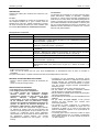
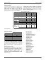
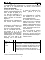
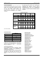
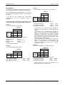
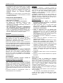
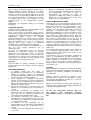
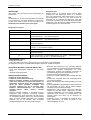
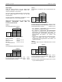
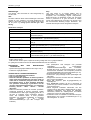
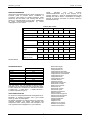
COMPOSITION DES REACTIFS DU COFFRET (30 TESTS) :
30 cartouches HPY
STR Prêtes à l'emploi.
30 cônes HPY (1 x 30)
SPR Prêts à l'emploi. Cônes sensibilisées par de l’antigène H. pylori purifié.
Standard HPY
S1 Prêt à l'emploi.
Sérum humain* contenant des anticorps anti-H. pylori + azoture de sodium 1 g/l.
(1 x 2 ml)
L’intervalle de confiance en "Relative Fluorescence Value" (RFV) est indiqué sur
la carte MLE avec la mention : "Standard (S1) RFV Range".
Contrôle positif HPY
C1 Prêt à l'emploi.
Sérum humain* contenant des anticorps anti-H. pylori + azoture de sodium 1 g/l.
(1 x 1,5 ml)
L’intervalle de confiance en Valeur Test (TV) est indiqué sur la carte MLE avec la
mention : "Control C1 Test Value Range".
Contrôle négatif HPY
C2 Prêt à l'emploi.
(1 x 1,5 ml)
Sérum humain* ne contenant pas d’ anticorps anti-H. pylori + azoture de sodium
1 g/l. L’intervalle de confiance en Valeur Test (TV) est indiqué sur la carte MLE
avec la mention : "Control C2 Test Value Range".
1 Carte MLE
Fiche de spécifications contenant les données usine nécessaires à la calibration
du test.
1 Notice
* L'absence d'antigène HBs de surface, d'anticorps anti-VIH1, anti-VIH2 et d'anticorps anti-VHC a été vérifiée. Cependant aucun test ne pouvant
apporter une garantie absolue, ce produit doit être manipulé avec les précautions d'usage relatives aux produits potentiellement infectieux.
bioMérieux
®
sa
Français - 1
VIDAS® H. pylori IgG
10988 D - FR - 2004/09
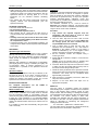
DESCRIPTION
Chaque test VIDAS HPY nécessite une cartouche et un
cône HPY.
La cartouche :
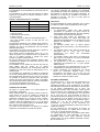
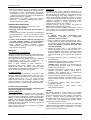
La cartouche est composée de 10 puits recouverts d'une
feuille d'aluminium scellée et étiquetée. L’étiquette
comporte un code à barres reprenant principalement le
code du test, le numéro de lot et la date de péremption du
coffret. Le premier puits comporte une partie prédécoupée
pour faciliter l'introduction de l'échantillon. Le dernier puits
est une cuvette permettant la lecture en fluorimétrie. Les
différents réactifs nécessaires à l’analyse sont contenus
dans les puits intermédiaires.
Le cône :
Le cône est sensibilisé au moment de la fabrication par
des antigènes purifiés de H. pylori. Chaque cône est
identifié par le code HPY. Utiliser uniquement le nombre
de cônes nécessaire et laisser les cônes inutilisés dans
leur sachet. Bien refermer le sachet après ouverture.
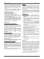
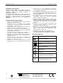
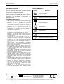
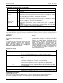
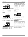
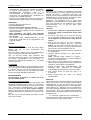
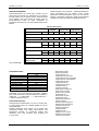
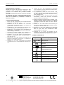
Description de la cartouche
Puits
Réactifs
1
Puits échantillon.
2
Diluant échantillon : Tampon TRIS (0,05 mol/l, pH 7,4) + stabilisateurs protéiques +
azoture de sodium 1 g/l (600 µl).
3
Tampon de prélavage : Tampon TRIS (0,05 mol/l, pH 7,4) + stabilisateurs
protéiques + azoture de sodium 1 g/l (400 µl).
4-5-7
Tampon de lavage : Tampon TRIS (0,05 mol/l) + détergent + azoture de sodium
1 g/l (600 µl).
6
Conjugué : Mélange titré d'anticorps monoclonaux de souris anti-IgG humaines
marqués à la phosphatase alcaline + azoture de sodium 1 g/l (400 µl).
8
Tampon de lavage : Tampon DEA (360 mmol/l) + azoture de sodium 1 g/l (600 µl).
9
Diluant échantillon : Tampon TRIS (0,05 mol/l, pH 7,4) + stabilisants protéiques +
azoture de sodium 1 g/l (300 µl).
10
Cuvette de lecture avec substrat : 4-Méthyl-ombelliferyl phosphate (0,6 mmol/l) +
diéthanolamine DEA* (0,62 mol/l soit 6,6 %) pH 9,2 + azoture de sodium 1 g/l
(300 µl).
* Réactif IRRITANT :
- R 36 : irritant pour les yeux.
- S 26 : en cas de contact avec les yeux, laver immédiatement et abondamment avec de l'eau et consulter un
spécialiste.
Pour plus d'informations, consulter la fiche de données sécurité disponible sur demande.
• Considérer tous les échantillons de patients comme
potentiellement infectieux et respecter les précautions
d'usage. Eliminer les composants utilisés et autres
matériaux contaminés selon les procédures en vigueur
pour les produits d'origine humaine potentiellement
infectieux (10-12).
• Ne pas utiliser les cônes dont le sachet est percé.
• Ne pas utiliser de cartouches visiblement altérées
(feuille aluminium ou plastique endommagé).
• Ne pas utiliser les réactifs après la date de péremption
indiquée sur l'étiquette étui.
• Ne pas mélanger les réactifs (ou consommables) de lots
différents.
• Ne pas utiliser de gants talqués, le talc pouvant
entraîner de faux résultats pour certains tests
immunoenzymatiques.
• Les réactifs du coffret contiennent un conservateur
(azoture de sodium), susceptible de réagir avec les
tuyauteries en plomb ou en cuivre et de former des
azotures métalliques explosifs. Il est recommandé de
rincer à l'eau tout rejet.
• Le substrat (puits 10 de la cartouche) contient un agent
irritant (diéthanolamine 6,6 %). Prendre connaissance
de la phrase de risque “R” et des conseils de prudence
“S” cités ci-dessus.
MATERIEL NECESSAIRE MAIS NON FOURNI
- Pipette à embout jetable permettant la distribution de
100 µl.
- Gants non talqués à usage unique.
PRECAUTIONS D'UTILISATION
• Pour diagnostic in vitro uniquement
• Pour usage professionnel uniquement
• Ce coffret contient des composants d'origine
humaine.
Aucune
des
méthodes
d'analyse
actuellement connues ne peut garantir de façon
absolue
l'absence
d'agents
pathogènes
transmissibles. Il est par conséquent recommandé
de les manipuler avec les précautions d'usage
relatives aux produits potentiellement infectieux (se
reporter au Manuel de Sécurité Biologique en
Laboratoire - OMS - Genève - dernière édition).
• Ce coffret contient des composants d'origine animale.
La maîtrise de l'origine et/ou de l'état sanitaire des
animaux ne pouvant garantir de façon absolue que ces
produits ne contiennent aucun agent pathogène
transmissible, il est recommandé de les manipuler avec
les précautions d'usage relatives aux produits
potentiellement infectieux (ne pas ingérer ; ne pas
inhaler).
bioMérieux
®
sa
Français - 2
VIDAS® H. pylori IgG
10988 D - FR - 2004/09
• Les projections doivent être traitées avec un liquide
détergent ou une solution d'eau de Javel contenant au
moins 0,5 % d'hypochlorite de sodium. Se référer au
Manuel d'Utilisation pour éliminer les projections sur ou
à l'intérieur de l’instrument. Ne pas autoclaver de produit
javellisé.
• Les éléments du module analytique VIDAS et mini
VIDAS doivent être régulièrement nettoyés et
décontaminés (se reporter au Manuel d'Utilisation).
Calibration
La calibration, à l'aide du standard fourni dans le coffret,
doit être effectuée à l’ouverture de chaque nouveau lot
après entrée des spécifications du lot puis tous les
14 jours. Cette opération permet d'ajuster la calibration à
chaque instrument et à l'évolution éventuelle du réactif
dans le temps.
Le standard, identifié par S1, sera analysé en double
(voir Manuel d'Utilisation). La valeur du standard doit être
comprise dans les limites de RFV ("Relative Fluorescence
Value") fixées. Si ce n'est pas le cas : refaire une
calibration.
CONDITIONS DE STOCKAGE
• Conserver le coffret VIDAS HPY à 2-8°C.
• Ne pas congeler les réactifs.
• Laisser à 2-8°C les réactifs non utilisés.
• A l'ouverture du coffret, vérifier l’intégrité et la bonne
fermeture du(des) sachet(s) de cônes. Dans le cas
contraire, ne pas utiliser les cônes.
• Après chaque utilisation, bien refermer le sachet
avec son déshydratant pour maintenir la stabilité
des cônes et replacer la totalité du coffret à 2-8°C.
• Tous les composants sont stables jusqu'à la date de
péremption indiquée sur l'étiquette étui, s'ils sont
conservés dans les conditions préconisées.
Réalisation du test
1. Sortir uniquement les réactifs nécessaires, les
laisser 30 minutes à température ambiante avant
utilisation.
2. Utiliser une cartouche "HPY" et un cône "HPY" pour
chaque échantillon, contrôle ou standard à tester.
Vérifier que le sachet de cônes a bien été refermé
après chaque utilisation.
3. Taper ou sélectionner "HPY" sur l’instrument pour
entrer le code du test. Le standard identifié
obligatoirement par "S1", doit être utilisé en double. Si
le contrôle positif doit être testé, il sera identifié par
"C1". Si le contrôle négatif doit être testé, il sera
identifié par "C2".
4. Homogénéiser à l’aide d’un agitateur de type vortex le
standard, les contrôles et les échantillons.
5. Distribuer 100 µl de standard, d'échantillon ou de
contrôle dans le puits échantillon .
(NB : vérifier l'absence de bulles dans les puits
échantillons après pipetage. Le cas échéant, tapoter
les cartouches pour éliminer les bulles).
6. Placer dans l’instrument les cônes et les cartouches.
Bien vérifier la concordance des codes (couleurs et
lettres) entre le cône et la cartouche.
7. Démarrer l'analyse (voir Manuel d'Utilisation). Toutes
les étapes sont alors gérées automatiquement par
l’instrument. Les résultats sont obtenus en 35 minutes
environ.
8. A la fin de l’analyse, retirer les cônes et les cartouches
de l’instrument.
9. Eliminer les cônes et cartouches utilisés dans un
récipient approprié.
ECHANTILLONS
Nature et prélèvement des échantillons :
Le test VIDAS HPY doit être réalisé sur des sérums ou
plasmas (EDTA) non hémolysés et non contaminés.
Ne pas chauffer les sérums. Les échantillons contenant
des particules en suspension devront être clarifiés par
centrifugation ou filtration avant analyse.
Bien que les données n'indiquent aucune interférence
avec l'hémoglobine (500 mg/dl), les lipides (2,0 mg/ml) ou
la bilirubine (30 mg/dl), l'utilisation d'échantillons
hémolysés, ictériques ou lipémiques n'est pas
recommandée. Effectuer si possible un nouveau
prélèvement.
Stabilité des échantillons
Tout échantillon non testé le jour de son prélèvement doit
être conservé à 2-8°C pendant un maximum de 5 jours.
Au delà, il est possible de congeler l'échantillon à
-25 ± 6°C jusqu'à 2 mois. Ne pas excéder 2 cycles de
congélation / décongélation. Ne pas tester d'échantillons
présentant une contamination microbienne visible.
MODE OPERATOIRE
Pour des instructions complètes, se référer au Manuel
d'Utilisation du VIDAS ou du mini VIDAS.
RESULTATS
Saisie des données de la carte MLE
A l’ouverture d'un nouveau lot, les spécifications (ou
données usine) doivent être entrées dans l'instrument
(VIDAS ou mini VIDAS) à l'aide de la carte MLE (fiche de
spécifications) incluse dans chaque coffret. Si cette
opération n'était pas effectuée avant de commencer les
tests, l'instrument ne pourrait pas éditer de résultats. Ces
spécifications ne sont entrées qu'une seule fois pour
chaque lot.
Il est possible de saisir les spécifications manuellement
ou de façon automatique grâce à la carte MLE.
bioMérieux
®
Dès le test terminé, les résultats sont analysés
automatiquement par le système informatique. L'appareil
effectue deux mesures de fluorescence dans la cuvette
de lecture pour chacun des tests. La première lecture
prend en compte le bruit de fond dû à la cuvette substrat
avant mise en contact du substrat avec le cône. La
seconde lecture est effectuée après incubation du
substrat dans le cône. Le calcul de la valeur relative de
fluorescence (RFV) est le résultat de la différence entre
les deux mesures. Il apparaît sur la feuille de résultats.
La valeur du test est obtenue en divisant la RFV de
l’échantillon de celle du standard.
TV= Valeur test = RFV patient / RFV standard
La valeur du test est alors comparée à un seuil mémorisé
par l'instrument et le résultat final est interprété.
sa
Français - 3
VIDAS® H. pylori IgG
10988 D - FR - 2004/09
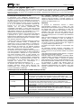
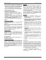
L'interprétation des résultats en fonction de la valeur du
test est la suivante :
Les résultats comportant des valeurs de test inférieures
au seuil indiquent que le patient ne présente pas
d'anticorps anti-H. pylori détectables.
Les valeurs attendues du standard sont également
mémorisées par l'intermédiaire de la carte MLE. Si les
valeurs du standard s'écartent des valeurs attendues,
elles seront signalées sur la feuille de résultats. Le
système ne calculera pas la valeur test (VT) ni la RFV
pour les patients et les contrôles.
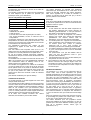
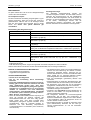
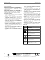
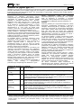
Seuil et interprétation des résultats
Valeur du test
Interprétation
TV < 0,75
Négatif
0,75 ≤ TV < 1,00
Equivoque
TV ≥ 1,00
Positif
Remarque
Il est de la responsabilité de l’utilisateur de s’assurer que
le contrôle de qualité est mis en œuvre conformément à la
législation locale en vigueur.
LIMITES DU TEST
1. Le test VIDAS HPY doit être utilisé uniquement sur
des patients présentant des signes cliniques de
maladies gastroduodénales. Il n'est pas destiné à
être utilisé sur des patients asymptomatiques.
2. Comme pour tout test de diagnostic, les résultats
obtenus avec VIDAS HPY doivent être interprétés
parallèlement aux résutats d’autres tests de
laboratoires, et aux données cliniques disponibles.
3. Un résultat positif HPY n'apporte aucune distinction
entre une infection et une colonisation avec H. pylori.
4. Un résultat positif HPY indique la présence
d'anticorps IgG H. pylori, mais n’indique pas
forcément qu’il existe une maladie gastroduodénale.
5. Un résultat négatif HPY indique soit l’absence d’IgG
H. pylori, soit leur présence à un taux non détectable
par le test HPY.
6. Les performances n’ont pas été établies dans le
cadre d’un suivi de traitement contre H. pylori
(thérapie antimicrobienne).
7. Les performances du test n'ont pas été établies pour
des patients de moins de 18 ans.
8. Une interférence peut être rencontrée avec certains
sérums contenant des anticorps dirigés contre des
composants du réactif, c’est pourquoi les résultats de
ce test doivent être interprétés en tenant compte du
contexte clinique.
Sont imprimés :
- le type de test,
- l'identification du patient,
- la date et l'heure,
- le numéro de lot et la date de péremption du coffret,
- pour chaque échantillon, la RFV, la valeur du test et
l'interprétation.
L’imprécision inhérente à toute méthode rend incertaine
l’interprétation des valeurs tests proches des seuils ; c’est
pourquoi une zone équivoque a été établie sur la base de
la connaissance de cette imprécision.
Les échantillons présentant des valeurs de test
supérieures ou égales au seuil haut sont interprétés
comme positifs.
Pour des valeurs seuil comprises entre 0,75 et 1,00,
refaire le test avec le prélèvement initial si possible. Sinon
procéder à un nouveau prélèvement et répéter le test.
En cas de nouveaux résultats équivoques, l'utilisation
d'informations cliniques et autres tests de laboratoire doit
être envisagée.
Les résultats sont non valides si la lecture du bruit de fond
(BDF) est au-dessus d’un seuil prédéterminé (indiquant
une contamination faible du substrat). Dans ce cas,
répéter l’essai avec le prélèvement initial.
Les résultats sont également non valides si le standard
n’a pas été validé pour le numéro de lot de la cartouche
test du patient. Dans ce cas, tester un standard en double
sur des cartouches HPY présentant le même numéro de
lot que le test patient non validé. Le résultat du test
patient pourra alors être recalculé à l'aide du standard
mémorisé.
(Voir Manuel d'Utilisation pour plus de détails).
VALEURS ATTENDUES
Les infections à H. pylori ont été observées dans le
monde entier, mais il existe cependant une grande
disparité géographique des prévalences. Elle est toujours
plus élevée dans les pays en développement (70 à 90%)
que dans les pays industrialisés (20 à 30%). Les
prévalences élevées étant associées à de faible niveaux
socio-économiques. Dans la population caucasienne
(Etats Unis et autres pays industrialisés), les infections à
H. pylori sont très peu fréquentes chez les enfants. La
prévalence augmente ensuite de 0,5 à 2% par année de
vie. Elle atteint 50 % chez les 60 ans et plus. La
prévalence semble être supérieure dans la population
noire et hispanique (13,14). Chez des patients présentant
des ulcères duodénaux, la fréquence des infections à
H. pylori est d'environ 80% pour toutes les tranches d'âge
(15).
Dans une population tout venant (200 donneurs de sang),
le pourcentage de positifs avec VIDAS HPY a été de
27,5% avec un taux d'équivoques de 5,5%.
Les valeurs attendues pour une population donnée
doivent être établies par chaque laboratoire. Le taux de
positivité peut varier selon différents facteurs (pays, âge,
sexe de la population étudiée, saison, type de
prélèvement, etc…).
CONTROLE DE QUALITE
Un contrôle positif et un contrôle négatif sont inclus dans
chaque coffret VIDAS HPY.
Ces contrôles doivent être utilisés à l'ouverture de chaque
nouveau coffret afin de vérifier l'absence d'altération des
réactifs. Chaque calibration doit être également vérifiée à
l'aide de ces contrôles. Pour que l'instrument puisse
vérifier la valeur des contrôles, il faut les identifier par C1
et C2. Le contrôle positif et le contrôle négatif doivent être
analysés selon les bonnes pratiques de laboratoire.
Si la valeur des contrôles s'écartent des valeurs
attendues, les résultats ne peuvent être validés.
bioMérieux
®
sa
Français - 4
VIDAS® H. pylori IgG
10988 D - FR - 2004/09
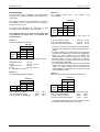
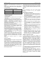
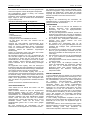
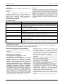
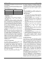
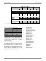
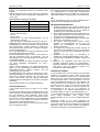
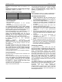
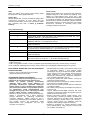
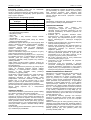
PERFORMANCES
Un total de 247 sérums congelés et 100 sérums frais a
été utilisé pour évaluer la sensibilité et la spécificité de
VIDAS HPY.
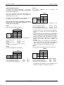
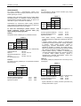
Tableau 3
247 sérums testés
commercialisée.
Ces mêmes 247 sérums congelés et 100 sérums frais
ont également été testés avec une méthode EIA
commercialisée.
Culture
VIDAS
9
E
0
1
+
-
+
118
11**
VIDAS
E***
1
1
HPY
-
2*
114
% de concordance positifs
% de concordance négatifs
% de concordance des résultats
Tableau 1
103
Sensibilité clinique
98,10%
IC 95%
93,12 % - 99,77 %
Spécificité clinique
90,82%
IC 95%
83,28% - 95,71%
IC : Intervalle de confiance
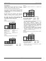
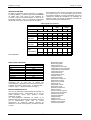
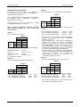
Tableau 2
43 sérums déterminés par histologie
Tableau 4
EIA
Histologie
VIDAS
+
17
0
HPY
-
0
26
% de concordance positifs
% de concordance négatifs
% de concordance des résultats
98,3%
91,2%
94,7%
Tableau 4
100 sérums frais testés avec une autre méthode EIA
Tableau 2
-
118/120
114/125
232/245
* 2 sérums positifs avec la méthode EIA commercialisée
et négatifs avec VIDAS HPY ont été trouvés négatifs par
culture.
**7 sérums négatifs avec la méthode EIA commercialisée
et positifs avec VIDAS HPY ont été trouvés négatifs par
culture. Un sérum négatif avec la méthode EIA
commercialisée et positif avec VIDAS HPY a été trouvé
positif par culture. 3 sérums négatifs avec la méthode
EIA commercialisée et positifs avec VIDAS HPY ont été
trouvés positifs par histologie.
***Un sérum positif avec la méthode EIA commercialisée
et équivoque avec VIDAS HPY a été trouvé négatif par
culture. Un sérum négatif avec la méthode EIA
commercialisée et équivoque avec VIDAS HPY a été
trouvé négatif par culture.
HPY
2
89
1 résultat VIDAS équivoque (non inclus dans les calculs)
+
EIA
2 résultats VIDAS équivoques (non inclus dans les
calculs)
Tableau 1
204 sérums déterminés par culture
+
méthode
EIA
43 sérums ont été testés par histologie : 26 sont
considérés comme négatifs et 17 comme positifs.
-
une
Tableau 3
204 sérums congelés ont été caractérisés par culture :
99 échantillons sont considérés comme négatifs et
105 positifs pour H. pylori.
+
avec
17/17
26/26
43/43
+
-
+
30
6
VIDAS
E
1
0
HPY
-
1
62
1 résultat VIDAS équivoque (non inclus dans les calculs)
% de concordance positifs
30/31
96,8%
% de concordance négatifs
62/68
91,2%
% de concordance des résultats
92/99
92,9%
100 %
100 %
100 %
Les résultats discordants n'ont pas été analysés à l’aide
d’une autre méthode de détection d'H. pylori.
bioMérieux
®
sa
Français - 5
VIDAS® H. pylori IgG
10988 D - FR - 2004/09
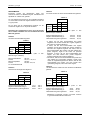
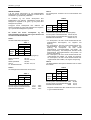
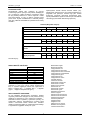
REPRODUCTIBILITE
Un panel de 6 pools d'échantillons (2 négatifs, 2 positifs
faibles et 2 positifs) a été utilisé pour la reproductibilité de
VIDAS
HPY.
Ce
panel
a
été
testé
sur
3 sites. Chaque pool a été testé 10 fois sur un instrument
pendant 3 jours. Les résultats combinés de précision
intra-série et totale sont reportés dans le Tableau 5.
La valeur test (VT) VIDAS a été utilisée. Le coefficient de
variation (%CV) n'est pas présenté pour les pools
d'échantillons négatifs, l'écart type (ET) représentant alors
la mesure de variabilité. Les résultats sont présentés
selon les normes du National Committee for Clinical
Laboratory Standards (NCCLS).
Tableau 5 (Tous les Sites)
Négatif
N
Moyenne des
VT
Positif
faible
90
90
Positif
fort
90
90
90
90
0,32
0,22
1,75
1,33
9,39
5,39
Précision
Intra-série
ET
%CV
0,02
5,8
0,01
6,3
0,07
3,9
0,06
4,7
0,25
2,7
0,20
3,8
Précision
Inter-jours
ET
%CV
0,05
16,1
0,02
11,0
0,08
4,5
0,08
6,0
0,44
4,6
0,78
14,4
Précision
totale
Inter-sites
ET
0,05
0,12
0,25
0,21
1,67
0,78
%CV
NA
NA
14,2
15,5
17,8
14,4
NA=Non applicable
REACTIONS CROISEES
F.R. +
A.N.A.+
MNI +
Grippe +
HSV +
Toxoplasmose IgG +
Syphilis +
CMV IgG +
Lupus érythémateux +
Bacteroides fragilis
Borrelia burgdorferi
Campylobacter coli
Campylobacter fetus fetus
Campylobacter fetus venerealis
Campylobacter hyointestinalis
Campylobacter jejuni
Campylobacter lari
Campylobacter rectus
Candida albicans
Citrobacter freundii
Enterobacter aerogenes
Enterobacter cloacae
Enterococcus faecalis
Escherichia coli
Helicobacter cinaedi
Helicobacter pylori
Klebsiella pneumoniae
Proteus vulgaris
Pseudomonas aeruginosa
Pseudomonas fluorescens
Salmonella minnesota
Serratia liquefaciens
Shigella flexneri
Shigella sonnei
Staphylococcus aureus
Wolinella succinogenes
Yersinia enterocolitica
VIDAS HPY
1/18
2/14
0/19
0/11
1/16
0/14
0/10
0/12
0/3
Au total 122 échantillons négatifs en anticorps
anti- H. pylori (avec une méthode EIA commercialisée)
ont été analysés : 5 résultats VIDAS HPY équivoques ont
été exclus du tableau (1 équivoque FR +, 2 équivoques
ANA +, 1 équivoque grippe +, 1 équivoque toxoplasmose
IgG +).
SPECIFICITE ANALYTIQUE
Afin de déterminer la spécificité du test, des microorganismes ont été pré-incubés dans un pool de sérums
humains anti-H. pylori positifs. Il ont été testés en triple.
Les micro-organismes testés sont listés ci-après et ont
présentés des résultats conformes avec le test
VIDAS HPY : les sérums séropositifs sont restés positifs
après absorption avec chaque micro-organisme, excepté
la souche d'H. pylori.
bioMérieux
®
sa
Français - 6
VIDAS® H. pylori IgG
10988 D - FR - 2004/09
ELIMINATION DES DECHETS
Eliminer les réactifs utilisés ou non utilisés ainsi que les
matériels à usage unique contaminés en suivant les
procédures relatives aux produits infectieux ou
potentiellement infectieux.
Il incombe à chaque laboratoire de gérer les déchets et
les effluents qu'il produit selon leur nature et leur
dangerosité, et d'en assurer (ou faire assurer) le
traitement et l'élimination selon les réglementations
applicables.
9. TALLEY, N.J. et al. 1991. Serodiagnosis of Helicobacter
pylori: Comparison of enzyme-linked immunosorbent assays.
J. Clin. Microbiol. 29:1635-1639.
10. U.S. Department of Heath and Human Services. 1999.
th
Biosafety in Microbiological and Biomedical Laboratories, 4
ed. HHS Publication (CDC) 1999. Government Printing
Office, Washington D.C.
11. U.S. Department of Labor, Occupational Safety and Health
Administration, 29CFR Part 1910.1030, Occupational
Exposure to Blood borne Pathogens.
1. BUCK, G.E. 1990. Campylobacter pylori and gastroduodenal
disease. Clin. Microbiol. Rev. 3:1-12.
12. National Committee for Clinical Laboratory Standards. 1997.
Approved Standard, M29-A. Protection of Laboratory
Workers from Instrument Biohazard and /Infectious Disease
Transmitted by Blood, Body Fluids, and Tissue. NCCLS,
Wayne Pa.
2. MARSHALL, B.J. and J.R. WARREN. 1984. Unidentified
curved bacilli in the stomach of patients with gastritis and
peptic ulceration. Lancet I:1311-1314.
13. DOOLEY, C.P. et al. 1989. Prevalence of Helicobacter pylori
infection and histologic gastritis in asymptomatic persons.
New England J. Med. 321:1562-1566.
3. PETERSON, W.L. 1991. Helicobacter pylori and peptic ulcer
disease. New England J. Med. 324(15):1043-1048.
14. GRAHAM, D.Y. et al. 1991. Epidemiology of Helicobacter
pylori in an asymptomatic population in the United States.
Effect of age, race, and socioeconomic status.
Gastroenterology. 100:1495-1501.
REFERENCES BIBLIOGRAPHIQUES
4. BLASER, M.J. 1992. Hypothesis on the Pathogenesis and
Natural History of Helicobacter pylori-Induced Inflammation.
Gastroenterology. 102:720-727.
5. TAYLOR, D.N. and M.J. BLASER. 1991. Epidemiology of
Helicobacter pylori infection. Epidemiol. Rev. 13:42-59.
6. MARSHALL. B.J. et al. 1988. Carbon-14 urea breath test for
diagnosis of Campylobacter pylori associated gastritis. J.
Nucl. Med. 29:11-16.
7. WILCOX, M.H. et al. 1996. Accuracy of serology for the
diagnosis of Helicobacter pylori infection - a comparison of
eight kits. J. Clin. Pathol. 49:373-376.
8. GRAHAM, D.Y. et al. 1987. Campylobacter pylori detected
non-invasively by the C-13 urea breath test. Lancet 23:11741177.
15. LOFFELD, R.J., et al. 1991. The prevalence of antiHelicobacter (Campylobacter) pylori antibodies in patients
and healthy blood donors. J. Med. Microbiol. 32:105-109.
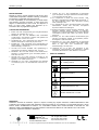
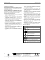
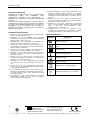
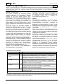
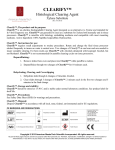
TABLE DES SYMBOLES
Signification
Symbole
ou
REF
Référence du catalogue
Dispositif médical de diagnostic in vitro
Fabricant
Limites de température
Utiliser jusque
Code du lot
Consulter les instructions d'utilisation
Contenu suffisant pour "n" tests
bioMérieux® sa
au capital de 11 879 045 €
673 620 399 RCS LYON
69280 Marcy-l'Etoile / France
Tél. 33 (0)4 78 87 20 00
Fax 33 (0)4 78 87 20 90
http://www.biomerieux.com
Imprimé en France
Le logo est une marque déposée et protégée qui est la propriété exclusive de bioMérieux sa ou de l’une de ses filiales.
REF 30 192
VIDAS® H. pylori IgG (HPY)
10988 D - GB - 2004/09
IVD
®
VIDAS H. pylori IgG (HPY) is an automated qualitative test for use on the VIDAS instruments, for the detection of antiHelicobacter pylori IgG antibodies in human serum or plasma (EDTA) using the ELFA technique (Enzyme Linked
Fluorescent Assay). The VIDAS HPY assay is intended as an aid in diagnosis of H. pylori infection in an adult
symptomatic population.
SUMMARY AND EXPLANATION
It has been shown that infection with Helicobacter
(formerly Campylobacter) pylori, a spiral shaped
microaerophilic bacillus, leads to inflammation of the
stomach mucosa (gastritis) and in some infected persons,
ulcers. The infection may also play a role in the
development of stomach cancer (1, 2, 3). Symptomatic
patients with H. pylori are considered infected, while
asymptomatic people with H. pylori are considered
colonized. Most people who are colonized with H. pylori
never develop ulceration and remain asymptomatic
despite colonization for years, probably even decades
(4, 5).
There are invasive and non-invasive methods for
determining presence of H. pylori. Biopsy by endoscopy
has traditionally been used to obtain gastric or duodenal
tissue specimens for subsequent stain, culture, and/or
direct urease detection.
With biopsy, false negative results can occur in infected
individuals due to non-uniform distribution of H. pylori in
the sample or by obtaining tissue with non-viable or nonurease producing H. pylori (6). Also, invasive methods
such as endoscopy involve patient discomfort, risk, and
are costly to perform.
Non-invasive methods include urea breath tests and
serological methods. Urea breath tests detect H. pylori
presence via its highly active urease. Urea labeled with
carbon-14 or carbon-13 is ingested by the patient, and
presence of exhaled carbon dioxide is determined via
scintillation or mass spectrometry. Patient exposure to
radioisotopes and expensive equipment are drawbacks to
urea breath testing (6, 7, 8).
Patients infected with H. pylori develop serum antibodies,
which are correlated with the presence of histologically
confirmed H. pylori infection.
Serological methods (such as enzyme immunoassays)
are non-invasive, inexpensive, quick, easy to perform, and
compared to invasive methods, the major advantage is
that serological methods do not rely on the accuracy of
the sampling (7, 9).
PRINCIPLE
The assay principle combines a 2-step enzyme
immunoassay sandwich method with a final fluorescent
detection (ELFA). All of the assay steps as well as the
assay temperature are controlled automatically by the
instrument. The Solid Phase Receptacle (SPR) serves as
the solid phase as well as the pipetting device for the
assay. Reagents for the assay are ready-to-use and predispensed in the sealed reagent strips.
After preliminary wash and sample dilution steps, the
sample is cycled in and out of the SPR for a specified
length of time. IgG antibodies to H. pylori present in the
specimen will bind to the H. pylori antigen coating the
interior of the SPR. Unbound sample components are
washed away.
Anti-human IgG antibodies conjugated with alkaline
phosphatase are cycled in and out of the SPR and will
attach to any human IgG bound to the SPR wall. A final
wash step removes unbound anti-human antibody
conjugate.
During the final detection step, the substrate (4-Methylumbelliferyl phosphate) is cycled in and out of the SPR.
The conjugate enzyme catalyzes the hydrolysis of this
substrate into a fluorescent product (4-Methylumbelliferone) the fluorescence of which is measured at
450 nm. The intensity of fluorescence is measured by the
optical scanner in the VIDAS instrument. At the end of the
assay, results are automatically calculated by the
instrument, a test value is generated and a report is
printed for each sample.
CONTENT OF THE KIT (30 TESTS) :
30 HPY strips
STR Ready-to-use.
30 HPY SPRs (1 x 30)
SPR Ready-to-use. Interior of SPRs coated with purified H. pylori antigen.
S1 Ready-to-use.
Human* serum containing anti-H. pylori antibodies + 1 g/l sodium azide.
The range is indicated in "Relative Fluorescence Value" (RFV) on the MLE card
after the following mention : "Standard (S1) RFV Range".
C1 Ready-to-use.
Human* serum containing anti-H. pylori antibodies + 1 g/l sodium azide.
The "Test Value" (TV) range is indicated on the MLE card after the following
mention : "Control C1 Test Value Range".
C2 Ready-to-use.
Human* serum containing no anti-H. pylori antibodies + 1 g/l sodium azide.
HPY Standard
(1 x 2 ml)
HPY Positive control
(1 x 1.5 ml)
HPY Negative control
(1 x 1.5 ml)
The "Test Value" (TV) range is indicated on the MLE card after the following
mention : "Control C2 Test Value Range".
1 MLE card
Specifications sheet containing the factory master calibration data required to
calibrate the test.
1 Package insert
* This product has been tested and shown to be negative for HBs surface antigen, antibodies to HIV1, HIV2 and HCV. However, since
no existing test method can totally guarantee their absence, this product must be treated as potentially infectious. Therefore, usual
safety procedures should be observed when handling.
bioMérieux
®
sa
English - 1
VIDAS® H. pylori IgG
10988 D - GB - 2004/09
DESCRIPTION
Each VIDAS HPY test requires one HPY Reagent Strip
and one HPY SPR.
The strip
The strip consists of 10 wells covered with a labeled, foil
seal. The label comprises a bar code which mainly
indicates the assay code, kit lot number and expiration
date. The foil of the first well is perforated to facilitate the
introduction of the sample. The last well of each strip is a
cuvette in which the fluorometric reading is performed.
The wells in the center section of the strip contain the
various reagents required for the assay.
The SPR
The interior of the SPR is coated during production with
purified H. pylori antigens. Each SPR is identified by the
HPY code. Only remove the required number of SPRs
from the pouch and reseal the pouch correctly after
opening.
Description of the strip
Wells
Reagents
1
Sample Well.
2
Sample Diluent: TRIS buffered saline (0.05 mol/l, pH 7.4) + protein stabilizers + 1 g/l
sodium azide (600 µl).
3
Pre-wash buffer: TRIS buffered saline (0.05 mol/l, pH 7.4) + protein stabilizers +
1 g/l sodium azide (400 µl).
4-5-7
Wash buffer: TRIS buffered saline (0.05 mol/l) + detergent + 1g/l sodium azide
(600 µl).
6
Conjugate: a titered mixture of alkaline phosphatase labeled mouse monoclonal
anti-human IgG + 1 g/l sodium azide (400 µl).
8
Wash buffer: DEA buffer (360 mmol/l) + 1g/l sodium azide (600 µl).
9
Sample diluent: TRIS buffer (0.05 mol/l, pH 7.4) + protein stabilizers + 1 g/l sodium
azide (300 µl).
10
Cuvette with substrate: 4-Methyl-umbelliferyl phosphate (0.6 mmol/l) +
diethanolamine DEA* (0.62 mol/l or 6.6%) pH 9.2 + 1 g/l sodium azide
(300 µl).
* IRRITANT reagent:
- R 36 : Irritating to eyes.
S 26 : In case of contact with eyes, rinse immediately with plenty of water and seek medical advice.
For further information, refer to the Safety Data Sheet available on request.
• Consider all patient specimens potentially infectious and
observe routine biosafety precautions. Dispose of all
used components and other contaminated materials by
acceptable procedures for potentially biohazardous
human blood products (10-12).
• Do not use the SPRs if the pouch is pierced.
• Do not use visibly deteriorated STRs (damaged foil or
plastic).
• Do not use reagents after the expiration date indicated
on the label.
• Do not mix reagents (or disposables) from different lots.
• Use powderless gloves, as powder has been reported
to cause false results for certain enzyme immunoassay
tests.
• Kit reagents contain sodium azide which can react with
lead or copper plumbing to form explosive metal azides.
If any liquid containing sodium azide is disposed of in
the plumbing system, drains should be flushed with
water to avoid build-up.
• The substrate in well 10 contains an irritant agent (6.6%
diethanolamine). Refer to the risk phrase “R” and the
precautions “S” above.
MATERIAL REQUIRED BUT NOT PROVIDED
- Pipette with disposable tip calibrated to dispense 100 µl.
- Powderless, disposable gloves.
WARNINGS AND PRECAUTIONS
• For in vitro diagnostic use only.
• For professional use only.
• This kit contains products of human origin. No
known analysis method can totally guarantee the
absence of transmissible pathogenic agents. It is
therefore recommended that these products be
treated as potentially infectious and handled
observing the usual safety precautions (see
Laboratory biosafety manual - WHO - Geneva - latest
edition).
• This kit contains products of animal origin. Certified
knowledge of the origin and/or sanitary state of the
animals does not totally guarantee the absence of
transmissible pathogenic agents. It is therefore
recommended that these products be treated as
potentially infectious and handled observing the usual
safety precautions (do not ingest or inhale).
bioMérieux
®
sa
English - 2
VIDAS® H. pylori IgG
10988 D - GB - 2004/09
• Spills should be wiped up thoroughly after treatment
with liquid detergent and a solution of household bleach
containing at least 0.5% sodium hypochlorite. See the
Operator’s Manual for cleaning spills on or in the
instrument. Do not autoclave solutions containing
bleach.
• The VIDAS and mini VIDAS instruments should be
regularly cleaned and decontaminated (see the
Operator’s Manual).
Calibration
Calibration, using the standard provided in the kit, must be
performed each time a new lot of reagents is opened,
after the master lot data have been entered. Calibration
should then be performed every 14 days. This operation
provides instrument-specific calibration curves and
compensates for possible minor variations in assay signal
throughout the shelf-life of the kit.
The standard, identified by S1, must be tested in
duplicate (see Operator’s Manual). The standard value
must be within the set RFV "Relative Fluorescence Value"
range. If this is not the case, recalibrate.
STORAGE CONDITIONS
• Store the VIDAS HPY kit at 2-8°C.
• Do not freeze reagents.
• Store all unused reagents at 2-8°C.
• After opening the kit, check that the SPR pouch is
correctly sealed and undamaged. If not, do not use the
SPRs.
• Carefully reseal the pouch with the desiccant inside
after use to maintain stability of the SPRs and return
the complete kit to 2-8°C.
• If stored according to the recommended conditions, all
components are stable until the expiration date indicated
on the label.
Procedure
1. Only remove the required reagents from the
refrigerator and allow them to come to room
temperature for at least 30 minutes.
2. Use one “HPY” strip and one “HPY” SPR for each
sample, control or standard to be tested. Make sure
the storage pouch has been resealed after the
required SPRs have been removed.
3. Type or select "HPY" to enter the test code. The
standard must be identified by "S1", and tested in
duplicate. If the positive control is to be tested, it
should be identified by "C1". If the negative control
needs to be tested, it should be identified by “C2”.
4. Mix the standard, controls and samples using a
Vortex-type mixer.
5. Pipette 100 µl of standard, sample or control into the
sample well.
(NOTE: Check the sample wells for bubbles after
pipetting and tap gently to remove any present.).
6. Insert the SPRs and strips into the instrument. Check
to make sure the color labels with the assay code on
the SPRs and the Reagent Strips match.
7. Initiate the assay as directed in the Operator’s Manual.
All the assay steps are performed automatically by the
instrument. The assay will be completed within
approximately 35 minutes.
8. After the assay is completed, remove the SPRs and
strips from the instrument.
9. Dispose of the used SPRs and strips into an
appropriate receptacle.
SPECIMENS
Specimen type and collection:
VIDAS HPY test should be performed on sera or plasma
(EDTA), that should not be hemolyzed or contaminated.
Do not heat the serum. Samples containing particulate
matter should be clarified by centrifugation or filtration
prior to testing.
Although data indicated no interference to hemoglobin
(500 mg/dl), lipids (2.0 mg/ml), or bilirubin (30 mg/dl), use
of hemolyzed, icteric, or lipemic specimens is not
recommended. If possible, a new specimen should be
collected.
Specimen stability
If a specimen is not tested on the day of collection, it can
be stored at 2-8°C for up to 5 days; if longer storage is
required, freeze at -25 ± 6°C for up to 2 months. Do not
exceed two freezing and thawing cycles. Do not test
specimens with obvious microbial contamination.
INSTRUCTIONS FOR USE
For complete instructions, see
mini VIDAS Operator’s Manual
the
VIDAS
or
RESULTS
Once the assay is completed, results are analyzed
automatically by the computer. Fluorescence is measured
twice in the Reagent Strip’s reading cuvette for each
sample tested. The first reading is a background reading
of the substrate cuvette before the SPR is introduced into
the substrate. The second reading is taken after
incubating the substrate with the enzyme remaining on
the interior of the SPR. The RFV (Relative Fluorescence
Value) is calculated by subtracting the background
reading from the final result.
TV= Test Value = patient RFV / standard RFV
The Test Value is then compared to a threshold stored by
the instrument and a final result is interpreted.
Master lot data entry
Before each new lot of reagents is used, specifications (or
factory master calibration curve data) must be entered
into the instrument (VIDAS or mini VIDAS) using the
master lot entry (MLE) card (specifications sheet) included
in each kit. If this operation is not performed before
initiating the tests, the instrument will not be able to print
results. The master lot data need only be entered once for
each lot.
It is possible to enter data automatically using the MLE
card or manually.
bioMérieux
®
sa
English - 3
VIDAS® H. pylori IgG
10988 D - GB - 2004/09
Interpretation of test results based on Test Value is as
follows:
Results with test values less than the lower threshold
indicate that the patient does not have detectable antiH. pylori antibodies.
The expected value range for the kit standard is entered
into the VIDAS system via the MLE card. Out-of-range
standard values will be flagged as invalid and the system
is unable to calculate control and patient RFV and Test
Value (TV).
Thresholds and Interpretation of Results
Note
It is the responsibility of the user to perform Quality
Control in accordance with any local applicable
regulations.
Test Value Threshold
Interpretation
TV < 0.75
Negative
0.75 ≤ TV < 1.00
Equivocal
TV ≥ 1.00
Positive
LIMITATIONS OF THE METHOD
1. The VIDAS HPY assay should be used only to
evaluate patients with clinical signs and symptoms of
gastroduodenal disease and is not intended for use
with asymptomatic patients.
2. As with any diagnostic test, results from the VIDAS
HPY IgG test should be interpreted in conjunction
with other laboratory and clinical data available to the
clinician.
3. A positive HPY assay result does not distinguish
between active infection and colonization with H.
pylori.
4. A positive HPY assay result only indicates presence
of IgG antibodies to H. pylori and does not
necessarily indicate that gastroduodenal disease is
present.
5. A negative HPY assay result indicates either that IgG
antibodies to H. pylori are not present or that they are
at a level not detectable by the HPY assay.
6. Performance has not been demonstrated for
monitoring the effects of antimicrobial therapy for
treatment of H. pylori.
7. The assay has not been established for patients
under 18 years of age.
8. Interference may be encountered with certain sera
containing antibodies directed against reagent
components. For this reason, assay results should be
interpreted taking into consideration the patient's
history, and the results of any other tests performed.
A report is printed which records:
- the type of test performed,
- the sample identification,
- the date and time,
- the lot number and expiration date of the kit reagent
being used,
- each sample’s RFV, test value and interpreted result.
The imprecision inherent in any method implies a lack of
confidence in samples with test values very close to the
thresholds.
Consequently, an equivocal zone is
established between the thresholds based on a statistical
understanding of this imprecision.
Samples with test values that are greater than or equal to
the high threshold are reported as positive.
Samples with test values between 0.75 and 1.00 should
be repeated with the original specimen, if available. If the
original specimen is not available, obtain a fresh
specimen and repeat the assay.
If the sample repeats as an equivocal, clinical information
and other available laboratory tests should be considered.
Invalid results are reported when the background reading
is above a predetermined cutoff (indicating low-level
substrate contamination). In this case, repeat the assay
with the original specimen.
An invalid result is also seen if there is no standard
available for the lot number of the patient test strip. In this
case, run a standard in duplicate in HPY strips with the
same lot number as the invalid patient test. The patient
test result can then be recalculated using the new stored
standard.
(Refer to the Operator's Manual for a detailed
explanation).
RANGE OF EXPECTED VALUES
H. pylori infection has been found worldwide, but
geographical distribution of prevalence varies widely. It is
always higher in developing countries (70 - 90%) than in
industrialized countries (20 - 30%), higher prevalence
being associated with low socio-economic levels. In
Caucasian populations in the United States and other
industrialized countries, H. pylori infection is infrequent in
childhood but with each year of age the prevalence
increases 0.5 - 2%, reaching about 50% in those who are
60 or older. Prevalence rates appear to be higher in
blacks and Hispanics than in whites (13, 14). The
frequency of H. pylori infection in patients diagnosed with
duodenal ulcers is approximately 80% in all age groups
(15).
QUALITY CONTROL
One positive control and one negative control are included
in each VIDAS HPY kit.
These controls must be performed immediately after
opening a new kit to ensure that reagent performance has
not been altered. Each calibration must also be checked
using these controls. The instrument will only be able to
check the control values if they are identified by C1 and
C2. The positive and negative controls must be tested
following Good Laboratory Practices.
Results cannot be validated if the control values deviate
from the expected values.
bioMérieux
®
In a random population of 200 apparently healthy blood
donors tested using VIDAS HPY, the positive rate was
27.5% with an equivocal rate of 5.5%.
Expected values for a given population should be
determined for each laboratory. The positivity rate for any
test may vary depending upon factors such as
geographical location, age, sex of population studied,
season of year, specimen collection and handling
procedures, etc.
sa
English - 4
VIDAS® H. pylori IgG
10988 D - GB - 2004/09
Table 3
247 Serum samples evaluated by a competitor EIA
method.
PERFORMANCE
A total of 247 frozen serum specimens and 100 freshly
collected serum samples were used to evaluate
VIDAS HPY sensitivity and specificity performance.
Table 3
Two hundred and four frozen serum specimens were well
characterized by culture. Ninety-nine specimens were
considered negative for H. pylori and 105 specimens were
considered positive.
EIA
Forty-three frozen serum specimens were defined by
histology. Twenty-six specimens were considered
negative and 17 specimens were considered positive.
Culture
VIDAS
9
E
0
1
HPY
2
89
1 VIDAS equivocal (not included in calculations)
Clinical Sensitivity
95% CI
Clinical Specificity
95% CI
CI: Confidence Interval
118
11**
VIDAS
E***
1
1
HPY
-
2*
114
98.10%
93.12% - 99,77%
90.82%
83.28% - 95.71%
Table 4
EIA
Table 2
Histology
-
VIDAS
+
17
0
HPY
-
0
26
Percent Positive Agreement
Percent Negative Agreement
Concordance of Results
98.3%
91.2%
94.7%
Table 4
100 Freshly collected serum samples defined by another
EIA method
Table 2
43 Histology stain defined serum samples
+
118/120
114/125
232/245
* Two serum specimens, competitor EIA positive/VIDAS
HPY negative, were negative by culture.
**Seven
serum
specimens,
competitor
EIA
negative/VIDAS HPY positive, were negative by culture.
One serum specimen, competitor EIA negative/VIDAS
HPY positive, was positive by culture. Three serum
specimens, competitor EIA negative/VIDAS HPY
positive, were positive by histology.
***One serum specimen, competitor EIA positive/VIDAS
HPY equivocal, was negative by culture. One serum
specimen, competitor EIA negative/VIDAS HPY
equivocal, was negative by culture.
Table 1
+
+
Percent Positive Agreement
Percent Negative Agreement
Concordance of Results
Table 1
204 Culture defined serum samples
103
-
2 VIDAS equivocals (not included in calculations)
The same 247 frozen serum specimens and
100 freshly collected serum specimens were also
evaluated by a competitor EIA method.
+
+
+
-
+
30
6
VIDAS
E
1
0
HPY
-
1
62
1 VIDAS equivocal (not included in calculations)
Percent Positive Agreement
30/31 96.8%
Percent Negative Agreement
62/68 91.2%
Concordance of Results
92/99 92.9%
17/17
26/26
43/43
Discrepant results were not reevaluated by another
H. pylori detection method.
100%
100%
100%
bioMérieux
®
sa
English - 5
VIDAS® H. pylori IgG
10988 D - GB - 2004/09
REPRODUCIBILITY
VIDAS HPY reproducibility was demonstrated using a
6-member panel of pooled specimens, consisting of
2 negative, 2 low positive and 2 positive pools. This panel
was run at three sites. Each pool was run 10 times in one
instrument run over three days. The results of combined
within run and total imprecision are shown in Table 5.
The VIDAS test value (TV) was used. Coefficient of
Variation (%CV) is not presented for the negative pools,
instead the Standard Deviation (SD) is presented as the
measure of variability. Results are presented according to
the National Committee for Clinical Laboratory Standards
(NCCLS).
Table 5 (All Sites)
Negative
N
Low positive
High positive
90
90
90
90
90
90
0.32
0.22
1.75
1.33
9.39
5.39
SD
0.02
0.01
0.07
0.06
0.25
0.20
%CV
5.8
6.3
3.9
4.7
2.7
3.8
Between
SD
0.05
0.02
0.08
0.08
0.44
0.78
Day
%CV
16.1
11.0
4.5
6.0
4.6
14.4
Total imprecision
SD
0.05
0.12
0.25
0.21
1.67
0.78
Between sites
%CV
NA
NA
14.2
15.5
17.8
14.4
Mean TV
With-in run
NA=Not applicable
CROSS REACTIVITY
R.F. +
A.N.A.+
MNI +
Influenza +
HSV +
Toxoplasmosis IgG +
Syphilis +
CMV IgG +
Lupus erythematosus +
Bacteroides fragilis
Borrelia burgdorferi
Campylobacter coli
Campylobacter fetus fetus
Campylobacter fetus venerealis
Campylobacter hyointestinalis
Campylobacter jejuni
Campylobacter lari
Campylobacter rectus
Candida albicans
Citrobacter freundii
Enterobacter aerogenes
Enterobacter cloacae
Enterococcus faecalis
Escherichia coli
Helicobacter cinaedi
Helicobacter pylori
Klebsiella pneumoniae
Proteus vulgaris
Pseudomonas aeruginosa
Pseudomonas fluorescens
Salmonella minnesota
Serratia liquefaciens
Shigella flexneri
Shigella sonnei
Staphylococcus aureus
Wolinella succinogenes
Yersinia enterocolitica
VIDAS HPY
1/18
2/14
0/19
0/11
1/16
0/14
0/10
0/12
0/3
A total of 122 samples negative for anti- H. pylori
antibodies (using a competitor EIA method) were tested:
5 equivocal VIDAS HPY results were excluded from the
table (1 equivocal RF +, 2 equivocal ANA +, 1 equivocal
influenza +, 1 equivocal toxoplasmosis IgG +).
ASSAY SPECIFICITY
To test for assay specificity, test organisms were preincubated in a pool of human anti-H. pylori positive serum
and tested in triplicate.
The tested organisms are listed below and showed no
unexpected results in the VIDAS HPY assay. The
seropositive serum remained positive after absorption with
every organism except H. pylori strain.
bioMérieux
®
sa
English - 6
VIDAS® H. pylori IgG
10988 D - GB - 2004/09
WASTE DISPOSAL
Dispose of used or unused reagents as well as any other
contaminated disposable materials following procedures
for infectious or potentially infectious products.
It is the responsibility of each laboratory to handle waste
and effluents produced according to their nature and
degree of hazardousness and to treat and dispose of
them (or have them treated and disposed of) in
accordance with any applicable regulations.
LITERATURE REFERENCES
1. BUCK, G.E. 1990. Campylobacter pylori and gastroduodenal
disease. Clin. Microbiol. Rev. 3:1-12.
2. MARSHALL, B.J. and J.R. WARREN. 1984. Unidentified
curved bacilli in the stomach of patients with gastritis and
peptic ulceration. Lancet I:1311-1314.
3. PETERSON, W.L. 1991. Helicobacter pylori and peptic ulcer
disease. New England J. Med. 324(15):1043-1048.
4. BLASER, M.J. 1992. Hypothesis on the Pathogenesis and
Natural History of Helicobacter pylori-Induced Inflammation.
Gastroenterology. 102:720-727.
5. TAYLOR, D.N. and M.J. BLASER. 1991. Epidemiology of
Helicobacter pylori infection. Epidemiol. Rev. 13:42-59.
6. MARSHALL. B.J. et al. 1988. Carbon-14 urea breath test for
diagnosis of Campylobacter pylori associated gastritis. J.
Nucl. Med. 29:11-16.
7. WILCOX, M.H. et al. 1996. Accuracy of serology for the
diagnosis of Helicobacter pylori infection - a comparison of
eight kits. J. Clin. Pathol. 49:373-376.
8. GRAHAM, D.Y. et al. 1987. Campylobacter pylori detected
non-invasively by the C-13 urea breath test. Lancet 23:11741177.
9. TALLEY, N.J. et al. 1991. Serodiagnosis of Helicobacter
pylori: Comparison of enzyme-linked immunosorbent assays.
J. Clin. Microbiol. 29:1635-1639.
10. U.S. Department of Heath and Human Services. 1999.
th
Biosafety in Microbiological and Biomedical Laboratories, 4
ed. HHS Publication (CDC) 1999. Government Printing
Office, Washington D.C.
11. U.S. Department of Labor, Occupational Safety and Health
Administration, 29CFR Part 1910.1030, Occupational
Exposure to Blood borne Pathogens.
12. National Committee for Clinical Laboratory Standards. 1997.
Approved Standard, M29-A. Protection of Laboratory
Workers from Instrument Biohazard and /Infectious Disease
Transmitted by Blood, Body Fluids, and Tissue. NCCLS,
Wayne Pa.
13. DOOLEY, C.P. et al. 1989. Prevalence of Helicobacter pylori
infection and histologic gastritis in asymptomatic persons.
New England J. Med. 321:1562-1566.
14. GRAHAM, D.Y. et al. 1991. Epidemiology of Helicobacter
pylori in an asymptomatic population in the United States.
Effect of age, race, and socioeconomic status.
Gastroenterology. 100:1495-1501.
15. LOFFELD, R.J., et al. 1991. The prevalence of antiHelicobacter (Campylobacter) pylori antibodies in patients
and healthy blood donors. J. Med. Microbiol. 32:105-109.
INDEX OF SYMBOLS
Meaning
Symbol
or
REF
GB : Catalogue number
US : Catalog number
In Vitro Diagnostic Medical Device
Manufacturer
Temperature limitation
Use by
Batch code
Consult Instructions for Use
Contains sufficient for <n> tests
WARRANTY
bioMérieux disclaims all warranties, express or implied, including any implied warranties of MERCHANTABILITY AND
FITNESS FOR A PARTICULAR USE. bioMérieux shall not be liable for any incidental or consequential damages. IN NO
EVENT SHALL BIOMERIEUX’S LIABLITY TO CUSTOMER UNDER ANY CLAIM EXCEED A REFUND OF THE
AMOUNT PAID TO BIOMERIEUX FOR THE PRODUCT OR SERVICE WHICH IS THE SUBJECT OF THE CLAIM.
VIDAS is a registered trademark of bioMérieux.
bioMérieux® sa
au capital de 11 879 045 €
673 620 399 RCS LYON
69280 Marcy-l'Etoile / France
Tel. 33 (0)4 78 87 20 00
Fax 33 (0)4 78 87 20 90
http://www.biomerieux.com
The logo is a registered and protected trademark of bioMérieux sa or one of its subsidiaries.
Printed in France
REF 30 192
VIDAS® H. pylori IgG (HPY)
10988 D - DE - 2004/09
IVD
VIDAS® H. pylori IgG (HPY) ist ein automatisierter Test für die VIDAS-Linie zum qualitativen Nachweis von Heliobacter
pylori IgG Antikörpern in Humanserum oder -plasma (EDTA) durch die ELFA-Technik (Enzyme Linked Fluorescent
®
Assay). VIDAS HPY dient als diagnostisches Hilfsmittel für den Nachweis von H. pylori Infektionen bei
symptomatischen Erwachsenen.
EINFÜHRUNG UND TESTERKLÄRUNG
Helicobacter pylori (früher als Campylobacter pylori
bezeichnet) ist ein mikroaerophiles, spiralförmiges
Bakterium, das Entzündungen der Magenschleimhaut
(Gastritis) und bei manchen Patienten auch Geschwüre
hervorruft. Die Infektion kann außerdem an der
Entstehung von Magenkrebs beteiligt sein (1, 2, 3).
Patienten mit einer H. pylori Symptomatik gelten als
infiziert, während man bei asymptomatischen H. pylori
Trägern von Besiedelung spricht. Die Mehrzahl der
Patienten mit H. pylori Besiedelung entwickeln nie
Geschwüre und bleiben asymptomatisch, trotz der
jahrelangen, wahrscheinlich sogar jahrzehntelangen
Besiedelung (4, 5).
Es gibt invasive und nicht invasive Methoden zum
Nachweis von H. pylori. Gewebebiopsien aus der
Magenschleimhaut oder dem Zwölffingerdarm werden
werden normalerweise endoskopisch gewonnen und
anschließend gefärbt, kultiviert und/oder auf die
Anwesenheit von Urease hin untersucht.
Serologische Methoden (wie Enzymimmunoassays) sind
nicht-invasiv, kostengünstig, schnell, einfach durchführbar
und - wichtigster Vorteil im Vergleich zu den invasiven
Methoden
unabhängig
von
der
Güte
der
Probengewinnung (7, 9).
PRINZIP
Das Testprinzip kombiniert eine immunenzymatische
Methode mit einer abschließenden Fluoreszenzmessung
(ELFA). Alle Reaktionsschritte sowie die Testtemperatur
werden automatisch vom Gerät kontrolliert. Der
Festphasenrezeptor dient gleichzeitig als Festphase und
Pipettiersystem für den Test. Die Testreagenzien befinden
sich gebrauchsfertig im Reagenzienriegel.
Nach einem ersten Wasch- und Probenverdünnungsschritt wird die Probe über einen bestimmten Zeitraum
mehrfach in den FPR aspiriert und wieder abgegeben. In
der Probe vorhandene H. pylori IgG Antikörper binden
spezifisch an das im FPR fixierte H. pylori Antigen.
Ungebundene Bestandteile werden durch Waschen
entfernt.
Mit alkalischer Phosphatase markierte Anti-humane IgG
Antikörper werden mehrfach in den FPR aspiriert und
wieder abgegeben und binden an das im FPR gebundene
humane IgG.
Ein letzter Waschschritt entfernt ungebundenes antihumanes Antikörperkonjugat.
Während des letzten Nachweisschrittes wird das Substrat
(4-Methyl-umbelliferyl-phosphat) mehrfach in den FPR
aspiriert und wieder abgegeben. Das Enzymkonjugat
katalysiert die Hydrolyse des Substrates in ein
fluoreszierendes Produkt (4-Methyl-umbelliferon), dessen
Fluoreszenz bei 450 nm gemessen wird. Die Intensität der
Fluoreszenz wird von dem optischen Scanner im VIDAS
Gerät gemessen. Nach Beendigung des Tests werden die
Ergebnisse automatisch vom VIDAS-Gerät analysiert. Für
jede Probe wird ein Testwert berechnet und ein
Ergebnisbericht ausgedruckt.
Eine ungleichmäßige Besiedelung der Schleimhaut mit
H. pylori oder die Gewinnung von Gewebeproben mit
nicht lebensfähigen oder nicht Urease-bildenden H. pylori
kann bei infizierten Personen zu falsch negativen
Ergebnissen führen (6). Invasive Methoden wie die
Endoskopie sind für den Patienten unangenehm, mit
Risiken verbunden und darüber hinaus kostenintensiv.
Zu den nicht invasiven Methoden gehören HarnstoffAtemtests und serologische Methoden. Der HarnstoffAtemtest weist die Anwesenheit von H. pylori über dessen
starke Ureaseaktivität nach. Mit C-14 oder C-13
markierter Harnstoff wird vom Patienten eingenommen,
anschließend wird über die Atemluft ausgeschiedenes
Kohlendioxid radiometrisch oder massenspektrometrisch
bestimmt. Nachteile dieser Methode sind die Exposition
des Patienten gegenüber Radioisotopen und die
erforderliche kostspielige Laborausrüstung (6, 7, 8).
Patienten, die mit H. pylori infiziert sind, bilden
Serumantikörper, die mit dem Vorliegen histologisch
bestätigter H. pylori Infektionen korrelieren.
PACKUNGSINHALT (30 TESTS):
30 HPY Reagenzienriegel
STR Gebrauchsfertig.
30 HPY FestphasenSPR Gebrauchsfertig. Mit gereinigtem H. pylori Antigen beschichtete FPR.
rezeptoren (1 x 30)
HPY Standard
S1 Humanserum*, das H. pylori Antikörper enthält + Natriumazid 1 g/l.
Der Vertrauensbereich in RFV (Relative Fluorescence Value) ist auf der MLE
(1 x 2 ml)
Karte nach dem Vermerk : "Standard (S1) RFV Range" angegeben.
HPY Positivkontrolle
C1 Gebrauchsfertig.
Humanserum*, das H. pylori Antikörper enthält + Natriumazid 1 g/l. Der Testwert(1 x 1,5 ml)
Bereich (TV) ist auf der MLE Karte nach dem Vermerk : "Control C1 Test Value
Range" angegeben.
HPY Negativkontrolle
C2 Gebrauchsfertig.
Humanserum*, das keine H. pylori Antikörper enthält + Natriumazid 1 g/l.
(1 x 1,5 ml)
Der Testwert-Bereich (TV) ist auf der MLE Karte nach dem Vermerk : "Control C2
Test Value Range" angegeben.
1 MLE Karte
Karte mit den für die Testkalibration erforderlichen Master Lot Daten.
1 Arbeitsanleitung
* Die Abwesenheit von HBs-Oberflächenantigen, HIV-1, HIV-2 und HCV-Antikörpern wurde überprüft. Da keine Testmethode die
Abwesenheit dieser Agenzien völlig gewährleisten kann, muss dieses Produkt als potenziell infektiös betrachtet und unter Beachtung
entsprechender Vorsichtsmaßnahmen sachgemäß behandelt werden.
bioMérieux
®
sa
Deutsch - 1
VIDAS® H. pylori IgG
10988 D - DE - 2004/09
BESCHREIBUNG
Für jeden VIDAS HPY Test ist ein HPY Reagenzienriegel
und ein HPY FPR erforderlich.
Der Reagenzienriegel
Der
etikettierte
Reagenzienriegel
besteht
aus
10 folienversiegelten Küvetten. Auf dem Etikett sind der
Testcode, die Chargennummer und das Verfallsdatum
des Kits im Barcode festgehalten. Die erste Küvette ist
perforiert, um das Einpipettieren der Probe zu erleichtern.
In der letzten, durchsichtigen Küvette wird die
photometrische Messung durchgeführt. Die mittleren
Küvetten beinhalten die verschiedenen Testreagenzien.
Der Festphasenrezeptor:
Der FPR wird bei der Herstellung mit gereinigtem H. pylori
Antigen beschichtet. Jeder FPR ist mit dem Code HPY
gekennzeichnet. Nehmen Sie nur die benötigte Anzahl
FPR aus dem Beutel. Nicht benötigte FPR im Beutel
lassen und diesen nach jedem Gebrauch gut
verschließen.
Beschreibung des Reagenzienriegels
Küvetten
Reagenzien
1
Probenküvette.
2
Probendiluent: TRIS Puffer (0,05 mol/l, pH 7,4) + Proteinstabilisatoren +
Natriumazid 1 g/l (600 µl).
3
Vorwaschpuffer: TRIS Puffer (0,05 mol/l, pH 7,4) + Proteinstabilisatoren +
Natriumazid 1 g/l (400 µl).
4-5-7
Waschpuffer:
1 g/l (600 µl).
TRIS
Puffer
(0,05
mol/l)
+
Detergenz
+
Natriumazid
6
Konjugat: titrierte Mischung aus monoklonalen Anti-humanen IgG Antikörpern
(Maus), die mit alkalischer Phosphatase markiert sind + Natriumazid 1 g/l (400 µl).
8
Waschpuffer: DEA Puffer (360 mmol/l) + Natriumazid 1 g/l (600 µl).
9
Probendiluent: TRIS Puffer (0,05 mol/l, pH 7,4) + Proteinstabilisatoren +
Natriumazid 1 g/l (300 µl).
10
Messküvette mit Substrat: 4-Methyl-umbelliferyl-phosphat (0,6 mmol/l) +
Diäthanolamin DEA* (0,62 mol/l bzw. 6,6 %) pH 9,2 + Natriumazid 1 g/l
(300 µl).
*REIZENDES Reagenz:
- R 36: Reizt die Augen.
- S 26: Bei Berührung mit den Augen sofort gründlich mit Wasser abspülen und Arzt konsultieren.
Weitere Informationen entnehmen Sie dem Sicherheitsdatenblatt, das auf Anfrage erhältlich ist.
• Alle Patientenproben müssen als potenziell infektiös betrachtet und unter Beachtung der üblichen Vorsichtsmaßnahmen behandelt werden. Entsorgen Sie gebrauchte Bestandteile und andere kontaminierte Materialien gemäß den für potenziell infektiöse humane
Produkte geltenden Vorsichtsmaßnahmen (10-12).
• FPR aus beschädigten (perforierten) Beuteln nicht
verwenden.
• Reagenzienriegel mit sichtbaren Beschädigungen (der
Aluminiumfolie oder des Kunststoffes) nicht verwenden.
• Die Reagenzien nach Ablauf des auf der Verpackung
angegebenen Verfallsdatums nicht mehr verwenden.
• Reagenzien (oder Verbrauchsmaterialien) aus unterschiedlichen Chargen nicht mischen.
• Verwenden Sie ungepuderte Einweg-Handschuhe, da
Puder bei einer Reihe von Enzymimmunoassays zu
falschen Ergebnissen geführt hat.
• Die Reagenzien enthalten ein Konservierungsmittel
(Natriumazid), das mit Blei- oder Kupferrohren zu
explosiven Metallaziden reagieren kann. Beim Ableiten
in die Kanalisation sollten die Reagenzien immer mit
reichlich Wasser verdünnt werden.
• Das Substrat (Küvette 10) enthält ein reizendes Agenz
(6,6% Diäthanolamin). Beachten Sie den oben genannten Gefahrenhinweis „R” und die Sicherheitsratschläge „S”.
ZUSÄTZLCH ERFORDERLICHE MATERIALIEN
- 100 µl Pipette mit Einwegspitzen.
- Ungepuderte Einweghandschuhe.
VORSICHTSMASSNAHMEN
• Nur für die in vitro Diagnostik
• Nur für die Verwendung durch Fachkundige
bestimmt.
• Dieser Kit enthält Bestandteile humanen Ursprungs.
Bislang gibt es kein bekanntes Verfahren mit dem
völlig gewährleistet werden kann, dass diese
Produkte keine übertragbaren pathogenen Agenzien
enthalten. Es ist empfehlenswert, sie als potenziell
infektiös zu betrachten und unter Beachtung
entsprechender Vorsichtsmaßnahmen sachgemäß
zu behandeln (siehe Laboratory biosafety manual WHO - Geneva - letzte Ausgabe).
• Dieser Kit enthält Bestandteile tierischen Ursprungs. Da
durch die Kontrolle der Herkunft und/oder des
Gesundheitszustandes der Tiere nicht völlig gewährleistet werden kann, dass diese Produkte keine
übertragbaren pathogenen Agenzien enthalten, ist es
empfehlenswert, diese als potenziell infektiös zu
betrachten und unter Beachtung entsprechender
Vorsichtsmaßnahmen zu behandeln (nicht einnehmen,
nicht einatmen).
bioMérieux
®
sa
Deutsch - 2
VIDAS® H. pylori IgG
10988 D - DE - 2004/09
• Verschüttete oder übergelaufene Flüssigkeiten mit
flüssigem Detergenz oder einer Haushaltsbleichlösung
mit
mindestens
0,5%
Natriumhypochlorit
zur
Inaktivierung infektiösen Materials behandeln. Bei
Verunreinigung auf oder im VIDAS-Gerät folgen Sie
bitte den Anweisungen des Handbuches. Lösungen, die
Bleichmittel enthalten, dürfen nicht in den Autoklaven
gestellt werden.
• Das VIDAS- und das mini VIDAS-Gerät regelmäßig
reinigen und dekontaminieren (siehe Benutzerhandbuch).
KALIBRATION
Für jede neue Reagenziencharge muss mit dem in der
Packung enthaltenen Standard nach der Eingabe der
chargenspezifischen Daten (MLE-Karte) eine Kalibration
durchgeführt werden. Die Rekalibration wird anschließend
14-tägig durchgeführt. Dadurch wird die Eichkurve
gerätespezifisch justiert und eventuelle lagerungsbedingte
Veränderungen des Reagenzes kompensiert.
Der mit S1 identifizierte Standard wird doppelt getestet
(siehe Benutzerhandbuch). Der Wert des Standards muss
innerhalb des festgelegten RFV-Bereiches („Relativer
Fluoreszenz Wert“) liegen. Ist dies nicht der Fall,
wiederholen Sie die Kalibration.
LAGERUNGSBEDINGUNGEN
• Den VIDAS HYP Kit bei 2-8°C lagern.
• Die Reagenzien nicht einfrieren.
• Nicht gebrauchte Reagenzien bei 2-8°C lagern.
• Überprüfen Sie nach dem Öffnen des Kits den (die)
FPR-Beutel. Beutel, die nicht korrekt verschlossen oder
die beschädigt sind, dürfen nicht verwendet werden.
• Den FPR-Beutel mit dem Trockenmittel nach jedem
Gebrauch wieder sorgfältig verschließen, um die
Haltbarkeit der FPR nicht zu beeinträchtigen und
den gesamten Kit wieder bei 2-8°C lagern.
• Unter den empfohlenen Lagerungsbedingungen sind
alle Kitbestandteile bis zu dem auf der Verpackung
angegebenen Verfallsdatum haltbar.
Testdurchführung
1. Nehmen Sie nur die erforderlichen Reagenzien aus
dem Kühlschrank und lassen Sie sie vor Gebrauch
mindestens 30 min. bei Raumtemperatur stehen.
2. Verwenden Sie für jede zu testende Probe bzw.
Kontrolle oder Standard einen „HPY“ Reagenzienriegel und einen „HPY“ FPR. Vergewissern Sie sich,
dass der Beutel mit den FPR nach jedem
Gebrauch wieder gut verschlossen wird.
3. Drücken oder wählen Sie „HPY“, um den Testcode
einzugeben. Der Standard muss unbedingt mit „S1“
identifiziert und doppelt getestet werden. Wenn die
Positivkontrolle getestet werden soll, identifizieren Sie
diese mit C1. Wenn die Negativkontrolle getestet
werden soll, identifizieren Sie diese mit C2.
4. Den Standard, die Kontrollen und die Proben auf dem
Vortex homogenisieren.
5. Pipettieren Sie 100 µl Standard, Probe oder Kontrolle
in die Probenküvette.
(Hinweis: Vergewissern Sie sich, dass nach dem
Pipettieren keine Bläschen in der Probenküvette sind.
Klopfen Sie gegebenenfalls leicht gegen den
Reagenzienriegel, um Bläschen zu entfernen).
6. Führen Sie die FPR und Reagenzienriegel in das
Gerät ein. Vergewissern Sie sich, dass die Farb- und
Buchstabencodierung auf dem FPR-Etikett mit der auf
dem Reagenzienriegel übereinstimmt.
PROBEN
Probenart und Probengewinnung:
Für den VIDAS HPY Test muss hämolysefreies und nicht
kontaminiertes Serum oder Plasma (EDTA) verwendet
werden.
Die Seren nicht erhitzen. Proben, die Partikel enthalten,
müssen vor der Testung zentrifugiert oder filtriert werden.
Obwohl keine Interferenzen mit Hämoglobin (500 mg/dl),
Lipiden (2.0 mg/ml), oder Bilirubin (30 mg/dl) festgestellt
wurden, ist der Gebrauch hämolysierter, ikterischer oder
lipämischer Proben nicht empfehlenswert. Fordern Sie in
diesem Fall falls möglich eine neue Probe an.
Haltbarkeit der Proben
Proben, die nicht am Tag der Probenentnahme getestet
werden, können bei 2-8°C maximal für 5 Tage gelagert
werden. Zur längeren Aufbewahrung frieren Sie die Probe
bei -25 ± 6°C bis zu 2 Monate ein. Nicht mehr als zweimal
Einfrieren und Auftauen. Proben mit sichtbarer
mikrobieller Verunreinigung sollten nicht getestet werden.
TESTDURCHFÜHRUNG
Ausführliche Informationen entnehmen Sie
VIDAS oder mini VIDAS Benutzerhandbuch.
7. Starten Sie den Test (siehe Benutzerhandbuch). Alle
weiteren Schritte werden automatisch vom VIDASGerät durchgeführt. Die Ergebnisse liegen nach ca.
35 min vor.
8. Nehmen Sie nach Beendigung des Tests die FPR und
Riegel aus dem Gerät.
9. Entsorgen Sie die FPR und Reagenzienriegel nach
Gebrauch in einem geeigneten Abfallbehälter.
dem
ERGEBNISSE
Nach Beendigung des Tests werden die Ergebnisse
automatisch vom VIDAS-Gerät ausgewertet. Für jede
Probe werden zwei Fluoreszenzmessungen in der
Messküvette des Reagenzienriegels durchgeführt. Die
erste Messung ist eine Hintergrundmessung der Küvette
und des Substrates, bevor der FPR in das Substrat
eintaucht. Die zweite Messung erfolgt nach der Inkubation
des Substrates mit dem im FPR vorhandenen
Enzymkonjugat. Aus der Differenz beider Messungen
ergibt sich ein relativer Fluoreszenzwert (RFV), der auf
dem Ergebnisblatt ausgedruckt wird.
Zur Berechnung des Testwertes wird der RFV der Probe
durch den RFV des Standards dividiert:
Eingabe der Master Lot Daten
Für jede neue Charge müssen vor Testbeginn die
Master Lot Daten (Master Lot Entry) mit der mitgelieferten
MLE-Karte in das Gerät (VIDAS oder mini VIDAS)
eingegeben werden, andernfalls kann das Gerät keine
Ergebnisse ausdrucken. Die Master Lot Daten werden für
jede Charge nur einmal eingegeben.
Die Dateneingabe mit der MLE-Karte kann automatisch
oder manuell durchgeführt werden.
TV = Testwert = RFV Probe/ RFV Standard
bioMérieux
®
sa
Deutsch - 3
VIDAS® H. pylori IgG
10988 D - DE - 2004/09
Die Sollwerte des Standards werden ebenfalls mit der
MLE-Karte eingegeben. Liegen die Werte des Standards
außerhalb des Sollbereichs, werden diese als ungültig
angezeigt. Das System kann den Testwert (TV) und den
RFV der Kontrollen und Proben nicht berechnen.
Der Testwert wird anschließend mit einem gespeicherten
Grenzwert verglichen und das Endergebnis interpretiert.
Die Testergebnisse werden in Bezug zum Testwert
folgendermaßen interpretiert:
Ergebnisse, deren Testwerte unterhalb des Grenzwertes
liegen, zeigen an, dass der Patient keine nachweisbaren
H.pylori Antikörper hat.
Anmerkung
Es liegt in der Verantwortung des Anwenders, die
Qualitätskontrolle in Übereinstimmung mit den jeweils
gültigen Vorschriften durchzuführen.
Grenzwert und Interpretation der Ergebnisse
Testwert
Interpretation
TV < 0,75
negativ
0,75 ≤ TV < 1,00
grenzwertig
TV ≥ 1,00
positiv
LIMITIERUNGEN
1. Der VIDAS HPY Test darf nur bei Patienten mit
klinischen
Anzeichen
einer
gastroduodenalen
Erkrankung und nicht bei asymptomatischen
Patienten verwendet werden.
2. Wie bei jeder diagnostischen Methode, müssen die
Ergebnisse des VIDAS HPY IgG Tests in Verbindung
mit den Ergebnissen anderer verfügbarer Labortests
und der Klinik des Patienten interpretiert werden.
3. Ein positives HPY Testergebnis differenziert nicht
zwischen einer aktiven Infektion und einer
Besiedelung mit H. pylori.
4. Ein positives HPY Testergebnis zeigt nur die
Anwesenheit von H. pylori IgG Antikörpern und nicht
unbedingt das Vorliegen einer gastroduodenalen
Erkrankung an.
5. Ein negatives HPY Ergebnis zeigt an, dass entweder
keine H. pylori IgG Antikörper vorhanden sind, oder
dass diese unterhalb der Nachweisgrenze des HPY
Tests liegen.
6. Die Performance des Tests für die Therapiekontrolle
von H. pylori Infektionen (antibakterielle Therapie)
wurde nicht ermittelt.
7. Die Testperformance wurde nicht für Patienten unter
18 Jahren ermittelt.
8. Bei einigen Seren, die Antikörper enthalten, die
gegen die Bestandteile des Reagenzes gerichtet
sind, kann es zu Interferenzen kommen. Die
Ergebnisse dieses Tests müssen deshalb unter
Berücksichtigung des klinischen Hintergrundes
interpretiert werden.
Der Ergebnisausdruck enthält folgende Angaben:
- Testart,
- Probenidentifikation,
- Datum und Uhrzeit,
- Chargennummer und Verfallsdatum des Kits,
- für jede Probe: den RFV, den Testwert und die
Interpretation
Aufgrund der jeder Methode innewohnenden Ungenauigkeit sollten Testwerte, die nahe am Grenzbereich
liegen, mit Vorsicht interpretiert werden; entsprechend
wurde ein Grenzbereich zwischen den Grenzwerten
geschaffen, welcher die statistische Ungenauigkeit
berücksichtigt.
Proben mit Testwerten größer oder gleich dem oberen
Grenzwert werden als positiv interpretiert.
Bei Testwerten zwischen 0,75 und 1,00 wiederholen Sie
den Test falls möglich mit der Originalprobe. Falls dies
nicht möglich ist, fordern Sie eine neue Probe an und
wiederholen Sie den Test. Ergibt sich erneut ein zweifelhaftes Ergebnis, müssen klinische Informationen und
andere Labortests mit in die Beurteilung einbezogen
werden.
Die Ergebnisse sind ungültig, wenn die Hintergrundfluoreszenz (HGF) über dem festgelegten cut-off liegt (der
eine geringe Substratkontamination anzeigt). Wiederholen
Sie in diesem Fall den Test mit der Originalprobe.
Die Ergebnisse sind ebenfalls ungültig, wenn für die
Chargennummer des Reagenzienriegels kein gespeicherter Standard vorliegt. Führen Sie in diesem Fall mit HYP
Reagenzienriegeln derselben Chargennummer eine Kalibration durch (Doppelansatz). Das Testergebnis kann
dann mit diesem neu gespeicherten Standard berechnet
werden.
(Weitere Informationen siehe Benutzerhandbuch).
ERWARTUNGSWERTE
H. pylori Infektionen kommen weltweit vor. Die Prävalenz
zeigt jedoch große geographische Schwankungen. In den
Entwicklungsländern ist sie immer höher (70-90%) als in
den Industrieländern (20-30%), da höhere Prävalenzen
mit
niedrigen
sozio-ökonomischen
Grundlagen
zusammenhängen. In der weißen Bevölkerung der
Vereinigten Staaten und anderen industrialisierten
Ländern treten bei Kindern nur sehr selten H. pylori
Infektionen auf. Die Prävalenz steigt jedoch mit jedem
Lebensjahr um 0,5 bis 2% und erreicht ca. 50% bei
Personen, die 60 Jahre oder älter sind. In der schwarzen
und hispanischen Bevölkerung ist die Prävalenz größer
(13,14). Bei Patienten mit duodenalen Geschwüren
beträgt die Häufigkeit von H. pylori Infektionen für alle
Altersklassen ca. 80% (15).
In einer zufällig ausgewählten Population (200 Blutspender), die mit dem VIDAS HPY getestet wurde, lag die
Positivrate bei 27,5% (5,5% der Ergebnisse lagen im
Graubereich).
Jedes Labor sollte für seine Patientenpopulation eigene
Sollwerte festlegen. Die Positivrate ist von verschiedenen
Faktoren abhängig (Land, Alter, Geschlecht der
untersuchten Population, Jahreszeit, Probentyp etc).
QUALITÄTSKONTROLLE
Jeder VIDAS HPY Kit enthält eine Positiv- und eine
Negativkontrolle.
Diese Kontrollen müssen für jede neu angebrochene
Packung getestet werden, um einen lieferungs- und
lagerungsbedingten Qualitätsverlust der Reagenzien
auszuschließen. Zur Qualitätssicherung müssen diese
Kontrollen außerdem nach jeder Rekalibration getestet
werden. Damit das Gerät den Wert der Kontrollen
überprüfen kann, müssen die Kontrollen mit C1 und C2
identifiziert werden. Testen Sie die Positiv- und
Negativkontrolle gemäß den in Ihrem Labor gültigen
Richtlinien.
Bei einer Abweichung der Kontrollwerte von den
Sollwerten können die Ergebnisse nicht validiert werden.
bioMérieux
®
sa
Deutsch - 4
VIDAS® H. pylori IgG
10988 D - DE - 2004/09
PERFORMANCE
Insgesamt wurden 247 tiefgefrorene Seren und
100 frische Seren zur Bestimmung der Sensitivität und
Spezifität von VIDAS HYP getestet.
Tabelle 3
247 Seren wurden mit einem kommerziellen EIA getestet.
Tabelle 3
Für 204 tiefgefrorene Seren lag ein kulturelles Ergebnis
vor: 99 Proben wurden als negativ bewertet und 105 als
H. pylori positiv.
EIA
Für 43 Seren lag ein histologisches Ergebnis vor: 26
wurden als negativ und 17 als positiv bewertet.
VIDAS
Tabelle1
VIDAS
103
9
E
0
1
HPY
2
89
1 grenzwertiges VIDAS Ergebnis
Berechnungen enthalten)
klinische Sensitivität
KI 95%
klinische Spezifität
KI 95%
KI = Konfidenzintervall
E***
1
1
(nicht
in
den
98,10%
93,12 % - 99,77 %
90,82%
83,28% - 95,71%
+
17
0
HPY
-
0
26
Positive Übereinstimmung %
Negative Übereinstimmung %
Übereinstimmung der Ergebnisse
98,3%
91,2%
94,7%
EIA
Histologie
VIDAS
den
Tabelle 4
Tabelle 2
-
118/120
114/125
232/245
in
Tabelle 4
100 frische Seren wurden mit einer anderen EIA Methode
getestet.
Tabelle 2
43 Seren mit histologischem Ergebnis
+
(nicht
* 2 Seren, die mit dem kommerziellen EIA positiv
reagierten und mit dem VIDAS HPY negativ, ergaben in
der Kultur ein negatives Ergebnis.
**7 Seren, die mit dem kommerziellen EIA negativ
reagierten und mit dem VIDAS HPY positiv, ergaben in
der Kultur ein negatives Ergebnis. Ein Serum, das mit
dem kommerziellen EIA negativ und dem VIDAS HPY
positiv reagierte, ergab in der Kultur ein positives
Ergebnis. 3 Seren, die mit dem kommerziellen EIA
negativ und dem VIDAS HPY positiv reagierten,
ergaben histologisch ein positives Ergebnis.
***Ein Serum, das mit dem kommerziellen EIA positiv und
dem VIDAS HPY grenzwertig reagierte, ergab in der
Kultur ein negatives Ergebnis. Ein Serum, das mit dem
kommerziellen EIA negativ und dem VIDAS HPY
grenzwertig reagierte, war in der Kultur negativ.
Kultur
+
11**
Positive Übereinstimmung %
Negative Übereinstimmung %
Übereinstimmung der Ergebnisse
Tabelle1
204 Seren mit kulturellem Ergebnis
-
-
118
HPY
2*
114
2 grenzwertige VIDAS Ergebnisse
Berechnungen enthalten)
Dieselben 247 tiefgefrorenen Seren und 100 frische
Seren wurden ebenfalls mit einer kommerziellen EIA
Methode getestet.
+
+
+
17/17
26/26
43/43
+
-
+
30
6
VIDAS
E
1
0
HPY
-
1
62
1 VIDAS grenzwertiges Ergebnis (nicht in den
Berechnungen berücksichtigt)
Positive Übereinstimmung %
30/31
96,8%
Negative Übereinstimmung %
62/68
91,2%
Übereinstimmung der Ergebnisse
92/99
92,9%
100 %
100 %
100 %
Diskrepante Ergebnisse wurden mit keiner anderen
H. pylori Nachweismethode untersucht.
bioMérieux
®
sa
Deutsch - 5
VIDAS® H. pylori IgG
10988 D - DE - 2004/09
REPRODUZIERBARKEIT
Die Reproduzierbarkeit des VIDAS HPY Tests wurde mit
einem Panel aus 6 Probenpools (2 negative, 2 schwach
positive und 2 positive Seren) ermittelt. Dieses Panel
wurde in 3 Labors getestet. Jeder Pool wurde 3 Tage
10
Mal
auf
einem
Gerät
getestet.
Die
zusammengefassten
Ergebnisse
der
Intra-Assay
Präzision und die Gesamtpräzision sind in Tabelle 5
angegeben.
Es wurde der VIDAS Testwert (TV) verwendet. Die
Variationskoeffizient (VK%) ist für die negativen
Probenpools nicht angegeben, dagegen wird die
Standardabweichung
(SA)
als
Variabilitätsmaß
angegeben. Die Ergebnisse werden gemäß dem National
Committee for Clinical Laboratory Standards (NCCLS)
angegeben.
Tabelle 5 (Alle Labore)
negativ
N
90
90
schwach
positiv
90
90
TV Mittelwert
0,32
0,22
1,75
1,33
9,39
5,39
SA
0,02
0,01
0,07
0,06
0,25
0,20
%VK
5,8
6,3
3,9
4,7
2,7
3,8
Präzision
Im Tagesvergleich
SA
%VK
0,05
16,1
0,02
11,0
0,08
4,5
0,08
6,0
0,44
4,6
0,78
14,4
Gesamtpräzision aller
Labore
SA
0,05
0,12
0,25
0,21
1,67
0,78
%VK
NA
NA
14,2
15,5
17,8
14,4
Intra-Assay
Präzision
stark
positiv
90
90
NA=nicht anwendbar
KREUZREAKTIONEN
RF. +
A.N.A.+
IM +
Grippe +
HSV +
Toxoplasmose IgG +
Syphilis +
CMV IgG +
Lupus erythematosus +
Bacteroides fragilis
Borrelia burgdorferi
Campylobacter coli
Campylobacter fetus fetus
Campylobacter fetus venerealis
Campylobacter hyointestinalis
Campylobacter jejuni
Campylobacter lari
Campylobacter rectus
Candida albicans
Citrobacter freundii
Enterobacter aerogenes
Enterobacter cloacae
Enterococcus faecalis
Escherichia coli
Helicobacter cinaedi
Helicobacter pylori
Klebsiella pneumoniae
Proteus vulgaris
Pseudomonas aeruginosa
Pseudomonas fluorescens
Salmonella minnesota
Serratia liquefaciens
Shigella flexneri
Shigella sonnei
Staphylococcus aureus
Wolinella succinogenes
Yersinia enterocolitica
VIDAS HPY
1/18
2/14
0/19
0/11
1/16
0/14
0/10
0/12
0/3
Insgesamt wurden 122 H. pylori Antikörper negative
Proben (mit einem kommerziellen EIA Test) getestet:
5 VIDAS HPY grenzwertige Ergebnisse wurden aus der
Tabelle genommen (1 grenzwertiges RF +, 2 grenzwertige
ANA +, 1 grenzwertiges Grippe +, 1 grenzwertiges
Toxoplasmose IgG +).
ANALYTISCHE SPEZIFITÄT
Um die Spezifität des Tests zu bestimmen, wurden
Mikroorganismen in einem Pool anti-H.pylori positiver
Humanseren
vorinkubiert
und
anschließend
in
Dreifachbestimmungen getestet.
Die getesteten Organismen sind im folgenden aufgeführt
und zeigten mit dem VIDAS HPY konforme Testergebnisse: Das seropositive Serum blieb nach der
Absorption mit jedem Keim bis auf den H. pylori-Stamm
positiv.
bioMérieux
®
sa
Deutsch - 6
VIDAS® H. pylori IgG
10988 D - DE - 2004/09
BESEITIGUNG DER ABFÄLLE
Entsorgen Sie alle gebrauchten und nicht gebrauchten
Reagenzien sowie kontaminierte Einwegmaterialien
gemäß den für infektiöse oder potenziell infektiöse
Materialien geltenden Bestimmungen.
Es liegt in der Verantwortung jedes Labors, die
entstandenen Fest- und Flüssigabfälle gemäß der
jeweiligen Risikogruppe zu behandeln und deren
Entsorgung in Übereinstimmung mit den gültigen
gesetzlichen Bestimmungen sicherzustellen.
LITERATUR
1. BUCK, G.E. 1990. Campylobacter pylori and gastroduodenal
disease. Clin. Microbiol. Rev. 3:1-12.
2. MARSHALL, B.J. and J.R. WARREN. 1984. Unidentified
curved bacilli in the stomach of patients with gastritis and
peptic ulceration. Lancet I:1311-1314.
3. PETERSON, W.L. 1991. Helicobacter pylori and peptic ulcer
disease. New England J. Med. 324(15):1043-1048.
4. BLASER, M.J. 1992. Hypothesis on the Pathogenesis and
Natural History of Helicobacter pylori-Induced Inflammation.
Gastroenterology. 102:720-727.
5. TAYLOR, D.N. and M.J. BLASER. 1991. Epidemiology of
Helicobacter pylori infection. Epidemiol. Rev. 13:42-59.
6. MARSHALL. B.J. et al. 1988. Carbon-14 urea breath test for
diagnosis of Campylobacter pylori associated gastritis. J.
Nucl. Med. 29:11-16.
7. WILCOX, M.H. et al. 1996. Accuracy of serology for the
diagnosis of Helicobacter pylori infection - a comparison of
eight kits. J. Clin. Pathol. 49:373-376.
8. GRAHAM, D.Y. et al. 1987. Campylobacter pylori detected
non-invasively by the C-13 urea breath test. Lancet 23:11741177.
9. TALLEY, N.J. et al. 1991. Serodiagnosis of Helicobacter
pylori: Comparison of enzyme-linked immunosorbent assays.
J. Clin. Microbiol. 29:1635-1639.
10. U.S. Department of Heath and Human Services. 1999.
th
Biosafety in Microbiological and Biomedical Laboratories, 4
ed. HHS Publication (CDC) 1999. Government Printing
Office, Washington D.C.
11. U.S. Department of Labor, Occupational Safety and Health
Administration, 29CFR Part 1910.1030, Occupational
Exposure to Blood borne Pathogens.
12. National Committee for Clinical Laboratory Standards. 1997.
Approved Standard, M29-A. Protection of Laboratory
Workers from Instrument Biohazard and /Infectious Disease
Transmitted by Blood, Body Fluids, and Tissue. NCCLS,
Wayne Pa.
13. DOOLEY, C.P. et al. 1989. Prevalence of Helicobacter pylori
infection and histologic gastritis in asymptomatic persons.
New England J. Med. 321:1562-1566.
14. GRAHAM, D.Y. et al. 1991. Epidemiology of Helicobacter
pylori in an asymptomatic population in the United States.
Effect of age, race, and socioeconomic status.
Gastroenterology. 100:1495-1501.
15. LOFFELD, R.J., et al. 1991. The prevalence of antiHelicobacter (Campylobacter) pylori antibodies in patients
and healthy blood donors. J. Med. Microbiol. 32:105-109.
SYMBOLE
Bedeutung
Symbol
oder
REF
Bestellnummer
In Vitro Diagnostikum
Hersteller
Zulässiger Temperaturbereich
Verwendbar bis
Chargenbezeichnung
Gebrauchsanweisung beachten
Ausreichend für "n" Ansätze
bioMérieux® sa
au capital de 11 879 045 €
673 620 399 RCS LYON
69280 Marcy-l'Etoile / France
Tel. 33 (0)4 78 87 20 00
Fax 33 (0)4 78 87 20 90
http://www.biomerieux.com
Gedruckt in Frankreich
Das Logo ist eine eingetragene und geschützte Marke von bioMérieux sa oder einer ihrer Filialen.
REF 30 192
VIDAS® H. pylori IgG (HPY)
10988 D - ES - 2004/09
IVD
®
VIDAS H. pylori IgG (HPY) es una determinación cualitativa automatizada en los sistemas VIDAS, que permite la
detección de los anticuerpos IgG anti-Helicobacter pylori en suero o plasma humano (EDTA), por técnica ELFA (Enzyme
®
Linked Fluorescent Assay). VIDAS HPY se utiliza para el diagnóstico de las infecciones por H. pylori en una población
adulta asintomática.
INTRODUCCION Y OBJETIVO DE LA PRUEBA
Helicobacter pylori (llamado a veces Campylobacter
pylori) es un bacilo microaerófilo con forma espiral que
produce una inflamación de la mucosa estomacal
(gastritis) y úlceras en las personas infectadas. La
infección puede igualmente ser el origen de cánceres de
estómago (1, 2, 3). Las personas con síntomas de
infección por H. pylori se consideran como infectadas,
mientras que las personas asintomáticas se consideran
como colonizadas. La mayoría de las personas
colonizadas no desarrollan jamás úlceras y permanecen
asintomáticas a pesar de la colonización por H. pylori
después de años, e incluso decenas de años. (4, 5).
Existen métodos invasivos y no invasivos para detectar la
presencia de H. pylori. La biopsia por endoscopia es
utilizada tradicionalmente para obtener muestras de
tejidos gástricos o duodenales para tinción, cultivo y/o
detección directa de la ureasa.
La biopsia puede originar resultados falsos negativos en
individuos infectados debido al reparto no uniforme de
H. pylori en el tejido gástrico o duodenal o cuando los
tejidos son portadores de H. pylori no viables o no
productores de ureasa (6). Los métodos invasivos tales
como la endoscopia son incómodos, presentan riesgos
para el paciente y son costosos de realizar.
Los métodos no invasivos comprenden las pruebas
respiratorias como la urea y los métodos serológicos. Las
pruebas respiratorias como la urea permiten la detección
de H. pylori por medio de su ureasa fuertemente activa: la
urea marcada con carbono-14 o carbono-13 es ingerida
por el paciente y, la presencia del dióxido de carbono
exhalado
se
determina
por
escintilización
o
espectrofotometría de masa. Los inconvenientes de tal
método son la exposición de los pacientes a
radioisótopos y el costoso equipamiento necesario (6, 7,
8).
Los pacientes que sufren infecciones por H. pylori
producen anticuerpos séricos. Estos anticuerpos se
correlacionan con la presencia de infecciones por
H. pylori confirmados por histología.
Los métodos serológicos (tales como las pruebas
inmunoenzimáticas) son no invasivos, poco costosos,
rápidos y fáciles de realizar. Su principal ventaja, respecto
a los métodos invasivos, es que no dependen de la
precisión de la muestra (7, 9).
PRINCIPIO
El principio de determinación asocia el método
inmunoenzimático tipo sandwich en 2 etapas con una
detección final por fluorescencia (ELFA). Todas las
etapas de la reacción así como la temperatura de la
determinación están controladas automáticamente por el
sistema. El cono de un solo uso sirve a la vez de fase
sólida y de sistema de pipeteo. El resto de los reactivos
de la reacción inmunológica están listos al empleo y se
distribuyen previamente en el cartucho.
Después de una etapa preliminar de lavado, y de dilución
de la muestra, ésta sufre unos ciclos de aspiración y
expulsión durante un tiempo predeterminado. La primera
etapa permite a los anticuerpos anti-H. pylori presentes
en la muestra unirse específicamente a los antígenos
H. pylori ya fijados en las paredes del cono.
Las etapas de lavado eliminan los componentes no
fijados.
Los anticuerpos anti-IgG humanos conjugados con la
fosfatasa alcalina son entonces aspirados/expulsados del
cono y se unen a las IgG humanas fijadas sobre las
paredes del cono.
Una etapa final de lavado permite eliminar el conjugado
de anticuerpos anti-humanos no fijado.
Durante la etapa final de revelado, el substrato (4-Metilumbeliferil fosfato) es aspirado y después es expulsado
del cono; el enzima del conjugado cataliza la reacción de
hidrólisis de este substrato en un producto (4-Metilumbeliferona) cuya fluorescencia emitida es leída a
450 nm. El valor de la señal de fluorescencia es medida
por el sistema óptico del sistema VIDAS. Al finalizar la
prueba, los resultados son calculados automáticamente
por el sistema. Se obtiene un valor del test y se imprime
un resultado para cada muestra.
COMPOSICION DE LOS REACTIVOS DEL EQUIPO (30 DETERMINACIONES) :
30 cartuchos HPY
STR Listos al empleo.
30 conos HPY (1 x 30)
SPR Listos al empleo. Conos sensibilizados con el antígeno H. pylori purificado.
Estándar HPY
S1 Listo al empleo.
Suero humano* con anticuerpos anti-H. pylori + azida sódica 1 g/l.
(1 x 2 ml)
El intervalo de confianza en "Valor de Fluorescencia Relativa" (RFV) se indica
en la tarjeta MLE con la indicación : "Standard (S1) RFV Range".
Control positivo HPY
C1 Listo al empleo.
Suero humano* con anticuerpos anti-H. pylori + azida sódica 1 g/l.
(1 x 1,5 ml)
El intervalo de confianza en Valor del test (VT) está indicado en la tarjeta MLE
con la indicación : "Control C1 Test Value Range".
Control negativo HPY
C2 Listo al empleo.
Suero humano* sin anticuerpos anti-H. pylori + azida sódica 1 g/l.
(1 x 1,5 ml)
El intervalo de confianza en Valor del Test (VT) está indicado en la tarjeta MLE
con la indicación : "Control C2 Test Value Range".
1 Tarjeta MLE
Ficha de especificaciones con los datos de fabricación necesarios para la
calibración de la determinación.
1 Ficha técnica
* Se ha verificado la ausencia de antígeno HBs, de anticuerpos anti-VIH1, VIH2 y anticuerpos anti-VHC. Sin embargo, ninguna prueba puede
aportar una garantía absoluta, este producto debe ser manipulado con las precauciones de los productos potencialmente infecciosos.
bioMérieux
®
sa
Español - 1
VIDAS® H. pylori IgG
10988 D - ES - 2004/09
DESCRIPCION
Cada determinación VIDAS HPY necesita un cartucho y
un cono HPY.
El cartucho:
El cartucho está compuesto de 10 pocillos recubiertos de
una hoja de aluminio sellada y etiquetada. La etiqueta
contiene un código de barras donde se incluye el código
de la prueba, el número de lote y la fecha de caducidad
del equipo. El primer pocillo tiene una parte precortada
para facilitar la introducción de la muestra. El último
pocillo es una cubeta que permite la lectura por
fluorescencia. Los diferentes reactivos necesarios para el
análisis están contenidos en los pocillos intermedios.
El cono :
El cono sensibilizado durante la fabricación con antígenos
purificados de H. pylori. Cada cono está identificado por
el código HPY. Utilizar únicamente el número de conos
necesarios y dejar los conos no utilizados en su bolsa.
Cerrar correctamente la bolsa después de su
apertura.
Descripción del cartucho
Pocillos
Reactivos
1
Pocillo de muestra.
2
Diluyente de la muestra: Tampón TRIS (0,05 mol/l, pH 7,4) + estabilizantes
proteicos + azida sódica 1 g/l (600 µl).
3
Tampón de prelavado: Tampón TRIS (0,05 mol/l, pH 7,4) + estabilizantes proteicos
+ azida sódica 1 g/l (400 µl).
4-5-7
Tampón de lavado: Tampón TRIS (0,05 mol/l) + detergente + azida sódica
1 g/l (600 µl).
6
Conjugado: Mezcla titulada de anticuerpos monoclonales de ratón anti-IgG
humanas marcados con la fosfatasa alcalina + azida sódica 1 g/l (400 µl).
8
Tampón de lavado: Tampón DEA (360 mmol/l) + azida sódica 1 g/l (600 µl).
9
Diluyente de muestra: Tampón TRIS (0,05 mol/l, pH 7,4) + estabilizantes proteicos
+ azida sódica 1 g/l (300 µl).
10
Cubeta de lectura con substrato: 4-Metil umbeliferil fosfato (0,6 mmol/l) +
dietanolamina DEA* (0,62 mol/l es decir 6,6 %) pH 9,2 + azida sódica 1 g/l
(300 µl).
* Reactivo IRRITANTE :
- R 36: irritante para los ojos.
- S 26: en caso de contacto con los ojos, lavar con agua inmediatamente y abundantemente y, consultar un especialista.
Para más información, consultar la ficha de seguridad disponible bajo pedido.
• Considerar todas las muestras de pacientes como
potencialmente infecciosas y respetar las precauciones
de uso. Eliminar los componentes utilizados y otros
materiales contaminados según los procedimientos en
vigor para los productos de origen humano
potencialmente infecciosos (10-12).
• No utilizar los conos cuya bolsa esté perforada.
• No utilizar cartuchos visiblemente alterados (hoja de
aluminio o plástico dañados).
• No utilizar los reactivos después de la fecha de
caducidad indicada en la etiqueta del envase.
• No mezclar los reactivos (o consumibles) de lotes
diferentes.
• No utilizar guantes con talco, este puede producir
resultados
falsos
para
ciertas
pruebas
inmunoenzimáticas.
• Los reactivos del equipo contienen un conservante
(azida sódica) susceptible de reaccionar con las
tuberías de plomo o cobre y formar azidas metálicas
explosivas. Se recomienda enjuagar abundantemente
todo desecho para evitar su acumulación.
• El substrato (pocillo 10 del cartucho) contiene un agente
irritante (dietanolamina 6,6 %). Tener en cuenta la frase
de riesgo “R” y los consejos de prudencia “S” citados
anteriormente.
MATERIAL NECESARIO NO SUMINISTRADO
- Pipeta de punta desechable de 100 µl.
- Guantes sin talco de un solo uso.
PRECAUCIONES DE UTILIZACION
• Para diagnóstico in vitro únicamente
• Para uso profesional únicamente
• Este equipo contiene componentes de origen
humano. Ninguno de los métodos actualmente
conocido puede garantizar de forma absoluta que
estos productos no contienen ningún agente
patógeno transmisible. Se recomienda manipularles
con las precauciones de uso relativas a los
productos potencialmente infecciosos (consultar el
Manual de bioseguridad en el laboratorio – OMS Ginebra - última edición).
• Este equipo contiene componentes de origen animal. El
certificado de origen y/o el estado sanitario de los
animales no puede garantizar de forma absoluta que
estos productos no contienen ningún agente patógeno
transmisible, se recomienda manipularlos con las
precauciones de uso relativas a los productos
potencialmente infecciosos (no ingerir; no inhalar).
bioMérieux
®
sa
Español - 2
VIDAS® H. pylori IgG
10988 D - ES - 2004/09
• Las proyecciones deben ser tratadas con un líquido
detergente o una solución con lejía que contenga al
menos un 0,5 % de hipoclorito sódico. Consultar el
Manual de Utilización para eliminar las proyecciones
sobre o en el interior del sistema. No autoclavar los
productos con lejía.
• Los elementos del módulo analítico VIDAS y mini
VIDAS
deben
limpiarse
y
descontaminarse
regularmente (consultar en el Manual de Utilización).
Calibración
La calibración, con la ayuda del calibrador suministrado
en el equipo, debe efectuarse cuando llegue un nuevo
lote después de introducir las especificaciones del lote, y
después cada 14 días. Esta operación permite ajustar la
calibración a cada sistema y a la eventual evolución del
reactivo con el tiempo.
El calibrador, identificado por S1, se analizará en doble
(ver Manual de Utilización). El valor del calibrador debe
estar comprendido en los límites de RFV ("Valor de
Fluorescencia Relativa") fijados. Si no es el caso: repetir
la calibración.
CONDICIONES DE CONSERVACION
•
•
•
•
Conservar el equipo VIDAS HPY a 2-8°C.
No congelar los reactivos.
Dejar a 2-8°C los reactivos no utilizados.
Cuando se abra un nuevo equipo, verificar la integridad
y el correcto cierre de (de las) bolsa(s). En caso
contrario, no utilizar los conos.
• Después de cada utilización, cerrar la bolsa con su
deshidratante para mantener la estabilidad de los
conos y mantener el equipo completo a 2-8°C.
• Todos los componentes son estables hasta la fecha de
caducidad indicada en la etiqueta del envase, si se
conservan en las condiciones recomendadas.
Realización de la prueba
1. Sacar únicamente los reactivos necesarios,
dejarlos 30 minutos a temperatura ambiente antes
de utilizar.
2. Utilizar un cartucho "HPY" y un cono "HPY" para cada
muestra, control o estándar a analizar. Verificar que
la bolsa de conos ha sido correctamente cerrada
después de cada utilización.
3. Teclear o seleccionar "HPY" en el sistema para
introducir el código de la prueba. El estándar
identificado obligatoriamente por
"S1", debe ser
utilizado en doble. Si el control positivo debe ser
analizado, se identificará por "C1". Si el control
negativo debe analizarse, se identificará por "C2".
4. Homogeneizar con la ayuda de un agitador de tipo
vórtex el calibrador, los controles y las muestras.
5. Distribuir 100 µl de estándar, muestra o control en el
pocillo de muestra.
(NOTA: verificar la ausencia de burbujas en los
pocillos de muestra después de pipetear. Llegado el
caso, golpear los cartuchos para eliminar las
burbujas).
6. Colocar en el sistema los conos y los cartuchos.
Verificar la correcta concordancia de los códigos
(colores y letras) entre el cono y el cartucho.
7. Iniciar el análisis (ver Manual de Utilización). Todas
las etapas se generan automáticamente por el
sistema. Los resultados se obtienen en 35 minutos
aproximadamente.
MUESTRAS
Naturaleza y toma de muestra:
La determinación VIDAS HPY debe realizarse en sueros
o plasmas (EDTA) no hemolizados y no contaminados.
No calentar los sueros. Las muestras con partículas en
suspensión
deben
ser
clarificadas
mediante
centrifugación o filtración antes de analizar.
Aunque los datos indican que no existen interferencias
con la hemoglobina (500 mg/dl), los lípidos (2,0 mg/ml) o
la bilirrubina (30 mg/dl), no se recomienda la utilización de
muestras hemolizadas, ictéricas o lipémicas. Efectuar si
es posible una nueva extracción.
Estabilidad de las muestras
Toda muestra no analizada el día de su extracción debe
ser conservada a 2-8°C durante un máximo de 5 días. Si
esto no es posible, congelar la muestra a
-25 ± 6°C hasta 2 meses. No exceder 2 ciclos de
congelación/descongelación. No analizar muestras que
presenten una contaminación microbiana visible.
8. Al finalizar el análisis, retirar los conos y los cartuchos
del sistema.
9. Eliminar los conos y cartuchos utilizados en un
recipiente adecuado.
TECNICA
Para instrucciones completas, consultar el Manual de
Utilización de VIDAS o mini VIDAS.
RESULTADOS
Una vez finalizada la determinación, los resultados se
analizan automáticamente por el sistema informático. El
sistema efectúa dos lecturas de fluorescencia en la
cubeta de lectura para cada prueba. La primera lectura
tiene en cuenta el ruido de fondo debido a la cubeta del
substrato antes de contactar con el cono. La segunda
lectura se efectúa después de la incubación del substrato
con el enzima presente en el cono. El cálculo del RFV
(Valor de Fluorescencia Relativo) es el resultado de la
diferencia de las dos lecturas. Aparece en la hoja de
resultados.
El valor del test se obtiene dividiendo el RFV de la
muestra por el del estándar.
TV= Valor del test = RFV paciente / RFV estándar
Introducción de los datos de la tarjeta MLE
Cuando se abra un nuevo lote, las especificaciones (o
datos de calibración) deben ser introducidos en el sistema
(VIDAS o mini VIDAS) con la ayuda de la tarjeta MLE
(ficha de especificaciones) incluida en cada equipo. Si
esta operación no se efectúa antes de comenzar las
pruebas, el sistema no podrá editar los resultados. Estas
especificaciones solo se introducen una vez para cada
lote.
Es posible introducir las especificaciones manualmente o
de forma automática gracias a la tarjeta MLE.
El valor del test se compara con un umbral memorizado
por el sistema y el resultado final es interpretado.
bioMérieux
®
sa
Español - 3
VIDAS® H. pylori IgG
10988 D - ES - 2004/09
La interpretación de los resultados en función del valor es
la siguiente:
Los resultados con valores del test inferiores al umbral
indican que el paciente no presenta anticuerpos anti-H.
pylori detectables.
Los valores esperados del estándar son igualmente
memorizados por medio de la tarjeta MLE. Si los valores
del estándar se desvían de los valores esperados, se
señalarán en la hoja de los resultados. El sistema no
calculará el valor del test (VT) ni el RFV para los
pacientes y los controles.
Umbral e interpretación de los resultados
Valor del test
Interpretación
TV < 0,75
Negativo
0,75 ≤ TV < 1,00
Dudoso
TV ≥ 1,00
Positivo
Advertencia
Es responsabilidad del usuario garantizar que el control
de calidad se realiza según la legislación local en vigor.
LIMITES DE LA PRUEBA
1. La determinación VIDAS HPY debe utilizarse
únicamente en pacientes con signos clínicos de
enfermedades gastroduodenales. No está destinado
a ser utilizado con pacientes asintomáticos.
2. Como para toda prueba de diagnóstico, los
resultados obtenidos con VIDAS HPY deben ser
interpretados paralelamente a los resultados de otras
pruebas de laboratorio, y a los datos clínicos
disponibles.
3. Un resultado positivo HPY no aporta ninguna
distinción entre una infección y una colonización por
H. pylori.
4. Un resultado positivo HPY indica la presencia de
anticuerpos IgG H. pylori, pero no indica
forzosamente
que
exista
una
enfermedad
gastroduodenal.
5. Un resultado negativo HPY indica o la ausencia de
IgG H. pylori, o su presencia a un nivel no detectable
por la determinación HPY.
6. Las prestaciones no han sido establecidas en el
seguimiento de tratamiento de H. pylori (terapia
antimicrobiana).
7. Las prestaciones de la determinación no han sido
establecidas para pacientes menores de 18 años.
8. Puede encontrarse una interferencia con ciertos
sueros con anticuerpos dirigidos frente a los
componentes del reactivo, es por esto que los
resultados de esta prueba deben ser interpretados
teniendo en cuenta el contexto clínico.
Se imprimen:
- el tipo de prueba,
- la identificación del paciente,
- la fecha y la hora,
- el número de lote y la fecha de caducidad del equipo,
- para cada muestra, el RFV, el valor del test y la
interpretación.
La imprecisión inherente a todo método hace incierta la
interpretación de los valores del tests próximos a los
umbrales; por esto se ha establecido una zona dudosa
dada esta imprecisión.
Las muestras con valores del test superiores o iguales al
umbral alto se interpretan como positivas.
Para valores umbral comprendidos entre 0,75 y 1,00,
repetir la prueba con la muestra inicial si es posible. Si
esto no es posible, proceder a realizar una nueva
extracción y repetir la determinación.
En caso de obtener de nuevo resultados dudosos, debe
considerarse la utilización de información clínica y otras
pruebas de laboratorio.
Los resultados no son válidos si la lectura del ruido de
fondo (BDF) está por encima de un umbral
predeterminado (indicando una contaminación débil del
substrato). En este caso, repetir la determinación con la
muestra inicial.
Los resultados son igualmente no válidos si el estándar
no ha sido validado para el número de lote del cartucho
de la determinación del paciente. En este caso, procesar
un estándar en doble con cartuchos HPY del mismo
número de lote que el del paciente no válido. El resultado
de la prueba del paciente podrá ser entonces recalculado
con la ayuda del estándar memorizado.
(Ver Manual de Utilización para más detalles).
VALORES ESPERADOS
Las infecciones por H. pylori han sido observadas en el
mundo entero, pero existe sin embargo una gran
disparidad geográfica de las prevalencias. Es siempre
más elevada en los países en desarrollo (70 al 90%) que
en los países industrializados (20 al 30%). Las
prevalencias elevadas están asociadas a bajos niveles
socio-económicos. En la población caucasiana (Estados
Unidos y otros países industrializados), las infecciones
por H. pylori son muy poco frecuentes en niños. La
prevalencia aumenta en seguida de 0,5 al 2% por año de
vida. Afecta al 50 % de sujetos con 60 años o más. La
prevalencia parece ser superior en la población negra e
hispana (13, 14). En los pacientes con úlceras
duodenales, la frecuencia de las infecciones por
H. pylori es de aproximadamente el 80% para todos los
rangos de edad (15).
En una población de 200 donantes de sangre, el
porcentaje de positivos con VIDAS HPY ha sido del
27,5% con un nivel de dudosos del 5,5%.
Los valores esperados para una población dada deben
ser establecidos por cada laboratorio. El nivel de
positividad puede variar según diferentes factores
(países, edad, sexo de la población estudiada, estación,
tipo de muestra, etc…).
CONTROL DE CALIDAD
Se incluye un control positivo y un control negativo en
cada equipo VIDAS HPY.
Estos controles deben ser utilizados cuando se abra un
nuevo equipo con el fin de verificar la ausencia de
alteración de los reactivos. Cada calibración debe ser
igualmente verificada con la ayuda de estos controles.
Para que el sistema pueda verificar el valor de los
controles, deben ser identificados por C1 y C2. El control
positivo y el control negativo deben ser analizados según
las correctas prácticas de laboratorio.
Si el valor de los controles no se ajusta a los valores
esperados, los resultados no pueden ser válidos.
bioMérieux
®
sa
Español - 4
VIDAS® H. pylori IgG
10988 D - ES - 2004/09
PRESTACIONES TECNICAS
Un total de 247 sueros congelados y 100 sueros
recientes han sido utilizados para evaluar la sensibilidad y
especificidad de VIDAS HPY.
Tabla 3
247 sueros analizados
comercializado.
Estos mismos 247 sueros congelados y 100 sueros
recientes han sido igualmente analizados por un
método EIA comercializado.
Cultivo
VIDAS
9
E
0
1
-
+
118
11**
VIDAS
E***
1
1
HPY
-
2*
114
98,10%
93,12 % - 99,77 %
90,82%
83,28% - 95,71%
Tabla 4
100 sueros recientes analizados con otro método EIA
Tabla 2
43 sueros determinados por histología
Tabla 4
EIA
Tabla 2
+
-
+
30
6
VIDAS
E
1
0
HPY
-
1
62
Histología
+
-
VIDAS
+
17
0
HPY
-
0
26
% de concordancia positivos
% de concordancia negativos
% de concordancia de resultados
98,3%
91,2%
94,7%
* 2 sueros positivos con el método EIA comercializado y
negativos con VIDAS HPY han sido negativos por
cultivo.
**7 sueros negativos con el método EIA comercializado y
positivos con VIDAS HPY fueron negativos por cultivo.
Un suero negativo con el método EIA comercializado y
positivo con VIDAS HPY fue positivo por cultivo.
3 sueros negativos con el método EIA comercializado y
positivos con VIDAS HPY fueron positivos por
histología.
***Un suero positivo con el método EIA comercializado y
dudoso con VIDAS HPY fue negativo por cultivo. Un
suero negativo con el método EIA comercializado y
dudoso con VIDAS HPY fue negativo por cultivo.
HPY
2
89
1 resultado VIDAS dudoso (no incluido en los cálculos)
Sensibilidad clínica
IC 95%
Especificidad clínica
IC 95%
IC: Intervalo de confianza
+
% de concordancia positivos
118/120
% de concordancia negativos
114/125
% de concordancia de los resultados 232/245
Tabla 1
103
EIA
2 resultados VIDAS dudosos (no incluidos en los
cálculos)
Tabla 1
204 sueros determinados por cultivo
+
método
EIA
43 sueros han sido analizados por histología: 26 se
consideraron como negativos y 17 como positivos.
-
un
Tabla 3
204 sueros congelados han sido caracterizados por
cultivo: 99 muestras se consideraron como negativas y
105 positivas para H. pylori.
+
con
17/17
26/26
43/43
1 resultado VIDAS dudoso (no incluido en los cálculos)
% de concordancia positivos
30/31
96,8%
% de concordancia negativos
62/68
91,2%
% de concordancia de resultados
92/99
92,9%
100 %
100 %
100 %
Los resultados discordantes no fueron analizados con la
ayuda de otro método de detección de H. pylori.
bioMérieux
®
sa
Español - 5
VIDAS® H. pylori IgG
10988 D - ES - 2004/09
REPRODUCTIBILIDAD
Se utilizó un panel de 6 pools de muestras (2 negativas,
2 positivas débiles y 2 positivas) para la reproductibilidad
de VIDAS HPY. Este panel ha sido analizado en
3 centros. Cada pool ha sido analizado 10 veces en un
instrumento durante 3 días. Los resultados combinados
de precisión intra-serie y total se indican en la Tabla 5.
Se ha utilizado el valor del test (VT) VIDAS. El coeficiente
de variación (%CV) no se ha presentado para los pools
de muestras negativas, por tanto la desviación típica (ET)
representa la medida de variabilidad. Los resultados se
presentan según las normas del National Committee for
Clinical Laboratory Standards (NCCLS).
Tabla 5 (Todos los Centros)
Negativo
N
Medias de VT
Positivo
débil
90
90
Positivo
fuerte
90
90
90
90
0,32
0,22
1,75
1,33
9,39
5,39
Precisión
Intra-serie
ET
%CV
0,02
5,8
0,01
6,3
0,07
3,9
0,06
4,7
0,25
2,7
0,20
3,8
Precisión
Inter-días
ET
%CV
0,05
16,1
0,02
11,0
0,08
4,5
0,08
6,0
0,44
4,6
0,78
14,4
Precisión
total
Inter-centros
ET
0,05
0,12
0,25
0,21
1,67
0,78
%CV
NA
NA
14,2
15,5
17,8
14,4
NA=No aplicable
REACCIONES CRUZADAS
F.R. +
A.N.A.+
MNI +
Gripe +
HSV +
Toxoplasmosis IgG +
Sífilis +
CMV IgG +
Lupus eritematoso +
Bacteroides fragilis
Borrelia burgdorferi
Campylobacter coli
Campylobacter fetus fetus
Campylobacter fetus venerealis
Campylobacter hyointestinalis
Campylobacter jejuni
Campylobacter lari
Campylobacter rectus
Candida albicans
Citrobacter freundii
Enterobacter aerogenes
Enterobacter cloacae
Enterococcus faecalis
Escherichia coli
Helicobacter cinaedi
Helicobacter pylori
Klebsiella pneumoniae
Proteus vulgaris
Pseudomonas aeruginosa
Pseudomonas fluorescens
Salmonella minnesota
Serratia liquefaciens
Shigella flexneri
Shigella sonnei
Staphylococcus aureus
Wolinella succinogenes
Yersinia enterocolitica
VIDAS HPY
1/18
2/14
0/19
0/11
1/16
0/14
0/10
0/12
0/3
Se han analizado un total de 122 muestras negativas en
anticuerpos anti- H. pylori (con un método EIA
comercializado): 5 resultados VIDAS HPY dudosos fueron
excluidos de la tabla (1 dudoso FR +, 2 dudosos ANA +,
1 dudoso gripe +, 1 dudoso toxoplasmosis IgG +).
ESPECIFICIDAD ANALITICA
Con el fin de determinar la especificidad de la prueba, los
microorganismos fueron preincubados en un pool de
sueros humanos anti-H. pylori positivos. Fueron
analizados en triple.
Los microorganismos analizados se indican en la
siguiente lista y han presentado los resultados conformes
con la determinación VIDAS HPY: los sueros
seropositivos se mantienen positivos después de
absorpción con cada microorganismo, excepto la cepa de
H. pylori.
bioMérieux
®
sa
Español - 6
VIDAS® H. pylori IgG
10988 D - ES - 2004/09
ELIMINACION DE RESIDUOS
Eliminar los reactivos utilizados o no utilizados así como
los materiales de un solo uso contaminados siguiendo los
procedimientos relativos a los productos infecciosos o
potencialmente infecciosos.
Es responsabilidad de cada laboratorio la gestión de sus
desechos y efluentes según su naturaleza y peligrosidad,
garantizando (o haciendo garantizar) su tratamiento y
eliminación, según las reglamentaciones aplicables.
REFERENCIAS BIBLIOGRAFICAS
1. BUCK, G.E. 1990. Campylobacter pylori and gastroduodenal
disease. Clin. Microbiol. Rev. 3:1-12.
2. MARSHALL, B.J. and J.R. WARREN. 1984. Unidentified
curved bacilli in the stomach of patients with gastritis and
peptic ulceration. Lancet I:1311-1314.
3. PETERSON, W.L. 1991. Helicobacter pylori and peptic ulcer
disease. New England J. Med. 324(15):1043-1048.
4. BLASER, M.J. 1992. Hypothesis on the Pathogenesis and
Natural History of Helicobacter pylori-Induced Inflammation.
Gastroenterology. 102:720-727.
5. TAYLOR, D.N. and M.J. BLASER. 1991. Epidemiology of
Helicobacter pylori infection. Epidemiol. Rev. 13:42-59.
6. MARSHALL. B.J. et al. 1988. Carbon-14 urea breath test for
diagnosis of Campylobacter pylori associated gastritis. J.
Nucl. Med. 29:11-16.
7. WILCOX, M.H. et al. 1996. Accuracy of serology for the
diagnosis of Helicobacter pylori infection - a comparison of
eight kits. J. Clin. Pathol. 49:373-376.
8. GRAHAM, D.Y. et al. 1987. Campylobacter pylori detected
non-invasively by the C-13 urea breath test. Lancet 23:11741177.
9. TALLEY, N.J. et al. 1991. Serodiagnosis of Helicobacter
pylori: Comparison of enzyme-linked immunosorbent assays.
J. Clin. Microbiol. 29:1635-1639.
10. U.S. Department of Heath and Human Services. 1999.
th
Biosafety in Microbiological and Biomedical Laboratories, 4
ed. HHS Publication (CDC) 1999. Government Printing
Office, Washington D.C.
11. U.S. Department of Labor, Occupational Safety and Health
Administration, 29CFR Part 1910.1030, Occupational
Exposure to Blood borne Pathogens.
12. National Committee for Clinical Laboratory Standards. 1997.
Approved Standard, M29-A. Protection of Laboratory
Workers from Instrument Biohazard and /Infectious Disease
Transmitted by Blood, Body Fluids, and Tissue. NCCLS,
Wayne Pa.
13. DOOLEY, C.P. et al. 1989. Prevalence of Helicobacter pylori
infection and histologic gastritis in asymptomatic persons.
New England J. Med. 321:1562-1566.
14. GRAHAM, D.Y. et al. 1991. Epidemiology of Helicobacter
pylori in an asymptomatic population in the United States.
Effect of age, race, and socioeconomic status.
Gastroenterology. 100:1495-1501.
15. LOFFELD, R.J., et al. 1991. The prevalence of antiHelicobacter (Campylobacter) pylori antibodies in patients
and healthy blood donors. J. Med. Microbiol. 32:105-109.
TABLA DE SIMBOLOS
Significado
Símbolo
o REF Número de catálogo
Producto sanitario para diagnóstico in
vitro
Fabricante
Limite de temperatura
Fecha de caducidad
Codigo de lote
Consulte las instrucciones de uso
Contenido suficiente para <n> ensayos
bioMérieux® sa
au capital de 11 879 045 €
673 620 399 RCS LYON
69280 Marcy-l'Etoile / France
Tél. 33 (0)4 78 87 20 00
Fax 33 (0)4 78 87 20 90
http://www.biomerieux.com
Impreso en Francia
El logotipo es una marca registrada y protegida, propiedad exclusiva de bioMérieux sa o de cada una de sus filiales.
REF 30 192
VIDAS® H. pylori IgG (HPY)
10988 D - IT - 2004/09
IVD
®
VIDAS H. pylori IgG (HPY) è un test qualitativo, automatizzato sugli strumenti VIDAS, che permette la ricerca degli
anticorpi IgG anti-Helicobacter pylori nel siero o nel plasma umano (EDTA) con la tecnica ELFA (Enzyme-Lynked
®
Fluorescent Assay). Il VIDAS HPY è utilizzato come ausilio alla diagnosi delle infezioni da H. pylori in una popolazione
adulta sintomatica.
INTRODUZIONE E OBIETTIVO DEL TEST
Helicobacter
pylori
(denominato
nel
passato
Campylobacter pylori) è un bacillo microaerofilo di forma
spiralata
che
causa,
nelle
persone
infettate,
un’infiammazione della mucosa dello stomaco (gastrite) e
delle ulcere. L’infezione può provocare inoltre tumori
gastrici (1, 2, 3). Le persone che presentano una
sintomatologia a seguito dell’infezione da H. pylori sono
considerate infettate, mentre i soggetti asintomatici sono
considerati colonizzati. La maggior parte dei soggetti
colonizzati non sviluppa mai ulcere e rimarrà asintomatica
nonostante che la colonizzazione da H. pylori possa
durare per anni o, addirittura, per decine di anni (4, 5).
Per rilevare la presenza di H. pylori esistono metodi
invasivi e non invasivi. Tradizionalmente, si utilizza il
prelievo bioptico in corso di gastroscopia per ottenere dei
campioni di tessuto gastrico o duodenale su cui eseguire
la colorazione, la coltura e/o la ricerca diretta dell’ureasi.
La biopsia può dare risultati falsamente negativi in
individui infettati a causa della ripartizione non uniforme
dell’H. pylori nel tessuto gastrico o duodenale o, ancora,
quando nei tessuti sono presenti H. pylori non vitali o non
produttori di ureasi (6). I metodi invasivi, come
l’endoscopia, sono fastidiosi e presentano dei rischi per il
paziente e sono costosi.
I metodi non invasivi comprendono i test respiratori
all’urea ed i metodi sierologici. I test respiratori all’urea
permettono di ricercare l’H. pylori grazie alla sua elevata
attività ureasica: al paziente viene fatta ingerire urea
marcata con carbonio-14 o con carbonio-13 ed il biossido
di carbonio presente nelle esalazioni viene analizzato con
la scintigrafia o con la spettrofotometria di massa. Gli
inconvenienti di questo metodo sono l’esposizione dei
pazienti a radioisotopi ed il costo elevato della
strumentazione necessaria (6, 7, 8).
I pazienti colpiti da un’infezione da H. pylori producono
degli anticorpi sierici. Questi anticorpi sono correlati alla
presenza di infezioni da H. pylori confermate con
l’istologia.
I metodi sierologici (come i test immunoenzimatici) non
sono invasivi, sono poco costosi, rapidi e di facile
esecuzione. Il loro principale vantaggio, in rapporto ai
metodi invasivi, è che non dipendono dalla accuratezza
del prelievo (7, 9).
PRINCIPIO
Il principio del dosaggio associa il metodo immunoenzimatico sandwich in 2 fasi ad una rivelazione finale in
fluorescenza (ELFA). Tutte le fasi della reazione, come
pure la temperatura del test, vengono gestite
automaticamente dallo strumento. I coni, monouso,
servono sia da fase solida che da sistema di
pipettamento. Gli altri reattivi della reazione immunologica
sono pronti per l'uso e pre-distribuiti nella cartuccia.
Il campione, dopo una fase preliminare di lavaggio, viene
diluito e quindi sottoposto a cicli di aspirazione/rilascio da
parte del cono per un tempo determinato. La prima fase
permette agli anticorpi anti-H. pylori presenti nel campione
di legarsi in maniera specifica agli antigeni H. pylori già
fissati sulle pareti del cono.
I componenti non legati vengono eliminati tramite fasi di
lavaggio.
Anticorpi anti-IgG umane coniugati con fosfatasi alcalina
vengono quindi aspirati/rilasciati dal cono e si vanno a
legare alle IgG umane fissate sulle pareti del cono.
Una fase finale di lavaggio permette di eliminare gli
anticorpi anti-umani coniugati che non si sono fissati.
Nella fase finale di rivelazione il substrato (4-Metilumbelliferil fosfato) viene aspirato/rilasciato dal cono:
l'enzima del coniugato ne catalizza l'idrolisi in un prodotto
fluorescente (4-Metil-umbelliferone) la cui fluorescenza
emessa viene misurata a 450 nm. Il valore del segnale di
fluorescenza viene misurato dal sistema ottico dello
strumento VIDAS.
Al termine del test i risultati sono analizzati automaticamente dallo strumento che, per ogni campione, calcola e
stampa un valore del test.
COMPOSIZIONE DELLA CONFEZIONE (30 DETERMINAZIONI) :
30 cartucce HPY
30 coni HPY (1 x 30)
Standard HPY
(1 x 2 ml)
Controllo positivo HPY
(1 x 1,5 ml)
Controllo negativo HPY
(1 x 1,5 ml)
1 Scheda MLE
STR Pronte per l’uso.
SPR Pronti per l’uso. Coni sensibilizzati con antigene H. pylori purificato.
S1 Pronto per l’uso.
Siero umano* contenente anticorpi anti-H. pylori + 1 g/l di sodio azide.
L'ambito dei valori in "Relative Fluorescence Value" (RFV) è indicato sulla card
MLE con la menzione : "Standard (S1) RFV Range".
C1 Pronto per l’uso.
Siero umano* contenente anticorpi anti-H. pylori + 1 g/l di sodio azide.
L'ambito dei valori in Valore del Test (TV) è indicato sulla card MLE con la
menzione : "Control C1 Test Value Range".
C2 Pronto per l’uso.
Siero umano* non contenente anticorpi anti-H. pylori + 1 g/l di sodio azide.
L'ambito dei valori in Valore del Test (TV) è indicato sulla card MLE con la
menzione : "Control C2 Test Value Range".
Scheda delle specifiche tecniche contenente i dati forniti dalla casa produttrice
per la calibrazione del test.
1 Scheda tecnica
* È stata verificata l'assenza di antigene HBs, di anticorpi anti-HIV1, HIV2 e di anticorpi anti-HCV. Tuttavia, poiché nessun test può darne
garanzia assoluta, questo prodotto deve essere manipolato con le precauzioni d'uso relative ai prodotti potenzialmente infettivi.
bioMérieux
®
sa
Italiano - 1
VIDAS® H. pylori IgG
10988 D - IT - 2004/09
DESCRIZIONE
Ogni test VIDAS HPY richiede un cono ed una cartuccia
HPY.
La cartuccia :
La cartuccia è composta da 10 pozzetti ricoperti da un
foglio di alluminio sigillato ed etichettato. Sopra vi è
stampato un codice a barre che corrisponde al tipo di test
da realizzare, al numero di lotto utilizzato ed alla data di
scadenza della confezione. L'etichetta, in corrispondenza
del primo pozzetto, è ritagliata per facilitare l'introduzione
del campione. L'ultimo pozzetto è una cuvetta che
consente la lettura in fluorimetria. I pozzetti intermedi
contengono i diversi reattivi necessari all'analisi.
Il cono :
Durante la produzione il cono viene sensibilizzato con
antigeni purificati di H. pylori. Ogni cono è contrassegnato
dal codice HPY. Prelevare dal sacchetto soltanto il
numero di coni necessari. Richiudere accuratamente il
sacchetto dopo l'apertura.
Descrizione della cartuccia
Pozzetto
Reattivi
1
Pozzetto del campione.
2
Diluente del campione : Tampone TRIS (0,05 mol/l, pH 7,4) + stabilizzanti proteici +
1 g/l di sodio azide (600 µl).
3
Tampone di prelavaggio : Tampone TRIS (0,05 mol/l, pH 7,4) + stabilizzanti proteici
+ 1 g/l di sodio azide (400 µl).
4-5-7
Tampone di lavaggio : Tampone TRIS (0,05 mol/l) + detergente + 1 g/l di sodio
azide (600 µl).
6
Coniugato : Miscela titolata di anticorpi monoclonali di topo anti-IgG umane marcati
con fosfatasi alcalina + 1 g/l di sodio azide (400 µl).
8
Tampone di lavaggio: Tampone DEA (360 mmol/l) + 1 g/l di sodio azide (600 µl).
9
Diluente del campione: Tampone TRIS (0,05 mol/l, pH 7,4) + stabilizzanti proteici +
1 g/l di sodio azide (300 µl).
10
Cuvetta di lettura contenente il substrato : 4 Metil-umbelliferil fosfato (0,6 mmol/l)+
dietanolamina DEA* (0,62 mol/l, ossia il 6,6 %) pH 9,2 + 1 g/l di sodio azide
(300 µl).
* Reattivo IRRITANTE :
- R 36: Irritante per gli occhi.
- S 26: In caso di contatto con gli occhi, lavare immediatamente e abbondantemente con acqua e consultare un medico.
Per informazioni più complete consultare la Scheda di Sicurezza disponibile su richiesta.
• Considerare tutti i campioni come potenzialmente
infettivi ed osservare le precauzioni d’uso. Provvedere
allo smaltimento di tutti i componenti e dell’altro
materiale contaminato con le procedure previste per i
prodotti di origine umana potenzialmente infettivi (1012).
• Non utilizzare i coni il cui sacchetto sia forato.
• Non utilizzare le cartucce visibilmente alterate (foglio di
alluminio o plastica danneggiati).
• Non utilizzare i reattivi dopo la data di scadenza indicata
sull’etichetta della confezione.
• Non mescolare i reattivi od i consumabili di lotti
differenti.
• I guanti non devono essere talcati poiché il talco, in
alcuni test immunoenzimatici, può causare falsi risultati.
• I reattivi della confezione contengono un conservante
(sodio azide) suscettibile di reagire con le tubature dei
lavelli in piombo o in rame formando azidi metalliche
esplosive. Si raccomanda di sciacquare con acqua dopo
ogni operazione di scarico.
• Il substrato (pozzetto 10 della cartuccia) contiene un
agente irritante (dietanolamina al 6,6 %). Prestare
attenzione alle frasi di rischio “R” ed ai consigli di
prudenza “S” sopra riportati.
MATERIALE NECESSARIO MA NON FORNITO
- Pipetta, con puntali monouso, per la distribuzione di
100 µl.
- Guanti monouso senza talco.
AVVERTENZE E PRECAUZIONI
• Unicamente per diagnostica in vitro.
• Esclusivamente per uso professionale.
• Questa confezione contiene componenti di origine
umana. Poiché nessuno dei metodi di analisi
attualmente conosciuti può garantire in maniera
assoluta che questi prodotti non contengano
nessun
agente
patogeno
trasmissibile,
si
raccomanda di manipolarli con le precauzioni d'uso
riservate ai prodotti potenzialmente infettivi
(consultare Laboratory biosafety manual - WHO –
Geneva - ultima edizione).
• Questa confezione contiene dei componenti di origine
animale. Poiché i controlli sull’origine e/o sullo stato
sanitario degli animali non possono garantire in maniera
assoluta che questi prodotti non contengano nessun
agente patogeno trasmissibile, si raccomanda di
manipolarli con le precauzioni d’uso relative ai prodotti
potenzialmente infettivi (non ingerire, non inalare).
bioMérieux
®
sa
Italiano - 2
VIDAS® H. pylori IgG
10988 D - IT - 2004/09
• In caso di versamento di liquidi si deve trattare con
detergenti o con una soluzione di ipoclorito di sodio allo
0,5 %. Per eliminare liquidi versati all'interno o
sull’apparecchio, consultare il Manuale d'Uso. Non
autoclavare i prodotti trattati con ipoclorito di sodio.
• Gli elementi del modulo analitico VIDAS e mini VIDAS
devono essere regolarmente puliti e decontaminati
(consultare il Manuale d'Uso).
Calibrazione
La calibrazione, con lo standard fornito nella confezione,
deve essere effettuata all’apertura di ogni nuovo lotto,
dopo aver memorizzato le specifiche del lotto stesso, e
deve essere ripetuta ogni 14 giorni. Questa operazione
permette di aggiustare la calibrazione ad ogni strumento
ed all'evoluzione eventuale dei reattivi nel tempo.
Lo standard, identificato con S1, deve essere analizzato
in doppio (consultare il Manuale d'Uso). Il valore dello
standard deve essere compreso nei limiti di RFV (Relative
Fluorescence Value) fissati. In caso contrario occorrerà
eseguire una nuova calibrazione.
CONDIZIONI DI CONSERVAZIONE
•
•
•
•
Conservare la confezione VIDAS HPY a 2-8°C.
Non congelare i reattivi.
Lasciare a 2-8°C i reattivi non utilizzati.
All'apertura della confezione, verificare l'integrità e la
corretta chiusura del(dei) sacchetto(i) dei coni. In caso
contrario, non utilizzare i coni.
• Per conservare la stabilità dei coni, richiudere
accuratamente, dopo ogni utilizzazione, il sacchetto
che li contiene con il suo disidratante e rimettere
tutta la confezione a 2-8°C.
• Tutti i componenti del kit, se correttamente conservati
alle condizioni prescritte, sono stabili fino alla data di
scadenza indicata sull'etichetta della confezione.
Esecuzione del test
1. Prelevare dal frigorifero soltanto i reattivi
necessari e lasciarli a temperatura ambiente per
almeno 30 minuti prima dell’uso.
2. Utilizzare una cartuccia "HPY" ed un cono "HPY" per
ogni campione, controllo o standard da analizzare.
Dopo l’uso, verificare di aver ben richiuso il
sacchetto dei coni.
3. Digitare o selezionare sullo strumento "HPY" per
inserire il codice del test. Lo standard, identificato
obbligatoriamente con "S1", dovrà essere inserito in
doppio. Se si deve esaminare il controllo positivo,
dovrà essere identificato con “C1”. Se si deve
esaminare il controllo negativo, dovrà essere
identificato con “C2”.
4. Omogeneizzare con un agitatore tipo vortex lo
standard, i controlli ed i campioni.
5. Distribuire 100 µl dello standard, del campione o del
controllo nel pozzetto del campione delle cartucce.
(NB : controllare che non vi siano bolle nel pozzetto
dei campioni dopo il pipettamento. Se ve ne sono,
picchiettare la cartuccia per eliminarle).
6. Inserire nello strumento i coni e le cartucce. Verificare
attentamente la concordanza dei codici (colori e
lettere) dei coni e delle cartucce..
7. Avviare l’analisi (vedere il Manuale d’Uso). Tutte le
fasi
del
procedimento
vengono
gestite
automaticamente dallo strumento. I risultati si
ottengono in circa 35 minuti.
CAMPIONI
Natura e prelievo dei campioni :
Il test VIDAS HPY deve essere realizzato su sieri o plasmi
(EDTA) non emolizzati e non contaminati.
Non riscaldare i sieri. I campioni che contengono delle
particelle in sospensione devono essere chiarificati, prima
di essere analizzati, centrifugandoli o filtrandoli.
Sebbene i dati mostrino che non si hanno interferenze
con l’emoglobina (500 mg/dl), con i lipidi (2,0 mg/ml) o
con la bilirubina (30 mg/dl), non è raccomandato l’uso di
campioni emolizzati, itterici o lipemici. In questi casi, se
possibile, eseguire un nuovo prelievo.
Stabilità dei campioni
Tutti i campioni che non vengono analizzati il giorno del
prelievo devono essere conservati, per un massimo di
5 giorni, a 2-8°C. Per conservazioni più lunghe, al
massimo di 2 mesi, è possibile congelare il campione a 25 ± 6°C. Non congelare / scongelare più di 2 volte. Non
esaminare campioni che presentino una visibile
contaminazione microbica.
8. Alla fine dell’analisi estrarre dallo strumento i coni e le
cartucce.
9. Eliminare i coni e le cartucce utilizzati in un idoneo
recipiente.
PROCEDIMENTO
Per le istruzioni complete, far riferimento al Manuale
d’Uso del VIDAS o del mini VIDAS.
Registrazione dei dati della scheda MLE
All’apertura di un nuovo lotto, prima di eseguire le analisi,
devono essere registrate nello strumento (VIDAS o
mini VIDAS), tramite la scheda MLE (scheda delle
specifiche acclusa ad ogni confezione), le specifiche (dati
forniti dal fabbricante) del lotto. Se questa operazione non
è stata fatta prima di avviare gli esami, lo strumento non
potrà fornire i risultati.
La registrazione delle specifiche va fatta una sola volta
per lotto; può essere effettuata manualmente od in
maniera automatica mediante la scheda MLE.
bioMérieux
®
RISULTATI
Una volta terminato il test, i risultati vengono analizzati
automaticamente dal sistema informatico dello strumento.
L’apparecchio esegue per ogni test due misure di
fluorescenza nella cuvetta di lettura. La prima lettura
prende in considerazione il rumore di fondo della cuvetta
del substrato prima che il substrato venga a contatto con il
cono. La seconda lettura è eseguita dopo l'incubazione
del substrato con l’enzima presente nel cono. Il calcolo
del valore relativo di fluorescenza (RFV) è il risultato della
differenza delle due misure. Viene stampato sul foglio dei
risultati.
Il valore del test è ottenuto dividendo l'RFV del campione
con quello dello standard.
TV = Valore del test = RFV campione / RFV standard
Il valore del test viene quindi comparato a una soglia
memorizzata dallo strumento ed il risultato finale viene
interpretato.
sa
Italiano - 3
VIDAS® H. pylori IgG
10988 D - IT - 2004/09
L'interpretazione dei risultati, in base al valore del test, è
la seguente :
I risultati con valori del test inferiori alla soglia indicano
che il paziente non ha anticorpi anti-H. pylori rilevabili.
Anche i valori attesi per lo standard vengono memorizzati
dallo strumento VIDAS tramite la scheda MLE. Se i valori
dello standard si discostano dai valori attesi, ciò verrà
segnalato sul foglio dei risultati. Il sistema non calcolerà il
valore del test (TV) né l’RFV per i campioni ed i controlli.
Valore soglia e interpretazione dei risultati
Valore del test
Interpretazione
TV < 0,75
Negativo
0,75 ≤ TV < 1,00
Dubbio
TV ≥ 1,00
Positivo
Nota
E’ responsabilità dell’utilizzatore assicurarsi che il
controllo di qualità corrisponda a quanto previsto dalla
legislazione locale vigente.
LIMITI DEL METODO
1. Il test VIDAS HPY deve essere utilizzato unicamente
per pazienti che presentino sintomi clinici di malattie
gastro-duodenali. Non è destinato ad essere
utilizzato per pazienti asintomatici.
2. Come per tutti gli esami diagnostici, i risultati ottenuti
con il VIDAS HPY devono essere interpretati in
parallelo con i risultati di altri test di laboratorio e con i
dati clinici disponibili.
3. Un risultato HPY positivo non permettere di
differenziare una infezione da una colonizzazione da
H. pylori.
4. Un risultato HPY positivo indica la presenza di
anticorpi IgG anti-H. pylori, ma non indica
necessariamente che sia in atto una malattia gastroduodenale.
5. Un risultato HPY negativo può significare sia che
sono assenti le IgG anti-H. pylori, sia che sono
presenti ad un titolo non rilevabile con il test HPY.
6. Non sono state determinate le performance nel
quadro del monitoraggio di una terapia anti-H. pilori
(terapia antimicrobica).
7. Le performance del test non sono state determinate
per pazienti con meno di 18 anni
8. Poiché è possibile avere interferenze con alcuni sieri
che contengono anticorpi diretti contro qualche
componente del reattivo, i risultati di questo test
devono essere interpretati tenendo conto del contesto
clinico.
Vengono stampati :
- il tipo di test,
- l'identificativo del paziente,
- la data e l’ora,
- il numero di lotto e la data di scadenza della confezione,
- per ogni campione, l’RFV, il valore del test e
l’interpretazione.
L’imprecisione inerente ad ogni metodo rende incerta
l’interpretazione dei valori del test vicini alle soglie; per
questo motivo è stata definita tra una zona dubbia, basata
sulla conoscenza statistica di questa imprecisione.
I campioni con valori del test superiori od uguali alla soglia
alta vengono interpretati come positivi.
Nel caso di valori compresi tra 0,75 ed 1,00 ripetere, se
possibile, l’esame con il prelievo iniziale. Se ciò non è
possibile eseguire un nuovo prelievo e ripetere il test.
Se si ottengono nuovamente risultati dubbi, si devono
analizzare i dati clinici ed i risultati di altri esami di
laboratorio disponibili.
I risultati non sono validi se la lettura del rumore di fondo
(RDF) è superiore ad una soglia predeterminata (indice di
una debole contaminazione del substrato). In questo caso
ripetere l’esame con il prelievo iniziale.
I risultati sono ugualmente non validi se non vi è uno
standard disponibile per il numero di lotto della cartuccia
del test del paziente. In questo caso eseguire uno
standard in doppio su cartucce HPY dello stesso numero
lotto di quelle dell’esame del paziente non validato. Il
risultato del test del paziente potrà essere ricalcolato con
lo standard memorizzato.
(Per maggiori dettagli consultare il Manuale d’Uso).
VALORI ATTESI
Le infezioni da H. pylori sono state osservate in tutto il
mondo, ma esiste una grande variabilità geografica per le
prevalenze. La prevalenza è sempre più alta nei Paesi in
via di sviluppo (70-90%) rispetto ai Paesi industrializzati
(20-30%). Le prevalenze elevate sono associate a bassi
livelli socio-economici. Nella popolazione caucasica (Stati
Uniti ed altri Paesi industrializzati), le infezioni da H. pylori
sono molto poco frequenti nei bambini. La prevalenza
aumenta in seguito del 0,5-2% per ogni anno di vita per
raggiungere, nei soggetti di 60 o più anni, il 50%. La
prevalenza sembra essere superiore nella popolazione
nera ed ispanica (13, 14). Nei pazienti che presentano
ulcere duodenali, la frequenza delle infezioni da H. pylori
è circa dell’80%, indipendentemente dall’età (15).
CONTROLLO DI QUALITÀ
In ogni confezione VIDAS HPY sono inclusi un controllo
positivo ed un controllo negativo.
Questi controlli devono essere utilizzati all'apertura di ogni
nuova confezione per verificare che i reattivi non siano
alterati. Anche ogni calibrazione deve essere verificata
tramite questi controlli. Affinché lo strumento possa
verificare il valore dei controlli, bisogna identificarli con C1
e con C2. lI controllo positivo ed il controllo negativo
devono essere analizzati seguendo le norme di buona
pratica di laboratorio.
Se il valore dei controlli è al di fuori dell'ambito indicato i
risultati non possono essere ritenuti validi.
bioMérieux
®
In una popolazione non selezionata (200 donatori di
sangue), la percentuale di positivi con il VIDAS HPY è
stata del 27,5% con il 5,5% di risultati dubbi.
I valori attesi su una data popolazione devono essere
determinati da ogni Laboratorio. Le percentuali di
positività possono variare in base a svariati fattori (Paese,
età, sesso della popolazione studiata, stagione, tipo di
prelievo, ecc…).
sa
Italiano - 4
VIDAS® H. pylori IgG
10988 D - IT - 2004/09
Tabella 3
PERFORMANCE
Per valutare la sensibilità e la specificità del VIDAS HPY
sono stati utilizzati 247 sieri congelati e 100 sieri freschi.
I 247 sieri testati con un metodo EIA del commercio.
Tabella 3
204 sieri congelati sono stati caratterizzati con la coltura :
99 campioni sono considerati negativi e 105 positivi per
l’H. pylori.
EIA
4 Gli altri 43 sieri congelati sono stati testati
istologicamente : 26 sono considerati negativi e
17 positivi.
+
-
+
118
11**
VIDAS
E***
1
1
HPY
-
2*
114
Gli stessi 247 sieri congelati e 100 sieri freschi sono
stati inoltre testati con un metodo EIA del commercio.
2 risultati VIDAS dubbi (non inclusi nei calcoli)
Tabella 1
I 204 sieri caratterizzati con la coltura
% di positivi concordanti
% di negativi concordanti
% di risultati concordanti
Tabella 1
VIDAS
-
+
103
9
E
0
1
2 sieri positivi con il metodo EIA del commercio e
negativi con il VIDAS HPY sono risultati negativi alla
coltura.
** 7 sieri negativi con il metodo EIA del commercio e
positivi con il VIDAS HPY sono risultati negativi alla
coltura. Un siero negativo con il metodo EIA del
commercio e positivo con il VIDAS HPY è risultato
positivo alla coltura. 3 sieri negativi con il metodo EIA
del commercio e positivi con il VIDAS HPY sono
risultati positivi all’esame istologico.
*** Un siero positivo con il metodo EIA del commercio e
dubbio con il VIDAS HPY è risultato negativo alla
cultura. Un siero negativo con il metodo EIA del
commercio e dubbio con il VIDAS HPY è risultato
negativo alla coltura.
HPY
2
89
1 risultato VIDAS dubbio (non incluso nei calcoli)
Sensibilità clinica
98,10%
IC 95%
93,12 % - 99,77 %
Specificità clinica
90,82%
IC 95%
83,28% - 95,71%
IC : Intervallo di confidenza
Tabella 4
I 100 sieri freschi testati con un altro metodo EIA
Tabella 2
I 43 sieri determinati istologicamente
Tabella 4
EIA
Tabella 2
+
-
+
30
6
Istologia
+
-
VIDAS
+
17
0
VIDAS
E
1
0
HPY
-
0
26
HPY
-
1
62
% di positivi concordanti
% di negativi concordanti
% di risultati concordanti
98,3%
91,2%
94,7%
*
Coltura
+
118/120
114/125
232/245
17/17
26/26
43/43
1 risultato VIDAS dubbio (non incluso nei calcoli)
% di positivi concordanti
30/31
96,8%
% di negativi concordanti
62/68
91,2%
% di risultati concordanti
92/99
92,9%
100 %
100 %
100 %
I risultati discordanti non sono stati analizzati con un
altro metodo per la ricerca dell’H. pilori.
bioMérieux
®
sa
Italiano - 5
VIDAS® H. pylori IgG
10988 D - IT - 2004/09
RIPRODUCIBILITÀ
Un pannello costituito da 6 pool di campioni (2 negativi, 2
positivi deboli e 2 positivi) è stato utilizzato per definire la
riproducibilità del VIDAS HPY. Questo pannello è stato
analizzato in 3 Centri. Ogni pool è stato testato 10 volte
su uno strumento nell’arco di 3 giorni. I risultati combinati
della precisione intra-serie e totale sono riportati nella
Tabella 5.
E’ stato utilizzato il valore del test (TV) VIDAS. Poiché il
coefficiente di variazione (CV %) per il pool dei campioni
negativi non è presentato, è la Deviazione Standard (DS)
che rappresenta la misura della variabilità. I risultati
vengono presentati secondo le norme del National
Committee for Clinical Laboratory Standards (NCCLS).
Tabella 5 (tutti i Centri)
Negativo
N
Media dei TV
Positivo
debole
90
90
Positivo
forte
90
90
90
90
0,32
0,22
1,75
1,33
9,39
5,39
Precisione
Intra-serie
DS
CV %
0,02
5,8
0,01
6,3
0,07
3,9
0,06
4,7
0,25
2,7
0,20
3,8
Precisione
Inter-giorno
DS
CV %
0,05
16,1
0,02
11,0
0,08
4,5
0,08
6,0
0,44
4,6
0,78
14,4
Precisione
totale
Inter-Centro
DS
0,05
0,12
0,25
0,21
1,67
0,78
CV %
NA
NA
14,2
15,5
17,8
14,4
NA = Non applicabile
REAZIONI CROCIATE
F.R. +
A.N.A.+
MNI +
Influenza +
HSV +
Toxoplasmosi IgG +
Sifilide +
CMV IgG +
Lupus eritematoso +
Bacteroides fragilis
Borrelia burgdorferi
Campylobacter coli
Campylobacter fetus fetus
Campylobacter fetus venerealis
Campylobacter hyointestinalis
Campylobacter jejuni
Campylobacter lari
Campylobacter rectus
Candida albicans
Citrobacter freundii
Enterobacter aerogenes
Enterobacter cloacae
Enterococcus faecalis
Escherichia coli
Helicobacter cinaedi
Helicobacter pylori
Klebsiella pneumoniae
Proteus vulgaris
Pseudomonas aeruginosa
Pseudomonas fluorescens
Salmonella minnesota
Serratia liquefaciens
Shigella flexneri
Shigella sonnei
Staphylococcus aureus
Wolinella succinogenes
Yersinia enterocolitica
VIDAS HPY
1/18
2/14
0/19
0/11
1/16
0/14
0/10
0/12
0/3
In totale sono stati analizzati 122 campioni negativi per gli
anticorpi anti-H. pylori con un metodo EIA del commercio :
5 risultati dubbi con il VIDAS HPY (1 FR +, 2 ANA +,
1 influenza +, 1 toxoplasmosi IgG +) sono stati esclusi
dalla tabella.
SPECIFICITÀ ANALITICA
Per determinare la specificità del test, dei microrganismi
sono stati pre-incubati in un pool di sieri umani positivi per
gli anti-H. pylori. Sono stati testati in triplo.
I microrganismi saggiati sono elencati di seguito ed hanno
fornito dei risultati conformi con il test VIDAS HPY : i sieri
sieropositivi sono rimasti positivi dopo l’adsorbimento con
ogni microrganismo, ad eccezione di quelli adsorbiti con il
ceppo di H. pylori.
bioMérieux
®
sa
Italiano - 6
VIDAS® H. pylori IgG
10988 D - IT - 2004/09
SMALTIMENTO DEI RIFIUTI
Smaltire i reattivi utilizzati o non utilizzati ed i materiali
monouso contaminati seguendo le procedure relative ai
prodotti infettivi o potenzialmente infettivi.
E’ responsabilità di ogni laboratorio gestire i rifiuti e gli
effluenti prodotti a seconda della loro natura e della loro
pericolosità ed assicurarne (o farne assicurare) il
trattamento e lo smaltimento conformemente alla
legislazione vigente.
RIFERIMENTI BIBLIOGRAFICI
1. BUCK, G.E. 1990. Campylobacter pylori and gastroduodenal
disease. Clin. Microbiol. Rev. 3:1-12.
TABELLA DEI SIMBOLI
Significato
Simbolo
o
REF
Numero di catalogo
Dispositivo medico-diagnostico in vitro
Fabbricante
2. MARSHALL, B.J. and J.R. WARREN. 1984. Unidentified
curved bacilli in the stomach of patients with gastritis and
peptic ulceration. Lancet I:1311-1314.
Limiti di temperatura
3. PETERSON, W.L. 1991. Helicobacter pylori and peptic ulcer
disease. New England J. Med. 324(15):1043-1048.
Utilizzare entro
4. BLASER, M.J. 1992. Hypothesis on the Pathogenesis and
Natural History of Helicobacter pylori-Induced Inflammation.
Gastroenterology. 102:720-727.
5. TAYLOR, D.N. and M.J. BLASER. 1991. Epidemiology of
Helicobacter pylori infection. Epidemiol. Rev. 13:42-59.
Codice del lotto
6. MARSHALL. B.J. et al. 1988. Carbon-14 urea breath test for
diagnosis of Campylobacter pylori associated gastritis. J.
Nucl. Med. 29:11-16.
Consultare le istruzioni per l'uso
7. WILCOX, M.H. et al. 1996. Accuracy of serology for the
diagnosis of Helicobacter pylori infection - a comparison of
eight kits. J. Clin. Pathol. 49:373-376.
Contenuto sufficiente per "n" saggi
8. GRAHAM, D.Y. et al. 1987. Campylobacter pylori detected
non-invasively by the C-13 urea breath test. Lancet 23:11741177.
9. TALLEY, N.J. et al. 1991. Serodiagnosis of Helicobacter
pylori: Comparison of enzyme-linked immunosorbent assays.
J. Clin. Microbiol. 29:1635-1639.
10. U.S. Department of Heath and Human Services. 1999.
th
Biosafety in Microbiological and Biomedical Laboratories, 4
ed. HHS Publication (CDC) 1999. Government Printing
Office, Washington D.C.
11. U.S. Department of Labor, Occupational Safety and Health
Administration, 29CFR Part 1910.1030, Occupational
Exposure to Blood borne Pathogens.
12. National Committee for Clinical Laboratory Standards. 1997.
Approved Standard, M29-A. Protection of Laboratory
Workers from Instrument Biohazard and /Infectious Disease
Transmitted by Blood, Body Fluids, and Tissue. NCCLS,
Wayne Pa.
13. DOOLEY, C.P. et al. 1989. Prevalence of Helicobacter pylori
infection and histologic gastritis in asymptomatic persons.
New England J. Med. 321:1562-1566.
14. GRAHAM, D.Y. et al. 1991. Epidemiology of Helicobacter
pylori in an asymptomatic population in the United States.
Effect of age, race, and socioeconomic status.
Gastroenterology. 100:1495-1501.
15. LOFFELD, R.J., et al. 1991. The prevalence of antiHelicobacter (Campylobacter) pylori antibodies in patients
and healthy blood donors. J. Med. Microbiol. 32:105-109.
bioMérieux® sa
au capital de 11 879 045 €
673 620 399 RCS LYON
69280 Marcy-l'Etoile / France
Tél. 33 (0)4 78 87 20 00
Fax 33 (0)4 78 87 20 90
http://www.biomerieux.com
Stampato in Francia
Il logo è un marchio depositato e protetto di proprietà esclusiva di bioMérieux sa o di una delle sue filiali.
REF 30 192
VIDAS® H. pylori IgG (HPY)
10988 D - PT - 2004/09
IVD
®
O VIDAS H. pylori IgG (HPY) é um teste qualitativo automatizado nos aparelhos VIDAS, que permite a detecção dos
anticorpos IgG anti-Helicobacter pylori no soro ou no plasma humano (EDTA), pela técnica ELFA (Enzyme Linked
®
Fluorescent Assay). O VIDAS HPY é utilizado para auxiliar o diagnóstico das infecções por H. pylori numa população
adulta sintomática.
INTRODUÇÃO E OBJECTIVO DO TESTE
O Helicobacter pylori (designado anteriormente por
Campylobacter pylori) é um bacilo microaerófilo em forma
de espiral que provoca uma inflamação da mucosa do
estômago (gastrite) e úlceras, nas pessoas infectadas. A
infecção pode também dar origem a cancro do estômago
(1, 2, 3). As pessoas que apresentem sintomas de
infecção por H. pylori são consideradas infectadas,
enquanto que as pessoas assintomáticas são
consideradas colonizadas. A maior parte das pessoas
colonizadas nunca desenvolvem úlceras e permanecem
assinomáticas apesar de uma colonização por H. pylori
desde há anos, ou mesmo, dezenas de anos. (4, 5).
Existem métodos invasivos e não invasivos para detectar
a presença de H. pylori. A biopsia por endoscopia é
tradicionalmente utilizada para obter amostras de tecidos
gástricos ou duodenais para efectuar a coloração, cultura
e/ou detecção directa da urease.
A biopsia pode levar a resultados falsamente negativos
nas pessoas infectadas devido à distribuição não
uniforme de H. pylori no tecido gástrico ou duodenal ou
ainda, quando os tecidos contêm H. pylori não viáveis ou
não produtores de urease (6). Os métodos invasivos
como a endoscopia são desconfortáveis, apresentam
riscos para o doente e são onerosos de realizar.
Os métodos não invasivos englobam testes respiratórios
com ureia e métodos serológicos. Os testes respiratórios
com ureia permitem a detecção de H. pylori através da
sua urease fortemente activa: a ureia marcada com
carbono-14 ou com carbono-13 é ingerida pelo doente e a
presença de dióxido de carbono exalada é determinada
por cintilação ou por espectrofotometria de massa. Os
inconvenientes de um método deste género são a
exposição dos doentes aos radio-isótopos e o
equipamento dispendioso necessário (6, 7, 8).
Os doentes que sofrem de infecções por H. pylori
produzem anticorpos séricos. Estes anticorpos são
correlacionados com a presença de infecções por
H. pylori confirmadas por histologia.
Os
métodos
serológicos
(como
os
testes
imunoenzimáticos) são não invasivos, pouco onerosos,
rápidos e fáceis de efectuar. A principal vantagem em
relação aos métodos invasivos é que não dependem da
precisão da colheita/coleta (7, 9).
PRINCÍPIO
O princípio do doseamento associa o método
imunoenzimático sandwich em 2 etapas com uma
detecção final em fluorescência (ELFA). Todas as etapas
da reacção bem como a temperatura do teste são
controladas automaticamente pelo aparelho. O cone de
utilização única serve tanto de fase sólida como de
suporte de pipetagem. Os outros reagentes da reacção
imunológica estão prontos a ser usados e pré-distribuídos
na barrete.
Após uma etapa preliminar de lavagem e de diluição da
amostra, esta é submetida a ciclos de aspiração e
dispensação durante um determinado tempo. A primeira
etapa permite que os anticorpos anti-H. pylori presentes
na amostra se liguem especificamente aos antigénios
H. pylori já fixados na parede do cone.
As etapas de lavagem eliminam os componentes não
fixados.
Os anticorpos anti-IgG humanos conjugados com
fosfatase alcalina são então aspirados e dispensados no
cone e vão fixar-se às IgG humanas fixadas nas paredes
do cone.
Uma etapa final de lavagem permite eliminar o conjugado
de anticorpos anti-humano não fixado.
Durante a etapa final de revelação, o substrato (4-Metilumbeliferil fosfato) é aspirado e dispensado pelo cone; a
enzima do conjugado catalisa a reacção de hidrólise
deste substrato num produto (4-Metil-umbeliferona) cuja
fluorescência emitida é medida a 450 nm. O valor do sinal
de fluorescência é medido pelo sistema óptico do
aparelho VIDAS. Terminado o teste, os resultados são
calculados automaticamente pelo aparelho. Para cada
amostra é impresso um valor de teste e o respectivo
resultado.
COMPOSIÇÃO DOS REAGENTES DA EMBALAGEM (30 TESTES):
30 barretes HPY
STR Prontas a usar.
30 cones HPY (1 x 30)
SPR Prontos a usar. Cones sensibilizados com antigénio H. pylori purificado.
Calibrador HPY
S1 Pronto a usar.
Soro humano* que contém anticorpos anti-H. pylori + azida sódica 1 g/l.
(1 x 2 ml)
O intervalo de confiança em "Relative Fluorescence Value" (RFV) está
indicado no cartão MLE com a menção : "Standard (S1) RFV Range".
Controlo positivo HPY
C1 Pronto a usar.
Soro humano* que contém anticorpos anti-H. pylori + azida sódica 1 g/l.
(1 x 1,5 ml)
O intervalo de confiança em Valor de Teste (TV) está indicado no cartão MLE
com a menção : "Control C1 Test Value Range".
Controlo negativo HPY
C2 Pronto a usar.
Soro humano* que não contém anticorpos anti-H. pylori + azida sódica 1 g/l.
(1 x 1,5 ml)
O intervalo de confiança em Valor de Teste (TV) está indicado no cartão MLE
com a menção : "Control C2 Test Value Range".
1 Cartão MLE
Ficha de especificações que contém os dados de fabrico necessários para a
calibração do teste.
1 Folheto Informativo
*
Foi verificada a ausência de antigénio HBs, de anticorpos anti-VIH1, anti-VIH2 e de anticorpos anti-VHC. No entanto, não podendo nenhum
teste dar uma garantia absoluta, deve este produto ser manipulado com as precauções de utilização relativas aos produtos potencialmente
infecciosos.
bioMérieux
®
sa
Português - 1
VIDAS® H. pylori IgG
10988 D - PT - 2004/09
DESCRIÇÃO
Cada teste VIDAS HPY necessita de uma barrete e de
um cone HPY.
O cone:
O cone é sensibilizado na altura do fabrico com
antigénios purificados de H. pylori. Cada cone é
identificado com o código HPY. Utilizar unicamente o
número de cones necessário e deixar os restantes na
saqueta/sachet.
Fechar
correctamente
a
saqueta/sachet após abertura.
A barrete
A barrete é composta por 10 poços cobertos por uma
folha de alumínio selada e etiquetada. A etiqueta tem um
código de barras com informação relativa ao tipo de teste
realizado, ao número de lote e à data de validade. O
primeiro poço apresenta uma parte perfurada para
facilitar a introdução da amostra. O último poço é uma
cuvete que permite a leitura em fluorimetria. Os poços
intermédios contêm os diferentes reagentes necessários
à análise.
Descrição da barrete
Poços
Reagentes
1
Poço-amostra.
2
Diluente da amostra: Tampão TRIS (0,05 mol/l, pH 7,4) + estabilizantes proteicos +
azida sódica 1 g/l (600 µl).
3
Tampão de pré-lavagem: Tampão TRIS (0,05 mol/l, pH 7,4) + estabilizantes
proteicos + azida sódica 1 g/l (400 µl).
4-5-7
Tampão de lavagem: Tampão TRIS (0,05 mol/l) + detergente + azida sódica
1 g/l (600 µl).
6
Conjugado: Mistura titulada de anticorpos monoclonais de rato anti-IgG humanas
marcados com fosfatase alcalina + azida sódica 1 g/l (400 µl).
8
Tampão de lavagem: Tampão DEA (360 mmol/l) + azida sódica 1 g/l (600 µl).
9
Diluente da amostra: Tampão TRIS (0,05 mol/l, pH 7,4) + estabilizantes proteicos +
azida sódica 1 g/l (300 µl).
10
Cuvete de leitura com substrato: 4-metil-umbeliferil fosfato (0,6 mmol/l) +
dietanolamina DEA* (0,62 mol/l, ou seja, 6,6%) pH 9,2 + azida sódica 1 g/l
(300 µl).
* Reagente IRRITANTE:
- R 36: irritante para os olhos.
- S 26: em caso de contacto com os olhos, lavar imediata e abundantemente com água e consultar um especialista.
Para mais informações, consultar a ficha de segunça disponível a pedido.
MATERIAL NECESSÁRIO MAS NÃO FORNECIDO
- Pipeta de ponta descartável que permite a distribuição
de 100 µl.
- Luvas sem pó de talco descartáveis.
PRECAUÇÕES DE UTILIZAÇÃO
• Somente para uso em diagnóstico in vitro.
• Unicamente para uso profissional.
• Este dispositivo contém componentes de origem
humana. Nenhum dos métodos de análise
actualmente conhecidos pode garantir de maneira
absoluta que estes produtos não contenham
nenhum agente patogénico transmissível. É
aconselhável manipulá-los com as precauções de
utilização relativas aos produtos potencialmente
infecciosos (consultar o manual : Laboratory
biosafety manual - WHO - Geneva - última edição).
• Este dispositivo contém componentes de origem animal.
O controlo da origem e/ou do estado sanitário dos
animais não pode garantir de maneira absoluta que
estes produtos não contenham nenhum agente
patogénico transmissível, é aconselhável manipulá-los
com as precauções de utilização relativas aos produtos
potencialmente infecciosos (não ingerir; não inalar).
bioMérieux
®
sa
• Considerar todas as amostras de pacientes como
potencialmente infecciosas e respeitar as precauções
de utilização. Eliminar os componentes utilizados e
outros materiais contaminados em conformidade com
os procedimentos em vigor para os produtos de origem
humana potencialmente infecciosos (10-12).
• Não utilizar os cones se a saqueta/sachet estiver
danificada.
• Não utilizar as barretes visivelmente alteradas (folha de
alumínio ou de plástico danificada).
• Não utilizar os reagentes após a data de validade
indicada na etiqueta da embalagem.
• Não misturar os reagentes (ou consumíveis)
provenientes de números de lotes diferentes.
• Não utilizar luvas com pó de talco, o talco pode levar a
falsos resultados para alguns testes imunoenzimáticos.
• Os reagentes da embalagem contêm um conservante
(azida sódica), susceptível de reagir com as
canalizações de chumbo ou de cobre dos lavatórios,
formando azidas metálicas explosivas. É aconselhável
passar por água qualquer produto de rejeição.
• O substrato (poço nº 10 da barrete) contém um agente
irritante (dietanolamina 6,6%). Ter em atenção a frase
de risco “R” e os conselhos de prudência “S” supra
citados.
Português - 2
VIDAS® H. pylori IgG
10988 D - PT - 2004/09
• As projecções devem ser tratadas com um líquido
detergente ou uma solução de lixívia/água sanitária
contendo, pelo menos 0,5 % de hipoclorito de sódio.
Consultar o Manual de Utilização para eliminar as
projecções sobre ou no interior do VIDAS. Não
autoclavar produtos que contenham lixívia/água
sanitária.
• Os elementos do módulo VIDAS e mini VIDAS devem
ser regularmente limpos e descontaminados (consultar
o Manual de utilização).
CONDIÇÕES DE ARMAZENAMENTO
•
•
•
•
Conservar a embalagem VIDAS HPY a 2° - 8° C.
Não congelar os reagentes.
Deixar a 2° - 8° C os reagentes não utilizados.
Na abertura da embalagem, verificar se a(s)
saqueta(s)/sachet(s)
dos
cones
está(ão)
bem
fechada(s) e se não está(ão) danificada(s). Se não for o
caso, não utilizar os cones.
• Após cada utilização, fechar bem a saqueta/sachet
com o desidratante para manter a estabilidade dos
cones e colocar novamente a embalagem a 2° - 8° C.
• Todos os componentes permanecem estáveis até à
data de validade indicada na etiqueta da embalagem,
se forem conservados nas condições exigidas.
AMOSTRAS
Natureza e colheita/coleta das amostras:
O teste VIDAS HPY deve ser efectuado com soro ou
plasma (EDTA) não hemolisados e não contaminados.
Não aquecer os soros. As amostras que contêm
partículas em suspensão devem ser clarificadas por
centrifugação ou filtração antes da análise.
Mesmo não indicando os dados nenhuma interferência
com a hemoglobina (500 mg/dl), os lípidos (2,0 mg/ml) ou
a bilirrubina (30 mg/dl), não é aconselhada a utilização de
amostras hemolisadas, ictéricas ou lipémicas. Efectuar,
se possível, uma nova colheita/coleta.
Estabilidade das amostras
Qualquer amostra não testada no dia da colheita/coleta
deve ser conservada a 2° - 8° C durante o máximo de 5
dias. Para além desse período, é possível congelar a
amostra a -25° ± 6° C durante 2 meses. Não efectuar
mais do que 2 ciclos de congelação e descongelação.
Não testar amostras que apresentem uma contaminação
microbiana visível.
PROCEDIMENTO
Para instruções completas, consultar o Manual de
Utilização do VIDAS ou do mini VIDAS.
Introdução dos dados do cartão MLE
Na recepção de um novo lote, as especificações (ou
dados de fabrico) devem ser introduzidas no aparelho
(VIDAS ou mini VIDAS) usando o cartão MLE (ficha de
especificações) incluído em cada embalagem. Se esta
operação não for efectuada antes de começar os
testes, o aparelho não poderá editar os resultados. Estas
especificações introduzem-se uma única vez para cada
lote.
É possível introduzir as especificações manual ou
automaticamente com o cartão MLE.
bioMérieux
®
sa
Calibração
A calibração, utilizando o calibrador fornecido na
embalagem, deve ser efectuada na abertura de cada
novo lote após a introdução das especificações do lote e
todos os 14 dias. Esta operação permite ajustar a
calibração a cada aparelho e à evolução eventual do
reagente no decorrer do tempo.
O calibrador, identificado por S1, será analisado em
duplicado (consultar o Manual de Utilização). O valor do
calibrador deve estar compreendido nos limites de RFV
("Relative Fluorescence Value") fixados. Se não for o
caso: efectuar novamente uma calibração.
Realização do teste
1. Tirar do frigorífico apenas os reagentes
necessários, deixá-los 30 minutos à temperatura
ambiente antes de os utilizar.
2. Utilizar uma barrete "HPY" e um cone "HPY" para
cada amostra, controlo ou calibrador a testar.
Verificar se a saqueta/sachet de cones está bem
fechada após cada utilização.
3. Digitar ou seleccionar "HPY" no aparelho para
introduzir o código do teste. O calibrador identificado
obrigatoriamente por "S1", deve ser utilizado em
duplicado. Se o controlo positivo tiver de ser testado,
será identificado por "C1". Se o controlo negativo tiver
de ser testado, será identificado por "C2".
4. Homogeneizar em vortex o calibrador, os controlos e
as amostras.
5. Pipetar 100 µl de calibrador, de amostra ou de
controlo no poço-amostra .
(NOTA: verificar a ausência de bolhas de ar no poçoamostra após a pipetagem. Caso contrário, bater
levemente nas barretes para eliminar as bolhas).
6. Colocar no aparelho os cones e as barretes nos locais
indicados no ecrã/tela. Verificar a concordância dos
códigos (cores e letras) entre o cone e a barrete.
7. Começar a análise (consultar o Manual de Utilização).
Todas as etapas são geridas automaticamente pelo
aparelho. A duração do teste é de cerca de
35 minutos.
8. Terminada a análise, retirar os cones e as barretes do
aparelho.
9. Eliminar os cones e barretes utilizados num recipiente
apropriado.
RESULTADOS
Terminado o teste, os resultados são analisados
automaticamente pelo sistema informático. O aparelho
efectua duas medidas de fluorescência na cuvete de
leitura para cada teste. A primeira leitura corresponde ao
branco da cuvete antes do substrato entrar em contacto
com o cone. A segunda leitura é efectuada após
incubação do substrato no cone.
O cálculo do RFV (Relative Fluorescence Value) é o
resultado da diferença das duas medidas. Este aparece
na folha de resultados.
O valor do teste é obtido dividindo o RFV da amostra pelo
RFV do calibrador.
TV= Valor teste = RFV paciente / RFV calibrador
O valor do teste é então comparado com um limiar
memorizado pelo aparelho e o resultado final é
interpretado.
Português - 3
VIDAS® H. pylori IgG
10988 D - PT - 2004/09
A interpretação dos resultados em função do valor do
teste é a seguinte:
Os resultados com valores de teste inferiores ao limiar
indicam que o paciente não apresenta anticorpos anti-H.
pylori detectáveis.
Limiar e interpretação dos resultados
Valor do teste
Interpretação
TV < 0,75
Negativo
0,75 ≤ TV < 1,00
Equívoco
TV ≥ 1,00
Positivo
Nota
É da responsabilidade do utilizador garantir que o
controlo da qualidade é efectuado em conformidade com
a legislação local em vigor.
LIMITES DO TESTE
São impressos:
- o tipo de teste,
- a identificação do doente,
- a data e a hora,
- o número de lote e a data de validade da embalagem,
- para cada amostra, o RFV, o valor do teste e a
interpretação.
A imprecisão inerente a qualquer método implica
considerar incerta a interpretação de valores próximos do
limiar; consequentemente, foi estabelecida uma zona
equívoca com base no conhecimento desta imprecisão.
As amostras que apresentem valores de teste superiores
ou iguais ao limiar alto são interpretados como positivos.
Para os valores compreendidos entre 0,75 e 1,00,
efectuar novamente o teste com a colheita/coleta inicial,
se possível. Se não for possível, efectuar uma nova
colheita/coleta e repetir o teste.
Se houver novos resultados equívocos, deve recorrer-se
a utilização de informações clínicas e outros testes de
laboratório.
Os resultados não serão válidos se a leitura do branco
(BDF) estiver acima de um limiar predeterminado (o que
indica uma ligeira contaminação do substrato). Neste
caso, repetir a análise com a colheita/coleta inicial.
Os resultados também não serão válidos se o calibrador
não tiver sido validado para o número de lote da barrete
do teste do doente. Neste caso, testar um calibrador em
duplicado com barretes HPY que apresentem o mesmo
número de lote que o teste do doente não validado. O
resultado do teste do doente poderá então ser novamente
calculado com o calibrador memorizado.
(Consultar o Manual de Utilização para informações
complementares).
CONTROLO DE QUALIDADE
Um controlo positivo e um controlo negativo estão
incluídos em cada embalagem VIDAS HPY.
Estes controlos devem ser utilizados na abertura de cada
nova embalagem para verificar a ausência de alteração
de reagentes. Cada calibração deve também ser
verificada utilizando estes controlos. Para que o aparelho
possa verificar o valor dos controlos, é necessário
identificá-los por C1 e C2. O controlo positivo e o controlo
negativo devem ser analisados em conformidade com as
boas práticas de laboratório.
Se o valor dos controlos se afastar dos valores
esperados, os resultados não podem ser validados.
bioMérieux
Os valores esperados do calibrador também são
memorizados no cartão MLE. Se os valores do calibrador
se afastarem dos valores esperados, serão assinalados
na folha de resultados. O sistema não calculará o valor de
teste (VT) nem o RFV para os doentes e para os
controlos.
®
sa
1.
2.
3.
4.
5.
6.
7.
8.
O teste VIDAS HPY deve ser utilizado unicamente
em pacientes que apresentem sinais clínicos de
doença gastroduodenal. Não deve ser utilizado em
doentes assintomáticos.
Como para qualquer teste de diagnóstico, os
resultados obtidos com VIDAS HPY devem ser
interpretados paralelamente com os resultados de
outros testes laboratoriais, e com os dados clínicos
disponíveis.
Um resultado positivo HPY não fornece nenhuma
distinção entre uma infecção e uma colonização por
H. pylori.
Um resultado positivo HPY indica a presença de
anticorpos IgG H. pylori, mas não indica
forçosamente
que
existe
uma
doença
gastroduodenal.
Um resultado negativo HPY indica quer a ausência
de IgG H. pylori, quer a sua presença a uma taxa não
detectável pelo teste HPY.
O comportamento funcional não foi estabelecido no
âmbito do seguimento do tratamento contra H. pylori
(terapia antimicrobiana).
O comportamento funcional do teste não foi
estabelecido para doentes com menos de 18 anos.
Pode ser detectada uma interferência com alguns
soros contendo anticorpos dirigidos contra os
componentes do reagente, por isso, os resultados
deste teste devem ser interpretados tendo em conta o
contexto clínico.
VALORES ESPERADOS
As infecções por H. pylori foram observadas no mundo
inteiro, mas existe, no entanto, uma grande disparidade
geográfica de prevalências. É sempre mais elevada nos
países em vias de desenvolvimento (70% a 90%) que nos
países industrializados (20% a 30%), estando as
prevalências elevadas associadas a fracos níveis socioeconómicos. Na população caucasiana (Estados Unidos
e outros países industrializados), as infecções por
H. pylori são muito raras nas crianças. A prevalência
aumenta em seguida de 0,5% a 2% por ano de vida.
Atinge os 50% nas pessoas com 60 anos ou mais. A
prevalência parece ser superior na população negra e
hispânica (13,14). Nos doentes que apresentavam
úlceras duodenais, a frequência das infecções por
H. pylori é de, aproximadamente, 80% para todas as
faixas etárias (15).
Numa população de rotina (200 dadores de sangue), a
percentagem de positivos com VIDAS HPY foi de 27,5%
com uma percentagem de equívocos de 5,5%.
Os valores esperados para uma determinada população
devem ser estabelecidos por cada laboratório. A
percentagem de positividade pode variar em função de
diferentes factores (país, idade, sexo da população
estudada, estação do ano, tipo de colheita/coleta, etc…).
Português - 4
VIDAS® H. pylori IgG
10988 D - PT - 2004/09
COMPORTAMENTO FUNCIONAL
Foi utilizado um total de 247 soros congelados e
100 soros frescos para avaliar a sensibilidade e a
especificidade de VIDAS HPY.
Quadro 3
247 soros testados com um método EIA comercializado.
Quadro 3
Foram caracterizados por cultura 204 soros congelados:
99 amostras foram consideradas negativas e
105 positivas para H. pylori.
Foram testados 43 soros por histologia : 26 foram
considerados negativos e 17 positivos.
EIA
+
-
+
118
11**
VIDAS
E***
1
1
HPY
-
2*
114
Estes 247 soros congelados e 100 soros recentes
foram também testados com outro método EIA
comercializado.
2 resultados VIDAS equívocos (não incluídos nos
cálculos)
Quadro 1
204 soros testados por cultura
% de concordância de positivos
% de concordância de negativos
% de concordância de resultados
Quadro 1
-
+
103
9
VIDAS
E
0
1
HPY
-
2
89
1 resultado VIDAS equívoco (não incluído nos cálculos)
Sensibilidade clínica
98,10%
IC 95%
93,12 % - 99,77 %
Especificidade clínica
90,82%
IC 95%
83,28% - 95,71%
IC : Intervalo de confiança
Quadro 4
100 soros recentes testados com outro método EIA
Quadro 2
43 soros de pacientes analisados por histologia
Quadro 4
EIA
Quadro 2
+
-
+
30
6
VIDAS
E
1
0
HPY
-
1
62
Histologia
+
-
VIDAS
+
17
0
HPY
-
0
26
% de concordância de positivos
% de concordância de negativos
% de concordância de resultados
98,3%
91,2%
94,7%
* 2 soros positivos com o método EIA comercializado e
negativos com VIDAS HPY foram considerados
negativos por cultura.
**7 soros negativos com o método EIA comercializado e
positivos com VIDAS HPY foram considerados
negativos por cultura. Um soro negativo com o método
EIA comercializado e positivo com VIDAS HPY foi
considerado positivo por cultura. 3 soros negativos com
o método EIA comercializado e positivos com VIDAS
HPY foram considerados positivos por histologia.
***Um soro positivo com o método EIA comercializado e
equívoco com VIDAS HPY foi considerado negativo por
cultura. Um soro negativo com o método EIA
comercializado e equívoco com VIDAS HPY foi
considerado negativo por cultura.
Cultura
+
118/120
114/125
232/245
17/17
26/26
43/43
1 resultado VIDAS equívoco (não incluído nos cálculos)
% de concordância de positivos
30/31
96,8%
% de concordância de negativos
62/68
91,2%
% de concordância de resultados
92/99
92,9%
100 %
100 %
100 %
Os resultados discordantes não foram analisados com
outro método de detecção de H. pylori.
bioMérieux
®
sa
Português - 5
VIDAS® H. pylori IgG
10988 D - PT - 2004/09
REPRODUTIBILIDADE
Foi utilizado um painel de 6 pools de amostras
(2 negativas, 2 positivas fracas e 2 positivas) para a
reprodutibilidade de VIDAS HPY. Este painel foi testado
em 3 locais. Cada pool foi testado 10 vezes num aparelho
durante 3 dias. Os resultados combinados de precisão
intra-série e total encontram-se no Quadro 5.
Foi utilizado o valor de teste (VT). O coeficiente de
variação (%CV) não foi apresentado para os pools de
amostras negativas, o desvio-padrão (DP) representa
então a medida de variabilidade. Os resultados
apresentam-se em conformidade com as normas do
National Committee for Clinical Laboratory Standards
(NCCLS).
Quadro 5 (Todos os Locais)
Negativo
N
Média dos VT
Positivo
fraco
90
90
Positivo
forte
90
90
90
90
0,32
0,22
1,75
1,33
9,39
5,39
Precisão
Intra-série
DP
%CV
0,02
5,8
0,01
6,3
0,07
3,9
0,06
4,7
0,25
2,7
0,20
3,8
Precisão
Inter-dias
DP
%CV
0,05
16,1
0,02
11,0
0,08
4,5
0,08
6,0
0,44
4,6
0,78
14,4
Precisão
total
Inter-locais
DP
0,05
0,12
0,25
0,21
1,67
0,78
%CV
NA
NA
14,2
15,5
17,8
14,4
NA=Não aplicável
REACÇÕES CRUZADAS
F.R. +
A.N.A.+
MNI +
Gripe +
HSV +
Toxoplasmose IgG +
Sífilis +
CMV IgG +
Lupus eritematoso +
VIDAS HPY
1/18
2/14
0/19
0/11
1/16
0/14
0/10
0/12
0/3
Foram analisadas no total 122 amostras negativas em
anticorpos anti-H. pylori (com um método EIA
comercializado): 5 resultados VIDAS HPY equívocos
foram excluídos do quadro (1 equívoco FR +, 2 equívocos
ANA +, 1 equívoco gripe +, 1 equívoco toxoplasmose
IgG +).
ESPECIFICIDADE ANALÍTICA
Para determinar a especificidade do teste, os
microrganismos foram pré-incubados num pool de soros
humanos anti-H. pylori positivos. Foram testados em
triplicado.
Os microrganismos analisados encontram-se na lista
seguinte e apresentam resultados conformes com o teste
VIDAS HPY : os soros seropositivos permaneceram
positivos após absorção de cada microrganismo, com
excepção da estirpe/cepa de H. pylori.
bioMérieux
®
sa
Bacteroides fragilis
Borrelia burgdorferi
Campylobacter coli
Campylobacter fetus fetus
Campylobacter fetus venerealis
Campylobacter hyointestinalis
Campylobacter jejuni
Campylobacter lari
Campylobacter rectus
Candida albicans
Citrobacter freundii
Enterobacter aerogenes
Enterobacter cloacae
Enterococcus faecalis
Escherichia coli
Helicobacter cinaedi
Helicobacter pylori
Klebsiella pneumoniae
Proteus vulgaris
Pseudomonas aeruginosa
Pseudomonas fluorescens
Salmonella minnesota
Serratia liquefaciens
Shigella flexneri
Shigella sonnei
Staphylococcus aureus
Wolinella succinogenes
Yersinia enterocolitica
Português - 6
VIDAS® H. pylori IgG
10988 D - PT - 2004/09
ELIMINAÇÃO DE RESÍDUOS
Eliminar os reagentes utilizados ou não utilizados e os
materiais descartáveis contaminados em conformidade
com os procedimentos relativos aos produtos infecciosos
ou potencialmente infecciosos.
É da responsabilidade de cada laboratório gerir os
resíduos e os efluentes que este produz consoante a sua
natureza e o seu perigo, e assegurar (ou fazer assegurar)
o tratamento e a eliminação em conformidade com as
regulamentações aplicáveis.
9. TALLEY, N.J. et al. 1991. Serodiagnosis of Helicobacter
pylori: Comparison of enzyme-linked immunosorbent assays.
J. Clin. Microbiol. 29:1635-1639.
10. U.S. Department of Heath and Human Services. 1999.
th
Biosafety in Microbiological and Biomedical Laboratories, 4
ed. HHS Publication (CDC) 1999. Government Printing
Office, Washington D.C.
11. U.S. Department of Labor, Occupational Safety and Health
Administration, 29CFR Part 1910.1030, Occupational
Exposure to Blood borne Pathogens.
1. BUCK, G.E. 1990. Campylobacter pylori and gastroduodenal
disease. Clin. Microbiol. Rev. 3:1-12.
12. National Committee for Clinical Laboratory Standards. 1997.
Approved Standard, M29-A. Protection of Laboratory
Workers from Instrument Biohazard and /Infectious Disease
Transmitted by Blood, Body Fluids, and Tissue. NCCLS,
Wayne Pa.
2. MARSHALL, B.J. and J.R. WARREN. 1984. Unidentified
curved bacilli in the stomach of patients with gastritis and
peptic ulceration. Lancet I:1311-1314.
13. DOOLEY, C.P. et al. 1989. Prevalence of Helicobacter pylori
infection and histologic gastritis in asymptomatic persons.
New England J. Med. 321:1562-1566.
3. PETERSON, W.L. 1991. Helicobacter pylori and peptic ulcer
disease. New England J. Med. 324(15):1043-1048.
14. GRAHAM, D.Y. et al. 1991. Epidemiology of Helicobacter
pylori in an asymptomatic population in the United States.
Effect of age, race, and socioeconomic status.
Gastroenterology. 100:1495-1501.
REFERÊNCIAS BIBLIOGRÁFICAS
4. BLASER, M.J. 1992. Hypothesis on the Pathogenesis and
Natural History of Helicobacter pylori-Induced Inflammation.
Gastroenterology. 102:720-727.
5. TAYLOR, D.N. and M.J. BLASER. 1991. Epidemiology of
Helicobacter pylori infection. Epidemiol. Rev. 13:42-59.
6. MARSHALL. B.J. et al. 1988. Carbon-14 urea breath test for
diagnosis of Campylobacter pylori associated gastritis. J.
Nucl. Med. 29:11-16.
7. WILCOX, M.H. et al. 1996. Accuracy of serology for the
diagnosis of Helicobacter pylori infection - a comparison of
eight kits. J. Clin. Pathol. 49:373-376.
8. GRAHAM, D.Y. et al. 1987. Campylobacter pylori detected
non-invasively by the C-13 urea breath test. Lancet 23:11741177.
15. LOFFELD, R.J., et al. 1991. The prevalence of antiHelicobacter (Campylobacter) pylori antibodies in patients
and healthy blood donors. J. Med. Microbiol. 32:105-109.
QUADRO DE SÍMBOLOS
Significado
Símbolo
ou
REF
Referência de catálogo
Dispositivo médico para diagnóstico in
vitro
Fabricante
Limites de temperatura
Prazo de validade
Código do lote
Consulte as instruções de utilização
Conteúdo suficiente para “n” ensaios
Brasil: Distribuído por bioMérieux Brasil S.A. - Estrada do Mapuá, 491 - Jacarepaguá - R.J. - CEP 22710-261
CNPJ: 33.040.635/0001-71
Atendimento ao Consumidor Tel.: 0800-264848
Prazo de Validade, N° de Lote, N° de Registro de Ministério da Saúde e Responsável Técnico:
VIDE EMBALAGEM
bioMérieux® sa
au capital de 11 879 045 €
673 620 399 RCS LYON
69280 Marcy-l'Etoile / France
Tel. 33 (0)4 78 87 20 00
Fax 33 (0)4 78 87 20 90
http://www.biomerieux.com
Impresso em França
O logotipo é uma marca registada e protegida, propriedade exclusiva da bioMérieux sa ou de uma das suas filiais.
REF 30 192
VIDAS® H. pylori IgG (HPY)
10988 D - GR - 2004/09
IVD
®
ȉȠ VIDAS H. pylori IgG (HPY) İȓȞĮȚ µȚĮ ĮȣIJȠµĮIJȠʌȠȚȘµȑȞȘ ʌȠȚȠIJȚțȒ İȟȑIJĮıȘ ȖȚĮ ȤȡȒıȘ ıIJĮ ȩȡȖĮȞĮ VIDAS, ȖȚĮ IJȘȞ
ĮȞȓȤȞİȣıȘ ĮȞIJȚıȦµȐIJȦȞ anti-Helicobacter pylori IgG ıİ ĮȞșȡȫʌȚȞȠ Ƞȡȩ Ȓ ʌȜȐıµĮ (EDTA) ȤȡȘıȚµȠʌȠȚȫȞIJĮȢ IJȘȞ IJİȤȞȚțȒ
®
ELFA (Enzyme Linked Fluorescent Assay). Ǿ ĮȞȐȜȣıȘ VIDAS HPY ʌȡȠȠȡȓȗİIJĮȚ ȦȢ ȕȠȒșȘµĮ ıIJȘ įȚȐȖȞȦıȘ ȜȠȓµȦȟȘȢ
Įʌȩ H. pylori ıİ İȞȒȜȚțȠ ıȣµʌIJȦµĮIJȚțȩ ʌȜȘșȣıµȩ.
ȆǼȇǿȁǾȌǾ Ȁǹǿ ǼȆǼȄǾīǾȈǾ
DzȤİȚ ȕȡİșİȓ ȩIJȚ Ș ȜȠȓµȦȟȘ Įʌȩ IJȠ Helicobacter (ʌȡȫȘȞ
Campylobacter) pylori, ȑȞĮ ıʌİȚȡȠİȚįȑȢ µȚțȡȠĮİȡȩijȚȜȠ
ȕĮțIJȒȡȚȠ, ȠįȘȖİȓ ıİ ijȜİȖµȠȞȒ IJȠȣ ȕȜİȞȞȠȖȩȞȠȣ IJȠȣ
ıIJȠµȐȤȠȣ (ȖĮıIJȡȓIJȚįĮ) țĮȚ ıİ ȠȡȚıµȑȞĮ µȠȜȣıµȑȞĮ ȐIJȠµĮ,
ıİ ȑȜțȘ. Ǿ ȜȠȓµȦȟȘ ȓıȦȢ İʌȓıȘȢ ȞĮ ʌĮȓȗİȚ țȐʌȠȚȠ ȡȩȜȠ
ıIJȘȞ ĮȞȐʌIJȣȟȘ țĮȡțȓȞȠȣ IJȠȣ ıIJȠµȐȤȠȣ (1, 2, 3). ȅȚ
ıȣµʌIJȦµĮIJȚțȠȓ ĮıșİȞİȓȢ µİ H. pylori șİȦȡİȓIJĮȚ ȩIJȚ ȑȤȠȣȞ
µȠȜȣȞșİȓ, İȞȫ IJĮ ĮıȣµʌIJȦµĮIJȚțȐ ȐIJȠµĮ µİ H. pylori
șİȦȡİȓIJĮȚ ȩIJȚ ȑȤȠȣȞ ĮʌȠȚțȚıIJİȓ. ȅȚ ʌİȡȚııȩIJİȡȠȚ ȐȞșȡȦʌȠȚ
ʌȠȣ ȑȤȠȣȞ ĮʌȠȚțȚıIJİȓ Įʌȩ IJȠ H. pylori įİȞ ĮȞĮʌIJȪııȠȣȞ
ʌȠIJȑ ȑȜțȠȢ țĮȚ ʌĮȡĮµȑȞȠȣȞ ĮıȣµʌIJȦµĮIJȚțȠȓ ʌĮȡȐ IJȠȞ
ĮʌȠȚțȚıµȩ ȖȚĮ ʌȠȜȜȐ ȤȡȩȞȚĮ, ĮțȩµĮ țĮȚ įİțĮİIJȓİȢ (4, 5).
ȊʌȐȡȤȠȣȞ İʌݵȕĮIJȚțȑȢ țĮȚ µȘ İʌݵȕĮIJȚțȑȢ µȑșȠįȠȚ ȖȚĮ
IJȠȞ ʌȡȠıįȚȠȡȚıµȩ IJȘȢ ʌĮȡȠȣıȓĮȢ IJȠȣ H. pylori. Ǿ ȕȚȠȥȓĮ
µİ İȞįȠıțȩʌȘıȘ ȤȡȘıȚµȠʌȠȚİȓIJĮȚ ʌĮȡĮįȠıȚĮțȐ ȖȚĮ ȞĮ
ʌȡȠțȪȥȠȣȞ įİȓȖµĮIJĮ ȖĮıIJȡȚțȠȪ Ȓ įȦįİțĮįĮțIJȣȜȚțȠȪ
ȚıIJȠȪ ȖȚĮ İʌĮțȩȜȠȣșȘ ȤȡȫıȘ, țĮȜȜȚȑȡȖİȚĮ, Ȓ/țĮȚ
ĮʌİȣșİȓĮȢ ĮȞȓȤȞİȣıȘ ȠȣȡİȐıȘȢ.
Ȃİ IJȘ ȕȚȠȥȓĮ, ȥİȣįȫȢ ĮȡȞȘIJȚțȐ ĮʌȠIJİȜȑıµĮIJĮ µʌȠȡİȓ ȞĮ
ʌȡȠțȪȥȠȣȞ ıİ µȠȜȣıµȑȞĮ ȐIJȠµĮ ȜȩȖȦ IJȘȢ µȘ
ȠµȠȚȩµȠȡijȘȢ țĮIJĮȞȠµȒȢ IJȠȣ H. pylori ıIJȠ įİȓȖµĮ Ȓ µİ IJȘ
ȜȒȥȘ ȚıIJȠȪ µİ µȘ ȕȚȫıȚµȠ H. pylori Ȓ H. pylori ʌȠȣ įİȞ
ʌĮȡȐȖİȚ ȠȣȡİȐıȘ (6). ǼʌȚʌȜȑȠȞ, ȠȚ İʌݵȕĮIJȚțȑȢ µȑșȠįȠȚ
ȩʌȦȢ Ș İȞįȠıțȩʌȘıȘ, ʌȡȠțĮȜȠȪȞ įȣıijȠȡȓĮ ıIJȠȞ
ĮıșİȞȒ, İȞȑȤȠȣȞ țȓȞįȣȞȠ țĮȚ İȓȞĮȚ įĮʌĮȞȘȡȑȢ ıIJȘȞ
İțIJȑȜİıȘ.
ȅȚ µȘ İʌݵȕĮIJȚțȑȢ µȑșȠįȠȚ ʌİȡȚȜĮµȕȐȞȠȣȞ İȟİIJȐıİȚȢ
İțʌȞȠȒȢ ȠȣȡȓĮȢ țĮȚ ȠȡȠȜȠȖȚțȑȢ µİșȩįȠȣȢ. ȅȚ İȟİIJȐıİȚȢ
İțʌȞȠȒȢ ȠȣȡȓĮȢ ĮȞȚȤȞİȪȠȣȞ IJȘȞ ʌĮȡȠȣıȓĮ IJȠȣ H. pylori
µȑıȦ IJȘȢ ȑȞIJȠȞĮ İȞİȡȖȒȢ ȠȣȡİȐıȘȢ. ȋȠȡȘȖİȓIJĮȚ ıIJȠȞ
ĮıșİȞȒ ȠȣȡȓĮ ıȘµĮıµȑȞȘ µİ ȐȞșȡĮțĮ-14 Ȓ ȐȞșȡĮțĮ-13,
țĮȚ ʌȡȠıįȚȠȡȓȗİIJĮȚ Ș ʌĮȡȠȣıȓĮ IJȠȣ İțʌȞİȩµİȞȠȣ
įȚȠȟİȚįȓȠȣ IJȠȣ ȐȞșȡĮțĮ µȑıȦ ıʌȚȞșȘȡȚıµȠȪ Ȓ
ijĮıµĮIJȠµİIJȡȓĮȢ µȐȗĮȢ. Ǿ ȑțșİıȘ IJȠȣ ĮıșİȞȒ ıİ
ȡĮįȚȠȧıȩIJȠʌĮ țĮȚ Ƞ įĮʌĮȞȘȡȩȢ İȟȠʌȜȚıµȩȢ ĮʌȠIJİȜȠȪȞ
µİȚȠȞİțIJȒµĮIJĮ IJȘȢ İȟȑIJĮıȘȢ İțʌȞȠȒȢ ȠȣȡȓĮȢ (6, 7, 8).
ȅȚ ĮıșİȞİȓȢ ʌȠȣ ȑȤȠȣȞ µȠȜȣȞșİȓ Įʌȩ IJȠ H. pylori
ĮȞĮʌIJȪııȠȣȞ ĮȞIJȚıȫµĮIJĮ ȠȡȠȪ, IJĮ ȠʌȠȓĮ ıȣıȤİIJȓȗȠȞIJĮȚ
µİ IJȘȞ ʌĮȡȠȣıȓĮ ȚıIJȠȜȠȖȚțȐ İʌȚȕİȕĮȚȦµȑȞȘȢ µȩȜȣȞıȘȢ
Įʌȩ IJȠ H. pylori.
bioMérieux
®
sa
ȅȚ ȠȡȠȜȠȖȚțȑȢ µȑșȠįȠȚ (ȩʌȦȢ ȠȚ ĮȞȠıȠİȞȗȣµȚțȑȢ
ĮȞĮȜȪıİȚȢ) İȓȞĮȚ µȘ İʌݵȕĮIJȚțȑȢ, ȠȚțȠȞȠµȚțȑȢ, IJĮȤİȓİȢ,
İȪțȠȜİȢ ıIJȘȞ İțIJȑȜİıȘ țĮȚ ıİ ıȪȖțȡȚıȘ µİ IJȚȢ İʌݵȕĮIJȚțȑȢ
µİșȩįȠȣȢ, IJȠ țȪȡȚȠ ʌȜİȠȞȑțIJȘµĮ İȓȞĮȚ ȩIJȚ ȠȚ ȠȡȠȜȠȖȚțȑȢ
µȑșȠįȠȚ įİȞ ıIJȘȡȓȗȠȞIJĮȚ ıIJȘȞ ĮțȡȓȕİȚĮ IJȘȢ įİȚȖµĮIJȠȜȘȥȓĮȢ
(7, 9).
ǹȇȋǾ ȂǼĬȅǻȅȊ
Ǿ ĮȡȤȒ IJȘȢ ĮȞȐȜȣıȘȢ ıȣȞįȣȐȗİȚ µȚĮ ĮȞȠıȠİȞȗȣµȚțȒ
µȑșȠįȠ sandwich 2 ıIJĮįȓȦȞ µİ µȚĮ IJİȜȚțȒ ĮȞȓȤȞİȣıȘ
ijșȠȡȚıµȠȪ (ELFA). ǵȜĮ IJĮ ıIJȐįȚĮ IJȘȢ ĮȞȐȜȣıȘȢ țĮșȫȢ
țĮȚ Ș șİȡµȠțȡĮıȓĮ IJȘȢ ĮȞȐȜȣıȘȢ İȜȑȖȤȠȞIJĮȚ ĮȣIJȩµĮIJĮ
Įʌȩ IJȠ ȩȡȖĮȞȠ. ȅ ȊʌȠįȠȤȑĮȢ ȈIJİȡİȐȢ ĭȐıȘȢ (SPR)
ȤȡȘıȚµİȪİȚ ȦȢ ıIJİȡİȐ ijȐıȘ țĮșȫȢ țĮȚ ȦȢ ȡȪȖȤȠȢ
ĮȞĮȡȡȩijȘıȘȢ ȖȚĮ IJȘȞ ĮȞȐȜȣıȘ. ȉĮ ĮȞIJȚįȡĮıIJȒȡȚĮ ȖȚĮ IJȘȞ
ĮȞȐȜȣıȘ İȓȞĮȚ ȑIJȠȚµĮ ʌȡȠȢ ȤȡȒıȘ țĮȚ ʌȡȠįȚĮȞݵȘµȑȞĮ
ıIJȚȢ ıijȡĮȖȚıµȑȞİȢ IJĮȚȞȓİȢ ĮȞIJȚįȡĮıIJȘȡȓȦȞ.
ȂİIJȐ IJȠ ʌȡȠțĮIJĮȡțIJȚțȩ ıIJȐįȚȠ ȑțʌȜȣıȘȢ țĮȚ IJĮ ıIJȐįȚĮ
ĮȡĮȓȦıȘȢ IJȠȣ įİȓȖµĮIJȠȢ, IJȠ įİȓȖµĮ ĮȞĮȡȡȠijȐIJĮȚ țĮȚ
İțȡȠijȐIJĮȚ Įʌȩ IJȠȞ SPR ȖȚĮ ȠȡȚıµȑȞȠ ȤȡȠȞȚțȩ įȚȐıIJȘµĮ.
ȉĮ ĮȞIJȚıȫµĮIJĮ IgG IJȠȣ H. pylori ʌȠȣ ȕȡȓıțȠȞIJĮȚ ıIJȠ
įİȓȖµĮ șĮ ıȣȞįİșȠȪȞ µİ IJȠ ĮȞIJȚȖȩȞȠ H. pylori IJȠ ȠʌȠȓȠ
İʌȚțĮȜȪʌIJİȚ IJȠ İıȦIJİȡȚțȩ IJȠȣ SPR. ȂȘ ıȣȞįİįݵȑȞĮ
ıȣıIJĮIJȚțȐ IJȠȣ įİȓȖµĮIJȠȢ ĮʌȠµĮțȡȪȞȠȞIJĮȚ µİ ȑțʌȜȣıȘ.
ȉĮ ıȣȗİȣȖµȑȞĮ µİ ĮȜțĮȜȚțȒ ijȦıijĮIJȐıȘ anti-human
ĮȞIJȚıȫµĮIJĮ IgG ĮȞĮȡȡȠijȫȞIJĮȚ țĮȚ İțȡȠijȫȞIJĮȚ Įʌȩ IJȠȞ
SPR țĮȚ ıȣȞįȑȠȞIJĮȚ µİ ĮȞșȡȫʌȚȞĮ IgG IJĮ ȠʌȠȓĮ İȓȞĮȚ
ıȣȞįİįݵȑȞĮ ıIJȠ IJȠȓȤȦµĮ IJȠȣ SPR. DzȞĮ IJİȜȚțȩ ıIJȐįȚȠ
ȑțʌȜȣıȘȢ ĮʌȠµĮțȡȪȞİȚ IJȠ µȘ ıȣȞįİįݵȑȞȠ ıȪȗİȣȖµĮ antihuman ĮȞIJȓıȦµĮ.
ȀĮIJȐ IJȘ įȚȐȡțİȚĮ IJȠȣ IJİȜȚțȠȪ ıIJĮįȓȠȣ ĮȞȓȤȞİȣıȘȢ, IJȠ
ȣʌȩıIJȡȦµĮ
(ĭȦıijȠȡȚțȒ
4-ȂİșȣȜ-ȠȣµʌİȜȚijİȡȪȜȘ)
ĮȞĮȡȡȠijȐIJĮȚ țĮȚ İțȡȠijȐIJĮȚ Įʌȩ IJȠȞ SPR. ȉȠ ıȣȗİȣȖµȑȞȠ
ȑȞȗȣµȠ țĮIJĮȜȪİȚ IJȘȞ ȣįȡȩȜȣıȘ ĮȣIJȠȪ IJȠȣ ȣʌȠıIJȡȫµĮIJȠȢ
ıİ ȑȞĮ ijșȠȡȓȗȠȞ ʌȡȠȧȩȞ (4-ȂİșȣȜ-ȠȣµʌİȜȚijİȡȩȞȘ), Ƞ
ijșȠȡȚıµȩȢ IJȠȣ ȠʌȠȓȠȣ µİIJȡȐIJĮȚ ıIJĮ 450 nm. Ǿ ȑȞIJĮıȘ
IJȠȣ ijșȠȡȚıµȠȪ µİIJȡȐIJĮȚ Įʌȩ IJȠȞ ȠʌIJȚțȩ ıĮȡȦIJȒ IJȠȣ
ȠȡȖȐȞȠȣ VIDAS. ȈIJȠ IJȑȜȠȢ IJȘȢ ĮȞȐȜȣıȘȢ, IJĮ
ĮʌȠIJİȜȑıµĮIJĮ ȣʌȠȜȠȖȓȗȠȞIJĮȚ ĮȣIJȩµĮIJĮ Įʌȩ IJȠ ȩȡȖĮȞȠ,
įȘµȚȠȣȡȖİȓIJĮȚ µȚĮ IJȚµȒ İȟȑIJĮıȘȢ țĮȚ İțIJȣʌȫȞİIJĮȚ µȚĮ
ĮȞĮijȠȡȐ ȖȚĮ țȐșİ įİȓȖµĮ.
ǼȜȜȘȞȚțȐ - 1
VIDAS® H. pylori IgG
10988 D - GR - 2004/09
ȆǼȇǿǼȋȅȂǼȃȅ ȉǾȈ ȈȊȈȀǼȊǹȈǿǹȈ (30 ǼȄǼȉǹȈǼǿȈ) :
30 ȉĮȚȞȓİȢ HPY
STR DzIJȠȚµİȢ ʌȡȠȢ ȤȡȒıȘ.
30 SPRs HPY (1 x 30)
SPR DzIJȠȚµȠȚ ʌȡȠȢ ȤȡȒıȘ. ȉȠ İıȦIJİȡȚțȩ IJȦȞ SPRs İʌȚțĮȜȪʌIJİIJĮȚ µİ țĮșĮȡȩ ĮȞIJȚȖȩȞȠ
H. pylori.
ȆȡȩIJȣʌȠ įȚȐȜȣµĮ HPY S1 DzIJȠȚµȠ ʌȡȠȢ ȤȡȒıȘ.
ǹȞșȡȫʌȚȞȠȢ* ȠȡȩȢ ʌȠȣ ʌİȡȚȑȤİȚ ĮȞIJȚıȫµĮIJĮ anti-H. pylori + 1 g/l ĮȗȓįȚȠ IJȠȣ ȞĮIJȡȓȠȣ.
(1 x 2 ml)
ȉȠ įȚȐıIJȘµĮ ݵʌȚıIJȠıȪȞȘȢ ıİ "Relative Fluorescence Value" ıȘµİȚȫȞİIJĮȚ ʌȐȞȦ
ıIJȘȞ țȐȡIJĮ MLE µİ IJȘȞ ĮȞĮijȠȡȐ : "Standard (S1) RFV Range".
ĬİIJȚțȩȢ ȠȡȩȢ İȜȑȖȤȠȣ
C1 DzIJȠȚµȠȢ ʌȡȠȢ ȤȡȒıȘ.
ǹȞșȡȫʌȚȞȠȢ* ȠȡȩȢ ʌȠȣ ʌİȡȚȑȤİȚ ĮȞIJȚıȫµĮIJĮ anti-H. pylori + 1 g/l ĮȗȓįȚȠ IJȠȣ ȞĮIJȡȓȠȣ.
HPY
ȉȠ įȚȐıIJȘµĮ ݵʌȚıIJȠıȪȞȘȢ «ȉȚµȒȢ İȟȑIJĮıȘȢ» (TV) ıȘµİȚȫȞİIJĮȚ ʌȐȞȦ ıIJȘȞ țȐȡIJĮ
(1 x 1,5 ml)
MLE µİ IJȘȞ ĮȞĮijȠȡȐ : "Control C1 Test Value Range".
ǹȡȞȘIJȚțȩȢ ȠȡȩȢ
C2 DzIJȠȚµȠȢ ʌȡȠȢ ȤȡȒıȘ.
ǹȞșȡȫʌȚȞȠȢ* ȠȡȩȢ ʌȠȣ įİȞ ʌİȡȚȑȤİȚ ĮȞIJȚıȫµĮIJĮ anti-H. pylori + 1 g/l ĮȗȓįȚȠ IJȠȣ
İȜȑȖȤȠȣ HPY
ȞĮIJȡȓȠȣ.
(1 x 1,5 ml)
ȉȠ įȚȐıIJȘµĮ ݵʌȚıIJȠıȪȞȘȢ «ȉȚµȒȢ İȟȑIJĮıȘȢ» (TV) ıȘµİȚȫȞİIJĮȚ ʌȐȞȦ ıIJȘȞ țȐȡIJĮ
MLE µİ IJȘȞ ĮȞĮijȠȡȐ : "Control C2 Test Value Range".
1 ȀȐȡIJĮ MLE
ĭȪȜȜȠ ʌȡȠįȚĮȖȡĮijȫȞ ʌȠȣ ʌİȡȚȑȤİȚ IJĮ İȡȖȠıIJĮıȚĮțȐ ʌȡȩIJȣʌĮ įİįȠµȑȞĮ
ȕĮșµȠȞȩµȘıȘȢ IJĮ ȠʌȠȓĮ ĮʌĮȚIJȠȪȞIJĮȚ ȖȚĮ IJȘ ȕĮșµȠȞȩµȘıȘ IJȘȢ İȟȑIJĮıȘȢ.
1 ǼıȫțȜİȚıIJȠ ȠįȘȖȚȫȞ
* ȉȠ ʌȡȠȧȩȞ ĮȣIJȩ ȑȤİȚ İȟİIJĮıIJİȓ țĮȚ ȑȤİȚ ݵijĮȞȚıIJİȓ ȞĮ İȓȞĮȚ ĮȡȞȘIJȚțȩ ȦȢ ʌȡȠȢ IJȠ ĮȞIJȚȖȩȞȠ İʌȚijĮȞİȓĮȢ HBs, țĮȚ IJĮ ĮȞIJȚıȫµĮIJĮ HIV1,
HIV2 țĮȚ HCV. ǼȞIJȠȪIJȠȚȢ, İijȩıȠȞ țĮµȓĮ ȣʌȐȡȤȠȣıĮ µȑșȠįȠȢ İȟȑIJĮıȘȢ įİȞ µʌȠȡİȓ ȞĮ İȖȖȣȘșİȓ ʌȜȒȡȦȢ IJȘȞ ĮʌȠȣıȓĮ IJȠȣȢ, ĮȣIJȩ IJȠ
ʌȡȠȧȩȞ ʌȡȑʌİȚ ȞĮ ĮȞIJȚµİIJȦʌȓȗİIJĮȚ ȦȢ įȣȞȘIJȚțȫȢ µȠȜȣıµĮIJȚțȩ. īȚ’ ĮȣIJȩ, ʌȡȑʌİȚ ȞĮ IJȘȡȠȪȞIJĮȚ ȠȚ ıȣȞȒșİȚȢ įȚĮįȚțĮıȓİȢ ĮıijĮȜİȓĮȢ, țĮIJȐ
IJȠȞ ȤİȚȡȚıµȩ IJȠȣ.
ȆǼȇǿīȇǹĭǾ
ȀȐșİ İȟȑIJĮıȘ VIDAS® HPY ĮʌĮȚIJİȓ
ǹȞIJȚįȡĮıIJȘȡȓȠȣ HPY țĮȚ ȑȞĮ SPR HPY.
µȚĮ
ȉĮȚȞȓĮ
ȅ SPR
ȉȠ İıȦIJİȡȚțȩ IJȠȣ SPR İʌȚțĮȜȪʌIJİIJĮȚ țĮIJȐ IJȘȞ ʌĮȡĮȖȦȖȒ
µİ țĮșĮȡȐ ĮȞIJȚȖȩȞĮ H. pylori. ȀȐșİ SPR IJĮȣIJȠʌȠȚİȓIJĮȚ
Įʌȩ IJȠȞ țȦįȚțȩ HPY. ǹijĮȚȡȑıIJİ µȩȞȠ IJȠȞ ĮʌĮȚIJȠȪµİȞȠ
ĮȡȚșµȩ SPRs Įʌȩ IJȠ ijȐțİȜȠ țĮȚ ȟĮȞĮıijȡĮȖȓıIJİ
İȡµȘIJȚțȐ IJȠ ijȐțİȜȠ µİIJȐ IJȠ ȐȞȠȚȖµĮ.
Ǿ IJĮȚȞȓĮ
Ǿ IJĮȚȞȓĮ ĮʌȠIJİȜİȓIJĮȚ Įʌȩ 10 țȣȥȑȜİȢ țĮȜȣµµȑȞİȢ µİ
ıȘµĮıµȑȞȠ ijȪȜȜȠ ĮȜȠȣµȚȞȓȠȣ. Ǿ ıȒµĮȞıȘ ʌİȡȚȜĮµȕȐȞİȚ
ȖȡĮµµȠțȫįȚțĮ, Ƞ ȠʌȠȓȠȢ ȣʌȠįİȚțȞȪİȚ țȣȡȓȦȢ IJȠȞ țȦįȚțȩ
ĮȞȐȜȣıȘȢ, IJȠȞ ĮȡȚșµȩ ʌĮȡIJȓįĮȢ ıȣıțİȣĮıȓĮȢ țĮȚ IJȘȞ
ȘµİȡȠµȘȞȓĮ ȜȒȟȘȢ. ȉȠ ijȪȜȜȠ ĮȜȠȣµȚȞȓȠȣ IJȘȢ ʌȡȫIJȘȢ
țȣȥȑȜȘȢ İȓȞĮȚ įȚĮIJİIJȡȘµȑȞȠ ʌȡȠȢ įȚİȣțȩȜȣȞıȘ IJȘȢ
İȚıĮȖȦȖȒȢ IJȠȣ įİȓȖµĮIJȠȢ. Ǿ IJİȜİȣIJĮȓĮ țȣȥȑȜȘ țȐșİ
IJĮȚȞȓĮȢ İȓȞĮȚ µȚĮ țȣȕȑIJIJĮ ıIJȘȞ ȠʌȠȓĮ ʌȡĮȖµĮIJȠʌȠȚİȓIJĮȚ Ș
ĮȞȐȖȞȦıȘ ijșȠȡȚıµȠȪ. ȅȚ țȣȥȑȜİȢ ıIJȘȞ țİȞIJȡȚțȒ ʌİȡȚȠȤȒ
IJȘȢ IJĮȚȞȓĮȢ ʌİȡȚȑȤȠȣȞ IJĮ įȚȐijȠȡĮ ĮȞIJȚįȡĮıIJȒȡȚĮ ʌȠȣ
ĮʌĮȚIJȠȪȞIJĮȚ ȖȚĮ IJȘȞ ĮȞȐȜȣıȘ.
ȆİȡȚȖȡĮijȒ IJȘȢ IJĮȚȞȓĮȢ
ȀȣȥȑȜİȢ
ǹȞIJȚįȡĮıIJȒȡȚĮ
1
ȀȣȥȑȜȘ ǻİȓȖµĮIJȠȢ.
2
ǹȡĮȚȦIJȚțȩ ǻİȓȖµĮIJȠȢ: ȇȣșµȚıIJȚțȩ įȚȐȜȣµĮ ijȣıȚȠȜȠȖȚțȠȪ ȠȡȠȪ TRIS (0,05 mol/l,
pH 7,4) + ʌȡȦIJİȧȞȚțȠȓ ıIJĮșİȡȠʌȠȚȘIJȑȢ + 1 g/l ĮȗȓįȚȠ IJȠȣ ȞĮIJȡȓȠȣ (600 µl).
3
ȇȣșµȚıIJȚțȩ įȚȐȜȣµĮ ʌȡȠ-ȑțʌȜȣıȘȢ: ȇȣșµȚıIJȚțȩ įȚȐȜȣµĮ ijȣıȚȠȜȠȖȚțȠȪ ȠȡȠȪ TRIS
(0,05 mol/l, pH 7,4) + ʌȡȦIJİȧȞȚțȠȓ ıIJĮșİȡȠʌȠȚȘIJȑȢ + 1 g/l ĮȗȓįȚȠ IJȠȣ ȞĮIJȡȓȠȣ
(400 µl).
4-5-7
ȇȣșµȚıIJȚțȩ įȚȐȜȣµĮ ȑțʌȜȣıȘȢ: ȇȣșµȚıIJȚțȩ įȚȐȜȣµĮ ijȣıȚȠȜȠȖȚțȠȪ ȠȡȠȪ TRIS
(0,05 mol/l) + ĮʌȠȡȡȣʌĮȞIJȚțȩ +1g/l ĮȗȓįȚȠ IJȠȣ ȞĮIJȡȓȠȣ (600 µl).
6
ȈȪµʌȜȠțȠ: ıȘµĮıµȑȞȠ µİ ĮȜțĮȜȚțȒ ijȦıijĮIJȐıȘ IJȚIJȜȠįȠIJȘµȑȞȠ
µȠȞȠțȜȦȞȚțȠȪ anti-human IgG ʌȠȞIJȚțȠȪ + 1 g/l ĮȗȓįȚȠ IJȠȣ ȞĮIJȡȓȠȣ (400 µl).
8
ȇȣșµȚıIJȚțȩ įȚȐȜȣµĮ ȑțʌȜȣıȘȢ: ȇȣșµȚıIJȚțȩ įȚȐȜȣµĮ DEA (360 mmol/l) +1g/l ĮȗȓįȚȠ
IJȠȣ ȞĮIJȡȓȠȣ (600 µl).
9
ǹȡĮȚȦIJȚțȩ įİȓȖµĮIJȠȢ: ȇȣșµȚıIJȚțȩ įȚȐȜȣµĮ TRIS (0,05 mol/l, pH 7,4) + ʌȡȦIJİȧȞȚțȠȓ
ıIJĮșİȡȠʌȠȚȘIJȑȢ + 1 g/l ĮȗȓįȚȠ IJȠȣ ȞĮIJȡȓȠȣ (300 µl).
10
ȀȣȕȑIJIJĮ µİ ȣʌȩıIJȡȦµĮ: ĭȦıijȠȡȚțȒ 4-ȂİșȣȜ-ȠȣµʌİȜȚijİȡȪȜȘ (0,6 mmol/l) +
įȚĮȚșĮȞȠȜĮµȓȞȘ DEA* (0,62 mol/l Ȓ 6,6%), pH 9,2 + 1 g/l ĮȗȓįȚȠ IJȠȣ ȞĮIJȡȓȠȣ (300 µl).
* ǼȇǼĬǿȈȉǿȀȅ ĮȞIJȚįȡĮıIJȒȡȚȠ:
- R 36 : ǼȡİșȓȗİȚ IJĮ µȐIJȚĮ.
- S 26 : Ȉİ ʌİȡȓʌIJȦıȘ İʌĮijȒȢ µİ IJĮ µȐIJȚĮ ʌȜȪȞİIJȑ IJĮ ĮµȑıȦȢ µİ ȐijșȠȞȠ Ȟİȡȩ țĮȚ ȗȘIJȒıIJİ ȚĮIJȡȚțȒ ıȣµȕȠȣȜȒ.
īȚĮ İʌȚʌȜȑȠȞ ʌȜȘȡȠijȠȡȓİȢ, ĮȞĮijİȡșİȓIJİ ıIJȠ ǻİȜIJȓȠ ǻİįȠµȑȞȦȞ ǹıijĮȜİȓĮȢ, įȚĮșȑıȚµȠ İijȩıȠȞ ȗȘIJȘșİȓ.
bioMérieux
®
sa
ǼȜȜȘȞȚțȐ - 2
µİȓȖµĮ
VIDAS® H. pylori IgG
10988 D - GR - 2004/09
ǹȆǹǿȉȅȊȂǼȃǹ ȂǾ ȆǹȇǼȋȅȂǼȃǹ ȊȁǿȀǹ
- ȆȚʌȑIJIJĮ µİ ĮȞĮȜȫıȚµȠ ȡȪȖȤȠȢ, ȕĮșµȠȞȠµȘµȑȞȘ ȞĮ
įȚĮȞȑµİȚ 100 µl.
- īȐȞIJȚĮ µȚĮȢ ȤȡȒıȘȢ, ȤȦȡȓȢ IJĮȜț.
ȆȇȅǼǿǻȅȆȅǿǾȈǼǿȈ Ȁǹǿ ȆȇȅĭȊȁǹȄǼǿȈ
• ǹʌȠțȜİȚıIJȚțȐ ȖȚĮ in vitro įȚĮȖȞȦıIJȚțȒ ȤȡȒıȘ.
• ǹʌȠțȜİȚıIJȚțȐ ȖȚĮ İʌĮȖȖİȜµĮIJȚțȒ ȤȡȒıȘ.
• ǹȣIJȒ Ș ıȣıțİȣĮıȓĮ ʌİȡȚȑȤİȚ ʌȡȠȧȩȞIJĮ ĮȞșȡȫʌȚȞȘȢ
ʌȡȠȑȜİȣıȘȢ. ȀĮµȓĮ ȖȞȦıIJȒ µȑșȠįȠȢ ĮȞȐȜȣıȘȢ įİ
µʌȠȡİȓ ȞĮ İȖȖȣȘșİȓ ʌȜȒȡȦȢ IJȘȞ ĮʌȠȣıȓĮ
µİIJĮįȚįȩµİȞȦȞ ʌĮșȠȖȩȞȦȞ ʌĮȡĮȖȩȞIJȦȞ. īȚ’ ĮȣIJȩ
ıȣȞȚıIJȐIJĮȚ ĮȣIJȐ IJĮ ʌȡȠȧȩȞIJĮ ȞĮ ĮȞIJȚµİIJȦʌȓȗȠȞIJĮȚ
ȦȢ įȣȞȘIJȚțȫȢ µȠȜȣıµĮIJȚțȐ țĮȚ µİ IJȒȡȘıȘ IJȦȞ
ıȣȞȒșȦȞ µȑIJȡȦȞ ĮıijĮȜİȓĮȢ (ȕȜȑʌİ Laboratory
biosafety manual - WHO - Geneva – ȉİȜİȣIJĮȓĮ
ȑțįȠıȘ).
• ǹȣIJȒ Ș ıȣıțİȣĮıȓĮ ʌİȡȚȑȤİȚ ʌȡȠȧȩȞIJĮ ȗȦȚțȒȢ
ʌȡȠȑȜİȣıȘȢ. ȆȚıIJȠʌȠȚȘµȑȞȘ ȖȞȫıȘ IJȘȢ ʌȡȠȑȜİȣıȘȢ
Ȓ/țĮȚ IJȘȢ ȣȖİȚȠȞȠµȚțȒȢ țĮIJȐıIJĮıȘȢ IJȦȞ ȗȫȦȞ įİȞ
İȖȖȣȐIJĮȚ ʌȜȒȡȦȢ IJȘȞ ĮʌȠȣıȓĮ µİIJĮįȚįȩµİȞȦȞ
ʌĮșȠȖȩȞȦȞ ʌĮȡĮȖȩȞIJȦȞ. īȚ’ ĮȣIJȩ ıȣȞȚıIJȐIJĮȚ ĮȣIJȐ IJĮ
ʌȡȠȧȩȞIJĮ
ȞĮ
ĮȞIJȚµİIJȦʌȓȗȠȞIJĮȚ
ȦȢ
įȣȞȘIJȚțȫȢ
µȠȜȣıµĮIJȚțȐ țĮȚ µİ IJȒȡȘıȘ IJȦȞ ıȣȞȒșȦȞ µȑIJȡȦȞ
ĮıijĮȜİȓĮȢ (ȞĮ µȘȞ ȜĮµȕȐȞȠȞIJĮȚ Įʌȩ IJȘȞ ʌİʌIJȚțȒ Ȓ IJȘȞ
ĮȞĮʌȞİȣıIJȚțȒ Ƞįȩ).
• ĬİȦȡȒıIJİ ȩȜĮ IJĮ įİȓȖµĮIJĮ ĮıșİȞȫȞ ȦȢ įȣȞȘIJȚțȫȢ
µȠȜȣıµĮIJȚțȐ țĮȚ IJȘȡȒıIJİ IJȚȢ ıȣȞȒșİȚȢ ʌȡȠijȣȜȐȟİȚȢ
ȕȚȠȜȠȖȚțȒȢ
ĮıijȐȜİȚĮȢ.
ǹʌȠȡȡȓȥIJİ
ȩȜĮ
IJĮ
ȤȡȘıȚµȠʌȠȚȘµȑȞĮ ıȣıIJĮIJȚțȐ țĮȚ IJĮ ȐȜȜĮ İʌȚµȠȜȣıµȑȞĮ
ȣȜȚțȐ µİ ĮʌȠįİțIJȑȢ įȚĮįȚțĮıȓİȢ ȖȚĮ įȣȞȘIJȚțȫȢ
İʌȚțȓȞįȣȞĮ ȕȚȠȜȠȖȚțȐ ʌȡȠȧȩȞIJĮ ĮȞșȡȫʌȚȞȠȣ ĮȓµĮIJȠȢ
(10-12).
• ȂȘ ȤȡȘıȚµȠʌȠȚİȓIJİ IJȠȣȢ SPRs ıİ ʌİȡȓʌIJȦıȘ ʌȠȣ Ƞ
ijȐțİȜȠȢ İȓȞĮȚ IJȡȣʌȘµȑȞȠȢ.
• ȂȘ ȤȡȘıȚµȠʌȠȚİȓIJİ STRs ʌȠȣ ʌĮȡȠȣıȚȐȗȠȣȞ ȠȡĮIJȑȢ
ijșȠȡȑȢ (țĮIJİıIJȡĮµµȑȞȠ ijȪȜȜȠ ĮȜȠȣµȚȞȓȠȣ Ȓ ʌȜĮıIJȚțȩ).
• ȂȘ ȤȡȘıȚµȠʌȠȚİȓIJİ ĮȞIJȚįȡĮıIJȒȡȚĮ µİIJȐ IJȘȞ ȘµİȡȠµȘȞȓĮ
ȜȒȟȘȢ ʌȠȣ ĮȞĮȖȡȐijİIJĮȚ ıIJȘȞ İIJȚțȑIJĮ.
• ȂȘȞ ĮȞĮµİȚȖȞȪİIJİ ĮȞIJȚįȡĮıIJȒȡȚĮ (Ȓ ĮȞĮȜȫıȚµĮ) Įʌȩ
įȚĮijȠȡİIJȚțȑȢ ʌĮȡIJȓįİȢ.
• ȋȡȘıȚµȠʌȠȚİȓIJİ ȖȐȞIJȚĮ ȤȦȡȓȢ IJĮȜț, țĮșȫȢ ȑȤİȚ
ĮȞĮijİȡșİȓ ȩIJȚ IJȠ IJĮȜț µʌȠȡİȓ ȞĮ ʌȡȠțĮȜȑıİȚ İıijĮȜµȑȞĮ
ĮʌȠIJİȜȑıµĮIJĮ ıİ ȠȡȚıµȑȞİȢ ĮȞȠıȠİȞȗȣµȚțȑȢ İȟİIJȐıİȚȢ.
• ȉĮ ĮȞIJȚįȡĮıIJȒȡȚĮ IJȘȢ ıȣıțİȣĮıȓĮȢ ʌİȡȚȑȤȠȣȞ ĮȗȓįȚȠ IJȠȣ
ȞĮIJȡȓȠȣ ʌȠȣ µʌȠȡİȓ ȞĮ ĮȞIJȚįȡȐıİȚ µİ µȩȜȣȕįȠ Ȓ ȤĮȜțȩ
ȣįȡĮȣȜȚțȫȞ ıȦȜȘȞȫıİȦȞ ıȤȘµĮIJȓȗȠȞIJĮȢ İțȡȘțIJȚțȐ
ĮȗȓįȚĮ µİIJȐȜȜȦȞ. ǹȞ ȠʌȠȚȠįȒʌȠIJİ ȣȖȡȩ ʌȠȣ ʌİȡȚȑȤİȚ
ĮȗȓįȚȠ IJȠȣ ȞĮIJȡȓȠȣ ĮʌȠȡȡȓʌIJİIJĮȚ ıIJȠ ȣįȡĮȣȜȚțȩ
ıȪıIJȘµĮ, ȠȚ ıȦȜȘȞȫıİȚȢ ʌȡȑʌİȚ ȞĮ ȟİʌȜȑȞȠȞIJĮȚ µİ Ȟİȡȩ
ȖȚĮ ȞĮ ĮʌȠijİȪȖİIJĮȚ Ș ıȣııȫȡİȣıȘ.
• ȉȠ ȣʌȩıIJȡȦµĮ ıIJȘȞ țȣȥȑȜȘ 10 ʌİȡȚȑȤİȚ ȑȞĮȞ İȡİșȚıIJȚțȩ
ʌĮȡȐȖȠȞIJĮ (6,6% įȚĮȚșĮȞȠȜĮµȓȞȘ). ǹȞĮIJȡȑȟIJİ ıIJȚȢ
ijȡȐıİȚȢ țȚȞįȪȞȠȣ "R" țĮȚ ĮıijĮȜİȓĮȢ "S" ʌĮȡĮʌȐȞȦ.
• ȉȣȤȩȞ țȘȜȓįİȢ Įʌȩ ȤȣµȑȞĮ ĮȞIJȚįȡĮıIJȒȡȚĮ țĮȚ įİȓȖµĮIJĮ
șĮ ʌȡȑʌİȚ ȞĮ ıțȠȣʌȓȗȠȞIJĮȚ İʌȚıIJĮµȑȞȦȢ µİIJȐ Įʌȩ
ȤȡȒıȘ ĮʌȠȜȣµĮȞIJȚțȠȪ ȣȖȡȠȪ țĮȚ įȚĮȜȪµĮIJȠȢ ȠȚțȚĮțȒȢ
ȤȜȦȡȓȞȘȢ µİ ʌİȡȚİțIJȚțȩIJȘIJĮ IJȠȣȜȐȤȚıIJȠȞ 0,5% ıİ
ȣʌȠȤȜȦȡȚȫįİȢ ȞȐIJȡȚȠ. ǺȜȑʌİ IJȠ ǼȖȤİȚȡȓįȚȠ ȋȡȒıȘȢ
VIDAS ȖȚĮ IJȠȞ țĮșĮȡȚıµȩ țȘȜȓįȦȞ İʌȐȞȦ Ȓ µȑıĮ ıIJȠ
ȩȡȖĮȞȠ. ȂȘȞ IJȠʌȠșİIJİȓIJİ ıIJȠ ĮȣIJȩțĮȣıIJȠ įȚĮȜȪµĮIJĮ
ʌȠȣ ʌİȡȚȑȤȠȣȞ ȤȜȦȡȓȞȘ.
• ȉĮ ȩȡȖĮȞĮ VIDAS țĮȚ mini VIDAS șĮ ʌȡȑʌİȚ ȞĮ
țĮșĮȡȓȗȠȞIJĮȚ țĮȚ ȞĮ ĮʌȠȜȣµĮȓȞȠȞIJĮȚ IJĮțIJȚțȐ (ȕȜȑʌİ IJȠ
ǼȖȤİȚȡȓįȚȠ ȋȡȒıȘȢ).
bioMérieux
®
sa
ȈȊȃĬǾȀǼȈ ĭȊȁǹȄǾȈ
• ĭȣȜȐııİIJİ IJȘ ıȣıțİȣĮıȓĮ VIDAS HPY ıIJȠȣȢ 2-8°C.
• ȂȘȞ țĮIJĮȥȪȤİIJİ IJĮ ĮȞIJȚįȡĮıIJȒȡȚĮ.
• ĭȣȜȐııİIJİ
ȩȜĮ
IJĮ
µȘ
ȤȡȘıȚµȠʌȠȚȘµȑȞĮ
ĮȞIJȚįȡĮıIJȒȡȚĮ ıIJȠȣȢ 2-8°C.
• ȂİIJȐ IJȠ ȐȞȠȚȖµĮ IJȘȢ ıȣıțİȣĮıȓĮȢ, İȜȑȖȟIJİ ĮȞ Ƞ ijȐțİȜȠȢ
ʌȠȣ ʌİȡȚȑȤİȚ IJȠȣȢ SPRs İȓȞĮȚ İȡµȘIJȚțȐ ıijȡĮȖȚıµȑȞȠȢ
țĮȚ ȐșȚțIJȠȢ. Ȉİ ĮȞIJȓșİIJȘ ʌİȡȓʌIJȦıȘ, µȘ ȤȡȘıȚµȠʌȠȚİȓIJİ
IJȠȣȢ SPRs.
• ȄĮȞĮıijȡĮȖȓıIJİ ʌȡȠıİțIJȚțȐ IJȠ ijȐțİȜȠ µİ IJȠȞ
ĮijȣȖȡĮȞIJȒ ıIJȠ İıȦIJİȡȚțȩ IJȠȣ µİIJȐ IJȘ ȤȡȒıȘ ȫıIJİ
ȞĮ įȚĮIJȘȡİȓIJĮȚ Ș ıIJĮșİȡȩIJȘIJĮ IJȦȞ SPRs țĮȚ
İʌĮȞĮIJȠʌȠșİIJİȓıIJİ
IJȘȞ
ʌȜȒȡȘ
ıȣıțİȣĮıȓĮ
ıIJȠȣȢ 2-8°C.
• ǹȞ Ș ıȣıțİȣĮıȓĮ ijȣȜȐııİIJĮȚ ıIJȚȢ ıȣȞȚıIJȫµİȞİȢ
ıȣȞșȒțİȢ, ȩȜĮ IJĮ ıȣıIJĮIJȚțȐ İȓȞĮȚ ıIJĮșİȡȐ µȑȤȡȚ IJȘȞ
ȘµİȡȠµȘȞȓĮ ȜȒȟȘȢ ʌȠȣ ĮȞĮȖȡȐijİIJĮȚ ıIJȘȞ İIJȚțȑIJĮ.
ǻǼǿīȂǹȉǹ
ȉȪʌȠȢ įİȓȖµĮIJȠȢ țĮȚ ıȣȜȜȠȖȒ:
Ǿ İȟȑIJĮıȘ VIDAS HPY șĮ ʌȡȑʌİȚ ȞĮ İțIJİȜİȓIJĮȚ ıİ ȠȡȠȪȢ Ȓ
ʌȜȐıµĮ (EDTA), ȠȚ ȠʌȠȓȠȚ įİȞ ʌȡȑʌİȚ ȞĮ İȓȞĮȚ
ĮȚµȠȜȣµȑȞȠȚ Ȓ µȠȜȣıµȑȞȠȚ.
ȂȘ șİȡµĮȓȞİIJİ IJȠȞ Ƞȡȩ. ȉĮ įİȓȖµĮ ʌȠȣ ʌİȡȚȑȤȠȣȞ
ıȦµĮIJȚįȚĮțȩ ȣȜȚțȩ șĮ ʌȡȑʌİȚ ȞĮ įȚĮȤȦȡȓȗȠȞIJĮȚ µİ
ijȣȖȠțȑȞIJȡȘıȘ Ȓ įȚȒșȘıȘ ʌȡȚȞ Įʌȩ IJȘȞ İȟȑIJĮıȘ.
ȆĮȡȩȜȠ ʌȠȣ IJĮ įİįȠµȑȞĮ įİȞ ȣʌȠįİȚțȞȪȠȣȞ ʌĮȡݵȕȠȜȒ
ıIJȘȞ ĮȚµȠıijĮȚȡȓȞȘ (500 mg/dl), IJĮ ȜȚʌȓįȚĮ (2,0 mg/ml), Ȓ
IJȘ ȤȠȜİȡȣșȡȓȞȘ (30 mg/dl), įİȞ ıȣȞȚıIJȐIJĮȚ Ș ȤȡȒıȘ
ĮȚµȠȜȣµȑȞȦȞ, ȚțIJİȡȚțȫȞ Ȓ ȜȚʌĮȚµȚțȫȞ įİȚȖµȐIJȦȞ. ǼȐȞ İȓȞĮȚ
įȣȞĮIJȩȞ, șĮ ʌȡȑʌİȚ ȞĮ ıȣȜȜȑȖİIJĮȚ ȑȞĮ ȞȑȠ įİȓȖµĮ.
ȈIJĮșİȡȩIJȘIJĮ įİȓȖµĮIJȠȢ
ǼȐȞ ȑȞĮ įİȓȖµĮ įİȞ İȟİIJĮıIJİȓ IJȘȞ ȘµȑȡĮ IJȘȢ ıȣȜȜȠȖȒȢ,
µʌȠȡİȓ ȞĮ ijȣȜȐııİIJĮȚ ıIJȠȣȢ 2-8°C µȑȤȡȚ 5 ȘµȑȡİȢ. ǼȐȞ
ĮʌĮȚIJİȓIJĮȚ ʌȚȠ İțIJİIJĮµȑȞȘ ijȪȜĮȟȘ, țĮIJĮȥȪȟIJİ ıIJȠȣȢ
-25 ± 6°C µȑȤȡȚ 2 µȒȞİȢ. ȂȘȞ ȣʌİȡȕĮȓȞİIJİ IJȠȣȢ įȪȠ
țȪțȜȠȣȢ țĮIJȐȥȣȟȘȢ țĮȚ ĮʌȩȥȣȟȘȢ. ȂȘȞ İȟİIJȐȗİIJİ
įİȓȖµĮIJĮ µİ ݵijĮȞȒ µȚțȡȠȕȚĮțȒ İʌȚµȩȜȣȞıȘ.
ȅǻǾīǿǼȈ ȋȇǾȈǾȈ
īȚĮ ʌȜȒȡİȚȢ ȠįȘȖȓİȢ, ȕȜȑʌİ IJȠ ǼȖȤİȚȡȓįȚȠ ȋȡȒıȘȢ
VIDAS Ȓ mini VIDAS.
ǼȚıĮȖȦȖȒ ʌȡȩIJȣʌȦȞ įİįȠµȑȞȦȞ ʌĮȡIJȓįĮȢ
ȆȡȚȞ Įʌȩ IJȘ ȤȡȒıȘ țȐșİ ȞȑĮȢ ʌĮȡIJȓįĮȢ ĮȞIJȚįȡĮıIJȘȡȓȦȞ,
ȠȚ ʌȡȠįȚĮȖȡĮijȑȢ (Ȓ IJĮ ʌȡȩIJȣʌĮ İȡȖȠıIJĮıȚĮțȐ įİįȠµȑȞĮ
țĮµʌȪȜȘȢ ȕĮșµȠȞȩµȘıȘȢ) ʌȡȑʌİȚ ȞĮ İȚıȐȖȠȞIJĮȚ ıIJȠ
ȩȡȖĮȞȠ (VIDAS Ȓ mini VIDAS) ȤȡȘıȚµȠʌȠȚȫȞIJĮȢ IJȘȞ
țȐȡIJĮ (MLE) İȚıĮȖȦȖȒȢ ʌȡȩIJȣʌȦȞ įİįȠµȑȞȦȞ ʌĮȡIJȓįĮȢ
(ijȪȜȜȠ ʌȡȠįȚĮȖȡĮijȫȞ) ʌȠȣ ıȣµʌİȡȚȜĮµȕȐȞİIJĮȚ ıİ țȐșİ
ıȣıțİȣĮıȓĮ. ǹȞ įİȞ įȚİȟĮȤșİȓ ĮȣIJȒ Ș ȜİȚIJȠȣȡȖȓĮ ʌȡȚȞ Įʌȩ
IJȘȞ ȑȞĮȡȟȘ IJȦȞ İȟİIJȐıİȦȞ, IJȠ ȩȡȖĮȞȠ įİȞ șĮ µʌȠȡİȓ ȞĮ
İțIJȣʌȫıİȚ IJĮ ĮʌȠIJİȜȑıµĮIJĮ. ȉĮ ʌȡȩIJȣʌĮ įİįȠµȑȞĮ
ʌĮȡIJȓįĮȢ ȤȡİȚȐȗİIJĮȚ ȞĮ İȚıĮȤșȠȪȞ µȩȞȠ µȚĮ ijȠȡȐ ȖȚĮ țȐșİ
ʌĮȡIJȓįĮ.
ǼȓȞĮȚ įȣȞĮIJȩȞ IJĮ įİįȠµȑȞĮ ȞĮ İȚıĮȤșȠȪȞ ĮȣIJȩµĮIJĮ
ȤȡȘıȚµȠʌȠȚȫȞIJĮȢ IJȘȞ țȐȡIJĮ MLE Ȓ ȤİȚȡȠțȓȞȘIJĮ.
ǼȜȜȘȞȚțȐ - 3
VIDAS® H. pylori IgG
10988 D - GR - 2004/09
ǺĮșµȠȞȩµȘıȘ
Ǿ ȕĮșµȠȞȩµȘıȘ, µİ IJȘ ȤȡȒıȘ IJȠȣ ʌȡȩIJȣʌȠȣ įȚĮȜȪµĮIJȠȢ
ʌȠȣ ʌĮȡȑȤİIJĮȚ ıIJȘ ıȣıțİȣĮıȓĮ, ʌȡȑʌİȚ ȞĮ įȚİȟȐȖİIJĮȚ
țȐșİ
ijȠȡȐ
ʌȠȣ
ĮȞȠȓȖİIJĮȚ
µȚĮ
ȞȑĮ
ʌĮȡIJȓįĮ
ĮȞIJȚįȡĮıIJȘȡȓȦȞ, ĮijȠȪ ȑȤȠȣȞ İȚıĮȤșİȓ IJĮ ʌȡȩIJȣʌĮ
įİįȠµȑȞĮ ʌĮȡIJȓįĮȢ. ȀĮIJȩʌȚȞ șĮ ʌȡȑʌİȚ ȞĮ įȚİȟȐȖİIJĮȚ
ȕĮșµȠȞȩµȘıȘ țȐșİ 14 ȘµȑȡİȢ. Ǿ ȜİȚIJȠȣȡȖȓĮ ĮȣIJȒ
İȟĮıijĮȜȓȗİȚ
İȚįȚțȑȢ
ȖȚĮ
IJȠ
ȩȡȖĮȞȠ
țĮµʌȪȜİȢ
ȕĮșµȠȞȩµȘıȘȢ țĮȚ ĮȞIJȚıIJĮșµȓȗİȚ ʌȚșĮȞȑȢ İȜȐııȠȞİȢ
µİIJĮȕȠȜȑȢ ıIJȠ ıȒµĮ IJȘȢ ĮȞȐȜȣıȘȢ țĮș’ ȩȜȘ IJȘ įȚȐȡțİȚĮ
ȗȦȒȢ IJȘȢ ıȣıțİȣĮıȓĮȢ.
ȉȠ ʌȡȩIJȣʌȠ įȚȐȜȣµĮ, IJĮȣIJȠʌȠȚȠȪµİȞȠ ȦȢ S1, ʌȡȑʌİȚ ȞĮ
İȟİIJȐȗİIJĮȚ İȚȢ įȚʌȜȠȪȞ (ȕȜȑʌİ ǼȖȤİȚȡȓįȚȠ ȋȡȒıȘȢ). Ǿ IJȚµȒ
IJȠȣ ʌȡȩIJȣʌȠȣ įȚĮȜȪµĮIJȠȢ ʌȡȑʌİȚ ȞĮ ȕȡȓıțİIJĮȚ İȞIJȩȢ IJȦȞ
ȠȡȓȦȞ IJȘȢ ȠȡȚȗȩµİȞȘȢ RFV "Relative Fluorescence Value".
Ȉİ
ʌİȡȓʌIJȦıȘ
ʌȠȣ
ĮȣIJȩ
įİȞ
İʌȚIJİȣȤșİȓ,
İʌĮȞĮȕĮșµȠȞȠµȒıIJİ.
ǻȚĮįȚțĮıȓĮ
1. ǺȖȐȜIJİ Įʌȩ IJȠ ȥȣȖİȓȠ µȩȞȠ IJĮ ĮʌĮȚIJȠȪµİȞĮ
ĮȞIJȚįȡĮıIJȒȡȚĮ țĮȚ ĮijȒıIJİ IJĮ IJȠȣȜȐȤȚıIJȠȞ
30 ȜİʌIJȐ ȫıIJİ ȞĮ ȑȜșȠȣȞ ıİ șİȡµȠțȡĮıȓĮ
įȦµĮIJȓȠȣ.
2. ȋȡȘıȚµȠʌȠȚȒıIJİ µȚĮ IJĮȚȞȓĮ “HPY” țĮȚ ȑȞĮȞ SPR “HPY”
ȖȚĮ țȐșİ įİȓȖµĮ, Ƞȡȩ İȜȑȖȤȠȣ Ȓ ʌȡȩIJȣʌȠ įȚȐȜȣµĮ ʌȠȣ
ʌȡȩțİȚIJĮȚ ȞĮ İȟİIJĮıIJİȓ. ǺİȕĮȚȦșİȓIJİ ȩIJȚ Ƞ ijȐțİȜȠȢ
ijȪȜĮȟȘȢ ȟĮȞĮıijȡĮȖȓȗİIJĮȚ µİIJȐ IJȘȞ ĮijĮȓȡİıȘ IJȦȞ
ĮʌĮȚIJȠȪµİȞȦȞ SPRs.
3. ȆȜȘțIJȡȠȜȠȖȒıIJİ Ȓ İʌȚȜȑȟIJİ "HPY" ʌȡȠțİȚµȑȞȠȣ ȞĮ
İȚıȐȖİIJİ IJȠȞ țȦįȚțȩ İȟȑIJĮıȘȢ. ȉȠ ʌȡȩIJȣʌȠ įȚȐȜȣµĮ
ʌȡȑʌİȚ ȞĮ IJĮȣIJȠʌȠȚȘșİȓ ȦȢ "S1", țĮȚ ȞĮ İȟİIJĮıIJİȓ İȚȢ
įȚʌȜȠȪȞ. ǹȞ ʌȡȩțİȚIJĮȚ ȞĮ İȟİIJĮıIJİȓ Ƞ șİIJȚțȩȢ ȠȡȩȢ
İȜȑȖȤȠȣ, șĮ ʌȡȑʌİȚ ȞĮ IJĮȣIJȠʌȠȚȘșİȓ ȦȢ "C1". ǹȞ
ȤȡİȚȐȗİIJĮȚ ȞĮ İȟİIJĮıIJİȓ Ƞ ĮȡȞȘIJȚțȩȢ ȠȡȩȢ İȜȑȖȤȠȣ, șĮ
ʌȡȑʌİȚ ȞĮ IJĮȣIJȠʌȠȚȘșİȓ ȦȢ “C2”.
4. ǹȞĮįİȪıIJİ ȑȞIJȠȞĮ IJȠ ʌȡȩIJȣʌȠ įȚȐȜȣµĮ, IJȠȣȢ ȠȡȠȪȢ
İȜȑȖȤȠȣ, țĮȚ IJĮ įİȓȖµĮIJĮ ȤȡȘıȚµȠʌȠȚȫȞIJĮȢ ȑȞĮȞ
ʌİȡȚįȚȞȘIJȒ IJȪʌȠȣ Vortex.
5. ǼȚıȐȖİIJİ 100 µl ʌȡȩIJȣʌȠȣ įȚĮȜȪµĮIJȠȢ, įİȓȖµĮIJȠȢ Ȓ
ȠȡȠȪ İȜȑȖȤȠȣ ıIJȘȞ țȣȥȑȜȘ įİȓȖµĮIJȠȢ.
(ȈǾȂǼǿȍȈǾ: ǼȜȑȖȟIJİ IJȚȢ țȣȥȑȜİȢ įİȓȖµĮIJȠȢ ȖȚĮ
ijȣıĮȜȓįİȢ µİIJȐ IJȘȞ İȚıĮȖȦȖȒ țĮȚ ȤIJȣʌȒıIJİ İȜĮijȡȐ
ȖȚĮ ȞĮ ĮijĮȚȡİșȠȪȞ IJȣȤȩȞ ȣʌȐȡȤȠȣıİȢ ijȣıĮȜȓįİȢ).
6. ǼȚıȐȖİIJİ IJȠȣȢ SPRs țĮȚ IJȚȢ IJĮȚȞȓİȢ ıIJȠ ȩȡȖĮȞȠ.
ǼȜȑȖȟIJİ, ȞĮ ȕİȕĮȚȦșİȓIJİ, ȩIJȚ ȠȚ ȑȖȤȡȦµİȢ İIJȚțȑIJİȢ µİ
IJȠȞ țȦįȚțȩ ĮȞȐȜȣıȘȢ ıIJȠȣȢ SPRs țĮȚ ıIJȚȢ ȉĮȚȞȓİȢ IJȦȞ
ǹȞIJȚįȡĮıIJȘȡȓȦȞ IJĮȚȡȚȐȗȠȣȞ.
7. ȄİțȚȞȒıIJİ IJȘȞ ĮȞȐȜȣıȘ ȩʌȦȢ ʌİȡȚȖȡȐijİIJĮȚ ıIJȠ
ǼȖȤİȚȡȓįȚȠ ȋȡȒıȘȢ. ǵȜĮ IJĮ ıIJȐįȚĮ IJȘȢ ĮȞȐȜȣıȘȢ
İțIJİȜȠȪȞIJĮȚ ĮȣIJȩµĮIJĮ Įʌȩ IJȠ ȩȡȖĮȞȠ. Ǿ ĮȞȐȜȣıȘ șĮ
ȠȜȠțȜȘȡȦșİȓ ıİ ʌİȡȓʌȠȣ 35 ȜİʌIJȐ.
8. ǹijȠȪ ȠȜȠțȜȘȡȦșİȓ Ș ĮȞȐȜȣıȘ, ĮʌȠµĮțȡȪȞİIJİ IJȠȣȢ
SPRs țĮȚ IJȚȢ IJĮȚȞȓİȢ Įʌȩ IJȠ ȩȡȖĮȞȠ.
9. ǹʌȠȡȡȓȥIJİ IJȠȣȢ ȤȡȘıȚµȠʌȠȚȘµȑȞȠȣȢ SPRs țĮȚ IJȚȢ
IJĮȚȞȓİȢ ıİ țĮIJȐȜȜȘȜȠ įȠȤİȓȠ.
bioMérieux
®
sa
ǹȆȅȉǼȁǼȈȂǹȉǹ
ǹijȠȪ ȠȜȠțȜȘȡȦșİȓ Ș ĮȞȐȜȣıȘ, IJĮ ĮʌȠIJİȜȑıµĮIJĮ
ĮȞĮȜȪȠȞIJĮȚ ĮȣIJȩµĮIJĮ Įʌȩ IJȠȞ ȣʌȠȜȠȖȚıIJȒ. ȅ ijșȠȡȚıµȩȢ
µİIJȡȐIJĮȚ įȣȠ ijȠȡȑȢ ıIJȘȞ țȣȕȑIJIJĮ ĮȞȐȖȞȦıȘȢ IJȘȢ ȉĮȚȞȓĮȢ
ǹȞIJȚįȡĮıIJȘȡȓȠȣ ȖȚĮ țȐșİ İȟİIJĮȗȩµİȞȠ įİȓȖµĮ. Ǿ ʌȡȫIJȘ
ĮȞȐȖȞȦıȘ ĮʌȠIJİȜİȓ IJȘȞ ĮȡȤȚțȒ µȑIJȡȘıȘ (background) IJȘȢ
țȣȕȑIJIJĮȢ IJȠȣ ȣʌȠıIJȡȫµĮIJȠȢ ʌȡȚȞ Įʌȩ IJȘȞ İȚıĮȖȦȖȒ IJȠȣ
SPR ıIJȠ ȣʌȩıIJȡȦµĮ. Ǿ įİȪIJİȡȘ ĮȞȐȖȞȦıȘ ȜĮµȕȐȞİIJĮȚ
ĮijȠȪ İʌȦĮıIJİȓ IJȠ ȣʌȩıIJȡȦµĮ µİ IJȠ ȑȞȗȣµȠ ʌȠȣ
ʌĮȡĮµȑȞİȚ ıIJȠ İıȦIJİȡȚțȩ IJȠȣ SPR. Ǿ RFV (ȈȤİIJȚțȒ ȉȚµȒ
ĭșȠȡȚıµȠȪ) ȣʌȠȜȠȖȓȗİIJĮȚ ĮijĮȚȡȫȞIJĮȢ IJȘȞ ĮȡȤȚțȒ µȑIJȡȘıȘ
Įʌȩ IJȠ IJİȜȚțȩ ĮʌȠIJȑȜİıµĮ.
TV=ȉȚµȒ ǼȟȑIJĮıȘȢ = RFV ĮıșİȞȠȪȢ / RFV ʌȡȩIJȣʌȠȣ
įȚĮȜȪµĮIJȠȢ
Ǿ ȉȚµȒ ǼȟȑIJĮıȘȢ ıȣȖțȡȓȞİIJĮȚ µİIJȐ µİ ȑȞĮ ȩȡȚȠ ʌȠȣ
ĮʌȠșȘțİȪİIJĮȚ Įʌȩ IJȠ ȩȡȖĮȞȠ țĮȚ IJȠ IJİȜȚțȩ ĮʌȠIJȑȜİıµĮ
İȡµȘȞİȪİIJĮȚ.
Ǿ İȡµȘȞİȓĮ IJȦȞ ĮʌȠIJİȜİıµȐIJȦȞ IJȘȢ İȟȑIJĮıȘȢ ʌȠȣ
ȕĮıȓȗİIJĮȚ ıIJȘȞ ȉȚµȒ ǼȟȑIJĮıȘȢ ȑȤİȚ ȦȢ İȟȒȢ:
ǹʌȠIJİȜȑıµĮIJĮ µİ IJȚµȑȢ İȟȑIJĮıȘȢ µȚțȡȩIJİȡİȢ Įʌȩ IJȠ
ȤĮµȘȜȩIJİȡȠ ȩȡȚȠ ȣʌȠįİȚțȞȪȠȣȞ ȩIJȚ Ƞ ĮıșİȞȒȢ įİȞ ȑȤİȚ
ĮȞȚȤȞİȪıȚµĮ ĮȞIJȚıȫµĮIJĮ anti-H. pylori.
ǵȡȚĮ țĮȚ ǼȡµȘȞİȓĮ ǹʌȠIJİȜİıµȐIJȦȞ
ǵȡȚȠ ȉȚµȒȢ ǼȟȑIJĮıȘȢ
ǼȡµȘȞİȓĮ
TV < 0,75
ǹȡȞȘIJȚțȩ
0,75 ≤ TV < 1,00
ǹµijȓȕȠȜȠ
TV ≥ 1,00
ĬİIJȚțȩ
ǼțIJȣʌȫȞİIJĮȚ µȚĮ ĮȞĮijȠȡȐ Ș ȠʌȠȓĮ țĮIJĮȖȡȐijİȚ:
- IJȠȞ IJȪʌȠ IJȘȢ İȟȑIJĮıȘȢ ʌȠȣ ʌȡĮȖµĮIJȠʌȠȚȒșȘțİ,
- IJȘȞ IJĮȣIJȠʌȠȓȘıȘ IJȠȣ įİȓȖµĮIJȠȢ,
- IJȘȞ ȘµİȡȠµȘȞȓĮ țĮȚ IJȘȞ ȫȡĮ,
- IJȠȞ ĮȡȚșµȩ ʌĮȡIJȓįĮȢ țĮȚ IJȘȞ ȘµİȡȠµȘȞȓĮ ȜȒȟȘȢ IJȘȢ
ıȣıțİȣĮıȓĮȢ ĮȞIJȚįȡĮıIJȘȡȓȠȣ ʌȠȣ ȤȡȘıȚµȠʌȠȚİȓIJĮȚ,
- IJȘȞ RFV IJȠȣ įİȓȖµĮIJȠȢ, IJȘȞ IJȚµȒ İȟȑIJĮıȘȢ țĮȚ IJȘȞ
İȡµȘȞİȓĮ IJȠȣ ĮʌȠIJİȜȑıµĮIJȠȢ.
Ǿ İȖȖİȞȒȢ ĮȞĮțȡȓȕİȚĮ ıİ ȠʌȠȚĮįȒʌȠIJİ Įʌȩ IJȚȢ µİșȩįȠȣȢ
ıȣȞİʌȐȖİIJĮȚ ȑȜȜİȚȥȘ ݵʌȚıIJȠıȪȞȘȢ ıIJĮ įİȓȖµĮIJĮ µİ IJȚµȑȢ
İȟȑIJĮıȘȢ ʌȠȜȪ țȠȞIJȐ ıIJĮ ȩȡȚĮ. ȈȣȞİʌȫȢ, țĮșȚİȡȫȞİIJĮȚ
µȚĮ ĮµijȓȕȠȜȘ ȗȫȞȘ µİIJĮȟȪ IJȦȞ ȠȡȓȦȞ ʌȠȣ ȕĮıȓȗİIJĮȚ ıİ
ıIJĮIJȚıIJȚțȒ țĮIJĮȞȩȘıȘ ĮȣIJȒȢ IJȘȢ ĮȞĮțȡȓȕİȚĮȢ.
ǻİȓȖµĮIJĮ µİ IJȚµȑȢ İȟȑIJĮıȘȢ µİȖĮȜȪIJİȡİȢ Įʌȩ Ȓ ȓıİȢ µİ IJȠ
ȣȥȘȜȩ ȩȡȚȠ ĮȞĮijȑȡȠȞIJĮȚ ȦȢ șİIJȚțȐ.
ǻİȓȖµĮIJĮ µİ IJȚµȑȢ İȟȑIJĮıȘȢ µİIJĮȟȪ 0,75 țĮȚ 1,00 șĮ
ʌȡȑʌİȚ ȞĮ İʌĮȞĮȜĮµȕȐȞȠȞIJĮȚ µİ IJȠ ĮȡȤȚțȩ įİȓȖµĮ, İȐȞ
İȓȞĮȚ įȚĮșȑıȚµȠ. ǼȐȞ įİȞ İȓȞĮȚ įȚĮșȑıȚµȠ IJȠ ĮȡȤȚțȩ įİȓȖµĮ,
ȜȐȕİIJİ ȑȞĮ įİȓȖµĮ ʌȡȩıijĮIJȘȢ ıȣȜȜȠȖȒȢ țĮȚ İʌĮȞĮȜȐȕİIJİ
IJȘȞ ĮȞȐȜȣıȘ.
ǼȐȞ IJȠ įİȓȖµĮ İȟĮțȠȜȠȣșİȓ ȞĮ įȓȞİȚ ĮµijȓȕȠȜȠ ĮʌȠIJȑȜİıµĮ,
șĮ ʌȡȑʌİȚ ȞĮ µİȜİIJȘșȠȪȞ IJĮ țȜȚȞȚțȐ įİįȠµȑȞĮ țĮȚ ȐȜȜİȢ
įȚĮșȑıȚµİȢ İȡȖĮıIJȘȡȚĮțȑȢ İȟİIJȐıİȚȢ.
ȂȘ ȑȖțȣȡĮ ĮʌȠIJİȜȑıµĮIJĮ ĮȞĮijȑȡȠȞIJĮȚ ȩIJĮȞ Ș ĮȡȤȚțȒ
µȑIJȡȘıȘ İȓȞĮȚ ʌȐȞȦ Įʌȩ µȓĮ ʌȡȠțĮșȠȡȚıµȑȞȘ IJȚµȒ
(ȣʌȠįȘȜȫȞİȚ ȣʌȩıIJȡȦµĮ ȤĮµȘȜȠȪ İʌȚʌȑįȠȣ µȩȜȣȞıȘȢ).
Ȉİ ĮȣIJȒ IJȘȞ ʌİȡȓʌIJȦıȘ, İʌĮȞĮȜȐȕİIJİ IJȘȞ ĮȞȐȜȣıȘ µİ IJȠ
ĮȡȤȚțȩ įİȓȖµĮ.
ǼȜȜȘȞȚțȐ - 4
VIDAS® H. pylori IgG
10988 D - GR - 2004/09
DzȞĮ µȘ ȑȖțȣȡȠ ĮʌȠIJȑȜİıµĮ ݵijĮȞȓȗİIJĮȚ İʌȓıȘȢ İȐȞ įİȞ
ȣʌȐȡȤİȚ įȚĮșȑıȚµȠ ʌȡȩIJȣʌȠ įȚȐȜȣµĮ ȖȚĮ IJȠȞ ĮȡȚșµȩ
ʌĮȡIJȓįĮȢ IJȘȢ IJĮȚȞȓĮȢ İȟȑIJĮıȘȢ IJȠȣ ĮıșİȞȒ. Ȉİ ĮȣIJȒ IJȘȞ
ʌİȡȓʌIJȦıȘ, İțIJİȜȑıIJİ ȑȞĮ ʌȡȩIJȣʌȠ įȚȐȜȣµĮ İȚȢ įȚʌȜȠȪȞ
ıİ IJĮȚȞȓİȢ HPY µİ IJȠȞ ȓįȚȠ ĮȡȚșµȩ ʌĮȡIJȓįĮȢ ȩʌȦȢ țĮȚ ıIJȘȞ
µȘ ȑȖțȣȡȘ İȟȑIJĮıȘ ĮıșİȞȒ. ȉȠ ĮʌȠIJȑȜİıµĮ IJȘȢ İȟȑIJĮıȘȢ
IJȠȣ ĮıșİȞȒ µʌȠȡİȓ µİIJȐ ȞĮ ȣʌȠȜȠȖȚıIJİȓ İț ȞȑȠȣ
ȤȡȘıȚµȠʌȠȚȫȞIJĮȢ IJȠ ȞȑȠ ĮʌȠșȘțİȣµȑȞȠ ʌȡȩIJȣʌȠ
įȚȐȜȣµĮ.
(ǹȞĮijİȡșİȓIJİ ıIJȠ ǼȖȤİȚȡȓįȚȠ ȋȡȒıȘȢ ȖȚĮ ȜİʌIJȠµİȡȒ
İʌİȟȒȖȘıȘ).
ȆȅǿȅȉǿȀȅȈ ǼȁǼīȋȅȈ
Ȉİ țȐșİ ıȣıțİȣĮıȓĮ VIDAS HPY ıȣµʌİȡȚȜĮµȕȐȞİIJĮȚ ȑȞĮȢ
șİIJȚțȩȢ țĮȚ ȑȞĮȢ ĮȡȞȘIJȚțȩȢ ȠȡȩȢ İȜȑȖȤȠȣ.
ǹȣIJȠȓ ȠȚ ȠȡȠȓ ʌȡȑʌİȚ ȞĮ İȜȑȖȤȠȞIJĮȚ ĮµȑıȦȢ µİIJȐ IJȠ
ȐȞȠȚȖµĮ µȚĮȢ ȞȑĮȢ ıȣıțİȣĮıȓĮȢ ȫıIJİ ȞĮ İȟĮıijĮȜȓȗİIJĮȚ ȩIJȚ
Ș ĮʌȩįȠıȘ IJȠȣ ĮȞIJȚįȡĮıIJȘȡȓȠȣ įİȞ ȑȤİȚ µİIJĮȕȜȘșİȓ. ȀȐșİ
ȕĮșµȠȞȩµȘıȘ
ʌȡȑʌİȚ
İʌȓıȘȢ
ȞĮ
İȜȑȖȤİIJĮȚ
ȤȡȘıȚµȠʌȠȚȫȞIJĮȢ ĮȣIJȠȪȢ IJȠȣȢ ȠȡȠȪȢ İȜȑȖȤȠȣ. ȉȠ ȩȡȖĮȞȠ
șĮ µʌȠȡİȓ ȞĮ İȜȑȖȟİȚ IJȚȢ IJȚµȑȢ IJȦȞ ȠȡȫȞ İȜȑȖȤȠȣ, µȩȞȠ ĮȞ
ĮȣIJȠȓ IJĮȣIJȠʌȠȚȘșȠȪȞ ȦȢ C1 țĮȚ C2. ȅȚ șİIJȚțȠȓ țĮȚ
ĮȡȞȘIJȚțȠȓ ȠȡȠȓ İȜȑȖȤȠȣ ʌȡȑʌİȚ ȞĮ İȟİIJȐȗȠȞIJĮȚ
ĮțȠȜȠȣșȫȞIJĮȢ IJȚȢ ȅȡșȑȢ ǼȡȖĮıIJȘȡȚĮțȑȢ ȆȡĮțIJȚțȑȢ.
ȉĮ ĮʌȠIJİȜȑıµĮIJĮ įİȞ µʌȠȡȠȪȞ ȞĮ İʌȚțȣȡȦșȠȪȞ ĮȞ ȠȚ
IJȚµȑȢ IJȦȞ ȠȡȫȞ İȜȑȖȤȠȣ ĮʌȠțȜȓȞȠȣȞ Įʌȩ IJȚȢ
ĮȞĮµİȞȩµİȞİȢ IJȚµȑȢ.
ȉȠ İȪȡȠȢ IJȦȞ ĮȞĮµİȞȩµİȞȦȞ IJȚµȫȞ IJȠȣ ʌȡȩIJȣʌȠȣ
įȚĮȜȪµĮIJȠȢ IJȘȢ ıȣıțİȣĮıȓĮȢ İȚıȐȖİIJĮȚ ıIJȠ ıȪıIJȘµĮ
VIDAS µȑıȦ IJȘȢ țȐȡIJĮȢ MLE. ȅȚ IJȚµȑȢ IJȠȣ ʌȡȩIJȣʌȠȣ
įȚĮȜȪµĮIJȠȢ ʌȠȣ įİȞ ȕȡȓıțȠȞIJĮȚ ıİ ĮȣIJȩ IJȠ İȪȡȠȢ șĮ
İʌȚıȘµĮȓȞȠȞIJĮȚ ȦȢ µȘ ȑȖțȣȡİȢ țĮȚ IJȠ ıȪıIJȘµĮ įİȞ µʌȠȡİȓ
ȞĮ ȣʌȠȜȠȖȓıİȚ IJȘȞ IJȚµȒ RFV IJȠȣ ȠȡȠȪ İȜȑȖȤȠȣ țĮȚ IJȠȣ
ĮıșİȞȒ țĮșȫȢ țĮȚ IJȘȞ ȉȚµȒ ǼȟȑIJĮıȘȢ (TV).
ȈȘµİȓȦıȘ
ǹʌȠIJİȜİȓ İȣșȪȞȘ IJȠȣ ȤȡȒıIJȘ ȞĮ įȚİȟȐȖİȚ IJȠȞ ȆȠȚȠIJȚțȩ
DzȜİȖȤȠ ıȪµijȦȞĮ µİ IJȠȣȢ İțȐıIJȠIJİ IJȠʌȚțȠȪȢ ȚıȤȪȠȞIJİȢ
țĮȞȠȞȚıµȠȪȢ.
ȆǼȇǿȅȇǿȈȂȅǿ ȂǼĬȅǻȅȊ
1. Ǿ ĮȞȐȜȣıȘ VIDAS HPY șĮ ʌȡȑʌİȚ ȞĮ
ȤȡȘıȚµȠʌȠȚİȓIJĮȚ ĮʌȠțȜİȚıIJȚțȐ ȖȚĮ IJȘȞ ĮȟȚȠȜȩȖȘıȘ
ĮıșİȞȫȞ µİ țȜȚȞȚțȑȢ İȞįİȓȟİȚȢ țĮȚ ıȣµʌIJȫµĮIJĮ
ȖĮıIJȡȠįȦįİțĮįĮțIJȣȜȚțȒȢ ȞȩıȠȣ țĮȚ įİȞ ʌȡȠȠȡȓȗİIJĮȚ
ȖȚĮ ȤȡȒıȘ ıİ ĮıȣµʌIJȦµĮIJȚțȠȪȢ ĮıșİȞİȓȢ.
2. ǵʌȦȢ ıȣµȕĮȓȞİȚ µİ ȠʌȠȚĮįȒʌȠIJİ įȚĮȖȞȦıIJȚțȒ
İȟȑIJĮıȘ, IJĮ ĮʌȠIJİȜȑıµĮIJĮ IJȘȢ İȟȑIJĮıȘȢ VIDAS HPY
IgG șĮ ʌȡȑʌİȚ ȞĮ İȡµȘȞİȪȠȞIJĮȚ ıİ ıȣȞįȣĮıµȩ µİ
ȐȜȜĮ İȡȖĮıIJȘȡȚĮțȐ țĮȚ țȜȚȞȚțȐ įİįȠµȑȞĮ ʌȠȣ ȑȤİȚ ıIJȘ
įȚȐșİıȒ IJȠȣ Ƞ țȜȚȞȚțȩȢ ȚĮIJȡȩȢ.
3. DzȞĮ șİIJȚțȩ ĮʌȠIJȑȜİıµĮ IJȘȢ ĮȞȐȜȣıȘȢ HPY įİȞ
įȚĮțȡȓȞİȚ IJȘȞ İȞİȡȖȒ ȜȠȓµȦȟȘ Įʌȩ IJȠȞ ĮʌȠȚțȚıµȩ µİ
H. pylori.
4. DzȞĮ șİIJȚțȩ ĮʌȠIJȑȜİıµĮ IJȘȢ ĮȞȐȜȣıȘȢ HPY
ȣʌȠįȘȜȫȞİȚ µȩȞȠ IJȘȞ ʌĮȡȠȣıȓĮ ĮȞIJȚıȦµȐIJȦȞ IgG
ȖȚĮ H. pylori țĮȚ įİȞ ȣʌȠįȘȜȫȞİȚ ĮʌĮȡĮȓIJȘIJĮ IJȘȞ
ʌĮȡȠȣıȓĮ ȖĮıIJȡȠįȦįİțĮįĮțIJȣȜȚțȒȢ ȞȩıȠȣ.
5. DzȞĮ ĮȡȞȘIJȚțȩ ĮʌȠIJȑȜİıµĮ IJȘȢ ĮȞȐȜȣıȘȢ HPY
ȣʌȠįȘȜȫȞİȚ İȓIJİ ȩIJȚ ĮȞIJȚıȫµĮIJĮ IgG ȖȚĮ IJȠ H. pylori
įİȞ ȣʌȐȡȤȠȣȞ Ȓ ȩIJȚ ȕȡȓıțȠȞIJĮȚ ıİ µȘ ĮȞȚȤȞİȪıȚµĮ ȖȚĮ
IJȘȞ ĮȞȐȜȣıȘ HPY İʌȓʌİįĮ.
6. Ǿ ĮʌȩįȠıȘ įİȞ ȑȤİȚ ĮʌȠįİȚȤșİȓ ȖȚĮ IJȘȞ
ʌĮȡĮțȠȜȠȪșȘıȘ IJȦȞ ĮʌȠIJİȜİıµȐIJȦȞ ĮȞIJȚµȚțȡȠȕȚĮțȒȢ
șİȡĮʌİȣIJȚțȒȢ ĮȖȦȖȒȢ ȖȚĮ IJȘ șİȡĮʌİȓĮ Įʌȩ IJȠ
H. pylori.
7. Ǿ ĮȞȐȜȣıȘ įİȞ ȑȤİȚ țĮșȚİȡȦșİȓ ȖȚĮ ĮıșİȞİȓȢ țȐIJȦ
IJȦȞ 18 İIJȫȞ.
bioMérieux
®
sa
8.
ȂʌȠȡİȓ ȞĮ ʌĮȡĮIJȘȡȘșİȓ ʌĮȡݵȕȠȜȒ ıİ ȠȡȚıµȑȞȠȣȢ
ȠȡȠȪȢ ʌȠȣ ʌİȡȚȑȤȠȣȞ ĮȞIJȚıȫµĮIJĮ ȑȞĮȞIJȚ ıȣıIJĮIJȚțȫȞ
IJȠȣ ĮȞIJȚįȡĮıIJȘȡȓȠȣ. īȚĮ ĮȣIJȩȞ IJȠ ȜȩȖȠ, IJĮ
ĮʌȠIJİȜȑıµĮIJĮ IJȘȢ ĮȞȐȜȣıȘȢ șĮ ʌȡȑʌİȚ ȞĮ
İȡµȘȞİȪȠȞIJĮȚ ȜĮµȕȐȞȠȞIJĮȢ ȣʌȩȥȘ IJȠ ȚıIJȠȡȚțȩ IJȠȣ
ĮıșİȞȒ țĮȚ IJĮ ĮʌȠIJİȜȑıµĮIJĮ Įʌȩ ȩʌȠȚİȢ ȐȜȜİȢ
İȟİIJȐıİȚȢ ȑȤȠȣȞ ʌȡĮȖµĮIJȠʌȠȚȘșİȓ.
ǼȊȇȅȈ ǹȃǹȂǼȃȅȂǼȃȍȃ ȉǿȂȍȃ
Ǿ ȜȠȓµȦȟȘ Įʌȩ IJȠ H. pylori ıȣȞĮȞIJȐIJĮȚ ʌĮȖțȠıµȓȦȢ ĮȜȜȐ
Ș ȖİȦȖȡĮijȚțȒ țĮIJĮȞȠµȒ İʌȚʌȠȜĮıµȠȪ įȚĮijȑȡİȚ İȣȡȑȦȢ.
ǼȓȞĮȚ ʌȐȞIJĮ ȣȥȘȜȩIJİȡȘ ıIJȚȢ ĮȞĮʌIJȣııȩµİȞİȢ ȤȫȡİȢ
(70-90%) Įʌȩ ȩIJȚ ıIJȚȢ ȕȚȠµȘȤĮȞȠʌȠȚȘµȑȞİȢ ȤȫȡİȢ
(20-30%), țĮȚ Ƞ ȣȥȘȜȩIJİȡȠȢ İʌȚʌȠȜĮıµȩȢ ıȤİIJȓȗİIJĮȚ µİ
ȤĮµȘȜȩIJİȡĮ
țȠȚȞȦȞȚțȠȠȚțȠȞȠµȚțȐ
İʌȓʌİįĮ.
ȈIJȠȣȢ
ȀĮȣțȐıȚȠȣȢ ʌȜȘșȣıµȠȪȢ IJȦȞ ǾȞȦµȑȞȦȞ ȆȠȜȚIJİȚȫȞ țĮȚ ıİ
ȐȜȜİȢ ȕȚȠµȘȤĮȞȠʌȠȚȘµȑȞİȢ ȤȫȡİȢ, Ș ȜȠȓµȦȟȘ H. pylori
İȓȞĮȚ ıʌȐȞȚĮ ıIJȘȞ ʌĮȚįȚțȒ ȘȜȚțȓĮ ĮȜȜȐ ȖȚĮ țȐșİ ȑIJȠȢ
ȘȜȚțȓĮȢ Ƞ İʌȚʌȠȜĮıµȩȢ ĮȣȟȐȞİIJĮȚ țĮIJȐ 0,5-2%, ijșȐȞȠȞIJĮȢ
50% ʌİȡȓʌȠȣ ıİ ȐIJȠµĮ ȘȜȚțȓĮȢ 60 İIJȫȞ țĮȚ ȐȞȦ. ȉĮ
ʌȠıȠıIJȐ İʌȚʌȠȜĮıµȠȪ ijĮȓȞİIJĮȚ ȞĮ İȓȞĮȚ ȣȥȘȜȩIJİȡĮ ıİ
ȑȖȤȡȦµȠȣȢ țĮȚ ǿıʌĮȞȩijȦȞȠȣȢ Įʌȩ ȩIJȚ ıIJȠȣȢ ȜİȣțȠȪȢ
(13,14). Ǿ ıȣȤȞȩIJȘIJĮ ݵijȐȞȚıȘȢ IJȘȢ ȜȠȓµȦȟȘȢ Įʌȩ IJȠ
H. pylori ıİ ĮıșİȞİȓȢ ʌȠȣ ȑȤȠȣȞ įȚĮȖȞȦıIJİȓ µİ ȑȜțȠȢ IJȠȣ
įȦįİțĮįȐțIJȣȜȠȣ İȓȞĮȚ ʌİȡȓʌȠȣ 80% ıİ ȩȜİȢ IJȚȢ ȘȜȚțȚĮțȑȢ
ȠµȐįİȢ (15).
Ȉİ ȑȞĮ IJȣȤĮȓȠ ʌȜȘșȣıµȩ 200 ݵijĮȞȫȢ ȣȖȚȫȞ ĮȚµȠįȠIJȫȞ
ʌȠȣ İȟİIJȐıIJȘțĮȞ ȤȡȘıȚµȠʌȠȚȫȞIJĮȢ VIDAS HPY, IJȠ
ʌȠıȠıIJȩ șİIJȚțȫȞ ĮʌȠIJİȜİıµȐIJȦȞ ȒIJĮȞ 27,5% µİ ȑȞĮ
ĮµijȓȕȠȜȠ ʌȠıȠıIJȩ 5,5%.
ȅȚ ĮȞĮµİȞȩµİȞİȢ IJȚµȑȢ ȖȚĮ ȑȞĮ įİįȠµȑȞȠ ʌȜȘșȣıµȩ șĮ
ʌȡȑʌİȚ ȞĮ ʌȡȠıįȚȠȡȓȗȠȞIJĮȚ ȖȚĮ țȐșİ İȡȖĮıIJȒȡȚȠ. ȉȠ
ʌȠıȠıIJȩ șİIJȚțȩIJȘIJĮȢ ȖȚĮ ȠʌȠȚĮįȒʌȠIJİ İȟȑIJĮıȘ µʌȠȡİȓ ȞĮ
ʌȠȚțȓȜİȚ ĮȞȐȜȠȖĮ µİ ʌĮȡȐȖȠȞIJİȢ ȩʌȦȢ Ș ȖİȦȖȡĮijȚțȒ
șȑıȘ, Ș ȘȜȚțȓĮ, IJȠ ijȪȜȠ IJȠȣ ȣʌȩ µİȜȑIJȘ ʌȜȘșȣıµȠȪ, Ș
İʌȠȤȒ IJȠȣ ȤȡȩȞȠȣ, ȠȚ įȚĮįȚțĮıȓİȢ ıȣȜȜȠȖȒȢ țĮȚ ȤİȚȡȚıµȠȪ
IJȠȣ įİȓȖµĮIJȠȢ, țȜʌ.
ǹȆȅǻȅȈǾ
ȋȡȘıȚµȠʌȠȚȒșȘțĮȞ ıȣȞȠȜȚțȐ 247 țĮIJİȥȣȖµȑȞĮ įİȓȖµĮIJĮ
ȠȡȠȪ țĮȚ 100 įİȓȖµĮIJĮ ȠȡȠȪ ʌȡȩıijĮIJȘȢ ıȣȜȜȠȖȒȢ ȖȚĮ IJȘȞ
ĮȟȚȠȜȩȖȘıȘ IJȘȢ ĮʌȩįȠıȘȢ İȣĮȚıșȘıȓĮȢ țĮȚ İȚįȚțȩIJȘIJĮȢ
IJȠȣ VIDAS HPY.
ǻȚĮțȩıȚĮ
IJȑııİȡĮ
țĮIJİȥȣȖµȑȞĮ
įİȓȖµĮIJĮ
ȠȡȠȪ
ȤĮȡĮțIJȘȡȓıIJȘțĮȞ İʌĮȡțȫȢ µİ țĮȜȜȚȑȡȖİȚĮ. ǼȞİȞȒȞIJĮ
İȞȞȑĮ įİȓȖµĮIJĮ șİȦȡȒșȘțĮȞ ĮȡȞȘIJȚțȐ ȖȚĮ H. pylori țĮȚ
105 įİȓȖµĮIJĮ șİȦȡȒșȘțĮȞ șİIJȚțȐ.
ȈĮȡȐȞIJĮ IJȡȓĮ țĮIJİȥȣȖµȑȞĮ įİȓȖµĮIJĮ ȠȡȠȪ ȠȡȓıIJȘțĮȞ µİ
ȚıIJȠȜȠȖȓĮ. ǼȓțȠıȚ ȑȟȚ įİȓȖµĮIJĮ șİȦȡȒșȘțĮȞ ĮȡȞȘIJȚțȐ țĮȚ
17 įİȓȖµĮIJĮ șİȦȡȒșȘțĮȞ șİIJȚțȐ.
ȉĮ ȓįȚĮ 247 țĮIJİȥȣȖµȑȞĮ įİȓȖµĮIJĮ ȠȡȠȪ țĮȚ
100
įİȓȖµĮIJĮ
ȠȡȠȪ
ʌȡȩıijĮIJȘȢ
ıȣȜȜȠȖȒȢ
ĮȟȚȠȜȠȖȒșȘțĮȞ İʌȓıȘȢ µİ µȚĮ ĮȞIJĮȖȦȞȚıIJȚțȒ µȑșȠįȠ
EIA.
ǼȜȜȘȞȚțȐ - 5
VIDAS® H. pylori IgG
10988 D - GR - 2004/09
ȆȓȞĮțĮȢ 1
204 įİȓȖµĮIJĮ ȠȡȠȪ ȠȡȓıIJȘțĮȞ µİ țĮȜȜȚȑȡȖİȚĮ
ȆȓȞĮțĮȢ 1
ȀĮȜȜȚȑȡȖİȚĮ
VIDAS
+
-
+
103
9
E
0
1
HPY
2
89
1 ĮµijȓȕȠȜȠ ĮʌȠIJȑȜİıµĮ VIDAS (įİȞ ıȣµʌİȡȚȜĮµȕȐȞİIJĮȚ
ıIJȠȣȢ ȣʌȠȜȠȖȚıµȠȪȢ)
ȀȜȚȞȚțȒ ǼȣĮȚıșȘıȓĮ
98,10%
95% CI
93,12% - 99,77%
ȀȜȚȞȚțȒ ǼȚįȚțȩIJȘIJĮ
90,82%
95% CI
83,28% - 95,71%
CI: ǻȚȐıIJȘµĮ ǼµʌȚıIJȠıȪȞȘȢ
* ǻȪȠ įİȓȖµĮIJĮ ȠȡȠȪ, șİIJȚțȐ µİ ĮȞIJĮȖȦȞȚıIJȚțȒ
EIA/ĮȡȞȘIJȚțȐ µİ VIDAS HPY, ȒIJĮȞ ĮȡȞȘIJȚțȐ µİ
țĮȜȜȚȑȡȖİȚĮ.
**ǼʌIJȐ įİȓȖµĮIJĮ ȠȡȠȪ, ĮȡȞȘIJȚțȐ µİ ĮȞIJĮȖȦȞȚıIJȚțȒ
EIA/șİIJȚțȐ µİ VIDAS HPY, ȒIJĮȞ ĮȡȞȘIJȚțȐ µİ
țĮȜȜȚȑȡȖİȚĮ.
DzȞĮ
įİȓȖµĮ
ȠȡȠȪ,
ĮȡȞȘIJȚțȩ
µİ
ĮȞIJĮȖȦȞȚıIJȚțȒ EIA/șİIJȚțȩ µİ VIDAS HPY, ȒIJĮȞ șİIJȚțȩ µİ
țĮȜȜȚȑȡȖİȚĮ. ȉȡȓĮ įİȓȖµĮIJĮ ȠȡȠȪ, ĮȡȞȘIJȚțȐ µİ
ĮȞIJĮȖȦȞȚıIJȚțȒ EIA/șİIJȚțȐ µİ VIDAS HPY, ȒIJĮȞ șİIJȚțȐ
ıIJȘȞ ȚıIJȠȜȠȖȚțȒ İȟȑIJĮıȘ.
***DzȞĮ įİȓȖµĮ ȠȡȠȪ, șİIJȚțȩ µİ ĮȞIJĮȖȦȞȚıIJȚțȒ
EIA/ĮµijȓȕȠȜȠ µİ VIDAS HPY, ȒIJĮȞ ĮȡȞȘIJȚțȩ µİ
țĮȜȜȚȑȡȖİȚĮ.
DzȞĮ
įİȓȖµĮ
ȠȡȠȪ,
ĮȡȞȘIJȚțȩ
µİ
ĮȞIJĮȖȦȞȚıIJȚțȒ EIA/ĮµijȓȕȠȜȠ µİ VIDAS HPY, ȒIJĮȞ
ĮȡȞȘIJȚțȩ µİ țĮȜȜȚȑȡȖİȚĮ.
ȆȓȞĮțĮȢ 4
100 įİȓȖµĮIJĮ ȠȡȠȪ ʌȡȩıijĮIJȘȢ ıȣȜȜȠȖȒȢ ȠȡȓıIJȘțĮȞ µİ
µȓĮ ȐȜȜȘ µȑșȠįȠ EIA
ȆȓȞĮțĮȢ 4
ȆȓȞĮțĮȢ 2
43 įİȓȖµĮIJĮ ȠȡȠȪ ȠȡȓıIJȘțĮȞ µİ ȤȡȫıȘ ȚıIJȠȜȠȖȓĮȢ
EIA
+
-
+
30
6
VIDAS
E
1
0
HPY
-
1
62
ȆȓȞĮțĮȢ 2
ǿıIJȠȜȠȖȓĮ
+
-
VIDAS
+
17
0
HPY
-
0
26
ȆȠıȠıIJȩ ĬİIJȚțȒȢ ȈȣµijȦȞȓĮȢ
ȆȠıȠıIJȩ ǹȡȞȘIJȚțȒȢ ȈȣµijȦȞȓĮȢ
ȈȣµijȦȞȓĮ ǹʌȠIJİȜİıµȐIJȦȞ
17/17
26/26
43/43
1 ĮµijȓȕȠȜȠ ĮʌȠIJȑȜİıµĮ VIDAS (įİȞ ıȣµʌİȡȚȜĮµȕȐȞİIJĮȚ
ıIJȠȣȢ ȣʌȠȜȠȖȚıµȠȪȢ)
ȆȠıȠıIJȩ ĬİIJȚțȒȢ ȈȣµijȦȞȓĮȢ
30/31 96,8%
ȆȠıȠıIJȩ ǹȡȞȘIJȚțȒȢ ȈȣµijȦȞȓĮȢ
62/68 91,2%
ȈȣµijȦȞȓĮ ǹʌȠIJİȜİıµȐIJȦȞ
92/99 92,9%
100%
100%
100%
ȆȓȞĮțĮȢ 3
247 įİȓȖµĮIJĮ ȠȡȠȪ ĮȟȚȠȜȠȖȒșȘțĮȞ µİ ĮȞIJĮȖȦȞȚıIJȚțȒ
µȑșȠįȠ EIA.
ȆȓȞĮțĮȢ 3
EIA
+
-
+
118
11**
VIDAS
E***
1
1
HPY
-
2*
114
2 ĮµijȓȕȠȜĮ ĮʌȠIJİȜȑıµĮIJĮ VIDAS (įİȞ ıȣµʌİȡȚȜĮµȕȐȞȠȞIJĮȚ
ıIJȠȣȢ ȣʌȠȜȠȖȚıµȠȪȢ)
ȆȠıȠıIJȩ ĬİIJȚțȒȢ ȈȣµijȦȞȓĮȢ
118/120
ȆȠıȠıIJȩ ǹȡȞȘIJȚțȒȢ ȈȣµijȦȞȓĮȢ 114/125
ȈȣµijȦȞȓĮ ǹʌȠIJİȜİıµȐIJȦȞ
232/245
98,3%
91,2%
94,7%
bioMérieux
®
sa
ȉĮ ĮȞIJȚijĮIJȚțȐ ĮʌȠIJİȜȑıµĮIJĮ įİȞ ĮȟȚȠȜȠȖȒșȘțĮȞ İț ȞȑȠȣ
µİ µȓĮ ȐȜȜȘ µȑșȠįȠ ĮȞȓȤȞİȣıȘȢ H. pylori.
ǼȆǹȃǹȁǾȌǿȂȅȉǾȉǹ
Ǿ İʌĮȞĮȜȘȥȚµȩIJȘIJĮ IJȠȣ VIDAS HPY ʌĮȡȠȣıȚȐıșȘțİ
ȤȡȘıȚµȠʌȠȚȫȞIJĮȢ µȓĮ 6-µİȜȒ ȠµȐįĮ µİȓȖµĮIJȠȢ įİȚȖµȐIJȦȞ
ʌȠȣ ĮʌȠIJİȜȠȪȞIJĮȚ Įʌȩ µİȓȖµĮ 2 ĮȡȞȘIJȚțȫȞ, 2 ĮıșİȞȫȢ
șİIJȚțȫȞ țĮȚ 2 șİIJȚțȫȞ įİȚȖµȐIJȦȞ. ǹȣIJȒ Ș ȠµȐįĮ
İțIJİȜȑıIJȘțİ ıİ IJȡİȚȢ įȚĮijȠȡİIJȚțȠȪȢ IJȩʌȠȣȢ. ȀȐșİ µİȓȖµĮ
İțIJİȜȑıIJȘțİ 10 ijȠȡȑȢ ıIJȘȞ ȓįȚĮ ıİȚȡȐ ĮȞȐȜȣıȘȢ IJȠȣ
ȠȡȖȐȞȠȣ ȖȚĮ IJȡİȚȢ ȘµȑȡİȢ. ȉĮ ĮʌȠIJİȜȑıµĮIJĮ ʌȠȣ
ʌȡȠȑțȣȥĮȞ Įʌȩ IJȠȞ ıȣȞįȣĮıµȩ İȞIJȩȢ IJȘȢ ıİȚȡȐȢ țĮȚ IJȘȢ
ıȣȞȠȜȚțȒȢ ĮȞĮțȡȓȕİȚĮȢ ʌĮȡȠȣıȚȐȗȠȞIJĮȚ ıIJȠȞ ȆȓȞĮțĮ 5.
ȋȡȘıȚµȠʌȠȚȒșȘțİ Ș IJȚµȒ İȟȑIJĮıȘȢ VIDAS (TV). ȅ
ȈȣȞIJİȜİıIJȒȢ ǻȚĮțȪµĮȞıȘȢ (%CV) įİȞ ʌĮȡȠȣıȚȐȗİIJĮȚ ȖȚĮ
IJĮ ĮȡȞȘIJȚțȐ µİȓȖµĮIJĮ, ĮȜȜȐ ʌĮȡȠȣıȚȐȗİIJĮȚ Ș ȆȡȩIJȣʌȘ
ǹʌȩțȜȚıȘ (SD) ȦȢ µȑIJȡȠ įȚĮțȪµĮȞıȘȢ. ȉĮ ĮʌȠIJİȜȑıµĮIJĮ
ʌĮȡȠȣıȚȐȗȠȞIJĮȚ ıȪµijȦȞĮ µİ IJȘȞ National Committee for
Clinical Laboratory Standards (NCCLS).
ǼȜȜȘȞȚțȐ - 6
VIDAS® H. pylori IgG
10988 D - GR - 2004/09
ȆȓȞĮțĮȢ 5 (ǵȜȠȚ ȠȚ ȉȩʌȠȚ)
ǹȡȞȘIJȚțȩ
N
ǹıșİȞȫȢ șİIJȚțȩ
DzȞIJȠȞĮ șİIJȚțȩ
90
90
90
90
90
90
0,32
0,22
1,75
1,33
9,39
5,39
SD
0,02
0,01
0,07
0,06
0,25
0,20
%CV
5,8
6,3
3,9
4,7
2,7
3,8
ȂİIJĮȟȪ
SD
0,05
0,02
0,08
0,08
0,44
0,78
ǾµȑȡĮ
%CV
16,1
11,0
4,5
6,0
4,6
14,4
ȈȣȞȠȜȚțȒ ĮȞĮțȡȓȕİȚĮ
SD
0,05
0,12
0,25
0,21
1,67
0,78
ȂİIJĮȟȪ IJȩʌȦȞ
%CV
NA
NA
14,2
15,5
17,8
14,4
ȂȑıȘ TV
ǼȞIJȩȢ IJȘȢ ȈİȚȡȐȢ
NA= ǻİȞ İijĮȡµȩȗİIJĮȚ
ǻǿǹȈȉǹȊȇȅȊȂǼȃǾ ǹȃȉǿǻȇǹȈȉǿȀȅȉǾȉǹ
R.F. +
A.N.A.+
MNI +
īȡȓʌȘ +
HSV +
ȉȠȟȠʌȜȐıµȦıȘ IgG +
ȈȪijȚȜȘ +
CMV IgG +
Lupus erythematosus +
VIDAS HPY
1/18
2/14
0/19
0/11
1/16
0/14
0/10
0/12
0/3
ǼȟİIJȐıIJȘțĮȞ ıȣȞȠȜȚțȐ 122 įİȓȖµĮIJĮ ĮȡȞȘIJȚțȐ ȖȚĮ
ĮȞIJȚıȫµĮIJĮ anti- H. pylori (ȤȡȘıȚµȠʌȠȚȫȞIJĮȢ µȓĮ
ĮȞIJĮȖȦȞȚıIJȚțȒ µȑșȠįȠ EIA): 5 ĮµijȓȕȠȜĮ ĮʌȠIJİȜȑıµĮIJĮ µİ
VIDAS HPY İȟĮȚȡȑșȘțĮȞ Įʌȩ IJȠȞ ʌȓȞĮțĮ (1 ĮµijȓȕȠȜȠ RF +,
2 ĮµijȓȕȠȜĮ ANA +, 1 ĮµijȓȕȠȜȠ ȖȡȓʌȘȢ +, 1 ĮµijȓȕȠȜȠ
IJȠȟȠʌȜȐıµȦıȘȢ IgG +).
ǼǿǻǿȀȅȉǾȉǹ ȉǾȈ ǹȃǹȁȊȈǾȈ
īȚĮ ȞĮ İȟİIJĮıIJİȓ Ș İȚįȚțȩIJȘIJĮ IJȘȢ ĮȞȐȜȣıȘȢ, ȠȚ ȠȡȖĮȞȚıµȠȓ
ʌȠȣ ʌȡȩțİȚIJĮȚ ȞĮ İȟİIJĮıIJȠȪȞ ʌȡȠ-İʌȦȐıIJȘțĮȞ µİ ȑȞĮ
µİȓȖµĮ ĮȞșȡȫʌȚȞȠȣ anti-H. pylori șİIJȚțȠȪ ȠȡȠȪ țĮȚ
İȟİIJȐıIJȘțĮȞ İȚȢ IJȡȚʌȜȠȪȞ.
ȅȚ İȟİIJĮȗȩµİȞȠȚ ȠȡȖĮȞȚıµȠȓ ĮȞĮȖȡȐijȠȞIJĮȚ ʌĮȡĮțȐIJȦ țĮȚ
įİȞ ݵijȐȞȚıĮȞ µȘ ĮȞĮµİȞȩµİȞĮ ĮʌȠIJİȜȑıµĮIJĮ µİ IJȘȞ
ĮȞȐȜȣıȘ VIDAS HPY. ȅ ȠȡȠșİIJȚțȩȢ ȠȡȩȢ ʌĮȡȑµİȚȞİ
șİIJȚțȩȢ µİIJȐ IJȘȞ ĮʌȠȡȡȩijȘıȘ µİ țĮșȑȞĮ Įʌȩ IJȠȣȢ
ȠȡȖĮȞȚıµȠȪȢ µİ İȟĮȓȡİıȘ IJȠ ıIJȑȜİȤȠȢ H. pylori.
bioMérieux
®
sa
Bacteroides fragilis
Borrelia burgdorferi
Campylobacter coli
Campylobacter fetus fetus
Campylobacter fetus venerealis
Campylobacter hyointestinalis
Campylobacter jejuni
Campylobacter lari
Campylobacter rectus
Candida albicans
Citrobacter freundii
Enterobacter aerogenes
Enterobacter cloacae
Enterococcus faecalis
Escherichia coli
Helicobacter cinaedi
Helicobacter pylori
Klebsiella pneumoniae
Proteus vulgaris
Pseudomonas aeruginosa
Pseudomonas fluorescens
Salmonella minnesota
Serratia liquefaciens
Shigella flexneri
Shigella sonnei
Staphylococcus aureus
Wolinella succinogenes
Yersinia enterocolitica
ǼȜȜȘȞȚțȐ - 7
VIDAS® H. pylori IgG
10988 D - GR - 2004/09
ǹȆȅȇȇǿȌǾ ǹȆȅǺȁǾȉȍȃ
ǹʌȠȡȡȓȥIJİ IJĮ ȤȡȘıȚµȠʌȠȚȘµȑȞĮ Ȓ µȘ ȤȡȘıȚµȠʌȠȚȘµȑȞĮ
ĮȞIJȚįȡĮıIJȒȡȚĮ
țĮșȫȢ
țĮȚ
ȠʌȠȚĮįȒʌȠIJİ
ȐȜȜĮ
İʌȚµȠȜȣıµȑȞĮ ĮȞĮȜȫıȚµĮ ȣȜȚțȐ ĮțȠȜȠȣșȫȞIJĮȢ IJȚȢ
įȚĮįȚțĮıȓİȢ ȖȚĮ µȠȜȣıµĮIJȚțȐ Ȓ įȣȞȘIJȚțȫȢ µȠȜȣıµĮIJȚțȐ
ʌȡȠȧȩȞIJĮ.
ǹʌȠIJİȜİȓ İȣșȪȞȘ țȐșİ İȡȖĮıIJȘȡȓȠȣ ȞĮ ĮȞIJȚµİIJȦʌȓȗİȚ IJĮ
ĮʌȩȕȜȘIJĮ țĮȚ IJĮ ȣȖȡȐ İțȡȠȒȢ ʌȠȣ ʌĮȡȐȖȠȞIJĮȚ ıȪµijȦȞĮ
µİ IJȘ ijȪıȘ țĮȚ IJȠ ȕĮșµȩ İʌȚțȚȞįȣȞȩIJȘIJȐȢ IJȠȣȢ țĮȚ ȞĮ IJĮ
įȚĮȤİȚȡȓȗİIJĮȚ țĮȚ ȞĮ IJĮ ĮʌȠȡȡȓʌIJİȚ (Ȓ ȞĮ ĮȞĮșȑIJİȚ IJȘ
įȚĮȤİȓȡȚıȘ țĮȚ ĮʌȩȡȡȚȥȒ IJȠȣȢ) ıȪµijȦȞĮ µİ IJȠȣȢ İțȐıIJȠIJİ
ȚıȤȪȠȞIJİȢ țĮȞȠȞȚıµȠȪȢ.
ǹȃǹĭȅȇǼȈ ǹȇĬȇȅīȇǹĭǿȍȃ
1. BUCK, G.E. 1990. Campylobacter pylori and gastroduodenal
disease. Clin. Microbiol. Rev. 3:1-12.
12. National Committee for Clinical Laboratory Standards. 1997.
Approved Standard, M29-A. Protection of Laboratory
Workers from Instrument Biohazard and /Infectious Disease
Transmitted by Blood, Body Fluids, and Tissue. NCCLS,
Wayne Pa.
13. DOOLEY, C.P. et al. 1989. Prevalence of Helicobacter pylori
infection and histologic gastritis in asymptomatic persons.
New England J. Med. 321:1562-1566.
14. GRAHAM, D.Y. et al. 1991. Epidemiology of Helicobacter
pylori in an asymptomatic population in the United States.
Effect of age, race, and socioeconomic status.
Gastroenterology. 100:1495-1501.
15. LOFFELD, R.J., et al. 1991. The prevalence of antiHelicobacter (Campylobacter) pylori antibodies in patients
and healthy blood donors. J. Med. Microbiol. 32:105-109
ȆǿȃǹȀǹȈ ȈȊȂǺȅȁȍȃ
2. MARSHALL, B.J. and J.R. WARREN. 1984. Unidentified
curved bacilli in the stomach of patients with gastritis and
peptic ulceration. Lancet I:1311-1314.
ȈȪµȕȠȜȠ
3. PETERSON, W.L. 1991. Helicobacter pylori and peptic ulcer
disease. New England J. Med. 324(15):1043-1048.
REF
Ȓ
ǼʌİȟȒȖȘıȘ
ǹȡȚșµȩȢ țĮIJĮȜȩȖȠȣ
4. BLASER, M.J. 1992. Hypothesis on the Pathogenesis and
Natural History of Helicobacter pylori-Induced Inflammation.
Gastroenterology. 102:720-727.
In Vitro ǻȚĮȖȞȦıIJȚțȩ ǿĮIJȡȠIJİȤȞȠȜȠȖȚțȩ
ʌȡȠȧȩȞ
5. TAYLOR, D.N. and M.J. BLASER. 1991. Epidemiology of
Helicobacter pylori infection. Epidemiol. Rev. 13:42-59.
ȀĮIJĮıțİȣĮıIJȒȢ
6. MARSHALL. B.J. et al. 1988. Carbon-14 urea breath test for
diagnosis of Campylobacter pylori associated gastritis. J.
Nucl. Med. 29:11-16.
ȆİȡȚȠȡȚıµȠȓ șİȡµȠțȡĮıȓĮȢ
7. WILCOX, M.H. et al. 1996. Accuracy of serology for the
diagnosis of Helicobacter pylori infection - a comparison of
eight kits. J. Clin. Pathol. 49:373-376.
ǾµİȡȠµȘȞȓĮ ȜȒȟȘȢ
8. GRAHAM, D.Y. et al. 1987. Campylobacter pylori detected
non-invasively by the C-13 urea breath test. Lancet 23:11741177.
ǹȡȚșµȩȢ ȆĮȡIJȓįĮȢ
9. TALLEY, N.J. et al. 1991. Serodiagnosis of Helicobacter
pylori: Comparison of enzyme-linked immunosorbent assays.
J. Clin. Microbiol. 29:1635-1639.
ȈȣµȕȠȣȜİȣIJİȓIJİ IJȚȢ ȠįȘȖȓİȢ ȤȡȒıȘȢ
10. U.S. Department of Heath and Human Services. 1999.
th
Biosafety in Microbiological and Biomedical Laboratories, 4
ed. HHS Publication (CDC) 1999. Government Printing
Office, Washington D.C.
ȆİȡȚİȤȩµİȞȠ İʌĮȡțȑȢ ȖȚĮ «Ȟ» İȟİIJȐıİȚȢ
11. U.S. Department of Labor, Occupational Safety and Health
Administration, 29CFR Part 1910.1030, Occupational
Exposure to Blood borne Pathogens.
bioMérieux® sa
au capital de 11 879 045 €
673 620 399 RCS LYON
69280 Marcy-l'Etoile / France
ȉȘȜ. 33 (0)4 78 87 20 00
Fax 33 (0)4 78 87 20 90
http://www.biomerieux.com
ǼțIJȣʌȫșȘțİ ıIJȘ īĮȜȜȓĮ
ȉȠ ȜȠȖȩIJȣʌȠ ĮʌȠIJİȜİȓ țĮIJĮȤȦȡȘµȑȞȠ țĮȚ ʌȡȠıIJĮIJİȣȩµİȞȠ ݵʌȠȡȚțȩ ıȒµĮ IJȘȢ bioMérieux sa Ȓ µȚĮȢ İț IJȦȞ șȣȖĮIJȡȚțȫȞ IJȘȢ.
REF 30 192
VIDAS® H. pylori IgG (HPY)
10988 D - SE - 2004/09
IVD
®
VIDAS H. pylori IgG (HPY) är ett automatiserat kvalitativt test för användning med VIDAS-instrument för detektering av
anti-Helicobacter pylori IgG-antikroppar i humanserum eller -plasma (EDTA) med hjälp av ELFA-teknik (Enzyme Linked
®
Fluorescent Assay) VIDAS HPY-analysen är avsedd som ett hjälpmedel vid diagnostisering av H. pylori-infektion i en
vuxen symptomatisk population.
SAMMANFATTNING OCH FÖRKLARING
Det har visats att infektion av Helicobacter (tidigare
Campylobacter) pylori, en spiralformad mikroaerofil stav,
orsakar inflammation i magens slemhinnor (gastrit) och
hos vissa infekterade personer, magsår. Infektionen kan
också spela en roll i utveckling av magcancer (1, 2, 3).
Symptomatiska patienter med H. pylori anses infekterade
medan asymptomatiska personer med H. pylori anses
som koloniserade. Många personer som är koloniserade
med H. pylori utvecklar aldrig magsår och fortsätter vara
asymptomatiska trots kolonisation sedan flera år, troligen
årtionden (4, 5).
Det finns invasiva och icke-invasiva metoder för att påvisa
H. pylori. Endoskopisk biopsi har traditionellt använts för
att erhålla vävnadsprover från mage och tolvfingertarm för
efterföljande färgning, odling och/eller direkt detektering
av ureas.
Med biopsi kan falskt negativa resultat inträffa för
infekterade individer på grund av ojämn distribution av H.
pylori i provet eller att vävnaden innehåller livsodugliga
eller icke-ureasbildande H. pylori (6). Invasiva metoder
som endoskopi involverar också obekvämlighet för
patienter, risk och är dyra att utföra.
Icke-invasiva metoder inkluderar andningsprov för
urinämne och serologiska metoder. Andningsprov för
urinämne detekterar H. pylori-förekomst via dess
högaktiva ureas. Urinämne märkt med kol-14 eller kol-13
sväljs av patienten och förekomst av utandad koldioxid
bestäms
via
skintillation
eller
masspektrometri.
Patientens exponering för radioisotoper och dyr utrustning
är nackdelar för andningstest för urinämne (6, 7, 8).
Patienter som är infekterade med H. pylori utvecklar
serumantikroppar som hänger samman med förekomst av
histologiskt konfirmerad H. pylori-infektion.
Serologiska metoder (som enzymatiska immunanalyser)
är icke-invasiva, billiga, snabba, lätta att utföra och den
största fördelen är att serologiska metoder, jämförda med
invasiva metoder, inte är beroende av noggrannhet vid
provtagningen (7, 9).
METOD
Analysprincipen kombinerar en tvåstegs enzymatisk
immunanalys
av
sandwichtyp
med
en
slutlig
fluorescensdetektering (ELFA). Alla analyssteg och
analystemperaturer
kontrolleras
automatiskt
av
instrumentet. Fastfasbehållaren (SPR) fungerar både som
fastfas och pipett för analysen. Reagenserna för analysen
är färdiga för bruk och i förväg doserade i de förseglade
reagensstripsen.
Efter preliminära tvätt- och provspädningssteg, förs provet
cykliskt in i och ut ur SPR under en specificerad
tidsperiod. IgG-antikroppar mot H. pylori som finns i
provet kommer att bindas till H. pylori-antigen belagda på
insidan av SPR. Obundna provkomponenter tvättas bort.
Anti-humana IgG-antikroppar konjugerade med alkaliskt
fosfatas cirkuleras in i och ut ur SPR och kommer att
bindas till humana IgG bundna till SPR-väggen.
Obundet anti-human-antikroppskonjugat avlägsnas under
det slutliga tvättningssteget.
Under det sista steget i detekteringen förs substratet (4metylumbelliferylfosfat) cykliskt in i och ut ur SPR. Det
konjugerade enzymet katalyserar substratets hydrolys till
en fluorescerande produkt (4-metylumbelliferon), vars
fluorescens mäts vid 450 nm. Intensiteten av
fluorescensen mäts av den optiska scannern i VIDASinstrumentet. När analysen är färdig analyseras resultaten
automatiskt av instrumentet, ett testvärde genereras och
en rapport skrivs ut för varje prov.
KITETS INNEHÅLL (30 TESTER):
30 HPY-strips
STR Färdiga att använda.
30 HPY-SPR
(1 x 30)
SPR Färdiga att använda. Insidan av SPR belagd med renat H. pylori-antigen.
HPY standard
(1 x 2 ml)
S1
HPY positiv kontroll
(1 x 1,5 ml)
C1
HPY negativ kontroll
(1 x 1,5 ml)
C2
1 MLE-kort
Färdig att använda.
Humanserum* innehållande anti-H. pylori-antikroppar + 1 g/l natriumazid.
Intervallet anges i ”relativt fluorescensvärde” (RFV) på MLE-kortet på följande
sätt : "Standard (S1) RFV Range".
Färdig att använda.
Humanserum* innehållande anti-H. pylori-antikroppar + 1 g/l natriumazid.
Konfidensintervallet ”testvärdet” (TV) är angivet på MLE-kortet på följande sätt :
"Control C1 Test Value Range".
Färdig att använda.
Humanserum* innehållande anti-H. pylori-antikroppar + 1 g/l natriumazid.
Konfidensintervallet ”testvärdet” (TV) är angivet på MLE-kortet på följande sätt :
"Control C2 Test Value Range".
Specifikationskort med tillverkarens baskalibreringsdata som krävs för att
kalibrera testet.
1 bipacksedel
* Denna produkt har testats och visat sig vara negativ för HBs-ytantigen och antikroppar mot HIV1, HIV2 samt HCV. Denna produkt
måste ändå behandlas som potentiellt infektiös eftersom ingen känd testmetod kan garantera total frånvaro av dessa smittämnen.
Därför bör sedvanliga säkerhetsåtgärder iakttas vid användning.
bioMérieux
®
sa
Svenska - 1
VIDAS® H. pylori IgG
10988 D - SE - 2004/09
BESKRIVNING
®
Varje VIDAS HPY-test kräver ett HPY-reagensstrips och
en HPY-SPR.
Reagensstripset
Stripset består av 10 brunnar täckta med en märkt
förslutning av folie. Märkningen består av en sifferkod
som visar analyskoden, kitets batchnummer och sista
förbrukningsdatum. Folien över den första brunnen är
perforerad för att underlätta provsättningen. Den sista
brunnen på varje strips är en kyvett, där den fluorimetriska
avläsningen utförs. Brunnarna i stripsets mittdel innehåller
de olika reagenser som analysen kräver.
SPR
Vid tillverkningen har SPR invändigt belagts med renade
H. pylori-antigener. Varje SPR identifieras av HPY-koden.
Ta inte ut fler SPR ur påsen än vad som behövs och
återförslut påsen korrekt när den varit öppen.
Beskrivning av stripset
Brunnar
Reagenser
1
Provbrunn.
2
Provspädningsvätska: TRIS-buffrad koksaltlösning
proteinstabilisatorer + 1 g/l natriumazid (600 µl).
3
Förtvättsbuffert: TRIS-buffrad koksaltlösning
proteinstabilisatorer + 1 g/l natriumazid (400 µl).
(0,05
(0,05
mol/l,
mol/l,
pH
pH
7,4)
7,4)
+
+
4-5-7
Tvättbuffert: TRIS-buffrad koksaltlösning (0,05 mol/l) + rengöringsmedel + 1g/l
natriumazid (600 µl).
6
Konjugat: en titrerad blandning av alkaliskt fosfatasmärkt monoklonal anti-human
mus-lgG + 1 g/l natriumazid (400 µl).
8
Tvättbuffert: DEA-buffert (360 mmol/l) + 1g/l natriumazid (600 µl).
9
Provspädningsvätska: TRIS-buffert (0,05 mol/l, pH 7,4) + proteinstabilisatorer + 1 g/l
natriumazid (300 µl).
10
Kyvett med substrat: 4-metylumbelliferylfosfat (0,6 mmol/l) + dietanolamin DEA*
(0,62 mol/l eller 6,6%) pH 9,2 + 1 g/l natriumazid (300 µl).
* IRRITERANDE reagens:
- R 36: Irriterar ögonen.
- S 26: Vid kontakt med ögonen, spola genast med mycket vatten och kontakta läkare.
För ytterligare information, se säkerhetsföreskrifterna. Dessa kan erhållas på begäran.
• Behandla alla patientprover som potentiellt infektiösa
och uppmärksamma rutinmässiga biosäkerhetsåtgärder.
Kassera alla använda komponenter och annat
kontaminerat material med godkända procedurer för
potentiellt biofarliga humana blodprodukter (10-12).
• Använd inte SPR med skadad påse.
• Använd inte synbart försämrad STR (skadad folie eller
plast).
• Använd inga reagenser efter sista förbrukningsdatum
som anges på etiketten.
• Blanda inte reagenser (eller engångsartiklar) från olika
batcher.
• Använd opudrade engångshandskar, eftersom det finns
fall rapporterade där puder ska ha orsakat felaktiga
resultat för vissa enzymimmunanalyser.
• Reagenserna i kitet innehåller natriumazid, vilket kan
reagera med bly eller koppar i ledningsrör och bilda
explosiva metallazider. Om någon vätska innehållande
natriumazid skulle komma ut i avloppssystemet, ska
avloppet spolas med vatten för att förebygga en
ackumulation.
• Substratet i brunn 10 innehåller ett irriterande ämne
(6,6% dietanolamin). Se vidare om risker ”R” och
säkerhetsåtgärder ”S” ovan.
NÖDVÄNDIGT MATERIEL (SOM INTE MEDFÖLJER)
- Pipett med engångsspets kalibrerad för att dosera
100 µl.
- Opudrade engångshandskar.
FÖRSIKTIGHETSÅTGÄRDER
• Endast för in vitro-diagnostik.
• Endast för professionell användning.
• Detta kit innehåller produkter av humant ursprung.
Ingen känd analys kan garantera total frånvaro av
överförbara patogena agens. Det rekommenderas
därför att dessa produkter behandlas som
potentiellt infektiösa och att de handhas enligt
allmänna säkerhetsföreskrifter. (se Laboratory
biosafety manual - WHO - Geneva - senaste
upplagan).
• Detta kit innehåller produkter av animaliskt ursprung.
Certifierade data angående ursprunget och/eller
hälsotillståndet hos djuren garanterar inte total frånvaro
av överförbara patogena agens. Det rekommenderas
därför att dessa produkter behandlas som potentiellt
infektiösa och att de handhas med sedvanliga
försiktighetsåtgärder (får inte förtäras eller inandas).
bioMérieux
®
sa
Svenska - 2
VIDAS® H. pylori IgG
10988 D - SE - 2004/09
• Utspillt material ska torkas upp noggrant efter tillförsel
av flytande rengöringsmedel och en lösning av
hushållsblekmedel
innehållande
minst
0,5
%
natriumhypoklorit. För att tvätta upp spill på eller i
instrumentet, se användarmanualen. Autoklavera inte
lösningar som innehåller blekmedel.
• VIDAS- och mini-VIDAS-instrumenten bör rengöras och
dekontamineras regelbundet (se i användarmanualen).
Kalibrering
Kalibrering med användning av standarden i kitet måste
utföras varje gång en ny batch reagenser öppnas efter att
basdata har matats in. Kalibrering skall sedan ske
varannan vecka. Detta förfarande ger instrumentspecifika
kalibreringskurvor och kompenserar för tänkbara mindre
variationer i analyssignalen under kitets livstid.
Standarden, som identifieras med S1, måste testas
dubbelt (se användarmanualen). Standardvärdet måste
ligga inom intervallet för det bestämda RFV (relativa
fluorescensvärdet). Om så inte blir fallet, måste
omkalibrering ske.
FÖRVARING
• Förvara VIDAS HPY-kitet vid 2-8°C.
• Frys inte reagenserna.
• Förvara alla oanvända reagenser vid 2-8°C.
• När kitet har öppnats, kontrollera att SPR-påsen är
korrekt försluten och oskadad. Om den är skadad, ska
SPR inte användas.
• Efter användning ska påsen med torkmedel
återförslutas för att hålla SPR stabila. Kitet förvaras
åter vid 2-8°C.
• Vid förvaring enligt rekommendationerna är alla
komponenter stabila fram till det utgångsdatum som
anges på etiketten.
Utförande
1. Ta inte ut fler reagenser än nödvändigt från kylen,
och låt dem uppnå rumstemperatur under minst
30 minuter.
2. Använd ett ”HPY”-strips och en ”HPY”-SPR för varje
prov, kontroll eller standard som ska testas. Se till att
förvaringspåsen har återförslutits sedan SPR har
tagits ut.
3. Skriv in eller välj ”HPY” för att mata in testkoden.
Standarden ska identifieras med "S1", och testas
dubbelt. Om den positiva kontrollen ska testas, ska
den identifieras med "C1". Om den negativa kontrollen
behövs testas, ska den identifieras med ”C2”.
4. Blanda standarden, kontroller och prover med hjälp av
en virvelblandare.
5. Pipettera 100 µl standard, prov eller kontroll i
provbrunnen.
(OBS: Undersök möjlig förekomst av bubblor i
provbrunnar efter pipettering och knacka försiktigt för
att ta bort om det finns några.)
6. Sätt in SPR och strips i instrumentet. Kontrollera för
säkerhets skull att färgetiketterna med analyskoderna
på SPR och reagensstrips överensstämmer.
7. Påbörja analysen enligt användarmanualen. Alla
analyssteg utförs automatiskt av instrumentet.
Analysförloppet är fullbordat inom ungefär 35 minuter.
8. Efter slutförd analys tas SPR och strips ut ur
instrumentet.
9. Kasta använda SPR och strips i en lämplig
avfallsbehållare.
PROVER
Provtyp och provtagning:
VIDAS HPY-test bör utföras med sera eller plasma
(EDTA) som inte får vara hemolyserade eller
kontaminerade.
Värm inte serum. Klargör prover med partikelmaterial
genom centrifugering eller filtrering före testning.
Även om data inte påvisar någon interferens med
hemoglobin (500 mg/dl), lipider (2,0 mg/ml) eller bilirubin
(30 mg/dl) rekommenderas inte användning av
hemolyserade, ikteriska eller lipemiska prov. Om möjligt
bör ett nytt prov tas.
Provstabilitet
Om ett prov inte kan testas på provtagningsdagen, förvara
vid 2-8°C i upp till fem dagar. Om längre förvaring behövs,
frys ned vid -25 ± 6°C i upp till två månader. Överskrid
inte över två nedfrysnings- och upptiningscykler. Testa
inte prov med tydlig mikrobiell kontamination.
BRUKSANVISNING
För fullständiga instruktioner se VIDAS’ eller miniVIDAS’ användarmanual.
RESULTAT
När analysen är klar, analyseras resultaten automatiskt av
datorn. Fluorescensen mäts två gånger i reagensstripsets
avläsningskyvett för varje prov som testas. Den första
avläsningen mäter bakgrundsvärdet för substratkyvetten,
innan SPR förs ner i substratet. Den andra avläsningen
sker sedan substratet inkuberats med enzymet som finns
kvar invändigt i SPR. RFV (relativa fluorescensvärdet)
beräknas genom att bakgrundsvärdet subtraheras från det
slutliga resultatet.
TV = Testvärde = patientens RFV / standard-RFV
Inmatning av batchens basdata
Innan varje ny reagensbatch används måste specifik data
(företagets baskalibreringskurva) föras in i instrumentet
(VIDAS eller mini-VIDAS) genom att använda ”master lot
entry” (MLE)-kortet (specifikationskort) som medföljer
varje kit. Om detta inte görs innan analysen påbörjas,
kommer instrumentet inte att kunna skriva ut resultaten.
Batchens basdata behöver bara föras in en gång för varje
batch.
Man kan föra in data automatiskt med hjälp av MLEkortet, eller manuellt.
bioMérieux
®
Testvärdet jämförs sedan med gränsvärden lagrade av
instrumentet och ett slutligt resultat tolkas.
sa
Svenska - 3
VIDAS® H. pylori IgG
10988 D - SE - 2004/09
Tolkning av testresultat baseras på testvärdet enligt
följande:
Resultat med testvärden lägre än det lägre gränsvärdet
visar att patienten inte har detekterbara antikroppar mot
anti-H. pylori.
Det förväntade intervallet för kitets standardvärden matas
in i VIDAS-systemet via MLE-kortet. Standardvärden
utanför intervallet larmas som ogiltiga och systemet kan
inte beräkna kontroll- och patient-RFV och testvärde (TV).
Obs
Det är användarens ansvar att utföra kvalitetskontroll i
enlighet med lokalt tillämpliga bestämmelser.
Gränsvärden och tolkning av resultat
Gränsvärde för testvärde
Tolkning
TV < 0,75
Negativt
0,75 ≤ TV < 1,00
Osäkert
TV ≥ 1,00
Positivt
METODENS BEGRÄNSNINGAR
1. VIDAS HPY-analysen bör endast användas för att
utvärdera patienter med kliniska tecken och symptom
på gastroduodenal sjukdom och är inte avsedd för
användning för asymptomatiska patienter.
2. Som med alla diagnostiska test bör VIDAS HPY IgGtestet tolkas tillsammans med andra laboratorie- och
kliniska data som är tillgängliga för en kliniker.
3. Ett positivt HPY-analysresultat skiljer inte mellan aktiv
infektion och kolonisation av H. pylori.
4. Ett positivt HPY-analysresultat påvisar endast
förekomst av IgG-antikroppar mot H. pylori och
påvisar inte nödvändigtvis att gastroduodenal
sjukdom förekommer.
5. Ett negativt HPY-analysresultat påvisar antingen att
det inte finns IgG-antikroppar mot H. pylori eller att de
ligger på en nivå som inte är detekterbar med HPYanalysen.
6. Prestanda har inte visats för övervakning av
effekterna av antimikrobiell behandling mot H. pylori.
7. Analysen har inte fastställts för patienter under 18 års
ålder.
8. Interferens kan förekomma med vissa sera som
innehåller antikroppar riktade mot beståndsdelar i
reagenset. Därför bör tolkning av testresultaten göras
med hänsyn tagen till patientens anamnes och
resultaten av andra utförda tester.
En rapport skrivs ut med följande noteringar:
- typen av test som utförts,
- prov-ID,
- datum och tid,
- batchnummer och sista förbrukningsdatum för det
reagenskit som används,
- RFV, testvärdet och det tolkade resultatet för varje prov.
Ofullständigheten förbunden med varje metod medför en
konfidensbrist för prov med testvärden som ligger väldigt
nära gränsvärdena. Därför fastställs en osäker zon
mellan gränsvärdena baserad på en statistisk förståelse
för denna ofullständighet.
Prover med testvärden som är högre än eller lika med det
höga gränsvärdet tolkas som positiva.
Prover med testvärden mellan 0,75 och 1,00 bör köras om
med det ursprungliga provet, om tillgängligt. Om det
ursprungliga provet inte är tillgängligt, ta ett färskt prov
och upprepa analysen.
Om provet fortfarande är osäkert bör klinisk information
och andra tillgängliga laboratorietest tas med i
beräkningen.
Ogiltiga resultat rapporteras när bakgrundsavläsningen
ligger över det förutbestämda gränsvärdet (visar låg
substratkontamination). Upprepa analysen med det
ursprungliga provet i detta fall.
Ett ogiltigt resultat visas också om det inte finns någon
standard tillgänglig för batchnumret på teststripset. Kör i
detta fall en standard dubbelt med HPY-strips med
samma batchnummer som det ogiltiga patienttestet.
Testresultatet kan sedan räknas om med den nya lagrade
standarden.
(Se användarmanualen för en detaljerad förklaring).
FÖRVÄNTADE VÄRDEN
H. pylori-infektion finns över hela världen men geografisk
distribution av förekomst varierar mycket. Den är alltid
högre i utvecklingsländer (70-90 %) än i industrialiserade
länder (20-30 %); högre förekomst associeras med låga
socio-ekonomiska nivåer. I vita populationer i USA och
andra industrialiserade länder är H. pylori-infektion inte
vanlig i barndomen men förekomsten stiger med 0,5-2 %
för varje år och når c:a 50 % hos de som är 60 år eller
äldre. Graderna av förekomst är högre hos färgade och
latinamerikaner än hos vita (13, 14). Frekvensen av
H. pylori-infektion hos patienter diagnostiserade med
duodenalt magsår är c:a 80 % i alla åldersgrupper (15).
I en slumpmässig population på 200 personer testades till
synes
friska
blodgivare
med
VIDAS
HPY.
Positivitetsgraden var 27,5 % med en osäkerhetsgrad på
5,5 %.
Förväntade värden för en given population bör
bestämmas för varje laboratorium. Positivitetsgraden för
ett test kan variera beroende på faktorer som geografisk
lokalisering, ålder, kön hos den studerade populationen,
tid på året, provtagning och hanteringsförfarande, etc.
KVALITETSKONTROLL
I varje VIDAS HPY-kit ingår en positiv och en negativ
kontroll.
Dessa kontroller måste utföras omedelbart sedan ett nytt
kit har öppnats för att säkerställa att reagensets prestanda
inte har förändrats. Varje kalibrering måste också
kontrolleras med dessa kontroller. Instrumentet kommer
bara att kunna validera kontrollvärdena om de identifieras
som C1 och C2. De positiva och negativa kontrollerna
måste testas enligt god laboratoriesed.
Resultaten kan inte valideras om kontrollvärdena avviker
från de förväntade värdena.
bioMérieux
®
sa
Svenska - 4
VIDAS® H. pylori IgG
10988 D - SE - 2004/09
PRESTANDA
Totalt 247 frusna serumprov och 100 nyligen tagna
serumprov användes för att utvärdera VIDAS HPYanalysens känslighet och specificitet.
Tabell 3
247 serumprov utvärderade med en konkurrerande EIAmetod.
Tabell 3
204 frusna serumprov karakteriserades väl med odling.
99 prov ansågs negativa för H. pylori och 105 prov
ansågs positiva.
EIA
43 frusna serumprov definierades med histologi. 26 prov
ansågs negativa och 17 prov ansågs positiva.
+
-
+
118
11**
VIDAS
E***
1
1
HPY
-
2*
114
Samma 247 frusna serumprov och 100 nyligen tagna
serumprov
utvärderades
också
med
en
konkurrerande EIA-metod.
2 VIDAS-osäkra (ingår inte i beräkningar)
Tabell 1
204 odlingsdefinierade serumprov
Procentandel positiv överensstämmelse 118/120 98,3 %
Procentandel negativ överensstämmelse 114/125 91,2 %
Överensstämmelse av resultat
232/245 94,7 %
Tabell 1
* Två serumprov, konkurrerande EIA-positiva/VIDAS
HPY-negativa, var negativa med odling.
**Sju serumprov, konkurrerande EIA-negativa/VIDAS
HPY-positiva, var negativa med odling. Ett serumprov,
konkurrerande EIA-negativt/VIDAS HPY-positivt, var
positivt med odling. Tre serumprov, konkurrerande EIAnegativa/VIDAS HPY-positiva, var positiva med
histologi.
***Ett serumprov, konkurrerande EIA-positivt/VIDAS HPYosäkert, var negativt med odling. Ett serumprov,
konkurrerande EIA-negativt/VIDAS HPY-osäkert, var
negativt med odling.
Odling
VIDAS
+
-
+
103
9
E
0
1
HPY
2
89
1 VIDAS-osäker (ingår inte i beräkningar)
Klinisk sensitivitet
95 % CI
Klinisk specificitet
95 % CI
CI: Konfidensintervall
98,10%
93,12% - 99,77%
90,82%
83,28% - 95,71%
Tabell 4
100 nyligen tagna serumprov definierade med en annan
EIA-metod
Tabell 2
43 histologifärgningsdefinierade serumprov
Tabell 4
EIA
+
-
+
30
6
VIDAS
E
1
0
HPY
-
1
62
Tabell 2
Histologi
+
-
VIDAS
+
17
0
HPY
-
0
26
Procentandel positiv överensstämmelse 17/17
Procentandel negativ överensstämmelse 26/26
Överensstämmelse av resultat
… 43/43
1 VIDAS-osäker (ingår inte i beräkningar)
Procentandel positiv överensstämmelse 30/31 96,8 %
Procentandel negativ överensstämmelse 62/68 91,2 %
Överensstämmelse av resultat
92/99 92,9 %
100 %
100 %
100 %
bioMérieux
®
Avvikande resultat utvärderades inte igen med en annan
H. pylori-detekteringsmetod.
sa
Svenska - 5
VIDAS® H. pylori IgG
10988 D - SE - 2004/09
REPRODUCERBARHET
Reproducerbarheten för VIDAS HPY visades med en
panel med sex poolade prov, bestående av två negativa,
två lågt positiva och två positiva pooler. Panelen kördes
på tre platser. Varje pool kördes tio gånger under en
instrumentkörning under tre dagar. Resultaten av
kombinerad inom körning- och total ofullständighet visas i
tabell 5.
VIDAS-testvärdet (TV) användes. Variationskoefficienten
(%CV) presenteras inte för negativa pooler, istället
presenteras standardavvikelsen (SD) som ett mått på
variation. Resultaten presenteras i enlighet med National
Committee for Clinical Laboratory Standards (NCCLS).
Tabell 5 (Alla platser)
Negativt
N
Lågt positivt
Högt positivt
90
90
90
90
90
90
0,32
0,22
1,75
1,33
9,39
5,39
SD
0,02
0,01
0,07
0,06
0,25
0,20
% CV
5,8
6,3
3,9
4,7
2,7
3,8
SD
0,05
0,02
0,08
0,08
0,44
0,78
% CV
16,1
11,0
4,5
6,0
4,6
14,4
Total ofullständighet
SD
0,05
0,12
0,25
0,21
1,67
0,78
Mellan platser
% CV
NA
NA
14,2
15,5
17,8
14,4
Medel-TV
Inom körning
Mellan dagar
NA=Inte tillämpligt
KORSREAKTIVITET
R.F. +
A.N.A.+
MNI +
Influensa +
HSV +
Toxoplasmos-IgG +
Syfilis +
CMV-IgG +
Lupus erytematosus +
Bacteroides fragilis
Borrelia burgdorferi
Campylobacter coli
Campylobacter fetus fetus
Campylobacter fetus venerealis
Campylobacter hyointestinalis
Campylobacter jejuni
Campylobacter lari
Campylobacter rectus
Candida albicans
Citrobacter freundii
Enterobacter aerogenes
Enterobacter cloacae
Enterococcus faecalis
Escherichia coli
Helicobacter cinaedi
Helicobacter pylori
Klebsiella pneumoniae
Proteus vulgaris
Pseudomonas aeruginosa
Pseudomonas fluorescens
Salmonella minnesota
Serratia liquefaciens
Shigella flexneri
Shigella sonnei
Staphylococcus aureus
Wolinella succinogenes
Yersinia enterocolitica
VIDAS HPY
1/18
2/14
0/19
0/11
1/16
0/14
0/10
0/12
0/3
Totalt 122 prov som var negativa för anti-H. pyloriantikroppar (med konkurrerande EIA-metod) testades:
Fem osäkra VIDAS HPY-resultat uteslöts från tabellen (1
osäker-RF +, 2 osäkra-ANA +, 1 osäker-influensa +,
1 osäker-toxoplasmos-IgG +).
ANALYSSPECIFICITET
Testorganismer förinkuberades i en pool av humant antiH. pylori-positivt serum och testades dubbelt för test av
analysspecificitet.
Testade organismer finns nedan och de visade inga
oförväntade resultat med VIDAS HPY-analysen. Det
seropositiva serumet var fortfarande positivt efter
absorption med varje organism med undantag för
H. pylori-stam.
bioMérieux
®
sa
Svenska - 6
VIDAS® H. pylori IgG
10988 D - SE - 2004/09
AVFALLSHANTERING
Avfallshantering av använda eller oanvända reagenser,
liksom av andra kontaminerade engångsmaterial, ska ske
i enlighet med procedurer för infektiösa eller potentiellt
infektiösa produkter.
Det är varje laboratoriums ansvar att handha avfalls- och
avloppsprodukter enligt typ och farlighetsgrad och
behandla och avlägsna dem (eller få dem behandlade och
avlägsnade) i enlighet med alla tillämpliga föreskrifter.
REFERENSLITTERATUR
1. BUCK, G.E. 1990. Campylobacter pylori and gastroduodenal
disease. Clin. Microbiol. Rev. 3:1-12.
2. MARSHALL, B.J. and J.R. WARREN. 1984. Unidentified
curved bacilli in the stomach of patients with gastritis and
peptic ulceration. Lancet I:1311-1314.
3. PETERSON, W.L. 1991. Helicobacter pylori and peptic ulcer
disease. New England J. Med. 324(15):1043-1048.
4. BLASER, M.J. 1992. Hypothesis on the Pathogenesis and
Natural History of Helicobacter pylori-Induced Inflammation.
Gastroenterology. 102:720-727.
5. TAYLOR, D.N. and M.J. BLASER. 1991. Epidemiology of
Helicobacter pylori infection. Epidemiol. Rev. 13:42-59.
6. MARSHALL. B.J. et al. 1988. Carbon-14 urea breath test for
diagnosis of Campylobacter pylori associated gastritis. J.
Nucl. Med. 29:11-16.
7. WILCOX, M.H. et al. 1996. Accuracy of serology for the
diagnosis of Helicobacter pylori infection - a comparison of
eight kits. J. Clin. Pathol. 49:373-376.
8. GRAHAM, D.Y. et al. 1987. Campylobacter pylori detected
non-invasively by the C-13 urea breath test. Lancet 23:11741177.
9. TALLEY, N.J. et al. 1991. Serodiagnosis of Helicobacter
pylori: Comparison of enzyme-linked immunosorbent assays.
J. Clin. Microbiol. 29:1635-1639.
10. U.S. Department of Heath and Human Services. 1999.
th
Biosafety in Microbiological and Biomedical Laboratories, 4
ed. HHS Publication (CDC) 1999. Government Printing
Office, Washington D.C.
11. U.S. Department of Labor, Occupational Safety and Health
Administration, 29CFR Part 1910.1030, Occupational
Exposure to Blood borne Pathogens.
12. National Committee for Clinical Laboratory Standards. 1997.
Approved Standard, M29-A. Protection of Laboratory
Workers from Instrument Biohazard and /Infectious Disease
Transmitted by Blood, Body Fluids, and Tissue. NCCLS,
Wayne Pa.
13. DOOLEY, C.P. et al. 1989. Prevalence of Helicobacter pylori
infection and histologic gastritis in asymptomatic persons.
New England J. Med. 321:1562-1566.
14. GRAHAM, D.Y. et al. 1991. Epidemiology of Helicobacter
pylori in an asymptomatic population in the United States.
Effect of age, race, and socioeconomic status.
Gastroenterology. 100:1495-1501.
15. LOFFELD, R.J., et al. 1991. The prevalence of antiHelicobacter (Campylobacter) pylori antibodies in patients
and healthy blood donors. J. Med. Microbiol. 32:105-109.
SYMBOLER
Symbol
eller
REF
Betydelse
Artikelnummer
Medicinsk utrustning för in vitro-diagnostik
Tillverkad av
Temperaturbegränsning
Används före
Batchnummer
Se bruksanvisningarna
Innehållet räcker till <n> tester
bioMérieux® sa
au capital de 11 879 045 €
673 620 399 RCS LYON
69280 Marcy-l'Etoile / France
Tel. 33 (0)4 78 87 20 00
Fax 33 (0)4 78 87 20 90
http://www.biomerieux.com
Tryckt i Frankrike
Logotypen är ett registrerat och skyddat varumärke för bioMérieux sa eller ett av dess dotterbolag.
REF 30 192
VIDAS® H. pylori IgG (HPY)
10988 D - DK - 2004/09
IVD
®
VIDAS H. pylori IgG (HPY) er en automatiseret kvalitativ test til brug med VIDAS-instrumenterne til detektion af antiHelicobacter pylori IgG antistoffer i humant serum eller plasma (EDTA) ved hjælp af ELFA-teknikken (Enzyme Linked
®
Fluorescent Assay). VIDAS HPY -analysen er tænkt som hjælp til diagnosticering af H. pylori infektion i en voksen,
symptomatisk population.
RESUMÉ OG FORKLARING
Det er påvist, at infektion med Helicobacter (tidligere
Campylobacter) pylori, en spiralformet mikroaerofil bacille,
medfører inflammation af mavesækkens slimhinde
(gastritis) og hos visse smittede personer ulcus.
Infektionen kan også spille en rolle for udvikling af cancer
i mavesækken (1, 2, 3). Symptomatiske patienter med H.
pylori betragtes som smittede, mens asymptomatiske
personer med H. pylori betragtes som koloniserede. De
fleste mennesker, der er koloniseret med H. pylori,
udvikler aldrig sår og forbliver asymptomatiske i årevis til
trods for koloniseringen, sandsynligvis i et tocifret antal år
(4, 5).
Der findes invasive og non-invasive metoder til
bestemmelse af tilstedeværelse af H. pylori. Biopsi ved
endoskopi har traditionelt været anvendt for at udtage
gastriske eller duodenale vævsprøver til efterfølgende
farvnings-, dyrknings-og/eller direkte urease-detektion.
Ved biopsi kan der forekomme falsk negative resultater
hos smittede personer på grund af uensartet fordeling af
H. pylori i prøven eller ved udtagning af væv med ikkeanvendelig eller ikke-ureasedannende H. pylori (6).
Desuden medfører invasive metoder som endoskopi
ubehag og risiko for patienten, og de er dyre at foretage.
Non-invasive metoder omfatter urea-åndedrætstests og
serologiske metoder. Urea-åndedrætstests detekterer
tilstedeværelse af H. pylori via dens stærkt aktive urease.
Urea mærket med kulstof-14 eller kulstof-13 indtages af
patienten, og tilstedeværelse af udåndet kuldioxid
bestemmes via scintillation eller massespektrometri.
Patienteksponering med radioisotoper og dyrt udstyr er
ulemperne ved urea-åndedrætstestning (6, 7, 8).
Patienter, der er smittet med H. pylori, udvikler serumantistoffer, som er korreleret med tilstedeværelsen af
histologisk bekræftet H. pylori-infektion.
Serologiske metoder (så som enzymimmunanalyse) er
non-invasive, økonomiske, hurtige at foretage, og
sammenlignet med invasive metoder er den største fordel,
at serologiske metoder ikke er afhængige af
prøveudtagningsnøjagtigheden (7, 9).
PRINCIP
Analyseprincippet kombinerer en 2-trins enzymimmunanalyse (sandwichmetode) med en afsluttende
fluorescensdetektion (ELFA). Alle trin i analysen og
analysetemperaturen
kontrolleres
automatisk
af
instrumentet.
Fast-fase-beholderen
(Solid
Phase
Receptacle (SPR) tjener både som fast fase og som
pipetteringsudstyr for analysen. Reagenserne til analysen
er brugsklare og prædispenserede i de forseglede
reagensstrips.
Efter indledende vaske- og fortyndingstrin føres prøven
cyklisk ind og ud af SPR’en i et specificeret tidsrum. IgGantistoffer mod H. pylori i prøven vil bindes til det H. pylori
-antigen, der coater indersiden af SPR’en. Ikke-bundne
prøvekomponenter skylles bort.
Anti-humane IgG-antistoffer konjugeret med alkalisk
fosfatase drives cyklisk ind og ud af SPR’en og vil binde
sig til et hvilket som helst humant IgG, der er bundet til
SPR-væggen. Et afsluttende vasketrin fjerner ubundet
anti-humant antistofkonjugat.
Under det sidste detektionstrin føres substrat (4-Methylumbelliferylfosfat) cyklisk ind i og ud af SPR’en.
Konjugatet katalyserer enzymatisk hydrolysen af dette
substrat til et fluorescerende produkt (4-Methylumbelliferon), hvis fluorescens måles ved 450 nm.
Intensiteten af fluorescensen måles i den optiske scanner
i VIDAS-instrumentet. I slutningen af analysen beregnes
resultaterne automatisk af instrumentet, der genereres en
testværdi, og der udskrives en rapport for hver prøve.
KITTETS INDHOLD (30 PRØVER):
30 HPY-strips
STR Brugsklar.
30 HPY SPRs (1 x 30)
SPR Brugsklar. Det indre af SPR belagt med renset H. pylori -antigen.
S1 Brugsklar.
Humant* serum indeholdende anti-H. pylori –antistoffer + 1 g/l natriumazid.
Konfidensintervallet i “Relativ Fluoresence Value” er beskrevet på MLE kortet
efter følgende meddelelse : "Standard (S1) RFV Range".
C1 Brugsklar.
Humant* serum indeholdende anti-H. pylori –antistoffer + 1 g/l natriumazid.
Konfidensintervallet "Testværdi" (TV) er beskrevet på MLE kortet efter følgende
meddelelse : ”Control C1 Test Value Range”.
HPY standard
(1 x 2 ml)
HPY Positiv kontrol
(1 x 1,5 ml)
HPY Negativ kontrol
(1 x 1,5 ml)
C2
Brugsklar.
Humant* serum uden anti-H. pylori –antistoffer + 1 g/l natriumazid.
Konfidensintervallet "Testværdi" (TV) er beskrevet på MLE kortet efter følgende
meddelelse : ”Control C2 Test Value Range”.
1 MLE kort.
Specifikationsark med de kalibreringsdata fra producenten, der er nødvendige
til kalibrering af testen.
1 Indlægsseddel
* Produktet er testet og fundet negativt for HBs-overfladeantigen, antistoffer mod HIV1, HIV 2 og HCV. Alligevel skal produktet
behandles som potentielt smittefarligt, da ingen eksisterende test med absolut sikkerhed kan garantere at de ikke er til stede. Derfor
skal sædvanlige sikkerhedsprocedurer iagttages under håndteringen.
bioMérieux
®
sa
Dansk - 1
VIDAS® H. pylori IgG
10988 D - DK – 2004/09
Strip
Hver strip består af 10 brønde dækket med en
folieforsegling med mærkat. Mærkaten består af en
stregkode, som hovedsagelig angiver analysekoden,
kittets lotnummer og udløbsdato. Folien over den første
brønd er perforeret for at gøre det lettere at indføre
prøven. Den sidste brønd i hver strip er en kuvette, hvori
den fluorometriske aflæsning foregår. Brøndene i den
centrale del af strip'en rummer reagenser til analysen.
BESKRIVELSE
Hver VIDAS® HPY test kræver en HPY Reagensstrip og
en HPY SPR.
SPR
Det indre af SPR’en bliver under fremstillingen coatet med
rensede H. pylori -antigener. Hver SPR identificeres ved
hjælp af HPY-koden. Tag kun det ønskede antal SPRer
ud fra posen og forsegl posen korrekt igen efter
åbning.
Beskrivelse af strip'en
Brønde
Reagenser
1
Prøvebrønd.
2
Prøvediluent: TRIS bufret saltvand (0,05 mol/l) pH 7,4 +proteinstabilisatorer + 1 g/l
natriumazid (600 µl).
3
Buffer til indledende vask: TRIS bufret saltvand (0,05 mol/l) pH 7,4 +
proteinstabilisatorer + 1g/l natriumazid (400 µl).
4-5-7
Vaskebuffer: TRIS pufret saltvand (0,05 mol/l) + detergent + 1g/l natriumazid
(600 µl).
6
Konjugat: En titreret blanding af alkalisk fosfatase-mærkede monoklonale antihumane IgG antistoffer (mus-) +1 g/l natriumazid (400 µl).
8
Vaskebuffer: DEA buffer (360 mmol/l) + 1g/l natriumazid (600 µl).
9
Prøvediluent: TRIS buffer (0,05 mol/l) pH 7,4
natriumazid (300 µl).
10
Kuvette med substrat: 4-Methyl-umbelliferylfosfat (0,6 mmol/l) + diætanolamin DEA*
(0,62 mol/l eller 6,6%) pH 9,2 + 1 g/l natriumazid (300 µl).
+proteinstabilisatorer + 1g/l
* IRRITERENDE reagens:
- R 36 : Irriterer øjnene.
S 26: Kommer stoffet i øjnene, skylles der straks grundigt med vand, og læge kontaktes.
For yderligere information henvises til Sikkerhedsdatabladet, der kan rekvireres.
• Alle patientprøver skal betragtes som potentielt
smittefarlige,
og
rutinemæssige
miljøsikkerhedsforanstaltninger
skal
overholdes.
Bortskaf alle brugte og ubrugte komponenter og andre
kontaminerede materialer med acceptable procedures
for potentielt miljøfarlige humane blodprodukter (10–12).
• Brug ikke SPR'erne hvis posen er brudt.
• Anvend ikke synligt ødelagte STR'er (beskadiget folie
eller plast).
• Reagenserne må ikke anvendes efter den udløbsdato,
der er angivet på etiketten.
• Bland ikke reagenser (eller engangsprodukter) fra
forskellige lots.
• Brug pudder-frie handsker, da det er rapporteret, at
pudder kan give forkerte resultater i visse enzymatiske
immunanalyse-tests.
• Kittets reagenser indeholder natriumazid, som kan
reagere med bly eller kobber i afløbssystemet og danne
eksplosive metalazider. Hvis væske, der indeholder
natriumazid, hældes ud i afløbssystemet, skal afløbet
skylles med vand for at undgå akkumulering.
• Substratet i brønd 10 indeholder et irriterende stof (6,6%
diætanolamin). Der henvises til risikosætningen “R” og
forholdsreglerne “S” ovenfor.
NØDVENDIGE
MEN
IKKE
MEDFØLGENDE
MATERIALER
- Pipette med engangsspids kalibreret til at afgive 100 µl.
- Pudder-frie engangshandsker.
ADVARSLER OG FORSIGTIGHEDSREGLER
• Kun til in vitro-diagnostisk anvendelse.
• Kun til professionel brug.
• Dette kit indeholder produkter af human oprindelse.
Ingen kendt analysemetode kan garantere fuldt ud,
at der ikke er overførbare patogene stoffer til stede.
Det anbefales derfor, at disse produkter behandles
som potentielt smittefarlige og håndteres med de
normale sikkerhedsforanstaltninger (se Laboratory
biosafety manual – WHO - Geneva – Seneste
udgivelse).
• Dette kit indeholder produkter af animalsk oprindelse.
Certificeret kendskab til dyrenes oprindelse og/eller
sundhedstilstand er ikke nogen fuldgyldig garanti for, at
der ikke er indeholdt nogen overførbare patogene
stoffer. Det anbefales derfor, at disse produkter
behandles som potentielt smittefarlige og håndteres
under
iagttagelse
af
de
normale
sikkerhedsforanstaltninger (må ikke indtages eller
indåndes).
bioMérieux
®
sa
Dansk - 2
VIDAS® H. pylori IgG
10988 D - DK – 2004/09
• Spild skal tørres omhyggeligt op efter behandling med
flydende sæbe og en opløsning af blegemiddel med
mindst 0,5% natriumhypoklorit. Se brugervejledningen
vedrørende spild på eller i instrumentet. Opløsninger,
der indeholder blegemiddel, må ikke autoklaveres.
• VIDAS- og mini-VIDAS-instrumenter skal regelmæssigt
rengøres og dekontamineres (se Brugervejledningen).
Kalibrering
Kalibrering ved hjælp af den kalibrator, der følger med
kittet, skal udføres, hver gang et nyt lot reagenser åbnes,
efter at masterlotdataene er indlæst. Kalibrering bør
udføres hver 14. dag. Denne funktion giver
instrumentspecifikke kalibreringskurver og kompenserer
for eventuelle mindre variationer i analysesignalet i løbet
af kittets holdbarhedsperiode.
Den standard, der identificeres af S1, skal testes dobbelt
(se Brugervejledningen). Standardværdien skal ligge
inden for området for den indstillede relative
fluorescensværdi, RFV "Relative Fluorescence Value" . Er
dette ikke tilfældet, må man rekalibrere.
OPBEVARINGSBETINGELSER
• VIDAS HPY-kittet skal opbevares ved 2-8°C.
• Reagenserne må ikke nedfryses.
• Alle ubrugte reagenser skal opbevares ved 2-8°C.
• Kontroller at SPR-posen er korrekt forseglet og
ubeskadiget, når kittet
åbnes. I modsat fald må
SPR'erne ikke bruges.
• Efter brug forsegles posen påny omhyggeligt, med
tørremidlet i, for at bevare SPR'ernes stabilitet,
hvorefter hele kittet stilles tilbage i 2-8°C.
• Ved den anbefalede opbevaring er alle komponenter
stabile indtil den udløbsdato, der er anført på etiketten.
Procedure
1. Tag kun de nødvendige reagenser ud af
køleskabet og lad dem stå i stuetemperatur i
mindst 30 minutter.
2. Brug én "HPY"-strip og én "HPY"-SPR for hver prøve,
kontrol eller standard, der skal testes. Kontroller at
opbevaringsposen bliver forseglet igen, efter at de
nødvendige SPR'er er taget ud.
3. Indtast eller vælg "HPY" for at indlæse testkoden.
Standarden skal identificeres som "S1" og testes
dobbelt. Hvis den positive kontrol skal testes, skal
den identificeres med "C1". Hvis det er nødvendigt at
teste den negative kontrol , skal den identificeres med
"C2".
4. Bland standard, kontroller og prøver med en mikser af
Vortex-typen.
5. Pipettér 100 µl standard, prøve eller kontrol i
prøvebrønden.
(BEMÆRK: Kontrollér prøvebrøndene for bobler efter
pipettering og bank forsigtigt for at fjerne eventuelle
eksisterende bobler).
6. Indsæt SPR'er og strips i instrumentet. Kontroller for
en sikkerheds skyld, at de farvede mærkater med
analysekoden på SPR'er og reagensstrips stemmer
overens.
7. Start analysen som angivet i brugervejledningen. Alle
trin i analysen udføres automatisk af instrumentet.
Analysen er færdig i løbet af cirka 35 minutter.
8. Fjern SPR'er og strips fra instrumentet, når analysen
er færdig.
9. Bortskaf de brugte SPR'er og strips i en egnet
beholder.
PRØVER
Prøvetype og –indsamling:
VIDAS HPY-test bør udføres på sera eller plasma
(EDTA), som ikke må være hæmolyseret eller
kontamineret.
Serumet må ikke opvarmes. Prøver, der indeholder
partikler, skal gøres klare ved centrifugering eller filtrering,
inden de testes.
Selv om data tydede på, at der ikke er nogen interferens
med hæmoglobin (500 mg/dl), lipider (2,0 mg/ml) eller
billirubin (30 mg/dl), kan anvendelse af hæmolyserede,
ikteriske eller lipæmiske prøver ikke anbefales. Om
muligt bør der udtages en ny prøve.
Prøvernes stabilitet
Hvis en prøve ikke testes på indsamlingsdagen, kan den
opbevares ved 2-8°C i op til 5 dage; hvis det er
nødvendigt med længere opbevaringstid, nedfryses den
ved –25 ± 6°C i op til 2 måneder. Undgå mere end to
frysninger og optøninger. Test ikke prøver med tydelig
mikrobiel kontamination.
BRUGSANVISNING
Se VIDAS eller mini VIDAS brugervejledning for
fuldstændig vejledning.
Indlæsning af masterlotdata
Inden hvert nyt lot af reagenser tages i anvendelse, skal
specifikationer (eller masterlotdata til kalibreringskurven)
indlæses i instrumentet (VIDAS eller mini VIDAS) ved
hjælp af det kort til indlæsning af masterlotdata (MLE)
(specifikationsark), der er indeholdt i hvert kit. Hvis dette
ikke udføres inden testene påbegyndes, vil instrumentet
ikke kunne udskrive resultater. Masterlotdataene behøver
kun at indlæses én gang for hvert lot.
Data kan indlæses automatisk ved hjælp af MLE-kortet
eller manuelt.
bioMérieux
®
RESULTATER
Når analysen er gennemført, analyseres resultaterne
automatisk af computeren. Fluorescens måles to gange i
reagensstrip'ens aflæsningskuvette for hver testet prøve.
Den første aflæsning er en baggrundsaflæsning af
substratkuvetten, inden SPR'en indføres i substratet. Den
anden aflæsning foretages, efter at substratet er inkuberet
med det enzym, der er tilbage på det indre af SPR'en.
RFV (Relativ Fluorescensværdi) beregnes ved subtraktion
af baggrundsaflæsningen fra slutresultatet.
TV= Testværdi = patient RFV / standard RFV
Testværdien
sammenlignes
dernæst
med
en
tærskelværdi, der er lagret af instrumentet, og et endeligt
resultat fortolkes.
sa
Dansk - 3
VIDAS® H. pylori IgG
10988 D - DK – 2004/09
Fortolkning af testresultater baseret på testværdi er som
følger:
Resultater med testværdier, der ligger under den nederste
tærskelværdi, indikerer, at patienten ikke har nogen
detektérbare antistoffer mod H. pylori .
Det forventede værdiområde for kit'ets standard indlæses
i VIDAS-systemet via MLE-kortet. Værdier, der ligger
uden for standardværdiområdet, vil blive mærket som
ugyldige, og systemet kan ikke beregne kontrol- og
patient RFV og testværdi (TV).
Tærskler og fortolkning af resultater
Bemærk
Det er brugerens ansvar at foretage kvalitetskontrol i
overensstemmelse med lokalt gældende bestemmelser.
Testværditærskel
Fortolkning
TV < 0,75
Negativ
0,75 ≤ TV < 1,00
Usikker
TV > 1,00
Positiv
METODENS BEGRÆNSNINGER
1. VIDAS HPY-analysen bør kun anvendes til at
evaluere patienter med kliniske tegn og symptomer
på gastroduodenal sygdom og er ikke beregnet til
brug på asymptomatiske patienter.
2. Som ved enhver diagnostisk test bør resultater fra
VIDAS HPY IgG-testen fortolkes i forbindelse med
andre laboratorie- og kliniske data, som klinikeren har
til rådighed.
3. En positiv HPY-analyse skelner ikke mellem aktiv
infektion og kolonisering med H. pylori.
4. Et positivt HPY-analyseresultat tyder blot på, at der er
IgG antistoffer mod H. pylori til stede og antyder ikke
nødvendigvis, at der er nogen gastroduodenal
sygdom.
5. Et negativt HPY-analyseresultat angiver enten, at der
ikke er IgG antistoffer mod H. pylori til stede, eller at
de er på et niveau, der ikke kan detekteres af HPYanalysen.
6. Præstationen er ikke blevet demonstreret for
overvågning af virkningerne af antimikrobiel
behandling mod H. pylori.
7. Annalysen er ikke blevet etableret for patienter under
18 år.
8. Der kan forekomme interferens med visse sera, der
indeholder antistoffer rettet mod reagenskomponenter.
Derfor skal der ved fortolkning af testresultaterne
tages højde for patientens sygehistorie og resultaterne
af eventuelle andre udførte undersøgelser.
Der udskrives en rapport med angivelse af:
- den anvendte type test,
- prøvens identifikation,
- dato og klokkeslæt,
- lotnummer og udløbsdatoen for det anvendte
reagenskit.
- hver prøves RFV, testværdi og fortolket resultat.
Den unøjagtighed, der ligger i enhver metode, medfører
en manglende pålidelighed for prøver med testværdier,
der ligger meget tæt på tærskelværdierne. Der opstår
følgelig en usikker zone mellem tærskelværdierne baseret
på en statistisk forståelse af denne unøjagtighed.
Prøver med testværdier, der er større end eller lig med
den øverste tærskelværdi, rapporteres som positive.
Prøver med testværdier mellem 0,75 og 1,00 bør
gentages med den oprindelige prøve, hvis den er
tilgængelig. Hvis der ikke er adgang til den oprindelige
prøve, udtages en ny prøve, og analysen gentages.
Hvis prøven stadig testes usikker, skal der tages hensyn
til kliniske informationer og andre tilgængelige
laboratorietests.
Ugyldige
resultater
indrapporteres,
når
baggrundsaflæsningen er over en forudbestemt
grænseværdi (som tegn på en substratkontamination på
lavt niveau). I så fald gentages analysen med den
oprindelige prøve.
Et ugyldigt resultat ses også, hvis der ikke er nogen
tilgængelig standard for lotnummeret på patientens teststrip. I så fald køres der en dobbelt standard i HPY-strips
med samme lot-nummer som den ugyldige patienttest.
Patienttestesultatet kan derefter genberegnes ved hjælp
af den nye lagrede standard.
(Se Brugervejledningem for en mere detaljeret forklaring).
FORVENTEDE VÆRDIER
H. pylori-infektion er fundet verden over, men den
geografisk spredning af forekomsten er stærkt varierende.
Den er altid højere i udviklingslande (70-90%) end i
industrilande (20-30%), højere forekomst forbindes med
lavere socio-økonomiske niveauer. Hos europide
populationer i USA og andre industrialiserede lande er H.
pylori-infektion ikke hyppig i barndommen, men for hvert
leveår øges forekomsten 0,5-2%, og når op på omkring
50% hos personer på 60 år eller derover. Forekomsten
lader til at være højere hos den farvede og
latinamerikanske del af befolkningen end blandt hvide
(13,14). Hyppigheden af H. pylori-infektion hos patienter
med diagnosticeret duodenalt ulcus er cirka 80% i alle
aldersgrupper (15).
I en tilfældig population på 200 tilsyneladende raske
bloddonorer, der blev testet med VIDAS HPY, var den
positive andel 27,5% med en usikkerhedsrate på 5,5%.
Forventede værdier for en given population bør
bestemmes
for
hvert
enkelt
laboratorium.
Positivitetsandelen for enhver test kan variere, afhængigt
af faktorer som geografisk placering, alder og køn hos
den undersøgte population, årstid, prøveindsamlings- og
håndteringsprocedurer etc.
KVALITETSKONTROL
En positiv kontrol og én negativ kontrol er indeholdt i hvert
VIDAS HPY –sæt.
Disse kontroller skal analyseres straks efter åbningen af
et nyt kit til sikring af, at reagensets ydeevne ikke er
ændret. Hver kalibrering skal også kontrolleres ved hjælp
af disse kontroller. Instrumentet kan kun kontrollere
kontrolværdierne, hvis de er identificeret ved C1 og C2.
De positive og negative ontroller skal testes under
anvendelse af god laboratoriepraksis.
Resultater kan ikke godkendes, hvis kontrolværdierne
afviger fra de forventede værdier.
bioMérieux
®
sa
Dansk - 4
VIDAS® H. pylori IgG
10988 D - DK – 2004/09
PRÆSTATIONER
I alt 247 frosne serumprøver og 100 friskindsamlede
serumprøver blev anvendt til evaluering af VIDAS HPY's
sensitivitets- og specificitets-præstation.
Tabel 3
247 Serumprøver evalueret ved en konkurrerende EIA
metode.
To hundrede og fire frosne serumprøver blev
karakteriseret ved dyrkning. Nioghalvfems prøver blev
betragtet sm negative for H. pylori, og 105 prøver blev
betragtet som positive.
Tabel 3
EIA
Treogfyrre frosne serumprøver blev defineret ved
histologi. Seksogtyve prøver blev betragtet som negative,
og 17 prøver blev betragtet som positive.
Kultur
103
9
VIDAS
E
0
1
HPY
-
2
89
1 VIDAS-usikker (ikke inkluderet i beregningerne).
Klinisk sensitivitet
95% CI
Klinisk specificitet
95% CI
CI : Konfidensinterval
118
11**
VIDAS
E***
1
1
HPY
-
2*
114
* To serumprøver, der blev fundet konkurrerende EIApositive/VIDAS HPY-negative, var negative ved
dyrkning.
**Syv serumprøver, der blev fundet konkurrerende EIAnegative/VIDAS HPY-positive, var negative ved
dyrkning.
En
serumprøve,
der
blev
fundet
konkurrerende EIA-negativ/VIDAS HPY-positiv var
positiv ved dyrkning. Tre serumprøver, der blev fundet
konkurrerende EIA-negative/VIDAS HPY-positive var
positive ved histologi.
***En serumprøve, der blev fundet konkurrerende EIApositiv/VIDAS HPY-usikker, var negativ ved dyrkning.
En serumprøve, der blev fundet konkurrerende EIAnegativ/VIDAS HPY-usikker, var negativ ved dyrkning.
Tabel 1
+
+
Procent positiv overensstemmelse 118/120 98,3%
Procent negativ overensstemmelse 114/125 91,2%
Resultaternes konkordans
232/245 94,7%
Tabel 1
204 kulturdefinerede serumprøver
-
-
2 VIDAS-usikre (ikke inkluderet i beregningerne).
De samme 247 frosne serumprøver og 100
friskindsamlede serumprøver blev også evalueret ved
en konkurrerende EIA-metode.
+
+
98,10%
93,12% - 99,77%
90,82%
83,28% - 95,71%
Tabel 4
100 friskindsamlede serumprøver defineret ved en anden
EIA metode.
Tabel 4
Tabel 2
43 Histologistamme-definerede serumprøver
EIA
+
-
+
30
6
VIDAS
E
1
0
HPY
-
1
62
Tabel 2
Histologi
+
-
VIDAS
+
17
0
HPY
-
0
26
Procent positiv overensstemmelse
Procent negativ overensstemmelse
Resultaternes konkordans
17/17
26/26
43/43
1 VIDAS-usikker (ikke inkluderet i beregningerne).
Procent positiv overensstemmelse 30/31 96,8%
Procent negativ overensstemmelse 62/68 91,2%
Resultaternes konkordans
92/99 92,9%
100%
100%
100%
bioMérieux
Afvigende resultater blev ikke reevalueret med en anden
H. pylori detektionsmetode.
®
sa
Dansk - 5
VIDAS® H. pylori IgG
10988 D - DK – 2004/09
VIDAS
testværdi
(TV)
blev
anvendt.
Variationskoefficienten (CV) er ikke fremlagt for de
negative samlinger, i stedet er standarddeviationen (SD)
fremlagt som mål for varians. Resultaterne er fremlagt i
henhold til National Committee for Clinical Laboratory
Standards (NCCLS).
REPRODUCÉRBARHED
VIDAS HPY-reproducérbarhed blev påvist ved hjælp af et
6-prøvers panel af poolede prøver, bestående af
2 negative, 2 lavt positive og 2 positive samlinger. Dette
panel blev kørt tre steder: Hver samling blev kørt 10
gange i én instrumentkørsel over tre dage. Resultaterne
af
både
inderfor-kørsel-unøjagtighed
og
samlet
unøjagtighed er vist i Tabel 5.
Tabel 5 (Alle centre)
Negativ
N
Svagt positiv
Stærkt positiv
90
90
90
90
90
90
0,32
0,22
1,75
1,33
9,39
5,39
SD
0,02
0,01
0,07
0,06
0,25
0,20
% CV
5,8
6,3
3,9
4,7
2,7
3,8
Mellem
SD
0,05
0,02
0,08
0,08
0,44
0,78
Dage
% CV
16,1
11,0
4,5
6,0
4,6
14,4
Total unøjagtighed
SD
0,05
0,12
0,25
0,21
1,67
0,78
Mellem centre
% CV
NA
NA
14,2
15,5
17,8
14,4
Gennemsnitlig TV
Inden-for-kørsel
NA=Ikke aktuelt
KRYDSREAKTIVITET
R.F. +
A.N.A.+
MNI +
Influenza +
HSV +
Toxoplasmose IgG +
Syfilis +
CMV IgG +
Lupus erythematosus +
Bacteroides fragilis
Borrelia burgdorferi
Campylobacter coli
Campylobacter fetus fetus
Campylobacter fetus venerealis
Campylobacter hyointestinalis
Campylobacter jejuni
Campylobacter lari
Campylobacter rectus
Candida albicans
Citrobacter freundii
Enterobacter aerogenes
Enterobacter cloacae
Enterococcus faecalis
Escherichia coli
Helicobacter cinaedi
Helicobacter pylori
Klebsiella pneumoniae
Proteus vulgaris
Pseudomonas aeruginosa
Pseudomonas fluorescens
Salmonella minnesota
Serratia liquefaciens
Shigella flexneri
Shigella sonnei
Staphylococcus aureus
Wolinella succinogenes
Yersinia enterocolitica
VIDAS HPY
1/18
2/14
0/19
0/11
1/16
0/14
0/10
0/12
0/3
I alt 122 prøver, som var negative for anti-H. pylori –
antistoffer (ved en konkurrerende EIA-metode) blev testet.
5 usikre VIDAS HPY-resultater blev udelukket fra tabellen
(1 usikker RF +, 2 usikre ANA +, 1 usikker influenza +,
1 usikker toxoplasmose IgG +).
ANALYSESPECIFICITET
For at teste for analysespecificitet blev testorganismer forinkuberet i en samling af humant anti-H. pylori positivt
serum og testet i tre eksemplarer.
De testede organismer er angivet nedenfor og viste ingen
uventede resultater ved VIDAS HPY-analysen Det
seropositive serum forblev positivt efter absorption ved
hver organisme med undtagelse af H. pylori-stammen.
bioMérieux
®
sa
Dansk - 6
VIDAS® H. pylori IgG
10988 D - DK – 2004/09
BORTSKAFFELSE AF AFFALD
Bortskaf alle brugte eller ubrugte komponenter samt
eventuelle andre kontaminerede materialer efter
procedurer for infektiøse eller potentielt infektiøse
produkter.
Det er ethvert laboratoriums ansvar at håndtere det affald
og spildevand, der opstår, i overensstemmelse med dets
art og grad af farlighed, og at behandle og bortskaffe det
(eller få det behandlet og bortskaffet) i henhold til
gældende forskrifter.
9. TALLEY, N.J. et al. 1991. Serodiagnosis of Helicobacter
pylori: Comparison of enzyme-linked immunosorbent assays.
J. Clin. Microbiol. 29:1635-1639.
10. U.S. Department of Heath and Human Services. 1999.
th
Biosafety in Microbiological and Biomedical Laboratories, 4
ed. HHS Publication (CDC) 1999. Government Printing
Office, Washington D.C.
11. U.S. Department of Labor, Occupational Safety and Health
Administration, 29CFR Part 1910.1030, Occupational
Exposure to Blood borne Pathogens.
1. BUCK, G.E. 1990. Campylobacter pylori and gastroduodenal
disease. Clin. Microbiol. Rev. 3:1-12.
12. National Committee for Clinical Laboratory Standards. 1997.
Approved Standard, M29-A. Protection of Laboratory
Workers from Instrument Biohazard and /Infectious Disease
Transmitted by Blood, Body Fluids, and Tissue. NCCLS,
Wayne Pa.
2. MARSHALL, B.J. and J.R. WARREN. 1984. Unidentified
curved bacilli in the stomach of patients with gastritis and
peptic ulceration. Lancet I:1311-1314.
13. DOOLEY, C.P. et al. 1989. Prevalence of Helicobacter pylori
infection and histologic gastritis in asymptomatic persons.
New England J. Med. 321:1562-1566.
3. PETERSON, W.L. 1991. Helicobacter pylori and peptic ulcer
disease. New England J. Med. 324(15):1043-1048.
14. GRAHAM, D.Y. et al. 1991. Epidemiology of Helicobacter
pylori in an asymptomatic population in the United States.
Effect of age, race, and socioeconomic status.
Gastroenterology. 100:1495-1501.
LITTERATURHENVISNINGER
4. BLASER, M.J. 1992. Hypothesis on the Pathogenesis and
Natural History of Helicobacter pylori-Induced Inflammation.
Gastroenterology. 102:720-727.
5. TAYLOR, D.N. and M.J. BLASER. 1991. Epidemiology of
Helicobacter pylori infection. Epidemiol. Rev. 13:42-59.
6. MARSHALL. B.J. et al. 1988. Carbon-14 urea breath test for
diagnosis of Campylobacter pylori associated gastritis. J.
Nucl. Med. 29:11-16.
7. WILCOX, M.H. et al. 1996. Accuracy of serology for the
diagnosis of Helicobacter pylori infection - a comparison of
eight kits. J. Clin. Pathol. 49:373-376.
8. GRAHAM, D.Y. et al. 1987. Campylobacter pylori detected
non-invasively by the C-13 urea breath test. Lancet 23:11741177.
15. LOFFELD, R.J., et al. 1991. The prevalence of antiHelicobacter (Campylobacter) pylori antibodies in patients
and healthy blood donors. J. Med. Microbiol. 32:105-109.
SYMBOLFORTEGNELSE
Symbol
eller REF
Betydning
Katalognummer
Medicinsk udstyr til in vitrodiagnostik
Producent
Temperaturbegrænsning
Holdbar til
Lotnummer
Se brugsanvisning
Indeholder tilstrækkeligt til "n" test
bioMérieux® sa
au capital de 11 879 045 €
673 620 399 RCS LYON
69280 Marcy-l'Etoile / France
Tel. 33 (0)4 78 87 20 00
Fax 33 (0)4 78 87 20 90
http://www.biomerieux.com
Trykt i Frankrig
Logoet er et registreret og beskyttet varemærke tilhørende bioMérieux sa eller et af dettes datterselskaber
REF 30 192
VIDAS® H. pylori IgG (HPY)
10988 D - PL - 2004/09
IVD
VIDAS® H. pylori IgG (HPY) jest automatycznym jakoĞciowym testem w systemie VIDAS do wykrywania przeciwciaá IgG
przeciwko Helicobacter pylori w ludzkiej surowicy lub osoczu (EDTA) z zastosowaniem techniki ELFA (metoda enzymo®
immunofluorescencyjna). Oznaczenie VIDAS HPY ma pomagaü w rozpoznawaniu zakaĪenia wĞród dorosáej populacji, u
której wystĊpują objawy zakaĪenia.
WPROWADZENIE
Metody
serologiczne
(takie
jak
oznaczenia
Wykazano,
Īe
zakaĪenie
Helicobacter
(dawne
immunoenzymatyczne) są nieinwazyjne, tanie, szybkie,
Campylobacter) pylori, mikroaerofilną paáeczką spiralną,
áatwe do wykonania w porównaniu z metodami
doprowadza do zapalenia báony Ğluzowej Īoáądka
inwazyjnymi, gáówną zaletą metod serologicznych jest to,
(gastritis), a u niektórych zakaĪonych osób – wrzodów.
Īe nie zaleĪą od dokáadnoĞci pobrania materiaáu (7, 9).
ZakaĪenie moĪe takĪe odgrywaü rolĊ w rozwoju raka
ZASADA
Īoáądka (1, 2, 3). Pacjenci objawowi z H. pylori uwaĪani
Podstawą badania jest enzymoimmunologiczna metoda
są za zakaĪonych, a pacjenci bezobjawowi z H. pylori
kanapkowa,
przebiegająca dwuetapowo z koĔcowym
uwaĪani są za skolonizowanych. WiĊkszoĞü ludzi
odczytem fluorescencji (ELFA). Wszystkie etapy jak i
skolonizowanych H. pylori nigdy nie bĊdzie miaáo
temperatury oznaczenia są automatycznie kontrolowane
wrzodów i przez lata, a czasami dziesiątki lat pomimo
przez aparat. Pipetka SPR (Solid Phase Receptacle)
kolonizacji bĊdą oni pozostawaü bezobjawowi (4, 5).
sáuĪy jako faza staáa, jak równieĪ jako urządzenie
Istnieją inwazyjne i nieinwazyjne metody stwierdzania
pipetujące w czasie oznaczenia. Odczynniki niezbĊdne do
obecnoĞci H. pylori. Biopsja uzyskiwana drogą endoskopii
przeprowadzenia oznaczenia są gotowe do uĪycia i
uĪywana byáa tradycyjnie celem uzyskania próbek tkanki
naáoĪone na zapieczĊtowane paski testowe.
Īoáądka lub dwunastnicy do dalszego barwienia, posiewu
Po
wstĊpnym páukaniu i etapach rozcieĔczenia próbki,
i/lub bezpoĞredniego wykrycia ureazy.
próbka jest cyklicznie wprowadzana do i pobierana z
W przypadku biopsji u osobników zakaĪonych mogą
pipetki SPR przez okreĞlony czas. Przeciwciaáa przeciwko
wystĊpowaü wyniki faászywie ujemne ze wzglĊdu na
H. pylori obecne w próbce zwiąĪą siĊ z antygenem
niejednorodne rozmieszczenie H. pylori w próbce lub z
H. pylori opáaszczonym na wewnĊtrznej powierzchni SPR.
powodu uzyskania próbki z nieĪywymi lub nie
Niezwiązane skáadniki próbki są wypáukiwane.
produkującymi ureazy H. pylori (6). Poza tym metody
Do pipetki SPR są cyklicznie wprowadzane i pobierane
inwazyjne jak endoskopia wiąĪą siĊ z dyskomfortem dla
przeciwciaáa anty-ludzkim IgG wyznakowane fosfatazą
pacjenta, ryzykiem oraz są kosztowne.
alkaliczną, wiąĪą siĊ one z ludzkimi przeciwciaáami IgG
Do metod nieinwazyjnych zalicza siĊ mocznikowy test
związanymi z powierzchnią Ğcianki SPR. KoĔcowe
oddechowy oraz metody serologiczne. Testy oddechowe
páukanie powoduje usuniĊcie niezwiązanych skáadowych
wykrywają obecnoĞü H. pylori dziĊki wysokoaktywnej
koniugatu anty-ludzkim przeciwciaáom.
ureazie. Mocznik wyznakowany wĊglem-14 lub wĊglemPodczas ostatniego etapu, substrat (fosforan 4 - Metylo13 poáykany jest przez pacjenta, a obecnoĞü dwutlenku
umbeliferylu) jest dynamicznie inkubowany wewnątrz
wĊgla w wydychanym powietrzu mierzy siĊ metodą
pipetki SPR. Enzym koniugatu katalizuje hydrolizĊ
scyntylacyjną lub na drodze spektrometrii masowej.
substratu
do fluoryzującego produktu (4-MetyloMinusem testów oddechowych jest ekspozycja pacjenta
umbeliferonu),
nastĊpnie mierzona jest fluorescencja przy
na dziaáanie radioizotopów i drogi sprzĊt (6, 7, 8).
dáugoĞci fali 450 nm. IntensywnoĞü fluorescencji jest
Pacjenci zakaĪeni H. pylori wytwarzają przeciwciaáa w
mierzona przez skaner optyczny aparatu VIDAS.
surowicy,
które
korelują
z
wystĊpowaniem
Na koniec oznaczenia wyniki analizowane są
potwierdzonego histopatologicznie zakaĪenia H. pylori.
automatycznie przez komputer, wyliczana jest wartoĞü
testowa i dla kaĪdej próbki drukowany jest raport.
ZAWARTOĝû ZESTAWU (30 TESTÓW) :
30 pasków
testowych HPY
STR Gotowe do uĪycia.
30 pipetek SPR HPY SPR Gotowe do uĪycia. WnĊtrze pipetek SPR opáaszczone oczyszczonym antygenem H. pylori.
(1 x 30)
HPY Standard
S1 Gotowy do uĪycia.
Ludzka* surowica zawierająca przeciwciaáa anty-H. pylori + 1 g/l azydek sodu.
(1 x 2 ml)
Przedziaá ufnoĞci dla „WartoĞci fluorescencji wzglĊdnej (RFV)” podany jest na karcie MLE
po nastĊpującym zdaniu : "Standard (S1) RFV Range".
HPY Kontrola
C1 Gotowa do uĪycia.
Ludzka* surowica zawierająca przeciwciaáa anty-H. pylori + 1 g/l azydek sodu.
dodatnia
Przedziaá ufnoĞci "WartoĞci testowej" (TV) podany jest na karcie MLE po nastĊpującym
(1 x 1,5 ml)
zdaniu : "Control C1 Test Value Range".
HPY Kontrola
C2 Gotowa do uĪycia.
Ludzka* surowica niezawierająca przeciwciaá anty-H. pylori + 1 g/l azydek sodu.
ujemna
Przedziaá ufnoĞci "WartoĞci testowej" (TV) podany jest na karcie MLE po nastĊpującym
(1 x 1,5 ml)
zdaniu : "Control C2 Test Value Range".
1 karta MLE
Arkusz specyfikacji zawierający fabryczne dane kalibracyjne wymagane do skalibrowania
testu.
1 Ulotka techniczna
* Niniejszy produkt zostaá poddany testom i uznany za wolny od antygenu HBS, przeciwciaá przeciwko HIV1, HIV2 i HCV. Jednak
poniewaĪ nie istnieje metoda mogąca zapewniü caákowitą pewnoĞü jeĪeli chodzi o wykluczenie obecnoĞci tych patogenów, niniejszy
produkt powinien byü traktowany jako potencjalnie zakaĨny. Z tego wzglĊdu naleĪy przestrzegaü obowiązujących Ğrodków
bezpieczeĔstwa.
bioMérieux
®
sa
Polski - 1
VIDAS® H. pylori IgG
10988 D - PL - 2004/09
OPIS
®
KaĪdy test VIDAS HPY wymaga uĪycia jednego paska
testowego HPY i jednej pipetki SPR HPY.
Pasek testowy
Pasek testowy skáada siĊ z 10 zakrytych folią studzienek.
Naklejka naklejona na folii zawiera kod paskowy, który
okreĞla typ wykonywanego testu, numer serii i datĊ
waĪnoĞci testu. Aby uáatwiü wprowadzenie badanej próbki
do pierwszej studzienki, folia zakrywająca tą studzienkĊ
jest perforowana. Ostatnią studzienką w kaĪdym zestawie
jest kuweta pomiarowa, w której dokonywany jest pomiar
fluorescencji. Studzienki w centralnej czĊĞci paska
zawierają wymagane odczynniki.
Pipetka SPR
WnĊtrze pipetki SPR w czasie produkcji jest opáaszczane
oczyszczonym antygenem H. pylori. KaĪdy SPR jest
oznaczony kodem HPY. Powinno siĊ wyjmowaü z torebki
tylko potrzebną iloĞü SPR, a potem ją dokáadnie
zamknąü.
Opis paska testowego
Studzienki
Odczynniki
1
Próbka.
2
RozcieĔczalnik próbki: TRIS buforowany solą fizjologiczną (0,05 mol/l, pH 7,4) +
stabilizatory biaákowe + 1 g/l azydek sodu (600 µl).
3
Bufor wstĊpnego páukania: TRIS buforowany solą fizjologiczną (0,05 mol/l, pH 7,4) +
stabilizatory biaákowe + 1 g/l azydek sodu (400 µl).
4-5-7
Bufor páuczący: TRIS buforowany solą fizjologiczną (0,05 mol/l) + detergent + 1 g/l
azydek sodu (600 µl).
6
Koniugat: miareczkowana mieszanina mysich monoklonalnych przeciwciaá anty-ludzkim
IgG wyznakowanych fosfatazą alkaliczną + 1 g/l azydek sodu (400 µl).
8
Bufor páuczący: bufor DEA (360 mmol/l) + 1g/l azydek sodu (600 µl).
9
RozcieĔczalnik próbki: bufor TRIS (0,05 mol/l, pH 7,4) + stabilizatory biaákowe + 1 g/l
azydek sodu (300 µl).
10
Kuweta z substratem: fosforan 4-Metyloumbelliferylu (0,6 mmol/l) + dietanoloamina
DEA* (0,62 mol/l lub 6,6 %) pH 9,2 + 1 g/l azydku sodowego (300 µl).
* Odczynnik DRAĩNIĄCY:
- R 36 : DraĪni oczy.
- S 26 : W przypadku kontaktu z oczami, przepáukaü natychmiast duĪą iloĞcią wody i szukaü pomocy medycznej.
W celu uzyskania dokáadniejszych informacji, naleĪy zwróciü siĊ o KartĊ BezpieczeĔstwa dostĊpną na Īyczenie.
• Wszelkie próbki pochodzące od pacjentów naleĪy uznaü
za potencjalnie zakaĨne z tego wzglĊdu naleĪy
przestrzegaü rutynowych Ğrodków ostroĪnoĞci. Wszelkie
zuĪyte elementy oraz inne materiaáy zanieczyszczone,
naleĪy utylizowaü w sposób zgodny z procedurami
dotyczącymi pozbywania siĊ potencjalnie niebezpiecznych produktów ludzkiej krwi (10-12).
• Nie uĪywaü pipetek SPR jeĪeli torebka, w której siĊ
znajdują jest przedziurawiona.
• Nie uĪywaü wizualnie uszkodzonych pasków testowych
STR (zniszczona folia albo plastik).
• Nie uĪywaü odczynników po dacie waĪnoĞci wskazanej
na etykiecie pudeáka.
• Nie mieszaü odczynników (lub materiaáów zuĪywalnych)
z róĪnych serii.
• UĪywaü rĊkawiczek bez talku, poniewaĪ talk moĪe
wpáywaü na poprawnoĞü wyników w przypadku pewnych
badaĔ immunoenzymatycznych.
• Zestaw odczynników zawiera azydek sodowy, który
moĪe reagowaü z oáowiem lub miedzią prowadząc do
tworzenia wybuchowych azydków metali. JeĪeli jakiĞ
roztwór zawierający azydek sodowy jest usuwany z
systemu, sączki powinny byü opáukiwane wodą dla
unikniĊcia niekorzystnego wpáywu na armaturĊ.
• Substrat w studzience 10 zawiera szkodliwy Ğrodek
(6,6% dietanoloamina). Patrz powyĪej na temat ryzyka
"R" i Ğrodków ostroĪnoĞci "S".
WYPOSAĩENIE WYMAGANE LECZ NIE WCHODZĄCE
W SKàAD ZESTAWU
- Pipeta na 100 µl z jednorazowymi koĔcówkami.
- RĊkawiczki jednorazowe bez talku
OSTRZEĩENIA I ĝRODKI OSTROĩNOĝCI
• Wyáącznie do zastosowania w diagnostyce in vitro.
• Wyáącznie do specjalistycznego zastosowania.
• Zestaw
ten
zawiera
produkty
pochodzenia
ludzkiego. ĩadne znane analizy caákowicie nie
gwarantują nieobecnoĞci czynników zakaĨnych.
Dlatego zaleca siĊ, aby te produkty byáy traktowane
jako potencjalnie zakaĨne. Z tego powodu powinno
siĊ przestrzegaü zasad bezpieczeĔstwa przy
posáugiwaniu siĊ nimi ( patrz “Laboratory biosafety
manual” – WHO – Geneva – ostatnie wydanie ).
• Zestaw ten zawiera produkty pochodzenia zwierzĊcego.
Wiedza o pochodzeniu i/lub o stanie sanitarnym
zwierząt caákowicie nie gwarantują nieobecnoĞci
czynników zakaĨnych. Dlatego zaleca siĊ, aby te
produkty byáy traktowane jako potencjalnie zakaĨne. Z
tego powodu powinno siĊ przestrzegaü zasad
bezpieczeĔstwa przy posáugiwaniu siĊ nimi (nie
spoĪywaü i nie wdychaü ).
bioMérieux
®
sa
Polski - 2
VIDAS® H. pylori IgG
10988 D - PL - 2004/09
• Zabrudzenia aparatu powinny byü dokáadnie wycierane
przy uĪyciu roztworu detergentu lub przygotowanego,
wybielającego roztworu zawierającego przynajmniej
0,5 % podchloryn sodu. Zobacz - Instrukcja UĪytkownik
- czyszczenie zabrudzeĔ lub aparat. Nie autoklawowaü
substancji zawierających wybielacz.
• Aparaty VIDAS i mini VIDAS powinny byü regularnie
czyszczone i poddawane dekontaminacji (patrz Instrukcja UĪytkownika VIDAS).
Rekalibracja
Rekalibracja przy uĪyciu standardu doáączonego do
zestawu, musi byü przeprowadzona po otrzymaniu nowej
serii odczynników, ale dopiero po wprowadzeniu danych
krzywej kalibracyjnej (karta MLE). NastĊpnie co 14 dni
naleĪy przeprowadzaü rekalibracjĊ. DziĊki tej operacji
uzyskuje siĊ krzywe kalibracyjne specyficzne dla danego
urządzenia, operacja ta równieĪ kompensuje moĪliwe
niewielkie odchylenia w sygnale oznaczenia aĪ do
osiągniĊcia przez zestaw daty waĪnoĞci.
Standard oznaczony jako S1 powinien byü badany w
dublecie (patrz Instrukcja UĪytkownika).
Poziom RFV standardu "Relative Fluorescence Value"
musi zawieraü siĊ w wyznaczonym przedziale. JeĪeli tak
nie jest, naleĪy ponownie wykonaü rekalibracjĊ.
WARUNKI PRZECHOWYWANIA
0
• Zestaw VIDAS HPY przechowywaü w temp. 2-8 C.
• Nie zamraĪaü odczynników.
• Wszystkie niezuĪyte odczynniki przechowywaü w
0
temp. 2-8 C
• Po otwarciu zestawu naleĪy sprawdziü czy torebka SPR
jest zapieczĊtowana i czy nie jest uszkodzona. JeĪeli
jest uszkodzona nie naleĪy stosowaü takich pipetek
SPR.
• TorebkĊ naleĪy dokáadnie zamknąü pamiĊtając aby
w Ğrodku pozostaá osuszacz. Tak zabezpieczony
0
zestaw moĪna dalej przechowywaü w temp. 2-8 C.
• JeĪeli
speánione
zostaną
wszystkie
warunki
przechowywania, zestaw jest waĪny aĪ do osiągniĊcia
daty waĪnoĞci na etykiecie.
Procedura
1. Z lodówki wyjąü tylko odpowiednią iloĞü
odczynników i umieĞciü je w temperaturze
pokojowej na okoáo 30 minut.
2. Wyjąü z pudeáka po jednym pasku testowym "HPY" i
jednej pipetce SPR "HPY" dla kaĪdej próbki, kontroli
lub standardu, które bĊdą badane. NaleĪy zwróciü
uwagĊ, aby torebka z pipetkami SPR byáa stale,
szczelnie zamkniĊta.
3. Wpisaü lub wybraü "HPY" w celu okreĞlenia kodu
badania. Standard musi byü oznaczony jako "S1" i
badany w dublecie. JeĪeli ma byü testowana kontrola
dodatnia, powinna byü oznaczona "C1". JeĪeli ma byü
testowana kontrola ujemna, powinna byü oznaczona
"C2".
4. Wymieszaü standard, kontrolĊ i próbki przy uĪyciu
mieszadáa Vortex.
5. Odpipetowaü 100 µl standardu, próbki lub kontroli do
pierwszych studzienek pasków testowych.
(UWAGA: NaleĪy sprawdziü czy w studzienkach nie
ma pĊcherzyków powietrza, jeĪeli są to delikatnie
stukając naleĪy je usunąü.).
6. UmieĞciü paski testowe i pipetki SPR w aparacie.
SprawdĨ czy kolorowe nalepki z kodem testu na
pipetkach SPR zgadzają siĊ z analogicznymi
umieszczonymi na paskach testowych.
7. Rozpocząü badanie zgodnie z Instrukcją UĪytkownika.
Wszystkie etapy badania są przeprowadzane przez
aparat. Badanie zostanie wykonane w ciągu okoáo
35 minut.
8. Po zakoĔczeniu badania usunąü paski testowe i
pipetki SPR z aparatu.
9. ZuĪyte pipetki SPR i paski testowe naleĪy
odpowiednio potraktowaü.
MATERIAàY
Typy materiaáów i ich pobieranie:
Oznaczenie VIDAS HPY powinno byü przeprowadzone na
próbkach surowicy lub osocza (EDTA), które nie powinny
byü zhemolizowane lub zanieczyszczone.
Nie podgrzewaü surowicy. Próbki zawierające cząsteczki
zanieczyszczeĔ
powinny
byü
odwirowane
lub
przefiltrowane przed wykonaniem oznaczenia.
Mimo, iĪ dane wskazują na brak interferencji z
hemoglobiną (500 mg/dl), lipidami (2,0 mg/ml) lub
bilirubiną (30 mg/dl), nie naleĪy stosowaü próbek
zhemolizowanych, Īóátaczkowych lub lipemicznych. JeĪeli
jest to moĪliwe, naleĪy pobraü nową próbkĊ.
TrwaáoĞü materiaáu
JeĪeli próbka nie jest poddawana oznaczeniu w dniu
pobrania, moĪe byü przechowywana w temp 2-8°C do 5
dni; jeĪeli niezbĊdne jest dáuĪsze przechowywanie, naleĪy
zamroziü próbkĊ w temp. -25° ± 6°C, moĪna ją wtedy
przechowywaü nawet do 2 miesiĊcy. MoĪliwe są dwa
cykle rozmraĪania i zamraĪania. Nie oznaczaü próbek
zanieczyszczonych w wyraĨny sposób mikroorganizmami.
INSTRUKCJE UĩYTKOWANIA
W celu uzyskania peánych instrukcji, patrz PodrĊcznik
UĪytkownika VIDAS lub mini VIDAS.
Krzywa kalibracyjna
Przed kaĪdym uĪyciem odczynników nowej serii naleĪy
wprowadziü do pamiĊci aparatu dane przygotowanej
fabrycznie krzywej kalibracyjnej, zawarte na karcie MLE
(Master Lot Entry). Karta MLE doáączana jest do kaĪdego
zestawu odczynnikowego. Bez wprowadzenia danych
MLE test nie zostanie wykonany. Dane o krzywej
kalibracyjnej naleĪy wprowadziü do pamiĊci aparatu raz
dla danej serii odczynników.
Dane z karty MLE moĪna wprowadzaü do aparatu
automatycznie lub rĊcznie.
bioMérieux
®
WYNIKI
Dla kaĪdej testowanej próbki odbywa siĊ podwójny
pomiar fluorescencji w kuwecie pomiarowej pasków
testowych. Pierwszy odczyt jest odczytem táa wzglĊdem
kuwety i substratu przed wprowadzeniem SPR do
substratu. Drugi odczyt odbywa siĊ po inkubacji substratu
w pipetce SPR. RFV obliczane jest poprzez odjĊcie
odczytu táa od wyniku koĔcowego.
TV= WartoĞü testowa = RFV pacjenta/ RFV standardu
WartoĞü testowa jest nastĊpnie porównywana do punktu
odciĊcia przechowywanych w komputerze i na koĔcu
interpretowany jest wynik koĔcowy.
sa
Polski - 3
VIDAS® H. pylori IgG
10988 D - PL - 2004/09
Interpretacja wyników oparta jest na wartoĞciach
testowych jak przedstawiono poniĪej:
Wyniki o wartoĞciach testowych poniĪej dolnego punktu
odciĊcia wskazują na niewykrywalny poziom przeciwciaá
anty-H. pylori u pacjenta.
Zakres oczekiwanych wartoĞci dla standardu z zestawu
jest wprowadzany do systemu VIDAS za pomocą karty
MLE. WartoĞci standardu poza zakresem bĊdą
oznaczone jako niewaĪne i system nie bĊdzie w stanie
wyliczyü wartoĞci RFV kontroli i pacjentów i wartoĞci
testowych (TV).
Punkty odciĊcia i interpretacja wyników
Punkty odciĊcia wartoĞci
testowych
Interpretacja
TV < 0,75
Ujemni
0,75 ≤ TV < 1,00
Wątpliwy
TV ≥ 1,00
Dodatni
Uwaga
Obowiązkiem uĪytkownika jest przeprowadzenie kontroli
jakoĞci zgodnie z lokalnymi uregulowaniami.
OGRANICZENIA BADANIA
1. Oznaczenie
VIDAS
HPY
powinno
byü
wykorzystywane jedynie do oceny pacjentów z
objawami choroby wrzodowej dwunastnicy, nie jest
natomiast przewidziany do badania pacjentów nie
mających objawów.
2. Podobnie jak w przypadku wszelkich testów
diagnostycznych, wyniki uzyskane w teĞcie VIDAS
HPY powinny byü interpretowane áącznie z innymi
danymi laboratoryjnymi i klinicznymi dostĊpnymi
lekarzowi.
3. Dodatni wynik oznaczenia HPY nie róĪnicuje aktywnej
infekcji i kolonizacji bakterią H. pylori.
4. Dodatni wynik oznaczenia HPY wskazuje jedynie na
obecnoĞü przeciwciaá IgG przeciwko H. pylori, ale
niekoniecznie wskazuje na obecnoĞü choroby
Īoáądkowo - jelitowej.
5. Ujemny wynik oznaczeni HPY wskazuje albo na brak
obecnoĞci przeciwciaá przeciwko H. pylori albo Īe
przeciwciaáa te są na tak niskim poziomie, Īe nie
mogą byü wykryte za pomocą oznaczenia HPY.
6. Nie moĪna stosowaü testu do oceny skutecznoĞci
leczenia eradykacyjnego H. pylori.
7. Oznaczenie nie jest przewidziane dla pacjentów
poniĪej 18 roku Īycia.
8. Interferencje mogą byü spotykane w pewnych
surowicach zawierających przeciwciaáa skierowane
przeciwko skáadnikom odczynników. Z tego powodu
wyniki powinny byü interpretowane w porównaniu z
historią choroby pacjenta i wynikami innych badaĔ.
Drukowany raport zawiera :
- typ przeprowadzonego badania,
- identyfikator próbki,
- datĊ i czas,
- numer serii i datĊ waĪnoĞci uĪytego zestawu
odczynników,
- poziom RFV dla kaĪdej próbki, odczyt táa, wartoĞü
testową i interpretacjĊ wyniku.
NiedokáadnoĞü typowa dla róĪnych metod sugeruje brak
ufnoĞci w próbkach, których wartoĞci testowe są bliskie
wartoĞciom
odciĊcia.
Konsekwentnie,
pomiĊdzy
wartoĞciami odciĊcia ustala siĊ strefĊ niepewnych
wyników
ustalanych
na
podstawie
zrozumienia
statystycznego braku precyzji.
Próbki z wartoĞciami równymi lub wyĪszymi niĪ górny
punkt odciĊcia uwaĪane są za dodatnie.
Próbki o wartoĞci testowej pomiĊdzy 0,75 a 1,00 powinny
byü zbadane ponownie z wykorzystaniem oryginalnego
materiaáu, jeĪeli jest dostĊpny. JeĪeli nie ma juĪ
oryginalnego materiaáu, naleĪy uzyskaü nowy materiaá I
powtórzyü badanie.
JeĪeli próbka ponownie bĊdzie wątpliwa, powinno siĊ
rozpatrzyü informacje kliniczne i wykonaü inne dostĊpne
testy laboratoryjne.
Wyniki báĊdne zgáaszane są wówczas, gdy odczyt táa jest
wyĪszy niĪ okreĞlona wstĊpnie wartoĞü odciĊcia
(wskazując na zanieczyszczenie substratu niskiego
stopnia). W takim przypadku naleĪy powtórzyü
oznaczenie z oryginalną próbką.
Wynik báĊdny wystepuje równieĪ tam, gdzie dla danego
numeru serii paska testowego pacjenta nie byáo
dostĊpnego standardu. W takim przypadku, naleĪy
przeprowadziü oznaczenie standardu w dublecie na
paskach HPY o tym samym numerze serii co wynik
báĊdny u pacjenta. Wynik oznaczenia u pacjenta moĪna
przeliczyü ponownie przy uĪyciu nowego standardu.
(W celu zapoznania siĊ z dokáadnymi procedurami analizy
danych, naleĪy zapoznaü siĊ z PodrĊcznikiem
UĪytkownika).
ZAKRES SPODZIEWANYCH WARTOĝCI
ZakaĪenie H. pylori stwierdzane jest na caáym Ğwiecie, ale
geograficzny
rozkáad
wystĊpowania
jest
bardzo
róĪnorodny. Zawsze jest on wyĪszy w krajach rozwijających siĊ (70-90%) niĪ w krajach uprzemysáowionych
(20-30%), wyĪszy procent wystĊpowania związany jest z
niskim poziomem socjo-ekonomicznym. W populacjach
kaukaskich w Stanach Zjednoczonych i innych krajach
uprzemysáowionych, zakaĪenie H. pylori jest rzadkie w
dzieciĔstwie, ale z kaĪdym rokiem zapadalnoĞü zwiĊksza
siĊ o 0,5 – 2% osiągając okoáo 50% u osób 60-cio letnich i
starszych. WskaĨniki zapadalnoĞci wydają siĊ byü wyĪsze
u rasy czarnej i Hiszpanów niĪ u rasy biaáej (13, 14).
CzĊstoĞü zakaĪenia H. pylori u pacjentów z rozpoznanymi
wrzodami dwunastnicy wynosi okoáo 80% we wszystkich
grupach wiekowych (15).
W przypadkowo dobranej populacji 200 zdrowych
dawców krwi poddanych testowi VIDAS HPY, wskaĨnik
dodatnich wyników wynosiá 27,5 %, przy 5,5 % wyników
wątpliwych.
WartoĞci referencyjne dla danej populacji powinny byü
okreĞlane przez kaĪde laboratorium. WskaĨnik dodatni w
przypadku kaĪdego testu moĪe róĪniü siĊ w zaleĪnoĞci od
czynników takich, jak poáoĪenie geograficzne, wiek, páeü
badanej populacji, pory roku, pobranie próbki oraz
procedury obróbki próbki, i.t.d.
KONTROLA JAKOĝCI
Jedna kontrola dodatnia i jedna kontrola ujemna znajdują
siĊ w kaĪdym zestawie odczynników VIDAS HPY.
Kontrola, przy pomocy tych odczynników musi byü
wykonana po otwarciu nowego zestawu odczynników dla
zapewnienia poprawnoĞci wyników. Kontrole muszą byü
sprawdzone podczas kaĪdej rekalibracji. Aparat bĊdzie w
stanie sprawdziü wartoĞü kontroli tylko wtedy gdy bĊdą
one oznaczone jako C1 i C2. Kontrole dodatnia u ujemna
muszą byü badane zgodnie z zasadami Dobrej Praktyki
Laboratoryjnej.
Wyniki nie mogą zostaü zatwierdzone, jeĪeli nie zawierają
siĊ w oczekiwanych przedziaáach.
bioMérieux
®
sa
Polski - 4
VIDAS® H. pylori IgG
10988 D - PL - 2004/09
Tabela 3
WIARYGODNOĝû
Do oceny czuáoĞci i specyficznoĞci VIDAS HPY
wykorzystano 247 zamroĪonych próbek surowic oraz 100
ĞwieĪo pobranych surowic.
247 zamroĪonych próbek surowic oceniano przy uĪyciu
metody kompetycyjnej EIA.
Tabela 3
DwieĞcie cztery zamroĪone próbki surowic zostaáy dobrze
scharakteryzowane na podstawie hodowli komórkowej.
DziewiĊüdziesiąt dziewiĊü próbek uznano za ujemne na
obecnoĞü H. pylori, 105 uznano za dodatnie.
EIA
CzterdzieĞci trzy zamroĪone próbki zostaáy okreĞlone
przez histologów. DwadzieĞcia szeĞü uznano za ujemne a
siedemnaĞcie próbek uznano za dodatnie.
Tabela 1
VIDAS
9
E
0
1
HPY
2
89
1 wynik VIDAS wątpliwy (nieuwzglĊdniony w obliczeniach)
CzuáoĞü kliniczna
95% PU
SpecyficznoĞü kliniczna
95% PU
PU: Przedziaá ufnoĞci
118
11**
VIDAS
E***
1
1
HPY
-
2*
114
98,10%
93,12% - 99,77%
90,82%
83,28% - 95,71%
+
17
0
HPY
-
0
26
Procent zgodnoĞci dodatniej
Procent zgodnoĞci ujemnej
ZgodnoĞü wyników
98,3%
91,2%
94,7%
EIA
+
-
+
30
6
VIDAS
E
1
0
HPY
-
1
62
Histologia
VIDAS
118/120
114/125
232/245
w
Tabela 4
Tabela 2
-
(nieuwzglĊdnione
Tabela 4
100 ĞwieĪo pobranych próbek surowic oznaczonych inną
metodą EIA
Tabela 2
43 próbek surowicy okreĞlonych na podstawie barwienia
laboratoryjnego
+
wątpliwe
* Dwie próbki surowicy, dodatnia w kompetycyjnej
metodzie EIA/ujemna VIDAS HPY okazaáy siĊ ujemne w
hodowli.
** Siedem próbek surowicy, ujemna w kompetycyjnej
metodzie EIA/ dodatnia VIDAS HPY okazaáy siĊ ujemne
w hodowli. Jedna próbka surowicy, ujemna w
kompetycyjnej metodzie EIA/dodatnia VIDAS HPY
okazaáa siĊ dodatnie w hodowli. Trzy próbki surowicy,
ujemne w kompetycyjnej metodzie EIA/ dodatnie VIDAS
HPY okazaáy siĊ ujemne w badaniu histologicznym.
***Jedna próbka surowicy, dodatnia w kompetycyjnej
metodzie EIA/wątpliwa VIDAS HPY okazaáa siĊ ujemna
w hodowli. Jedna próbka surowicy, ujemna w
kompetycyjnej metodzie EIA/wątpliwa VIDAS HPY
okazaáa siĊ ujemna w hodowli.
Hodowla
+
+
Procent zgodnoĞci dodatniej
Procent zgodnoĞci ujemnej
ZgodnoĞü wyników
Tabela 1
204 próbki surowic okreĞlonych na podstawie hodowli
103
-
2 wyniki VIDAS
obliczeniach)
Te same 247 zamroĪonych próbek surowic i 100
ĞwieĪo pobranych surowic oceniano takĪe przy
uĪyciu metody kompetycyjnej EIA.
+
+
17/17
26/26
43/43
1 wynik VIDAS wątpliwy (nieuwzglĊdniony w obliczeniach)
Procent zgodnoĞci dodatniej
30/31 96,8%
Procent zgodnoĞci ujemnej
62/68 91,2%
ZgodnoĞü wyników
92/99 92,9%
100%
100%
100%
Wyniki rozbieĪne nie zostaáy ocenione ponownie inną
metodą oznaczania H. pylori.
bioMérieux
®
sa
Polski - 5
VIDAS® H. pylori IgG
10988 D - PL - 2004/09
POWTARZALNOĝû
PowtarzalnoĞü VIDAS HPY wykazano za pomocą
6-czáonowego panelu pewnych serii próbek, skáadających
siĊ z 2 ujemnych, 2 sáabo dodatnich i 2 dodatnich. Panel
ten byá poddany badaniom w trzech miejscach. KaĪda
seria byáa badana 10 razy na jednym aparacie przez
ponad 3 dni. Wyniki niedokáadnoĞci w zakresie jednego
cyklu jak i caákowita niedokáadnoĞü przedstawione zostaáy
w tabeli 5.
Wykorzystana zostaáa wartoĞü testowa VIDAS (TV).
Wspóáczynnik wariancji (%CV) nie zostaá przedstawiony w
przypadku serii ujemnych, zamiast tego jako miernik
zmiennoĞci wystĊpuje odchylenie standardowe (SD).
Wyniki przedstawiono zgodnie z Klinicznymi Standardami
Laboratoryjnymi Komitetu Narodowego (NCCLS).
Tabela 5 (Wszystkie miejsca)
Ujemny
N
Sáabo dodatni
Silnie dodatni
90
90
90
90
90
90
0,32
0,22
1,75
1,33
9,39
5,39
SD
0,02
0,01
0,07
0,06
0,25
0,20
%CV
5,8
6,3
3,9
4,7
2,7
3,8
PomiĊdzy
SD
0,05
0,02
0,08
0,08
0,44
0,78
Dniami
%CV
16,1
11,0
4,5
6,0
4,6
14,4
Caákowita niedokáadnoĞü
SD
0,05
0,12
0,25
0,21
1,67
0,78
MiĊdzy miejscami
%CV
NA
NA
14,2
15,5
17,8
14,4
ĝrednie TV
W obrĊbie oznaczenia
NA=Nie dotyczy
REAKTYWNOĝû KRZYĩOWA
R.F. +
A.N.A.+
MNI +
Grypa +
HSV +
Toksoplazmoza IgG +
Kiáa +
CMV IgG +
RumieĔ wielonarządowy +
Bacteroides fragilis
Borrelia burgdorferi
Campylobacter coli
Campylobacter fetus fetus
Campylobacter fetus venerealis
Campylobacter hyointestinalis
Campylobacter jejuni
Campylobacter lari
Campylobacter rectus
Candida albicans
Citrobacter freundii
Enterobacter aerogenes
Enterobacter cloacae
Enterococcus faecalis
Escherichia coli
Helicobacter cinaedi
Helicobacter pylori
Klebsiella pneumoniae
Proteus vulgaris
Pseudomonas aeruginosa
Pseudomonas fluorescens
Salmonella minnesota
Serratia liquefaciens
Shigella flexneri
Shigella sonnei
Staphylococcus aureus
Wolinella succinogenes
Yersinia enterocolitica
VIDAS HPY
1/18
2/14
0/19
0/11
1/16
0/14
0/10
0/12
0/3
Zbadano 122 próbki ujemne jeĪeli chodzi o przeciwciaáa
przeciwko H. pylori (przy uĪyciu metody kompetycyjnej
EIA): 5 wyników wątpliwych VIDAS HPY wykluczono z
tabeli (1 wątpliwe RF +, 2 wątpliwe ANA +, 1 wątpliwe
grypa +, 1 wątpliwa toksoplazmoza IgG +).
SPECYFICZNOĝû OZNACZENIA
W celu zbadania specyficznoĞci oznaczenia, badane
organizmy inkubowano wstĊpnie w serii próbek surowicy
dodatnich na obecnoĞü H. pylori i oznaczono w triplecie.
Poddawane testowi organizmy wymienione poniĪej, nie
wykazywaáy Īadnych nieoczekiwanych wyników jeĪeli
chodzi o oznaczenie testem VIDAS HPY. Surowice
dodatnie pozostaáy dodatnie po zaabsorbowaniu kaĪdego
z organizmów z wyjątkiem szczepu H. Pylori.
bioMérieux
®
sa
Polski - 6
VIDAS® H. pylori IgG
10988 D - PL - 2004/09
SKàADOWANIE ODPADÓW
Skáadowanie
wszystkich
zuĪytych
i
niezuĪytych
skáadników i innych skaĪonych materiaáów naleĪy
przeprowadzaü zgodnie z procedurą dotyczącą produktów
potencjalnie zakaĨnych.
Za obchodzenie siĊ i skáadowanie wytworzonych
odpadów oraz Ğcieków odpowiedzialne jest laboratorium,
które musi traktowaü je i skáadowaü (lub powierzyü do
skáadowania)
stosownie
do
stopnia
ich
niebezpieczeĔstwa oraz zgodnie z odpowiednimi
regulacjami prawnym.
LITERATURA
1. BUCK, G.E. 1990. Campylobacter pylori and gastroduodenal
disease. Clin. Microbiol. Rev. 3:1-12.
2. MARSHALL, B.J. and J.R. WARREN. 1984. Unidentified
curved bacilli in the stomach of patients with gastritis and
peptic ulceration. Lancet I:1311-1314.
3. PETERSON, W.L. 1991. Helicobacter pylori and peptic ulcer
disease. New England J. Med. 324(15):1043-1048.
4. BLASER, M.J. 1992. Hypothesis on the Pathogenesis and
Natural History of Helicobacter pylori-Induced Inflammation.
Gastroenterology. 102:720-727.
5. TAYLOR, D.N. and M.J. BLASER. 1991. Epidemiology of
Helicobacter pylori infection. Epidemiol. Rev. 13:42-59.
6. MARSHALL. B.J. et al. 1988. Carbon-14 urea breath test for
diagnosis of Campylobacter pylori associated gastritis. J.
Nucl. Med. 29:11-16.
7. WILCOX, M.H. et al. 1996. Accuracy of serology for the
diagnosis of Helicobacter pylori infection - a comparison of
eight kits. J. Clin. Pathol. 49:373-376.
8. GRAHAM, D.Y. et al. 1987. Campylobacter pylori detected
non-invasively by the C-13 urea breath test. Lancet 23:11741177.
9. TALLEY, N.J. et al. 1991. Serodiagnosis of Helicobacter
pylori: Comparison of enzyme-linked immunosorbent assays.
J. Clin. Microbiol. 29:1635-1639.
10. U.S. Department of Heath and Human Services. 1999.
th
Biosafety in Microbiological and Biomedical Laboratories, 4
ed. HHS Publication (CDC) 1999. Government Printing
Office, Washington D.C.
11. U.S. Department of Labor, Occupational Safety and Health
Administration, 29CFR Part 1910.1030, Occupational
Exposure to Blood borne Pathogens.
12. National Committee for Clinical Laboratory Standards. 1997.
Approved Standard, M29-A. Protection of Laboratory
Workers from Instrument Biohazard and /Infectious Disease
Transmitted by Blood, Body Fluids, and Tissue. NCCLS,
Wayne Pa.
13. DOOLEY, C.P. et al. 1989. Prevalence of Helicobacter pylori
infection and histologic gastritis in asymptomatic persons.
New England J. Med. 321:1562-1566.
14. GRAHAM, D.Y. et al. 1991. Epidemiology of Helicobacter
pylori in an asymptomatic population in the United States.
Effect of age, race, and socioeconomic status.
Gastroenterology. 100:1495-1501.
15. LOFFELD, R.J., et al. 1991. The prevalence of antiHelicobacter (Campylobacter) pylori antibodies in patients
and healthy blood donors. J. Med. Microbiol. 32:105-109
TABELA SYMBOLI
Znaczenie
Symbol
lub
REF
Numer katalogowy
Wyrób medyczny do diagnostyki in vitro
Producent
Przechowywaü w temperaturze
ZuĪyü do
Numer serii
OdnieĞ siĊ do instrukcji uĪycia
ZawartoĞü wystarczy do wykonania <n>
oznaczeĔ
bioMérieux® sa
au capital de 11 879 045 €
673 620 399 RCS LYON
69280 Marcy-l'Etoile / France
Tel. 33 (0)4 78 87 20 00
Fax 33 (0)4 78 87 20 90
http://www.biomerieux.com
Wydrukowano we Francji
Logo jest znakiem towarowym zarejestrowanym i zastrzeĪonym przez firmĊ bioMerieux sa lub jedno z jej
przedstawicielstw.