1
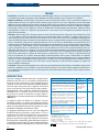
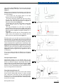
MED EMERGENCY / URGENCE ISSN 2222-9442 Wound or medical device protection: Secuderm® Factors behind delay in final disposition of patients La gangrène diabétique du membre inférieur Austere, remote and disaster medicine Occult elbow fractures in children Intoxication par la chloroquine Trimestriel How to prevent and treat vasovagal syncope at its early stage? Endorsed by September 2014 - N°20 Just because you are trained for something doesn't mean you are prepared for it… With the NSEC, direct your staff into the right path. NSEC offers a wide range of courses accredited by the Lebanese Ministry of Education. • BLS, ALS, PBLS, APLS • Emergency Medicine Techniques • Emergency preparedness • Disaster and Crisis Management • Forensic Emergencies • ED management • Intensive Care Techniques • Combat Medics: CLS, CMAST, Demining Medics • Road Safety • First Aid for passengers • Sport Emergency Medicine Techniques In addition to these standardized courses, NSEC can provide customized courses and workshops to mesh with our customers’ needs and requirements in their preferable language (English, Arabic and/or French). Fanar, P.O.Box: 90815 Jdeideh – Metn, Lebanon, T +961 1 888921, E [email protected], W www.newheathconcept.net E D I T O R ’ s N O T E When there is a will, there is a way .. MED Emergency, MJEM Mediterranean Journal of Emergency Medicine Publication of the Lebanese Resuscitation Council By New Health Concept P.O.Box 90.815 Jdeideh - Lebanon Tel: 00961.1.888921 Fax: 00.961.1.888922 Email: [email protected] Website: www.newhealthconcept.net Editorial board Editor in Chief Nagi SOUAIBY Managing editor Maria Frangieh Research Abdo KHOURY (France) Steve PHOTIOU (Italy) Jean-Cyrille PITTELOUD (Switzerland) Continuous Education Elvis CORDIER (France) Daryl MACIAS (USA) Karim BEN MILOUD (Switzerland) Innovation, Editing and Translation Guillaume Alinier (Qatar / UK) Karim FARAH (Lebanon) Hugues LEFORT (France) Online Publication and Design Ismaël HSSAIN (France) Alec KAZANDJIAN Mireille SROUR Nursing Lina AOUN CHOUEIRY Chantal SAADEH KHALIL Midwives Sabine Abou Malham (Canada) Students’ Forums and conferences Ziad KHOUEIRY (France) Paramedics and Ambulances Frédéric HOEPPLI (Switzerland) Juerg LINIGER (Switzerland) Administration and Marketing Georges KHALIL Alliances Fire Brigade of Paris – France Global Network Association of Emergency Medicine Global Emergency Medicine Literature Review Lebanese Society for Quality and Patient Safety advisory Committee Pierre ABI HANNA, Georges ABI SAAD, Nayla Abou Malham Doughane, Arthur ATCHABAHIAN, Omar AYACH, Abdelouahab BELLOU, Maria Paula GOMEZ, Thierry GROS, Maurice HADDAD, Berthe HACHEM, Mohamed HACHELAF, Jamil HALABI, Chokri HAMOUDA, Khalil HELOU, Aziz KOLEILAT, Bruno MEGARBANE, Ahmad OSMAN, Alissar RADY, Wassim RAFFOUL, Sami RICHA, Abdul Mohsen AL SAAWI, Karim TAZAROURTE, Youri YORDANOV. Med Emergency, MJEM – 2014, No 20 The region faces many challenges, crises and disasters, either manmade or natural. Recently, disaster and emergency medicine became a priority for hospitals and other healthcare facilities. And in the past years, a rapid evolution in emergency medicine has occurred in the region. In fact, researchers from the US and several European countries are collaborating with their peers from the Middle East and North Africa region to develop new research studies in emergency medicine field, gather more comprehensive related data and provide relevant training workshops to hospital and healthcare staff and other relevant stakeholders on the preparedness and management of various emergencies. In spite of the humanitarian crisis as well as the political and budgetary constraints, the region has much potential for growth and development in the health sector in general, and the emergency field in particular. Furthermore, conflict-affected countries where services pertaining to emergency medicine and management are particularly needed yet still understudied. Accordingly, more studies are required to reflect the true demands and needs in emergency field, whether in its technical, medical and/ or managerial aspect, to assess what is currently being done and to develop efficient relevant standards. As such, in MJEM Med Emergency, we make sure that we highlight recent concerns and challenges throughout original articles and studies performed in the MENA region. We also include research and latest studies performed in high income countries (the European and American experience) to benefit from their know-how. Furthermore, we reflect findings of research performed in various work environments, extreme conditions, different target population, etc. We try to tackle the different aspects of this specialty throughout our enriching original articles, continuous education, literature review, case reports, abstracts and relevant news sections. Last but not least, we support young talents and innovations; we follow up and actively take part in relevant conferences and training on a national and international level. …We strive to exceed our readers’ expectations… Nagi Souaiby, MD, MPH, MHM Chief Editor 1 C O N T E N T S Original Articles Wound or medical device protection: benefits of the waterproof dressing Secuderm® Lefort H, Bon O, Hersan O, Travers S, Bignand M, Tourtier JP ............................................................................. Factors behind delay in final disposition of patients from emergency department of a tertiary care center Kazi G, Siddiqui E, Habib I, Khan I, Khan B, Feroz A, Iqbal A ..................... Original Articles (French) La gangrène diabétique du membre inférieur : épidémiologie et facteurs de choix de la meilleure technique . . . . . . . . . . . . . . . anesthésique The gangrene diabetic of the lower limb: epidemiology and factors of choice of the best anesthetic technique Damghi N, Belkouch A, Sibou R, Nebhani T, Zidouh S, Belyamani L Continuous Education Austere, remote and disaster medicine-keeping everybody safe MACIAS D, WILLIAMS J ............................................................................................................................. Occult elbow fractures in children: some tips and tricks to read radiographs Courvoisier A, Calvelli N, Bourgeois E, Eid A, Griffet J How to prevent and treat vasovagal syncope at its early stage? Farrokhi P, Taboulet P, Boutmy DE .............................................................................................. .................................................................................................................................. Continuous Education (French) Intoxication par la chloroquine Chloroquine intoxication Mégarbane B ................................................................................................................................................................................................................ General information Recommendations for authors . . . . . . . . . . . . . . . . . . . . . . . . . . . . . . . . . . . . . . . . . . . . . . . . . . . . . . . . . . . . . . . . . . . . . . . . . . . . . . . . . . . . . . . . . . . . . . . . . . . . . . . . . . . . . . . . . . . . . . . . . . . . . . . . . . . . . . . . . . . . . . . . . . . . . . . . . . . . . . . . . . . . . . . . . . . . . . . . . . . . . . . . . . . . . . . . . . . . . . . . . . . . . . . . . . . Membership . . . . . . . . . . . . . . . . . . . . . . . . . . . . . . . . . . . . . . . . . . . . . . . . . . . . . . . . . . . . . . . . . . . . . . . . . . . . . . . . . . . . . . . . . . . . . . . . . . . . . . . . . . . . . . . . . . . . . . . . . . . . . . . . . . . . . . . . . . . . . . . . . . . . . . . . . . . . . . . . . . . . . . . . . . . . . . . . . . . . . . . . . . . . . . . . . . . . . . . . . . . . . . . . . . . . . . . . . . . . . . . . . . . . . . . . . . . . . . . . . . . . . . . . . . . . . . . . 2 p. 3 p. 9 p. 15 p. 23 p. 30 p. 35 p. 40 p. 46 p. 48 Med Emergency, MJEM – 2014, No 20 ORIGINAL ARTICLE Wound or medical device protection: benefits of the waterproof dressing Secuderm®. Protection des plaies ou d’un dispositif médical: intérêts du pansement étanche Secuderm®. Lefort H, Bon O, Hersan O, Travers S, Bignand M, Tourtier JP. Wound or medical device protection: benefits of the waterproof dressing Secuderm®. Med Emergency, MJEM 2014; 20:3-8. Mots clés : Secuderm®, pansement secondaire, cicatrisation, étanche, armée, polyuréthane, plaie Keywords: Secuderm®, secondary dressing, wound healing, waterproof, army, polyurethane, wound ABSTRACT Aim: The wound is the consequence of an acute skin aggression either limited or spreading, sometimes iatrogenic, which may be worsened by a delay in care in peculiar circumstances. The aim of this study is to present the benefits and give potential indications of a waterproof dressing. This dressing guarantees the protection during the healing process and increases the patient’s compliance to the treatment of his wounds, even in difficult situations. Methods: We used various dressings or means (primary adherent dressings, polyurethane film, cling film) to protect wounds in isolated or precarious care situations, but also in a more conventional context. Results: Secuderm® is the only dressing that is waterproof, reliable for acute and chronic protection for repeated exposures to water: excessive sweating, friction of clothing, projection or complete immersion under water. We report the use of Secuderm® in different exemption situations: in Guyana during French military missions by the Navy divers, in Cameroon for the treatment of Buruli ulcers, after arthroscopy, to protect medical devices (catheters, etc.) on acute wounds, or even during patient exposure to a nuclear, radiological, biological or chemical risk (NRBC). Discussion: To effectively protect a wound is difficult when the human resources and materials are limited. An efficient protection of the healing wound allows the resumption of professional, social and private activities in a certain comfort. Probably the only solution that is currently available, Secuderm® is a cunning waterproof protection for several days. Its indications are multiple whether in the treatment of acute, chronic or iatrogenic wounds, at the hospital, at home, in degraded situation or even when exposed to nuclear, radiological, biological or chemical risk. Prospective comparative studies are required. Authors’ affiliation: Correspondent author: Hugues LEFORT, MD Emergency Medical Service, Fire Brigade of Paris, Paris, France 1 place Jules Renard, 75017, Paris [email protected] Lefort H, MD1, Bon O, MD1, Hersan O, MD2, Travers S, MD1, Bignand M, MD1, Tourtier JP, MD1 1. Pre Hospital Care Department. Fire Brigade of Paris, France 2. SMPM, Paris, France Article history / info: Category: Original article Received: Nov 21, 2013 Revised: June 20, 2014 Accepted: July 23, 2014 Dr Hugues Lefort Conflict of interest statement: There is no conflict of interest to declare Med Emergency, MJEM – 2014, No 20 3 ORIGINAL ARTICLE Emergency Showstopper- Factors behind Delays in Final Disposition Kazi G, Siddiqui E, Habib I, Khan I, Khan B, Feroz A, Iqbal A. Emergency showstopper-sactors behind delays in final disposition. Med Emergency, MJEM 2014; 20:9-14. Key words: factors, delay, length of stay, emergency department ABSTRACT Introduction: Clinical management outcome of emergency patients with delays are directly related to blocked access to the next level of care from emergency department. It predicts delay to the definitive procedure plan to manage the patient and is also a marker of hospital functional flaws. Objective: To study the frequency and associated factor of delays behind final disposition of patients presenting to the Emergency Department of a tertiary care hospital in Pakistan. Methods: This is comparative cross sectional study, conducted at Aga Khan University Hospital. Both adult and pediatric patients were included. Comparison was done between delayed and non-delayed emergency department patients. Six hour was taken as cut-off. SPSS version 19 and MS excel 2010 were used for analysis. Results: Out of 365 cases, 133 (36%) were pediatric and 232 (64%) were adults patients. There were 184 (50%) males. More than six hour delay was noted in 94 patients (27%). Adult patients were delayed more than pediatric patients (p<0.001). Laboratorial, radiological test and generated consults were all found highly significant difference for the delays (p<0.001). 297 (81%) were discharged home, while 17 (5%) of them were admitted. Discussion: Overcrowding is common in Emergency Department (ED) and hence the chances of delay in disposition of patients from ED are very high which will ultimately compromise the patient care. Reducing the number of comparatively stable patients with effective triaging, ED clinics and diverting available resources towards more critical patients can reduce congestion, input and throughput. Reducing consults and unnecessary investigations with the provision of more experienced physicians & nurses is an important factor to reduce delays. Conclusion: Extended length of stay in ED may exceed the potential capability to deliver quality care within appropriate time frame; this may lead to drastic decrease in patient and family satisfaction, leading to compromised clinical care. Authors’ affiliation: Correspondent author: Sayyeda Ghazala Irfan Kazi, MD Assistant Professor Department of Emergency Medicine Aga Khan University, Pakistan [email protected] Kazi G, MD, Siddiqui E, MD, Habib I, MD, Khan I, MD, Khan B, MD, Feroz A, MS, Iqbal A Department of Emergency Medicine Aga Khan University Article history / info: Category: Original article Received: April 9, 2014 Revised: June 18, 2014 Accepted: July 28, 2014 Dr Ghazala Kazi Conflict of interest statement: The authors declare no conflict of interest. Med Emergency, MJEM – 2014, No 20 9 ORIGINAL ARTICLE La gangrène diabétique du membre inférieur : épidémiologie et facteurs de choix de la meilleure technique anesthésique The gangrene diabetic of the lower limb: epidemiology and factors of choice of the best anesthetic technique Damghi N, Belkouch A, Sibou R, Nebhani T, Zidouh S, Belyamani L. La gangrène diabétique du membre inférieur : épidémiologie et facteurs de choix de la meilleure technique anesthésique. Med Emergency, MJEM 2014; 20:15-22. Mots clés: gangrène, pied diabétique, anesthésie locorégionale, anesthésie générale, amputation Key words: gangrene, foot diabetic, locoregional anesthesia, general anesthesia, amputation ABSTRACT Objective: The identification of the epidemiological characteristics as well as the comparison of the anesthetic techniques of the patients presenting a gangrene of the foot diabetic (general anesthesia versus locoregional anesthesia). Material and method: Descriptive, retrospective study spread over the year 2013 and realized in the operating block of the emergencies of the military hospital of Rabat. The criteria of inclusion were an age of more than 18 years, and an amputation at the level of the lower limb further to a gangrene evolving in a context of known or inaugural diabetes. The incomplete files were excluded. Various epidemiological, clinical, biological and therapeutic parameters were collected. The various used anesthetic techniques were compared in term of modifications perioperative hémodynamiques, speed to reduce the hyperglycemia, arisen post-operative short-term complication, appeal in the analgesia operating comment as well as the total duration of intervention. Results: During year 2013, 118 patients were listed among which 109 only included. The average age of the patients was 58.3 ± 11 years old (extremes: 25-86 years) among which 81.6% were male (40 men, 9 women). The gangrene was localized at the level of a foot (61.5%), of a toe (34.9%), by a leg (2.7%) or of a thigh (0.9%). The biological characteristics of the patients in the admission were characterized by a hyperleucocytose, hyponatremia, a light acidose and a renal insufficiency. The quantity of the perfused solution in preoperative was 250 ± 132 mL with administration of an initial bolus of 10 UI of fast insulin. The antibiotic treatment was with amoxicillin/clavulanic acid (66.7%), of cefazolin (22.8%), of amoxicilline/clavulanic acid + metronidazol (6.3%) and of metronidazol only (4.2%). The used anesthetic techniques were a plexique block (62.2%), a spinal anesthesia (24.7%), a local anesthetic (8.5%) and a general anesthesia (4.6%). These various techniques were compared in term of modifications perioperative hémodynamiques, speed to reduce the hyperglycemia, arisen post-operative short-term complication, appeal in the analgesia operating comment as well as the total duration of intervention. The quantity of the perfusd solution it peropératoire was 775 ± 409 mL. The premedication was made by the midazolam. The hemodynamic variations were important in the group spinal anesthesia and local anesthetic. The reduction of hyperglycemia as well as the duration of intervention were more important in the group general anesthesia. The post-operative consequences were marked by the arisen of a hemorrhagic shock at three sick (3.3%) and with a toxic shock at a sick person (1.1%). Our study showed the advantage to realize plexique blocks compared with the other anesthetic techniques. Conclusion: Our therapeutic protocol which consists of an adapted hydro electrolytic resuscitation e metabolic preoperative and favors plexiques blocks allowed to reduce the incidence of the hemodynamics peroperative variations and the post-operative complications but would require to have patients’ more important staff and to be compared with other similar studies. Authors’ affiliation: Correspondent author: Nada Damghi, MD Pôle SAMU –SMUR-SAU, CHR Idrissi BP : 14020, Kénitra, Maroc [email protected] Damghi N, MD1, Belkouch A, MD2, Sibou R, MD2, Nebhani T, MD2, Zidouh S, MD2, Belyamani L, MD2 1. Pôle SAMU –SMUR-SAU, Hôpital Idrissi, CHR Kénitra, Maroc 2. Service d’anesthésiologie, hôpital d’instruction militaire Mohamed V, Rabat, Maroc Med Emergency, MJEM – 2014, No 20 Article history / info: Category: Original article Received: April 14, 2014 Revised: July 24, 2014 Accepted: Aug. 1, 2014 Conflict of interest statement: There is no conflict of interest to declare Dr Nada Damghi 15 original article RÉSUMÉ Introduction : L’identification des caractéristiques épidémiologiques ainsi que la comparaison des techniques anesthésique des patients prénsentant une gangrène du pied diabétique (anesthésie générale versus anesthésie loco-régionale). Matériel et méthode : Etude descriptive, rétrospective étalée sur l’année 2013 et réalisée au bloc opératoire des urgences de l’hôpital militaire de Rabat. Les critères d’inclusion étaient un âge de plus de 18 ans, et une amputation au niveau du membre inférieur suite à une gangrène évoluant dans un contexte de diabète connu ou inaugural. Ont été exclus les dossiers incomplets. Différents paramètres épidémiologiques, cliniques, biologiques et thérapeutiques ont été recueillis. Les différentes techniques anesthésiques utilisées ont été comparées en terme de modifications hémodynamiques péri-opératoires, rapidité de réduire l’hyperglycémie, survenue de complication post-opératoire à court terme, recours à l’analgésie post opératoire ainsi que la durée totale d’intervention. Résultats : Durant l’année 2013, 118 patients étaient recensés dont 109 étaient inclus. L’âge moyen des patients était de 58,3 ± 11 ans (extrêmes : 25-86 ans) dont 81,6% étaient de sexe masculin (40 hommes, 9 femmes). La gangrène était localisée au niveau d’un pied (61,5%), d’un orteil (34,9%), d’une jambe (2,7%) ou d’une cuisse (0,9%). Les caractéristiques biologiques des patients à l’admission étaient caractérisées par une hyperleucocytose, une hyponatrémie, une légère acidose et une insuffisance rénale. La quantité du soluté perfusé en préopératoire était de 250 ± 132 mL avec administration d’un bolus initial de 10 UI d’insuline rapide. L’antibiothérapie était à base d’amoxicilline/acide clavulanique (66,7%), de céfazoline (22,8%), d’amoxicilline / acide clavulanique + métronidazole (6,3%) et de métronidazole seul (4,2%). Les techniques anesthésiques utilisées étaient un bloc plexique (62,2%), une rachianesthésie (24,7%), une anesthésie locale (8,5%) et une anesthésie générale (4,6%). Ces différentes techniques ont été comparées en terme de modifications hémodynamiques péri-opératoires, rapidité de réduire l’hyperglycémie, survenue de complication post-opératoire à court terme, recours à l’analgésie post opératoire ainsi que la durée totale d’intervention. La quantité du soluté perfusé en peropératoire était de 775 ± 409 mL. La prémédication était faite par le midazolam. Les variations hémodynamiques étaient importantes dans le groupe rachianesthésie et anesthésie locale. La réduction de l’hyperglycémie ainsi que la durée d’intervention étaient plus importantes dans le groupe anesthésie générale. Les suites postopératoires étaient marquées par la survenue d’un choc hémorragique chez trois malades (3,3%) et d’un choc septique chez un malade (1,1%). Notre étude a montré l’avantage de réaliser des blocs plexiques par rapport aux autres techniques anesthésiques. Conclusion : Notre protocole thérapeutique qui consiste en une réanimation hydro électrolytique et métabolique préopératoire adaptées et privilégie les blocs plexiques a permis de réduire l’incidence des variations hémodynamiques peropératoires et des complications postopératoires mais nécessiterait d’avoir un effectif plus important de patients et d’être comparé à d’autres études similaires. INTRODUCTION L’infection complique l’évolution d’une plaie chronique du pied diabétique dans 25% des cas, en alourdit considérablement la prise en charge et augmente le risque d’amputation surtout lorsqu’elle est associée à une artérite des membres inférieurs et/ou une ostéite sous-jacente [1;2]. Le diagnostic de l’infection repose sur la présence d’au moins deux des signes suivants : augmentation de volume, induration, érythème péri-lésionnel, sensibilité locale ou douleur, chaleur locale ou présence de pus [3;4]. La sévérité de l’infection sera jugée d’après la classification du Consensus International sur le Pied Diabétique (Tableau I). La gangrène humide est définie par la présence de tissus nécrotiques noirâtres. Les lésions sont rapidement évolutives avec décollement et pus grisâtre d’odeur nauséabonde, pouvant aboutir à une dégradation rapide de l’état général avec sepsis, déséquilibre métabolique et insuffisance rénale. Les gangrènes diabétiques sont une cause non négligeable d’antibiothérapies non justifiées et participent à ce titre à l’aggravation de la résistance bactérienne et à son extension au travers des soins [5]. La fréquence et la sévérité de ces gangrènes trouvent leur origine dans l’altération des fonctions des polynucléaires neutrophiles, particulièrement marquée en cas d’hyperglycémie prolongée [6;7], dans l’anatomie particulière du pied [8] qui favorise la dissémination de l’infection et dans 16 Manifestations cliniques de l’infection Sévérité PEDIS Absence de pus et/ou de signes d’inflammation Pas d’infection 1 2 parmi les signes suivants sont présents : augmentation de volume, induration, érythèm entre 0,5 et 2 cm autour de la lésion, sensibilité ou douleur, chaleur locale, écoulement purulent 2 Infection légère : pas de mise en jeu du pronostic du pied ni vital Comme précédemment ; le patient ne présente pas de signe de sepsis* ni de déséquilibre métabolique mais présente du pronostic du pied mais pas vital, 1 parmi les Infection modérée : signes suivants : érythème > 2 cm autour mise en jeu de la plaie, lymphangite, atteinte des fascia superficiels, abcès profond,gangrène, extension aux structures ostéo-articulaires 3 Présence d’un sepsis* ou d’instabilité métabolique (fièvre, frissons, tachycardie, Infection sévère : hypotension, confusion, vomissements, pronostic vital en hyperleucocytose, acidose, hyperglycémie, jeu hyperazotémie) 4 * au moins deux signes parmi : – température < 36 °C ou > 38 °C – fréquence cardiaque > 90 batt/min – fréquence respiratoire > 20 cycles/min – PaCO2 < 32 mmHg – leucocytose > 12000/mm3 ou < 4000/mm3 ou ≥ 10% de formes immatures Table 1 : Définition et classification des infections du pied diabétique [3] Med Emergency, MJEM – 2014, No 20 C ontin u o u s E d u cation Austere, Remote and Disaster Medicine-Keeping Everybody Safe MACIAS D, WILLIAMS J. Austere, remote and disaster medicine-keeping everybody safe. Med Emergency, MJEM 2014; 20:23-9. Keywords: Wilderness medicine, disaster medicine, resource-limited, 7 P’S ABSTRACT Medical care in resource-limited environments (“austere” settings) can occur in the context of a disaster, wilderness, or a tactical field operation. Regardless of the type of environment, there are common organizational themes in most successful humanitarian missions that occur in harsh environmental conditions, be they natural, or man-made. These principles prioritize the initiation and execution of any given deployment in austere or remote settings, diverging from priorities that would occur in a situation where the medical structure is intact and operating well. Attention to these priorities not only helps providers with delivering medical care to the needy during a period of resource limitations, they also can keep a provider, teams, the public, and a patient safe during, and after a deployment. Authors’ affiliation: Correspondent author: Darryl MACIAS, MD Department of Emergency Medicine and Emergency Medical Services Academy, University of New Mexico School of Medicine, Albuquerque, NM 87131, USA. [email protected] Macias D, MD, Williams J Department of Emergency Medicine and Emergency Medical Services Academy, University of New Mexico School of Medicine, Albuquerque, NM 87131, USA. Article history / info: Category: Continuous Education Received: May 5, 2014 Revised: June 20, 2014 Accepted: July 4, 2014 Dr Darryl Macias Conflict of interest statement: There is no conflict of interest to declare INTRODUCTION Recent world catastrophic events and humanitarian crises have called for more assistance from relief workers than ever before. Physicians and other health care workers desire to answer the call more than ever, due to a sense of social responsibility, commitment to service, and increased educational offerings in humanitarian health [1]. However, some participants may not comprehend the difficulties inherent in working in austere environments. ”Austere medicine” is often used in the context of operational medicine, associated with combat, hazardous or tactical operations [2]. Austere medicine also encompasses resource-limited settings, where advanced hospital technology is not readily available, be it in a health care clinic in an underdeveloped region (Figure 1), in an air ambulance setting, during a wilderness expedition or wilderness rescue situation, during an in-flight or space mission emergency, or in a disaster Med Emergency, MJEM – 2014, No 20 Figure 1: Healthcare clinic in an underdeveloped setting 23 C ontin u o u s E d u cation Occult elbow fractures in children: some tips and tricks to read radiographs Courvoisier A, Calvelli N, Bourgeois E, Eid A, Griffet J. Occult elbow fractures in children: some tips and tricks to read radiographs. Med Emergency, MJEM 2014; 20:30-4. Keywords: elbow, traumatism, fracture, children ABSTRACT Elbow traumatisms are very common in children. X-rays play important roles in the diagnosis but are sometimes difficult to read considering the quality, the normal variations of bone ossification and the type of fracture. The key to a better understanding of these fractures is first to differentiate normality from abnormality. The purpose of this report is to provide tricks and tips to help the reader when dealing with a child with an elbow traumatism. The real problem is to miss a fracture that would need a surgical treatment. Simple geometric constructions and knowledge of the aspect of the different ossification steps of a growing elbow are sufficient. But there is no need to do comparative X-Rays. This report was focused on the X-rays but one must not forget the common sense and the clinical examination to orientate the diagnosis. Finally, in cases where the X-ray is thought to be normal, a cast immobilization and evaluation 10 days after the traumatism is necessary. Authors’ affiliation: Correspondent author: Aurélien COURVOISIER, MD Pediatric Orthopedic Department. Hôpital Couple-Enfant. Grenoble University Hospital Joseph Fourier University BP 217 38043 Grenoble Cedex 09 – France [email protected] Courvoisier A, MD, PhD, Calvelli N, MD, Bourgeois E, MD, MSc, Eid A, MD, Griffet J, MD, PhD Pediatric Orthopedic Department. Hôpital Couple-Enfant. Grenoble University Hospital Joseph Fourier University, France Article history / info: Category: Continuous Education Received: Jan. 17, 2014 Revised: Mars 13, 2014 Accepted: July 24, 2014 Dr Aurélien Courvoisier Conflict of interest statement: There is no conflict of interest to declare INTRODUCTION Elbow traumatisms are very common in children. X-rays play important roles in the diagnosis but are sometimes difficult to read considering the quality, the normal variations of bone ossification and the type of fracture. The key to a better understanding of these fractures is first to differentiate normality from abnormality. The purpose of this report is to provide tricks and tips to help the reader when dealing with a child with an elbow traumatism. First, let us describe the normality [1] From birth to skeletal maturity, X-ray changes and normal variations of bone ossification of the elbow need to be understood. At birth, only the distal humeral metaphysis is ossified (Figure 1). Then the elbow ossification progressively appears: at 2 years, the capitellum is ossified (Figure 2), at 4 years, it is the radial head (Figure 3), at 6 years, it is the medial epicondyle (Figure 4), at 8 30 Med Emergency, MJEM – 2014, No 20 C ontin u o u s E d u cation years, it is the trochlea (Figure 5), at 10 years, it is the olecranon (Figure 6) and finally at 12 years, it is the lateral epicondyle (Figure 7). Different lines and geometrical constructions on the X-ray may also help to detect an anomaly. The most important features are described hereafter: - On the lateral view, the anterior flexion of the distal humerus is between 30 and 40° (Figure 8). - The Storen construction: whatever the view on the X-rays, the axis of the radial diaphysis crosses the center of the capitellum: this point is very important for the diagnostic of dislocation of the radial head (Figure 9). - On the lateral view, the line of the anterior cortex of the humeral diaphysis crosses the middle of the capitellum (Figure 8). - The angle of Baumann: on an AP view, it is the angle between the line, which passes by the middle of the humeral diaphysis, and the one that passes through the growth plate at superior part of the capitellum. The angle is normally 72° ± 5° (Figure 10). - On an AP view: the line who is in the prolongation of the medial cortex of the humerus has to almost touch the superior part of the medial epicondyle (Figure 11). With these tools we are now able to proceed to the analysis of abnormal X-Rays. Two important signs need to be sought in all cases of elbow traumatism. The first sign is hemarthrosis. When there is blood in the joint, the capsule is distended. On a lateral view the distention of the capsule induces a modification of the position of the fat density triangle at the anterior part of the joint (Figure 12). The second sign is the edema. On an AP view the soft tissues close to the fracture are filled with hematoma and edema, which causes a differentiation of the soft tissues density (Figure 13). These two signs are very important in cases of occult fractures. Figure 1: lateral and AP view of the elbow at birth showing that only the distal humeral metaphysis is ossified. Figure 2: lateral and AP view of the elbow at 2 years showing the ossification of the capitellum. Figure 3: lateral and AP view at 4 years showing the ossification of the radial head. We are now armed to describe the different types of fractures of the elbow in children. But we will only focus on difficult diagnosis. The supracondylar fractures Supracondylar fractures are very frequent. Many are very displaced and the diagnosis is easy. Two types of supracondylar fracture are difficult to diagnose: supracondylar fracture with anterior flexion of the distal fragment and the non or slightly displaced supracondylar fracture in extension (Figures 14-15). In these two cases, the hemarthrosis sign is positive and on the tip is to use the geometrical constructions. Figure 4: lateral and AP view of the elbow at 6 years showing the ossification of the medial epicondyle. The medial epicondyle fracture [2] The medial epicondyle fracture is the second most common fracture after the supracondylar fracture. It is often associated with a dislocation of the elbow and the medial epicondyle fracture may be a sign of spontaneously reduced dislocation. Conversely, in case of an elbow dislocation a medial epicondyle fracture has to be sought. Med Emergency, MJEM – 2014, No 20 Figure 5: lateral and AP view of the elbow at 8 years showing the the ossification of the throchlea. 31 C ontin u o u s E d u cation How to prevent and treat vasovagal syncope at its early stage? Comment prévenir et traiter un malaise vagal à son début? Farrokhi P, Taboulet P, Boutmy D.E. How to prevent and treat vasovagal syncope at its early stage?. Med Emergency, MJEM 2014; 20:35-8. Keywords: syncope, vasovagal, emergency care, blood donation, prevention, treatment ABSTRACT Parasympathetic system activation is generally known as the origin of vasovagal syncope. As a matter of fact, the sympathetic system activation is the major trigger of vasovagal syncope. The association of hyperventilation or hyperexcitability neuromuscular syndrome can also play an important role as a trigger. A good knowledge of these mechanisms may help to prevent and treat the vasovagal syncope at its early stage. The treatment of vagal reaction relies on: supine position, contractions of the muscles of the lower limbs for about ten minutes and reduction of the ventilation rate. Authors’ affiliation: Correspondent author: Parviz Farrokhi, MD Etablissement Français du Sang d’Ile de France, Site Saint-Louis, 38 rue Bichat 75010, Paris, France [email protected] Farrokhi P, MD1, Taboulet P, MD2, Boutmy DE, MD, PhD2. 1. Etablissement Français du Sang d’Ile de France, Site Saint-Louis, 38 rue Bichat 75010, Paris, France 2. Hôpital Saint-Louis, Assistance-Publique-Hôpitaux de Paris, 1 avenue Claude Vellefaux, 75010, Paris, France Article history / info: Dr Parviz Farrokhi Category: Continuous Education Received: July 25, 2014 Accepted: Aug. 6, 2014 Conflict of interest statement: There is no conflict of interest to declare Acknowledgement: To the staff of Saint-Louis “Blood Collection Centre” RÉSUMÉ L’activation du système parasympathique est généralement connu comme l’origine de la syncope vaso-vagale. En fait, l’activation du système sympathique est la gâchette principal de la syncope vaso-vagale. L’association d’une hyperventilation avec hyperexcitabilité neuromusculaire peut également jouer un rôle important. Une bonne connaissance de ces mécanismes peut aider à prévenir et traiter la syncope vaso-vagale à son début. Le traitement de la réaction vagale repose sur : le décubitus dorsal, des contractions musculaires des membres inférieurs pendant environ dix minutes et la réduction de la fréquence ventilatoire. Med Emergency, MJEM – 2014, No 20 35 C ontin u o u s E d u cation Introduction Syncope is a common problem that occurs in general practice, in the emergency room and also before, during or after blood donation [1-5]. Approximately 1-3% of the emergency admissions to the hospitals and 1% of all hospitalizations [1;6] are related to syncope, the majority with vasovagal origin. The prognosis is good, but this at an extra cost to the public healthcare. The aim of this paper is to describe the pathophysiology of the vasovagal syncope (also called “vagal syncope” or VS) in order to understand and implement the steps required for its prevention and treatment. The main clinical VS symptoms are classically the results of the parasympathetic activation. Indeed, before parasympathetic activation, the sympathetic system, stress-related activation, plays a major role in the VS genesis [7-16]. Furthermore, the hyperventilation and neuromuscular excitability syndromes [17-23] are sometimes the prodromes of VS and they may warn the clinicians to prevent and treat the VS (Figure 1). Figure 1 : Triad syndrome Pathophysiology of the VS They are two pathways involved in the VS genesis [24]. A - The major and slower one is the hypothalamic-adrenal-medullae axis. In this case, VS occurs about six to ten minutes after triggering factor onset [24;25]. It could be called the “hormonal or peripheral pathway”. It begins with global sympathetic system activation. There are two modes for sympathetic system activation: partial and global [26]. - The partial activation is mediated by the postganglionic sympathetic nerves that release norepinephrine (NEP), a vasoconstrictive and cardiotonic agent. Thus, NEP is able to prevent the orthostatic syncope [1;12]. - The global activation is mediated by the hypothalamus, stimulated by stress (panic, fear or severe pain). It induces a global sympathetic activation called “mass discharge” (discharge simultaneous of almost all portions of sympathetic nervous system). The adrenal medulla stimulation by postganglionic sympathetic nerves releases large amount of catecholamines, about 80% epinephrine (EP) and 20% NEP into circulating blood. This activation has almost the same effects as those caused by direct sympathetic stimulation, but differs in duration by lasting five to ten times longer. According to a study by Sra [14], the patients exhibiting VS six minutes after the starting tilt test had significant increase of the EP and NEP release, whereas the control subjects only had an increase of NEP release. This study suggests that “mass discharge” phenomenon plays an important role in triggering VS (EP release). Mass discharge increases the ability of the body to perform vigorous muscle activity. Cardiovascular signs include: tachycardia, increased blood pressure, vasoconstriction in the gastrointestinal tract and the kidneys and a vasodilatation of the lower limb muscles (specific action beta-2 receptor of the EP). As a result, venous pooling of blood in the lower limbs [10;15;26] decreases the venous return. The consequence is an activation of the parasympathetic nervous system known as “the reflex of Bezold-Jarisch”. The clinical symptoms are sweating, bradycardia, hypotension, nausea, vomiting and consciousness loss. The decrease of the blood pressure reduces cerebral blood flow, which causes lipothymia and light-headedness. This phenomenon is termed by certain author as “hypotensive functional haemorrhage in spite of the absence of external or internal bleedings (haemorrhages) [15]. Baroreceptors system stimulation has been proposed as hypothesis to explain this reflex [1;27]. The decrease of the venous return is considered as a very important triggering factor, not only seen in VS genesis, but also in other kinds of syncope (i.e. during pregnancy with the supine inferior vena cava compression [28-30] or in case of intrathoracic pressure elevation linked to paroxysmal coughing). B - The second pathway involvement is less common, but more striking. Hence, clinical signs (blood pressure fall and bradycardia) arise without warnings or delay due to the direct role of the hypothalamic stimulation [26] on both the sympathetic center (inhibition), and the parasympathetic system (activation). One could name it the “central pathway”. In all cases, they are several associated factors that can be involved. The CO2 is one of the metabolic factors which has potent effect in controlling cerebral blood flow. Hyperventilation is a normal response of the body to eliminate an excessive muscular CO2 production during physical exercise. The hyperventilation acts quickly to avoid the hypercapnia (high pressure CO2 or PCO2 level), thus the increase of pH in the blood. In the absence of physical effort, hyperventilation is also an answer to stress situations. In that case, this hyperventilation results in hypocapnia and reduction in cerebral blood flow. For example, a decrease in cerebral tissue PCO2 about 20 mm Hg (normal value is 40 mm Hg) reduces about 40% cerebral blood flow, and vice versa [26]. The consequences of hyperventilation-induced hypocapnia on the cerebral blood flow may increase the drop of this blood flow induced by both the parasympathetic and sympathetic systems. 36 Med Emergency, MJEM – 2014, No 20 C ontin u o u s E d u cation Intoxication par la chloroquine Chloroquine intoxication Mégarbane B. Intoxication par la chloroquine. Med Emergency, MJEM 2014; 20:40-3. Mots clés : chloroquine, intoxication, effet stabilisant de membrane, choc, arrêt cardiaque, assistance circulatoire Keywords: chloroquine, intoxication, membrane stabilizing effect, shock, cardiac arrest, extracorporeal life support ABSTRACT Chloroquine poisoning is rare but may be life-threatening. Toxicity results from membrane stabilizing effect related to the blockage of sodium channels on myocardial contractile cells and conduction tissue. Bad prognosticators include the presumed ingested dose >4 g, the decrease in systolic blood pressure <100 mmHg, and the enlargement of QRS >0.10 s on the electrocardiogram. In the emergency department and even as soon as at the pre-hospital scene, management relies on tracheal intubation, epinephrine and diazepam infusion in severely poisoned patients, based on prognosticators, in order to prevent the onset of cardiac complications. Authors’ affiliation: Correspondent author: Bruno Mégarbane, MD, PhD Réanimation Médicale et Toxicologique, Hôpital Lariboisière, INSERM 1144, Université Paris-Diderot, 2 Rue Ambroise Paré, 75010 Paris [email protected] Article history / info: Category: Continuous Education Received: July 25, 2014 Accepted: Aug. 6, 2014 Conflict of interest statement: Pr Bruno Mégarbane There is no conflict of interest to declare RÉSUMÉ L’intoxication par la chloroquine est rare mais potentiellement grave. La sévérité de cette intoxication est liée à l’effet stabilisant de membrane qui résulte du blocage des canaux sodiques, à la surface des cellules contractiles et du tissu de conduction cardiaque. Les facteurs de mauvais pronostic sont la dose supposée ingérée >4 g, la baisse de la pression artérielle systolique <100 mmHg et l’élargissement des complexes QRS sur l’électrocardiogramme à >0,10 s. Aux urgences ou en pré-hospitalier, la stratégie thérapeutique est basée sur l’intubation trachéale, la mise sous adrénaline et l’administration de diazépam préventivement dès l’identification d’une forme sévère (en se basant sur les facteurs pronostiques) et avant la survenue de complications cardiaques. L’intoxication par la chloroquine (Nivaquine®, Savarine®) est rare mais potentiellement grave. En France, la publication dans les années 80 du livre « Suicide mode d’emploi » avait popularisé l’intoxication à la chloroquine. Depuis cette date, les facteurs pronostiques ont été identifiés et la stratégie thérapeutique optimisée. Malgré la réduction des prescriptions de chloroquine en raison des résistances acquises par l’agent du paludisme, cette intoxication persiste en France avec la facilitation récente de sa délivrance sans ordonnance sur internet Cas clinique Une jeune femme de 36 ans est admise au service d’accueil des urgences, amenée par son compagnon qui l’a vu ingérer des médicaments en excès 1h30 auparavant. Elle est dépressive depuis deux années, à la suite de plusieurs échecs professionnels. A l’admission aux urgences, la patiente est calme et parfaitement consciente. Sa pression artérielle est à 95/50 mmHg, sa fréquence cardiaque à 110 batt/min, sa fréquence respiratoire à 20 cycles/min et sa SpO2 à 97% en air ambiant. Dès le box d’accueil, un 40 Med Emergency, MJEM – 2014, No 20 The new C-MAC® Monitor AN 44 11/2013/A-LB Fine, Fast, Focused – Toggle Between the two Video Endoscopes KARL STORZ GmbH & Co. KG, Mittelstraße 8, 78532 Tuttlingen/Germany Phone: +49 (0)7461 708-0, Fax: + 49 (0)7461 708-105, E-Mail: [email protected] KARL STORZ Endoskope – East Mediterranean and Gulf S.A.L, Block M, 3rd Floor, Beirut Souks, Weygand Street, 2012 3301 Beirut/Lebanon, Phone: +961 (1) 999390, Fax: +961 (1) 999391, E-mail: [email protected] www.karlstorz.com R E C O M M E N D AT I O N S F O R A U T H O R S Med Emergency, MJEM The Mediterranean Journal of Emergency Medicine The Journal publishes articles in English and/or French pertaining to Emergency Medicine from its scientific aspect (research, case studies, clinical articles, orientation and practical conduct), administrative (Management and organization of Emergency Medicine), medical-legal and social aspects. It also accepts articles that deal with prevention of emergencies. Although it focuses more on practical issues of emergency medicine, the Journal accepts theoretical, methodological and analytical articles. It is also interested in communications, letters, commentaries and critiques of issues related to emergency. Authors can submit their original articles and the accompanying references to the editor: New Health Concept B.P. 90.815 JdeidehLebanon or via email. The article should be accompanied by a letter by the author/s that clearly states that joint authors of the article are aware of the application to publish and have agreed to allow free accessing of texts by New Health Concept Edition publication. Please create a separate file (indicating the name of the author) for all the photographs, tables and graphs you would like to be included in the article and send them to the following address: [email protected] All submissions will undergo a preliminary evaluation and an ethical revision by the editorial board to determine whether it will be allowed to appear in the journal. Articles that pass this preliminary evaluation will also be anonymously reviewed by two members of a scientific committee. Once the article has been approved for publication, a biography of 10 lines should be developed. Manuscript Preparation Articles are to be submitted in a typewritten format. Paragraphs are double spaced. Font size should be 12. The submitting author should send his contact details with the article such as telephone number or an email address. The original text of the article should be sent without illustrations in its original format (e.g. Microsoft Word). Pages should be numbered. Titles and subtitles of equal importance should be marked identically. Abbreviations should be explained when first encountered in the text. The articles should not exceed 2500 words or not more than 10 pages. Abstracts and Key Words: Each article should include an abstract In English (and in French for French articles) no longer than 300 words. Keywords (not more than 6 words) and the title of the article should also be presented in both languages. Text: The author needs to respect the following formatting procedures when submitting the article: • On the front page- the author’s name, affiliations, complete mailing address, telephone number and email address. The names and the affiliations of collaborators should be clearly indicated. Please ensure that this information is only presented on the front page and does not appear on the other pages of the article. • Bibliographic References need to appear in order of appearance in the text. They must be identified in the text by Arabic numbers in brackets. There should be about 10-30 references. They must conform to presentation norms applied in the scientific editing world (Vancouver style). • Photographs, figures, graphs and tables: these should be sent in separate files and need to be numbered and marked with the author’s name and commentary. They need to be numbered in chronological ordered when they are to be referred to in the text. The term “graph/table/figure/photo number x” should be used in order to avoid confusion with bibliographical references. • End notes should be listed separately at the end of the text and not at the end of each page. PS: It’s strongly recommended to add photography of the author who can also allow us to communicate his E-mail address. 46 For research original articles and review articles authors should clearly note the following: • If the study was approved by a local or international IRB (institutional review board), a government ministry, or a community group. • The design of a study: a randomized controlled trial or an observational study that includes a control group. • Discuss attempts to limit bias in the article. • The design of a review: formal meta-analysis or a systematic review that only includes studies with a control group how the review articles are selected. • Which statistical tests are used to analyze the data? ADDENDUM Conflict-of-Interest Statement* Conflict of interest exists when an author (or the author’s institution), reviewer, or editor has financial or personal relationships that inappropriately influence (bias) his or her actions (such relationships are also known as dual commitments, competing interests, or competing loyalties). These relationships vary from those with negligible potential to those with great potential to influence judgment, and not all relationships represent true conflict of interest. The potential for conflict of interest can exist whether or not an individual believes that the relationship affects his or her scientific judgment. Financial relationships (such as employment, consultancies, stock ownership, honoraria, paid expert testimony) are the most easily identifiable conflicts of interest and the most likely to undermine the credibility of the journal, the authors, and of science itself. However, conflicts can occur for other reasons, such as personal relationships, academic competition, and intellectual passion. Statement of Informed Consent* Patients have a right to privacy that should not be infringed without informed consent. Identifying information, including patients’ names, initials, or hospital numbers, should not be published in written descriptions, photographs, and pedigrees unless the information is essential for scientific purposes and the patient (or parent or guardian) gives written informed consent for publication. Informed consent for this purpose requires that a patient who is identifiable be shown the manuscript to be published. Authors should identify Individuals who provide writing assistance and disclose the funding source for this assistance. Identifying details should be omitted if they are not essential. Complete anonymity is difficult to achieve, however, and informed consent should be obtained if there is any doubt. For example, masking the eye region in photographs of patients is inadequate protection of anonymity. If identifying characteristics are altered to protect anonymity, such as in genetic pedigrees, authors should provide assurance that alterations do not distort scientific meaning and editors should so note. Statement of Human and Animal Rights* When reporting experiments on human subjects, authors should indicate whether the procedures followed were in accordance with the ethical standards of the responsible committee on human experimentation (institutional and national) and with the Helsinki Declaration of 1975, as revised in 2000 (5). If doubt exists whether the research was conducted in accordance with the Helsinki Declaration, the authors must explain the rationale for their approach, and demonstrate that the institutional review body explicitly approved the doubtful aspects of the study. When reporting experiments on animals, authors should be asked to indicate whether the institutional and national guide for the care and use of laboratory animals was followed. *International Committee of Medical Journal Editors ("Uniform Requirements for Manuscripts Submitted to Biomedical Journals") -- February 2006 Med Emergency, MJEM – 2014, No 20 On the occasion of the Third World Congress of Plastic Surgeon of Lebanese Descent The Euro-Mediterranean Council for Burns & Fire Disasters - MBC and The National School for Emergency Care Have the pleasure to invite you to attend the Special Burn session Saturday October 25, 2014 - Le Royal Palace Hotel - Dbaye 11:00-12:00| Cérémonie officielle. Lancement du projet: “Mise en Place d’un Plan Catastrophe National pour la Prise en Charge des Brûlés au Liban”. Projet du Conseil de Recherche de l’Université St Joseph • Introduction et présentation du projet: Dr Nagi Souaiby, Sara Mufarrij, Lina Deghaili • Plan Suisse: Professeur Wassim Raffoul • Mot de l’Ambassadeur Suisse • Mot du Recteur de l’USJ • Mot du Ministre de la Santé 12:00-12:30| MBC LECTURE 1: Burn Unit at the Lebanese Hospital – Geitawi. G. GHANIMÉ (Lebanon) 12:30-13:00| MBC LECTURE 2: Burn Care Program, a strategic national public health project. W. RAFFOUL (Switzerland) 13:00-13:30| Lunch box 13:30-13:40| Prehospital management of Burns. A. Khoury (France) 13:40-13:50| Les progrès en brûlologie chirurgicale les 50 dernières années. M. Costagliola (France) 13:50-14:00| Neck Burn Reconstruction. M. Costagliola (France) 14:00-14:10| Assessment of airway improvement following neck burn scar contracture. S. Hayek, A. El Khatib, G. Kanaze (Lebanon) 14:10-14:20| Application Smart Phone Pour Grand Brulés. A. Khoury (France) 14:20-14:30| Measures of Intervention in Burn Disasters: Preparedness and Crisis Management. B. Atiyeh, S. Dibo (Lebanon) 14:30-14:40| Evolution of biological bandages in burn treatment: L.A. Laurent-Applegate, C.Scaleea, N. Hirt-Burri, A. de Buys Roessingh, Y-A Que, P. Jafari, W. Raffoul (Switzerland) 14:40-15:00| Discussion 15:00-15:30| ROUND TABLE. W. Raffoul, M. Costagliola • Wound cleansing, topical, antiseptics and wound healing: F. Chahine, B. Atiyeh, S. Hayek (Lebanon) • Effect of Silver on burn wound infection control and healing: Review of the literature: J. Baroud, B. Atiyeh, M. Costagliola, S. Hayek, S. Dibo (Lebanon, France) • New technologies for burn wound closure and healing: Review of the literature: F. Chahine, B. Atiyeh, S. Hayek, S.W. Gunn (Lebanon) 15:30-17:00| WORKSHOP on Suturing techniques: N. Souaiby (Lebanon), W. Raffoul (Switzerland) CME accredited For information and registration: NSEC: Tel/Fax: +961 1 888921, Email: [email protected] Website: www.newhealthconcept.net Trust & Traders Intl: Tel/Fax: +961 1 333900, +961 1 333130, +961 1 333730, Email: [email protected] Website: www.trust-traders.com The Advertising Organizations: Soins – page 44. Karl-Storz – page 45. Special Burn session – page 47. Med Emergency MJEM – cover page 3. New Health Concept – back cover. Editor in Chief and Director Nagi Souaiby Managing editor Maria Frangieh Executive board ONLINE SUBSCRIPTION http://www.newhealthconcept.net/subscribe.asp Kindly fill and send by email to: Med Emergency, MJEM Publications, New Health Concept, Email: [email protected] Membership Individual Student Institution 4 Issues 1 Year (USD) Ismaël HSSAIN Abdo KHOURY Hugues LEFORT 8 Issues 2 Year (USD) 110 180 90 130 200 350 COVER PICTURE SSA\BSPP\MBignand Design Mireille SROUR: Creative Couple Webmaster E-copy Alec KAZANDJIAN: Web Sart Printed copy Name Surname Address P.O.Box Country Tel Profession PRINTING Melki print international S.A.L. Quarterly Journal ISSN No 2222-9442 Printed in Lebanon City Email Affiliation All rights reserved. Please note Med Emergency Publication copyright in all reprints. MED EMERGENCY Publications - P.O. Box 90.815 - Jdeideh - Lebanon Tel: +961-1-888921, Fax: +961-1-888922 ELECTROCARDIOGRAMME ELECTROCARDIOGRAMME Electrocardiogramme simplifié SIMPLIFIÉ SIMPLIFIÉ Un guide pratique pour tous guide pratique pour tous UnUn guide pratique pour tous ELECTROCARDIOGRAMME SIMPLIFIÉ Un guide pratique pour tous Tout professionnel exerçant dans le cadre des urgences se trouve confronté à répondre à une des deux questions simples suivantes : l’ECG est-il normal ? Sinon est-ce grave ? L’intérêt de cette méthode est d’offrir une réponse simple et rapide en un coup d’œil. L’objectif de ce travail est de proposer une méthode de lecture rapide et simple basée sur des repères clairs permettant au praticien de détecter des anomalies à l’ECG notamment celles prédictives de problèmes graves et nécessitant une prise en charge immédiate. Ce travail est l’aboutissement d’une revue deGottwalles la Gottwalles littérature et Yannick Yannick des guides pratiques proposés dans le commerce. Mais ilNicolas est Kempf Kempf Nicolas surtout le fruit d’une pratique quotidienne dans lesLefort services des Lefort Hugues Hugues urgences et en pré-hospitalier. Savineau Jean-Rémy Savineau Jean-Rémy Souaiby Souaiby NagiNagi Une approche systématique est proposée. Sont abordés successivement et sous forme de chapitres séparés facilitant la lecture les éléments suivants : les pré-requis anatomiques et électro physiologiques, l’onde P et l’espace PR, le QRS, l’onde T, les troubles du rythme, de la conduction et de la repolarisation, les données électriques et quelques pièges à éviter. Cet ouvrage sera sans doute le livre de chevet de tout professionnel désireux de mettre à profit toutes les informations que la lecture de l’ECG peut offrir pour une bonne approche diagnostique. Disponible : chez Sauramps Médical à partir de Novembre Co-Edition : Préfacé par: JUILLIÈRE SFC ; HOFFMAN O, CNCF ; CATTAN S, HANSSEN M, CNCH Préfacé par: JUILLIÈRE Y, SFCY,; HOFFMAN O, CNCF ; CATTAN S, HANSSEN M, CNCH Plus qu’une simple revue scientifique… MED EMERGENCY / URGENCE La revue Méditerranéenne de Médecine d’Urgence est l’un des meilleurs forums d’échanges entre les professionnels de l’urgence où le haut niveau, éthique et scientifique, des publications va de paire avec l’aspect pratique et iconographique de la lecture. [email protected] - www.newhealthconcept.net