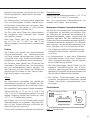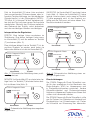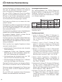
1
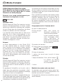
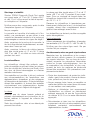
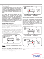
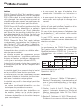
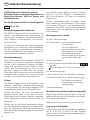
Ebola Rapid Lateral Flow Test Test rapide Schnelltest Rapid lateral flow test for the qualitative detection of the VP40 antigen of the Ebola virus in serum and throat swabs. For Professional In Vitro Diagnostic Use Only Kit de détection rapide de l‘antigène VP40 du virus Ebola dans le sérum humain et sur frottis pharyngé. Réservé à un usage exclusivement professionnel de diagnostic in vitro Schnelltest zum qualitativen Nachweis von EbolaVirus-Antigen VP40 im Serum und Rachenabstrich. Nur für die professionelle In-vitro-Diagnostik EN FR DE Instruction for use Mode d’emploi Gebrauchsanweisung EN Instruction for use STADA Diagnostik Ebola Rapid Lateral Flow Test Rapid lateral flow test for the qualitative detection of the VP40 antigen of the Ebola virus in serum and throat swabs For Professional In Vitro Diagnostic Use Only REF 2.13.1.122 Appearance of a colored line in the test region indicates a positive result (presence of Ebola antigen), while its absence indicates a negative result. For process control, a colored line will always appear in the control line region, indicating correct reaction conditions and procedure (proper volume of sample has been added and membrane wicking has occurred). Intended use Contents of the kit The STADA Diagnostik Ebola Rapid Lateral Flow Test is a rapid visually read chromatographic immunoassay for the qualitative detection of Ebola virus antigen in human serum and throat swabs. Kit for 20 determinations 20 test cassettes in aluminium pouch including desiccant bag The kit comprises particles coated with anti-Ebola antibody, anti-Ebola antibodies and anti-mouse IgG immobilized on nitrocellulose membrane. It is used to detect Ebola infection in febrile conditions. It is intended to be used only together with clinical information/methods and other diagnostic in vitro procedures. Summary 20 transfer pipettes Ebola hemorrhagic fever (Ebola HF) is a severe, often fatal disease of humans and nonhuman primates (monkeys, gorillas and chimpanzees) that has appeared sporadically. The virus is one of two members of a family of RNA viruses called the Filoviridae. There are five identified subtypes of Ebola virus. 90 % of the outbreaks are caused by the Ebola Zaire strain. 20 sterile polyester-tipped applicators/ swabs 0086, Puritan Medical Products Company LLC 20 extraction tubes 1 workstation for up to 8 extraction tubes 1 x 10 ml extraction buffer, ready-to-use The buffer solution contains detergent and preservative. Principle The STADA Diagnostik Ebola Rapid Lateral Flow Test is a rapid, qualitative, membrane-based immunoassay for detection of the VP40 antigen of the Ebola virus in human serum and throat swabs. After applying the sample to the sample well, it can react with particles coated with anti-Ebola antibodies. The mixture migrates upward on the membrane by capillary action. Upon reaching the test line, antigen bound to the particles can react with the capture reagent, an anti-Ebola antibody immobilized on the test line of the membrane, and generate a colored line. 2 1 package insert Equipment required but not provided Timer, for measuring the time between application of the sample and interpretation of the results after ten minutes. Sample collection device for serum. Storage and stability Store the STADA Diagnostik Ebola Rapid Lateral Flow Test unopened at +4 °C to +30 °C (+39 °F to +86 °F) and use it by the expiration date given on the label. Do not use any component past the labeled expiration date. Do not freeze. The test cassette is sensitive to light and to humidity, therefore do not use test devices that have become wet or cassettes whose pouch is damaged or shows signs of damage! After opening of the test cassette pouch, the test has to be performed immediately, within max. 1 hour. Warnings, notices and precautions Handle all samples and used test components as though they contain infectious agents. Observe established precautions against microbiological hazards throughout the procedure. Appropriate biosafety practices must be complied with when handling samples and test components. These precautions include, but are not limited to, the following. • Wear personal protective equipment: powder-free gloves, lab coat, eye protectors etc. After opening, the buffer vial can be stored at +4 °C to +30 °C (+39 °F to +86 °F) and safely be used for three months. • Do not eat, drink, smoke, apply cosmetics, or handle contact lenses in areas where these materials are handled. Samples • Clean and disinfect all spills of samples using suitable disinfectant, such as 0.5 % sodium hypochlorite. Samples should be collected, handled and shipped using appropriate infection control precautions for Ebola, other hemorrhagic fever viruses or other infectious diseases. Shipping may be performed according to the policies of the sender, customs regulations, and the requirements of the receiving laboratory. If samples are to be shipped, they should be packed in compliance with statutory regulations for transportation of etiologic agents. • Decontaminate and dispose of all samples, test components and other potentially contaminated materials in accordance with the local regulations. Testing procedure STADA Diagnostik Ebola xxxxxxxxxx Serum Only use human serum collected and prepared according to the instructions of the manufacturer of the sampling device for sample collection. Only clear, non-hemolyzed samples may be used. Serum may be stored at +2 °C to +8 °C (+35 °F to +46 °F) for up to 3 days. For long-term storage, samples may be kept below -20 °C/-4 °F. Bring samples to room temperature prior to testing. Frozen samples must be completely thawed and mixed well prior to testing. Samples should not be frozen and thawed repeatedly. Throat swabs Use sample at room temperature only (+15 °C to +30 °C/+59 °F to +86 °F). Only use fresh throat swabs. Do not store. Do not freeze. Result Window Sample Well Fig. 1: Test cassette Preparation Allow the kit components and sample to equilibrate to room temperature (+15 °C to +30 °C/+59 °F to +86 °F) prior to testing. • Remove the test cassette from the sealed pouch and use it as soon as possible. The test should be performed within 1 hour after opening the pouch. 3 EN Instruction for use • Place the test cassette on a clean and level surface. • Clearly indicate the patient/sample identification on the test device to avoid confusion. • Place the extraction tube in the workstation and indicate the patient/sample identification on the tube. Throat swabs • Hold the extraction buffer bottle vertically and add 8 to 10 drops of the extraction buffer solution to the extraction tube. Only use the buffer solution provided with this test kit. • Insert the provided sterile swab into the patient’s oral cavity. • Very gently push the swab until resistance is met at level of the pharynx. • Gently rotate the swab for a few times over the mucous membrane. • Immediately insert the swab with collected throat swab sample into the tube, compress the bottom of the tube and rotate the swab vigorously to mix the reagents for at least 1 minute. Press the swab against the inside of the tube, and squeeze as much liquid as possible from the swab by pressing and rotating the fiber portion against the wall of the tube. Discard the swab. • Let the mixture incubate for 1 to 15 minutes. • Mix the contents of the tube again by gentle swirling. • Now the mixture is ready to be tested. Fill the provided transfer pipette with the mixture. • Hold the extraction buffer bottle vertically and add 3 drops (approx. 100 µl) of the extraction buffer to the extraction tube. Only use the buffer included in the test kit. • Hold the provided transfer pipette vertically, transfer 3 drops of sample (approx. 100 µl) into the extraction tube and incubate for 1 to 15 minutes. • Now the mixture is ready to be tested. Fill the provided transfer pipette with the mixture, hold the pipette vertically over the sample well of the test cassette and drop the whole mixture into the sample well. Wait until each drop is absorbed before adding additional drops. • Start the timer. • Wait 10 minutes from addition of the sample/ buffer mixture and then read the results. Warning: Do not interpret the results after more than 15 minutes, to avoid misinterpretation. Quality control A process control is included in the test. A colored line appearing in the control region (C) is the internal process control. It confirms sufficient sample volume and correct technique. If the control line fails to appear (insufficient sample volume or incorrect techniques are the most likely reasons), the test result is INVALID (figure 4). Check the procedure and repeat the test with a new test cassette. If the problem persists, discontinue using the test kit immediately and contact your local distributor. Interpretation of results • Hold the provided transfer pipette over the sample well of the test cassette and squeeze 6 drops into the well. Wait until each drop is absorbed before adding additional drops. POSITIVE: Two distinct colored lines appear. One line should be in the control region (C), and another line should be in the test region (T) (figure 2). • Start the timer. Any visible colored line in the test region (T) should be interpreted as positive, even if the line in the test region (T) appears lighter or darker than the line in the control region (C). • Wait 10 minutes from addition of the sample/ buffer mixture and then read the results. Warning: Do not interpret the results after more than 15 minutes, to avoid misinterpretation. 4 Serum STADA Diagnostik Ebola STADA Diagnostik Ebola xxxxxxxxxx xxxxxxxxxx Control Line Region Test Line Region Control Line Region Test Line Region Fig. 2: Schematic illustration of a positive result Fig. 4: Schematic illustration of invalid results NEGATIVE: One colored line appears in the control region (C). No visible line appears in the test region (T) (figure 3). Limitations STADA Diagnostik Ebola xxxxxxxxxx Control Line Region Test Line Region Fig. 3: Schematic illustration of a negative result INVALID: Control line fails to appear (figure 4). If there is no colored line in the control region (C), even if a colored line appears in the test region (T), the result is invalid, and the test should be repeated with a new test cassette. STADA Diagnostik Ebola xxxxxxxxxx The STADA Diagnostik Ebola Rapid Lateral Flow Test is designed for qualitative in vitro detection of the VP40 antigen of the Ebola virus in human serum and throat swabs. Other body fluids and pooled samples may not give accurate results, and should not be used. The STADA Diagnostik Ebola Rapid Lateral Flow Test should be used only on samples from patients with suspected Ebola infections (febrile diseases) as an aid to detect Ebola hemorrhagic fever. It should not be used as the sole criterion in diagnosing Ebola infections. The STADA Diagnostik Ebola Rapid Lateral Flow Test will indicate presence of Ebola virions at densities of ≥106 PFU/ml in the sample. Therefore it should not be used to exclude Ebola infections. Results should be interpreted by a physician only. Results should be used only in synopsis with clinical information/symptoms/methods and other In Vitro Diagnostic procedures (e.g. PCR techniques). The intensity of the colored line in the test region does not correlate to the titer of antigen in the sample. A negative result does not exclude the possibility of Ebola infection at any time. Control Line Region Test Line Region 5 EN Instruction for use A positive result should be confirmed using a different method, and the result should be evaluated together with the overall clinical evaluation. False-positive or false-negative results may be produced by: • insufficient sample volume or incorrect techniques • use of inappropriate sample or materials (other buffer than the one included in the test kit, other volumes of samples and buffer than those indicated) • disregard of the incubation time of 1 minute of sample and extraction buffer • disregard of the waiting time of 10 minutes after applying the mixture to the test cassette Low titers of Ebola may result in a faint line appearing in the test region after a prolonged time. The “Hook effect” in case of very high levels of antigen in the sample can lead to false-negative results. Apart from EDTA, no substance has been demonstrated to trigger cross-reactions. Performance characteristics The performance of STADA Diagnostik Ebola Rapid Lateral Flow Test has been compared with that of the PCR system RealStar® Ebola virus RT-PCR Kit 1.0 based on samples of African patients from Conakry in Guinea. STADA Diagnostik Ebola Analytic sensitivity: ≤CT 23 Specificity: 98 % 6 References 1. Lucht A, Grunow R, Möller P, Feldmann H, Becker S: Development, characterization and use of monoclonal antibodies for the detection of Ebola virus. J Virol Methods 2003; 111:21-8 2. Grolla A, Lucht A, Dick D, Strong JE, Feldmann H: Laboratory diagnosis of Ebola and Marburg hemorrhagic fever. Bull Soc Pathol Exot 2005; 98:205-9 3. Becker S, Feldmann H, Will C, Slenczka W: Evidence of occurrence of filovirus antibodies in humans and imported monkeys: Do subclinical filovirus infections occur worldwide? Med Microbiol Immunol 1992; 181:43-55 4. World Health Organization. Outbreak(s) of Ebola haemorrhagic fever in the Republic of Congo, January-April 2003. Wkly Epidemiol Rec 2003; 78:285-9 5. Hartmann H Lübbers B, Cararetto M, Bautsch W, Klos A, Köhl J. Rapid quantification of C3a and C5a using a combination of chromatographic and immunoassay procedures. J Immunol Methods 1993; 166:35-44 6. Ksiazek TG, Rollin PE, Jahrling PB, Johnson E, Dalgard DW, Peters CJ: Enzyme immunosorbent assay for Ebola virus antigens in tissues of infected primates. J Clin Microbiol 1992; 30:947-50 7. National Committee for Clinical Standards Clinical Waste Management: Approved Guideline. NCCLS Document GPS-A. Villanova, PA: NCCLS 1993; 13:1-18, 29-42 8. Towner JS, Rollin PE, Bausch DG, Sanchez A, Crary SM, Vincent M, Lee WF, Spiropoulou CF, Ksiazek TG, Lukwiya M, Kaducu F, Downing R, Nichol ST: Rapid diagnosis of ebola hemorrhagic fever by reverse transcription-PCR in an outbreak setting and assessment of patient viral load as a predictor of outcome. J Virol 2004; 78:4330-41 Package contains a sterile product 0086, Puritan Medical Products Company LLC kit contains single-use items Senova Gesellschaft für Biowissenschaft und Technik mbH, Industriestr. 8, 99427 Weimar, Germany Distributed by/Distribué par/Vertrieb durch: STADApharm GmbH, 61118 Bad Vilbel, Germany, www.stada.de 25.02.2015 K22 7 FR Mode d‘emploi STADA Diagnostik Ebola Test rapide Kit de détection qualitative rapide de l‘antigène VP40 du virus Ebola dans le sérum humain et sur frottis pharyngé La présence d’une bande colorée dans la zone de test indique un résultat positif (présence de l’antigène Ebola), alors que son absence indique un résultat négatif. Réservé à un usage exclusivement professionnel de diagnostic in vitro Pour contrôler la procédure, une bande colorée apparaîtra toujours dans la zone de la ligne de contrôle, indiquant que les conditions de la réaction et la technologie ont bien fonctionné (le volume d’échantillon correct a été ajouté et l’ascension capillaire a bien eu lieu au niveau de la membrane). REF 2.13.1.122 Utilisation visée STADA Diagnostik Ebola Test rapide est un test immuno-chromatographique à lecture visuelle rapide pour la détection qualitative de l’antigène du virus Ebola dans des échantillons de sérum humain ou prélevés par frottis pharyngé. Il est destiné à être utilisé dans le cadre de la détection de la fièvre hémorragique à virus Ebola, dans les cas de maladies infectieuses avec fièvre. Il ne peut être utilisé qu’avec les informations / méthodes cliniques et autres procédures de diagnostic in vitro. Composants du kit / Contenu du kit Kit pour 20 déterminations 20 tests de type cassette Le test contient des particules recouvertes d’anticorps anti-Ebola et d‘anti-IgG de souris immobilisés sur une membrane en nitrocellulose. Résumé La fièvre hémorragique à virus Ebola (FHV) est une maladie grave et souvent mortelle pour l’homme et les primates (singes, gorilles et chimpanzés) qui se manifeste sporadiquement. Le virus est l’un des deux membres de la famille des virus à ARN appelés Filovirus. Cinq sous-types de virus Ebola ont été identifiés. 90 % des épidémies sont causées par la souche Ebola-Zaïre. Principe STADA Diagnostik Ebola Test rapide est un test immunologique qualitatif comportant une membrane pour la détection de l’antigène VP40 du virus Ebola dans le sérum humain et sur frottis pharyngé. 8 dans une poche en aluminium contenant un sachet de déshydratant. 20 pipettes de transfert 20 cotons-tiges en polyester stérile 0086, Puritan Medical Products Company LLC 20 tubes d’extraction 1 station de travail avec jusqu’à 8 tubes d‘extraction 1 tampon d’extraction 10 ml, prêt à l’emploi La solution du tampon contient un détergent et un conservateur. 1 notice En appliquant l’échantillon sur le puits, celui-ci réagit avec des particules recouvertes d’anticorps anti-Ebola. Par action capillaire, le mélange migre vers le haut sur la membrane. Matériel nécessaire mais non fourni En atteignant la zone de test, l’antigène lié aux particules peut réagir avec le réactif de capture, l’anticorps anti-Ebola, immobilisé sur la ligne de test de la membrane, et générer une bande colorée. Un appareil de collecte des échantillons (pour le sérum humain). Un minuteur, pour mesurer le temps entre l’application des échantillons et l’interprétation des résultats au bout de 10 minutes. Stockage et stabilité Stocker STADA Diagnostik Ebola Test rapide non ouvert entre +4 °C et +30 °C (entre +39 °F et +86 °F) et l’utiliser avant la date d‘expiration indiquée sur l’étiquette. N‘utiliser aucun des composants après la date d’expiration figurant sur l’étiquette. Ne pas congeler. La cassette est sensible à la lumière et à l’humidité ; par conséquent, ne pas utiliser si une cassette est devenue humide ou si la poche est endommagée ou montre des signes de dégât ! Après ouverture de la poche de la cassette, le test doit être effectué immédiatement, au maximum dans l’heure qui suit. Après ouverture, le flacon de solution tampon peut être stocké entre +4 °C et +30 °C (entre +39 °F et +86 °F) et utilisé en toute sécurité pendant trois mois. Les échantillons Les échantillons doivent être prélevés, manipulés et expédiés en utilisant toutes les précautions de contrôle de l’infection par Ebola, d’autres virus de fièvre hémorragique ou d’autres maladies infectieuses. Une expédition est possible si elle est conforme aux recommandations de l‘expéditeur, aux réglementations douanières et aux exigences du laboratoire de destination. Si des échantillons doivent être expédiés, ils doivent être emballés conformément aux réglementations légales relatives au transport d’agents étiologiques. Le sérum N’utiliser que du sérum humain prélevé et préparé conformément aux instructions du fabricant relatives à l’appareil de collecte des échantillons. Seuls des échantillons clairs et non hémolysés peuvent être utilisés. Le sérum peut être stocké entre +2 °C et +8 °C (entre +35 °F et +46 °F) pendant 3 jours maximum. Pour un stockage de longue durée, les échantillons doivent être conservés en dessous de -20 °C / -4 °F. Ramener les échantillons à température ambiante avant d‘effectuer le test. Les échantillons congelés doivent être complètement décongelés et bien mélangés avant le test. Les échantillons ne doivent pas être recongelés après décongélation. Frottis pharyngés Utiliser uniquement des échantillons à température ambiante (+15 °C à +30 °C / +59 °F à +86 °F). N’utiliser que des cotons-tiges neufs. Ne pas stocker. Ne pas congeler. Avertissements, mises en garde et précautions Manipuler tous les échantillons et les composants de tests utilisés comme s’ils contenaient des agents infectieux. Tout au long de la procédure, respecter les précautions nécessaires contre les risques microbiologiques. Lors de la manipulation des échantillons et des composants de tests, les pratiques de biosécurité doivent être respectées. Ces précautions comprennent entre autres : • Porter des équipements de protection individuelle : gants non poudrés, blouse de laboratoire, protections pour les yeux, etc. • Ne pas manger, boire, fumer, se maquiller ou mettre des lentilles de contact dans des zones où ces matières sont manipulées. • Nettoyer et désinfecter tout renversement d‘échantillons en utilisant un désinfectant adéquat approprié, tel que de l’hypochlorite de sodium à 0,5 % ou un autre désinfectant approprié. • Décontaminer et jeter tous les échantillons, composants de tests et autre matériel potentiellement contaminé, conformément aux réglementations locales. 9 FR Mode d‘emploi Procédure coton-tige contre l’intérieur du tube et extraire autant de liquide que possible du coton-tige en appuyant et en tournant la partie fibreuse contre la paroi du tube. Jeter le coton-tige. STADA Diagnostik Ebola xxxxxxxxxx • Laisser le mélange incuber pendant 1 à 15 minutes. Fenêtres de résultats Puits d‘échantillons Fig. 1: Cassette Préparation Laisser le temps aux composants de tests et aux échantillons de se stabiliser à la température ambiante (+15 °C à +30 °C / +59 °F à +86 °F) avant d‘effectuer le test. • Sortir la cassette de la poche scellée et l’utiliser dès que possible. Le test doit être effectué dans l’heure suivant l’ouverture de la poche. • Placer la cassette sur une surface propre et plane. • Indiquer clairement le nom du patient / de l‘échantillon sur la cassette afin d’éviter toute erreur. • Le mélange est maintenant prêt à être testé. Remplir la pipette de transfert fournie avec le mélange. • Tenir la pipette de transfert au-dessus du puits de la cassette et ajouter 6 gouttes dans le puits. Attendre que chaque goutte soit absorbée avant d’ajouter des gouttes supplémentaires. • Démarrer le minuteur. • Attendre 10 minutes à partir de l’ajout du mélange de l‘échantillon / la solution tampon, puis lire le résultat. Attention ! Ne pas lire le résultat au-delà de 15 minutes pour éviter des erreurs d‘interprétation. Le sérum Frottis pharyngé • Tenir verticalement le flacon de tampon d‘extraction et ajouter 3 gouttes (environ 100 µl) de solution tampon dans le tube d’extraction. N’utiliser que la solution tampon fournie dans le kit de test. • Tenir verticalement le flacon du tampon d‘extraction et ajouter 8 à 10 gouttes de solution tampon dans le tube d’extraction. N’utiliser que la solution tampon fournie dans le kit de test. • Tenir verticalement la pipette de transfert fournie, transférer 3 gouttes d’échantillon (environ 100 µl) dans le tube d’extraction et laisser incuber pendant 1 à 15 minutes. • Insérer le coton-tige stérile fourni dans la cavité buccale du patient. • Le mélange est maintenant prêt à être testé. Remplir la pipette de transfert fournie avec le mélange, tenir verticalement la pipette de transfert au-dessus du puits de la cassette et ajouter tout le mélange dans le puits. Attendre que chaque goutte soit absorbée avant d’ajouter des gouttes supplémentaires. • Placer le tube d’extraction sur la station de travail et indiquer le nom du patient / de l‘échantillon sur le tube. • Pousser très doucement le coton-tige jusqu´à rencontrer une résistance au niveau du pharynx. • Tourner doucement plusieurs fois le coton-tige sur la muqueuse. • Insérer immédiatement le coton-tige avec le prélèvement d‘échantillon dans le tube. Comprimer le fond du tube et faire tourner vigoureusement le coton-tige pour mélanger les réactifs au moins 1 minute. Appuyer le 10 • Mélanger le contenu du tube à nouveau en le tournant doucement. • Démarrer le minuteur. • Attendre 10 minutes à partir de l’ajout du mélange de l‘échantillon / de la solution tampon, puis lire le résultat. Attention ! Ne pas lire le résultat au-delà de 15 minutes pour éviter des erreurs d‘interprétation. Contrôle qualité STADA Diagnostik Ebola Un contrôle de procédure est inclus dans le test. La bande colorée qui apparaît dans la zone de contrôle (C) constitue le contrôle de procédure. Elle confirme que le volume d‘échantillon est suffisant et que la technique de procédure est correcte. Si la bande de contrôle n’apparaît pas (les raisons les plus probables sont un volume d‘échantillon insuffisant ou des techniques de procédure incorrectes), le résultat du test N‘EST PAS VALIDE (figure 4). Revoir la procédure et répéter le test avec une nouvellle cassette. Si le problème persiste, ne plus utiliser le kit de test et contacter le distributeur local. Interprétation du résultat POSITIF : Deux bandes colorées distinctes apparaissent. Une bande doit se trouver dans la zone de contrôle (C) et l’autre dans la zone de test (T) (figure 2). xxxxxxxxxx Zone de contrôle Zone de test Fig. 3 : Illustration schématique d‘un résultat négatif NON VALIDE : La bande de contrôle n’apparaît pas (figure 4). Si aucune bande colorée n’apparaît dans la zone de contrôle (C), même si une bande colorée apparaît dans la zone test (T), le résultat n‘est pas valide et le test doit être répété avec une nouvelle cassette. STADA Diagnostik Ebola xxxxxxxxxx Toute bande colorée visible dans la zone de test (T) doit être interprétée comme étant un résultat positif, même si la bande dans la zone de test (T) apparaît plus claire ou plus foncée que la bande dans la zone de contrôle (C). STADA Diagnostik Ebola Zone de contrôle Zone de test xxxxxxxxxx STADA Diagnostik Ebola xxxxxxxxxx Zone de contrôle Zone de test Fig. 2 : Illustration schématique d‘un résultat positif NÉGATIF : Une bande colorée apparaît dans la zone de contrôle (C). Aucune bande n’apparaît dans la zone de test (T) (figure 3). Zone de contrôle Zone de test Fig. 4 : Illustration schématique de résultats non valides 11 FR Mode d‘emploi Limites STADA Diagnostik Ebola Test rapide est conçu pour la détection qualitative in vitro de l’antigène VP40 d’Ebola dans le sérum humain et dans le frottis pharyngé. Les autres liquides corporels et échantillons groupés peuvent ne pas donner de résultats exacts et ne doivent pas être utilisés. STADA Diagnostik Ebola Test rapide ne doit être utilisé que pour des échantillons de patients soupçonnés d’infection par Ebola (maladies avec fièvre) afin de permettre la détection de la fièvre hémorragique à virus Ebola. Il ne doit pas être utilisé en tant que seul critère pour le diagnostic de l’infection par Ebola. STADA Diagnostik Ebola Test rapide indique la présence de concentration de l‘antigène du virus Ebola ≥106 PFU / ml dans l‘échantillon. Aussi ne doit-il pas être utilisé pour exclure les infections par Ebola. L’interprétation des résultats ne doit être effectuée que par un médecin. Les résultats doivent être uniquement utilisés avec des informations / symptômes / méthodes cliniques et d’autres procédures de diagnostic in vitro (par ex. techniques utilisant la PCR). • le non-respect du temps d‘incubation d‘une minute pour l‘échantillon ou l‘extraction du tampon • le non-respect du temps d’attente de 10 minutes après avoir appliqué le mélange sur la cassette Après une durée prolongée, la faible concentration d’Ebola peut entraîner l’apparition d’une ligne très claire dans la zone de test. En cas de très hauts niveaux d’antigènes dans les échantillons, l’« effet crochet » peut conduire à des faux-négatifs. A part l‘EDTA, aucune autre substance n‘est actuellement connue comme déclenchant des réactions croisées. Caractéristiques de performance La performance de STADA Diagnostik Ebola Test rapide a été comparée au système PCR RealStar-Ebolavirus RT-PCR Kit 1.0 au moyen d‘échantillons de partients de Conakry en Guinée. STADA Diagnostik Ebola L’intensité de la bande colorée dans la zone test ne correspond pas au titre de l’antigène dans l‘échantillon. Un résultat négatif à un moment donné n’exclut pas la possibilité d’une infection par Ebola. Sensibilité analytique : ≤CT 23 Spécificité : 98 % Un résultat positif doit être confirmé en utilisant une autre méthode et le résultat doit être interprété avec l’évaluation clinique globale. Références Un résultat faux-positif ou faux-négatif peut être causé par : • un volume insuffisant d‘échantillons ou des techniques de procédure incorrectes • une mauvaise utilisation des échantillons ou de matières (autres que la solution tampon qui est contenue dans le kit de test, d’autres volumes d‘échantillons et de solution tampon que ceux indiqués) 12 1. Lucht A, Grunow R, Möller P, Feldmann H, Becker S: Development, characterization and use of monoclonal antibodies for the detection of Ebola virus. J Virol Methods 2003; 111:21-8 2. Grolla A, Lucht A, Dick D, Strong JE, Feldmann H: Laboratory diagnosis of Ebola and Marburg hemorrhagic fever. Bull Soc Pathol Exot 2005; 98:205-9 3. Becker S, Feldmann H, Will C, Slenczka W: Evidence of occurrence of filovirus antibodies in humans and imported monkeys: Do subclinical filovirus infections occur worldwide? Med Microbiol Immunol 1992; 181:43-55 4. World Health Organization. Outbreak(s) of Ebola haemorrhagic fever in the Republic of Congo, January-April 2003. Wkly Epidemiol Rec 2003; 78:285-9 5. Hartmann H Lübbers B, Cararetto M, Bautsch W, Klos A, Köhl J. Rapid quantification of C3a and C5a using a combination of chromatographic and immunoassay procedures. J Immunol Methods 1993; 166:35-44 6. Ksiazek TG, Rollin PE, Jahrling PB, Johnson E, Dalgard DW, Peters CJ: Enzyme immunosorbent assay for Ebola virus antigens in tissues of infected primates. J Clin Microbiol 1992; 30:947-50 7. National Committee for Clinical Standards Clinical Waste Management: Approved Guideline. NCCLS Document GPS-A. Villanova, PA: NCCLS 1993; 13:1-18, 29-42 8. Towner JS, Rollin PE, Bausch DG, Sanchez A, Crary SM, Vincent M, Lee WF, Spiropoulou CF, Ksiazek TG, Lukwiya M, Kaducu F, Downing R, Nichol ST: Rapid diagnosis of ebola hemorrhagic fever by reverse transcription-PCR in an outbreak setting and assessment of patient viral load as a predictor of outcome. J Virol 2004; 78:4330-41 Le kit contient un produit stérile. 0086, Puritan Medical Products Company LLC Le kit contient des produits à usage unique Senova Gesellschaft für Biowissenschaft und Technik mbH, Industriestr. 8, 99427 Weimar, Germany Distributed by/Distribué par/Vertrieb durch: STADApharm GmbH, 61118 Bad Vilbel, Germany, www.stada.de 25.02.2015 K22 13 DE Gebrauchsanweisung STADA Diagnostik Ebola Schnelltest Schnelltest zum qualitativen Nachweis von Ebola-Virus-Antigen VP40 im Serum und Rachenabstrich Das Auftreten dieser gefärbten Linie im Testfeld zeigt ein positives Ergebnis (Vorhandensein des Ebola-Antigens), ihr Fehlen ein negatives Ergebnis an. Nur für die professionelle In-vitro-Diagnostik Zwecks Verfahrenskontrolle erscheint immer eine Farblinie im Kontrollbereich. Diese deutet auf korrekte Reaktions- und Verfahrensbedingungen hin (das heißt, das Probenvolumen war ausreichend und die Membran wurde ausreichend benetzt). REF 2.13.1.122 Bestimmungsgemäßer Gebrauch Der STADA Diagnostik Ebola Schnelltest ist ein visueller chromatografischer Immunoassay für den schnellen qualitativen Nachweis des Ebola Virus-Antigens in Humanserum- und Rachen abstrichproben. Er dient der Erkennung des hämorrhagischen Ebolafiebers bei infektiösen Fieberkrankheiten. Der Test sollte nur zusammen mit klinischen Informationen/Methoden und anderen In-vitro-Diagnoseverfahren eingesetzt werden. Zusammenfassung Beim hämorrhagischen Ebolafieber (Ebola HF) handelt es sich um eine sporadisch auftretende und oft tödlich verlaufende Krankheit bei Menschen und nichtmenschlichen Primaten (Affen, Gorillas und Schimpansen). Das Virus ist einer der zwei Erreger der RNA-Viren der Gruppe Filoviridae. Beim Ebola-Virus wird zwischen fünf Subtypen unterschieden. 90 % aller Ausbrüche werden vom Stamm Ebola Zaire verursacht. Prinzip Der STADA Diagnostik Ebola Schnelltest ist ein schneller qualitativer Membran-Immunoassay für den Nachweis des Ebola-Virus-Antigens VP40 in Humanserumproben und Rachenabstrich. Sobald die Probe auf dem Testträger auf gebracht wurde, kann sie mit den mit EbolaAntikörpern beschichteten Partikeln reagieren. Durch Kapillarkraft läuft das Gemisch über die Membran. Sobald das an die Partikel gebundene Antigen das Testfeld erreicht, reagiert es mit dem im Bereich der Testlinie der Membran immobilisierten Ebola-Antikörper (Fängerreagenz) und bildet eine farbige Linie. 14 Kit-Komponenten/-Inhalt Kit für 20 Bestimmungen 20 Testkassetten im Aluminiumbeutel mit Trockenmittel. Der Test enthält mit Ebola-Antikörpern beschichtete Partikel und auf einer Nitrozellulosemembran immobilisierte Ebola-Antikörper und Anti-Maus-IgG. 20 Transferpipetten 20 sterile Polyestertupfer/Wattestäbchen 0086, Puritan Medical Products Company LLC 20 Extraktionsröhrchen 1 Workstation für bis zu 8 Extraktionsröhrchen 1 Extraktionspuffer 10 ml, gebrauchsfertig Die Pufferlösung enthält Detergenzien und Konservierungsmittel. 1 Packungsbeilage Zusätzliche Ausrüstung (nicht bereitgestellt) Stoppuhr für die Messung der Zeit von der Aufgabe der Probe bis zur Interpretation der Ergebnisse nach zehn Minuten. Material zur Probensammlung (nur für Serum). Lagerung und Stabilität STADA Diagnostik Ebola Schnelltest ungeöffnet bei +4 °C bis +30 °C (+39 °F bis +86 °F) lagern und bis zum auf der Packung angegebenen Verfallsdatum verwenden. Niemals Komponenten nach Ablauf des auf dem Etikett angegebenen Verfallsdatum benutzen. Nicht einfrieren. Die Testkassetten sind empfindlich gegenüber Licht und Feuchtigkeit. Deshalb niemals feuchte Kassetten verwenden oder Kassetten, deren Aluminiumbeutel beschädigt ist oder Zeichen von Beschädigung erkennen lässt. Der Test sollte nach Öffnen des Aluminiumbeutels umgehend oder innerhalb einer Stunde durchgeführt werden. Nach dem Öffnen kann der Extraktionspuffer bei +4 °C bis +30 °C (+39 °F bis +86 °F) drei Monate gelagert und bedenkenlos angewendet werden. Proben Alle Proben sind gemäß den entsprechenden Vorsichtsmaßnahmen der Infektionskontrolle für Ebola, andere hämorrhagisches Fieber hervorrufende Viren oder Infektionskrankheiten zu entnehmen, zu handhaben und zu versenden. Der Versand kann gemäß den Richtlinien des Lieferanten, Zollrichtlinien bzw. den Anforderungen des empfangenden Labors erfolgen. Für den Transport sind die Proben im Einklang mit gesetzlichen Richtlinien für den Transport ätiologischer Erreger zu verpacken. Serum Nur Humanserum verwenden, das gemäß den Herstelleranweisungen für das Material zur Probensammlung hergestellt und vorbereitet wurde. Nur ungetrübte, hämolysefreie Proben verwenden. Serum kann bei +2 °C bis +8 °C (+35 °F bis +46 °F) bis zu 3 Tage gelagert werden. Für eine längere Lagerung können die Proben bei unter -20 °C (-4 °F) aufbewahrt werden. Alle Proben sind vor der Testdurchführung auf Raumtemperatur zu erwärmen. Gefrorene Proben vor der Testdurchführung vollständig auftauen und gut durchmischen. Proben nicht wiederholt einfrieren und auftauen. Rachenabstrich Proben nur bei Zimmertemperatur (+15 °C bis +30 °C/+59 °F bis +86 °F) verwenden. Nur frisch gewonnene Rachenabstriche verwenden. Nicht lagern. Nicht einfrieren. Warnungen, Hinweise, Vorsichtsmaßnahmen Alle Proben und gebrauchten Testkomponenten so behandeln, als enthielten sie infektiöse Erreger. Während des gesamten Tests etablierte Sicherheitsmaßnahmen gegen mikrobiologische Gefahren beachten. Bei der Handhabung von Proben und Testkomponenten angemessene Biosicherheitsmaßnahmen einhalten. Die Maßnahmen umfassen die nachfolgend genannten Faktoren, sind aber nicht nur auf diese beschränkt: • Das Tragen persönlicher Schutzausrüstung: puderfreie Handschuhe, Laborkittel, Augen- schutz etc. • Im Arbeitsbereich, in dem diese Materialien verwendet werden, nicht essen, trinken, rauchen, Kosmetika auftragen oder mit Kontaktlinsen in Berührung kommen. • Alle Verunreinigungen durch Proben mit geeignetem Desinfektionsmittel wie 0,5-prozentigem Natriumhypochlorit oder anderem geeigneten Desinfektionsmittel entfernen und die betroffenen Bereiche desinfizieren. • Alle Proben, Testkomponenten und andere potenziell kontaminierten Materialien gemäß lokalen Vorschriften dekontaminieren bzw. entsorgen. Testverfahren STADA Diagnostik Ebola xxxxxxxxxx Ergebnisfenster Probenmulde Abb. 1: Testkassette 15 DE Gebrauchsanweisung Vorbereitung Proben und Testkomponenten vor der Testdurchführung auf Zimmertemperatur (+15 °C bis +30 °C/+59 °F bis +86 °F) bringen. • Die Testkassette aus der versiegelten Verpackung nehmen und so bald wie möglich verwenden. Den Test binnen 1 Stunde nach Öffnen der Verpackung durchführen. • Die Testkassette auf einer sauberen und ebenen Oberfläche platzieren. • Die Testkassette mit den Patienten-/Probeninformationen versehen, um Missverständnisse zu vermeiden. • Das Extraktionsröhrchen in der Workstation platzieren und mit den Patienten-/Proben informationen versehen. Rachenabstrich • Extraktionspufferflasche senkrecht halten und 8 bis 10 Tropfen der Pufferlösung in das Extraktionsröhrchen einbringen. Nur die Pufferlösung aus dem Testkit verwenden. • Das beiliegende sterile Wattestäbchen in die Mundhöhle des Patienten einbringen. • Das Wattestäbchen vorsichtig einführen, bis es den Rachen des Patienten berührt. • Mit dem Wattestäbchen vorsichtig mehrere Male drehend über die Schleimhaut streichen. • Das Wattestäbchen mit dem Rachenabstrich unverzüglich in das Röhrchen einbringen, den Boden des Röhrchens zusammendrücken und das Stäbchen zwecks Mischen der Reagenzien mindestens 1 Minute lang kräftig drehen. Dann durch wiederholtes Abstreifen an der Gefäßwand so viel Flüssigkeit wie möglich aus dem Wattestäbchen herauspressen. Wattestäbchen entsorgen. • Die Mischung 1 bis 15 Minuten lang inkubieren lassen. • Den Inhalt des Röhrchens erneut durch sanftes Schwenken mischen. • Die Mischung ist nun für den Test bereit. Die 16 Mischung mit der beiliegenden Transferpipette aufnehmen. • Die Transferpipette über die Probenmulde der Testkassette halten und 6 Tropfen in die Mulde geben. Nach jedem Tropfen warten, bis dieser vollständig absorbiert ist, bevor ein neuer Tropfen aufgebracht wird. • Die Stoppuhr starten. • Nach Hinzufügen der Probe/Puffermischung 10 Minuten warten und dann die Ergebnisse ablesen. Warnung: Nach mehr als 15 Minuten sollten keine Ergebnisse mehr abgelesen werden, um Fehlinterpretationen vorzubeugen. Serum • Die Extraktionspufferflasche senkrecht halten und 3 Tropfen (ca. 100 µl) der Lösung in das Extraktionsröhrchen einbringen. Nur die Pufferlösung aus diesem Testkit verwenden. • Die beiliegende Transferpipette senkrecht halten und 3 Tropfen der Probe (ca. 100 µl) in das Extraktionsröhrchen einbringen und dann 1 bis 15 Minuten lang inkubieren lassen. • Die Mischung ist nun für den Test bereit. Die Mischung mit der beiliegenden Transferpipette aufnehmen, die Transferpipette senkrecht über die Probenmulde der Testkassette halten und den gesamten Inhalt in die Mulde geben. Nach jedem Tropfen warten, bis dieser vollständig absorbiert ist, bevor ein neuer Tropfen aufgebracht wird. • Die Stoppuhr starten. • Nach Hinzufügen der Probe/Puffermischung 10 Minuten warten und dann die Ergebnisse ablesen. Warnung: Nach mehr als 15 Minuten keine Ergebnisse mehr ablesen, um Fehlinterpretationen vorzubeugen. Qualitätskontrolle Der Test beinhaltet eine interne Verfahrenskontrolle. Diese wird durch eine farbige Linie im Kontrollfeld (C) repräsentiert. Ihr Erscheinen bestätigt ein ausreichendes Probenvolumen und eine korrekte Testdurchführung. Falls im Kontrollfeld (C) keine Linie erscheint (kein ausreichendes Probenvolumen oder inkorrekte Testdurchführung sind die häufigsten Gründe hierfür), ist das Testergebnis UNGÜLTIG (Abb. 4). In diesem Fall das Verfahren prüfen und den Test mit einer neuen Testkassette wiederholen. Besteht das Problem weiterhin, den Gebrauch des Testkits sofort abbrechen und den zuständigen Händler verständigen. UNGÜLTIG: Im Kontrollfeld (C) erscheint keine Linie (Abb. 4). Wenn im Kontrollfeld (C) keine farbige Linie erscheint, auch wenn im Testfeld (T) eine angezeigt wird, ist das Ergebnis ungültig und der Test muss mit einer neuen Testkassette wiederholt werden. STADA Diagnostik Ebola xxxxxxxxxx Interpretation der Ergebnisse POSITIV: Zwei farbige Linien erscheinen im Sichtfenster. Eine dieser farbigen Linien muss im Kontrollfeld (C) und die andere im Testfeld (T) erscheinen (Abb. 2). Eine sichtbare farbige Linie im Testfeld (T) ist als positives Ergebnis zu werten, auch wenn sie heller oder dunkler ist als die im Kontrollfeld (C). Testfeld STADA Diagnostik Ebola STADA Diagnostik Ebola xxxxxxxxxx xxxxxxxxxx Kontrollfeld Kontrollfeld Testfeld Kontrollfeld Testfeld Abb. 2: Schematische Abbildung eines positiven Ergebnisses Abb. 4: Schematische Abbildung eines ungültigen Ergebnisses NEGATIV: Im Kontrollfeld (C) erscheint eine farbige Linie. Im Testfeld (T) erscheint keine deutlich sichtbare Linie (Abb. 3). Einschränkungen STADA Diagnostik Ebola xxxxxxxxxx Der STADA Diagnostik Ebola Schnelltest wurde für den qualitativen In-vitro-Nachweis des Ebola-Antigens VP40 im Humanserum und in Rachenabstrichproben entwickelt. Andere Körperflüssigkeiten und Probenmischungen können zu ungenauen Ergebnissen führen und sollten nicht verwendet werden. Der STADA Diagnostik Ebola Schnelltest sollte nur für Proben von Patienten mit Verdacht auf Ebolainfektion bei Erkrankungen mit Fieber als Hilfsmittel zur Erkennung des hämorrha- Kontrollfeld Testfeld Abb. 3: Schematische Abbildung eines negativen Ergebnisses 17 DE Gebrauchsanweisung gischen Ebolafiebers verwendet werden. Der Test darf nicht als alleiniges Kriterium zur D iagnose einer Ebolainfektion herangezogen werden. Mit dem STADA Diagnostik Ebola Schnelltest können Ebola-Antigenkonzentrationen ≥106 PFU/ml in der Probe nachgewiesen werden. Der Test darf deshalb nicht zum Ausschluss einer Ebolainfektion herangezogen werden. Leistungscharakteristika Die Leistungsfähigkeit des STADA Diagnostik Ebola Schnelltests wurde mit dem PCR-System RealStar® Ebolavirus RT-PCR Kit 1.0 anhand von Proben afrikanischer Patienten aus Conakry in Guinea verglichen. STADA Diagnostik Ebola Die Ergebnisse dürfen nur von einem Arzt interpretiert werden. Die Ergebnisse sollten nur zusammen mit klinischen Informationen/Symptomen/Methoden und anderen In-vitro-Diagnoseverfahren (z. B. PCR-Techniken) verwendet werden. Die Intensität der gefärbten Linie im Testfeldbereich korreliert nicht mit dem Antigentiter der Probe. Ein negatives Ergebnis schließt zu keiner Zeit die Möglichkeit einer Ebolainfektion aus. Positive Ergebnisse sind mithilfe anderer M ethoden zu bestätigen und das Ergebnis wiederum ist unter Berücksichtigung der allgemeinen klinischen Beurteilung zu bewerten. Falsch positive bzw. negative Ergebnisse können verursacht werden durch: • unzureichendes Probenvolumen oder inkorrekte Testdurchführung • ungeeignete Proben oder Materialien (anderer Puffer als der im Testkit enthaltene, andere Proben- und Puffermengen als angegeben) • Nichtbeachtung der Inkubationszeit von 1 Minute für Probe und Extraktionspuffer • Nichtbeachtung der Wartezeit von 10 Minuten nach dem Auftragen der Mischung Niedrige Ebolatiter können nach längerer Wartezeit zu einer schwachen Linie im Testfeld führen. Bei einem hohen Antigengehalt der Probe kann ein „Hook-Effekt“ zu falsch negativen Ergebnissen führen. Bis auf EDTA sind momentan keine Substanzen bekannt, die Kreuzreaktionen verursachen. 18 Analytische Sensitivität: ≤CT 23 Spezifität: 98 % Quellenverzeichnis 1. Lucht A, Grunow R, Möller P, Feldmann H, Becker S: Development, characterization and use of monoclonal antibodies for the detection of Ebola virus. J Virol Methods 2003; 111:21-8 2. Grolla A, Lucht A, Dick D, Strong JE, Feldmann H: Laboratory diagnosis of Ebola and Marburg hemorrhagic fever. Bull Soc Pathol Exot 2005; 98:205-9 3. Becker S, Feldmann H, Will C, Slenczka W: Evidence of occurrence of filovirus antibodies in humans and imported monkeys: Do subclinical filovirus infections occur worldwide? Med Microbiol Immunol 1992; 181:43-55 4. World Health Organization. Outbreak(s) of Ebola haemorrhagic fever in the Republic of Congo, January-April 2003. Wkly Epidemiol Rec 2003; 78:285-9 5. Hartmann H Lübbers B, Cararetto M, Bautsch W, Klos A, Köhl J. Rapid quantification of C3a and C5a using a combination of chromatographic and immunoassay procedures. J Immunol Methods 1993; 166:35-44 6. Ksiazek TG, Rollin PE, Jahrling PB, Johnson E, Dalgard DW, Peters CJ: Enzyme immunosorbent assay for Ebola virus antigens in tissues of infected primates. J Clin Microbiol 1992; 30:947-50 7. National Committee for Clinical Standards Clinical Waste Management: Approved Guideline. NCCLS Document GPS-A. Villanova, PA: NCCLS 1993; 13:1-18, 29-42 8. Towner JS, Rollin PE, Bausch DG, Sanchez A, Crary SM, Vincent M, Lee WF, Spiropoulou CF, Ksiazek TG, Lukwiya M, Kaducu F, Downing R, Nichol ST: Rapid diagnosis of ebola hemorrhagic fever by reverse transcription-PCR in an outbreak setting and assessment of patient viral load as a predictor of outcome. J Virol 2004; 78:4330-41 Packung enthält ein steriles Produkt 0086, Puritan Medical Products Company LLC Packung enthält Produkte zur einmaligen Verwendung Senova Gesellschaft für Biowissenschaft und Technik mbH, Industriestr. 8, 99427 Weimar, Germany Distributed by/Distribué par/Vertrieb durch: STADApharm GmbH, 61118 Bad Vilbel, Germany, www.stada.de 25.02.2015 K22 19 9264560 1502 25.02.2015 K22 Senova Gesellschaft für Biowissenschaft und Technik mbH Industriestraße 8, 99427 Weimar, Germany Distributed by/Distribué par/Vertrieb durch: STADApharm GmbH, 61118 Bad Vilbel, Germany, www.stada.de