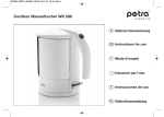
Download Gebrauchsanweisung Instruction for Use Mode d`emploi
Transcript
Gebrauchsanweisung Instruction for Use Mode d’emploi JOTEC GmbH Lotzenäcker 23 D-72379 Hechingen T +49 (0) 7471/922 – 0 F +49 (0) 7471 / 922 – 100 [email protected] www.jotec.net JOTEC Gebrauchsanweisung E-vita® thoracic Deutsch S. 2 English S. 15 Français S. 26 2 JOTEC Gebrauchsanweisung E-vita® thoracic Vor dem Gebrauch bitte sorgfältig alle Anweisungen lesen. Bitte alle Warnhinweise und Vorsichtsmaßnahmen beachten, die in dieser Anweisung aufgeführt sind. Eine Missachtung kann zu erheblichen Komplikationen führen. 1.0 Produktbeschreibung Die konventionelle Behandlung von Thorakalen Aorten Aneurysmen (TAA) und Thorakalen Aorten Dissektionen ist eine in hohem Maße invasive chirurgische Maßnahme. Wie bei jeder größeren Operation ist das mit einem chirurgischen Eingriff verbundene Risiko hoch. ® Die endoluminalen Stentgraftprothesen der JOTEC GmbH stellen Alternativen zu konventionellen Operationen und Behandlungsmöglichkeiten zur Behebung von Gefäßaneurysmen, Dissektionen und Aortenrupturen in der thorakalen Aorta dar. ® Das JOTEC E-vita thoracic Stentgraft System wurde speziell für die endoluminale Behandlung von Läsionen, insbesondere Aneurysmen und Dissektionen, in der thorakalen Aorta descendens entwickelt. ® Das E-vita thoracic System besteht aus zwei Hauptkomponenten: • dem selbstexpandierenden Nitinolstent mit Polyesterummantelung und Platin-Iridium Röntgenmarkern, sowie • dem Einführsystem, auf dem der E-vita thoracic Stentgraft montiert ist. ® ® Das Einführsystem mit dem E-vita thoracic Stentgraft kann durch eine geeignete 20F, 22F oder 24F E-asy Schleuse oder direkt über einen 0,035 Zoll (0,89mm) steifen, beschichteten Führungsdraht in den Körper eingeführt und bis zum geschädigten Abschnitt der thorakalen Aorta vorgeschoben werden. Durch Zurückziehen der Textilhülle des Einführsystems und der separaten Freigabe der proximalen Stentfeder wird der selbstexpandierende Mechanismus des E® vita thoracic Stentgrafts ausgelöst. ® Der E-vita thoracic Stentgraft legt sich über den geschädigten Gefäßabschnitt. Damit wird der Blutfluss durch das Lumen des Stentgrafts geleitet und von der Läsion abgeschirmt. Falls erforderlich kann mit Hilfe eines separaten Ballonkatheters ® der E-vita thoracic Stentgraft schrittweise an die Gefäßwand anmodelliert werden. ® 1.1 E-vita thoracic TAA Stentgraft Der TAA Stentgraft (Abb. 1 und 2) besteht aus einzelnen Stentfedern (NiTi-Legierung), die in einem Gewebeschlauch (PET) dauerhaft mit chirurgischem Nahtmaterial (PET, geflochten) eingenäht sind. Unterschiedliche Durchmesser und ® Formen der Stentfedern ermöglichen die Bereitstellung von E-vita thoracic Stentgrafts in Rohr- oder in Kegelform. Eine optimale Anpassung an den geschädigten Gefäßabschnitt ist somit möglich. ® E-vita thoracic Stentgrafts, die sehr weit in den Aortenbogen implantiert werden müssen, sind mit freien Stentfedern (Straight Open Design) versehen und können dort die abgehenden supraaortalen Gefäße, insbesondere die Arteria subclavia, offen halten. Im distalen Bereich der Prothese kann eine Offenhaltung abgehender Gefäße durch eine freie Stentfeder (Free Spring ® Design) erreicht werden. Bei besonders langen Läsionen können auch zwei oder mehrere E-vita thoracic Stentgrafts überlappend ineinander implantiert werden. Hier ist darauf zu achten, dass zwischen den beiden Stentgrafts ein Durchmesserunterschied von ca. 20%, allerdings nicht größer als 6mm, besteht und die Überlappungslänge mindestens 30 mm beträgt. ® Zur besseren Röntgensichtbarkeit bei der Positionierung und zur Nachkontrolle des E-vita thoracic Stentgrafts sind kleine Röntgenmarkerringe aus einer Pt-Ir-Legierung an das Polyestergewebe angenäht. Bei der Straight open-Straight Cut (SO) Konfiguration sind 5 Röntgenmarkerringe, bei der Twin Stent – Free Spring Konfiguration 3 und bei der Twin Stent – Straight Cut Konfiguration 4 Röntgenmarkerringe angebracht. 3 JOTEC Gebrauchsanweisung E-vita® thoracic Röntgenmarkierungen Straigth Open Design Straight Cut Design ® Abbildung 1: E-vita thoracic Stentgraft, Artikelbeispiel 70SOxxyySzz-00 Röntgenmarkierungen a) Free Spring Design Twin Stent Design b) Röntgenmarkierungen Twin Stent Design Straight Cut Design ® Abbildung 2: E-vita thoracic Stentgraft a) Artikelbeispiel 70STxxyyFzz-00 b) Artikelbeispiel 70STxxyySzz-00 1.2 Einführsystem Das Einführsystem (Abb. 3) besteht aus vier ineinander verschachtelten Kathetern: • einem innen liegenden Drahtführungskatheter • einem zwischenliegendem Auslösekatheter für die proximale Stentgraftfreisetzung • einem Pusher (Katheter) • und der äußeren Einführschleuse mit proximaler Textilummantelung. Wichtig: Der Schalter am Freisetzungsgriff muss bis zum Zeitpunkt der Freisetzung des Stentgrafts in der dafür vorgesehenen Stellung P verbleiben. Das Lumen des Drahtführungskatheters dient dem Führungsdraht. Am vorderen Ende des Drahtführungskatheters befindet ® sich eine weiche, röntgensichtbare Spitze aus Polyurethan (PU). Der E-vita thoracic Stentgraft ist am proximalen Ende des ® Einführsystems geladen, wobei der Pusher (Katheter) das distale Ende des E-vita thoracic Stentgrafts berührt (Abb. 3, Detail C). Durch Verschieben des Schalters am Freisetzungsgriff in Stellung D, wird das System in den Betriebsmodus versetzt und der Stentgraft kann freigesetzt werden (Abb. 3, Detail B). Dies geschieht durch kontrolliertes Zurückziehen der Textilhülle gegen den Pusher. Die Fixierung der ersten Stentfeder kann zu jedem Zeitpunkt der Freisetzung gelöst werden, indem der rote Auslöseknopf (Abb. 3, Detail A) am Pushergriff betätigt wird. Nach kompletter Freisetzung des Stentgraft wird das Einführsystem aus dem Gefäßsystem entfernt. 4 JOTEC Gebrauchsanweisung E-vita® thoracic Detail A Detail B Pushergriff Detail C Freisetzungsgriff Detail A: Pushergriff Auslöseknopf (rot) Sicherung (weiß) Spülventil Detail B: Freisetzungsgriff b) geöffneter Zustand Stellung D a) geschlossener Zustand Stellung P Hebelgriff geöffnet Schalter mit den Stellungen P, D und N Detail C Spülschlauch Einführungsschleuse mit geladenem Stentgraft Pusher Röntgenmarkierung derTextilhülle Weiche Spitze ® Abbildung 3: E-vita thoracic Stentgraft System 5 JOTEC Gebrauchsanweisung E-vita® thoracic 2.0 Indikation ® ® Der E-vita thoracic Stentgraft der JOTEC GmbH ist speziell für die Behandlung von Aneurysmen, Dissektionen und spezifischen Läsionen der thorakalen Aorta descendens entwickelt worden. Hauptindikationen für den Einsatz des TAA ® Stentgrafts der JOTEC GmbH liegen vor allem bei akuten vitalen Bedrohungen des Patienten durch • die Ruptur eines Aneurysmas • eine traumatische Aortenruptur • eine Aortendissektion • eine Plaqueruptur • ein penetrierendes aortales Ulkus (PAU) • ein intramurales Hämatom (IMH) • eine aortobronchiale Fistel oder eine aortoösophagale Fistel vor. Der Stentgraft ist für Patienten gedacht, bei denen entweder ein konventioneller chirurgischer Eingriff indiziert oder aufgrund von präexistenten Risikofaktoren kontraindiziert ist. 3.0 Kontraindikation ® Das E-vita thoracic Stentgraft System ist für folgende Patienten kontraindiziert: • Patienten, deren Gefäße stark gewunden sind. Im Zweifel kontaktieren Sie JOTEC. • Patienten, deren Gefäß- bzw. Aneurysmagröße für das E-vita thoracic Stentgraft System nicht geeignet ist. • Patienten, bei denen das Aneurysma oder die durch den Stentgraft auszuschaltende Aortenerkrankung Abgänge lebenswichtiger Gefäße (Carotiden, Viszeral- und Nierenarterien) einschließt. • Patienten mit Stenosen, die mit dem Einführsystem nicht durchquert werden können. • Patienten, deren Aorta am Absetzpunkt des distalen Ende des Stentgrafts eine starke Krümmung aufweist. • Patienten, deren arterielle Zugangsstelle für die zur Implantation des E-vita thoracic Stentgraft Systems zu eng ist. • Patienten, bei denen aufgrund der Arbeitslänge des Einführsystems eine situativ erforderliche proximale Positionierung in der Aorta unmöglich ist. • Patienten, bei denen die zur Implantation erforderlichen Materialien (s. u.) nicht eingesetzt werden können. ® ® Eine sorgfältige Abwägung der Behandlungsindikation ist erforderlich bei: • Patienten mit systemischen oder lokalen Infektionen, bei denen eine bakterielle Infektion des Stentgrafts möglich erscheint. • Patienten, bei denen Röntgen-Kontrastmittel, Antikoagulatien oder Heparinapplikation kontraindiziert ist. • Patienten, bei denen das E-vita thoracic Stentgraft System das Risiko einer Paraplegie oder eines sensoriellen Funktionsverlustes erhöhen würde. ® Achtung: Die endgültige Entscheidung über die Art der Behandlung liegt im Ermessen des Arztes. 6 JOTEC Gebrauchsanweisung E-vita® thoracic ® 4.0 Größenbestimmung für das E-vita thoracic Stentgraft System Vor Beginn des Eingriffes müssen Größe und Eigenschaften des erkrankten Aortenabschnittes sowie der Zugangsgefäße, wie Gefäßdurchmesser und -zustand (z. B. Verkalkungen und Stenosen, intraluminale Thromben) mit Hilfe geeigneter Bildgebung festgestellt werden. Hier empfiehlt es sich, Spiral-CT-Aufnahmen mit Kontrastmittel und 3D-Darstellung durchzuführen. Die Gefäßdurchmesser müssen im gesunden Aortenabschnitt gemessen werden. Der für das native Gefäß geeignete Stentgraftdurchmesser wird durch Überdimensionierung „Oversizing“ ermittelt. Die Richtgröße ist 20%, jedoch nicht größer als 6 mm, Bsp: 24 mm Gefäßdurchmesser + 20% = 28,8 mm. Dies entspricht einem 28 mm Stentgraftdurchmesser. Bei Dissektionen sollte die Überdimensionierung nicht mehr als 10% betragen. In die Längenbestimmung ist sowohl die proximale als auch die distale Verankerungszone mit einzubeziehen. Mindestens eine ummantelte Federreihe ® muss in der Verrankerungszone zum Liegen kommen. Das distale Ende des E-vita thoracic Stentgrafts darf nicht direkt an ® einer starken Krümmung (Knick) positioniert werden. Dies kann durch sorgfältige Festlegung der Länge des E-vita thoracic Stentgrafts vermieden werden. Vor Beginn des Eingriffes muss geprüft werden, ob die Arbeitslänge des Einführsystems ausreicht um die erforderliche, proximale Positionierung in der Aorta zu erreichen. ® Bei Verwendung mehrerer E-vita thoracic Stentgraft Systeme ist Folgendes zu beachten: • Der Überlappungsbereich zwischen zwei oder mehreren Stentgraft Systemen sollte mindestens 30 mm betragen. In winkeligen oder gekrümmten Bereichen wird ein größerer Überlappungsbereich empfohlen. • Die Stentgrafts sollten einen Durchmesserunterschied von 20% jedoch nicht größer als 6 mm aufweisen • Es darf nur der Stentgraft mit dem größeren Durchmesser in den Stentgraft mit dem kleineren Durchmesser platziert werden. Achtung: Die Auswahl der richtigen Größen liegt in der Verantwortung des behandelnden Arztes. Bei speziellen anatomischen Gegebenheiten können die proximalen und distalen Stentgraftanschlüsse optional kundenspezifisch hergestellt werden. Bei Fragen können Sie sich gerne unter +49 (0)7471-922 0 an unseren Customer Care oder einen Vertreter der JOTEC GmbH wenden. 5.0 Vorbereitung auf das thorakale Stentgraftverfahren ® 5.1 Erforderliche Ausrüstung für die Implantation eines E-vita thoracic Stentgrafts • Röntgenbildwandler mit digitaler Angiographiefunktion (Mobiles C-Bogen-Gerät oder stationäre Angiographieanlage). • Fluoroskopische Visualisierung und die Möglichkeit zum Aufzeichnen und Abrufen aller Bilder. • Operationssaal für den Fall, dass eine offene Operation notwendig werden sollte. Hinweis: Die Implantation sollte nur von einem in endovaskulären Techniken ausgebildeten Arzt (Herz-, Thorax- und Gefäßchirurg, interventioneller Radiologe oder Kardiologe) durchgeführt werden. Die personellen und organisatorischen Möglichkeiten für den einfachen oder erweiterten Gefäßzugang oder den Umstieg auf die offene Operation sollten zur Verfügung stehen. 7 JOTEC Gebrauchsanweisung E-vita® thoracic 5.2 Zur Implantation erforderliche Materialien • Verschiedene weiche Führungsdrähte mit mindestens 260 cm Länge • Ein steifer beschichteter Führungsdraht mit 0,035 Zoll (0,89 mm) Durchmesser und 300 cm Länge, vorzugsweise der ® JOTEC E -wire Draht. • Diverse Angiographie- und Angioplastie-Katheter. • Eine geeignete E-asy Einführschleuse, wahlweise 20F, 22F und 24F Hinweis: Bei der Verwendung einer E-asy Einführschleuse von JOTEC verkürzt sich die Arbeitslänge des E-vita thoracic Stentgraft Systems um 90 mm! • Sterile Einführschleusen: 7, 8 oder 9 French ( 3 French ≅ 1 mm). • Kontrastmittel • Punktionsnadel • Sterile Spritzen • Heparin und heparinisierte physiologische Kochsalzlösung • Ballonkatheter 6.0 Heparinisierung und Thrombozytenaggregationshemmung Zur Reduktion des Risikos einer Thromboembolie sollte das Blut der Patienten während des Eingriffs antikoaguliert werden. Wie bei einem gefäßchirugischen Eingriff sollten intraoperativ gewichtsadaptiert zwischen 5000 und 10000 IE Heparin appliziert werden. Bei Heparinunverträglichkeit können alternative Präparate wie z.B. Hirudin eingesetzt werden. Nach jeweiligem Ermessen des Arztes kann postoperativ eine Antithrombozyten-Therapie indiziert sein. Vorsicht: Ungenügende Heparinisierung kann zu Thrombose und Embolie führen. 7.0 Vorbereitung zur Implantation • Das Zugangsgefäß, A. iliaca oder A. femoralis, wird in üblicher chirurgischer Technik freigelegt. • Eine zweite Zugangsstelle kann für diagnostische Zwecke zum Legen eines Angiographiekatheters verwendet werden. Diese kann nach Ermessen des Arztes perkutan femoral, brachial o.ä. angelegt werden. • Unter Fluoroskopie kann über einen 0,035 Zoll (0,89mm) liegenden steifen Führungsdraht eine zum Einführungssystem passende E-asy Schleuse bis unmittelbar unter die Nierenarterien geschoben werden. • Führen Sie über die zweite Zugangsstelle einen Angiographiekatheter ein, um die Implantationsstelle zu lokalisieren. Der Angiographiekatheter kann während der Implantation an seiner Stelle bleiben und zur Orientierung eingesetzt werden. • • Nach Entfernen der beiden Schutzdrähte aus dem Einführsystem wird das Lumen des Drahtführungskatheters mit heparinisierter Kochsalzlösung gespült. Pushergriff Stellen Sie sicher, dass der Spülschlauch über die Textilhülle, in ® der der E-vita thoracic Stentgraft geladen ist, geschoben ist. Das Einführsystem wird mit dem proximalen Ende nach oben gehalten und über das eingelassene Spülventil am Pushergriff mit heparinisierter Kochsalzlösung gespült und entlüftet. Spülventil ® Achtung: Die E-vita thoracic darf ohne Führungsdraht nicht gebogen oder geknickt werden. Vergewissern Sie sich, dass der Katheter nicht beschädigt sind. Im Zweifel muss das System verworfen und durch ein Neues ersetzt werden. 8 JOTEC Gebrauchsanweisung E-vita® thoracic ® 7.1 Einführung des E-vita thoracic Stentgraft Systems • ® Das E-vita thoracic Stentgraft System wird über einen steifen beschichteten 0,035 Zoll (0,89 mm) Führungsdraht direkt oder bei verengten und/oder kalzifizierten Zugangsgefäßen durch eine passende E-asy Einführschleuse in das Gefäß eingeführt. • Der E-vita thoracic Stentgraft muss kurz vor dem Einführen in die Arterie bzw. in die Einführschleuse vollständig entlüftet sein. Geben Sie ggf. einen weiteren Bolus Spülflüssigkeit (heparinisierte Kochsalzlösung) über das eingelassene Spülventil am Pushergriff in den Stentgraft. Der Spülschlauch muss den geladenen Stentgraft abdecken. ® Führen Sie das E-vita thoracic Stentgraft System dann in die freigelegte Arterie bzw. in die vorgelegte Schleuse ein. Dabei wird der Spülschlauch von selbst zurückgedrückt. • Entfernen Sie diesen Spülschlauch nachdem die Textilhülle vollständig in das Zugangsgefäss bzw. Schleuse eingeführt wurde. • Das E-vita thoracic Stentgraft System wird langsam unter Fluoroskopie bis zur Implantationsstelle vorgeschoben. • Wird der Pushergriff rotiert, muss unter Fluoroskopie sicher gestellt werden, dass die Katheterspitze und das Implantat der Drehbewegung folgen. Sollte das System in sich verdreht werden, könnte dies zu einem Katheterbruch führen. • Vor der Freisetzung des E-vita thoracic Stentgrafts ist die genaue Position und Lage des geladenen Stentgrafts angiographisch zu überprüfen. Danach ist der Bildwandler zu fixieren und die Orientierungspunkte für die Freisetzung ® zu markieren (Road-map, Anzeichnen am Bildschirm oder externe Markierung z.B. durch Nadeln). Der E-vita thoracic Stentgraft besitzt am proximalen Ende Röntgenmarker. Die den Beginn der zirkulären Graftummantelung markieren ® ® ® Weitere Marker sind in der Mitte und am distalen Ende des Stentgrafts angebracht. • Soll der E-vita thoracic Stentgraft an ein abgehendes Gefäß, z.B. der A. subclavia, platziert werden, muss der erste Röntgenmarker distal des abgehenden Gefäßes platziert werden. • Sobald das E-vita thoracic Stentgraft System ausgerichtet und in seine genaue Position gebracht worden ist, kann die kurzfristige Senkung des mittleren arteriellen Blutdrucks des Patienten auf etwa 60 mmHg angebracht sein. Diese ® Maßnahme dient der Vermeidung eines versehentlichen Verschiebens des E-vita thoracic Stentgrafts bei der Freisetzung. ® ® • Entfernen Sie, sofern nicht bereits geschehen die Transportsicherung um den Freisetzungsgriff (weißes Klettband) . • Schieben Sie den Schalter am Freisetzungsgriff in Stellung D. • Der orangene Hebel am Griff springt auf. Das System ist nun bereit zur Freisetzung. Freisetzungsgriff geöffneter Zustand (Stellung D) • Hebelgriff geöffnet Durch gleichmäßiges Auf- und Abbewegen des orangenen Hebels am Freisetzungsgriff, unter gleichzeitiger Fixierung des Pushergriffes mit der anderen Hand, wird der Stentgraft von proximal nach distal freigesetzt. Wichtig: Die Position von Stentgraft und Katheterspitze wird durch den Pushergriff bestimmt. • Die Textilhülle mit Markerring wird unter Fluoroskopie schrittweise zurückgezogen bis die ersten drei Stentfedern freigesetzt sind (Abb. 4). Die erste Feder ist dabei noch proximal an der Spitze des Drahtführungskatheters fixiert. • Die Position des E-vita thoracic Stentgrafts in Bezug auf die Implantationsstelle sollte mit Hilfe einer Angiographie ® bzw. Fluoroskopie kontrolliert werden. Falls der E-vita thoracic Stentgraft zu weit oberhalb bzw. unterhalb der ® 9 JOTEC Gebrauchsanweisung E-vita® thoracic vorgesehenen Implantationsstelle teilentfaltet wurde, kann das gesamte Einführsystem behutsam nach distal bzw. proximal geschoben werden, bis der erste Röntgenmarker die gewünschte Position anzeigt. Achtung: Im Falle einer unbeabsichtigten Freisetzung der proximalen Stentfeder muss dieses Manöver unterbleiben! • Sicherung Die Freisetzung der proximalen Feder des Stentgrafts erfolgt nach Entfernen der weißen Sicherung am Pushergriff durch Drücken des roten Auslöseknopfes. Vergewissern Sie sich unter Fluoroskopie, dass die proximale Feder vollständig frei gegeben wurde. Wichtig: Setzen Sie die weiße Sicherung nach erfolgreicher Auslösung wieder an die vorgesehene Stelle am Pushergriff ein. Pushergriff • Die vollständige Freisetzung des E-vita thoracic Stentgrafts nach distal ist erreicht, wenn der Freisetzungsgriff an den fixierten Pushergriff stößt • Nach erfolgter Freisetzung wird der Schalter am Freisetzungsgriff auf Position N geschoben. Fixieren Sie nun mit einer Hand den Freisetzungsgriff und ziehen Sie am Pushergriff das Einführsystem wieder in die Ausgangposition. Danach können Sie das verschlossene Einführsystem aus der Aorta bzw. aus der Schleuse entfernen. • Beim Zurückziehen darf der Führungsdraht nicht mit zurückgezogen bzw. entfernt werden. • Zur Behandlung längerer Aortensegmente können weitere E-vita thoracic Stentgrafts verwendet werden. Hier ist ® ® darauf zu achten, dass eine Überlappung von mindestens 30 mm und ein Oversizing von 20%, allerdings nicht größer als 6 mm, eingehalten werden. Es darf nur der Stentgraft mit dem größeren Durchmesser in den Stentgraft mit dem kleineren Durchmesser platziert bzw. entfaltet werden. • Nach Ausschluss einer Endoleckage und Dokumentation der vollständigen Exklusion der Aortenläsion mittels abschließender Angiographie, werden die E-asy Schleuse, sofern verwendet, und der steife Führungsdraht entfernt. • Die unmittelbare, intraoperative Kontrolle des Therapieerfolges, also der Ausschluss von Leckagen und die Offenheit des Stentgrafts, erfolgt geeigneterweise mittels digitaler Subtraktionsangiographie (DSA). • Die arterielle Zugangsstelle wird in üblicher chirurgischer Technik verschlossen. 8.0 Warnhinweise und Vorsichtsmaßnahmen • Das E-vita thoracic Stentgraft System darf nicht bei Patienten verwendet werden, bei denen die notwendigen prä- und postoperativen bildgebenden Untersuchungen nicht durchgeführt werden können. Alle Patienten sollten sorgfältig überwacht und regelmäßig auf eine Änderung ihres Krankheitszustandes und die Funktion und Integrität des Stentgrafts hin untersucht werden. • Das E-vita thoracic Stentgraft System darf nicht verwendet werden, wenn die Arbeitslänge des Einführsystems nicht ausreichend ist. • Das E-vita thoracic Stentgraft System darf nicht eingesetzt werden, wenn die Aorta am Absetzpunkt des distalen Endes Stentgrafts eine starke Krümmung aufweist. • Das Produkt darf nicht erneut sterilisiert werden. Das Produkt darf nicht verwendet werden, wenn die Verpackung beschädigt ist oder wenn diese außerhalb des sterilen Bereichs geöffnet wurde. • ® ® ® Das Produkt darf nicht verwendet werden, wenn das proximale Ende des Textilschlauches von der Spitze abgerutscht ist. • Das Produkt darf nicht verwendet werden, wenn das proximale oder das distale Ende einen Knick aufweist (möglicher Transportschaden). • Ungenügende Heparinisierung kann zu Thrombose und Embolie führen. • Es ist auf eine mögliche Empfindlichkeit oder Allergieneigung des Patienten gegen Heparin oder/und Kontrastmittel zu achten. • Das E-vita thoracic Einführungssystem ist nach Verwendung gemäß den geltenden Krankenhaus-, Verwaltungs- oder ® behördlichen Vorschriften zu entsorgen. 10 JOTEC Gebrauchsanweisung E-vita® thoracic • Der E-vita thoracic Stentgraft im Einführsystem muss kurz vor dem Einbringen ins arterielle Gefäßsystem vollständig entlüftet sein. Geben Sie ggf. einen Bolus Spülflüssigkeit (heparinisierte Kochsalzlösung) über das eingelassene Ventil am Pushergriff in den Stentgraft mit übergezogenem Spülschlauch. Die Textilhülle des Einführsystems gewährleistet eine vollständige Entlüftung auch in horizontaler Position. • Der E-vita thoracic Stentgraft darf vor der Freisetzung nicht tordiert sein. • Der Schalter am Freisetzungsgriff muss bis zum Zeitpunkt der Freisetzung des Stentgrafts in Position P verbleiben. • Rotationsmanöver zur Ausrichtung des proximalen E-vita thoracic Stentgrafts dürfen nicht im Aortenbogen oder in stark gekrümmten Aortensegmenten durchgeführt werden. • Der E-vita thoracic Stentgraft darf nur innerhalb des ummantelten Bereiches mit einem Ballon anmodelliert werden. Beim Anmodellieren einer freien, nicht ummantelten Feder kann es zu einer Perforation der Aortenwand kommen. • Beim Zurückziehen des Drahtführungskatheters ist darauf zu achten, dass sich der distale Teil der Spitze nicht in der ® proximalen Stentfeder verhakt. Eine Dislokation oder Stauchung des E-vita thoracic Stentgrafts könnte die Folge sein. ® ® ® ® Beobachten Sie bitte während dieses Manövers genau die Lage des Stentgrafts. Prüfen Sie die Situation, falls Widerstände beim Zurückziehen auftreten. Kontrollieren Sie stets die Lage des Führungsdrahtes. • Leckagen sind die am häufigsten beobachteten Komplikationen bei endovaskulären Stentgraft Implantationen. Nach ® Abschluss der Implantation des E-vita thoracic Stentgrafts muss auf Leckagen am Stentgraft in das Aneurysma oder das falsche Lumen der Dissektion geachtet werden. Geringfügige, vorübergehende Leckagen durch das Prothesengewebe sind unter Umständen auf die intraoperative Heparinisierung zurückzuführen. Leckagen sind im Rahmen des Follow-up zu beobachten. • Mikroembolien – Die Mikroemboliegefahr erhöht sich mit zunehmender Manipulation und Dauer des Eingriffs. Daher sind übermäßige und langwierige endovaskuläre Manipulationen zu vermeiden. • Gefäßthrombose – Falten und Unebenheiten im Stentgraft können zu Thrombosebildung führen. Deshalb kann mit ® einem seperaten Ballonkatheter der E-vita thoracic Stentgraft nachgedehnt werden, um Faltenbildung und Unebenheiten auf ein Minimum zu reduzieren. Dabei ist stets darauf zu achten, dass der Ballon nicht zu stark ® expandiert bzw. aufgedehnt wird. Eine übermäßige Verengung des E-vita thoracic Stentgrafts kann ebenfalls zu ® Thrombosen führen. Nach der Platzierung des E-vita thoracic Stentgrafts muss intraoperativ mittels Angiographie ® oder postoperativ mittels Brustkorb-Röntgenaufnahme sicher gestellt werden, dass der implantierte E-vita thoracic Stentgraft nicht geknickt oder verengt ist. Stark verengte Bereiche sind unverzüglich zu behandeln. • ® Migration – Wenn der E-vita thoracic Stentgraft zu klein ist, kann dies zu einer distalen Stentmigration führen. Bei der ® Größenbestimmung des E-vita thoracic Stentgrafts sollte eine 20%-ige Überdimensionierung (aber nicht mehr als 6mm), bei Dissektionen nicht mehr als 10%, im Vergleich zum Gefäßdurchmesser erfolgen. Weitere Ursachen einer Migration sind ungenügende Fixierung in dem insgesamt zur Verfügung stehenden Aortenhals und die Expansion des proximalen Stentgrafts in einem mit Thromben gefüllten oder stark abgewinkelten Gefäß. Für die Auswahl der richtigen Größen ist der behandelnde Arzt verantwortlich. • Gefäßüberdehnung – Überdehnung und Schäden können infolge eines im Verhältnis zum Gefäßdurchmesser zu groß gewählten E-vita ® thoracic Stentgrafts oder bei zu starkem Aufdehnen/Anmodellieren während eines nachgeschalteten Ballonmanövers verursacht werden. • Schlaganfall – Emboliefragmente oder eine Blockierung der brachiozephalen Gefäße können einen Schlaganfall verursachen. • Darmischämie – Der vollständige oder teilweise Verschluss des Tr. coeliacus und der A. mesenterica superior durch ® einen falsch platzierten E-vita thoracic Stentgraft oder durch stentbedingte Embolien kann zu Ischämie der Oberbauchorgane und des Darmes führen. • ® Niereninsuffizienz – Der vollständige oder teilweise Verschluss der A. renalis durch einen falsch platzierten E-vita thoracic Stentgraft oder durch stentbedingte Embolien kann zu ernsthaften Nierenkomplikationen führen. Ein Übermaß an Kontrastmittel kann ebenfalls zu ernsthaften Nierenkomplikationen führen. 11 JOTEC Gebrauchsanweisung E-vita® thoracic 9.0 Mögliche Komplikationen und Nebenerscheinungen In der Literatur werden bei der Anwendung von Stentgrafts 30-Tage-Mortalitätsraten zwischen 7 und 10% angegeben. Beim Alternativverfahren, der offenen Operation, werden 30-Tage-Mortalitätsraten zwischen 12 und 31% genannt. Komplikationen können jederzeit während oder nach der Stentgraft-Implantation auftreten. Eine solche Behandlung sollte nur von Ärzten durchgeführt werden, die mit den möglichen Komplikationen vertraut sind. Es können außerdem Spätfolgen auftreten, die durch regelmäßige Nachuntersuchungen zu beobachten sind. Klinische Komplikationen: • Zu den Gefäßkomplikationen zählen: Thrombose, Thromboembolie, Verschluss (arteriell und venös), Dissektionen, Perforation, Verschluss kollateraler Gefäße, vaskuläre Ischämie, Gewebenekrose, Amputation • Gefäßruptur/Aneurysma • Zu den neurologischen Komplikationen zählen: Paralyse, Paraparese, zerebrovaskuläre Ereignisse, transitorischischämische Anfälle und Schlaganfall • Darmischämie • Infektion und Fieber • Gastrointestinale Komplikationen • Blutungen • Lungenkomplikationen • Komplikationen bei der Wundheilung • Ödem • Herzversagen/Infarkt • Umstieg auf offene Operation • Tod ® Mit dem E-vita thoracic Stentgraft System zusammenhängende Probleme: • Endoleckage • Migration • Klemmen verschiedener Komponenten des Einführsystems (Freisetzungsblockade) • Ablösung des Freisetzungsgriffes von der äußeren Einführschleuse bei Freisetzungsblockade • Verdrehen oder Knicken des Stentgrafts • Schwierigkeiten beim Einführen und Entfernen des Einführsystems • Expansionsschwierigkeiten (z.B. Stentgraft bleibt im Einführsystem stecken) • Stentgraft entfaltet sich nicht • Ungenaue Platzierung (z.B. falsche Orientierung des Stentgrafts) • Reißen des Graftmaterials • Bruch der Stentgraftfedern 10.0 MRT-Information Sicherheitsinformationen für Magnetresonanztomographie (MRT-Verfahren) beziehen sich auf die Verwendung von abgeschirmten MRT-Systemen mit statischen Magnetfeldern von 1,5 Tesla oder geringer, Gradient-Magnetfeldern von 20 Tesla/Sekunde oder geringer und einer maximalen am Ganzkörper gemittelten spezifischen Absorptionsrate (SAR) von 1,2W/kg für eine 30 minütige Visualisierung. Der Stentgraft ist MRT-sicher. Das bedeutet, dass der Stentgraft im Körper eines Patienten, der sich einem MRT-Verfahren unterzieht, keine zusätzliche Gefährdung darstellt, aber je nach Pulssequenz und dem zu evaluierenden Bereich die MRTBildqualität beeinflussen kann. 12 JOTEC Gebrauchsanweisung E-vita® thoracic 11.0 Lagerung und Verpackung ® Beim E-vita thoracic TAA Stentgraft System handelt es sich um ein mit Gammastrahlen sterilisiertes Einmalprodukt, das in einer Sterilverpackung geliefert wird und steril bleibt, solange die Verpackung nicht geöffnet oder beschädigt wird. Die Aufbewahrung des Stentgraft Systems muss an einem dunklen, trockenen und kühlen Ort bei Temperaturen zwischen 10°C und 38°C erfolgen. Direkte Sonneneinstrahlung muss vermieden werden - Hitze könnte erheblichen, negativen Einfluss auf die Funktionalität des Produktes nehmen. Setzen Sie das Stentgraft System keiner ionisierenden Strahlung oder ultraviolettem Licht aus. Garantie und Gewährleistung ® Die JOTEC GmbH garantiert, dass dieses Produkt den gesetzlichen Anforderungen, insbesondere denen der EG® Richtlinie für Medizinprodukte 93/42/EWG, entspricht. Daher wird die JOTEC GmbH für den Fall, dass das Produkt vor Ablauf des Verfallsdatums nachweislich Material- oder Verarbeitungsfehler aufweist, die während der Herstellung oder Verpackung entstanden sind: a) das Produkt durch ein bauartgleiches oder b) ein hinsichtlich der Funktion gleichwertiges Produkt ersetzen. Für die Inanspruchnahme der Garantie müssen sämtliche der folgenden Voraussetzungen vorliegen: a) Das Produkt darf nicht nach seinem Verfallsdatum benutzt worden sein. b) Der Fehler ist nicht zum ersten Mal nach dem Verfallsdatum des Produkts aufgetreten. ® c) Wenn es ein nur zur Einmalverwendung zugelassenes oder von der JOTEC GmbH nur zur Einmalverwendung vorgesehenes Produkt ist, muss es sich um die Erstverwendung handeln. ® d) Der Fehler ist unverzüglich der JOTEC GmbH oder ihrem Vertreter gemeldet worden. ® e) Das fehlerhafte Produkt ist unverzüglich nach der Fehlermeldung der JOTEC GmbH oder ihrem Vertreter übersandt worden. Die vorstehende Garantie beeinträchtigt keine der und gilt neben den zwingenden Regelungen der Gewährleistung des Verkäufers und des Produkthaftungsgesetzes. Im übrigen gilt die Garantie nur im vorstehend beschriebenen Umfang. Weitergehende Ansprüche aus der Garantie sind ausgeschlossen. Die Garantie sowie die genannten gesetzlichen Ansprüche gelten anstelle und unter Ausschluss jeglicher anderer mündlichen oder schriftlichen, ausdrücklichen, stillschweigenden oder sonstigen Zusagen durch die ® JOTEC GmbH. Keiner unserer Vertreter oder Mitarbeiter ist berechtigt, anderslautende Zusicherungen zu machen oder anders lautende Vereinbarungen zu treffen. ® Aufgrund zahlreicher Faktoren, die außerhalb der Kontrolle der JOTEC GmbH liegen, wie Versand, Lagerung, Handhabung durch den Anwender, Missbrauch oder Nichtbeachtung der Gebrauchsanleitung oder von Warnhinweisen, ® haftet die JOTEC GmbH insbesondere nicht für Schäden jeglicher Art, die direkt oder indirekt durch das Produkt oder seinen Gebrauch entstehen, wenn diese Schäden nicht nachweisbar durch Fehler des Produktes verursacht wurden. ® Die JOTEC GmbH übernimmt keine Haftung für eine falsche Größenbestimmung des Stentgrafts, eine nicht ® sachgerechte Verwendung des Produktes oder für eine falsche Platzierung des E-vita thoracic Stentgrafts. ® Die JOTEC GmbH haftet insbesondere auch nicht für Schäden, die durch Resterilisation oder Wiederverwendung von ® Produkten verursacht werden, die nur zur Einmalverwendung zugelassen oder von der JOTEC GmbH nur zur Einmalverwendung vorgesehen sind. 13 JOTEC Gebrauchsanweisung E-vita® thoracic Anhang: 1. 3. 2. 4. 5. 1. Vorbereitetes E-vita thoracic System 2. 3. 4. Öffnen des Freisetzungsmechanimus, indem der Schalter am Freisetzungsgriff von P auf D geschoben wird. Beginn der Freisetzung, indem der geöffnete orangene Hebel am Freisetzungsgriff gleichmäßig auf- und abbewegt wird. Der Pushergriff wird währenddessen fixiert. Lösen der proximalen Fixierung, nachdem die weiße Sicherung am Pushergriff entfernt ist, und der rote 5. Auslöseknof betätigt wurde. Anschließend wird die weiße Sicherung wieder an die vorgesehene Stelle am Pushergriff eingesetzt. Vollständige Freisetzung des Stentgrafts. ® Abbildung 4: Freisetzungsprozedur des E-vita thoracic Stentgrafts, Schritte 1 bis 5 14 JOTEC Instruction for use E-vita® thoracic Please read all instructions carefully before using the product. Please comply with all warnings and precautions included in these instructions. Non-compliance may result in serious complications. 1.0 Product description The conventional treatment of thoracic aortic aneurysm (TAA) and TA dissections is a highly invasive surgical procedure. As is always the case with major surgery, the level of risk is high. ® The endoluminal stentgraft prostheses from JOTEC GmbH represent an alternative to the conventional surgical methods and therapeutic practices otherwise applied in treatment of vascular aneurysms, dissections and aortic ruptures of the thoracic aorta. ® The JOTEC E-vita thoracic stentgraft system was specifically developed and designed for endoluminal treatment of lesions, in particular aneurysms and dissections in the thoracic aorta descendens. ® The E-vita thoracic system has two main components: • the self-expandable nitinol stent with polyester mantle and platinum-iridium radiopaque markers and • the delivery system carrying the E-vita thoracic Stentgraft. ® ® The delivery system with the E-vita thoracic Stentgraft is introduced into the body using a 20F, 22F or 24F E-asy sheath or directly via an 0.035" (0.89 mm) rigid, coated guidewire, then advanced up to the damaged section of the thoracic aorta. Retracting the outer shell of the delivery system and separate release of the proximal stent wire activates the self® expandable mechanism of the E-vita thoracic Stentgraft. ® The E-vita thoracic Stentgraft covers the damaged vessel segment surface so that the blood flow passes through the ® ® lumen of the E-vita thoracic Stentgraft and is shielded from contact with the lesion. If necessary, the E-vita thoracic Stentgraft can be modelled step by step onto the vascular wall with the help of a separate balloon catheter. ® 1.1 E-vita thoracic TAA Stentgraft The TAA stentgraft (Fig. 1 and 2) comprises individual stent wires (NiTi alloy), permanently sewn into a textile tube (PET) ® using surgical suture material (PET, woven). E-vita thoracic Stentgrafts can be supplied either in tubular or conical forms, which are realized by varying the diameter and form of the stent wires, providing for optimized adaptation to the damaged vascular segment. ® E-vita thoracic Stentgrafts, which have to be implanted far into the aortic arch, feature free wires (Straight Open Design) to hold open the supraaortal vessels, in particular the subclavian artery. A free wire at the distal end part of the prosthesis holds efferent vessels open. In particularly long lesions, two or more E® vita thoracic Stentgrafts can be implanted so as to overlap. In such cases, the diameters of two adjacent stentgrafts should differ by approximately 20%, at any rate not by more than 6 mm, and the overlap should be at least 30 mm long. Small radiopaque marker rings made of a Pt-Ir alloy have been sewn into the polyester textile to improve X-ray visibility for ® positioning and subsequent checking of E-vita thoracic stengrafts. In the Straight Open – Straight Cut (SO) configuration, 5 radiopaque marker rings are attached and in the Twin Stent – Free Spring / Straight Cut (ST) configuration 3 / 4 radiopaque marker rings are attached. 15 JOTEC Instruction for use E-vita® thoracic X-ray marker Straigth Open Design Straight Cut Design ® Figure 1: E-vita thoracic Stentgraft, e.g. article 70SOxxyySzz-00 X-ray marker a) Free Spring Design Twin Stent Design X-ray marker b) Twin Stent Design Straight Cut Design ® Figure 2: E-vita thoracic Stentgraft, e.g. a) article 70STxxyyFzz-00 b) artiche 70STxxyySzz-00 1.2 Delivery system The delivery system (Fig. 3) consists of four nesting catheters: • an internal guide wire catheter • an intermediate release catheter for proximal stentgraft release • a pusher (catheter) • and the outer delivery sheath with proximal textile mantle. Important: The switch on the release grip must remain in the P position as designated until the stentgraft is released. The lumen of the guide wire catheter is for the guide wire. The proximal end of the guide wire catheter is fitted with a soft, ® radiopaque polyurethane (PU) tip. The E-vita thoracic Stentgraft is loaded at the proximal end of the delivery system. The ® pusher (catheter) is in contact with the distal end of the E-vita thoracic Stentgraft. Shifting the switch on the release handle to position D switches the system to operating mode and the stentgraft can be released. This is realized by means of controlled retraction of the delivery sheath against the pusher. Fixation of the first stent wire can be released at any time during the process by pushing the actuation button (red) on the pusher handle. Following complete release of the stentgraft, the delivery system is removed from the vascular system. 16 JOTEC Instruction for use E-vita® thoracic Detail A Detail B Detail C Pusher handle Release handle Detail A: Pusher handle Retainer (white) Flushing valve Actuation button (red) Detail B: Release handle b) opened position D a) closed position P Lever handle opened Switch with positions P, D and N Flushing tube Detail C Delivery sheath with loaded stentgraft Soft tip Pusher Radiopaque marker ring on textile sheath ® Figure 3: E-vita thoracic Stentgraft System 17 JOTEC Instruction for use E-vita® thoracic 2.0 Indications ® ® The E-vita thoracic Stentgraft from JOTEC GmbH has been specially developed for treatment of aneurysms, dissections ® and specific lesions of the thoracic aorta descendens. The main indications for use of a TAA stentgraft from JOTEC GmbH would be, in particular, acutely life-threatening patient conditions due to • aneurysm rupture • traumatic aortic rupture • aortic dissection • a plaque rupture • a penetrating aortic ulcer (PAU) • an intramural haematoma (IMH) • an aortobronchial fistula or aortooesophageal fistula. The stentgraft was designed for patients for whom conventional surgery is either indicated or contraindicated due to preexistent risk factors. 3.0 Contraindications ® The E-vita thoracic Stentgraft system is contraindicated in the following patients: • Patients with highly tortuous vessels; contact JOTEC if in doubt. • Patients with blood vessels or aneurysms not suitable for treatment with the E-vita thoracic Stentgraft system • Patients in whom the aneurysm or aortic disease to be repaired by the stentgraft includes vitally important vascular exits (carotid arteries, visceral and renal arteries). • Patients with stenoses that cannot be crossed by the delivery system. • Patients with a heavy curve on the setting off point of the distal end of the E-vita thoracic Stentgraft. • Patients in whom the arterial approach site is too narrow for implantation of the E-vita thoracic Stentgraft System. • Patients in whom the necessary proximal positioning in the aorta is impossible due to the work length of the delivery system. • Patients in whom the required implantation materials (see below) cannot be used. ® ® ® The therapeutic indication must be very carefully considered in the following cases: • Patients with systemic or local infections in whom a bacterial infection of the stentgraft would appear to be a possibility • Patients in whom administration of radiological contrast agents, anticoagulants or heparin is contraindicated • Patients in whom the E-vita thoracic Stentgraft System would raise the risk of a paraplegia or loss of sensory function. ® Important: The final therapeutic decision must be made by the physician in charge of treatment. ® 4.0 Sizing of the E-vita thoracic Stentgraft System Before beginning with the procedure, size and properties of the diseased aortic section and access vessels, such as vessel diameter and status (e.g. calcifications and stenoses, intraluminal thrombi) must the determined with the aid of suitable imaging equipment. 3D spiral CT angiography with contrast agent is recommended. The vessel diameters must be measured in the healthy aortic segment. The stentgraft diameter suitable for the native vessel is determined by “oversizing“. 18 JOTEC Instruction for use E-vita® thoracic The guideline figure is 20%, but not more than 6 mm, for example vessel diameter 24 mm + 20% = 28.8 mm, corresponding to a stentgraft diameter of 28 mm. In dissections oversizing should not be more than 10%. When determining the length, both the proximal and distal anchoring length must be included in the calculation. At least one covered spring must be ® placed in the anchoring zone. The distal end of the E-vita thoracic Stentgraft must not be positioned directly in a ® pronounced kink. This can be avoided by means of careful determination of the length of the E-vita thoracic Stentgraft. Before beginning with the procedure it must be determined whether the working length of the delivery system is sufficient to reach the required proximal position in the aorta. ® Important in cases in which several E-vita thoracic Stentgraft Systems are used: • The overlap between two or more stentgraft systems should be at least 30 mm long. A longer overlap is recommended in angled or curved segments. • Further, the stentgrafts must have a diameter difference of 20%, but not exceeding 6 mm • The stentgraft with the larger diameter must always be placed in the stentgraft with the smaller diameter. Important: The physician in charge of treatment is responsible for selection of the correct sizes. To accommodate special anatomical situations, the proximal and distal stentgraft connections can also be custom-made to meet customer requirements. Please contact our Customer Service or a JOTEC representative if you have any questions. 5.0 Preparations for the thoracic stentgraft procedure ® 5.1 Required equipment for implantation of an E-vita thoracic Stentgraft: • Radiographic image converter with digital angiography function (mobile C-Arm device or stationary angiography system) • Fluoroscopic visualization and facilities for recording and callup of all images • Surgical theatre in case open surgery should become necessary. Important: The implantation procedure should only be carried out by a physician trained in endovascular techniques (cardiac and thoracic surgeon, vascular surgeon, interventional radiologist or cardiologist). Staff and facilities should be available for simple or complex vascular access or a transition to open surgery. 5.2 Materials required for the implantation procedure • Various different guide wires, length at least 260 cm • A rigid, coated guide wire measuring 0.035" (0.89 mm) in diameter and 300 cm in length, preferably the JOTEC Ewire • Various different angiography and angioplasty catheters • A suitable sized JOTEC E-asy delivery sheath Note: Using the JOTEC E-asy delivery sheath reduces the working length of the E-vita thoracic Stentgraft System by 90 mm! • Sterile delivery sheaths: 7, 8 or 9 French (3 French ≅ 1 mm). • Contrast agent • Puncture needle • Sterile syringes • Heparin and heparinized physiological saline solution • Balloon catheters 19 JOTEC Instruction for use E-vita® thoracic 6.0 Heparinization and inhibition of thrombocyte aggregation To reduce the risk of a thromboembolism, the patient's blood should be anticoagulated during the procedure. As in vascular surgery, between 5,000 and 10,000 IU of heparin, depending on body weight, should be applied intraoperatively. In cases of heparin intolerance, preparations such as hirudin, for example, can be used. Postoperative antithrombocyte therapy may be indicated as judged by the physician in charge of treatment. Caution: Insufficient heparinization may result in thrombosis and embolism. 7.0 Preparation for implantation • The access vessel, a. iliaca or a. femoralis, is exposed using standard surgical techniques. • A second point of access can be used for diagnostic purposes to lay an angiography catheter. This access point can • Under fluoroscopy, a fitting E-asy sheath for the delivery system can be inserted up to immediately below the renal arteries using an 0.035" (0.89 mm) guidewire. • Insert an angiography catheter through the second access point to localize the implantation site. The angiography catheter can stay put during the implantation procedure for orientation purposes. • After removing the two protective wires from the delivery system, the lumen of the guide wire catheter is flushed with heparinized saline solution. Pusher handle Make sure the flushing tube is covering the textile tube in which be located as per the judgement of the physician percutaneous, femoral or brachial, etc. • ® the E-vita thoracic Stentgraft is loaded. The delivery system is held with the proximal end up and is flushed with heparinized saline solution through the inserted flushing valves at the pusher handle. Flushing valve ® Important: The E-vita thoracic must not be bent or kinked without a guide wire; make sure the catheters have not been damaged. In case of doubt, the system must be discarded and replaced by an intact system. ® 7.1 Delivery of the E-vita thoracic Stentgraft System • The E-vita thoracic Stentgraft System is delivered into the vessel by means of a rigid, coated 0.035" (0.89 mm) guide wire. Use a matching E-asy sheath in case of calcified and/or stenosed vessels. • The E-vita thoracic Stentgraft must be completely deaerated just before it is introduced into the artery or delivery ® ® sheath. If necessary, introduce a further bolus of flushing liquid (heparinized saline solution) into the stentgraft through the inserted flushing valve on the pusher handle. The flushing tube must cover the loaded stentgraft. Then introduce ® the E-vita thoracic Stentgraft System into the exposed artery or in the indwelling sheath, whereby the flushing tube is pushed back of its own accord. • After the textile sheath is completely inserted into the access vessel or the sheath remove this flushing tube. • The E-vita thoracic Stentgraft System is slowly advanced to the implantation site under fluoroscopy. • When the pusher handle needs to be rotated, use fluoroscopy to make sure, tip and stent follow the rotation. Do not ® twist the catheter system as this could lead to catheter damage! • ® Prior to release of the E-vita thoracic Stentgraft, the exact position and situation of the stentgraft must be checked by means of angiography. Then fix the image converter and mark the orientation points for release (roadmap, mark on ® screen or external marking, e.g. with needles). The E-vita thoracic Stentgraft features two radiopaque marking rings at the proximal end (Fig. 1). These markers form the beginning of the circular graft mantle. Further markers are located in the middle and two others at the distal end of the stentgraft. 20 JOTEC Instruction for use E-vita® thoracic • If the E-vita thoracic Stentgraft is to be located on an efferent vessel, e.g. the a. subclavia, the first radiopaque ring must be positioned distally in relation to the efferent vessel. • As soon as the E-vita thoracic Stentgraft System has been precisely positioned, it may be appropriate to reduce the patient’s mean arterial blood pressure to about 60 mmHg. This measure is taken to prevent unintentional shifting of the ® E-vita thoracic Stentgraft upon release. ® ® • If still necessary, the transport retainer should now be removed (white Velcro band). • Push the switch on the release handle to position D. • The orange lever on the handle is raised. The system is ready for release. Release handle opened (Position D) • Lever raised By raising and lowering the orange lever on the release handle while retaining the pusher handle with the other hand the the stentgraft is released from proximal to distal. Important: The position of tip and stentgraft is controlled with the pusher handle. • The textile sheath with the marking ring is retracted until the first three stent wires are released (Fig. 4). This is checked under fluoroscopy. Thereby the first wire is still held proximally at the tip of the guide wire catheter. • The position of the E-vita thoracic Stentgraft in relation to the implantation site should be checked once again using ® angiography or fluoroscopy. If the E-vita thoracic Stentgraft has partially opened too far below or above the intended implantation site, the entire delivery system can be carefully pushed in distal or proximal direction until the first X-ray marker shows the desired position. ® Important: This manoeuvre cannot be performed if the proximal stent wire has been released unintentionally! • retainer The proximal stent wire can now be released from the delivery system by removing the white safety retainer from the pusher handle, then pushing the red actuation button. Check under fluoroscopy to make sure the proximal wire is completely released. Pusher handle Important: Replace the white safety retainer at the intended location on the pusher handle once release is realized. • The E-vita thoracic Stentgraft is completely released in distal direction until the release handle contacts the fixated pusher handle. • Finally, the switch on the release handle is pushed to position N. Now fixate the release handle and pull the delivery ® system back into the initial positions using the pusher handle. Then remove the closed delivery system from the aorta and sheath, respectively. • The guidewire must not be pulled back or removed when removing the E-vita thoracic delivery system. • For treatment of longer aortic segments, additional E-vita thoracic Stentgrafts can be used. Ensure overlapping of at least 30 mm and oversizing of 20%, although not more than 6 mm. The stentgraft with the larger diameter must be positioned / opened in the stentgraft with the smaller diameter. ® 21 JOTEC Instruction for use E-vita® thoracic • Having excluded an endoleakage and documented the complete exclusion of the aortic lesion in the final angiography, the delivery sheath, if used, is removed along with the stiff guide wire. • Direct intraoperative control of therapeutic success, i.e. exclusion of leakage and patency of the stentgraft, is best realized by means of digital subtraction angiography (DSA). • The arterial access point is closed using standard surgical techniques. 8.0 Warnings and precautions • Do not use the E-vita thoracic Stentgraft System with patients in whom the necessary preoperative and postoperative imaging examinations cannot be carried out. All patients should be carefully monitored and must undergo regular checkups to determine changes in their pathological status and the functionality and integrity of the stentgraft. • Do not use the E-vita thoracic Stentgraft if the work length of the delivery system is insufficient. • Do not use the E-vita thoracic Stentgraft System if the aorta is heavily curved on the setting off point of the distal end. • This product must not be resterilized. The product must not be used if the packaging is damaged or has been opened outside of the sterile area. • Do not use the E-vita thoracic Stentgraft System if the proximal end of the textile tube has slipped off the tip. • Do not use the E-vita thoracic Stentgraft System if the proximal or distal end is bent or buckled (possible transport damage). • Insufficient heparinization may result in a thrombosis or embolism. • Patient sensitivity or tendency to show an allergic reaction to heparin and/or contrast agent must be kept in mind. • The E-vita thoracic Delivery System must be disposed of in accordance with valid hospital, administrative and other official regulations. • The E-vita thoracic Stentgraft in the delivery system must be completely deaerated (ventilated) shortly before it is inserted into the arterial vascular system. If necessary, inject a bolus of flushing liquid (heparinized saline solution) into the stentgraft through the inlet valve on the pusher handle with the flushing tube pulled over it. The textile mantle of the • The E-vita thoracic Stentgraft must not be twisted prior to release. • The switch on the release handle must remain in position P until the stentgraft is released. • Rotational manoeuvres to position the proximal E-vita thoracic Stentgraft must not be carried out in the aortic arch or in other highly curved aortic segments. • Only the mantled segment of the E-vita thoracic Stentgraft may be modelled to the aortic wall with the balloon. Modelling of a freestanding, non-mantled wire may result in perforation of the aortic wall. • When retracting the guide wire, make sure the distal part of the tip does not become entangled in the proximal stent ® wire. Otherwise, the E-vita thoracic Stentgraft could be dislocated or collapsed. Please observe the position of the stentgraft carefully during this manoeuvre. Review the situation if resistance is felt during the retraction, always keep the position of the guidewire under observation. ® ® ® delivery system ensures complete deaeration, even in a horizontal position. • ® ® ® Leaks are the single most frequently observed complication in endovascular stentgraft implantations. Following ® implantation of the E-vita thoracic Stentgraft it is important to check for leakage at the stentgraft in the aneurysm or in the false dissection lumen. Minimal, temporary leaks through the prosthesis textile may be due to the intraoperative heparinization. Checks for leaks must be included in follow-up examinations. • Microembolisms – The risk of microembolism rises with the amount of manipulation and the duration of the procedure, so that excessively manipulative and long endovascular procedures should be avoided. • Vascular thrombosis – Folds and wrinkles in the stentgraft may cause thrombosis formation. Subsequent dilatation of ® the E-vita thoracic Stentgraft with a separate balloon should therefore be practised to reduce folds and wrinkles to a ® minimum. Always make sure the balloon does not expand or blow up too much. Excessive stenosing of the E-vita thoracic Stentgrafts may also lead to formation of thromboses. Following the stentgraft placement an intraoperative ® angiogram or postoperative chest x-ray must ensure that the implanted E-vita thoracic Stentgraft is neither buckled nor stenosed. Highly stenosed (narrowed) segments require immediate treatment. • ® Migration – If the E-vita thoracic Stentgraft is too small, distal stent migration is a possibility. When sizing the E-vita ® thoracic Stentgraft 20% oversizing (but no more than 6 mm), in case of dissections no more than 10% is recommended. Other causes of migration include insufficient fixation within the total available aortic neck length and 22 JOTEC Instruction for use E-vita® thoracic expansion of the proximal stentgraft in a vessel that is filled with thrombi or characterized by a sharp bend. The physician in charge of treatment is responsible for selecting the proper dimensions. • ® Overdilatation – Overstretching and damage may develop if the E-vita thoracic Stentgraft size used is too large in relation to the vessel diameter; such damage can also result from excessive expansion/modelling pressure applied by means of a subsequent balloon manoeuvre. • • Stroke – Embolism fragments or blockage of the brachiocephalic vessels may cause a stroke. Intestinal ischaemia – Complete or partial occlusion of the tr. coeliacus and a. mesenterica superior by an incorrectly ® positioned E-vita thoracic Stentgraft or stent-caused embolisms may lead to ischaemia of the organs of the upper abdomen and intestine. • ® Renal insufficiency – Complete or partial occlusion of the a. renalis by an incorrectly positioned E-vita thoracic Stentgraft or stent-caused embolisms may lead to severe renal complications. Excessive amounts of contrast agent may also cause serious renal complications. 9.0 Possible complications and side effects The literature reports 30-day mortality rates of between 7 and 10% following stentgraft implantations. The 30-day mortality rates for the alternative procedure, open surgery, are between 12 and 31%. Complications may occur at any time during or after the stentgraft implantation. Physicians intending to perform the procedure must be familiar with the possible complications. Late complications may also occur and the course should be monitored in regular aftercare checkups. Clinical complications: • Potential vascular complications include: thrombosis, thromboembolism, occlusion (arterial and venous), dissections, perforation, occlusion of collateral vessels, vascular ischaemia, tissue necrosis, amputation • Vascular rupture / aneurysm • Potential neurological complications include: paralysis, paraparesis, cerebrovascular events, transitory ischaemic episodes and stroke. • Intestinal ischaemia • Infection and fever • Gastrointestinal complications • Haemorrhaging • Pulmonary complications • Wound healing complications • Oedema • Cardiac failure / infarction • Transition to open surgery • Death ® Problems involving the E-vita thoracic Stentgraft System: • Endoleaks • Migration • Jamming, entanglement of various delivery system components (release blockade) • Disassembly of release handle from the outer introducer sheath in case of release blockade • Twisting or buckling of the stentgraft • Difficulties with delivery or removal of the delivery system • Expansion difficulties (e.g. stentgraft gets stuck in delivery system) • Stentgraft does not open up • Inexact positioning (e.g. false orientation of the stentgraft) • Tearing of the graft material • Breakage of stentgraft wires 23 JOTEC Instruction for use E-vita® thoracic 10.0 MRI Information Safety information concerning magnetic resonance imaging (MRI) refer to use of shielded MRI systems with static magnetic fields of 1.5 Tesla or less, gradient magnetic fields of 20 Tesla/second or less and a maximum mean total body specific absorption rate (SAR) of 1.2 W/kg during a 30-minute imaging session. The stentgraft is MRI-safe, that is the stentgraft in the body of a patient undergoing an MRI procedure does not represent an additional risk, although it may influence the MRI image quality depending on the pulse sequence and the area to be evaluated. 11.0 Storage and packaging ® The E-vita thoracic Stentgraft System is a single-use (disposable) product sterilized by gamma radiation which is supplied in a sterile package and remains sterile as long as the package has not been opened or damaged. The stentgraft system must be stored in a dark, dry, cool location at temperatures of between 10°C and 38°C. Avoid direct exposure to sunlight, excessive heat can have considerable negative effects on the functionality of the product. Do not expose the stentgraft system to ionizing radiation or ultraviolet light. Guarantee and warranty ® JOTEC GmbH guarantees that this product complies with the legal requirements, in particular the EU Directive on ® Medical Products 93/42/EEC. JOTEC GmbH will therefore, if the product demonstrably has material or manufacturing defects prior to its expiry date that have resulted during the manufacturing or packaging procedures: a) replace the product with a product of the same type and design or b) with a product that is of equal value as to functionality. All of the following conditions must be met before the guarantee applies: a) The product must not have been used after its expiry date. b) The defect did not occur for the first time after the product expiry date. ® c) For products approved for single use only or intended by JOTEC GmbH for single use only, product use revealing the defect must have occurred during first use. ® d) The defect must be reported to JOTEC GmbH or its representative without delay. ® f) The defective product must have been sent to JOTEC GmbH or its representative immediately following reporting of the defect. The above guarantee does not affect any legal regulations concerning warranty liability by the seller or provisions of product liability law and applies in addition to such regulations. Otherwise, the guarantee applies only in the scope described above. Further guarantee claims are excluded. The guarantee and the legal claims mentioned above apply in place of and to the exclusion of any other provisions by ® JOTEC GmbH, whether made orally, in writing, expressly or tacitly or in any other manner. None of our representatives or employees has the right to establish any other provisions or make agreements to contrary effect. ® Due to many factors beyond the influence of JOTEC GmbH such as shipping, storage, handling by users, misuse and ® non-compliance with instructions for use or warnings, JOTEC GmbH cannot be made liable for damages of any kind caused directly or indirectly by the product or its use if the damage is not demonstrably caused by product defects. ® JOTEC GmbH cannot assume liability for false sizing of the stentgraft, improper use of the product or false positioning ® ® of the E-vita thoracic Stentgraft.JOTEC GmbH also cannot be made liable in particular for damages caused by ® resterilization or reuse of products approved or intended by JOTEC GmbH for single use only. 24 JOTEC Instruction for use E-vita® thoracic Appendix: 1. 3. 2. 4. 5. 1. Prepared E-vita thoracic System 2. 3. 4. Opening of the release mechanism by pushing the switch on the release handle from P to D. Start of release by means of steady raising and lowering the opened orange lever. The pusher handle is held in place. Release of the proximal fixation after the white retainer has been removed from the pusher handle and the red 5. actuation button has been pushed. Afterwards, the white retainer is returned to its former position on the pusher handle. Complete release of stentgraft. ® Figure 4: Release procedure for E-vita thoracic Stentgraft, steps 1 to 5 25 JOTEC Mode d’emploi E-vita® thoracic Veuillez lire soigneusement cette notice d’instruction avant l’utilisation et respecter toutes les mises en garde et les mesures de précaution qui y figurent. Leur non-respect peut conduire à de graves complications. 1.0 Description du produit Le traitement conventionnel des anévrismes de l'aorte thoracique (AAT) et des dissections de l'aorte thoracique est une intervention chirurgicale hautement invasive. Comme pour toute opération importante, les risques liés à l’intervention chirurgicale sont grands. ® Les prothèses vasculaires endoluminales de JOTEC GmbH sont une alternative aux opérations et aux traitements conventionnels des anévrismes vasculaires, des dissections de l’aorte thoracique et des ruptures de l’aorte thoracique ® Le système d’endoprothèse E-vita thoracic de JOTEC a été développée spécialement pour le traitement endoluminal des lésions de l’aorte thoracique descendante et, en particulier, pour le traitement des anévrismes et des dissections. ® Le système d’endoprothèse E-vita thoracic est constitué de deux composants principaux: • l’endoprothèse vasculaire auto-expansible en nitinol munie d’une gaine en polyester et de marqueurs radio-opaques en platine-iridium et • le système d'introduction sur lequel est monté l’endoprothèse E-vita thoracic ® Le système d'introduction comprenant l’endoprothèse E-vita thoracic s’introduit dans le corps à travers un introducteur de 20, 22 ou 24F ou bien directement sans introducteur au moyen d’un fil guide enrobé rigide d’un diamètre de 0,035 pouces (0,87mm) et s’avance jusqu’à la section lésée de l’aorte thoracique. Le retrait de l’enveloppe extérieure du système d'introduction et la libération séparée du ressort proximal de l’endoprothèse vasculaire déclenchent le mécanisme d’auto® expansion de l’endoprothèse E-vita thoracic ® L’endoprothèse E-vita thoracic recouvre la section vasculaire lésée afin que le flux sanguin s’écoule par la lumière de ® ® l’endoprothèse E-vita thoracic et qu’il soit isolé de la lésion par cette dernière. L’endoprothèse E-vita thoracic peut être – ® si nécessaire – modelé à la paroi intimale à l’ aide d’un cathéter à ballonnet séparé1.1 L’endoprothèse E-vita thoracic ® L’endoprothèse E-vita thoracic (fig. 1 et 2) est constituée de ressorts individuels (alliage NiTi) cousus durablement dans une gaine de tissu (PET) avec un matériau de suture chirurgicale (PET tressé). Différents diamètres et différentes formes ® de ressort permettent de confectionner des endoprothèses E-vita thoracic de forme cylindrique ou conique pour assurer une adaptation optimale à la section vasculaire lésée. ® Les endoprothèses E-vita thoracic qui doivent être implantées très loin dans la crosse de l'aorte sont munies de ressorts découverts en partie (/Straight open Design) permettant de dégager les départs des vaisseaux supra-aortiques et, en particulier, de l’artère sous-clavière. Sur la partie distale de la prothèse, le dégagement des départs vasculaires s’obtient avec un ressort non couvert (Free ® Spring Design). Pour les lésions particulièrement longues, on peut aussi implanter deux ou plusieurs endoprothèses E-vita thoracic imbriquées les unes dans les autres. Il faut alors veiller à ce que les deux endoprothèses vasculaires présentent une différence de diamètre de 20% environ (maximum 6mm) et que la longueur de recouvrement soit de 30mm au moins. De petits marqueurs radio-opaques en alliage Pt-Ir sont cousus sur le tissu en polyester pour améliorer la visibilité ® radiologique lors du positionnement et des contrôles ultérieurs de l’endoprothèse E-vita thoracic. La configuration Straight Open – Straight Cut comporte 5 marqueurs suturés et la configuration Twin Stent-Free Spring et Twin Stent -Straight Cut comporte 3 et 4 marqueurs, respectivement. 26 JOTEC Mode d’emploi E-vita® thoracic marqueurs radio-opaques Straigth Open Design Straight Cut Design ® Figure 1: E-vita thoracic Stentgraft, e.g. article 70SOxxyySzz-00 marqueurs radio-opaques a) Free Spring Design Twin Stent Design marqueurs radio-opaques b) Twin Stent Design Straight Cut Design ® Figure 2: L’endoprothèse E-vita thoracic, e.g. a) article 70STxxyyFzz-00 b) article 70STxxyySzz-00 1.2 Système d'introduction Le système d'introduction (fig. 3) est constitué de quatre cathéters imbriqués les uns dans les autres: • un cathéter guide-fil à l’intérieur, • un cathéter intermédiaire de largage pour libération proximale de l’endoprothèse • un pousseur (cathéter) • et l’introducteur extérieur muni d’une gaine textile proximale. Attention: Le curseur de la poignée de largage doit rester sur la position P de la poignée jusqu’à ce que l’endoprothèse débute son déploiement La lumière du cathéter guide-fil contient le fil de guidage. L’extrémité proximale du cathéter guide-fil est munie d’une pointe souple et radio-opaque en polyuréthane (PU). ® L’endoprothèse E-vita thoracic est chargée à l’extrémité proximale du système d'introduction. Le pousseur (cathéter) ® touche l’extrémité distale de l’endoprothèse E-vita thoracic(Fig 3, détail C). Le glissement du curseur de la poignée de largage vers la position D de celle-ci déclenche le mode fonctionnel du système de largage et l’endoprothèse peut alors être déployée (fig. 3, détail B). Ceci se produit par le retrait contrôlé de l’enveloppe externe du système d’introduction contre le pousseur. Le déploiement du premier ressort peut être réalisée à tout moment en poussant le bouton déclencheur (rouge) situé à la base de la poignée de poussée. (Fig 3, détail A). Une fois l’endoprothèse totalement déployée , le système d’introduction est retiré. 27 JOTEC Mode d’emploi E-vita® thoracic Détail A Détail B Détail C Poignée du pousseur Poignée de largage Détail A: Poignée du pousseur Goupille (Blanche) Port de rincage Bouton déclencheur (rouge) Détail B: Poignée de largage b) Position Ouverte D a) Position Fermée P Levier ouvert Curseur à position P, D et N Détail C Tube de rincage Gaine d’introduction chargée de l’ endoprothèse Pointe atraumatique Pousseur Marqueur radio opaque sur la gaine textile ® Figure 3: Système d’endoprothèse E-vita thoracic 28 JOTEC Mode d’emploi E-vita® thoracic 2.0 Indications ® ® Les endoprothèses E-vita thoracic de JOTEC GmbH ont été développées spécialement pour le traitement d’anévrismes, de dissections et de lésions spécifiques de l’aorte thoracique descendante. L’utilisation des endoprothèses vasculaires AAT ® de JOTEC GmbH est principalement indiquée en cas de menace vitale aiguë pour le patient telle que : • rupture d’un anévrisme • rupture traumatique de l'aorte • dissection aortique • rupture de plaque • ulcère pénétrant de l'aorte • hématome intramural • fistule aorto-pulmonaire ou fistule aorto-œsophagienne L’endoprothèse vasculaire est prévue pour des patients pour lesquels une intervention chirurgicale conventionnelle est soit indiquée, soit contre-indiquée en raison de facteurs à risque déjà existants. 3.0 Contre-indications ® L’ endoprothèse- E-vita thoracic est contre-indiquée chez les patients suivants : • Patients ayant des vaisseaux très tortueux. En cas de doute contacter JOTEC. • patients chez lesquels la taille du vaisseau ou de l’anévrisme n’est pas adaptée au système ® d’ endoprothèses E-vita thoracic • patients chez lesquels l’anévrisme ou la lésion aortique à traiter avec l’endoprothèse vasculaire incluent le départ de vaisseaux vitaux (carotides, artères viscérales et rénales) • patients ayant des sténoses infranchissables avec le système d'introduction • patients ayant une tortuosité prononcée sur la zone distale de l’endoprothèse E-vita thoracic. • patients ayant un abord artériel trop étroit pour l’introduction du système d’endoprothèse E-vita thoracic • patients pour lesquels la longueur efficace du système de largage est insuffisante pour le placement de l’endoprothèse dans la zone cible proximale • patients pour lesquels les dispositifs d’implantations requis (voir ci-dessous) ne peuvent être utillisés Dans les cas suivants, les indications de traitement doivent être évaluées avec grand soin : • patients souffrant d’infections systémiques ou locales qui présentent un risque d’infection bactérienne de l’endoprothèse • patients chez lesquels l’utilisation de produits de contraste radiologique, d’anticoagulants ou d’héparine est contreindiquée • patients chez lesquels le système d’ endoprothèses E-vita thoracic peut augmenter le risque d’une paraplégie ou d’une perte de fonction sensorielle Attention: ® La décision définitive du traitement est laissée à l’appréciation du médecin. ® 4.0 Détermination de la taille du système d’ endoprothèses E-vita thoracic Avant le début de l’intervention, il convient d’examiner les caractéristiques de la section aortique malade et des vaisseaux d'accès, telles que la taille, le diamètre et l’état (calcification, sténose, thrombus intraluminal), au moyen d’un système d’imagerie approprié. Il est recommandé d’utiliser un scanner spiralé avec un produit de contraste et une reconstruction 3D. Les diamètres vasculaires doivent être mesurés dans la section aortique saine. Le diamètre de l’endoprothèse vasculaire adaptée au vaisseau natif se détermine par surdimensionnement (oversizing). La valeur indicative du surdimensionnement 29 JOTEC Mode d’emploi E-vita® thoracic de l’endoprothèse vasculaire par rapport au diamètre de l’aorte est de 20%, mais de 6mm au maximum (exemple : pour un vaisseau de 24mm de diamètre + 20 %, le diamètre de l’endoprothèse vasculaire sera de 28.8mm correspondant à une endoprothèse de 28 mm). Pour les dissections le surdimensionnement ne devrait pas être supérieur à 10 %. La longueur choisie doit, pour sa part, tenir compte de la longueur de l’ancrage aussi bien proximale que distale. La surface d’un ressort ® couvert au moins doit être placée dans la zone d’ancrage. L’extrémité distale de l’ endoprothèse E-vita thoracic ne doit pas être placée dans une courbure prononcée. Ceci peut être évité par une évaluation précise de la longueur nécessaire de l’ ® endoprothèse E-vita thoracic Avant le début de la procédure, il doit être évalué que la longueur efficace du système de largage puisse atteindre la zone proximale aortique requise. ® Les points suivants doivent être respectés pour l’utilisation de plusieurs systèmes d’ endoprothèses E-vita thoracic : • la zone de chevauchement entre deux ou plusieurs systèmes d’endoprothèse vasculaire devrait être de 30 mm au moins (dans les zones angulaires ou coudées, il est recommandé de prévoir une zone de chevauchement plus grande) • La différence de diamètre des systèmes doivent être de 20 %, sans dépasser 6 mm • Il est uniquement permis de placer l’endoprothèse vasculaire de diamètre supérieur dans l’endoprothèse vasculaire de diamètre inférieur Attention: Le médecin traitant est responsable du choix correct des tailles. En option, les extrémités proximales et distales des endoprothèses vasculaires peuvent être réalisées selon les exigences du client pour les cas anatomiques spéciaux. Pour vos questions, vous pouvez vous adresser à notre service clients ou à un représentant JOTEC 5.0 Préparation de l’implantation de l’endoprothèse vasculaire dans la région thoracique ® 5.1 Equipement nécessaire pour l’implantation d’ une endoprothèses E-vita thoracic • Convertisseur de radiographie à fonction d’angiographie numérique (appareil mobile à bras en C ou appareil d'angiographie stationnaire). • Visualisation fluoroscopique et moyen d’enregistrer et d’afficher toutes les images. • Salle d'opération pour le cas où une opération ouverte s’avérerait nécessaire. Note: L’implantation ne devrait être réalisée que par un médecin formé aux techniques endovasculaires (chirurgien cardio-vasculaire et thoracique, chirurgien vasculaire, radiologue ou cardiologue interventionnel). Le personnel et les moyens organisationnels disponibles devraient répondre aux exigences d’un abord vasculaire simple ou complexe, voire d’un passage à une opération ouverte. 5.2 Matériel nécessaire pour l’implantation • Différents fils de guidage souples de 260cm de long • Fil de guide enrobé rigide, diamètre 0,035 pouces (0,87mm), longueur 300cm, de préférence E-wire de JOTEC • Divers cathéters d’angiographie et d'angioplastie • Un introducteur E-asy de JOTEC au diamètre adapté, au choix 20, 22, 24F Note: L’utilisation d’un introducteur E-asy de JOTEC réduit la longueur efficace du système d’endoprothèse E-vita thoracic de 90mm! • Introducteurs stériles: 7, 8 ou 9 French (3 french = 1 mm) • Produit de contraste 30 ® JOTEC Mode d’emploi E-vita® thoracic • Aiguille de ponction • Seringues stériles • Héparine et solution physiologique héparinée • Cathéters à ballonet 6.0 Héparinisation et inhibition de l'agrégation plaquettaire Pour réduire le risque d’une thromboembolie, il est recommandé d’anticoaguler le sang du patient pendant l’intervention. Comme pour les interventions de chirurgie vasculaire, on procède à une application intra-opératoire d’héparine de 5000 à 10000 IE, selon le poids. En cas d’allergie à l'héparine, on utilise des préparations alternatives telles que l’hirudine. Une thérapie antiplaquettaire postopératoire peut être indiquée. Cette décision est laissée à l’appréciation du médecin. Attention; Une héparinisation insuffisante peut entraîner une thrombose ou une embolie. 7.0 Préparation de l’implantation • Dégager le vaisseau d'accès (aorte iliaque ou aorte fémorale) avec les techniques chirurgicales courantes. • Un deuxième abord peut être utilisé pour poser un cathéter d'angiographie à des fins de diagnostic. Le choix de la nature de cet abord (voie cutanée fémorale ou brachiale, etc.) est laissé à l’appréciation du médecin. • Sous fluoroscopie, l’introducteur E-asy approprié au système sur un fil de guidage de 0,035 pouces (0,87mm) peut être introduit jusque sous l’artère rénale. • Introduire un cathéter d'angiographie par le second abord pour localiser l’emplacement de l’implantation. Le cathéter d'angiographie peut rester en place pendant l’implantation et servir de repère. • Après le retrait des deux fils de protection du système d'introduction, rincer la lumière du cathéter guide-fil avec la solution physiologique héparinée. Poignée du pousseur S’assurer que le tube de rincage recouvre la gaine textile dans ® laquelle l’ endoprothèses E-vita thoracic est chargée. Le purgage • du système s’effectue en levant l’extrémité proximale vers le haut, et en injectant une solution physiologique héparinée par le port de rincage situé sur la poignée du pousseur. Port de rincage ® Important: L’ endoprothèse E-vita thoracic ne doit pas courbée ni pliée sans un fil guide à l’intérieur ; S’assurer que le cathéter n’est pas été endommagé. Dans le doute, le dispositif doit être remplacé par un autre, intact. ® 7.1 Introduction du système d’ endoprothèse E-vita thoracic • L’introduction du système d’endoprothèse E-vita thoracic dans l’aorte s’effectue au moyen d’un fil de guidage rigide et enrobé de 0,035 pouces, introduit dans la lumière centrale du système de largage. Le cas échéant et si les vaisseaux d’accès sont sténosés et/ou calcifiés, utiliser un introducteur E-asy adapté au diamètre externe de l’ endoprothèse ® E-vita thoracic • Avant son introduction dans l’abord artériel (ou dans l’introducteur) , le système d endoprothèse ® E-vita thoracic doit être entièrement purgé. Le cas échéant, ajouter un bolus de liquide de rinçage (solution physiologique héparinée) dans le système d’endoprothèse vasculaire par le port de rincage sur la poignée du pousseur. Le tube de rinçage doit recouvrir l’endoprothèse vasculaire chargée. Puis enfiler le système d’endoprothèse ® vasculaire E-vita dans l’artère exposée (ou dans l’introducteur). Le tube de rinçage est alors automatiquement poussé en arrière. • Lorsque la gaine textile est entièrement introduite dans l’abord vasculaire, enlever le tube de rinçage • Le système d’endoprothèse E-vita thoracic est lentement acheminé vers la zone d’implantation sous fluoroscopie ® ® 31 JOTEC Mode d’emploi E-vita® thoracic • Lorsque la poignée de poussée doit effectuer une rotation, utiliser l’imagerie par rayons X pour s’assurer que l’extrémité du système et le stent suivent cette rotation. Ne pas vriller le système de cathéter, ce qui est susceptible de l’endommager • Avant le largage de l’endoprothèse E-vita thoracic , la position et la situation exactes de l’endoprothèse doit être verifiée et confirmée par une angiographie. Fixer alors le tube image et marquer les repères du déploiement. (« Road ® map, marque sur écran de contrôle, ou marque externe au patients e.g aguilles) ® L’endoprothèse E-vita thoracic comporte des marqueurs radio opaques au bord proximal de l’endoprothèse. Ces marqueurs indiquent le début de la couverture circulaire prothétique. D’autres marqueurs sont situés au milieu et l’extrémité distale de l’endoprothèse. • Si l’on souhaite placer l’ endoprothèse E-vita thoracic sur un vaisseau efférent (par exemple l’artère sous-clavière), les marqueurs radio opaques proximaux doivent être placés distalement au vaisseauefférent • Dès que endoprothèse E-vita thoracic est placée précisément, il peut être utile de réduire la pression artérielle moyenne du patient vers 60 mm Hg afin de prévenir un déplacement vers l’aval lors du déploiement. • Le cas échéant, la bande de transport doit alors être retirée (bande Velcro® blanche) • Pousser le curseur de la poignée de largage sur la position D • Le levier orange se soulève. Le système est alors prêt pour le déploiement. ® ® Poignée de largage Ouverte (Position D) • Levier soulevé La montée et descente du levier orange sur la poignée de largage tout en immobilisant la poignée du pousseur avec l’autre main organise le largage de l’endoprothèse du bord proximale vers le bord distale. Important: La position de la pointe et de l’endoprothèse est contrôlée par la poignée du pousseur • La gaine textile et son anneau marqueur est retirée jusqu’à ce que les trois premiers ressorts soient déployés,(Fig.4) ce qui est confirmé par la fluoroscopie. Le premier ressort est alors toujours maintenu par le nez du dispositif. • La position de l’ endoprothèse E-vita thoracic par rapport à la zone de largage devrait être vérifiée à nouveau à l’aide ® d’une fluoroscopie ou d’une angiographie. Si l’ endoprothèse E-vita thoracic s’est partiellement déployée trop bas ou trop haut de la zone ciblée, le système complet peut prudemment être replacé en direction proximale ou distale, jusqu’à ce que le marqueur adéquat montre le placement correct ® . Important: Cette manœuvre ne peut pas être réalisée si le ressort proximal a été déployée de façon involontaire • Le ressort proximal peut alors être largué du système en ôtant la goupille de sécurité de la poignée du pousseur, et en poussant sur le bouton rouge déclencheur. Vérifier sous fluoroscopie que le ressort proximal est totalement déployé. Important:Replacer la goupille de sécurité sur le bouton déclencheur de la poignée du pousseur une fois le largage effectué goupille Poignée du pousseur • L’ endoprothèse E-vita thoracic est alors totalement déployée en direction distale jusqu’à ce que la poingée de largage touche la poignée du pousseur. • Le curseur est alors glissé sur la position N. Bloquer la poignée de largage et tirer vers vous le système complet de largage par la poignée du pousseur. Les poignées doivent se retrouver dans la position où elles étaient avant le ® largage. Retirer enfin le dispositif de largage clos de l’aorte (ou de l’introducteur). 32 JOTEC Mode d’emploi E-vita® thoracic • Le fil guide ne doit pas être retiré ou enlevé lors du retrait du système de l’endoprothèse. • Pour le traitement de lésions aortique plus longues, des dispositifs d’endoprothèses E-vita thoracic supplémentaires peuvent être utlisés. S’assurer d’une superposition d’au moins 30 mm et d’un surdimensionnement de 20 % qui n’excède pas 6 mm. L’endoprothèse au diamètre le plus large doit être placée/déployée à l’intérieur de l’endoprothèse de plus petit diamètre. • En l’absence d’endofuite et en présence d’une exclusion complète de la lésion aortique, documentée par l’angiographie finale, l’ introducteur (s’il a été utilisé ) peut alors être retiré ainsi que le fil guide sur lequel il est monté. • L’angiographie digitalisée par soustraction (DSA) est le meilleur moyen technique d’ assurer un contrôle immédiat peropératoire du succès thérapeutique c’est à dire exclusion de fuites et perméabilité de l’endoprothèse. • L’abord artériel est refermé selon les techniques chirurgicales courantes 8.0 ® Mises en garde et mesures de précaution • Ne pas utiliser le système d’ endoprothèse E-vita thoracic chez les patients pour lesquels il n’est pas possible de procéder aux examens nécessaires avant et après l’intervention par imagerie. Surveiller soigneusement et régulièrement tous les patients et contrôler l’évolution de leur maladie et le fonctionnement et l’intégrité de l’endoprothèse vasculaire. • Le système de l’endoprothèse E-vita thoracic ne doit pas être implantée si la longueur utile du système de largage n’est pas suffisante. • Ne pas restériliser le produit. Ne pas utiliser le produit si l’emballage est endommagé ou si celui-ci a été ouvert en ® ® dehors de la zone stérile. • Ne pas utiliser le produit si l’extrémité proximale du tube en tissu a glissé de la pointe • Ne pas utiliser le produit si les extrémités proximale ou distale sont pliées (détérioration éventuellement due au transport). • Une héparinisation insuffisante peut entraîner une thrombose ou une embolie. • Tenir compte d’une éventuelle sensibilité ou tendance allergique du patient à l’héparine et/ou aux produits de contraste. • Après son usage, éliminer le système d’endoprothèse E-vita thoracic conformément aux directives de l’hôpital ou de l’administration qui sont en vigueur. • Juste avant son introduction, le système d’ endoprothèse E-vita thoracic doit être entièrement purgé. Le cas échéant, ajouter un bolus de liquide de rinçage (solution physiologique héparinée) dans l’endoprothèse vasculaire recouverte avec la gaine en tissu par le port de rincage sur la poignée pousseur. La gaine en tissu du système d'introduction assure une purge complète même en position horizontale. • Avant sa libération, l’ endoprothèse E-vita thoracic ne doit pas être vrillée. • Le curseur doit rester sur la position P avant le déploiement de l’endoprothèse. • Ne pas procéder aux manœuvres de rotation servant à aligner l’endoprothèse vasculaire E-vita proximalement dans la crosse de l'aorte ou dans des segments fortement coudés de l’aorte. • Le modelage par un ballonet ne doit pas être effectué que sur la partie couverte de l’endoprothèse. Le risque existe de perforation de la paroi aortique lorsqu’un ressort non couvert est balloné.. • Lors du retrait du fil guide, s’assurer que la partie distale de la pointe ne s’accroche pas à la partie proximale du ressort. Dans le cas contraire, l’endoprothèse E-vita thoracic peut être déplacé ou comprimée. Oberver avec ® ® ® thoracic précaution la position de l’endoprothèse déployée pendant cette manœuvre. Si une résistance est perçue lors du retrait, analyser la situation et observer la position du fil guide. • Fuites – Les complications que l’on observe le plus fréquemment lors de l’implantation d’endoprothèses vasculaires ® sont les fuites. Après avoir terminé l’implantation de l’ endoprothèse E-vita thoracic contrôler si l’endoprothèse vasculaire présente des fuites dans l’anévrisme ou dans la fausse lumière de la dissection. De légères fuites passagères à travers le tissu de l’endoprothèse vasculaire peuvent résulter de l’héparinisation intra-opératoire. Observer les fuites dans le cadre du suivi. • Microembolies – Le risque de microembolie augmente plus on manipule et plus l’intervention dure. Il convient donc d’éviter les manipulations endovasculaires excessives et longues. • Thrombose vasculaire – Les plis et les irrégularités de l’endoprothèse vasculaire peuvent provoquer l’apparition de ® thromboses. Il est donc recommandé de dilater l’ endoprothèse E-vita thoracic avec un ballon après largage pour 33 JOTEC Mode d’emploi E-vita® thoracic réduire la formation de plis et d’irrégularités autant que possible. Veiller constamment à ne pas trop dilater le ballonnet. ® Un resserrement excessif de l’ endoprothèse E-vita thoracic peut également provoquer des thromboses. Lors de l’implantation avec l’aide de l’angiogramme intra-opératoire ou avant la sortie de l’hôpital, s’assurer au moyen d’une ® radiographie de la cage thoracique que l’ endoprothèse E-vita thoracic qui a été implantée n’est ni coudée ni serrée. Les parties fortement resserrées doivent être traitées sans délai. • ® Migration – Si l’ endoprothèse E-vita thoracic est trop étroite, ceci peut provoquer une migration distale de la ® prothèse. Pour déterminer la taille de endoprothèse E-vita thoracic, il convient d’appliquer un surdimensionnement de 20% (maximum 6mm) par rapport au diamètre du vaisseau. Une migration peut également résulter d’un ancrage insuffisant dans le segment aortique complet dont on dispose et d’une expansion proximale de l’endoprothèse vasculaire dans un vaisseau rempli de thrombus ou fortement coudé. La responsabilité du choix correct des tailles incombe au médecin traitant. • ® Dilatation excessive – Une dilatation et une lésion peuvent apparaître si l’ endoprothèse E-vita thoracic est trop large par rapport au diamètre du vaisseau. De telles lésions peuvent aussi apparaître lors d’expansion excessives ou de pressions de ballonet de modelage trop importantes. • Attaque cérébrale – Les fragments d’embolie ou un blocage des vaisseaux brachio-céphaliques peuvent provoquer une attaque cérébrale. • Ischémie intestinale – L’occlusion totale ou partielle du tronc cœliaque ou de l’aorte mésentérique supérieure par une endoprothèse vasculaire E-vita thoracic mal placée ou par des embolies provoquées par l’endoprothèse peut entraîner une ischémie des organes de l’épigastre et de l’intestin. • Insuffisance rénale – L’occlusion totale ou partielle de l’aorte rénale par une endoprothèse vasculaire E-vita thoracic mal placée ou par des embolies provoquées par l’endoprothèse peut entraîner de graves complications rénales. L’utilisation excessive de produit de contraste peut également provoquer de graves complications rénales. 9.0 Complications et effets secondaires possibles Pour l’utilisation d’endoprothèses vasculaires, la littérature fait état d’un taux de mortalité à 30 jours de 7 à 10%. Pour le procédé alternatif, soit l’opération ouverte, les taux de mortalité à 30 jours qui y sont indiqués se situent entre 12 et 31%. Les complications peuvent apparaître à tout moment pendant et après l’implantation de l’endoprothèse vasculaire. Un traitement devrait être effectué uniquement par un médecin qui connaît parfaitement les complications possibles. Il peut également y avoir des complications tardives qu’il convient d’observer à l’occasion des examens de contrôle réguliers. Complications cliniques • Parmi les complications vasculaires figurent les cas suivants : thrombose, thromboembolie, occlusion (artérielle et veineuse), dissection, perforation, occlusion des vaisseaux collatéraux, ischémie vasculaire, nécrose tissulaire, amputation • Rupture vasculaire, anévrisme • Parmi les complications neurologiques figurent les cas suivants : paralysie, paraparésie, évènements cérébrovasculaires, attaques ischémiques transitoires et attaque cérébrale • Ischémie intestinale • Infection et fièvre • Complications gastro-intestinales • Hémorragies • Complications pulmonaires • Complications de la cicatrisation • Œdème • Défaillance cardiaque/infarctus • Conversion en chirurgie ouverte • Décès ® Problèmes liés au système d’ endoprothèse E-vita thoracic 34 JOTEC Mode d’emploi E-vita® thoracic • Fuite • Migration • Grippage de divers composants du système d'introduction • Dislocation de la poignée de largage en cas de blocage complet du systéme • Torsion ou pli de l’endoprothèse vasculaire • Difficultés à enfiler et à retirer le système d'introduction • Difficultés lors de l’expansion (exemple : l’endoprothèse vasculaire reste coincée dans le système d'introduction) • L’endoprothèse vasculaire ne se déploie pas. • Positionnement imprécis (exemple : mauvaise orientation de l’endoprothèse vasculaire) • Rupture du matériau de l’endoprothèse vasculaire • Rupture des ressorts de l’endoprothèse vasculaire 10.0 Informations relatives aux IRM Les consignes de sécurité relatives à l’imagerie par résonance magnétique (IRM) se réfèrent à l’utilisation de systèmes d’IRM blindés ayant des champs magnétiques statiques de 1,5Tesla ou moins, des champs magnétiques gradients de 20Tesla/seconde ou moins et un taux moyen d’absorption spécifique (TAS) réparti sur tout le corps de 1,2W/kg au maximum pour une visualisation de 30 minutes. Lors des IRM, l’endoprothèse vasculaire ne présente aucun danger supplémentaire pour le patient dans lequel elle est implantée, bien qu’elle puisse influencer la qualité de l’IRM selon la séquence d’impulsions et la zone à évaluer. 11.0 Stockage et emballage ® Le système d’endoprothèse E-vita thoracic est un produit à usage unique stérilisé aux rayons gamma. Ce produit est livré dans un emballage stérile et reste stérile aussi longtemps que l’emballage n’est pas ouvert ou endommagé. Le système d’endoprothèse vasculaire doit être stockée à l’abri de la lumière, au sec et au frais à une température comprise entre 10°C et 38°C. Le produit ne doit pas être exposé directement au soleil, car la chaleur peut considérablement nuire à sa fonctionnalité. Ne pas exposer le système d’endoprothèse vasculaire à un rayonnement ionisant ou à une lumière ultraviolette. Garantie ® JOTEC GmbH garantit que ce produit correspond aux exigences légales et, en particulier, aux exigences de la directive européenne 93/42/CEE sur les dispositifs médicaux. Si le produit présente un défaut de matériau ou de ® fabrication dont il devra être prouvé qu’il est intervenu pendant la fabrication ou le conditionnement, JOTEC GmbH remplacera le produit : a) par un produit de type identique ou b) par un produit équivalent de par sa fonction. Pour tout recours en garantie, l’ensemble des conditions suivantes doit être réuni : a) le produit n’a pas été utilisé après sa date limite b) le défaut n’est pas apparu la première fois après la date limite du produit c) il s’agit de la première utilisation si le produit n’est approuvé que pour un usage unique ou s’il n’est prévu que pour ® un usage unique par JOTEC GmbH ® d) le défaut a été signalé sans délai à JOTEC GmbH ou à son représentant ® le produit défectueux a été envoyé sans délai à JOTEC GmbH ou à son représentant après le signalement du défaut 35 JOTEC Mode d’emploi E-vita® thoracic La garantie décrite ci-dessus ne porte atteinte à aucune règle de garantie obligatoire du vendeur ou de la loi sur la responsabilité du produit et s’applique en complément à ces règles. La garantie n’est en outre valable que pour l’étendue décrite ci-dessus. Tout autre recours en garantie est exclu. La garantie ainsi que les revendications légales citées sont valables en lieu et place et à l’exclusion de toute autre garantie ® orale, écrite, expresse, tacite ou autre de JOTEC GmbH. Aucun de nos représentants ou de nos collaborateurs n’est en droit d’accorder des garanties différentes ou de passer des accords différents. ® En raison de nombreux facteurs qui se trouvent hors du contrôle de JOTEC GmbH, tels que l’expédition, le stockage, ® la manipulation par l’utilisateur, l’abus ou le non-respect de la notice d’instruction et des mises en garde, JOTEC GmbH n’est responsable d’aucun dommage provoqué directement ou indirectement par le produit ou par son utilisation ® s’il n’est pas possible de prouver que le dommage a effectivement été causé par un défaut du produit. JOTEC GmbH n’assume aucune responsabilité pour l’utilisation d’une endoprothèse vasculaire de mauvaise taille, pour une utilisation ® non adéquate du produit ou pour un mauvais positionnement de l’endoprothèse E-vita thoracic ® JOTEC GmbH n’est en outre pas responsable des dommages causés par une restérilisation ou par une réutilisation de ® produits approuvés seulement pour un usage unique ou prévus seulement pour un usage unique par JOTEC GmbH. 36 JOTEC Mode d’emploi E-vita® thoracic Appendix: 1. 3. 1. 2. 3. 4. 5. 2. 4. 5. Système d’endoprothèse E-vita thoracic préparé Ouverture mécanisme en glissant curseur de P à D. le levier orange baissé et relevé débute le largage. Le pousseur ne bouge pas. Largage de la fixation proximale après retrait de la goupille blanche de la poignée du pousseur et pression sur le bouton déclencheur rouge. Ensuite, la goupille blanche est replacée sur la poigneée du pousseur. Largage totale de l’endoprothèse. ® Figure 4: Procedure de largage de l’endoprothèse E-vita thoracic, étapes 1 à 5 37 Symbol Identifizierung / Symbol legend / Légende des symboles Bitte beachten Sie die Gebrauchsanweisung Please note instruction for use Veuillez respecter le mode d’emploi Not intended for reuse Nicht zur Wiederverwendung Usage Unique. Ne doit pas être réutilisé Bestellnummer Order number N° de référence Chargennummer Batch designation Numéro de Lot Verwendbar bis Expiry date A utiliser avant Herstellungsdatum Date of manufacture Date de fabrication Sterilisation mit Gammastrahlung Gamma Sterilisation Stérilisation par ionisation aux rayons Gamma JOTEC GmbH Lotzenäcker 23 D-72379 Hechingen T +49 (0) 7471 / 922 – 0 F +49 (0) 7471 / 922 – 100 [email protected] 38 TAA/03.2008-2 Art.Nr.: 902375


