1
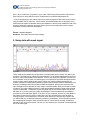
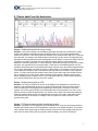
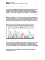
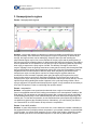
Genomic Platform
Institute of research in immunology and cancer, University of Montreal
www.genomique.iric.ca
Sequencing troubleshooting
If you have problems with your results, this is for you!
You’ve just received your results and you are all excited. You open the text file
and, surprise, there is full of “N” in you sequence, or worst there is only 5 of them
and that’s all. All that work for useless results!
Before calling us for a re-run, please take 2 minutes and check your
chomatogram! It might help you figure out why your reaction failed.
If you recognize your chromatogram pattern in one of this figure, take this
document (and read it). You can always e-mail us to discuss these features and
kindly ask for a re-run if you strongly think the failure was our fault.
1. No sequence data
2.1 Noisy data from the beginning
2.2 Noisy data from farther in the sequence
3. Homopolymeric regions
4. Truncated sequences
1
Genomic Platform
Institute of research in immunology and cancer, University of Montreal
www.genomique.iric.ca
Troubleshooting Your Data
This document was taken on the Roswell Park Institute website
http://www.roswellpark.org
The two most common causes for failure to get good or any sequence data for your samples are
purity and concentration of your template DNA. If it appears that you have done everything
correctly, then look below for some additional reasons why you might obtain less than optimal
DNA sequence data quality. We’ve listed various causes, solutions and, for some, pictorial
representations of what these specific problems might look like. Many causes and solutions may
look rather obvious and just involve common sense, but you’d be surprised how many times
we’ve heard "how could I have done that?"...
1. No sequence data
Cause: not enough or no DNA/primer in tube
Solutions: doublecheck your quantitations, stock concentrations and dilutions.. While our
sequencers are very sensitive and can detect a range of DNA concentrations, there is still a
"threshold" amount that must be reached to obtain any sequence data.
Cause: inhibitory contaminant
Solutions: the cycle sequencing reaction used to amplify samples for automated sequencing is
very sensitive to the presence of certain contaminants, some of which will completely inhibit our
sequencing enzyme. Please check the Contaminant section for a list of potential inhibitors. You
may need to reprep your sample to sufficiently remove one or more inhibitory components to
obtain any sequence data.
Cause: priming site not present
Solutions: if you’ve chosen one of the sequencing facility’s vector primers (T7+, T7-, T3, SP6,
2
Genomic Platform
Institute of research in immunology and cancer, University of Montreal
www.genomique.iric.ca
M13+, M13-), make sure it is present in your vector. While many of the primers we provide are
quite common to many different vectors. Doublecheck your plasmid maps/sequences.
- if you’ve designed your own custom primer from previous sequence data, make sure you were
using a reliable area of sequence - look for sharp, well-defined peaks with no ambiguity. Avoid
areas where the peaks are broader and not well separated - this will occur towards the end of the
sequence where the fragments are larger and the polymer cannot adequately resolve single
nucleotides, causing inaccurate basecalling.
Cause: expired reagents
Solutions: falls under common sense category
2. Noisy data with weak signal
"Noisy" data can be identified by the presence of multiple peaks and numerous "N"s within your
sequence. The Sequencing Analysis program assigns an "N’ as a base identification when there
are two or more peaks present at one position. This "N" may signify the legitimate occurrence of
two nucleotides, as in the case of a heterozygote, but may also be seen when background noise
is high or when multiple products are present. When your sample exhibits weak signal, the
software attempts to compensate by boosting up the signal of sample bands to detectable levels.
However, the background noise will also be artificially amplified, giving a poor signal-to-noise
ratio. Background noise appears as many smaller, undefined peaks under your sequence peaks
of interest. This noise is always present, but with well-prepared samples of good signal strength,
it will be undetectable. To determine if your noisy data may be due to weak signal, look at your
ABI trace file. If you are looking at a paper chromatogram, look towards the top and middle of
your trace for a line that says "Signal". If the file is on your computer, click the "A" radio button in
the bottom left-hand corner, which is visible when you have opened up the trace file within a
viewing program, such as EditView or Chromas. Scroll down to the line that says "Signal" and
you will see the four nucleotides followed by numbers in parentheses. These numbers represent
the average signal strength of each nucleotide and their values should, optimally, be between
200-400. If they are much less than 100, then you can assume your noisy data is at least partially
due to its weak signal.
3
Genomic Platform
Institute of research in immunology and cancer, University of Montreal
www.genomique.iric.ca
Cause: not enough DNA
Solutions: doublecheck your quantitations, stock concentrations, calculations and dilutions.
Make sure you’ve provided the appropriate amount of DNA and/or primer.
Cause: inhibitory contaminant e.g..salts, phenol
Solutions: the cycle sequencing reaction used to amplify samples for automated sequencing is
very sensitive to the presence of certain contaminants, some of which can partially or completely
inhibit our sequencing enzyme. You may need to re-purify your sample to sufficiently remove one
or more inhibitory components to obtain better sequence data.
Cause: degraded DNA from nucleases, repeated freeze-thaw, excessive UV light
exposure, bisulfite treatment.
Solutions: Nuclease contamination in a template preparation as well as repeated freeze-thaw
cycles can degrade DNA over time. Even low amounts of nucleases can extensively degrade
DNA depending on storage conditions and temperatures, as well as the length of time the DNA is
stored. Generally, re-isolation and purification of the template DNA will be necessary to obtain
good DNA sequence. When extracting PCR products from a gel, prolonged exposure to UV light
will degrade and nick the DNA. Limit the time and UV intensity as much as possible to prevent
degradation. When treating DNA with bisulfite for methylation experiments, it is important to avoid
long incubations at higher temperatures as substantial amounts of DNA will be degraded in this
process.
Cause: trend in worsening data?
Solutions: if you have previously been able to obtain good sequence data but begin to see a
deterioration in quality that gets progressively worse, you may have some contamination in one or
more reagents, or have some reagents that have reached the end of their usefulness. Make up
fresh stocks of commonly used reagents, such as buffers, and always use high quality distilled
water in your preparations.
Cause: inefficient primer binding (low Tm, degenerate primers, mismatch)
Solutions: the Tm of a primer is defined as the temperature at which 50% of the oligonucleotide
and its perfect complement are in duplex. The Tm of an oligo can be roughly calculated by using
the formula:
Tm = 2°C(A+T) + 4°C(G+C)
This is the most commonly used formula for calculating Tm, though it is not the most accurate as
it does not factor in salt or formamide concentrations. A good website to check out if you are
interested in some detailed theory behind Tm calculations is http://www.sigmagenosys.com/oligo_meltingtemp.asp.
In our cycle sequencing reaction, our primer/template annealing step occurs at 50ºC. Thus, if
your primer Tm is much lower than 50ºC, hybridization to its complementary template will be
much less efficient and a lesser number of extending fragments will be generated. Increase your
primer Tm by adding additional bases to the 5’ or 3’ end to raise the Tm to be within the range of
52ºC-58ºC. Degenerate primers and those with mismatched bases will also show decreased
hybridization efficiency due to reduction of the stability of primer binding, and if degeneracy or
mismatches occur at or near the 3’ end of your primer, it is highly likely that your sequencing
attempt will fail.
4
Genomic Platform
Institute of research in immunology and cancer, University of Montreal
www.genomique.iric.ca
2.1 Noisy data From the beginning
Cause: multiple priming sites involving vectors
Solution: your primer may have a secondary hybridization site that may be identical or closely
related, with different nucleotide sequences following each site, giving superimposed bands
within your sequence. If the priming sites are identical, (such as when more than one T7 promoter
site is present, for example), the double peaks will be strong from the outset. The fragments may
also show shifted migration so that the double peaks are not directly on top of one another but will
be offset to one side or the other due to the differing mobility patterns of the strands with
dissimilar nucleotide composition. In other instances, a secondary priming site may not be exactly
the same, but may differ by a few internal bases. In this case, the mismatched primer may not
hybridize as efficiently but can still anneal and extend, and give rise to less intense fragments that
can be seen underneath your peaks of interest. In both cases, it’s necessary to screen both your
vector and insert carefully to look for sequences that may match or be similar to your proposed
primer. You may need to choose another vector primer on the same end of the multiple cloning
site or redesign your custom primer. When choosing another primer is difficult, such as when
primer walking through a repetitive area, try to find a primer that has a 3’-base match specific to
your area of interest which can help act as an "anchor".
Cause: multiple priming sites in PCR
Solution: this may occur when one or both of the PCR primers hybridizes to more than one
position on the template DNA, giving rise to multiple PCR products. Often this will be obvious
when visualizing the PCR products on an agarose gel as there will be more than one band
present. In this case, gel purification of the desired product will be necessary. One can run into
difficulty, however, when the products are very similar in size, which may arise when amplifying
related or repetitive DNA, and do not separate well on the gel. In this case, optimization of the
PCR reaction may be necessary or redesign of the PCR primers in order to choose a more
specific priming site.
Cause: PCR primers acting as both forward and reverse
Solution: sometimes, a PCR product may be generated when one primer functions as both the
forward and reverse primer in the PCR reaction, giving rise to an artifactual product. This is fairly
easy to detect when sequencing the PCR product as one primer will give double peaks from the
start, while the other fails to give any sequence data. Redesign your set of PCR primers.
5
Genomic Platform
Institute of research in immunology and cancer, University of Montreal
www.genomique.iric.ca
Cause: residual PCR primers and/or dNTPs
Solution: as two primers are present in the PCR reaction, incomplete removal of these primers
can lead to double peaks within the sequencing data. Both primers will act as sequencing primers
and lead to superimposed bands which correspond to the complementary strands from opposite
orientations. It is critical to remove excess primers and dNTPs from the PCR reaction by
purification. If attempting to do direct sequencing of PCR products without purification by diluting
an aliquot of your PCR product with water to lower the concentration of residual primers and
dNTPS (a method which we do not recommend), then it is imperative to optimize your PCR
reaction so that primers and dNTPS are used in limiting amounts so that most are used up by the
end of the PCR.
Cause: primers with high Tm
Solution: primers that have a Tm much higher (>65ºC) than our suggested 52ºC-58ºC often do
not function well as sequencing primers. When primers have a Tm that high, it is often a result of
increased G-C content or because the primer is quite long, both factors that can increase the
potential for primer secondary structure formation. If possible, choose another primer with a lower
Tm.
Cause: primers with n-1 population
Solution: this problem is not uncommon and can result from poor quality synthesis of
sequencing primers. Primers are synthesized from the 3’end to the 5’end and when synthesis is
inefficient, there can be a significant population of less than full-length primers - n-1s, which are
full-length primers minus one base, plus other shorter derivatives. These primers have a common
3’end but different 5’ends, thus chains that terminate at the same position will have different
lengths and will run at different positions on the gel. Primers that have degraded from the 3’end
will also give this appearance. It is easy to spot this problem within the sequencing chromatogram
as each position will contain the true peak as well as the peak immediately to the right of it, giving
the appearance of "shadow" peaks. Whatever the cause of the n-1s, it will be necessary to
resynthesize the primer to obtain an oligo of suitable quality for sequencing. When high-quality
reagents and proper protocols are utilized during oligo synthesis, cartridge or HPLC purification of
the primers is usually not necessary for typical oligos (<30 bp), but sometimes additional
purification can be beneficial.
6
Genomic Platform
Institute of research in immunology and cancer, University of Montreal
www.genomique.iric.ca
2.2 Noisy data Begin farther into the sequence
Cause: mixed plasmid prep
Solution: a plasmid prep that is contaminated by more than one product, such as two vectors
with different inserts or vector with insert and vector without, will generally show an early section
of clean sequence data (common vector multiple cloning site sequence) followed by double
peaks. Occasionally, a plasmid may contain more than one vector molecule or may encounter
spontaneous deletions or insertions during growth. The point at which the double peaks begin
corresponds to the start of the insert cloning site. To avoid this problem, it’s important to carefully
pick a single colony from your growth plate, restreaking if necessary, to be sure that your colony
is completely clonal. You should follow this up with a restriction digest of your plasmid run out on
an agarose gel to ensure vector and insert are present as expected.
7
Genomic Platform
Institute of research in immunology and cancer, University of Montreal
www.genomique.iric.ca
3. Homopolymeric regions
Cause: homopolymeric regions
Solution: regions that contain long stretches of a single nucleotide can be difficult to sequence
through accurately. Short stretches of homopolymeric regions are generally not difficult to get
through, but longer sections can be challenging. Sequence data up to and including the
polynucleotide region may be fine, but the last base of the poly region and all peaks following it
may show a wave-like, stuttering pattern of double peaks that cannot be interpreted. This tends to
be more problematic in PCR products, but can also occur when sequencing plasmids, especially
when trying to sequence the polyA region of cDNA. This difficulty is thought to arise due to
enzyme "slippage" when the growing strand does not stay paired correctly with the template DNA
during polymerization through the homopolymer region, thus giving rise to fragments of varying
lengths that have the same sequence after this area. When sequencing cloned DNA with a
homopolymer region, several options can be tried. Sequencing the opposite strand can
sometimes be more successful, especially when going through a polyG region as the polyC
strand is often easier to get through. Sometimes designing a new primer that is closer to the
homopolymeric region can help, as nucleotide concentration and enzyme activity will be in a more
optimal range when extending the smaller fragments in the cycle sequencing reaction. When
trying to sequence PCR products with homopolymeric regions, it may sometimes be necessary to
clone the PCR product in order to read through the repetitive stretch.
Cause: compression
Solution: compressions can sometimes be observed when a region of secondary structure
forms in the amplified strand of DNA, leading to an alteration in the electrophoretic mobility of the
DNA strand. This can appear as overlapping fragments after a certain point and can resemble a
contaminated plasmid prep, but the contaminated prep will show double peaks beginning at the
insertion site. To relax this compression, we can sometimes alter cycle sequencing conditions or
use additives to denature the secondary structure. Alternatively, you can linearize your DNA or
use 7-deaza-dGTP in a PCR reaction to help relieve the compression.
Cause: frame shift mutation
Solution: a frame shift mutation can occur when one or more bases are inserted or deleted into
the template DNA and if multiple products are present in your sample, whether it be plasmid DNA
or PCR product, you will see clean sequence up to the point of the mutation, followed by double
peaks caused by the shift in the nucleotide sequence. In the case of plasmid DNA, it will be
8
Genomic Platform
Institute of research in immunology and cancer, University of Montreal
www.genomique.iric.ca
necessary to re-isolate your DNA to get a pure clone containing only one of the molecules. With
PCR products, you will need to gel purify the two products in order to separate them.
4. Truncated sequences
Truncated sequences can be characterized as abrupt or gradual. Abrupt truncations will show
strong, clean signal up to a point and then drop sharply down over the course of a few
nucleotides to much weaker or no detectable signal. Gradual truncations will show good
sequence data initially but then begins to taper off to progressively weaker, smaller peaks until
there is nothing but background noise. The nature of the truncation can sometimes help to
determine its cause.
Cause: secondary structure
Solution: G-C rich, and to a lesser degree, A-T rich, DNA is predisposed to secondary structure
formation, as strong hydrogen bonding between G and C nucleotides can cause the template
DNA to loop or bend and anneal to complementary sequences, forming hairpins that can restrict
the passage of the sequencing polymerase and thus be very difficult to sequence through
reliably. These hairpins may not melt at our cycle sequencing temperatures and can cause
premature termination of sequence data. Secondary structure may appear as a sharp termination
of signal with no sequence data after, or if the loop has been relaxed slightly, you may see strong
signal that drops abruptly but may have some weaker peaks following that are still quite accurate.
With the newest formulation of BigDye Terminator chemistries (v3.1), some G-C rich difficulties
have improved dramatically, but unfortunately it hasn’t solved everything. There is not one
solution that resolves every secondary structure problem, but there are couple you can try and
usually one will allow us to read through it. Placing a primer as close to the hairpin loop as
possible to help force its unwinding has also worked in the past. Sequencing the opposite strand
can sometimes lead to a huge improvement. If these solutions don’t work, we may suggest you
try linearizing your DNA with restriction enzymes to help relax the hairpin. And if you are trying to
PCR up a very G-C rich region, addition of betaine or DMSO to your PCR reaction can help, as
can substitution of 7-deaza dGTP for 75% of the dGTP in your PCR reaction. And if all else fails,
you can try manual radioactive sequencing as a last resort.
Cause: linearized DNA
Solution: if your DNA has been cut with one or more restriction enzymes, the sequence data will
sharply end at the recognition site of the enzyme that cut at the 3’ end of your insert. Did you
9
Genomic Platform
Institute of research in immunology and cancer, University of Montreal
www.genomique.iric.ca
accidentally send us digested DNA? Run it out on a gel to see.
Cause: too much DNA
Solution: while there is a range of DNA concentrations we can sequence reliably, too much DNA
will cause premature termination of signal. Overloading of DNA will exhibit early top-heavy peaks
followed by rapidly weakening peak height and strength. This occurs because the dNTPS in the
cycle sequencing reaction will be distributed among too many extending chains and will be
depleted early on, resulting in an excessive amount of short fragments.. In addition, if your
template is impure, higher concentrations of DNA can be accompanied by higher amounts of
contaminants that can further worsen your DNA sequence quality.
Cause: salts
Solution: excessive amounts of salts will also give rise to premature termination and may look
similar to DNA overloading, with strong signal followed by progressively weakening signal. Salts
have an inhibitory effect on the processivity of the sequencing Taq polymerase, which can lead to
an overabundance of short fragments, or if the salt concentration is too great, the enzyme will be
completely inhibited with no sequence data obtained. If salts are potentially a problem, perform
an ethanol precipitation for salt removal.
Cause: repetitive regions
10
Genomic Platform
Institute of research in immunology and cancer, University of Montreal
www.genomique.iric.ca
Solution: the nucleotide composition, as well as the size, of a repetitive region can play a large
role in the success of sequencing through such an area. In general, G-C and G-T (ofter seen in
bisulfite-treated DNA) repeats tend to be the most troublesome though, as mentioned before, the
newest version of Applied Biosystems BigDye Terminator v3.1 contains some modifications that
have allowed for some striking improvements in certain previously difficult templates. However,
there are still some that remain a pain. In general, one can sequence partially through the
repetitive region and the signal begins to fade and eventually becomes unreadable. This may be
due to premature dNTP depletion, secondary structure formation or enzyme slippage. Various
methods can be tried to sequence the repeat entirely, and many are similar to those we would
use for G-C rich templates that form secondary structures. If the repeat region is not excessively
large, sequencing from the opposite strand to complete the region can be successful, especially if
the complementary strand has a nucleotide composition that is more efficiently extended.
However, if the region is large, it may be difficult to complete its entire sequence and determine
the exact number of repeats present. Alternative methods, such as directed deletions or the use
of an in vitro transposon system may need to be utilized.
Plasmid templates
Considerations when cleaning up plasmid preps
Poor template quality is one of the most common reasons for bad sequence data, as mentioned
above, and is a prime consideration when choosing a plasmid cleanup method to give DNA of
optimal purity for automated sequencing. Plasmid template quality can be affected by a variety of
factors and contaminants including the following:
• Salts or organics left over from template preparation
* Presence of cellular components such as RNA, proteins, polysaccharides or
chromosomal DNA
* DNA that has degraded while in storage
* silica fines that carryover from template preparation kits that utilize loose resin or silica
solutions
Contaminants
Salts- the processivity of the Taq polymerase used in the cycle sequencing reaction declines in
the presence of high amounts of salts. Salt contamination in DNA preps may result from
coprecipitation of salts in alcohol precipitations, insufficient removal of supernatant after
precipitations or an incomplete wash of the pellet with 70% ethanol. Careful technique should be
used when precipitating with alcohol. It has also been demonstrated that acetate ions, as
opposed to sodium, potassium or chloride ions, are the most inhibitory in sequencing reactions.
When using potassium acetate or sodium acetate, concentrations over 20 mM led to complete
failure of the sequencing reactions, while concentrations of 60mM of sodium chloride were
required before complete inhibition.
Ethanol- ethanol contamination can occur when the sample is insufficiently dried after
precipitation or when carried over in an ethanol-containing wash buffer used in some DNA
isolation procedures. Contamination with 10% or greater concentrations of ethanol usually leads
to failure of the DNA sequencing reaction. Complete drying of the DNA samples is required to
remove these traces of ethanol.
Phenol- phenol may be carried over from DNA alkaline lysis methods that utilize phenol and
chloroform to remove proteins and other cellular contaminants from cell lysates. Phenol cannot
11
Genomic Platform
Institute of research in immunology and cancer, University of Montreal
www.genomique.iric.ca
be tolerated in the cycle sequencing reaction as it denatures proteins and will thus degrade the
Taq polymerase enzyme used in the cycle sequencing reaction. Chloroform does not have the
strong denaturing properties of phenol and doesn’t appear to adversely affect the sequencing
reaction.
EDTA - EDTA can chelate the magnesium required by the Taq polymerase in the cycle
sequencing reaction, so when submitting samples, it is best to always have them diluted or
resuspended in sterile ddH20 or 1X Tris buffer. Suspension in TE buffer is not recommended,
though people have done it and many times there is not a problem. However, providing template
DNA in water is an easy thing to do and if there is a problem with your sequence quality, the fact
that there is no EDTA in your sample is one potential problem we can eliminate right away.
Finally, always check your chromatogram to be sure that the base caller
did call true bases and not background peaks.
This document was taken on the Roswell Park Institute website
http://www.roswellpark.org
12