1
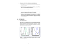
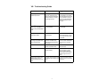
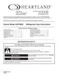
Enabling Discovery in Life Science® Lyso-ID® Green Detection Kit for microscopy Instruction Manual Cat. No. ENZ-51034-K500 For research use only. Rev. 1.0 May 2010 500 assays Notice to Purchaser The Lyso-ID® Green Detection Kit is a member of the CELLestial® product line, reagents and assay kits comprising fluorescent molecular probes that have been extensively benchmarked for live cell analysis applications. CELLestial® reagents and kits are optimal for use in demanding cell analysis applications involving confocal microscopy, flow cytometry, microplate readers and HCS/HTS, where consistency and reproducibility are required. This product is manufactured and sold by ENZO LIFE SCIENCES, INC. for research use only by the end-user in the research market and is not intended for diagnostic or therapeutic use. Purchase does not include any right or license to use, develop or otherwise exploit this product commercially. Any commercial use, development or exploitation of this product or development using this product without the express prior written authorization of ENZO LIFE SCIENCES, INC. is strictly prohibited. Limited Warranty These products are offered under a limited warranty. The products are guaranteed to meet appropriate specifications described in the package insert at the time of shipment. Enzo Life Sciences’ sole obligation is to replace the product to the extent of the purchase price. All claims must be made to Enzo Life Sciences, Inc. within five (5) days of receipt of order. Trademarks and Patents Enzo, CELLestial, and Lyso-ID are trademarks of Enzo Life Sciences, Inc. Lysotracker is a trademark of Molecular Probes. Several of Enzo’s products and product applications are covered by US and foreign patents and patents pending. Contents I. Introduction ............................................................... 1 II. Reagents Provided and Storage.............................. 2 III. Additional Materials Required ................................. 2 IV. Safety Warnings and Precautions........................... 2 V. Methods and Procedures ......................................... 3 A. Reagent Preparation .............................................. 3 B. Cell Preparations.................................................... 4 C. Staining Live, Adherent Cells ................................. 4 D. Staining Live Cells Grown in Suspension .............. 5 VI. Appendices ............................................................... 5 A. Filter Set Selection................................................ 5 B. Results .......................................................................... 6 VII. References ................................................................ 6 VIII. Troubleshooting Guide ........................................... 7 I. Introduction Enzo Life Sciences’ Lyso-ID® Green Detection Kit contains a novel acidic organelle-selective dye suitable for live-cell staining. Conventional fluorescent stains for acidic organelles, such as Acridine Orange (Catalog No. ENZ-52405), form metachromatic artifacts that interfere with multicolor imaging applications. Lyso-ID® Green dye generates emission profiles that can be multiplexed with other fluorophores. The dye accumulates in acidic compartments, such as endosomes, lysosomes, and secretory vesicles. Low micromolar concentrations of Lyso-ID® Green dye are sufficient for staining mammalian cells. This has been validated with the human cervical carcinoma cell line, HeLa, the human T-lymphocyte cell line, Jurkat, and the human bone osteosarcoma epithelial cell line, U2OS. One important application of Lyso-ID® Green dye is in fluorescence co-localization imaging with red fluorescent protein (RFP)-tagged proteins. This is a powerful approach for determining the targeting of molecules to intracellular compartments, and for screening of associations and interactions between these molecules. For example, dual-color fluorescence detection may be employed for the identification of vesicular compartments and the investigation of their dynamics and fusion during exocytosis. However, metachromatic artifacts wherein fluorescent dyes emit both in the red and green regions of the spectrum compromise such analyses, as observed with Acridine Orange dye. These observations have led to spurious results in fluorescent protein co-localization experiments.(1, 2) Additionally, many organelle-targeting probes photobleach rapidly, are subject to quenching when concentrated in organelles, are highly toxic, or only transiently associate with the target organelle, requiring imaging within a minute or two of dye addition.(3, 4) The Lyso-ID® Green dye, a new green-emitting, cell-permeable small organic probe molecule that spontaneously localizes to live cell acidic organelles, was developed to overcome the above problems. It can be readily used in combination with other common UV and visible light excitable fluorescent dyes and various fluorescent proteins in multicolor imaging and detection applications. The Lyso-ID® Green dye is suitable for both short-term and long-term tracking studies. It emits in the FITC region of the visible light spectrum, and is highly resistant to photobleaching, concentration quenching and photoconversion. The Lyso-ID® Green Detection Kit is specifically designed for use with RFP-expressing cell lines, as well as cells expressing blue, cyan or orange fluorescent proteins (BFPs, CFPs, OFPs). A lysosome perturbation agent, chloroquine, is provided as a positive control for monitoring changes in lysosome number and volume. A nuclear counterstain is also provided in the kit to highlight this organelle as well. 1 II. Reagents Provided and Storage All reagents are shipped on dry ice. Upon receipt, the kit should be stored at ≤-20°C, protected from light. When stored properly, these reagents are stable for at least twelve months. Avoid repeated freezing and thawing. Reagents provided in the kit are sufficient for approximately 500 assays using either live, adherent cells or cells in suspension. Reagent Quantity Lyso-ID® Green Detection Reagent 50 µL Hoechst 33342 Nuclear Stain 50 µL 7.5 µmol Chloroquine Control 10X Assay Buffer 15 mL III. Additional Materials Required Standard fluorescence microscope Calibrated, adjustable precision pipetters, preferably with disposable plastic tips Adjustable speed centrifuge with swinging buckets (for suspension cultures) Glass microscope slides Glass cover slips Deionized water Anhydrous DMSO (optional) Growth medium (e.g., Dulbecco’s Modified Eagle Medium, D-MEM) IV. Safety Warnings and Precautions This product is for research use only and is not intended for diagnostic purposes. The Lyso-ID® Green Detection Reagent contains DMSO which is readily absorbed through the skin. It is harmful if ingested or absorbed through the skin and may cause irritation to the eyes. Observe appropriate precautions when handling. Reagents should be treated as possible mutagens and should be handled with care and disposed of properly. Observe good laboratory practices. Gloves, lab coat, and protective eyewear should always be worn. Never pipet by mouth. Do not eat, drink or smoke in the laboratory areas. All blood components and biological materials should be treated as potentially hazardous and 2 handled as such. They should be disposed of in accordance with established safety procedures. To avoid photobleaching, perform all manipulations in low light environments or protected from light by other means. V. Methods and Procedures NOTE: Allow all reagents to thaw at room temperature before starting with the procedures. Upon thawing, gently hand-mix or vortex the reagents prior to use to ensure a homogenous solution. Briefly centrifuge the vials at the time of first use, as well as for all subsequent uses, to gather the contents at the bottom of the tube. A. REAGENT PREPARATION 1. Positive Control Chloroquine is a lysosomotropic agent. It accumulates preferentially in the lysosomes of cells.5 The pKa for the quinoline nitrogen of chloroquine is approximately 8.5. At physiological pH, it is roughly 10% deprotonated as calculated using the HendersonHasselbalch equation. This decreases to roughly 0.2% at a lysosomal pH of 4.6. Since the deprotonated form of the compound is more membrane permeable than the protonated form, chloroquine becomes quantitatively "trapped" in lysosomes. The chloroquine provided in the kit may be used as a positive control for increasing lysosome number and volume. It is supplied lyophilized (7.5 µmoles) and should be centrifuged briefly to gather the material at the bottom of the tube. Reconstitute the lyophilized material in 125 μL deionized water for a 60 mM stock solution. It is recommended that treatment with the agent be performed using 10-300 μM final concentration in order to observe changes in lysosomal morphology. Unused stock chloroquine may be stored in small aliquots at -20°C for several weeks. 2. 1X Assay Buffer Allow the 10X Assay Buffer to warm to room temperature. Make sure that the reagent is free of any crystallization before dilution. Prepare enough 1X Assay Buffer for the number of samples to be assayed by diluting each milliliter (mL) of the 10X Assay Buffer with 9 mL of deionized water. 3. Dual Detection Reagent The concentration of Lyso-ID® Green dye for optimal staining will vary depending upon the application. Suggestions are provided to use as guidelines, though some modifications may be required 3 depending upon the particular cell type employed and other factors such as the permeability of the dye to the cells or tissues. To reduce potential artifacts from overloading of the cells, the concentration of the dye should be kept as low as possible. Prepare sufficient amount of Dual Detection Reagent for the number of samples to be assayed as follows: To each milliliter of 1X Assay Buffer (see preparation in step 2) or cell culture medium, add 1 µL of Lyso-ID® Green Detection Reagent and 1 µL of Hoechst 33342 Nuclear Stain. Serum may be included, if preferred. NOTE: (a) The dyes may be combined into one staining solution or each may be used separately, if desired. (b) The Hoechst 33342 Nuclear Stain can be diluted further if its staining intensity is much stronger than the green lysosomal stain, Lyso-ID® Green. (c) When staining BFP- or CFP-expressing cells, the Hoechst 33342 Nuclear Stain should be omitted due to its spectral overlap with these fluorescent proteins. B. CELL PREPARATIONS Cells should be maintained via standard tissue culture practices. Positive control cells should be pretreated with the chloroquine control for 2-8 hours. Response to chloroquine is time and concentration dependent and may also vary significantly depending upon cell type and cell line. Negative control cells should be treated with a vehicle (DMSO, media or other solvent used to reconstitute or dilute an inducer or inhibitor) for an equal length of time under similar conditions. C. STAINING LIVE, ADHERENT CELLS 1. Grow cells on cover slips inside a Petri dish filled with the appropriate culture medium. When the cells have reached the desired level of confluence, carefully remove the medium. 2. Dispense sufficient volume of Dual Detection Reagent (see section V-A3, page 3) to cover the monolayer cells (~100 μL of labeling solution for cells grown on an 18 X 18 mm coverslip). 3. Protect samples from light and incubate for 30 minutes at 37°C. 4. Wash the cells with 100 μL 1X Assay Buffer. Remove excess buffer and place coverslip on slide. 5. Analyze the stained cells by wide-field fluorescence or confocal microscopy (60X magnification recommended). Use a standard FITC filter set for imaging the lysosomes. Optionally, image the nucleus using a DAPI filter set and the RFP-tagged protein using a Texas Red filter set. 4 D. STAINING LIVE CELLS GROWN IN SUSPENSION 1. Centrifuge cells for 5 minutes at 400 x g at room temperature (RT) to obtain a cell pellet. 2. Carefully remove the supernatant by aspiration and dispense sufficient volume of Dual Detection Reagent (see section V-A3, page 3) to cover the dispersed cell pellet. 3. Protect samples from light and incubate for 15 to 30 minutes at 37°C. 4. Wash the cells with 100 μL 1X Assay Buffer. Remove excess buffer. Resuspend cells in 100 μL 1X Assay Buffer, then apply the cells to a glass slide and overlay with a coverslip. 5. Analyze the stained cells by wide-field fluorescence or confocal microscopy (60X magnification recommended). Use a standard FITC filter set for imaging the lysosomes. Optionally, image the nucleus using a DAPI filter set and the RFP-tagged protein using a Texas Red filter set. VI. APPENDICES A. Filter Set Selection The selection of optimal filter sets for a fluorescence microscopy application requires matching the optical filter specifications to the spectral characteristics of the dyes employed in the analysis. Consult the microscope or filter set manufacturer for assistance in selecting optimal filter sets for your microscope. 400 500 600 700 Wavelength (nm) Absorbance Absorbance 300 Fluorescence Emission B Fluorescence Emission A 200 250 300 350 400 450 500 550 600 Wavelength (nm) Figure 1. Absorbance and fluorescence emission spectra for Lyso-ID® Green (panel A) and Hoechst 33342 (panel B) dyes. All spectra were determined in 1X Assay Buffer. 5 B. Results Lysosomes are membrane-bound organelles involved in the degradation of macromolecules and pathogens in diverse processes including endocytosis, phagocytosis and autophagy. Lysosomal morphology varies with the state of the cell and its degree of degradative activity, and the vesicles can have pieces of membranes, vacuoles, granules and even parts of mitochondria within them. They are typically spherical vesicles, ranging in size from 0.2 to 2 µm in diameter. The lumen of lysosomes and other acidic organelles is characterized by low pH generated via proton-pumping vacuolar ATPases. Lyso-ID® Green dye is selectively sequestered in acidic organelles by a mechanism that likely involves protonation and retention within the membranes of the organelles. However, staining is even feasible in cells pretreated with weakly basic cell-permeant compounds, such as chloroquine. HeLa and U2OS cells treated with 300 µM chloroquine for 4 hours show a dramatic increase in lysosome-like vesicle number and volume, confirming Lyso-ID® Green dye is associated with this subcellular compartment. Thus, Lyso-ID® Green stain can be employed to highlight lysosome-like organelles under certain conditions, such as chloroquine drug treatment, wherein cells produce lysosome-like bodies that contain most of the degradative enzymes of the lysosome, but are not as acidic as this organelle. VII. References 1. Freundt EC, Czapiga M and Lenardo MJ (2007) “Photoconversion of Lysotracker Red to a green fluorescent molecule” Cell Res. 17(11):956-958. 2. Nadrigny F, Li D, Kemnitz K, Ropert N, Koulakoff A, Rudolph S, Vitali M, Giaume C, Kirchhoff F and Oheim M (2007) “Systematic colocalization errors between acridine orange and EGFP in astrocyte vesicular organelles” Biophys J. 93(3):969-980. 3. Jaiswal JK, Fix M, Takano T, Nedergaard M, and Simon SM (2007) “Resolving vesicle fusion from lysis to monitor calcium-triggered lysosomal exocytosis in astrocytes” Proc Natl Acad Sci USA. 104(35):14151-14156. 4. Brunk UT, Dalen H, Roberg K and Hellquist HB (1997) “Photooxidative disruption of lysosomal membranes causes apoptosis of cultured human fibroblasts” Free Radical Biology and Medicine, 23(4):616-626. 5. Michihara, Toda, Kubo, Fujiwara, Akasaki and Tsuji “Disruptive effect of chloroquine on lysosomes in cultured rat hepatocytes” (2005) Biol. Pharm. Bull. 28(6):947-951. 6 VIII. Troubleshooting Guide Problem Potential Cause Suggestion Acidic organelles are not sufficiently stained. Very low concentration of Lyso-ID® Green dye was used or dye was incubated with the cells for an insufficient length of time. Either increase the labeling concentration or increase the time allowed for the dye to accumulate in the lysosome once the cells have been transferred to fresh medium. Lyso-ID® Green dye fails to stain acidic organelles in fixed and/or permeabilized cells. The dye is only suitable for live-cell staining. Use the dye only for livecell analysis. Precipitate is seen in the 10X Assay Buffer. Precipitate forms at low temperatures. Allow solution to warm to room temperature or 37°C, then vortex to dissolve all precipitate. Blue nuclear counterstain is too bright compared to the red lysosomal stain. Different microscopes, cameras and filters may make some signals appear very bright. Reduce the concentration of the nuclear counterstain or shorten the exposure time. Untreated cells do not stain. The lysosomal volume is small in untreated cells. Increase exposure time or add more dye. Cells do not appear healthy. Some cells require serum to remain healthy. Add serum to stain and wash solutions. Serum does not affect staining. Normal amounts of serum added range from 2% to 10%. Chloroquine treated cells appear dead or are no longer attached to the surface. The EC50 of chloroquine may Lower the dose of Chlorobe different with different cell quine, or reduce the time of lines. exposure. 7 www.enzolifesciences.com Enabling Discovery in Life Science® NORTH/SOUTH AMERICA GERMANY UK & IRELAND ENZO LIFE SCIENCES INTERNATIONAL, INC. 5120 Butler Pike Plymouth Meeting, PA 19462-1202 USA T 1-800-942-0430/(610) 941-0430 F (610) 941-9252 E [email protected] ENZO LIFE SCIENCES GMBH Marie-Curie-Strasse 8 DE-79539 Lörrach Germany T +49/0 7621 5500 526 Toll Free 0800 664 9518 F +49/0 7621 5500 527 E [email protected] www.enzolifesciences.com ENZO LIFE SCIENCES (UK) LTD. Palatine House Matford Court Exeter EX2 8NL UK T 0845 601 1488 (UK customers) T +44/0 1392 825900 (from overseas) F +44/0 1392 825910 E [email protected] www.enzolifesciences.com www.enzolifesciences.com SWITZERLAND & REST OF EUROPE BENELUX FRANCE ENZO LIFE SCIENCES AG Industriestrasse 17, Postfach CH-4415 Lausen Switzerland T +41/0 61 926 89 89 F +41/0 61 926 89 79 E [email protected] www.enzolifesciences.com ENZO LIFE SCIENCES BVBA Melkerijweg 3 BE-2240 Zandhoven Belgium T +32/0 3 466 04 20 F +32/0 3 466 04 29 E [email protected] www.enzolifesciences.com ENZO LIFE SCIENCES c/o Covalab s.a.s. 13, Avenue Albert Einstein FR -69100 Villeurbanne France T +33 472 440 655 F +33 437 484 239 E [email protected] www.enzolifesciences.com