1
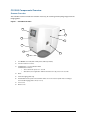
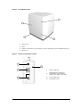
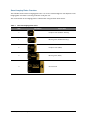
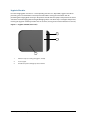
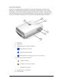
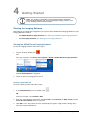
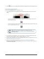
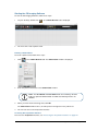
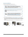
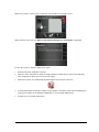
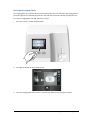
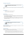
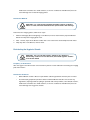
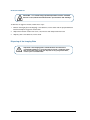
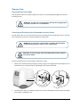
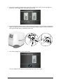
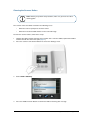
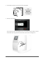
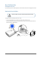
CS 7600
User Guide
Notice
CARESTREAM Systems are designed to meet international safety and performance standards. Personnel
operating the unit must have a thorough understanding of the proper operation of the system. This guide has been
prepared to aid dental professionals to understand and operate the system. Do not operate the system before
reading this guide and gaining a clear understanding of the operation of the system. If any part of this guide is not
clear, please contact your Carestream Health representative for clarification.
WARNING: Read the “Safety, Regulatory and the Technical
Specifications User Guide” and make sure you observe all the
Warnings and Cautions before using the CS 7600.
The CS 7600 is intended for digital dental radiography using an imaging plate (storage phosphor screen) for
radiographic diagnostic intraoral images.
The CS 7600 in addition, optionally provides the Scan & Go technology to enable dental practitioners prior to
acquisition to record the dental imaging plates with exam acquisition data.
No part of this guide may be reproduced without the express permission of Carestream Health, Inc.
U.S. Federal law restricts this device to sale by or on the order of a dentist or physician.
This document is originally written in English.
Guide Name: CS 7600 User Guide
Part Number: 8J4071
Revision Number: 01
Print Date: 2011-08
In this guide, all trademarks and registered trademarks are the property of their respective holders.
The brand names and logos reproduced in this guide are copyright.
CS 7600 complies with Directive 93/42/CEE relating to medical equipment.
0086
Manufacturer
Care stream Hea lth, Inc.
150 Verona Street
Roche ster NY 14 608
Authorized Representative in the European Community
EC REP
Carestream Health France
COPERNIC II
Immeuble Le Neptune
1, Rue Galilée
93192 Noisy-Le-Grand
FRANCE
Contents
Chapter 1
Conventions in this Guide. . . . . . . . . . . . . . . . . . . . . . . . . . . . . . .1
Conventions in this
Guide
Chapter 2
Introduction . . . . . . . . . . . . . . . . . . . . . . . . . . . . . . . . . . . . . . . . . . . .3
CS 7600 System
CS 7600 Components Overview. . . . . . . . . . . . . . . . . . . . . . . . .4
Overview
Scanner Overview. . . . . . . . . . . . . . . . . . . . . . . . . . . . . . . . . . .4
Smart Imaging Plates Overview . . . . . . . . . . . . . . . . . . . . . .6
Hygienic Sheaths . . . . . . . . . . . . . . . . . . . . . . . . . . . . . . . . . . .8
Scan & Go Overview . . . . . . . . . . . . . . . . . . . . . . . . . . . . . . .9
Shelf . . . . . . . . . . . . . . . . . . . . . . . . . . . . . . . . . . . . . . . . . . . . 10
Chapter 3
Computer System Requirements . . . . . . . . . . . . . . . . . . . . . . . 11
Software Overview
General Software Overview . . . . . . . . . . . . . . . . . . . . . . . . . . . . 11
Imaging Software Overview . . . . . . . . . . . . . . . . . . . . . . . . . . . . 11
CS 7600 Acquisition Interface Overview . . . . . . . . . . . . . . . . 11
Main Window . . . . . . . . . . . . . . . . . . . . . . . . . . . . . . . . . . . . 12
Settings. . . . . . . . . . . . . . . . . . . . . . . . . . . . . . . . . . . . . . . . . . 16
General settings tab. . . . . . . . . . . . . . . . . . . 16
Imaging plate information tab . . . . . . . . . . . . . . 18
CS 7600 Scanner Interface Overview . . . . . . . . . . . . . . 18
Scanner Interface Icons . . . . . . . . . . . . . . . . . . . . . . . . . . . 19
Chapter 4
Getting Started
Starting the Imaging Software . . . . . . . . . . . . . . . . . . . . . . . . . 21
Starting the KODAK Dental Imaging Software . . . . . . . 21
Creating a Patient Record . . . . . . . . . . . . . . . 21
Accessing the Acquisition Interface . . . . . . . . . . 22
Starting the CS Imaging Software. . . . . . . . . . . . . . . . . . 23
Creating a Patient Card . . . . . . . . . . . . . . . . . 23
Accessing the Acquisition Interface . . . . . . . . . . 23
Chapter 5
Introduction . . . . . . . . . . . . . . . . . . . . . . . . . . . . . . . . . . . . . . . . . . 25
Acquiring Images
Preparing for Acquisition . . . . . . . . . . . . . . . . . . . . . . . . . . . . . . 25
Preparing the Imaging Plates. . . . . . . . . . . . . . . . . . . . . . . 26
Performing the X-rays . . . . . . . . . . . . . . . . . . . . . . . . . . . . . .27
Scanning the Imaging Plates . . . . . . . . . . . . . . . . . . . . . . . 28
Processing the Images. . . . . . . . . . . . . . . . . . . . . . . . . . . . . 30
Storing the Images. . . . . . . . . . . . . . . . . . . . . . . . . . . . . . . . 30
Acquisition Related Procedures . . . . . . . . . . . . . . . . . . . . . 30
Clearing Scanner Memory . . . . . . . . . . . . . . . . . . . . . . . . . 30
CS 7600_User Guide (8J4071)_Ed01
iii
Retrieving No ID Images . . . . . . . . . . . . . . . . . . . . . . . . . . 32
Erasing the Imaging Plates. . . . . . . . . . . . . . . . . . . . . . . . . 33
Chapter 6
Introduction . . . . . . . . . . . . . . . . . . . . . . . . . . . . . . . . . . . . . . . . . . 35
Acquiring Images
Preparing for Acquisition . . . . . . . . . . . . . . . . . . . . . . . . . . . . . . 36
using Scan & Go
Preparing the Imaging Plates. . . . . . . . . . . . . . . . . . . . . . . 36
Performing X-rays . . . . . . . . . . . . . . . . . . . . . . . . . . . . . . . . . 38
Scanning the Imaging Plates . . . . . . . . . . . . . . . . . . . . . . . 39
Processing the Images. . . . . . . . . . . . . . . . . . . . . . . . . . . . . 41
Storing the Images. . . . . . . . . . . . . . . . . . . . . . . . . . . . . . . . 41
Acquisition Related Procedures . . . . . . . . . . . . . . . . . . . . . . . . 41
Clearing Scanner Memory. . . . . . . . . . . . . . . . . . . . . . . . . . 41
Re-recording imaging plates . . . . . . . . . . . . . . . . . . . . . . . . 42
Erasing the Imaging Plates. . . . . . . . . . . . . . . . . . . . . . . . . 42
Chapter 7
Maintenance
Imaging Plate Care . . . . . . . . . . . . . . . . . . . . . . . . . . . . . . . . . . . 43
Handling and Storage of Imaging Plates . . . . . . . . . . . . 43
Cleaning the Imaging Plate . . . . . . . . . . . . . . . . . . . . . . . . 44
Cleaning Materials . . . . . . . . . . . . . . . . . . . 44
Cleaning Method . . . . . . . . . . . . . . . . . . . . 44
Disinfecting Imaging Plates. . . . . . . . . . . . . . . . . . . . . . . . . 44
Frequency of Disinfection . . . . . . . . . . . . . . . 44
Disinfection Solutions . . . . . . . . . . . . . . . . . 44
Disinfection Method . . . . . . . . . . . . . . . . . . 45
Disinfecting the Hygienic Sheath . . . . . . . . . . . . . . . . . . 45
Frequency of Disinfection . . . . . . . . . . . . . . . 45
Disinfection Solutions . . . . . . . . . . . . . . . . . 45
Disinfection Method . . . . . . . . . . . . . . . . . . 46
Disposing of the Imaging Plate. . . . . . . . . . . . . . . . . . . . . 46
Scanner Care . . . . . . . . . . . . . . . . . . . . . . . . . . . . . . . . . . . . . . . . 47
Cleaning the Scanner Body. . . . . . . . . . . . . . . . . . . . . . . . 47
Cleaning and Disinfecting the Detachable
Insertion Panels . . . . . . . . . . . . . . . . . . . . . . . . . . . . . . . . . . . 47
Cleaning the Scanner Rollers . . . . . . . . . . . . . . . . . . . . . . 49
Scan & Go Device Care . . . . . . . . . . . . . . . . . . . . . . . . . . . . . . 52
Cleaning the Scan & Go . . . . . . . . . . . . . . . . . . . . . . . . . . 52
Replacing the Scan & Go Battery . . . . . . . . . . . . . . . . . . 52
Chapter 8
Quick Troubleshooting . . . . . . . . . . . . . . . . . . . . . . . . . . . . . . . . . 55
Troubleshooting
Appendix:
Consumables &
Accessories
iv
CS 7600 Consumables & Accessories . . . . . . . . . . . . . . . . . 57
1
Conventions in this Guide
Conventions in this Guide
The following special messages emphasize information or indicate potential risk to personnel or
equipment:
WARNING: Warns you to avoid injury to yourself or others by following
the safety instructions precisely.
CAUTION: Alerts you to a condition that might cause serious damage.
Important: Alerts you to a condition that might cause problems.
Note: Emphasizes important information.
Tip: Provides extra information and hints.
CS 7600_User Guide (8J4071)_Ed01
1
2
Chapter 1 Conventions in this Guide
2
CS 7600 System Overview
Introduction
Note: The CS 7600 is intended for dental professionals. You are required
to have training to use the CS 7600.
CS 7600 is a scanner system for intraoral X-ray exposed imaging plates.
The CS 7600 is intended for digital dental radiography using an imaging plate (storage phosphor
screen) for radiographic diagnostic intraoral images. Use the CS 7600 system to scan and review
intraoral dental X-ray images. When scanning the X-ray exposed imaging plate a digital image is
previewed on the scanner’s LCD and saved to the scanner’s internal memory. After scanning, the
scanner erases the imaging plate and ejects it. The imaging plate is ready for re-use.
The exam acquisition’s scanned image is sent over the network to the workstation’s Acquisition
interface. Using the Acquisition interface the image is processed and reviewed.
The Acquisition interface sends the images to the Kodak Dental Imaging Software, which archives the
acquired images.
The scanner connects directly to the network, or is connected peer-to-peer to the workstation.
The scanner can be optionally mounted on a wall, or placed on a desk/counter top using a special shelf
(adapter).
The system is composed of the following elements:
•
CS 7600 scanning device (page 4)
•
Smart imaging plates (page 6)
•
CS 7600 Acquisition interface (page 11)
•
Scan & Go device (optional, page 45)
Note: The scanner is supplied with a separate viewing and archiving
software package: Kodak Dental Imaging Software, approved by
Carestream Health.
Note: The CS 7600 User Guide is also provided as a PDF file on the
supplied CD-ROM.
CS 7600_User Guide (8J4071)_Ed01
3
CS 7600 Components Overview
Scanner Overview
The CS 7600 scanner includes the functions necessary for scanning and acquiring images from the
imaging plates.
Figure 1 CS 7600 Front View
1
8
2
7
3
4
6
5
1
3.5" QVGA color LCD (320 X 240 pixels, landscape matrix)
2
Function buttons for menus
3
On/Off button + power indication LED.
On/Off button functions:
•
Turn scanner On: press for 1 second
•
4
Shut down (close application and files and then turn off): press for 2 seconds.
4
Base
5
Intraoral imaging plates tray
6
Detachable insertion panel. The CS 7600 makes use of two insertion panel sizes according to
the inserted imaging plates size (0–3 or 4).
7
Front cover
8
Back covers
Chapter 2 CS 7600 System Overview
Figure 2 CS 7600 Rear View
4
1
2
3
1
Service door
2
Base
3
Cables compartment for power supply connection and interface ports (RJ-45 Ethernet port)
4
Back cover
Figure 3 Cables Compartment in detail
1
4
2
3
1
Power supply inlet
2
RJ-45 Ethernet port (Ethernet
activity LEDs: yellow indicates
speed, green indicates link status)
3
Service ONLY ports
4
Compartment cover
CS 7600_User Guide (8J4071)_Ed01
5
Smart Imaging Plates Overview
The CS 7600 works with five imaging plate sizes: 0 to 4. The scanned image file size depends on the
imaging plate size and the scanning resolution set by the user.
The smart function of the imaging plates is utilized when using the Scan & Go device.
Table 1
Intraoral imaging plates sizes
Size
6
Illustration
Application
0
Periapical exam (Pediatric dentistry)
1
Bitewing exam (Pediatric dentistry)
2
Periapical exam (Adult)
3
Bitewing exam (Adult)
4
Occlusal exam
Chapter 2 CS 7600 System Overview
The imaging plate has two sides: the active side for capturing the X-ray image and the inactive side.
The active side of the imaging plate includes an orientation mark ‘c’ to facilitate accurate positioning
within the patients’ mouth. The inactive side has a printed dot that corresponds to the location of the
orientation mark on the active side. This dot is visible through the transparent hygienic sheath and is
helpful too in correct positioning of the imaging plate in the patient’s mouth.
C
Active side
Inactive side
Important: Use only Carestream imaging plates and hygienic
sheaths!
CS 7600_User Guide (8J4071)_Ed01
7
Hygienic Sheaths
For each imaging plate size there is a corresponding one-time use, disposable, hygienic sheath for
preventing cross contamination. The transparent side allows viewing the orientation mark for
positioning the imaging plate correctly in the patient’s mouth while the opaque side protects the active
side of the inserted imaging plate from light damaging effects. The foam strip is a stopper that prevents
inserting the hygienic sheath by mistake into the scanner (as long as the insertion direction is correct).
Figure 4 Hygienic Sheath (front view)
1
2
3
8
1
Adhesive strip (for sealing the hygienic sheath)
2
Foam stopper
3
Sheath transparent and light protection barrier
Chapter 2 CS 7600 System Overview
Scan & Go Overview
Scan & Go is an optional device that enables you to link the imaging plate to a specific exam
acquisition (e.g. patient) prior to acquisition. This information is retrieved by the scanner and the
imaging plate is scanned automatically according to this information (resolution, target workstation,
etc.). Scanned images are then routed automatically to the correct workstation, associated to the
correct exam acquisition and placed in the correct frame in the CS 7600 Acquisition interface.
4
3
5
2
1
6
1
USB 2.0 port
2
On/Off button
3
On/Off/Recording status indicator LED (blue)
Turning On/Off, 3 short flashes
Standby (intermittent flashing)
Recording data to the imaging plate (successive short flashes)
4
Battery status indicator LED (amber):
Charging (static light)
Low battery (successive short flashes)
(light Off) battery charged
5
Recording surface for imaging plate
6
Mounting adapter
CS 7600_User Guide (8J4071)_Ed01
9
Shelf
The CS 7600 can be optionally mounted on the wall using an adapter.
10 Chapter 2 CS 7600 System Overview
3
Software Overview
Computer System Requirements
Important: It is MANDATORY to check that the computer system
configuration is compatible with the computer minimum system
requirements specified in the Safety, Regulatory and the Technical
Specifications User Guide. If necessary you must update your
computer system configuration.
General Software Overview
The CS 7600 intraoral imaging system operates with the following software:
•
Imaging Software
•
CS 7600 Acquisition interface
•
CS 7600 Scanner interface
In addition, the system can interface with patient data management software applications.
Imaging Software Overview
The Imaging Software is a user-friendly working interface that was designed and developed specifically
for radiological diagnosis. It is the common imaging platform for all our digital systems for dentistry.
The Imaging Software has the following features:
•
Patient management
•
Image management
CS 7600 Acquisition Interface Overview
The CS 7600 Acquisition interface is a user-friendly application designed and developed specifically
for the CS 7600 intraoral imaging system. It provides a rich array of image processing options in
addition to the acquisition functions.
The Acquisition interface provides two operation modes:
•
Full Mouth Series: predefined templates for full mouth X-ray images
•
Image Acquisition: automatic, dynamic addition of frames according to the number of images
acquired.
CS 7600_User Guide (8J4071)_Ed01 11
Main Window
Tip: Selected and active options are highlighted in blue.
1
8
6
4
2
7
3
5
19
9
12
13
10
14
18
15
11
7
12 Chapter 3 Software Overview
16
17
1
Exit
Click
to exit the CS 7600 Acquisition interface.
2
Settings
Click
to customize the default settings. See “Settings” on page 16.
3
Scan & Go
status
The Scan & Go’s active connection type (white color indicates connection, red
indicates no connection).
USB (connected)
4
Scanner area Allows browsing all connected scanners (if there are multiple scanners in the same
network).
Click
to list all scanners.
Green frame indicates a connected scanner.
Grey frame indicates a disconnected scanner.
Red frame indicates a scanner in error state (click the scanner icon to
display the error tool tip).
Click
scanner.
5
to display the images in the memory of the selected
Image
Click to enhance a different zone of interest:
enhancement
Click to optimize the display of periodontal tissues (Perio).
buttons
mp
p
Click to optimize the contrast over the entire image (Endo).
Click to optimize the contrast at the dentin to enamel junction (DEJ).
6
Patient
information
7
Tooth
selection
Displays the patient information.
or
reflects the selected template: FMS or image
acquisition, respectively.
When using an FMS template, the tooth associated with the selected frame is
highlighted green in the panoramic arch icon.
When using image acquisition, clicking the arrow allows selecting individual teeth in
either child
or adult
dentitions. (To clear the selection, click the tooth
again.)
CS 7600_User Guide (8J4071)_Ed01 13
8
Imaging
Software
Details
Imaging Software details.
9
Scan
resolution
The scan resolution to apply when recording information on the imaging plates with
the Scan & Go device. To change the scan resolution for the current exam
acquisition, click the required icon before you record the imaging plate:
High speed (HS)
High resolution (HR)
Super high resolution (SHR)
10
Frames area
This area is populated by frames in a layout determined by the selected template.
11
Image gallery Useful for storing images when rearranging the layout, or for discarding an image
panel
when necessary.
12
Orientation
buttons
Click to rotate the selected image 90 degrees clockwise
Click to rotate the selected image 180 degrees.
Click to rotate the selected image 90 degrees counter clockwise.
Click to flip the selected image vertically. (Mirror inversed images.)
Click to flip the selected image horizontally. (Mirror inversed images.)
13
Preview panel Displays the active frame’s image (if frame is populated). Apply the required
processing and comments to the preview image.
14
Full window
Click to display the image in a separate, full screen window (dark room
mode).
15
Comments &
Exposure
Values
Click to add a comment to the image. (The comments will be added to the
DICOM tags of the image and can be viewed later using the Imaging
Software or DICOM viewer.)
Click to enter custom X-ray exposure values. This information will be
inserted in the image’s DICOM header. Otherwise the default exposure
values as defined in the General settings tab are inserted in the DICOM
header.
14 Chapter 3 Software Overview
16
Selected
Image
Resolution
Indicates the selected image’s resolution level (preview image panel):
High speed (HS)
High resolution (HR)
Super high resolution (SHR)
17
X-ray
exposure
indication
Partly red, indicates underexposed image. Review the X-ray
technique and consider increasing the exposure values for better
image quality and diagnosis.
Green, indicates good exposure values.
All red, indicates the image is over exposed. Review the X-ray
technique and consider lower exposure values for better image
quality and diagnostics.
18
Delete
Click to delete ALL images from the Image Gallery panel.
Click to delete the selected image from the Image Gallery panel.
19
Image
brightness
and contrast
Use the brightness slider to manually adjust the image’s brightness.
Use the contrast slider to manually adjust the image’s contrast.
Click to apply an automatic optimization of brightness and contrast.
CS 7600_User Guide (8J4071)_Ed01 15
Settings
General settings tab
1
6
9
2
3
7
10
4
8
5
1
Tooth numbering system
Set the tooth numbering system (European or American). When using
individual tooth selection, the tooth number is stored in the image’s
DICOM tags.
2
Default scan resolution
Set the default scan resolution for identifying imaging plates with the
Scan & Go device.
Note that this scan resolution does not change the scanner’s default
resolution, which is set via the scanner’s settings.
3
Default orientation
By default, turn the image 90° clockwise (CW) or 90° counter clockwise
(CCW) to ensure correct orientation.
4
Output files format
The scanned image save file format. Scanned images are always saved
as DICOM files and there is an option to save the scanned image also in
JPEG format.
5
Workstation name
If needed, change the workstation name by entering the new name.
16 Chapter 3 Software Overview
6
Image processing
Default filter and preset
Set the default filter to be applied to the acquired image. (The installation
default is Endo.)
Perio (Periodontal) filter: Designed to assist in detecting disease or
inflammation in the interface between the gum (gingival part) and the
bone.
Endo (Endodontic) filter: Provides overall good contrast and look of the
image, and high quality view of the root canals. It visualizes any diseases
or injuries that affect the root tips or nerves in the teeth.
Dej (dentine to enamel junction) filter: Designed to improve the contrast
resolution between the dentine and the enamel. It enables the dentist to
detect small cracks and evaluate the inspected region.
Preset enables setting a default image processing parameters preset
that is applied after the image is acquired.
•
Sharp: Sharpening filter for the sharpest look, contrast
transition emphasis.
•
Soft: Soft look, smooth transition from high to low density.
•
Standard: Intermediate processing, between sharp to soft.
•
None: Raw image with no processing filters applied.
Adjustment mode: Imaging processing adjustment mode enables
setting the optimal filter and preset combination for each filter type.
7
Default exposure values
Enter the default X-ray exposure values to add to all scanned images
attributes (DICOM tags).
8
DICOM Character Set
Select the applicable DICOM character set encoding for the site to
ensure the information is rendered correctly on DICOM viewers,
regardless of the locale.
9
Default template selection
Set the template used when opening a new exam acquisition, or to
change the current template (applicable to a newly opened acquisition
session ONLY, before images have been retrieved from the scanner.)
10
Demo mode
This option allows putting the Acquisition interface in demonstration
mode for demonstration purposes.
Click Apply to apply your settings.
Click Cancel to exit to the main screen without saving changes.
Click OK to save the changes and exit to the main screen.
CS 7600_User Guide (8J4071)_Ed01 17
Imaging plate information tab
The Imaging plate Scan & Go information fields are populated with the imaging plate’s identifying
information when it is placed upon the Scan & Go device.
CS 7600 Scanner Interface Overview
The CS 7600 scanner interface is used primarily for scanning the exposed imaging plates and
provides easy-to-understand prompts.
The scanner interface also provides the option of changing the scanner default settings as described
in the CS 7600 installation guide.
All scanner functions are applied using the three buttons beneath the LCD.
The functional buttons are used for navigating the LCD screens, menus and used to apply your
selections.
18 Chapter 3 Software Overview
Scanner Interface Icons
The following table lists the icons that are part of the various LCD screens.
Stored images counter and available memory status. Blue background indicates
available scanner memory.
Stored images counter and available memory status. Red background indicates
scanner memory is either nearing its full capacity or is already used up.
Network connection state, connected.
Network connection state, disconnected.
Network connection state, limited connection.
Scan & Go state indicator. Blue indicates the scanner supports Scan & Go smart
scanning. When scanning recorded imaging plates this icon blinks.
Scan & Go state indicator. White indicates the scanner does not support Scan & Go
smart scanning, or that the scanned imaging plate does not contain Scan & Go
recorded information.
Scan
resolution
The image scan resolution.
High speed (HS)
High resolution (HR)
Super high resolution (SHR)
Press the scanner’s left function button to access the Settings menu. For related
Settings procedures refer to the Maintenance chapter and the CS 7600 installation
guide.
Press the scanner’s middle function button to manually erase the imaging plate. See
“Erasing the Imaging Plates” on page 33.
Press the scanner’s middle function button to return to the Insert Plate screen.
CS 7600_User Guide (8J4071)_Ed01 19
Press the scanner’s right function button to access scanner information such as
scanner name, IP address and total scans performed by the system, and the
system’s set date and time.
Press the scanner’s left function button to scroll down/forwards.
Press the scanner’s right function button to scroll up/backwards.
Press the scanner’s middle function button to confirm your selection.
To change the scan resolution for the current exam acquisition (when acquiring
images without Scan & Go device), press the right scanner function button and
toggle to the required scan resolution after inserting the imaging plate. (If the current
exam acquisition is changed, the scanner reverts to its default resolution.)
20 Chapter 3 Software Overview
4
Getting Started
Note: This chapter is applicable to CS Imaging Software and Kodak
Dental Imaging Software only. If using a 3rd party imaging software, refer
to the 3rd party software documentation.
Starting the Imaging Software
Depending on your particular configuration you may have either Kodak Dental Imaging Software or CS
Imaging Software installed.
•
For KODAK Dental Imaging Software see “Starting the KODAK Dental Imaging Software.”
•
For CS Imaging Software see “Starting the CS Imaging Software.”
Starting the KODAK Dental Imaging Software
To start the imaging software, follow these steps:
1
On your desktop, double-click
.
OR
From your computer, click Start > All Programs > Kodak > Kodak Dental Imaging Software.
A blank Patient Window is displayed.
2
Create or open an existing patient record.
Creating a Patient Record
To create a patient record, follow these steps:
1
In the Patient Window, from the toolbar, click
.
OR
From the menu bar, select Patient > New.
2
Enter the required patient information. The Last name, the First name the Date of birth, and the
Social Security Number fields are required.
3
Click OK to save. The patient record is automatically assigned a 7-digit number starting with a
letter (for example, M0000001).
CS 7600_User Guide (8J4071)_Ed01 21
4
Click
to access the Imaging window. (Alternatively, double-click the selected patient name.)
Accessing the Acquisition Interface
To access the Acquisition interface, follow these steps:
1
In the Imaging window, from the toolbar, click the Acquisition interface access icon on the top
toolbar:
•
To start the Acquisition software in CS 7600 FMS mode (Template mode), click the icon’s
expand triangle and click:
•
To start the Acquisition software in CS 7600 Acquisition mode (image acquisition mode), click
the icon’s expand triangle and click:
Note: Keep the Acquisition interface open throughout your exam
acquisition session: close it only after you have completed scanning and
optimizing all images.
2
For non Scan & Go configurations: If you need to change the scanning resolution for this exam
acquisition, do this through the scanner resolution setting (see page 29).
3
For Scan & Go configurations only: Verify that the Scan & Go device is connected (see “CS 7600
Acquisition Interface Overview” on page 11).
4
If you are not using a Scan & Go device, see Chapter 5—Acquiring Images to launch an
acquisition. If you are using a Scan & Go device, see Chapter 6—Acquiring Images using Scan &
Go to launch an acquisition.
22 Chapter 4 Getting Started
Starting the CS Imaging Software
To start the CS Imaging Software, follow these steps:
1
On your desktop, double-click
2
You must now create a patient card.
. The Patient Browser tab is displayed.
Creating a Patient Card
To create a patient card, follow these steps:
1
Click
in the Patient Browser tab. The Patient Card window is displayed.
2
Enter the patient information in the Patient Card window.
Note: The First Name and Last Name fields are mandatory. All other
fields are optional. Shaded fields are filled automatically and are not
editable.
3
When you have finished entering data, click OK.
The Patient Card window closes, and the patient now appears in the patient list.
4
You can now access the Acquisition interface.
Accessing the Acquisition Interface
To access the Acquisition interface, see “Accessing the Acquisition Interface” on page 22.
CS 7600_User Guide (8J4071)_Ed01 23
24 Chapter 4 Getting Started
5
Acquiring Images
Introduction
This chapter describes the acquisition procedure for configurations that do not include the optional
Scan & Go device.
Note: If your configuration includes the optional Scan & Go device, refer
to Chapter 6—Acquiring Images using Scan & Go.
A typical workflow consists of the following steps:
•
Registering the patient, choosing the tooth to X-ray.
•
Inserting the imaging plate into a hygienic sheath and sealing it.
•
Placing the imaging plate in the patient’s mouth behind the selected tooth or teeth.
•
Exposing the imaging plate to X-ray.
•
Removing the imaging plate from the patient’s mouth.
•
Disinfecting the hygienic sheath.
•
Scanning the imaging plate in the CS 7600 scanner (imaging plate is scanned without its
hygienic sheath).
•
Reviewing the scanned image on the Acquisition interface.
•
Storing the image in the Kodak Dental Imaging Software.
Preparing for Acquisition
Before starting the acquisition procedure, make sure you have:
•
Selected the patient record
•
Accessed the imaging window
•
Accessed the Acquisition interface
The above procedures are detailed in the previous chapter.
Note: Keep the Acquisition interface open throughout your exam
acquisition session: close it only after you have completed scanning and
optimizing all images.
CS 7600_User Guide (8J4071)_Ed01 25
Preparing the Imaging Plates
To prepare the intraoral imaging plate, follow these steps:
1
Choose an imaging plate of the right size for your examination.
Important: Use only CS 7600 intraoral imaging plates from
Carestream and hygienic sheaths from Carestream. The use of
plates or hygienic sheaths from a third party supplier may cause a
malfunction of the system and void the warranty.
Tip: If the imaging plate has not been used for a while, proceed to erase it
first. See “Erasing the Imaging Plates” on page 33.
Important: If you select a size 4 imaging plate, make sure the
scanner has the size 4 insertion slot panel installed. Instructions on
replacing the insertion panel are included in: “Cleaning and
Disinfecting the Detachable Insertion Panels” on page 47. There is no
need to power off the system when replacing insertion panels.
2
Insert the disinfected imaging plate (see “Disinfecting Imaging Plates” on page 44) into its
protective hygienic sheath with the inactive side facing the adhesive strip, so the imaging plate’s
size number is seen through the transparent side of the hygienic sheath and the orientation mark
visible in the bottom corner (A). Peel the adhesive strip’s protective paper to seal the hygienic
sheath (B).
Handle the imaging plate by holding the hygienic sheath’s empty edge (C) where the foam is.
B
A
26 Chapter 5 Acquiring Images
C
Performing the X-rays
Perform the required X-rays according to your clinical procedure.
It is recommended to continue using X-ray positioning techniques and tools to ensure the resulting
image is complete and includes all the information required for diagnosis. It is recommended to use the
Rinn Suresoft holder or equivalent products available on your market. To facilitate matching the
orientation of the image in the software to the clinical reality, we recommend to position the imaging
plate in the mouth of the patient with the orientation mark always towards the bottom.
Important: Make sure the active side of the imaging plate is facing
the patient’s teeth.
C
CS 7600_User Guide (8J4071)_Ed01 27
Scanning the Imaging Plates
1
Remove the imaging plate from the patient’s mouth. Do not remove the imaging plate from its
hygienic sheath. Wipe and disinfect the exposed imaging plate’s hygienic sheath (see
“Disinfecting the Hygienic Sheath” on page 45). Keep the imaging plate in its hygienic sheath to
minimize exposure to light.
Important: If the imaging plate is exposed to light, it could result in
degradation of image quality. Do not remove the imaging plate from
its hygienic sheath until you insert it in the scanner’s insertion slot.
2
At the scanner location, tear open the hygienic sheath (A) and insert the exposed imaging plate
into the scanner by positioning the open side of the sheath in the insertion panel slot (B) with the
foam side to the right. Squeeze out the tip of the imaging plate from its hygienic sheath (C). The
imaging plate (without its protective sheath) will be drawn into the insertion slot by the scanner.
The scanner preview shows the scan progress.
B
D
A
C
28 Chapter 5 Acquiring Images
3
Dispose of the used protective hygienic sheath (D in previous figure).
4
Select the patient from the list. If the required patient does not appear in the list, select the No ID
option. (See “Retrieving No ID Images” on page 32.)
5
Verify the scanning resolution is as desired–High Speed, High Resolution, or Super High
Resolution (A).
A
B
HS (High Speed)
HR (High Resolution)
SHR (Super High Resolution)
If it is not, you can change the scanning resolution for this specific scan by pressing the scanner’s
right function button (depicted in B) to toggle between the resolution levels. The resolution
displayed is the scan resolution.
6
Press the middle function button to scan the imaging plate.
7
Collect the ejected imaging plate from the lower tray after it is scanned.
Note: After scanning the image, the scanner erases the imaging plate
before ejecting it. The imaging plate is ready for re-use.
CS 7600_User Guide (8J4071)_Ed01 29
Processing the Images.
Important: The imaging plate has a marker attached to its inactive
side. It will indicate if the imaging plate has been exposed from the
wrong side contrary to its intended use. If the shadow of the marker
is visible in the X-ray image, the imaging plate was exposed from the
inactive side contrary to its intended use. The image orientation can
be corrected using the mirroring tool in the acquisition software. If a
diagnosis is not possible in the area of the marker, the X-ray should
be repeated.
To process the scanned images, from your workstation follow these steps:
1
The scanned image populates the first available frame. If there is no such frame (all frames are
occupied, applicable only when working with FMS mode), the image is routed to the Image Gallery
panel. In image acquisition mode, a new frame is added to the images layout.
For manual reordering of the frames, drag and drop the image to the desired frame. If the frame is
populated with an image, the former image in the frame will be moved automatically to the Image
Gallery panel when the new image is dropped within it.
Note: When exiting the Acquisition interface, images stored in the Gallery
panel are sent to the Imaging Software database.
2
Use the Acquisition interface processing options to optimize the image. See “CS 7600
Acquisition Interface Overview” on page 11.
3
Click the X icon (top-right corner) to exit the Acquisition interface and return to the Imaging
Software. Images are transferred from the CS 7600 Acquisition interface to the Imaging Software
automatically.
Storing the Images
Image management (storage and retrieval) is performed through the Imaging Software. Refer to the
Imaging Software documentation.
Acquisition Related Procedures
Clearing Scanner Memory
The scanner’s storage capacity is 25 images. Images are stored in the scanner’s memory in the
following cases:
•
When No ID is selected in the scanner’s Select patient screen. (See “Retrieving No ID
Images” on page 32.)
•
When the destination workstation is not available due to network problems and so forth.
30 Chapter 5 Acquiring Images
When the scanner’s memory nears its limit, the scan number icon changes to red.
When memory is full, error 071: Memory full. Retrieve images from workstation is displayed.
To clear the scanner’s memory, follow these steps:
1
Start the CS 7600 Acquisition interface.
2
Open the exam acquisition to which the images belong, or alternatively create a new temporary
exam acquisition in which you will save these images.
3
Select the scanner; it is indicated by the following full memory/error state icon:
4
In the Unclaimed panel select the images to clear (CTRL + left click to multi-select) and drag them
to their correct place in the template if applicable, or to the Image Gallery panel.
5
If images are not needed, delete them.
CS 7600_User Guide (8J4071)_Ed01 31
Retrieving No ID Images
“No ID” is a term for scanned images that are not associated with any exam acquisition. When you
scan a plate and select the No ID option in the scanner’s Select Patient screen, the scanned image is
stored as a No ID image in the scanner’s memory. No ID is usually selected when the required exam
acquisition is not available, such as when the Acquisition interface is closed on the workstation. These
images are routed to the scanner’s Unclaimed list for further operations.
To retrieve a No ID image follow these steps:
1
Access the CS 7600 Acquisition interface from the patient file.
2
In the Scanner list, select the applicable scanner. The list of No ID images for this scanner is
displayed.
3
Locate the image. Each image displays its scan time and date and can be previewed to aid in the
identification process.
Important: Make sure you have correctly identified the image before
retrieving it to a specific patient file.
4
Click to select the correct image from the No ID list and drag it to a frame of your choice. The exam
acquisition’s data is now associated to the image and the image is no longer “No ID”.
32 Chapter 5 Acquiring Images
Erasing the Imaging Plates
The imaging plates are automatically erased and ejected after they are scanned. If the imaging plate is
exposed to light for an extended period of time, the plate will need to be manually erased before use.
To erase the imaging plate manually, follow these steps:
1
Press the scanner’s middle function button
2
The LCD will display the Erase plate screen:
3
Insert the imaging plate in the scanner’s insertion slot (without using a hygienic sheath).
CS 7600_User Guide (8J4071)_Ed01 33
The Erasing screen is displayed:
4
The erased imaging plate is ejected to the tray and the scanner reverts to the Insert Plate screen:
Collect the imaging plate while taking care to maintain hygienic protocol and minimal exposure to
light.
34 Chapter 5 Acquiring Images
6
Acquiring Images using Scan & Go
Introduction
This chapter describes the acquisition procedure for configurations that include the optional Scan &
Go device.
Using the Scan & Go option streamlines and shortens the scanning process. The scanned image
reaches its destination automatically; no need to select the destination workstation when scanning the
imaging plate. In FMS mode, the image automatically populates its designated frame in the template.
The Scan & Go option allows multiple users to scan images from multiple patients simultaneously,
automatically and securely.
Note: If your configuration does not include the optional Scan & Go
device, refer to Chapter 5—Acquiring Images.
A typical workflow consists of the following steps:
•
Registering the patient, choosing the tooth to X-ray.
•
Recording the imaging plate with the exam acquisition information using the Scan & Go
device.
•
Inserting the imaging plate into a hygienic sheath and sealing it.
•
Placing the imaging plate in the patient’s mouth behind the selected tooth or teeth.
•
Exposing the imaging plate to X-ray.
•
Removing the imaging plate from the patient’s mouth.
•
Disinfecting the hygienic sheath.
•
Scanning the imaging plate in the CS 7600 scanner (imaging plate is scanned without its
hygienic sheath).
•
Reviewing the scanned image on the Acquisition interface.
•
Storing the image in the Kodak Dental Imaging Software.
CS 7600_User Guide (8J4071)_Ed01 35
Preparing for Acquisition
Before starting the acquisition procedure, make sure you have:
•
Verified that the Scan & Go device is connected
•
Selected the patient record
•
Accessed the imaging window
•
Accessed the Acquisition interface
The above procedures are detailed in “Getting Started” on page 21.
Note: Keep the Acquisition interface open throughout your exam
acquisition session: close it only after you have completed scanning and
optimizing all images.
Preparing the Imaging Plates
To prepare the intraoral imaging plate, follow these steps:
1
Choose an imaging plate of the right size for your examination.
Important: Use only CS 7600 intraoral imaging plates from
Carestream and hygienic sheaths from Carestream. The use of
plates or hygienic sheaths from a third party supplier may cause a
malfunction of the system and void the warranty.
Tip: If the imaging plate has not been used for a while, proceed to erase it
first. See “Erasing the Imaging Plates” on page 33.
Important: If you select a size 4 imaging plate, make sure the
scanner has the size 4 insertion slot panel installed. Instructions on
replacing the insertion panel are included in: “Cleaning and
Disinfecting the Detachable Insertion Panels” on page 47. There is no
need to power off the system when replacing insertion panels.
2
Insert the disinfected imaging plate (see “Disinfecting Imaging Plates” on page 44) into its
protective hygienic sheath with the inactive side facing the adhesive strip, so the imaging plate’s
size number is seen through the transparent side of the hygienic sheath and the orientation mark
36 Chapter 6 Acquiring Images using Scan & Go
visible in the bottom corner (A). Peel the adhesive strip’s protective paper to seal the hygienic
sheath (B).
Handle the imaging plate by holding the hygienic sheath’s empty edge (C) where the foam is.
B
A
C
3
In the Acquisition interface, select the frame to which the image exposed on this imaging plate
should be routed to. If no frame is selected, the scanned image will be routed to the first available
frame in the selected template.
4
Place the imaging plate on the Scan & Go device to record the exam acquisition data. An audible
beep and two successive blinks of the blue LED indicate the exam acquisition information was
recorded. Additionally, the CS 7600 Acquisition interface marks the corresponding frame as
recorded (frame color changes to indicate recorded status).
5
Perform steps 3 and 4 for each imaging plate.
Tip: To display the imaging plate’s recorded information at any time, place
the imaging plate on top of the Scan & Go device and check the Imaging
plate Scan & Go information in the Settings-Imaging plate information tab.
To record exam acquisition data on an imaging plate that was already recorded, refer to “Re-recording
imaging plates” on page 42.
CS 7600_User Guide (8J4071)_Ed01 37
Performing X-rays
Perform the required X-rays according to your clinical procedure.
It is recommended to continue using X-ray positioning techniques and tools to ensure the resulting
image is complete and includes all the information required for diagnosis. It is recommended to use the
Rinn Suresoft holder or equivalent products available on your market. To facilitate matching the
orientation of the image in the software to the clinical reality, we recommend to position the imaging
plate in the mouth of the patient with the orientation mark always towards the bottom.
Important: Make sure the active side of the imaging plate is facing
the patient’s teeth.
C
38 Chapter 6 Acquiring Images using Scan & Go
Scanning the Imaging Plates
1
Remove the imaging plate from the patient’s mouth. Do not remove the imaging plate from its
hygienic sheath. Wipe and disinfect the exposed imaging plate’s hygienic sheath (see
“Disinfecting the Hygienic Sheath” on page 45). Keep the imaging plate in its hygienic sheath to
minimize exposure to light.
Important: If the imaging plate is exposed to light, it could result in
degradation of image quality. Do not remove the imaging plate from
its hygienic sheath until you insert it in the scanner’s insertion slot.
2
At the scanner location, tear open the hygienic sheath (A) and insert the exposed imaging plate
into the scanner by positioning the open side of the sheath in the insertion panel slot (B) with the
foam side to the right. Squeeze out the tip of the imaging plate from its hygienic sheath (C). The
CS 7600_User Guide (8J4071)_Ed01 39
imaging plate (without its protective sheath) will be drawn into the insertion slot by the scanner
and scanning starts automatically. The scanner preview shows the scan progress and the scanned
patient’s details.
B
D
A
C
3
Dispose of the used protective hygienic sheath (D in previous figure).
4
Collect the ejected imaging plate from the lower tray after it is scanned.
Note: After scanning the image, the scanner erases the imaging plate
including its recorded information before ejecting it. The imaging plate is
ready for re-use.
40 Chapter 6 Acquiring Images using Scan & Go
Processing the Images.
Important: The imaging plate has a marker attached to its inactive
side. It will indicate if the imaging plate has been exposed from the
wrong side contrary to its intended use. If the shadow of the marker
is visible in the X-ray image, the imaging plate was exposed from the
inactive side contrary to its intended use. The image orientation can
be corrected using the mirroring tool in the acquisition software. If a
diagnosis is not possible in the area of the marker, the X-ray should
be repeated.
To process the scanned images, from your workstation follow these steps:
1
Images retrieved from recorded imaging plates populate the correct frame in the layout regardless
of the scanning sequence.
If the image is a new image (retake) of a previous image that already populates a frame, the new
image will populate the correct frame and the old image is moved to the Image Gallery panel.
2
Use the Acquisition interface processing options to optimize the scanned image, refer to “CS
7600 Acquisition Interface Overview” on page 11.
3
Click the X icon (top-right corner) to exit the Acquisition interface and return to the imaging
application. Images are transferred from the CS 7600 Acquisition interface to the Imaging
Software automatically.
Storing the Images
Image management (storage and retrieval) is performed through the Imaging Software. Refer to the
Imaging Software documentation.
Acquisition Related Procedures
Clearing Scanner Memory
When the CS 7600 Acquisition interface is open at the designated workstation, the scanned imaging
plate image is automatically routed to the correct exam acquisition.
If the CS 7600 Acquisition interface is closed, the scanned imaging plate image is stored in the
scanner’s memory. The scanner’s storage capacity is 25 images.
To clear the scanner’s memory, follow steps 1 through 5 on page 31, “Clearing Scanner Memory”.
CS 7600_User Guide (8J4071)_Ed01 41
Re-recording imaging plates
To record an imaging plate that was already recorded with exam acquisition information (re-recording),
follow these steps:
1
Place the imaging plate upon the Scan & Go device. The following prompt is displayed.
2
Click OK to re-record this imaging plate. The imaging plate is recorded with the current exam
acquisition and the current active frame information.
If you do not want to re-record the imaging plate, either remove the imaging plate from the Scan &
Go surface or click Cancel in the overwrite prompt. The current recorded data as displayed in this
prompt remains unchanged.
3
Wait until the exam acquisition information recording is completed as indicated by an audible
beep, successive short flashes of the blue LED and a change of color in the frame border and
number.
Erasing the Imaging Plates
The imaging plates are automatically erased and ejected after their information is scanned. Exposure to
light and so forth, requires erasing manually the imaging plates before use. To erase the imaging plate
manually, follow steps 1 through 4 on page 33, “Erasing the Imaging Plates”.
Note: Erasing the imaging plate manually does not erase the recorded
exam acquisition information.
42 Chapter 6 Acquiring Images using Scan & Go
7
Maintenance
Imaging Plate Care
If used with care, Carestream Health imaging plates can be used repeatedly. However, the imaging
plates will show wear over time due to continuous use. Inspect the imaging plates regularly and
replace them if they are scratched and/or show signs of excessive wear.
CAUTION: To prevent damage to the imaging plates and the
possibility of image artifacts, avoid contact between the imaging
plates and the following materials/solutions/solvents: Isopropyl
alcohol, hydrogen peroxide and other peroxides, citrus-based
cleaners, hand lotions and water-less hand sanitizers, as well as
surfactants and lubricants.
Handling and Storage of Imaging Plates
•
Imaging plates should be stored in their original packing or storage box when they are not in
use. Always store imaging plates in a dark and dry place.
•
Do not expose the imaging plates to light for long periods as this can have a degrading effect.
•
Do not store imaging plates in hot or moist conditions.
•
Do not fold, crease, or bend the imaging plates.
•
Avoid touching the active side of the imaging plates, and be careful not to drag the active side
of the imaging plate across any surface as this will damage the imaging plate.
•
Do not leave imaging plates where they can become damaged by liquid or chemical spills.
•
Do not autoclave.
CS 7600_User Guide (8J4071)_Ed01 43
Cleaning the Imaging Plate
CAUTION: Read and follow the instructions in Material Safety Data
Sheets (MSDS) for Kodak Screen Cleaner.
Clean the imaging plate to remove dirt, dust particles, finger prints and so forth that could affect image
quality.
Cleaning Materials
•
Clean, dry non-abrasive, lint-free wipes or cloths
•
Kodak Screen Cleaner, CAT No. 845-4977
Cleaning Method
To clean the imaging plates, follow these steps:
1
Fold a non-abrasive, lint-free wipe or cloth and dampen with a small amount of the solution. Be
careful not to pour the solution directly on the imaging plate. Excessive amounts of the screen
cleaner may damage the imaging plate.
2
Wipe the imaging plate thoroughly dry with a clean, dry, non-abrasive, lint-free cloth to remove
residual cleaner. DO NOT LEAVE THE imaging plate TO AIR-DRY. Apply pressure to remove
persistent dirt, if necessary.
Disinfecting Imaging Plates
CAUTION: Do not soak the imaging plate in any cleaning or
disinfecting solutions.
Frequency of Disinfection
Disinfect the imaging plate after every use (after the imaging plate is scanned), before it is inserted in
the hygienic sheath for the next use.
Disinfection Solutions
To disinfect the imaging plates use either a self-prepared solution, or a commercially prepared product
that adheres to the following detailed specifications and restrictions:
•
Diluted bleach solution: dilute one part 5.25% sodium hypochlorite with ten parts of water.
•
A commercially prepared equivalent solution of diluted bleach that does not contain any
ingredients (Isopropyl alcohol, hydrogen peroxide and other peroxides, citrus-based cleaners,
44 Chapter 7 Maintenance
hand lotions and water-less hand sanitizers, as well as surfactants and lubricants) that can
cause damage to the intraoral imaging plates.
Disinfection Method
CAUTION: If a commercially prepared equivalent solution of diluted
bleach is used, follow the manufacturer’s specifications and warnings.
To disinfect the imaging plates, follow these steps:
1
Moisten thoroughly (but not dripping) a non-abrasive, lint-free cloth with the prepared diluted
bleach and wipe the imaging plate clean.
2
After 1 minute, wipe off the bleach residue with a new soft lint-free cloth dampened with water.
3
Wipe dry with a non-abrasive, lint-free cloth.
Disinfecting the Hygienic Sheath
CAUTION: Do not soak the hygienic sheath in any cleaning or
disinfecting solutions. Do not autoclave; autoclaved hygienic sheaths
must be discarded.
Frequency of Disinfection
After the hygienic sheath has been removed from a patient’s mouth and before extracting the imaging
plate from within it.
Disinfection Solutions
•
Diluted bleach solution: dilute one part 5.25% sodium hypochlorite with ten parts of water.
•
A commercially prepared equivalent solution of diluted bleach that does not contain any
ingredients (Isopropyl alcohol, hydrogen peroxide and other peroxides, citrus-based cleaners,
hand lotions and water-less hand sanitizers, as well as surfactants and lubricants) that can
cause damage to the hygienic sheaths.
CS 7600_User Guide (8J4071)_Ed01 45
Disinfection Method
CAUTION: If a commercially prepared equivalent solution of diluted
bleach is used, follow the manufacturer’s specifications and warnings.
To disinfect the hygienic sheaths, follow these steps:
1
Moisten thoroughly (but not dripping) a non-abrasive, lint-free cloth with the prepared diluted
bleach and wipe the hygienic sheath clean.
2
Wipe off the bleach residue with a new, soft, lint-free cloth dampened with water.
3
Wipe dry with a non-abrasive, lint-free cloth.
Disposing of the Imaging Plate
Important: The imaging plate contains Barium and should be
considered hazardous or special waste in specific conditions at the
end of its useful service life. For disposal or recycling information,
contact your local authorities.
46 Chapter 7 Maintenance
Scanner Care
Cleaning the Scanner Body
Use disinfection wipes for medical equipment to clean the scanner body thoroughly from all sides,
including the tray.
WARNING: It is the user’s responsibility to disinfect the imaging plates
tray daily to prevent cross contamination.
Cleaning and Disinfecting the Detachable Insertion Panels
The CS 7600 makes use of two insertion panel sizes according to the inserted plates size (0–3 and 4).
The maintenance procedure is identical for both insertion panels.
WARNING: It is the user’s responsibility to disinfect the detachable
insertion panel(s) daily to prevent cross contamination.
CAUTION: Do not autoclave; autoclaved detachable insertion panel(s)
must be discarded.
To clean and disinfect the insertion panels, follow these steps:
1
Remove the installed insertion panel by inserting a finger in the designated removal depression at
the bottom of the insertion panel (A) and lifting out the insertion panel (B).
A
B
2
Use disinfection wipes for medical equipment to clean the detachable insertion panels thoroughly
from all sides including the slot.
CS 7600_User Guide (8J4071)_Ed01 47
3
The scanner displays a prompt to put in place an insertion panel, either a size 0-3 (left figure in
prompt) or a size 4 (right figure in prompt) insertion panel.
4
Install the cleaned and disinfected insertion panel by positioning the insertion panel at a 45 degree
angle to the right side of the insertion panel’s housing (A) and pressing the insertion slot panel in
from right to left (B) until it’s completely inserted (C).
B
C
A
5
The scanner displays an insertion confirmation.
Press the middle function button to confirm and return to the main screen.
48 Chapter 7 Maintenance
Cleaning the Scanner Rollers
Note: Before you perform this procedure, make sure you have the rollers
cleaning plate.
The scanner rollers should be cleaned in the following cases:
•
When the scanner prompts to clean the rollers.
•
When there are dirt-related artifacts on the scanned image.
To clean the scanner rollers, follow these steps:
1
Cleaning the rollers requires using the size 4 insertion slot. If it is not installed, replace the installed
insertion slot with the correct size. (See page 47.)
2
Press the scanner’s left function button to access the Settings menu.
3
Select Rollers Cleaning.
4
Press the middle function button to confirm the Rollers Cleaning due message.
CS 7600_User Guide (8J4071)_Ed01 49
5
Peel the adhesive protective sheet from the rollers cleaning plate.
6
Follow the scanner prompt:
and insert gently the rollers cleaning plate into the size 4 insertion slot with the adhesive side first
and the black stripe facing downwards until the rollers draw the rollers cleaning plate in.
50 Chapter 7 Maintenance
7
When the cleaning procedure is completed the scanner will display a message that the roller
cleaning is done.
Remove the roller cleaning plate from the insertion slot.
Tip: The cleaning procedure is to be repeated 3 times to ensure the
rollers are thoroughly cleaned.
CS 7600_User Guide (8J4071)_Ed01 51
Scan & Go Device Care
Cleaning the Scan & Go
Use disinfection wipes for medical equipment to clean the Scan & Go device thoroughly from all sides.
Replacing the Scan & Go Battery
Important: Replace only with a Carestream genuine BL-5C battery.
To replace the Scan & Go battery, follow these steps:
1
Press the On/Off button for 5 seconds to turn off the Scan & Go device (A).
(The blue LED will flash quickly 3 times (B) and then the device is powered off.) Disconnect the
Scan & Go device’s USB cable (C).
x3
B
5s
A
C
52 Chapter 7 Maintenance
2
Open the battery compartment by carefully pressing the clip outwards and lifting out the cover (A,
B).
A
C
D
B
3
Extract the battery as shown in the figure above (C, D) using the removal depression to lift the
battery out.
4
Insert the new battery by first pressing its contacts against the battery housing terminals (A) and
then place it in the housing (B).
B
A
D
C
5
Press the cover firmly to close it (C, D in above figure).
6
Connect the USB cable to the computer.
CS 7600_User Guide (8J4071)_Ed01 53
7
Charge for 3 hours.
12
9
3
6
3 hrs
B
A
8
Verify the device operates: press the On/Off button for 1 second and verify that the LEDs function
as described in the “Scan & Go Overview” on page 9.
9
Dispose of the old battery in accordance with all applicable federal, state, and local regulations.
54 Chapter 7 Maintenance
8
Troubleshooting
Quick Troubleshooting
The quick troubleshooting guides you through actions you need to take to correct malfunctions you
may encounter and that do not require authorized technical support. For errors and malfunctions not
detailed in this section, contact technical support.
The following table lists the malfunctions that are displayed on the scanner’s LCD and in the
Acquisition interface, and details the corrective action.
Table 1
Quick Troubleshooting
Malfunction
Possible Cause
Action
•
mp
or
on scanner display
and/or
in the Acquisition
interface
Limited or no connection to
scanner (physical connection
is intact, but the IP address
is not correct).
•
•
•
•
•
•
•
•
No connection to scanner
after launching Acquisition
interface.
•
•
•
•
Scanner Error # 071
Scanner memory full.
Imaging plate loading failure.
•
Scanner Error # 001
Imaging plate is not ejected
from the scanner into the
imaging plates tray.
Check that the scanner’s power supply
is connected.
Check the network cable is connected
to its RJ45 port in both the scanner and
workstation computer.
Check the local network configuration.
Check the computers’s firewall settings
and disable the firewall.
Exit the Acquisition interface and
relaunch the application.
Check the scanner’s network
configuration by checking in the
scanner’s Settings-IP Setup screen if
DHCP is enabled or not and consult
with your system administrator what the
scanner configuration should be.
Replace the network cable.
If the scanner icon frame does not
change to green within 40 s, restart the
scanner by turning it off and then on
again.
See “Clearing Scanner Memory” on
page 30.
•
Scanner Error # 073
Check the network cable is connected
to its RJ45 port in both the scanner and
workstation computer.
Check the local network connectivity.
Check the computers’s firewall settings
and disable the firewall.
Replace the network cable.
Check the scanner’s network
configuration.
Press the middle function button to
eject the imaging plate and then
reinsert the imaging plate again.
Replace imaging plate if reinsertion
does not work.
Remove the imaging plate manually from the
scanner’s insertion slot.
CS 7600_User Guide (8J4071)_Ed01 55
Table 1
Quick Troubleshooting (Continued)
Malfunction
Possible Cause
Action
•
•
Scanner Error # 008
Either imaging plate inserted
incorrectly, or the imaging
plate hygienic sheath is
inserted in the wrong
direction.
•
•
Make sure you are using Carestream
imaging plates.
Make sure you follow the preparations
and scanning instructions in the
Acquiring Images procedures to
prevent incorrect insertion of the
imaging plates.
Reinsert the imaging plate in the right
direction: with the active side facing
outwards.
Turn the hygienic sheath around to
switch between the open side of the
sheath with the foam to the sealed side,
then position it correctly into the
insertion slot.
Use the Acquisition interface to check the
selected scanner’s unclaimed images and
retrieve the required images. See “Retrieving
No ID Images” on page 32.
Scanned images do not populate
frame.
The scanned image was
routed to the scanner’s
memory.
Imaging plate is blocked when
inserting an imaging plate into the
insertion panel.
Incompatible insertion panel Check if the insertion panel size is
size.
compatible to the imaging plate size.
Artifacts (black dots or smeared
lines) on scanned image.
Scanner rollers have
accumulated grime.
See “Cleaning the Scanner Rollers” on
page 49.
•
•
For Scan & Go configurations only,
Acquisition interface displays:
56 Chapter 8 Troubleshooting
No USB connection to Scan
& Go device.
•
Press the Scan & Go device On/Off
button for 1.5 s. If the icon color
changes from red to white, connection
is established.
Disconnect the USB cable from the
Scan & Go device and reconnect it.
If the icon color changes from red to
white, connection is established.
Insert the Scan & Go device in an
alternative USB port in your
workstation. If this is the first time the
Scan & Go device is detected, you may
be prompted to reinstall the Scan & Go
device driver, which you should do.
When the icon color changes from red
to white, connection is established.
Appendix: Consumables & Accessories
CS 7600 Consumables & Accessories
P/N
Description
Quantity
1014364
CS 7600 hyg. sheaths size 0
200 pcs.
1014372
CS 7600 hyg. sheaths size 1
200 pcs.
1014380
CS 7600 hyg. sheaths size 2
200 pcs.
1014398
CS 7600 hyg. sheaths size 3
200 pcs.
1014406
CS 7600 hyg. sheaths size 4
200 pcs.
1014422
CS 7600 smart plate size 0
4 pcs.
1014430
CS 7600 smart plate size 1
4 pcs.
1014448
CS 7600 smart plate size 2
4 pcs.
1014455
CS 7600 smart plate size 3
4 pcs.
1014463
CS 7600 smart plate size 4
4 pcs.
1014315
CS 7600 Scan & Go pack, 1 license
1
1014331
CS 7600 Scan & Go pack, 3 licenses
1
1014513
CS 7600 imaging plate transport box & pad
1
1015098
CS 7600 imaging plate pad
1
1023704
CS 7600 slot panel size 0
1
1023712
CS 7600 slot panel size 4
1
1014497
CS 7600 wall mount adapter
1
1018894
CS 7600 cleaning plate
1
1020338
CS 7600 battery for Scan & Go device
1
1023720
CS 7600 quality control tool
1
CS 7600_User Guide (8J4071)_Ed01 57
58 Chapter Appendix: Consumables & Accessories