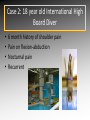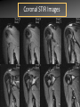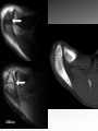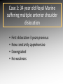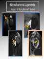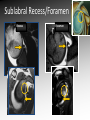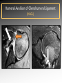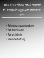
1
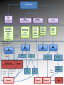
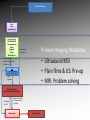
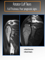
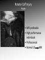
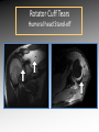
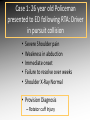
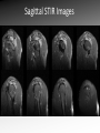
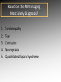
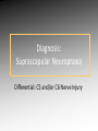
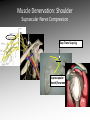
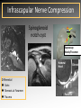
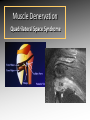
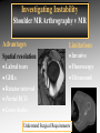
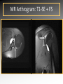
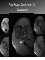
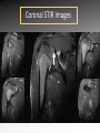
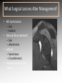
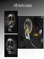
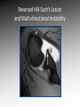
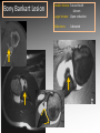
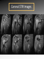
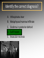
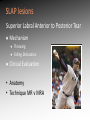
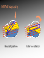
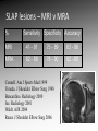
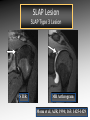
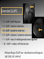
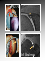
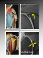
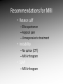
MRI shoulder: troubleshooting the cuff and instability Phil Hughes Plymouth Shoulder Pathways Pain (subacromial/cuff) General Practice Conservative Mx Stop aggravating factors Analgesia Physio Blind Injection Stiffness (Frozen shoulder/OA) No response To injection ICAT (Ortho) ICAT 1st or 2nd injection Failedt injection Candidate For SAD >60 XR <60 Adhesive casulitis Unresponsive to Injection Orthopaedics General practice Gross weakness Refer Ortho General Practice Conservative Mx Analgesia Physiotherapy Unable to inject or temporary response To injection U/S guided Injection (up to 2 U/S guided injections) Weakness (Query cuff tear) Instability General Practice Uncertain diagnosis General Practice Physio ICAT (Ortho) ICAT (Ortho) Traumatic Atraumatic Fluoroscopy injection No OA Failed Treatment OA Orthopaedics Orthopaedics Physio No Imaging Imaging Shoulder Pathways Pain (subacromial/cuff) General Practice Conservative Mx Stop aggravating factors Analgesia Physio Blind Injection Primary Imaging Modalities • Ultrasound MSI • Plain films & US: Pre-op • MRI: Problem solving No response To injection Unable to inject or temporary response To injection ICAT 1st or 2nd injection Failedt injection U/S guided Injection (up to 2 U/S guided injections) Candidate For SAD Adhesive casulitis Unresponsive to Injection Orthopaedics Diagnostic US Rotator Cuff Rotator Cuff Tears Does MR have an Advantage ? A C B D Rotator Cuff Tears Full Thickness: Poor prognostic signs Medial Retraction Muscle Atrophy Muscle Atrophy US v MRI RT Strobel. US in fatty atrophy of cuff muscles (2003) Accuracy 76 - 80% for grade 2 - 4 Rotator Cuff Injury Acute • MRI preferable • High performance individuals • Professionals • Normal US MRI Rotator Cuff Tears Humeral head Stand-off Case 1: 26 year old Policeman presented to ED following RTA: Driver in pursuit collision • • • • • Severe Shoulder pain Weakness in abduction Immediate onset Failure to resolve over weeks Shoulder X-Ray Normal • Provision Diagnosis – Rotator cuff Injury Coronal STIR and Proton Density Images Sagittal STIR Images Based on the MRI Imaging Most Likely Diagnosis? 1. 2. 3. 4. 5. Tendonopathy Tear Contusion Neuropraxia Quadrilateral Space Syndrome Based on the MRI Imaging Most Likely Diagnosis? 1. 2. 3. 4. 5. Tendonopathy Tear Contusion Neuropraxia Quadrilateral Space Syndrome Diagnosis: Suprascapular Neuropraxia Differential: C5 and/or C6 Nerve Injury Muscle Denervation: Shoulder Suprascular Nerve Compression C5 C6 C7 C8 T1 Sup Trans Scap Lig SS Suprascapular Notch/Foramen IS TM Infrascapular Nerve Compression Spinoglenoid notch cyst X X Suprascap Notch/Foramen Humeral Head Differential Varix Stenosis at Foramen Trauma Glenoid Posterior Muscle Denervation Quadrilateral Space Syndrome IS TM Case 2: 18 year old International High Board Diver • • • • 6 month history of shoulder pain Pain on flexion-abduction Nocturnal pain Recurrent Coronal STIR Images Sagittal Proton Density + Fat Suppression Select the most likely diagnosis based on the MRI ? 1. 2. 3. 4. 5. Bursitis Tendonopathy Tear Os Acromiale Superior labral tear anterior to posterior (SLAP) 2006 Jan Os Acromiale Normal Appearances Os Acromiale • Incidence 1-15% • Cadaveric 8% (33% Bilateral) • Black:White 2:1 Sammarco et al, JBJS, 2000 2005 April 2006 Jan Case 3: 34 year old Royal Marine suffering multiple anterior shoulder dislocation • • • • First dislocation 3 years previous Now constantly apprehensive Downgraded No weakness Investigating Instability Shoulder MR Arthrography v MR Advantages Limitations Spatial resolution Invasive Labral Fluoroscopy tears Ultrasound GHLs Rotator interval Partial RCTs Loose bodies Understand Surgical Requirements MR Arthrogram: T1-SE + FS Axial Proton Density with Fat Suppression Coronal STIR Images What Surgical Lesions Alter Management? • Hill-Sachs lesion – Size – location • Glenoid (Bony Bankart) – Size – attachments • Labrum – Varied tears – Circumferential • Humeral avulsion GHL (HAGL) Hill-Sachs Lesion Left >30% Richards et al, Rad; 1994; 190: 665-8 Right Reversed Hill-Sach’s Lesion and Multi-directional Instability Bony Bankart Lesion Smaller lesions: Sutured with labrum Larger lesions: Open reduction Malunions: Liberated Glenohumeral Ligaments Impact of Bony Bankart Lesion 1 3 2 4 Labral Tear Bankart lesion Classic Reattached Bankart ALPSA: Chronic Anterior labral and periosteal sleeve avulsion Sublabral Recess/Foramen Recess Foramen Buford Complex Normal variant Differential: Bankart Lesion Humeral Avulsion of Glenohumeral Ligament (HAGL) Case 4: 39 year old male patient presented to Orthopaedic Surgeon with intermittent pain • • • • Fallen onto an outstretched arm Pain with resistance Pain on abduction Intermittent catching Coronal STIR Images Identify the correct diagnosis? A. B. C. D. E. Infraspinatus tear Metaphyseal marrow infiltrate Oedema in posterior deltoid SLAP lesion Avascular necrosis SLAP lesions Superior Labral Anterior to Posterior Tear Mechanism Throwing Falling-Dislocation Clinical Evaluation • Anatomy • Technique MR v MRA MRArthrography SSP ISP Neutral position SSP ISP External rotation MRA technique Chan. SLAP lesions: MRA with arm traction AJR 1999; 173: 1117 • Cor Obl T1W fat sat • Cor Obl STIR • Sag Obl T1W fat sat • Axial Obl T1W fat sat Add Cor Obl T1W fat sat in external rotation Neutral External Rotation SLAP lesions – MRI v MRA % Sensitivity Specificity Accuracy MRI 41 - 91 75 - 89 63 - 98 MRA 82 - 89 78 - 90 82 - 90 Connell. Am J Sports Med 1999 Yoneda. J Shoulder Elbow Surg 1998 Bencardino. Radiology 2000 Jee. Radiology 2001 Waldt. AJR 2004 Reuss. J Shoulder Elbow Surg 2006 SLAP Lesion SLAP Type 3 Lesion STIR MR Arthrogram Monu et al, AJR, 1994; 163: 1425-1429 SLAP tears 1 2 Superior Labral Anterior to Posterior Tear 3 Snyder. SLAP lesions Arthroscopy 1990; 6: 274 4 2 Extended SLAPS • • • • • • 6 – SLAP 3 with flap tear 5 – SLAP + anterior extension 8 – SLAP + posterior extension 9 – SLAP + anterior / posterior extension 7 – SLAP + tear of middle glenohumeral ligament 10 – SLAP + rotator cuff interval tear Mohana-Borges SLAP tear: classification and diagnosis AJR 2003;181:1449-62. SLAP 1 SLAP 2 Sub-labral recess SLAP 2 Sub-labral recess Other MR signs “Double Oreo” sign Tuite. SLAP lesions: 3 signs on MR. Radiology 2000 Recommendations for MRI • Rotator cuff – Elite sportsmen – Atypical pain – Unresponsive to treatment • Instability – No option (CT?) – MR Arthrogram • SLAP – MR Arthrogram