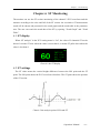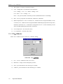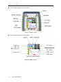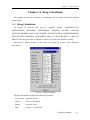
1
About the User’s Manual The User’s Manual serves for operating this instrument only. Our company shall not be responsible for any consequences and liabilities caused by using this user’s manual for other purposes. The User’s Manual containing special information is under copyright protection. All rights reserved. Copy, duplication or translation of the whole or any part of the User’s Manual is prohibited in case of no prior written consent from our company. Based on the need of product technical improvement or the file updates, we reserve the right to modify the contents contained in this manual , if the change do not involve safety issues, the contents are subject to amend without notification Due to technical upgrade or special requirements from users and with the precondition that the performance of the instrument will not be lowered, some components may vary from the description of configuration in the User’s Manual. The explain for tagging in this manual Warning : You should know the information for how to avoid the patients may suffer injury. edical staff Note: You should know the information for how to avoid possible damage to equipment. Version:1.0 2011---01 All rights reserved © Shenzhen Biocare Electronics Co., Ltd CONTENTS CONTENTS Chapter 1 Overview ....................................................................................................................................1 1.1 Overview.................................................................................................................................1 1.2 Screen layouts introduction.....................................................................................................4 1.3 Alarm ......................................................................................................................................6 1.4 Control panel...........................................................................................................................6 1.5 Sensor socket.........................................................................................................................15 1.6 Networks ...............................................................................................................................15 1.7 Rechargeable built-in battery ................................................................................................16 Chapter 2 Alarms ......................................................................................................................................17 2.1 Alarms overview ...................................................................................................................17 2.2 Alarms pausing .....................................................................................................................18 2.3 Alarms SILENCE..................................................................................................................18 2.4 Alarm Setting ........................................................................................................................19 2.5 Limits of the alarms ..............................................................................................................20 2.6 Physiology alarm information............................................................................................... 25 2.7 Technical alarm information .................................................................................................29 Chapter 3 Record ......................................................................................................................................33 3.1 Record setting .......................................................................................................................33 3.2 Record type ...........................................................................................................................34 3.3 Record content ......................................................................................................................34 Chapter 4 Admit/Discharge Patient...........................................................................................................35 4.1 Admit patient.........................................................................................................................35 4.2 Discharge patient...................................................................................................................36 Chapter 5 ECG Monitoring.......................................................................................................................37 5.1 Connecting ECG electrodes ..................................................................................................37 Chapter 6 ST Monitoring ..........................................................................................................................41 6.1 ST display..............................................................................................................................41 6.2 ST settings.............................................................................................................................41 Chapter 7 Arrhythmia Monitoring ............................................................................................................43 7.1 Arrhythmias types .................................................................................................................43 7.2 PVCs parameter ....................................................................................................................44 Chapter 8 Resp Monitoring.......................................................................................................................45 8.1 Principles of Respiration measurement.................................................................................45 8.2 Placing the electrodes............................................................................................................45 8.3 Resp settings .........................................................................................................................45 Chapter 9 SpO₂ Monitoring......................................................................................................................47 9.1 Measurement restrictions ......................................................................................................47 Operation Manual --I-- CONTENTS 9.2 SpO₂ setting.........................................................................................................................48 Chapter 10 NIBP monitoring ....................................................................................................................50 10.1 NIBP measurement step ........................................................................................................50 10.2 NIBP measurement limits .....................................................................................................50 10.3 NIBP settings ........................................................................................................................51 10.4 Blood pressure calibrations ...................................................................................................52 10.5 leakage examination..............................................................................................................52 Chapter 11 Temperature Monitoring .........................................................................................................54 11.1 Steps of temperature measurement........................................................................................54 11.2 Temperatures settings menu ..................................................................................................54 Chapter 12 IBP Monitoring.......................................................................................................................55 12.1 Steps of IBP measurement ....................................................................................................55 12.2 IBP Setting ............................................................................................................................56 12.3 IBP Zeroing ...........................................................................................................................56 12.4 IBP Calibration......................................................................................................................58 Chapter 13 CO₂ Monitoring ....................................................................................................................61 13.1 Introduction...........................................................................................................................61 13.2 CO₂ Measurement ................................................................................................................61 13.3 CO₂Setting............................................................................................................................61 13.4 CO₂calibrate .........................................................................................................................62 Chapter 14 IoC Monitoring.......................................................................................................................64 14.1 Discription.............................................................................................................................64 14.2 The patient cable ...................................................................................................................65 14.3 Skin preparation and connections .........................................................................................66 14.4 IoC module...........................................................................................................................67 14.5 Patient monitor screen...........................................................................................................71 14.6 IoC setting .............................................................................................................................71 Chapter 15 History Review.......................................................................................................................72 15.1 Trend Graph ..........................................................................................................................72 15.2 Trend table ............................................................................................................................73 15.3 Alarm Review .......................................................................................................................75 15.4 NIBP Review.........................................................................................................................76 15.5 Wave review..........................................................................................................................77 Chapter 16 Drug Calculation ....................................................................................................................79 16.1 Drug Calculation ...................................................................................................................79 16.2 Operating procedures ............................................................................................................80 16.3 Titration table ........................................................................................................................80 Chapter 17 Appendix - Product Specifications .........................................................................................82 --II-- Operation Manual CONTENTS 17.1 Classification.........................................................................................................................82 17.2 Accordant Standard ...............................................................................................................82 17.3 Power Supply ........................................................................................................................82 17.4 Battery...................................................................................................................................82 17.5 Environment..........................................................................................................................82 17.6 Signal Interface .....................................................................................................................82 17.7 ECG.......................................................................................................................................83 17.8 Respiration ............................................................................................................................84 17.9 NIBP .....................................................................................................................................85 17.10 SpO₂..................................................................................................................................86 17.11 Temperature.......................................................................................................................86 17.12 IBP(optional).....................................................................................................................87 17.13 CO₂(optional) ...................................................................................................................87 17.14 IoC (optional) ....................................................................................................................88 17.15 Recorder(optional) ............................................................................................................88 Operation Manual --III-- Chapter 1 Overview Chapter 1 Overview 1.1 Overview This monitor is suitable for adult, pediatric and neonate patient, may monitor the physical parameters, such as electrocardiograph (ECG), noninvasive blood pressure (NIBP), oxygen saturation (SpO₂), respiration rate (Resp), body temperature (Temp) , invasive blood pressure (IBP), carbon dioxide (CO₂), index of cerebral(IoC)and so on, can display maximum 11 waveforms and all information of the parameters monitored in the same screen. Below shows the monitoring functions of this monitor: 1) electrocardiograph (ECG), including: heart rate, 6 channels ECG waveforms, ST segment analysis, arrhythmia analysis; 2) oxygen saturation (SpO₂), including: oxygen saturation , pulse rate, pulse wave; 3) noninvasive blood pressure (NIBP), including: Systolic pressure, diastolic pressure, mean pressure; 4) body temperature (Temp): 2 channels body temperature data; 5) respiration (Resp): Breath rate, breath waveform; 6) invasive blood pressure (IBP): 2 channels IBP waves and Systolic pressure, diastolic pressure, mean pressure; 7) carbon dioxide (CO₂): 1 channel CO₂ wave and Breath rate, Et CO₂, FiCO₂. 8) Index of Cerebral(IoC): 1 channel EEG wave and IoC, EMG, SQI, ESR. Warning Monitor can only monitoring one patient at a time. Warning There could be hazard of electrical shock by opening the monitor casing. All servicing and future upgrading to this equipment must be carried out by personnel trained and authorized by Factory. Warning You must verify if the device and accessories can function safely and normally before use. Warning Possible explosion hazard if used in the presence of flammable anesthetics or other flammable substance in combination with air, oxygen-enriched environments, or nitrous oxide. . Warning You must customize the alarm setups according to individual patient situation and make sure that alarm sound can be activated when alarm occurs. Operation Manual --1-- Chapter 1 Overview Warning Do not touch the patient, table, or the device during defibrillation. Warning Do not use cellular phone in the vicinity of this device. High level electromagnetic radiation emitted from such devices may greatly affect the monitor performance. Warning Devices connected to the monitor shall form an equipotential system (protectively earthed). Warning When used with Electro-surgery equipment, you (doctor or nurse) must give top priority to the patient safety. Warning Do not place the monitor or external power supply in any position that might cause it to fall on the patient. Do not lift the monitor by the power supply cord or patient cable, use only the handle on the monitor. Warning Consult IEC-601-1-1 for system interconnection guidance. The specific requirements for system interconnection are dependent upon the device connected to the monitor and the relative locations of each device from the patient, and the relative location of the connected device to the medically used room containing the monitor. In all circumstance the monitor must be connected to a grounded AC power supply. The monitor is referred to as an IEC 601/F device in the summary of situations table contained in IEC 601-1-1. Warning Dispose of the packaging material, observing the applicable waste control regulations and keeping it out of children’s reach. Warning Grounding: Connect the monitor only to a three-wire, grounded, hospital-grade receptacle. The three-conductor plug must be inserted into a properly wired three-wire receptacle; if a three-wire receptacle is not available, a qualified electrician must install one in accordance with the governing electrical code. Do not under any circumstances remove the grounding conductor from the power plug. --2-- Operation Manual Chapter 1 Overview Do not use extension cords or adapters of any type. The power cord and plug must be intact and undamaged. If there is any doubt about the integrity of the protective earth conductor arrangement, operate the monitor on internal battery power until the AC power supply protective conductor is fully functional. Warning For continued safe use of this equipment, it is necessary that the listed instructions be followed. However, instructions listed in this manual in no way supersede established medical practices concerning patient care. Do not rely only on audible alarm system to monitor patient. When monitoring adjusting the volume to very low or completely muting the sound may result in the disaster to the patient. The most reliable way of monitoring the patient is at the same time of using monitoring equipment correctly, manual monitoring should be carried out. This multi-parameter patient monitor is intended for use only by medical professionals in health care institutions. To avoid electrical shock, you shall not open any cover by yourself. Service must be carried out by qualified personnel. Use of this device may affect ultrasonic imaging system in the presence of the interfering signal on the screen of ultrasonic imaging system. Keep the distance between the monitor and the ultrasonic imaging system as far as possible. It is dangerous to expose electrical contact or applicant coupler to normal saline, other liquid or conductive adhesive. Electrical contact and coupler such as cable connector, power supply and parameter module socket-inlet and frame must be kept clean and dry. Once being polluted by liquid, they must be thoroughly dried. If to further remove the pollution, please contact your biomedical department or Factory. Warning It is important for the hospital or organization that employs this equipment to carry out a reasonable maintenance schedule. Neglect of this may result in machine breakdown or injury of human health. Caution If you have any doubt to the grounding layout and its performance, you must use the built-in battery to power the monitor. Operation Manual --3-- Chapter 1 Overview 1.2 Screen layouts introduction The screen is divided into four sections: 1st information section; 2nd waveform section; 3rd parameters section; 4th menu section (as chart 1-1 shows). Chart 1-1 monitor demo interface Information section The information section is on top of the screen, displays current conditions of monitor and patient. The information in turn from left to right on the top is “patient information”, “technical alarm information”, “physiological alarm information”, “date and time” , “network state” and “battery state”. 1) Patient information: Bed number (refers to the hospital bed number of patient monitored); Type of patient (“Adult”, “Pediatric” or “Neonate”); Name of patient (if operator doesn’t input patient’s name, this position will displays “NO NAME”); 2) Technical alarm information: Reporting current condition of monitor or sensors, this section will display alarm information; --4-- Operation Manual Chapter 1 Overview 3) Physiological alarm information: If patient's physiological parameters exceed the alarm limit, this section will display alarm information; 4) Date and time: Updating current date and time every second; 5) Network connection state; 6) Battery state: current battery capacity or its condition. Parameters section Heart rate: heart rate (unit: beats per minute bpm) ST: ST segment (unit: millivolt mV) PVCs: times of premature ventricular constriction (unit: times/minute) NIBP: From right to left is: systolic pressure, diastolic pressure, mean pressure (unit: millimeter mercury column- mmHg or kilopascal- kPa) SpO₂: oxygen saturation SpO₂ (unit: %), Pulse rate (unit: pulses /minute) Respiration rate: respiration rate (unit: Breaths/Minute BrPM) Temperature: body temperature (unit: centigrade -℃ or Fahrenheit- ℉) invasive blood pressure (IBP): Systolic pressure, diastolic pressure, mean pressure; carbon dioxide (CO₂): Breath rate, Et CO₂ and FiCO₂. Index of Cerebral(IoC): IoC, EMG, SQI, ESR. The user may change the settings of above monitored parameters which will be introduced in later chapters in detail. Waveform section The waveform section displays 7 waveforms in standard screen layout, which from top to bottom respectively are: ECG1 waveform, ECG2 waveform, pulse wave, respiration waveform, 2 channels of IBP waveforms, CO₂ waveform (standard interface). Total 11 waveforms can be displayed if in “ECG Full Lead” screen layout. The name appears in upper left side of each waveform. The ECG waveform gain and filter mode will be also displayed besides the ECG wave name. On the right side of the ECG waveform stands a mark with the unit of 1 mV. The gain of breathing waveform is displayed on the right side of the name of breathing wave. The ruler mark of IBP waveform is displayed on the right side of the name of IBP wave. The ruler mark of CO₂ waveform is displayed on the right side of the name of CO₂ wave. Operation Manual --5-- Chapter 1 Overview When user push the keys of patient monitor, a window may pop-up in the waveform section. The waveform section will restore demo after the window is retreated. Menu section On the bottom of the screen there are 5 menu item: “Patient”, “Review”, “Setting”, “Alarm Limit” and “Service”. When no window displays on the screen, the user may visit these menus by rotating the knobs. When the cursor chooses anyone of the items, sublevel menus will popup. When the user presses down the knob once again, the corresponding dialog will pop-up and the user can change the settings in the dialog. 1.3 Alarm When the alarm occurs, the warning light will glitter or bright, the color represents certain level of the alarm. The detailed contents please refer to chapter2 “Alarm”. 1.4 Control panel The control panel is on the front panel. The total keys from left to right are listed below: 1) Power key: key to turning on and turning off of the power; 2) Alarm silence key: With this key pressed down, sound of the alarm will shut down, also the “ALARM SILENCE” will be displayed in the information section, and other sounds (key sound, palpitation sound and so on) will not be affected. Pressing down the key again will restore all the alarms. 3) Alarm pausing key: With this key pressed down, the alarm may hang up for 2 minutes (“1 minute”, “2 minutes” and “3 minutes” are optional), and the “ALARM PAUSE” will be displayed in the information section. All the alarm will be restored after this key is pressed again. 4) Freezing key: In the normal mode, all the waveforms on the screen will be frozen with this key pressed down. Pressing down this key once again will release the frozen waveforms; 5) Blood pressure key: Pressing down this key will start to charge the cuff with gas, and to measure the blood pressure. Pressing down the key once again can cancel the measurement; 6) Record/Stop key: If the monitor has a recorder, pressing down this key will start recording the real-time waveforms. Pressing the key again may stop recording; 7) Main menu key: press this key to returning to the main menu; --6-- Operation Manual Chapter 1 Overview 8) Knob key: With this key, the user may enter the menus and windows and change the monitor settings. Patients management By pushing the “patient” button, the user may choose to enter the window of “Admit new patients”, “Discharge Current Patient” or “Dose Calculation” the detailed introductions may refer to chapter4 “Admit and Discharge Patient”. Chart 1-3 patient management History review By selecting the “Review” button, the user may choose to enter the window of “Trend Graph”, “Trend Table”, “Alarm Review”,“NIBP review” or “Wave review”. The detailed introductions may refer to chapter12 “History Review”. Chart 1-4 history review Operation Manual --7-- Chapter 1 Overview Setting By selecting the “Setting” button, the user may choose to enter the window of “Alarm setting”, “Record setting”, “Screen layout”, “Adjust time”, “Miscellaneous”, “ECG setting”, “ST setting”, “SpO₂ setting”, “NIBP setting”, “Resp setting”, “Temp setting” , “IBP Setting”, “CO₂ Setting”, ““IOC Setting”” or “Load Default “. Chart 1-5 setting Alarm setting Detailed introductions can refer to Chapter2 “Alarm”. Recording setting Detailed introductions can refer to Chapter3 “Recording”. Screens layout After entering the screen layouts window, the user may change the current display interface by selecting the interfaces of 6 types of “Standard”, “ECG Full Lead”, “Big Font”, “OxyCRG”, “NIBP Trend”, “Trend Table”, and choose to turn on or turn off parameter or waveform in the “parameter switch” and “waveform switch”. The user can change trend resolution from Time” if the screen layout is set to “Trend Table”. --8-- Operation Manual “1 min” to “60 min” by setting “Trend Chapter 1 Overview The following chart shows the menu of screen layouts: Chart 1-6 screen layouts Screen layout Big font Standard ECG Full Lead OxyCRG Trend table NIBP trrend Operation Manual --9-- Chapter 1 Overview Adjust Time By entering the adjust time window, the user may choose the date format and adjust the current date and time, as the following chart shows: Chart 1-7 adjust time Miscellaneous By entering the miscellaneous windows, the user may change the key volume and the screen brightness. The adjusting scope of key volume is 0~10 (0 means volume closure); The adjusting scope of screen brightness is 1~10 (10 means the highest brightness). If “Wave Smooth” switch is “On”, the wave will be displayed as smooth mode. Chart 1-8 miscellaneous setting ECG setting Detailed introductions of ECG settings can refer to Chapter5 “ECG monitoring”. ST setting Detailed introductions of ST settings can refer to Chapter6 “ST monitoring”. Resp setting Detailed introductions of RESP settings can refer to Chapter8 “RESP monitoring”. --10-- Operation Manual Chapter 1 Overview SpO₂ setting Detailed introductions of oxygen saturation settings can refer to Chapter9 “SpO ₂ monitoring”. NIBP setting Detailed introductions of noninvasive blood pressure settings can refer to Chapter10 “NIBP monitoring”. Temperature setting Detailed introductions of body temperature settings can refer to chapter11 “temperature monitoring”. IBP setting Detailed introductions of IBP settings can refer to chapter12 “IBP monitoring”. CO₂ setting Detailed introductions of CO₂ settings can refer to chapter13 “CO₂ monitoring”. CO₂ setting Detailed introductions of IoC settings can refer to chapter14 “IoC monitoring”. Load default setting The following chart shows the window of Apply default settings: Chart 1-9 Load default settings If “Yes” is chosen, then the current settings will be replaced with default settings Operation Manual --11-- Chapter 1 Overview Alarm limit By selecting the “limits of alarm” button, the user may choose to enter the windows of “ECG Alarm Limit”, “ SpO₂ Alarm Limit”, “NIBP Alarm Limit”, “Resp Alarm Limit”, “Temp Alarm Limit”, “IBP Alarm Limit”, “CO₂ Alarm Limit”, “IOC Alarm Limit” or “Load default Alarm Limit”, the detailed introductions refer to chapter2 “alarms”. Chart 1-10 Alarm Limit Maintenance By selecting the “Service” button, the user may choose to enter the windows of “ECG calibration”, “ Temp Sensor Type”, “NIBP Pneumatic Test”, “NIBP calibration”, “NIBP Reset”, “IBP Zero”, “IBP Calibration”, “CO₂ Calibration”, “Demo mode”, “Version info”, “User setting”, “Factory Service” and so on. Chart 1-11 Service --12-- Operation Manual Chapter 1 Overview ECG calibration Entering the ECG calibration window, the user may turning on or turning off of the ECG calibration signal, as the following chart shows: Chart 1-12 ECG calibration Temp Sensor Type Entering the Temp Sensor Type window, the user may initialize the type of body temperature sensor: 10K or 2.25K, as the following chart shows: Chart 1-13 Temp Sensor Type Air leakage examination of noninvasive blood pressure Selecting the air leakage examination of noninvasive blood pressure, the user may examine whether the entire air way of blood pressure measurement leaks air or not. When the blood pressure cuff is connected, the user may start the air leakage test with this key, thus discover whether the airtight condition of gas route is good or not. The examination result is: If air leakage examination is passed, the system will not make any prompt; If isn’t, the corresponding failure prompts will be displayed in the noninvasive blood pressure information section. The detailed introductions refer to 10.5 the air leakage examination. NIBP calibration After selecting the noninvasive blood pressure calibration, the user enters the calibration mode, and at this time the user may calibrate, using a pressure gauge (or mercury sphygmomanometer) with a calibration precision higher than 1 mmHg after calibrated. If the “measure blood pressure” key is presses down during the calibration, the system will stop calibrating. The detailed introductions refer to 10.4 the blood pressure calibrations. Operation Manual --13-- Chapter 1 Overview NIBP reset After choosing the noninvasive blood pressure reset, the user may restore the blood pressure module to the initial settings. When the blood pressure measurement is abnormal, yet the monitor cannot prompt reasons of the problem, using this key is suggested. Because this causes the blood pressure module to reset, the blood pressure module may commit self-recovery when the abnormity of work is caused by accidental reasons. IBP Zero Detailed introductions of IBP zero can refer to 12.2 “IBP zero”. IBP Calibration Detailed introductions of IBP calibration can refer to 12.3 “IBP Calibration”. CO₂ Calibration Detailed introductions of CO₂ Calibration can refer to 12.2 “CO₂ Calibration”. Demo mode The user input the correct password, the monitor will enter demo mode, and in the centre of the screen a big “DEMO” label will be shown. The demo mode is a particular state just for demonstrating performance of the machine, helping user carry out trainings. In the actual clinical use, this function is forbidden, because it can possibly cause the medical staffs to take demo waveforms as the patient waveforms and parameters by mistake, to affect patient monitoring. Version information Choosing the “version information”, the user may look over the version information of the software installed in the monitor. User settings The user may carry out user maintenance in the user settings menu, by inputting the password. This item is merely open to the serviceman appointed by the factory. Factory service The user cannot implement functions of maintenance. This item is merely open to the serviceman appointed by the factory. --14-- Operation Manual Chapter 1 Overview 1.5 Sensor socket Chart 1-14 sensor socket The following shows sensor socket: T1: temperature channel 1 sensor socket; T2: temperature channel 2 sensor socket; SpO₂: Oxygen saturation sensor socket; NIBP: non-invasive blood pressure cuff socket; ECG: ECG cable socket; 1.6 Networks The network port of the monitor is the standard RJ45 network interface, may communicate with the central station through the ethernet cable, to achieve the function of remote monitoring. In the top right corner of the screen there is a network icon representing current network status. If the network electric cable is disconnected, the network condition icon shows as “ the icon shows as “ ”; After the monitor has established connection with the central station, ”; If the monitor communicates normally with the central monitoring system, the icon shows as “ ”. Operation Manual --15-- Chapter 1 Overview 1.7 Rechargeable built-in battery The monitor is equipped with a rechargeable built-in battery. In the top right corner of the screen exists one symbol “ “, indicating the state of the battery capacity, of which the green part denoting electric quantity of the battery. When the battery is charged, the charging condition is expressed with animation. After the battery is full-charged, the symbol will show as “ ”. When this monitor has not been installed the built-in battery, the symbol shows as “ ” indicating no battery. When running with power supply from battery, the monitor detects the volume of the battery, and alarms when the battery is insufficient, and prompts in the information section: “BAT LOW”. At this time, the AC power should be plug in, and immediately charge battery in time. If battery is still used for power supply, the monitor will power off automatically when the battery exhausted. --16-- Operation Manual Chapter 2 Alarms Chapter 2 Alarms 2.1 Alarms overview Types of the alarms The alarms can be divided into two types: physiology alarms and technical alarms. Physiology alarms: triggered by some of patient's physiological parameters exceed the limits, taking the body temperature exceeding temperature alarm limit as an example. Technical alarms: triggered by the abnormality of certain monitoring function or distortion of monitoring results caused by failure of system or sensors, taking ECG lead off as an example. Level of alarms The alarms have three levels: high, medium and low. The monitor has set levels for technical alarms and physiology alarms. Modes of the alarms When alarming, the monitor gives alarm prompts by three ways: sound alarm, light alarm, and alarm message description. The prompts of sound and light come from the speaker, the alarm indicator light and alarm message description. The alarm message description is displayed on the screen. The physiology alarm is displayed in the patient alarm information section, while the technical alarm displayed in the monitor alarm information section. When the physiology alarm occurs, which is caused by the measurement parameters exceeding the alarm limit, the color of high limit and low limit would change from dark to bright, besides the three means of alarm prompting mentioned above. When there is “*” before technical or the physiology information section, it means low level alarm. “**” means medium level alarm and the information bottom color will turn yellow. “***” means high level alarm and information bottom color will turn red. For example: The “** HR TOO HIGH” is the expression of medium alarm. Physical alarm has 2 kind of alarm mode: LATCH or Unlatch. LATCH means that once alarm occurs, the system will give alarm all the time until manual intervention (such as push the “SILENCE” button on the panel). UNLATCH means that the system will stop giving alarm once the alarm condition does not exist. Operation Manual --17-- Chapter 2 Alarms There are three levels of the alarm: high, midium, low, by using the different light and the sound. The following table shows in order: Alarm level alarm light High alarm light glitters red, the The flicker frequency is quick sound characteristic of the alarm pattern as “honk - honk - honk------honk - honk, honk - honk honk------honk - honk”, each 8 seconds occur once medium alarm light glitters yellow, the The pattern as “honk - honk - honk”, flicker frequency is slow Low each 25 seconds emit sound once alarm light is bright and The pattern as “honk -”, each 25 seconds always show yellow emit sound once 2.2 Alarms pausing Presses “PAUSE” key on the control panel, all alarm sound and light and the alarm message are closed. Then the system enters alarm suspend state. The suspension countdown time is displayed in the area of the technical alarm. Three options can be set about the alarm suspension time: 1 minute, 2 minute and 3 minutes. The user must enter the window of the alarm setting, chooses correspondingly the suspension time. After presses down of “PAUSE” key again, the system may restore to the normal state. 2.3 Alarms SILENCE Presses “SILENCE” key on the control panel, then may close the sound and the light alarm; when presses down “SILENCE” key again, will quit from alarm silence condition and reactivated correspondingly sound alarm, returns to normal alarm condition. If the alarm still exists under the condition of the silence state, then the information section display this alarm information. If there is no alarm exists under the condition of the silence state, then all the alarm will be eliminated. Attention When the system is under the “SILENCE” condition, any newly triggered alarm will terminate the silence condition, and then makes the system to restore to the normal alarm condition. --18-- Operation Manual Chapter 2 Alarms 2.4 Alarm Setting Enter the alarm setting window, the options below may be set. 1) Alarm Volume: The scope is 1~10 (10 is the highest volume). 2) Suspend Time: 1 minute, 2 minutes, 3 minutes. 3) Flash: if “On” is selected and there is physical alarm, corresponding parameter digit will flash to indicate that the parameter has alarm. 4) Para Alarm: 2 items: LATCH or Unlatch. LATCH means that once alarm occurs, the system will give alarm all the time until manual intervention (such as push the “SILENCE” button on the panel). UNLATCH means that the system will stop giving alarm once the alarm condition does not exist. 5) Alarm Record: If “On” is selected, the recorder will record the alarm event when physical alarm occurs, otherwise it will not record. 6) Voice Alarm: If “On” is selected and alarm event occurs, a human voice alarm will continuously notify the user, otherwise it will not notify by human voice. Chart 2-1 alarm setting Operation Manual --19-- Chapter 2 Alarms 2.5 Limits of the alarms Physiology alarm is triggered according to the settings limits. Various parameters limits are showed by the dark color in the parameter area of the upper corner on the left side. If the parameter exceeds the limits, then triggers the physiology alarm at this parameter by the bright color. For example: the low limit of the heart rate is 80, if at this time the heart rate is 60 pieces, then triggers “HR TOO LOW”, the low limit of the heart rate “80” will be a bright color, the following chart will show: Chart 2-2 Alarm Limit ECG Alarm Limit Choosing “ECG Alarm Limit” may enter “ECG Alarm Limit” window: Chart 2-3 Ecg Alarm Limit Following is the adjustment scope of the heart rate: Patient type Adult Pediatric Neonate HR high limit 300 350 350 HR low limit 15 15 15 adjustment scope of the ST:- 2.00mV~2.00mV. adjustment scope of the PVCs :0~10. --20-- Operation Manual Chapter 2 Alarms SpO₂ Alarm Limit Choosing “SpO₂ Alarm limit” may enter “SpO₂ Alarm Limit” window: Chart 2-4 SpO₂ Alarm Limit The SpO₂ limit adjustment scope is 0~100; The Pulse rate alarm limit adjustment scope are 20~300. NIBP Alarm Limit Choosing “NIBP Alarm Limit” may enter “NIBP Alarm Limit” window: Chart 2-5 NIBP Alarm Limit The NIBP Alarm Limit adjustment scope as follows: Patient type Adult Pediatric Neonate Systolic pressure high limit 280 220 135 Systolic pressure low limit 40 40 40 Diastolic pressure high limit 220 160 100 Diastolic pressure low limit 10 10 10 mean pressure high limit 240 170 110 mean pressure low limit 20 20 20 Operation Manual --21-- Chapter 2 Alarms Resp Alarm Limit Choosing “Resp Alarm Limit” may enter “Resp Alarm Limit” window: Chart 2-6 Resp Alarm Limit The Resp rate alarm limit adjustment scope is:7~120. Patient type Adult Pediatric Neonate RR high limit 120 150 150 RR low limit 7 7 7 Temp Alarm Limit Choosing “Temp Alarm Limit” may enter “Temp Alarm Limit” window: chart 2-7 Temp Alarm Limit The Temp alarm limit adjustment scope is:0~50℃ (32~122℉). --22-- Operation Manual Chapter 2 Alarms IBP Alarm Limit Choosing “IBP Alarm Limit” may enter “IBP Alarm Limit” window: chart 2-8 IBP Alarm Limit The IBP alarm limit adjustment scope is: -50~300mmHg. CO₂ Alarm Limit Chart 2-9 CO₂ Alarm Limit The Resp rate alarm limit adjustment scope is: 7~120; The EtCO₂alarm limit adjustment scope is: 0~100mmHg; The FiCO₂alarm limit adjustment scope is: 0~100mmHg. Operation Manual --23-- Chapter 2 Alarms IOC Alarm Limit Chart 2-10 CO₂ Alarm Limit The IoC rate alarm limit adjustment scope is: 0~100; Load Default Alarm Limit Choosing “Load Default Alarm Limit” can enter “load default Alarm limit” window: The chart 2-11 Load default Alarm Limit If chooses “Yes”, then the current alarm limit settings will be able to substituted by the default alarm limit settings. --24-- Operation Manual Chapter 2 Alarms 2.6 Physiology alarm information Below is all physiologies alarm tabulates: Alarm Information Trigger Condition ***ASYSTOLE Over 4 seconds non- palpitations signals *** APNEA In a setting time without breath signal *** NO PULSE Over 15 seconds without pulse signals ** HR TOO HIGH The heart rate exceeds the alarm high limit ** HR TOO LOW The heart rate is lower than the alarm low limit ** ST-I TO HIGH the ST value correlate with I surpass the upper alarm limit ** ST-I TOO LOW the ST value correlate with I surpass the lower alarm limit ** ST-II TO HIGH the ST value correlate with II surpass the upper alarm limit ** ST-II TOO LOW the ST value correlate with II surpass the lower alarm limit ** ST-III TO HIGH the ST value correlate with III surpass the upper alarm limit ** ST-III TOO LOW the ST value correlate with III surpass the lower alarm limit ** ST-AVR TOO HIGH the ST value correlate with AVR surpass the upper alarm limit ** ST-AVR TOO LOW the ST value correlate with AVR surpass the lower alarm limit ** ST-AVL TOO HIGH the ST value correlate with AVL surpass the upper alarm limit ** ST-AVL TOO LOW the ST value correlate with AVL surpass the lower alarm limit ** ST-AVF TOO HIGH the ST value correlate with AVF surpass the upper alarm limit ** ST-AVF TOO LOW the ST value correlate with AVF surpass the lower alarm limit ** ST-V TOO HIGH the ST value correlate with V surpass the upper alarm limit ** ST-V TOO LOW the ST value correlate with V surpass the lower alarm limit Operation Manual --25-- Chapter 2 Alarms ** PVCs TOO HIGH The PVCs value exceeds the alarm high limit ** SPO₂ TOO HIGH The oxygen saturation exceeds the alarm high limit ** SPO₂ TOO LOW The oxygen saturation is lower than the alarm low limit ** Pulse rate TOO HIGH The Pulse rate surpass the alarm high limit ** Pulse rate TOO LOW The Pulse rate are lower than the alarm low limit **NIBP SYS TOO HIGH NIBP systolic pressure exceeds the alarm high limit **NIBP SYS TOO LOW NIBP systolic pressure is lower than the lower alarm limit **NIBP MEAN TOO HIGH NIBP mean pressure exceeds the alarm high limit **NIBP MEAN TOO LOW NIBP mean pressure is lower than the alarm low limit **NIBP DIA TOO HIGH NIBP diastolic pressure exceeds the alarm high limit **NIBP DIA TOO HIGH NIBP diastolic pressure is lower than the alarm low limit ** RR TOO HIGH The Breath rate exceeds the alarm high limit ** RR TOO LOW The Breath rate is lower than the alarm low limit ** TEMP1 TOO HIGH The body temperature channel 1 exceeds the alarm high limit ** TEMP1 TOO LOW The body temperature channel 1 is lower than the alarm low limit ** TEMP2 TOO HIGH The body temperature channel 2 exceeds the alarm high limit ** TEMP2 TOO LOW The body temperature channel 2 is lower than the alarm low limit **ART SYS TOO HIGH ART systolic pressure exceeds the alarm high limit **ART SYS TOO LOW ART systolic pressure is lower than the low limit **ART MEAN TOO HIGH ART mean pressure exceeds the high **ART MEAN TOO LOW ART mean pressure is lower than the low limit **ART DIA TOO HIGH ART diastolic pressure exceeds the high limit **PA DIA TOO LOW PA diastolic pressure is lower than the low limit **PA SYS TOO HIGH PA limit --26-- Operation Manual limit systolic pressure exceeds the alarm high Chapter 2 Alarms **PA SYS TOO LOW PA systolic pressure is lower than the low limit **PA MEAN TOO HIGH PA mean pressure exceeds the high limit **PA MEAN TOO LOW PA mean pressure is lower than the low limit **PA DIA TOO HIGH PA diastolic pressure exceeds the high limit **PA DIA TOO LOW PA diastolic pressure is lower than the low limit **PA DIA TOO LOW PA diastolic pressure is lower than the low limit **CVP SYS TOO HIGH CVP systolic pressure exceeds the alarm high limit **CVP SYS TOO LOW CVP systolic pressure is lower than the low limit **CVP MEAN TOO HIGH CVP mean pressure exceeds the high **CVP MEAN TOO LOW CVP mean pressure is lower than the low limit **CVP DIA TOO HIGH CVP diastolic pressure exceeds the high limit **CVP DIA TOO LOW CVP diastolic pressure is lower than the low limit **LAP SYS TOO HIGH LAP systolic pressure exceeds the alarm high limit limit **LAP SYS TOO LOW LAP systolic pressure is lower than the low limit **LAP MEAN TOO HIGH LAP mean pressure exceeds the high limit **LAP MEAN TOO LOW LAP mean pressure is lower than the low limit **LAP DIA TOO HIGH LAP diastolic pressure exceeds the high limit **LAP DIA TOO LOW LAP diastolic pressure is lower than the low limit **RAP SYS TOO HIGH RAP systolic pressure exceeds the alarm high limit **RAP SYS TOO LOW RAP systolic pressure is lower than the low limit **RAP MEAN TOO HIGH RAP mean pressure exceeds the high **RAP MEAN TOO LOW RAP mean pressure is lower than the low limit **RAP DIA TOO HIGH RAP diastolic pressure exceeds the high limit **RAP DIA TOO LOW RAP diastolic pressure is lower than the low limit **ICP SYS TOO HIGH ICP systolic pressure exceeds the alarm high limit **ICP SYS TOO LOW ICP systolic pressure is lower than the low limit **ICP MEAN TOO HIGH ICP mean pressure exceeds the high limit **ICP MEAN TOO LOW ICP mean pressure is lower than the low limit **ICP DIA TOO HIGH ICP diastolic pressure exceeds the high limit **ICP DIA TOO LOW ICP diastolic pressure is lower than the low limit limit Operation Manual --27-- Chapter 2 Alarms **P1 SYS TOO HIGH P1 systolic pressure exceeds the alarm high limit **P1 SYS TOO LOW P1 systolic pressure is lower than the low limit **P1 MEAN TOO HIGH P1 mean pressure exceeds the high **P1 MEAN TOO LOW P1 mean pressure is lower than the low limit **P1 DIA TOO HIGH P1 diastolic pressure exceeds the high **P1 DIA TOO LOW P1 diastolic pressure is lower than the low limit **P2 SYS TOO HIGH P2 systolic pressure exceeds the alarm high limit **P2 SYS TOO LOW P2systolic pressure is lower than the low limit **P2 MEAN TOO HIGH P2mean pressure exceeds the high **P2 MEAN TOO LOW P2mean pressure is lower than the low limit **P2 DIA TOO HIGH P2diastolic pressure exceeds the high **P2 DIA TOO LOW P2diastolic pressure is lower than the low limit **EtCO₂TOO HIGH EtCO₂exceeds the high **EtCO₂ TOO LOW EtCO₂ is lower than the low limit **FiCO₂ TOO HIGH FiCO₂exceeds the high **FiCO₂ TOO LOW FiCO₂ is lower than the low limit **AwRR TOO HIGH AwRR exceeds the high **AwRR TOO LOW AwRR is lower than the low limit **IoC TOO HIGH IoC is higher than the low limit ** IoC TOO LOW IoC is lower than the low limit --28-- Operation Manual limit limit limit limit limit limit limit Chapter 2 Alarms 2.7 Technical alarm information Below is all technical alarm tabulates: Alarm Information Trigger Condition Process Method ** ECG LEAD OFF RL or more than 2 ECG leads check falls off connection RA lead fall off check ** ECG LEAD RA OFF the the ECG lead ECG lead ECG lead ECG lead ECG lead connection ** ECG LEAD LA OFF LA lead fall off check the connection ** ECG LEAD LL OFF LL lead fall off check the connection ** ECG LEAD V OFF V lead fall off check the connection ** MODULE INIT ERR Module self-checking mistake Restart the machine, if error still existed, contact the factory service ***MODULE COMM STOP ** MODULE COMM ERR ** PARA ALARM LMT ERR The module and the main Restart the machine, if error engine communication have still existed, contact the the problem factory service The module and the main Restart the machine, if error engine communication have still existed, contact the the problem factory service The parameter of the alarm contact the factory service limit is modified by the accident ** RANGE EXEED The parameter observed value contact the factory service has exceed the measurement scope which the system can carry on ** SpO₂ SENSOR OFF ** SpO₂ FINGER OFF SEARCHING PULSE... SpO ₂ sensor does not Check SpO ₂ sensor connected connection The finger fall off from SpO₂ Check SpO₂ sensor connect sensor with the finger SpO₂ sensor connect bad or Check the patient move the arm connection SpO ₂ situation sensor and patient's current condition Operation Manual --29-- Chapter 2 Alarms ** Temp1 SENSOR OFF The body temperature channel 1 sensor do not Check temperature sensor connection connect ** Temp2 SENSOR OFF The body temperature channel 2 sensor do not Check temperature sensor connection connect ** WATCHDOG ERR Main engine watch-dog self-checking defeat Restart the machine, if wrong still existed, contact the factory service ** SYSTEM TIME LOST The system clock has not set Change the system time as the current time, if error still existed, related the factory to carry on the service ** 12V HIGH ** 12V LOW ** 3.3V HIGH The 12V voltage examination Restart the machine, if error exceeds the normal voltage still existed, contact the scope factory service The 12V voltage examination Restart the machine, if error is lower than the normal still existed, contact the voltage scope factory service The 3.3V examination voltage exceeds the normal voltage scope ** 3.3V LOW **BAT HIGH The factory service Restart the machine, if error examination is lower than the still existed, contact the normal voltage scope factory service battery examination voltage exceeds the normal voltage scope **BAT LOW still existed, contact the voltage The 3.3V Restart the machine, if error The battery capacity Restart the machine, if error still existed, contact the factory service is insufficient Meets the alternating current to carry on the charge immediately to the battery * NIBP LOOSE CUFF The cuff has not connected * NIBP AIR LEAK Reconnects Operation Manual blood pressure cuff check the pipe connection The cuff has not connected situation or replace cuff, if good or the air course leaks the breakdown still existed, air please contact the factory service --30-- the Chapter 2 Alarms * NIBP DEFLATE ERR When blood pressure measurement deflates has the problem check the tube connection or replace cuff, if the error still existed, please contact the factory service * NIBP WEAK SIGNAL Examined the patient type When blood pressure set whether correctly, check measurement the pulse signal the too replace cuff, if the error still weak, is unable to calculate the blood pressure tube connection or existed, please contact the factory service * NIBP OUT OF RANGE When blood pressure blood check he tube connection or pressure or the pulse signal replace cuff, if the error still exceeds the normal range, is existed, please contact the unable factory service measurement to the carry on the measurement * NIBP MOVEMENT Patient arm move Check the patient situation or replace cuff, if the error still existed, please contact the factory service ** NIBP OVER PRESSURE The pressure value exceeds check the pipe connection the measurement scope situation or replace cuff, if the error still existed, please contact the factory service * NIBP SATURATE pressure Check the patient situation measurement the pulse signal or replace cuff, if the error exceeds the normal range, is still existed, please contact unable the factory service When blood to carry on the measurement * NIBP PNEUMATIC FAIL The cuff has not connected check the pipe connection good or the air course leaks situation or replace cuff, if air the error still existed, please contact the factory service ** NIBP SYSTEM ERR Blood pressure system self-check defeat Restart the machine, if the error still existed, please contact the factory service ** NIBP TIME OUT Blood pressure measurement Restart the machine, if the overtime error still existed, please Operation Manual --31-- Chapter 2 Alarms contact the factory service ** NIBP CUFF TYPE WRONG Patient type for adult when Check the patient type or has used the neonate cuff replace cuff, if the error still existed, please contact the factory service ** NIBP MEASURE FAIL ** NIBP RESET ERR This blood pressure Check the patient situation measurement has not been or replace cuff, if the error able to calculate the blood still existed, please contact pressure the factory service When blood measurement pressure Restart the machine, if the exceptionally error still existed, please reset **IBP1 SENSOR OFF IBP contact the factory service sensor does not connected **IBP2 SENSOR OFF IBP sensor Check IBP sensor IBP sensor connection does not Check connected connection **IBP1 NEED ZERO IBP1 has not been zeroed Zero IBP channel 1 sensor ** IBP1 NEED ZERO IBP2 has not been zeroed Zero IBP channel 2 sensor **CO₂ STANDBY CO₂ is on standby mode Set CO₂ to run mode ***CO₂ COMM STOP CO ₂ module and the main Restart the machine, if error engine communication have still existed, contact the the problem factory service IOC module and the main Restart the machine, if error engine communication have still existed, contact the the problem factory service **IOC COMM STOP **IOC SENSOR OFF IOC sensor does not connected IOC SQI LOW IOC sensor Check IOC sensor IOC sensor connection does connected or skin dirty not Check connection Attention 1. When different level of alarm simultaneously exists, the sound of the alarm is the highest level alarm. 2. In alarm suspend condition, Monitoring will not process any alarm information. --32-- Operation Manual Chapter 3 Record Chapter 3 Record 3.1 Record setting Chart 3-1 recording settings 1) record waveform 1, record waveform 2: there are five options of the waveform to be choose: off, ECG1, ECG2, pulse wave, respiratory wave, IBP1 wave, IBP2 wave, CO₂ wave. The user may choose simultaneously two waveforms to record, or choose one waveform to record while close another record wave. 2) record length: there are two options, which are “continuous”, “8 seconds”. “continuous” means that the record can continuously output the wave until presses down the “RECORD” key again. 3) record period: the time interval of two record outputs. There are 10 options which are: off, 10 minutes, 20 minutes, 30 minutes, 40 minutes, 50 minutes, 1 hour, 2 hours, 3 hour and 4 hours. The recording length is 8 seconds. 4) record speed: There are two options, which are “25.0mm/s” and “50.0mm/s”. 5) record grid: “Off” means non- grid output. “On” means grid output. Operation Manual --33-- Chapter 3 Record 3.2 Record type The monitor can carry on several kinds of types record: the continuously real-time records; 8 seconds real-time records; Automatic 8 second records. Real-time record This means that it starts the real-time recording waveform when presses down the recording key , and stops the record when press down the recording key again in the recording process . Auto record The monitor can trigger the record output according to the time interval. the recording length is 8 seconds. 3.3 Record content The record outputs contents: The recording type, the patient information, the parameter table, the record time, the waveform name, the waveform amplitude and waveforms. --34-- Operation Manual Chapter 4 Admit/Discharge Patient Chapter 4 Admit/Discharge Patient 4.1 Admit patient The step of receiving new patient is as follows: enter “Patient info” window by choosing the “Admit new patient” menu, and input the patient information (the following chart to show). Chart 4-1 admit new patient choose the”yes” button to quit, the patient’s informations is accepted. Operation Manual --35-- Chapter 4 Admit/Discharge Patient 4.2 Discharge patient Enter the “Discharge Patient” window by choosing ”Discharge Patient” menu, as the following chart to show. Chart 4-2 Discharge patient Carry on the following operations to relieve the patient: 1) Discharge all patients information; 2) Discharge all historical data (including trend graph, trend table, blood pressure review, waveform review data); Attention If do not relieve the patient firstly before receive new patient, new patient’s measurement data would be save in the preceding patient's data. The monitor can not distinguish the new patient data from the old one. --36-- Operation Manual Chapter 5 ECG Monitoring Chapter 5 ECG Monitoring The ECG monitors the heat electricity activity of the body, and shows the heart electricity waveform and the heart rate on the monitor. 5.1 Connecting ECG electrodes 1) make the patient’s skin preparation at first before place the electrode. A good signal at the electrode provides the monitor with valid information for ECG data processing. Clean the skin with the soap and the water (don’t use aether and pure alcohol, because this can increase skin impedance) or scratches the skin dry to increase the blood stream capillary of the organization, and remove skin filings and fat. If necessary, shave the hairs in which the electrode is placed. 2) place the electrode on the patient’s body. 3) Connect the ECG-lead with the patient cable. ECG electrode placement The position of the ECG electrode is as follows: The RA (right arm) electrode — place under the subclavian, approaching the right shoulder. The LA (left arm) electrode — places under the subclavian, approaching the left shoulder. The LL (left leg) electrode — places under the left abdomen. The RL (right leg) electrode — places under the right abdomen. The V (chest) electrode — places on the chest. Chart 5-1 The position of electrode Operation Manual --37-- Chapter 5 ECG Monitoring Connecting ECG leads recommended for surgical patients The position of ECG electrode is decided by the type of the operation. For example, regarding the chest operation, the electrode may be put on the chest side or the back. Sometimes in the operating room, because of using surgical equipment, the artifact possibly can affect the ECG waveform. In order to reduce the artifact, place the electrode on the left or right shoulder, approaching the left or right side of the abdomen, however, the chest leads can be placed on the center of the chest left side. Avoid to place the electrode on the upper arm, otherwise ECG signal can be very weak. A good characteristic of the ECG-waveform: the QRS wave height is great and narrow with no notchs. The R wave height is big and located completely above the baseline or under. The amplitude of the P wave and the T wave is smaller than 0.2mV. Chart 5-2 standard ECG-waveform ECG setting enter the” ECG setting” window by choosing the “ECG setting” menu, as can be seen from the following chart: Chart 5-3 ECG settings menu 1) Pacemaker: When it is turned on, the pacing signal, which is considered as the pacing --38-- Operation Manual Chapter 5 ECG Monitoring symbol, is shown as a vertical line above the ECG waveform l; When it is turned off, the pace maker will not be detected. 2) channel 1 lead, channel 2 lead, channel 3 lead: There are 7 leads: I, II, III, AVR, AVL, AVF, V. 3) channel 1 gain, channel 2 gain, channel 3 lead: There are four gains: “×0.25”, “× 0.5”, “×1”, “×2”. 1 millivolt ruler mark is displayed on the right of the ECG waveform, the height of which make a direct ratio with the wave amplitude. 4) Notch: work frequency suppression switch, when it is “On” will filter the AC disturbance of ECG signal. 5) Lead mode: 5 lead, 3 lead. 6) Filter mode: There are 3 filter modes, diagnostic, monitor and surgery. In “diagnostic” mode, The ECG wave without filtering is displayed; In “monitor” mode, the artifact which causes the false alarm, is filtered out; In “surgery” mode, the artifact and the disturbance caused by the electricity surgical equipment can be reduced. The filter modes can be displayed above the heart electricity waveform. 7) heart volume: the range is from 0 to10, “0” means that the sound of heartbeat is shuted, “10” means it is on the maximum volume. 8) wave speed: There are three levels of the ECG waveform tracing speed to be chosen, 12.5, 25.0 and 50.0 mm/s. 9) HR source:there are “Auto”, “Ecg”, “SpO₂”. When “Ecg” is selected, HR and heart sound are from ECG; when “SpO₂”is selected, HR and heart sound are from SpO₂;when “Auto” is selected, patient monitor will auto detect the ECG and SpO₂ signal, HR will from ECG when ECG signal exist, otherwise is from SpO₂; 10) ST switch: When it is “On”, the ST analysis is carried on; otherwise, it isn’t. 11) arrhythmia switch: When it is “On”, the arrhythmia analysis is carried on, which shows the PVCs parameter in the parameter area; Otherwise, the arrhythmia analysis isn’t carried on, and the PCVs parameter is not shown. Operation Manual --39-- Chapter 5 ECG Monitoring Attention When the Pace analysis is turned on, the arrhythmia which is related to PVC/ Premature Ventricular Contractions(including the PVCs computation), will not be detected, simultaneously, the ST section analysis is not carried on. Warning 1) Don’t touch the patient or the monitor in the period of defibrillating. 2) In order to ensure the patient safety, all leads must be connected to the patient 3) When the electricity surgical (ES) equipment is used, lay the ECG-lead in the middle of both the ES ground plate and ES to avoid burning. The cable of the electricity surgical equipment cannot twist with the ECG-cable. 4) When the electricity surgical (ES) equipment is used, don’t place the electrode on the ground plate near the electricity surgical equipment. Otherwise, the ECG-signal will be disturbed. 6) Regarding the pacemaker patient, the pacing switch must be “On”, otherwise, it is possibly to consider the pacing pulse as the normal QRS --40-- Operation Manual Chapter 6 ST Monitoring Chapter 6 ST Monitoring The monitor can use the ST section monitoring of the channel 2 ECG-waveform and the measure according to the raise and fall of the ST section. the correlative ST measurement result will be shown and presented in the trend graph and the trend table in the parameter area. The user can watch the trend data of the ST by opening “Trend Graph” and “Trend Table”. 6.1 ST display When “ST analysis” in the ECG setting menu is “On”, the value of 2 channels ST can be shown. It means ST raise when the value is more than 0; it means ST pulls down when the value is less than 0. Chart 6-1 the ST display 6.2 ST settings The ST value means the vertical height difference between the ISO point and the ST point. The ISO point shows the ECG-waveform’s baseline. The ST point shows the position of the ST section. Chart 6-2 the analysis point of ISO and ST Operation Manual --41-- Chapter 6 ST Monitoring Enter the “ST setting” window by choosing “ST Setting”, as the following chart can be seen. Chart 6-3 ST settings ISO (baseline point): set the baseline point. The default setting is 80 milliseconds (ms) ST (measuring point): set the measuring point. The default setting is: 112 milliseconds (ms) The reference point of setting the ST point is the R wave peak point. If the patient's heart rate has changed obviously, the ST measuring point must be adjusted. Attention When the ECG filter mode is “monitor” or “surgery” mode, ECG waveform can make a distortion, possibly causing the ST value to be distorted. It is suggested to set ECG filter mode as “diagnostic” when the ST monitoring works. Attention When the “Pace” switch is “On”, ST analysis doesn’t work. --42-- Operation Manual Chapter 7 Arrhythmia Monitoring Chapter 7Arrhythmia Monitoring When the arrhythmia analysis switch in the ECG setting menu is turned on, the monitor can carry on arrhythmia analysis. The arrhythmia analysis can be used in the clinical examining on the arrhythmia event or producing reports for the alarm. Doctor can evaluate the patient's condition according to the arrhythmia analysis result (including the heart rate, PVCs (the frequency of premature ventricular contraction), arrhythmia alarm and make a diagnosis or give treatment by this. 7.1 Arrhythmias types The monitor may carry on 13 kind of arrhythmias analyzes: Arrhythmia type Asystole patient type All Ventricular fibrillation /Ventricular alarm level conditions have not examined the palpitation High continuously in 4 seconds fibrillate wave continue 4 seconds or High No pace maker tachycardia continuous more than 5 ventricular beats and the heart rate more than 100 RUN No pace maker More than 3 clusters of PVC medium Pair No pace maker Two continuous PVC medium Bigeminy No pace maker PVC Bigeminy medium Trigeminy No pace maker PVC Trigeminy medium No pace maker One type of PVC: The R wave medium R on T appears in the T wave’s position No pace maker VPC medium not belong to the above types of PVC Tachy All Brady All Miss All Ventricular tachycardia: the heart rate medium is more than 120 Ventricular bradycardia: The heart rate medium is less than 40 Missed beat. Can not detecte the pulse medium over1.5 seconds Operation Manual --43-- Chapter 7 Arrhythmia Monitoring PNF pace maker Pace maker not work. No pacing pulse medium and the palpitation signal Pace maker not captured. In has medium PNC pace maker wrestles the pulse in the situation not no palpitation signal in the condition of pacing pulse suitable patient type: “All”:Include :Patients taking the pacemaker or not. “No pace maker”: The patient without the pacemaker “Pace maker”: The patient with the pacemaker 7.2 PVCs parameter The PCVs parameter means the frequency of appearing PVC in 1 minute. When PCVs value is over the range of the alarm settings, the monitor can alarm “PVCs Too High”. --44-- Operation Manual Chapter 8 Resp Monitoring Chapter 8 Resp Monitoring 8.1 Principles of Respiration measurement When the human body breathes, the chest impedance will change along with the breath, the monitor gets the breath signal through the chest impedance value from the RA and the LL electrodes at the chest. After amplify the signal of the impedance between the electrodes (as a result of the thorax activity), the breath wave will be displayed on the screen. 8.2 Placing the electrodes Connect the electrodes like the way that connect the heart electrodes at 5.1. 8.3 Resp settings Choose the “Resp settings” menu and enters “Resp setting” window. Chart 8-1 RESP settings 1) Apnea alarm: Setting the judgment time while the patient is asphyxiating, between 10 seconds and 40 seconds, if switch the settings off, indicate the asphyxiation alarm is closed. 2) Waveform speed: you can choose the waveform speed at 6.25mm/s, 12.5mm/s,25.0 mm/s . 3) Amplitude: The user may setting the amplitude’s enlargement factor, has ×0.25, ×0.5, ×1, ×2, ×4 altogether 5 levels. 4) RR Source: when “Ecg” is selected, RR is from ECG leads; when “CO₂” is selected, RR is from CO₂ module and AwRR is displayed on parameter area. Operation Manual --45-- Chapter 8 Resp Monitoring Attention Resp monitoring is not recommended on patient who moves a lot, because this possibly causes wrong alarm. Attention Place the RA and the LL electrode in the patient opposite angle of the body in order to obtain the best breath wave. Should avoid the liver area and the ventricle at the breath electrode’s lines, this may avoid the false difference to be caused by the heart beat or pulsing blood stream, this is specially important to the neonate. --46-- Operation Manual Chapter 9 SpO₂ Monitoring Chapter 9 SpO₂ Monitoring The Oxygen Saturation (SpO₂) parameter measurement the artery blood oxygen saturation, it is the percentage of the oxygen gathers hemoglobin .For example, if in the artery blood red blood cell, 97% hemoglobin combine with the oxygen, then this blood has 97% oxygen saturation, the value reading on the monitor should be 97%, this value demonstrated the percent of the carry oxygen hemoglobin molecule which forms the oxygen gathers hemoglobin. 9.1 Measurement restrictions In the operating process, following factors may affect the accuracy of the oxygen saturation measurement: 1) High-frequency electrical jam, such as the disturbance which is produced by monitor system oneself or comes from such as the electricity surgery instrument disturbance which connected with the system; 2) In magnetic resonance image formation scanning (MRI) period do not use the blood oxymeter and the blood oxygen sensor, the induced current possibly can cause the burn; 3) In vein dye; 4) Patient too frequently migration; 5) Outside ray radiation; 6) Sensor installment inappropriate or contact the improper position with the object; 7) Body temperature (best body temperature should in 28℃- 42℃); 8) Lay aside the sensor in the body has the blood pressure cuff, in the ductus 9) arteriosus or the cavity on the pipeline body; 10) The density of the non- function hemoglobin like carbon oxygen hemoglobin (COHb) and blood and iron hemoglobin (MetHb) and so on; 11) Oxygen saturation lowly; To be circular poured is not good at the test part; Shock, anemia, the low temperature and applies the vasoconstriction medicine and so on all possibly cause the artery blood stream to be reduced to the level which was unable to measurement; Operation Manual --47-- Chapter 9 SpO₂ Monitoring 12) Measurement is also decided on the oxygen gathers hemoglobin and the absorption situation of the return oxygen gathers hemoglobin to the special wave length light. If other substances which absorb the same wave length light exist, they can cause the measurement to appear pseudo or the low oxygen saturation value. For example: Carbonizes the hemoglobin, the blood and iron hemoglobin, the methylene blue, indigo carmine. 9.2 SpO₂ setting Chooses “SpO₂ settings” menu and enters “SpO₂ setting” window. Chart 9-1 SpO₂ settings 1) Pulse volume: the volume choice scope is the 0~10,0 denotes closure pulse sound, 10 denotes maximum volumes. 2) Sensitivity: the sensitivity for computing oxygen saturation value, has “high”, “medium”, “low” three options. 3) Wave speed: the waveform scanning velocity has 12.5 and 25mm/s ,two levels may choose. 4) Pulse rate: setting as “On”, in parameter area will show Pulse rate; otherwise the Pulse rate will not be displayed. 5) Wave Mode: when “Line” is selected, will use line mode to draw pleth wave; when “Fill” is selected, will use fill mode to draw pleth wave; 6) Pitch Tone: on and off. --48-- Operation Manual Chapter 9 SpO₂ Monitoring Warning 1) If it has the carbon oxygen hemoglobin, metahemoglobin or dye dilution chemicals, then the oxygen saturation value can have the deviation; 2) Electricity surgical department equipment electric cable cannot twine with the sensor cable in the same place; 3) Do not place the sensor at the body has the ductus arteriosus or the vein syringe ; 4) Guarantees the nail to block the lights. Sensor should at the back of hand; 5) Do not place SpO₂ or the blood pressure oversleeve blood pressure measurement on the same body, because in the blood pressure measurement process the blood stream unenlightened can affect the oxygen saturation reading. 6) Continually, the excessively long time monitor possibly can increase do not hope danger that the skin characteristic change occurs, for example exceptionally sensitive, changes red, bubbles or pressure necrosis, specially in the neonate or has pour barrier as well as the change or juvenility skin kind sickness person; 7) In the long time continuous monitoring process, about every 2 hours inspects the measurement SpO₂ the end circulation situation and the skin situation, if discovered changes not good, should change the measurement SpO₂ promptly, simultaneously should periodical inspection the sensor fastness situation, avoids the sensor fastness situation change caused by the moving and so on the factors affect the accuracy of the measurement; 8) If the test SpO₂ and the sensor cannot locate accurately, possibly causes the oxygen saturation reading inaccurate, even unable to search the pulse wave result in unable to carry on the blood oxygen monitor, this time should relocate; 9) Measurement SpO₂ move excessively possibly creates measurements inaccurate, this time should cause the patient peaceful or the replacement measurement SpO₂, reduces the influence of moves excessively to the measurement Operation Manual --49-- Chapter 10 NIBP Monitoring Chapter 10 NIBP monitoring 10.1 NIBP measurement step 1) Insert the gas tube into the blood pressure socket of the monitor; 2) Tie the blood pressure cuff on the patient upper arm or the thigh; 3) Use the suitable size cuff for the patient, guaranteed the symbol Ф is located above to the suitable artery. Guarantee the cuff to twine the body is not too tight, otherwise possibly causes the body far-end to change color even lacks the blood; 4) Inspects the edge of the cuff to fall in the range signed <->.If it is not this, exchange a more appropriate cuff; 5) Confirm the cuff deflated completely; 6) Cuff and gaseous tube coupling. The body which will be measured should put in the same horizontal position with the patient heart. If it is unable to achieve, must use the following adjustment method to make the revision to the measurement result If the cuff is higher than the heart horizontal position, each centimeter disparity should add 0.75mmHg(0.10kPa) in the value. If the cuff is lower than the heart horizontal position, each centimeter disparity should reduce 0.75mmHg(0.10kPa) in the value. 7) Confirm the patient type whether correct (patient type shows in the block of information on the monitor, the right side of bed number), if needs to change the patient type, please enter “the patient information” window, change “the patient type”; 8) Press down the blood pressure measurement button on the front panel, start to measures the blood pressure. 10.2 NIBP measurement limits This machine NIBP measuring technique is the vibration mothod, this kind of measuring technique basis has the certain limit according to difference metrical object. The user should realize at following several situations, the observed value changes unreliable, or the time measured press increases or the measurement is unable to carry on. 1) Patient movement: If the patient is moving, trembles or the convulsion; 2) Arrhythmia: the irregular heart beat caused by the arrhythmia; 3) Heart-lung machine: such as the patient uses the heart-lung machine connection; --50-- Operation Manual Chapter 10 NIBP Monitoring 4) Pressure variation: such as while in blood pressure measurement the patient blood pressure rapid change; 5) Serious shock: such as the patient is being in the serious shock or the hypothermia; 6) The heart rate exorbitant or lower: The heart rate is lower than 40bpm (heart beat/minute) and is higher than 240bpm (heart beat/minute), cannot carry on the blood pressure measurement; 7) Obese patient: The excessively thick fat stratum can reduce the accuracy of the measurement, because the fat can cause the artery pulse signal cannot arrive the cuff. 10.3 NIBP settings Chart 10-1 NIBP settings 1) Pressure unit: mmHg or kPa is optional. 2) measurements mode: have 3 kinds of mode: manual, automatic, STAT. Under the manual measurement way, presses down the blood pressure measurement button on the control panel, then starts the manual measurement once; Under the automatic measurement way, presses down the blood pressure measurement button on the control panel, then starts the automatic measurement once, afterwards the monitor can automatic start blood pressure measurement defer to the period; Under the STAT measurement way, presses down the blood pressure measurement button on the control panel, then starts to continuously measure for 5 minutes. While the blood pressure measuring, the user presses down the blood pressure measurement button on the control panel anytime, can stop the current blood pressure measurement. 3) The automatic sampling interval: If the measurement pattern setting as “automatically”, then the automatic sampling interval button will be available. The automatic sampling interval time can be chosen in 1 minute, 2 minutes, 3 minutes, 4 minutes, 5 minutes, 10 minutes, 15 minutes, 30 minutes, 60 minutes, 90 minutes, 2 hours, 3 hours, 4 hours, 8 hours. Operation Manual --51-- Chapter 10 NIBP Monitoring After choose the time interval, presses down the blood pressure measurement button will start the first automatic measurement charge, in order to finish the automatic measurement should choose the “manually” returns to the manual pattern while in sampling interval period. 10.4 Blood pressure calibrations Using the precision of the pressure gauge (or mercury sphygmomanometer) is higher than 1 mmHg after the calibration carries to carry on the calibration, choose “noninvasive blood pressure calibration” in the “the maintenance” menu to start to carry on the calibration, if presses down the blood pressure measurement button while calibrating , then the system will stop calibrating. Connect the pressure gauge, the cuff through a 3-way tube to the blood pressure trachea jack on the monitor, setting the monitor as “the calibration” pattern, then charge the cuff using a air pump, first make the pressure to 250 mmHg, then slowly deflates, when the monitor display 200, 150 and 50 mmHg, the disparity between the standard pressure gauge value and the monitor pressure value should in 3 mmHg. If the value exceeds 3 mmHg, please contact our company’s attendant. Attention: The cuff must entangle in the suitable big and small pillars. 10.5 leakage examination When the cuff is connected may use this function to start air course charge process, thus to discover whether the air way’s airtight condition is good or not. If the test passes, the system will not make any prompt; If do not passed, then in the noninvasive blood pressure parameter area will have the corresponding wrong prompt. The air leakage examination process: 1) Connect the cuff and the blood pressure socket on the monitor; 2) Wrap the cuff around a suitable cylinder; 3) Choose “NIBP Pneumatic Test” in “Service” menu, the noninvasive blood pressure parameter area diplays “Pneumatic test......”, indicated the system starting to carry out leak air examination; 5) After about 20 seconds, the system will turn on the valve automatically, marking leaks air examination is completed; --52-- Operation Manual Chapter 10 NIBP Monitoring 6) If in the noninvasive blood pressure parameter area does not prompt the information, indicate the system does not leak air. If “Pneumatic leak!” is displayed, indicate the air course possibly leaks air. The operator should check loose conditions and carry on the leaks air examination again after confirming all connections are ok. Warning 1) Can’t carry on the noninvasive blood pressure on the patient who have the sickle cell anemia or have the skin disrepair or will have damage. 2) To the patient who has the serious hemoglutination machine-made barrier, must according to the clinically appraise decided whether carries on the automatic blood pressure measurement, because the place where the body and the cuff friction will has have the haematoma danger. 3) Before start the measurement, you must confirm the patient type is correct(adult, pediatric, neonate). 4) Do not enwind the cuff to the body have the venous transfusion or inserted the drive pipe, while cuff charging period, when the transfusion reduces speed or stops up, possibly causes damage around the drive pipe. 5) If the time of the automatic pattern noninvasive blood pressure measurement pull too long, then the body connected with the cuff possibly have the purpura, lack the blood and the neuralgia. When guarding patient, must inspect the luster, the warmth and the sensitivity of the body far-end frequently. Once observes any exception, please immediately stop the blood pressure measurement. 6) The calibration of the noninvasive blood pressure measurement is supposed to be carried on one time every year. (Or according to the maintenance regulation of your hospital). 7) The cuff width should be 40% size of the body perimeter. (Neonate is 50%), or the 2/3 of the upper arm length. The length of the cuff charging part should long enough surround 50~80% of the body, the inappropriate size cuff can have the wrong reading. If the cuff size has the question, should use the bigger cuff to reduce the mistake. Operation Manual --53-- Chapter 11 Temperature Monitoring Chapter 11 Temperature Monitoring 11.1 Steps of temperature measurement 1) Insert temperature sensor directly into the socket. 2) Power on patient monitor. 11.2 Temperatures settings menu Chooses “Temperature setting” menu and enters “Temp setting” window: Chart 11-1 Temperature settings Temperature unit: Choose ℃ or ℉. Warning Before start to use the temperature measuring , please examine whether the sensor cable is normal. Unplug the temperature sensor cable from the socket, the screen will display the error message “Temp sensor off” and sends out the sound alarm. --54-- Operation Manual Chapter 12 IBP Monitoring Chapter 12 IBP Monitoring 12.1 Steps of IBP measurement 1) Plug the pressure cable into the IBP connector on the monitor and power on the monitor; 2) Prepare the flush solution. 3) Flush the system to exhaust all air from the tubing. Make sure that the transducer and stopcocks are free of air bubbles. 4) Connect the pressure line to the patient catheter. 5) Position the transducer so that it is level with the heart, approximately at the level of the midaxillary line. 6) Select IBP channel name. 7) Zeroing IBP transducer. Following is IBP sensor connection chart: Chart 12-1 IBP sensor connection Operation Manual --55-- Chapter 12 IBP Monitoring 12.2 IBP Setting Select “IBP Setting”menu will enter “IBP Setting” dialog: Chart 12-2 IBP Setting 1) Ch1 Name, Ch2 Name:set name of IBP channel 1 and 2: ART, PA, CVP, LAP , RAP , ICP , P1, P2. 2) Gain: when “Manual” is selected, the measure range is set by channel high or low gain mark ; when “Auto” is selected, when name is P1 or P2, the measure range is auto adjusted, otherwise is set by channel high or low gain mark. 3) Filter: when “normal” is selected, the IBP wave bandwidth is 40Hz; when “Smooth” is selected, the IBP wave bandwidth is 12.5Hz. 4) Wave Speed: 12.5mm/s or 25mm/s can be selected; 5) Unit:mmHg or kPa. 6) Ch1 High, Ch1 Low, Ch2 High, Ch2 Low:set channel 1 or channel 2 wave display range; 7) P1, P2:if“S/D(M)”is selected, systolic, mean and diastolic pressure are measured; if “MEAN” is selected, only mean pressure is measured. 12.3 IBP Zeroing Steps of IBP zeroing: 1) Turn off the stopcock to the patient; 2) Vent the transducer to the atmospheric, to compensate for the static and atmospheric pressure exceed on the transducer; 3) --56-- Enter “IBP Zero” dialog, push “IBP1 Zero” or “IBP2 Zero” button to start zeroing; Operation Manual Chapter 12 4) IBP Monitoring When zeroing finished, turn the stopcock to the patient. Chart 12-3 IBP Zero Dialog Prompt information of IBP zeroing: Prompt Cause Process Method IBP Zero Fail: IBP Lead Off connected sensor not Verify the transducer is connected to the monitor, and then perform zeroing again. If the monitor continues to give this prompt information, contact Customer Service. IBP Zero Fail: Pressure is not zero Over Range Verify the transducer is connected to the monitor and the 3-way stopcock is open to the atmosphere, then perform zeroing again. If the monitor continues to give this prompt information, contact Customer Service. IBP Zero Fail: Pressure is not stable Pulsatile Verify the transducer is connected to the monitor and the 3-way stopcock is open to the atmosphere, then perform zeroing again. If the monitor continues to give this prompt information, contact Customer Service. Warning 1、 Ensure zero the transducer before it is used to zero the monitor, otherwise will cause uncorrect measurement. 2、 Position the transducer at the same level with the patient’s heart, approximately mid-axillary line. 3、 Perform zeroing before monitoring and between monitoring (at least once per day). The zeroing should also be conducted once the transducer cable is changed. Operation Manual --57-- Chapter 12 IBP Monitoring 12.4 IBP Calibration Steps of IBP Calibration: 1) Disconnect the pressure transducer from the patient. Using a T connector to connect the 3-way stopcock, the sphygmomanometer and the inflation orb as Chart 12-5 shown below; 2) Perform the pressure transducer zeroing. If the zeroing succeeds, open the stopcock to the sphygmomanometer; 3) Inflate using the inflation orb, until the mercury volume of the sphygmomanometer rises to 100~250mmHg; 4) In the “IBP Calibrate” menu, set the value for calibration which should be the same with the sphygmomanometer; 5) Press “IBP1 Calibrate” or “IBP2 Calibrate” to start calibrate; 6) After calibration, disconnect the blood pressure tubing and the T-shape connector. Then, connect the pressure transducer with the patient for normal monitoring; --58-- Operation Manual Chapter 12 IBP Monitoring Chart 12- 4 IBP Calibrate Dialog Chart 12-5 IBP Calibrate IBP calibrate prompt message: Prompt Cause Process Method IBP need zero IBP have not zeroed Start zeroing IBP then calibrate again, if the message still exist, contact Customer Service. IBP calibrate fail:Lead off IBP sensor is not connected to the monitor Verify the transducer is connected to the monitor, and then perform calibrate again. If the monitor continues to give this prompt information, contact Customer Service. IBP calibrate IBP pressure is too high Verify the transducer is connected to the monitor and the 3-way stopcock is open to the atmosphere, then fail:Over Range perform zeroing again. If the monitor continues to give this prompt information, contact Customer Service. IBP calibrate Fail:Pulsatile IBP pressure is not stable Verify the transducer is connected to the monitor and the 3-way stopcock is open to the atmosphere, then perform zeroing again. If the monitor continues to give this prompt information, contact Customer Service. Operation Manual --59-- Chapter 12 IBP Monitoring Warning 1、 Ensure zero the transducer and perform zeroing before it is used to calibrate the monitor. 2、 Calibration should be performed before using a new pressure transducer. --60-- Operation Manual Chapter 13 CO₂ Monitoring Chapter 13 CO₂ Monitoring 13.1 Introduction The monitor can measure CO₂ of patient’s air way, displays 1 channel CO₂ wave form and EtCO₂(End Tidal CO₂), FiCO₂(Fraction of Inspired CO₂), AwRR(AwRR:Air Way Respiration Rate). 13.2 CO₂ Measurement 1) Connect the water trap to the socket and power up the monitor; 2) If 3) After monitor is power up, CO₂waveform and values will be displayed. prompt “CO₂STANDBY”, enter“CO₂Setting” and change work mode to “RUN”; 13.3 CO₂Setting Choose “CO₂ setting” menu to enter“CO₂ Setting” dialog: Chart 13-1 CO₂ Setting 1) Work Mode:when “Standby” is selected, CO₂ pump will be closed to lower the power consumption and extend the lifetime of CO₂ module; 2) Apnea Time:set delay time of apnea detection, from 10 seconds to 40 seconds. If the settings off, indicate the apnea alarm is closed. 3) Wave Scale:wave scale is 30~100mmHg; Operation Manual --61-- Chapter 13 CO₂ Monitoring 4) Wave Speed:6.25mm/s,12.5mm/s,25.0mm/s; 5) Unit:mmHg, kPa, %, the formula of unit list below: CO₂(mmHg)= CO₂(%)×Pbaro(mmHg)/100 CO₂(kPa)= CO₂(mmHg)/7.5 PCO₂ : CO₂ pressure; Pbaro: barometer pressure,standard barometer is 760mmHg. 6) Rate:set CO₂ pump flow rate:50ml/min, 100ml/min, 150ml/min; 7) BTPS compensation:if “On” is selected, CO₂ module will auto compensate BTPS; if “Off” is selected, CO₂ module will not compensate BTPS. BTPS(Body temperature and pressure, Saturated)means the CO₂ is measured at a temperature of 37℃, a relative humidity of 95% and a 47mmHg (pH2O) partial pressure of moisture. 8) N₂O Compensate:Off, 20%, 40%, 60%; 9) O₂ Compensate:Off, 40%, 60%, 80%; 13.4 CO₂calibrate Select “CO₂ Calibrate” menu to enter“CO₂ Calibrate” dialog: Chart 13-2 CO₂ calibrate --62-- 1) CO₂:set CO₂ calibration value to 5%~10%; 2) Barometer:display current atmosphere pressure; 3) Zero:press this button to start zeroing CO₂ module; 4) calibrate:press this button to start calibrating CO₂ module; Operation Manual Chapter 13 CO₂ Monitoring CO₂ zero procedure 1) Power on the monitor for 30 minutes; 2) Disconnect the filter line from the patient and make sure that the air in filter line have no CO₂; 3) Press “Zero” button in “CO₂ Zero”dialog; CO₂ calibrate procedure 1) Power on the monitor for 30 minutes; 2) Connect a gas bottle with a 3-way connector to the monitor as shown below; 3) Set the CO₂ value in “CO₂calibrate” dialog the same with the CO₂ gas bottle; Chart 13-3 CO₂calibrate Operation Manual --63-- Chapter 14 IoC Monitoring Chapter 14 IoC Monitoring 14.1 Discription The index of cerebral (IoC) has been designed to be used in the monitoring of the level of consciousness of a person during the application of general anaesthesia or in intensive care. This is accomplished by registering the electroencephalographic signal (EEG) bymeans of surface electrodes which is then analyzed by a digital process. As a result of the applied calculation, an index “IoC” is obtained, which serves as guidance to the experts who use it to determine the level of consciousness of the patient during surgery. IoC Clinical state 90-100 Awake 80-90 Drowsy 60-80 Light anaesthesia 40-60 Range consider as adequate for surgical anaesthesia 10-40 0-10 Deep anaesthesia, in most cases accompanied by BS (Burst Suppression) Close to coma. BS greater than 75. When IoC is below 3, the EEG is practically iso-electric Warning 1) Not to be used in the presence of flammable gases; explosion risk. 2) When used with High Frequency(HF) surgery please note the positioning of the sensors. In order to reduce the hazard of burns the sensors should not be located between the surgical site and the electro-surgical unit return sensor. 3) Pay attension if the monitor is connected to a patient connected to other equipment. The total of leakage current may exceed the allowable llimit and cause a possible hazard to the patient. 4) The conductive parts of sensors and ther connectors, incuding the neutral sensor, should not contact other conductive parts including earth. 5) The monitor will not accurate readings when used on patients with severe neurological disordersand patients under 2 years of age. 6) --64-- The use of pacemakers might cause either long periods of artifacts or elevated IoC values. Operation Manual Chapter 14 IoC Monitoring Cautions 1) The monitor should be used in conjuction with other patient monitoring parameters and clinical signs. This will ensure the optimum balance of the anaesthesia/sedation administration. 2) Do not use IoC monitor when cardiac defibrillator is used. 3) Patient cable are not protected against defibrillation. 14.2 The patient cable The patient cable in figure 14.1 is connected to the monitor at the patient cable connector; while the other end with three leads (red, yellow and green) is connected to the electrodes on the skin of the patient, see figure 14.2. Figure 14-1 patient cable The cable is of a design especially prepared for measuring the EEG with low levels of interference. Among its characteristics are accentuated: 1) Each lead of the cable is shielded individually until the yoke. 2) Short terminals that permit better rejection of both capacitive and inductive interference. 3) Long main cable. The connections have different colors to assist in the correct placement. Care should be taken when positioning the electrodes as they are identical except for the colors. The device will not work properly if the electrode leads are interchanged. Operation Manual --65-- Chapter 14 IoC Monitoring Figure 14-2– Application of the electrodes 14.3 Skin preparation and connections In order to obtain a correct measurement of the EEG signal, it is especially recommended to prepare the skin in order to reduce the contact impedance that exists in normal conditions. For this purpose, it is advised to use fine sandpaper dedicated to remove the surface layers of the skin in the areas where the electrodes are to be placed. 3.3.1. Application of the electrodes The patient-cable possesses three connections that should be connected to the electrodes placed on the surface of the patient’s previously prepared skin. The three electrodes should be placed as indicated in figure 3. The coloured spots correspond to the colours of the ends of the patient cable, red (+), yellow (reference) and green (-). Always follow the instructions provided by the manufacturer of the electrodes. Warning --66-- 1) If skin rash or other unusual symptoms develop, remove sensors from patient. 2) It is important to take specially care of patients with skin problems. 3) Do not place the electrodes on wounds Operation Manual Chapter 14 IoC Monitoring 14.4 IoC module Figure 14-3 IoC module 14.4.1: There are 2 buttons on IoC-View module: Button 1(On-off /Change screen): this button on the left side of the monitor is generally used to turn on, turn off, enter the Main Menu and exit from all other menus. Press the button during 3 seconds will switch on the IoC-view. Button 2 (Confirm value): this button on the right side of the monitor is used to move from one selection to another inside the same screen or to another menu. It is also used for certain procedures, such as the sensor check and confirms the different options. Operation Manual --67-- Chapter 14 14.4.2 IoC Monitoring Graphic user interface When the user switches on the IoC-View module, the screen of welcome will be displayed, figure 7. After the device has been turned on for three seconds the electrodes test screen appears. This shows the estimation of the impedance of every electrode. There are three different screens showing the clinical information, see fig 14.7, 14.8 and 14.9; each one will be described below. To change between them press button 2, as in the upper part of figure 10. In the same figure pressing 1C leads to the main menu. The procedure to change between windows or choose the main menu is shown in figure 14.4. Figure 14-4 IoC Mainscreens --68-- Operation Manual Chapter 14 14.4.3 IoC Monitoring Display mode (A) – Welcome screen When the monitor is turned on the welcome screen (figure 7) is shown for 2 seconds; after that the electrodes test screen is shown (figure 8). Figure 14-5 Welcome screen 14.4.4 Display mode (B)- Electrode test Figure 14-6 Electrode test screen 14.4.5 Display mode (C1) –main screen1 Figure 14-7 Main screen1 Operation Manual --69-- Chapter 14 14.4.6 IoC Monitoring Display mode (C2) –main screen2 Figure 14-8 Main screen2 14.4.7 Display mode (C3) –main screen3 Figure 14-9 Main screen3 --70-- Operation Manual Chapter 14 IoC Monitoring 14.5 Patient monitor screen Figure 14-10 Patient monitor screen EEG and 4 IoC trend waves (include IoC, SQI, BS, EMG) are displayed on wave area; 4 IoC parameters (include IoC, SQI, BS, EMG ) and white lead, black lead value are displayed on parameter area. 14.6 IoC setting Choose “IoC Setting” in “Setting” menu will enter “IoC Setting”dialog: Figure 14-11 IoC setting 1) EEG Wave Size:±10uV、±20uV、±40uV、±80uV、±100uV、±150uV、±200uV. 2) Wave Speed:6.25mm/s,12.5mm/s,25.0mm/s. Operation Manual --71-- Chapter 15 History Review Chapter 15 History Review The monitor can storage 72 hours trend data of the whole monitored parameters and 1000 noninvasive blood pressure measurement data. The monitor collects data of parameter every minute and preserves it in trend data, the operator may choose trend graph or trend table to examine the trend data. Every time the noninvasive blood pressure measurement data is obtained, it will be stored in the noninvasive trend data, the operator may choose the noninvasive blood pressure review to look over the noninvasive blood pressure trend data. 15.1 Trend Graph The trend graph permits operator observing the stored trend data in graph mode. The recent 72 hours trend data is displayed as a trend curve with a resolution of 1 second, 5 second, 1 minute, 2 minutes, 3 minutes, 4 minutes or 5 minutes. Choosing the “Trend Graph” in the “Review” menu will spring out the following window: Chart 12-1 Trend Graph In trend graph window, time shows underneath the X axis, recent time is displayed on the nearest right side, scope value of parameters is displayed on left side of the Y axis. Select parameters By selecting the “parameter” list box with cursor, the operator may choose the parameter trend that is to be displayed. After the anticipant parameter appears, its trend graph will show in the window by pressing down the revolving button. --72-- Operation Manual Chapter 15 History Review Set period By selecting the “period” option, the operator may choose a period of 1 second, 5 seconds, 1 minute, 2 minutes, 3 minutes, 4 minutes or 5 minutes. Adjust observing time With the button “ ” and “ ”, the operator may move the time of trend graph a second length forward or backward (current period). With the button “ ” and “ ”, the operator may move the time of trend graph a page forward or backward. By selecting the button “ ” the operator may move the time of trend graph 72 hours forward, and “ ” to current time. 15.2 Trend table The trend graph permits operator observing the trend data in tabulate mode. The recent 72 hours trend data is displayed as a trend curve with a resolution of 1 minute, 2 minutes, 3 minutes, 4 minutes, 5 minutes, 10 minutes, 15 minutes, 30 minutes or 60 minutes. Choosing the “Trend Graph” in the “Review” menu will spring out the following window: Chart 12-2 trend table menu In the window of trend table, the time shows underneath the parameter tabulates, the recent time is displayed on the nearest right side, the parameter name and the unit are displayed in the first column. The alarm events may also be observed in the trend table: The alarm time of parameter is saved in the trend data, if the parameter alarm, the trend data in the correspond alarm time period would be displayed with a yellow background color. Operation Manual --73-- Chapter 15 History Review Set period By selecting the “period” option with cursor, the operator may choose a period of 1 minute, 2 minutes, 3 minutes, 4 minutes, 5 minutes, 10 minutes, 15 minutes, 30 minutes or 60 minutes. Adjust observing time With the buttons “ ” and “ ”, the operator may move the time of trend graph a step length forward or backward (current period). With the buttons “ ” and “ ”, the operator may move the time of trend graph a page forward or backward. By selecting the button “ ”, the operator may move the time of trend graph 72 hours forward, and “ --74-- Operation Manual ”the current time. Chapter 15 History Review 15.3 Alarm Review When physical alarm occurs, the monitor will save all the parameters and 16 seconds waveforms in the alarm event database. the monitor can display 200 alarm event in the alarm review. Choosing the “Alarm Review” in the “Review” menu will display recent alarm event information, just as the following chart shows: z Sequence number: format is I/N which I means the index of alarm event and N means the total alarm event number in the database, as chart 12-4 shown. new alarm has smaller number, eg, No 1 means the closest alarm. z Alarm event’s time; z Alarm event’s type; z Parameters when alarm occurs; z 2 channels of waveform, 16 seconds for both channels; Chart 12-4 NIBP measurement review Operation Manual --75-- Chapter 15 History Review Alarm Type There are 8 types of alarm event: “All”, “ECG”, “NIBP”, “SpO2”, “RESP”, “TEMP”, “IBP”, “CO2”, “All” means all parameters. User can select the parameter’s alarm event to view. Choose Alarm User may use button “ “ and “ “ to choose alarm event. By selecting “ the previous event will be displayed, selecting “ “ button, “ button, the next event will be displayed, Select waveform With the buttons “ “ and “ “, the operator may move the alarm waveform a page forward or backward. Record The recorder will output current alarm event if the user pressed button ”Record”. 15.4 NIBP Review The monitor may display the recent 1000 pieces of noninvasive blood pressure measurement data in the NIBP review. Choosing the “NIBP Review” in the “Review” menu will display the results and time of the recent 10 pieces of noninvasive blood pressure measurement, just as the following window shows: Chart 12-4 NIBP measurement review --76-- Operation Manual Chapter 15 History Review The data is arranged in order according to the time, the recent measurement data is displayed on the topside, 10 measurement data can be displayed on the screen each time. The buttons “ ” and “ ”can display the pre or the next measurement data. With the buttons “ ” and “ ”, the operator may move the time of trend graph a page forward or backward. By selecting the button “ ”, the operator may see the earliest measurement data, and “ ”the most recent. 15.5 Wave review The monitor can display 1 hour waveform data in the waveform review. Choosing the “wave review” in the “history review” menu will display the recent measurement waveform, just as the following chart shows: Chart 12-5 waveform review Above the waveform shows the interrelated information: waveform scanning velocity, current review time, the currently reviewed parameter measurement tabulate. Select waveform By selecting “waveform 1” and “waveform 2” with cursor, the operator may choose the waveform that he wants to observe: ECG1, ECG2, pulse wave and resp wave. Operation Manual --77-- Chapter 15 History Review Adjust observing time With the buttons “ “ and “ “, the operator may move the waveform a page forward or backward. With the buttons “ “ and “ “, the operator may move the waveform one minute forward or backward. By selecting the button “ “, the operator may move the waveform time one hour backward, and “ “the current time. Attention The trend data can be preserved for 72 hours after the turning off of the monitor. If the monitor is turned on after 72 hours’ power-off, the trend data would be eliminated. The waveform review data can be preserved for 1 hour after the turning off of the monitor. If the monitor is turned on after 1 hour’s power-off, the waveform review data would be deleted. --78-- Operation Manual Chapter 16 Drug Calculation Chapter 16 Drug Calculation This monitor provides the function of computation for 21 kinds of medicines and the titration table. 16.1 Drug Calculation The kinds of medicine that can be computed include: AMINOPHYLLIN, DOBUTAMINE, DOPAMINE, EPINEPHRINE, HEPARIN, ISUPREL, INOCOR, INSULIN,INSUPREL,LIDOCAINE, NIPRIDE, NITROGLYCERIN, NOREPINEPHRINE, PITOCIN, PROCAINAMIDE, VASOPRESIN. DRUG A, DRUG B, DRUG C, DRUG D, DRUG E have been provided in addition to replace any kind of the medicine nimbly. Selecting the “Drug Calculate” in the menu will spring out window as the following chart shows: Chart 13-1 Drug Calculate The drug calculation can apply the following formulas: Concentrate = Amount/volume Inf rate = Dose / Concentrate Durate = Amount / Dose Dose = Inf rate × Concentrate Operation Manual --79-- Chapter 16 Drug Calculation 16.2 Operating procedures In the Drug Calculate window, first the operator should choose the name of the drug that is to be computed, then confirm the patient’s weight, and input other values that’s already known. Rotate the knob, move the cursor to each calculated item in the formula separately. Press down and rotate the knob, select the calculated values. After the selection, value of the calculated item will be displayed in the corresponding place. Drug name selection: move the cursor to the “drug name”, rotate the knob, may choose among the 21 kinds of medicines, AMINOPHYLLIN, DOBUTAMINE, DOPAMINE, EPINEPHRINE, HEPARIN, ISUPREL, INOCOR, INSULIN, INSUPREL, LIDOCAINE, NIPRIDE, NITROGLYCERIN, NOREPINEPHRINE, PITOCIN, PROCAINAMIDE, VASOPRESIN, DRUG A, DRUG B, DRUG C, DRUG D, DRUG E. Only one type of medicine can be computed each time. 16.3 Titration table Select the “Titration Table” in the “Drug Calculate” menu to turn into the interface of titration table. The following chart shows the interface of the titration table: Chart 13-2 titration table --80-- Operation Manual Chapter 16 Drug Calculation 1) Move the cursor to the “DoseType” option, press down the knob to choose dosage unit. 2) Move the cursor to the “Item” option, then press down the knob to choose “Dose”, “Inf Rate”. The selection of “Dose” will calculate the infusion rate taking the dose as the basis of calculation, otherwise the dose taking infusion rate as the basis of calculation. 3) Move the cursor to the “step” option, press down the knob to choose length of step. The optional scope is 1~10. 4) With the buttons “ ” and “ ”, the operator may move the titration table a step backward or forward. With the buttons “ ” and “ ”, the operator may move the table a page forward or backward. By selecting the button “ ”, the operator may display the minimum titration table data, and “ ” the maximum. 5) The recorder will output current titration table if “Record” button is pressed. 6) Move the cursor to the “Return” button, press down the knob to get back to the “Drug Calculate” menu. Operation Manual --81-- Chapter 17 Appendix-Product Specifications Chapter 17 Appendix - Product Specifications Warning The patient monitor may not meet its performance specification if stored or used outside the manufacturer’s specified temperature and humidity range. 17.1 Classification Anti-electroshock type Class I equipment Anti-electroshock degree CF defibrillation; CO2 and IoC are BF defibrillation. 17.2 Accordant Standard GB 9706.1-1995 YY0089-92 YY91079-1999 17.3 Power Supply 100~250 VAC, 50/60 Hz, Pmax=60VA 17.4 Battery 2.6 Ah 12V rechargeable battery Operating time after full charge is more than 1 hours Operating time after the first alarm of low battery will be about 5 minutes Maximum charging time is less than 5 hours. 17.5 Environment Temperature Working 0 ~ 40 °C Storage -20 ~ 50°C Working 15% - 90 % Storage 15% - 90 % (no coagulation) Humidity 17.6 Signal Interface Network interface --82-- Operation Manual standard RJ45 Socket Chapter 17 Appendix-Product Specifications 17.7 ECG Lead mode 3 Leads, RA、LA、LL lead mode: I,II,III 5 Leads, RA、LA、LL、RL、V lead mode: I, II, III, AVR, AVL, AVF, V Gain ×2.5mm/mV, 5.0mm/mV, 10mm/mV, 20mm/mV Heart rate Measure range: Adult 15 ~ 300 bpm Neonatal/Pediatric 15 ~ 350 bpm accuracy ± 1% resolution 1 bpm Sensitivity > 200 μV P-P Differential Input Impedance > 5 M ohm CMRR Diagnostic Mode >90 dB Monitor Mode >105 dB Surgery Mode >105 dB Electrode offset potential ±300mV Leakage Current < 10 μA PACE pulse detect range ±4~±700mV width 0.1~2ms rise time 10~100µs PACE pulse rejection range ±2~±700mV width 0.1~2ms rise time 10~100µs Operation Manual --83-- Chapter 17 Appendix-Product Specifications Baseline Recovery < 3 s After defibrillation. Signal Range ± 8 mV p-p Bandwidth Surgery 1 ~ 20 Hz Monitor 0.5 ~ 35 Hz Diagnostic 0.05 ~ 130 Hz Calibration Signal 1 mV p-p, ± 5% accuracy ST measurement range -2.0 ~ +2.0 mV Accuracy -0.8mV~+0.8mV: ±0.02mV or ±10%, which is greater Other range: unspecified 17.8 Respiration Method Impedance between RA-LL Differential Input Impedance >2.5 M ohm Respiration Impedance Range 0.3~3Ω Base Impedance Range 200Ω-4000Ω Bandwidth 0.3 ~ 2.5 Hz Gain ×0.25,×0.500,×1,×2,×4 Respiration Rate Measurement Range Adult 0 ~ 120 BrPM Neonatal / Pediatric 0 ~ 150 BrPM Resolution --84-- Operation Manual 1 BrPM Chapter 17 Accuracy Appendix-Product Specifications 0~6 BrPM: unspecified 7~150 BrPM: ±2 BrPM or ±2%, use the greater < 300 µA RMS max Inspiriting current Apnea Alarm 10 ~ 40 s 17.9 NIBP Method Oscillometry Measure mode Manual, Auto, STAT Measure Interval in AUTO Mode 1,2,3,4,5,10,15,30,60,90,120,180,240,480 min Measure Period in STAT Mode 5 min Pulse Rate Range 40 ~ 240 bpm Measure and Alarm Range Adult Mode SYS 40 ~ 280 mmHg DIA 10 ~ 220 mmHg MEAN 20 ~ 240 mmHg SYS 40 ~ 220 mmHg DIA 10 ~ 160 mmHg MEAN 20 ~ 170 mmHg SYS 40 ~ 135 mmHg DIA 10 ~ 100 mmHg MEAN 20 ~ 110 mmHg Pediatric Mode Neonatal Mode Static pressure accuracy ±3mmHg Resolution 1mmHg Operation Manual --85-- Chapter 17 Appendix-Product Specifications Accuracy Maximum Mean error ±5mmHg Maximum Standard deviation 8mmHg Overpressure Protection Adult 300 mmHg Pediatric 240 mmHg Neonatal 150 mmHg 17.10 SpO₂ Measurement Range 0 ~ 100 % Resolution 1% Accuracy 70% ~ 100% ±2 % 40% ~ 69% ±3 % 0% ~ 39% unspecified Pulse Rate Measure and Alarm Range 20~300bpm Resolution 1bpm Accuracy ±3bpm 17.11Temperature Channel 2 Measure and Alarm Range 0 ~ 50 °C Resolution 0.1°C Accuracy(no sensor) ± 0.1℃(25℃ – 45℃) ,± 0.2℃(other) Accuracy(include sensor) ± 0.2℃(32℃ - 42℃) ± 0.3℃(other) --86-- Operation Manual Chapter 17 Appendix-Product Specifications 17.12 IBP(optional) Sensor reusable sensors:OHMEDA P23XL or BD, EDWARD compatible once usage:OHMEDA DT-4812 or BD, EDWARD compatible excitation voltage:+5Vdc±2% sensitivity:5uV/V/mmHg Channels 2 channels Measuer Range -50~ 360(mmHg) Resolution 1mmHg Accuracy(no sensor) ±2% or ±1mmHg, use the greater Band Width Normal mode: DC~40Hz Smooth mode: DC~12.5Hz 17.13 CO₂(optional) Measuer Range 0% ~ 13% Resolution 1 mmHg Accuracy 2 mmHg @ < 5.0% CO₂ (at ATPS) Breath Rate Breath Rate: 3 - 150 bpm Operation Manual --87-- Chapter 17 Appendix-Product Specifications 17.14 IoC (optional) EEG sensitivity ±475 uV CMRR: > 100 dB Input Impedance > 50 Mohm IoC 0-100 ESR 0-100% EMG 0-100 17.15Recorder(optional) Paper width 48 mm speed 25/50 mm/s wave channel 2 channels --88-- Operation Manual