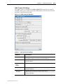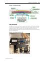
1
Imaging System User Guide Revision C ii DeltaVision OMX User Guide Legal Notices DeltaVision OMX User Guide. PN 04‐720120‐002, Revision C ©2009‐2011 Applied Precision, Inc. All rights reserved. No part of this manual may be reproduced, transmitted, stored in a retrieval system, or translated into any language in any form by any means without the written permission of Applied Precision, Inc. Information in this document is subject to change without notice. DeltaVision OMX, Applied Precision, and softWoRx are registered trademarks of Applied Precision, Inc. Nunc is a registered trademark of Nunc A/S Corporation and Lab‐Tek is a trademark of Nalge Nunc International Corporation. ibidi is a registered trademark of ibidi GmbH Corporation. All other registered names and trademarks referred to in this manual are the property of their respective companies. Applied Precision, Inc. 1040 12th Ave. NW Issaquah, WA 98027 (425) 557‐1000 FAX: (425) 557‐1055 www.appliedprecision.com Preface iii Contents Preface ................................................................................................................................ vi Document Audience ................................................................................................. vi Document Conventions ............................................................................................ vi User Interface Description Conventions ................................................................ vii Contacting Applied Precision, Inc. ................................................................................ vii Acknowledgements ......................................................................................................... vii Chapter 1. System Introduction 1.1 What is DeltaVision|OMX? ......................................................................................... 1.1 Modes of Operation ......................................................................................................... 1.3 Basic DeltaVision OMX Architecture ............................................................................ 1.3 Laser/Electronics Enclosure ............................................................................................ 1.6 Microscope Enclosure ...................................................................................................... 1.9 User Workstation ............................................................................................................. 1.10 Pre‐screening System ....................................................................................................... 1.11 Ionizing Air Gun .............................................................................................................. 1.11 Chapter 2. Lasers and System Safety 2.1 DeltaVision OMX Lasers ................................................................................................. 2.1 405nm Laser ‐ Power Technology ........................................................................... 2.1 488nm Laser ‐ Coherent 561nm Laser ‐ Coherent (Optional) ........................................................................ 2.2 Other Optional Fiber Lasers ‐ MPB Communications ......................................... 2.2 Laser Interlocks ................................................................................................................ 2.2 Safety Labeling ................................................................................................................. 2.4 Applied Precision Labels .......................................................................................... 2.4 Fiber Optic Cable Routing Safety Label ................................................................. 2.9 Individual Laser Switch Labels ............................................................................... 2.9 Laser Manufacturer’s Labels .................................................................................... 2.11 Proper System Component Placement ......................................................................... 2.14 Power Cable ................................................................................................................ 2.14 Camera Coolers .......................................................................................................... 2.14 Chemical Safety ................................................................................................................ 2.15 Safety Goggles .................................................................................................................. 2.15 Wiring Diagram ‐ Safety Interlock ................................................................................. 2.16 Chapter 3. Basic Operations 3.1 Safety Notices ................................................................................................................... 3.1 Electronics Rack ................................................................................................................ 3.2 Startup/Shutdown ............................................................................................................ 3.3 System Startup ........................................................................................................... 3.3 System Shutdown ...................................................................................................... 3.5 04-720120-002 RevC/0211 iv DeltaVision OMX User Guide Loading a Sample onto the Slide Holder ...................................................................... 3.6 Removing and Replacing the OMX Objective ............................................................. 3.7 Removing and Replacing the Dichroic Drawer ........................................................... 3.9 Chapter 4. Acquiring Images 4.1 Running an Experiment .................................................................................................. 4.1 Point Visiting .................................................................................................................... 4.7 Defining the Field of View and Finding Focus ............................................................ 4.12 Spiral Mosaic .............................................................................................................. 4.12 DV Points .................................................................................................................... 4.14 Helpful Tips for Finding the Area of Interest ........................................................ 4.17 Chapter 5. Analyzing Images 5.1 Overview ........................................................................................................................... 5.1 The OMX SI Reconstruction Tool .................................................................................. 5.1 The Task Builder .............................................................................................................. 5.7 Chapter 6. Creating OTFs 6.1 OTFs ................................................................................................................................... 6.1 Creating a Bead Slide ...................................................................................................... 6.2 Scanning the Bead Slide .................................................................................................. 6.4 Creating the OTF File ...................................................................................................... 6.7 Selecting the Correct Immersion Oil ............................................................................. 6.11 Chapter 7. Coordinate Mapping 7.1 About Coordinate Mapping ........................................................................................... 7.1 The Coordinate Mapping Slide ...................................................................................... 7.2 Finding the Features in the Mask Using the personalDV .......................................... 7.2 Finding the Features in the Mask Using the DeltaVision OMX ................................ 7.4 Calculating the Coordinate Transform ......................................................................... 7.4 Testing and Refining the Coordinate Values ............................................................... 7.6 Chapter 8. System Maintenance 8.1 Aligning the Dichroic Drawer ........................................................................................ 8.1 Defining the OMX Alignment Parameters ................................................................... 8.8 Aligning the OMX Image ................................................................................................ 8.12 Cleaning the DeltaVision OMX System Hardware ..................................................... 8.16 Dichroic Drawer(s) .................................................................................................... 8.16 Microscope and Camera Components ................................................................... 8.17 Camera Coolers .......................................................................................................... 8.17 Objective Lens ............................................................................................................ 8.17 Preface Chapter 9. Software Reference v 9.1 DeltaVision OMX Software ............................................................................................ 9.1 Main Program Window ............................................................................................ 9.1 Main Menus ................................................................................................................ 9.3 Light Settings .............................................................................................................. 9.6 Stage Positioning Tools ............................................................................................. 9.8 Experiment Settings .................................................................................................. 9.11 softWoRx Imaging Workstation Software .................................................................... 9.12 Starting softWoRx ...................................................................................................... 9.12 OMX‐Related Windows in softWoRx ..................................................................... 9.13 Appendix A. Facility Requirements A.1 Appendix B. TIRF Option B.1 What is TIRF? .................................................................................................................... B.1 The Evanescent Wave ............................................................................................... B.2 TIRF Hardware .......................................................................................................... B.3 TIRF‐specific Laser Safety Considerations ................................................................... B.5 Using TIRF ........................................................................................................................ B.5 Appendix C. DIC Option C.1 What is DIC? ..................................................................................................................... C.1 DIC Optics .................................................................................................................. C.2 Installing the DIC Module ........................................................................................ C.4 Aligning the DIC Module ......................................................................................... C.5 Appendix D. Live Cell Option D.1 What is the Live Cell Option? ........................................................................................ D.1 Live Cell Option Kit ......................................................................................................... D.1 Compatibility ............................................................................................................. D.3 Index 04-720120-002 RevC/0211 I.1 vi DeltaVision OMX User Guide Preface This manual describes how to perform the tasks necessary to safely operate your DeltaVision OMX Imaging System. This topic includes the following sections: Document Audience describes who should read this manual and what prerequisite knowledge the reader should have. Document Conventions explains the typography, symbols, and other conventions used in this manual. Contacting Applied Precision, Inc. provides information about how to contact customer support. Document Audience The information contained in this manual assumes that you are familiar with the basics of fluorescence microscopy. Correct operation of the microscope is fundamental to obtaining quality images with the DeltaVision OMX Imaging System. In addition, an understanding of image processing basics will help you use the system to its full potential. To manage the computer systems, some familiarity with Linux workstations and IBM‐type personal computers is helpful. Document Conventions In order to make the information in this manual as easy as possible for you to locate and use, the following conventions are observed. Lists and Procedures • Round bullets indicate items in a list, options in procedures, and single‐ step procedures. 1. Numbered items indicate sequential steps for completing a procedure. Square bullets indicate items in a list. Notes, Warnings, and Cautions Note Indicates information about the previous paragraph or step in a procedure. Important Indicates important or critical information about the previous paragraph or step in a procedure. Tip Indicates helpful advice. WARNING! Indicates important information regarding potential personal injury. Preface vii CAUTION! Indicates important information regarding potential damage to the equipment or software. WARNING! Indicates important information regarding potential personal injury due to hazardous radiation. User Interface Description Conventions Boldface indicates the names of buttons, menus, dialog box options/fields. Initial Capitals indicate the names of windows, dialog boxes, and tabs. ALL CAPITALS SAN SERIF indicates the name of a key on your keyboard, such as ENTER or DELETE. Uniform width font indicates text to enter on a command line. Contacting Applied Precision, Inc. If you have questions about DeltaVision OMX Imaging System, first refer to this manual. If you donʹt find the information you need, contact Applied Precision using the following information. Customer Service Hotline Phone: 800‐862‐5166 email: [email protected] Hours: 8:00 AM ‐ 5:00 PM, Pacific Time, Monday ‐ Friday Corporate Headquarters Applied Precision, Inc. 1040 12th Avenue NW Issaquah, WA USA Phone: (425) 557‐1000 Fax: (425) 557‐1055 Internet address: www.appliedprecision.com Acknowledgements Applied Precision would like to thank the laboratories led by Drs. Sedat, Agard, and Gustafsson of the University of California, San Francisco for their expertise and cooperation in helping to make this technology available to the Life Sciences research community. 04-720120-002 RevC/0211 viii DeltaVision OMX User Guide 1. System Introduction Provides an overview of the DeltaVision OMX Imaging System Defines facility requirements and system footprint specifications Describes basic system architecture and main system components What is DeltaVisionOMX? The DeltaVision OMX Imaging System is a super‐resolution microscopy system that surpasses previous resolution limits and allows you to image beyond the surface of the coverslip with multiple probes. The system’s structured illumination technology images more of your biology and resolves features invisible with traditional microscopy. Using the DeltaVision OMX, you can image from five to twenty microns into cells and tissues; and, since the DeltaVision OMX works with conventional fluorochromes, there is no need to genetically engineer novel or complex photoswitchable probes. You use the same preparation methods and apply the same fluorescent labeling reagents (antibodies and protein tags) already used in the lab. 04-720120-002 RevC/0211 1.2 DeltaVision OMX User Guide Figure 1.1 Comparison - Standard Widefield (Left) and DeltaVision OMX (Right) Two isoforms of beta‐tubulin in a cultured neuron ‐ Image courtesy of Stephanie Kaech Petrie and Aurelie Snyder, Advanced Light Microcopy Core at The Jungers Institute Oregon Health & Sciences University Three-Dimensional Structured Illumination The resolution of a microscope is limited by the amount of spatial information that can pass successfully through the optics. In Figure 1.2, this is represented by the fine grid pattern shown in Panel 1. If that high‐resolution information is mixed with a known signal that we can resolve (Panel 2), we generate a new pattern, the moiré pattern seen in Panel 3. In Panel 3, the pattern that we see is the difference between the two patterns and can easily be represented without high‐resolution methods. Figure 1.2 The Moiré Pattern Similarly, when the DeltaVision OMX is in three‐dimensional structured illumination mode, the system superimposes a three dimensional illumination pattern onto the sample. This pattern generates a new pattern (a moiré pattern) that contains both the illumination and the sample data. By carefully reconstructing the sample data from the moiré pattern, it is possible to create a super‐resolution three‐dimensional image of the original sample. Because the illumination pattern is three‐dimensional, the image that is created contains both the lateral (2D) and axial (3D) data. This illumination pattern can be efficiently generated with multiple excitation wavelengths, allowing multiple fluorochromes to be used in the same sample. Chapter 1: System Overview 1.3 Modes of Operation The three principal modes of operation for the DeltaVision OMX Imaging System are Live Cell Imaging, Structured Illumination, and optionally, TIRF (Total Internal Reflection Fluorescence). • Live Cell Imaging involves using multiple lasers to illuminate the sample with different wavelengths of laser light. This can excite multiple dyes at once, allowing multi‐color images to be generated simultaneously. Efficient optics minimize photo‐toxic effects on living cells. It can also be used to study processes that occur very rapidly. In this mode of operation, the full power of all lasers in use can be combined into one multi‐color beam. • Structured Illumination diffracts the laser beam into three parallel beams which are combined by the objective to produce 3‐D interference fringe patterns in the sample. Multiple images of the sample are produced by shifting and rotating the fringe pattern, and by stepping through the sample in the vertical direction (Z). The mathematical algorithm at the heart of OMX recovers a super‐resolution image by solving large systems of linear equations with data from the many captured images. Because the sample must remain absolutely motionless during imaging, this technique is not currently practical for live cells. • TIRF is an optical sectioning technique that limits fluorescence imaging to a thin area at the surface of a specimen, typically only to a depth of 100 ‐ 200nm, resulting in an enhanced signal‐to‐noise ratio and increased imaging contrast. TIRF capability is optional on a DeltaVision OMX. (Refer to Appendix B, “TIRF Option” on page B.1 for additional information.) Other operating modes, such as DIC (Differential Interference Contrast), can be used in addition to the three principal operating modes described above. The DeltaVision OMX Imaging System is typically bundled with other microscope products from Applied Precision to help identify sample regions of interest at lower magnification and automatically transfer the coordinates to the DeltaVision OMX. Basic DeltaVision OMX Architecture The DeltaVision OMX Imaging System will often take up a small room. It consists of four major subsystems: • a Laser/Electronics Enclosure • a Microscope Enclosure • a pre‐screening system (usually a personalDV) • and the User Workstation. 04-720120-002 RevC/0211 1.4 DeltaVision OMX User Guide Figure 1.3 Main System Components The following illustration provides a basic functional description of the system. Chapter 1: System Overview Figure 1.4 DeltaVision OMX Functional System Layout 04-720120-002 RevC/0211 1.5 1.6 DeltaVision OMX User Guide Laser/Electronics Enclosure The Laser/Electronics Enclosure houses the laser heads and the optics that prepare the beams for delivery to the Microscope Enclosure. The optics include reflective notch filters for combining the beams and ND filters for adjusting the power level of the beams. Fast shutters control the exposures and a flip mirror (a movable mirror driven with a motor via software commands) launches the laser beam into either a large multimode fiber (for Live Cell Imaging) or a smaller fiber (for Structured Illumination). The laser heads and attendant optics are all mounted on an optical table. As the user does not require access to these components, the work surface of this table is encased in a protective housing and secured for laser safety. Figure 1.5 Laser/Electronics Enclosure - Laser Table Chapter 1: System Overview Figure 1.6 Laser/Electronics Enclosure (table and cabinet open) Figure 1.7 Laser/Electronics Enclosure (table and cabinet closed) 04-720120-002 RevC/0211 1.7 1.8 DeltaVision OMX User Guide The lasers are all continuous wave lasers. All lasers can be used at the same time, so the maximum laser power available is the sum of all the individual laser powers. Laser power supplies and controllers are rack‐mounted in the cabinet beneath the Laser Table. The Laser/Electronics Enclosure laser safety power switch is shown in the following figure. Figure 1.8 Laser Power Supplies and Controllers (inside cabinet) The Laser/Electronics Enclosure cabinet contains: • Camera PCs • DeltaVision OMX Instrument Controller (OMXIC) • OMXIC PC • Nanomotion 3 Chassis • Piezo Controller for Z axis • Primary and Secondary Laser Control Modules • Network Switch • System Power Supply • TIRF Controller (Optional. (Refer to Appendix B, “TIRF Option” on page B.1 for additional information.) The Primary Laser Control Module houses the power and controllers for the 405nm and 488nm lasers, as well as the safety interlock and indicator light controls. The Secondary Laser Control Module houses all optional lasers. No radiation is emitted from the cabinet. Chapter 1: System Overview 1.9 Microscope Enclosure The laser fibers deliver the beam to the Microscope Enclosure, which consists of an illumination system, high‐speed cameras, and microscope housed within a protective enclosure. This enclosure is not easily moved, so it is very important to determine a permanent location for it before setting up the DeltaVision OMX system. Figure 1.9 Microscope Enclosure Optics guide the light into the microscope. The microscope is custom‐designed for the DeltaVision OMX and includes: • An objective lens to focus the light on the sample. • A stage to hold the sample and move it to precise locations. • Two or more cameras to capture the fluorescent light emitted from the dyes. • User‐changeable dichroic drawers to guide the light or separate it into various channels. 04-720120-002 RevC/0211 1.10 DeltaVision OMX User Guide Figure 1.10 Microscope Assembly Open laser beams are accessible at the illumination system and microscope. These include both collimated beams and light that is strongly divergent after it has passed through the objective (included angles of over 90 degrees). The entire microscope assembly is encased in a protective enclosure. Besides laser safety, this also provides a dark and dust‐free environment for the system to function at peak performance. User access to the microscope and into the protective enclosure is through the big front door. This door is interlocked so that laser light cannot exit when the door is opened. There are also service‐only access doors on each side of the Microscope Enclosure. These doors are locked at all times except during service. Users should not open these service doors at any time! User Workstation The User Workstation is a computer desk and two computers from which the operator controls the DeltaVision OMX Imaging System. One computer runs software that controls all aspects of hardware operation and image acquisition, including stage motion, imaging, modes of operation, laser selection, and power. The other computer runs software that manages various post‐imaging processing activities (deconvolution and reconstruction, calibration, animation, and storage). The User Workstation is outside all protective housings, and no access to the beam is required to operate the DeltaVision OMX Imaging System from the workstation. Chapter 1: System Overview 1.11 Pre-screening System The pre‐screening system (usually a personalDV) incorporates multiple objectives and a larger field of view as compared to the DeltaVision OMX system. The user locates areas of interest on the slide using the pre‐screening system, then transfers the sample to the DeltaVision OMX stage. Stage coordinates are mapped on both systems, making it easy move the sample between systems. Once you determine the location of interest on the slide using the pre‐screening system, you can switch the slide to the DeltaVision OMX, where the system can then easily move to the same location on the DeltaVision OMX stage. Ionizing Air Gun The TopGun ionizing air gun is included with the DeltaVision OMX system to provide an efficient method of cleaning system optics while simultaneously decreasing static charge. WARNING! Always wear appropriate eye protection to keep your eyes safe from flying debris that may be disturbed by the air gun. Figure 1.11 Ionizing Air Gun All functionality is built into the gun, including a flow control value for adjusting the airflow, a balance adjustment for calibration, and a two‐level LED. The air gun has a filter at its exit to ensure that out‐flowing air is clean. WARNING! The air gun should be used only as directed for cleaning. The air gun is not designed for any other purpose. 04-720120-002 RevC/0211 1.12 DeltaVision OMX User Guide 2. Lasers and System Safety Provides critical information regarding OMX laser safety Describes safety‐specific information regarding equipment, power cabling, interlocks, and chemicals Illustrates system safety labeling DeltaVision OMX Lasers The DeltaVision OMX Imaging System is a Class I laser system. No access to laser radiation is permitted during operation or maintenance. During service, however, Class IV radiation is accessible. WARNING! Due to the potential for personal injury, particularly to the eyes, service on the DeltaVision OMX Imaging System should ONLY be performed by Applied Precision personnel or persons trained by Applied Precision specifically for this purpose. Unauthorized service by any other personnel may violate the warranty. This section describes the lasers and associated radiation accessible during service. Note that all beams are continuous wave. 405nm Laser - Power Technology The 405nm laser is a state‐of‐the‐art diode laser. Maximum power output is 600mW. 04-720120-002 RevC/0211 2.2 DeltaVision OMX User Guide 488nm Laser - Coherent 561nm Laser - Coherent (Optional) The 488nm and the optional 561nm lasers are optically pumped, frequency‐doubled VECSEL (vertical external cavity surface emitting lasers). Semiconductor lasers at 808nm pump quantum well structures to emit at 976nm, which is frequency‐doubled in an external cavity with a non‐linear crystal to produce 488nm or 561nm. Maximum power output is 200mW. Other Optional Fiber Lasers - MPB Communications In addition to the 561nm laser from Coherent, three other optional lasers are available for the DeltaVision OMX, all from MPB Communications: 514nm, 300mW Laser 592.5nm, 300mW Laser 642nm, 300mW Laser The visible light from each of these optional fiber lasers is emitted by a frequency‐ doubling crystal. The light source for the crystal is optically pumped, proprietary doped fiber. The desired IR wavelength is produced by stimulated Raman scattering and selected by Bragg gratings etched into the fiber. Note Only three of the four optional lasers can be included with a DeltaVision OMX system. The system can support a maximum of five lasers in total. Laser Interlocks The interlock system on the DeltaVision OMX Imaging System is designed to promote safe operation and service. The Class I system includes a protective housing that must be accessed often for maintenance. In addition, the enclosure must be open and the lasers enabled during service. A safety shutter in the Laser/Electronics Enclosure is open whenever the system is in operating mode. This shutter is based on a solenoid that must be continually powered in order to remain open. Two conditions can interrupt the current to the solenoid, which will close the shutter and block all laser radiation: • The door on the Microscope Enclosure is opened. An interlock on the enclosure door senses this condition. • The key switch on the Primary Laser Control Module is switched off. This permits service personnel additional control over laser operation, particularly when working in the Laser/Electronics Enclosure. All of the circuitry in the interlock circuit is redundant and/or fail‐safe to comply with safety requirements. Chapter 2: Lasers and System Safety 2.3 The interlock system also controls status indicator lights located on both the Laser/ Electronics and Microscope Enclosures. Figure 2.1 Laser/Electronics Enclosure Status Lights The various colors indicate the following system status: • GREEN ‐ lasers are OFF. • AMBER ‐ laser power is ON and safety shutter is closed. No laser light can get through to the Microscope Enclosure. • RED ‐ lasers are ON and safety shutter is open. Laser light can get through to the Microscope Enclosure. Refer to Figure 2.20 at the end of this chapter to view a simplified view of the wiring for the safety interlock. 04-720120-002 RevC/0211 2.4 DeltaVision OMX User Guide Safety Labeling The following sections list and illustrate the laser safety labels attached at various locations to the DeltaVision OMX Imaging system and to the lasers within the system. Applied Precision Labels Safety Label #1 (5 X 2ʺ) is attached to the front of the Laser/Electronics Enclosure cabinet and both side doors of the Microscope Enclosure. Figure 2.2 Laser Safety Label #1 and Locations on System Chapter 2: Lasers and System Safety 2.5 Safety Label #2 (5 X 2ʺ) is attached to the front, user access door of the Microscope Enclosure. Figure 2.3 Safety Label #2 and Location on System 04-720120-002 RevC/0211 2.6 DeltaVision OMX User Guide The following CE label (4 X 2.88ʺ), containing specific system information, is attached to the back of the Laser\Electronics Enclosure, on the left rear post (as shown). Figure 2.4 CE Label Location - Laser/Electronics Enclosure (left rear post) Chapter 2: Lasers and System Safety 2.7 The following CE label (4 X 2.88ʺ), containing specific system information, is attached to the back of the Microscope Enclosure cabinet, on the right rear corner (as shown). Figure 2.5 CE Label Location - Microscope Enclosure (right rear corner) Note that this label will be slightly different for installations in the UK, Europe, or Asia (as shown in the following figure). Figure 2.6 Microscope Enclosure CE Label Varies Depending on Geographic Location 04-720120-002 RevC/0211 2.8 DeltaVision OMX User Guide The following laser safety labels (2 X 2ʺ) are attached (as appropriate) to the inside of the left side door of the Microscope Enclosure. The labels applied will depend on which lasers are included in the system. Figure 2.7 Laser-Specific Safety Labels Figure 2.8 Laser-Specific Labels - Microscope Enclosure (inside left side door) Chapter 2: Lasers and System Safety 2.9 Fiber Optic Cable Routing Safety Label The following label is attached to the conduit that runs between the Laser/Electronics Enclosure and the Microscope Enclosure. Figure 2.9 Fiber Optic Cable Routing Label Individual Laser Switch Labels The following labels (2 X 5ʺ) are attached (as appropriate) above the individual laser switches on the Primary and Secondary Laser Control Modules (inside the Laser/ Electronics Enclosure cabinet). The labels applied will depend on which optional lasers are included in the system. Figure 2.10 Laser Switch Labels (for specific lasers) 04-720120-002 RevC/0211 2.10 DeltaVision OMX User Guide Figure 2.11 Laser Switch Label Locations (5 Lasers Maximum) Chapter 2: Lasers and System Safety 2.11 Laser Manufacturer’s Labels The laser safety labels attached to the available laser heads are shown in this section. Figure 2.12 405nm Laser Head, 600mW Laser Figure 2.13 488nm Laser Head, 200mW Laser 04-720120-002 RevC/0211 2.12 DeltaVision OMX User Guide The following laser safety label is attached to the optional laser heads. Figure 2.14 Laser Safety Label for Optional Laser Heads Figure 2.15 514nm Optional Laser Head Figure 2.16 592.5nm Optional Laser Head Chapter 2: Lasers and System Safety Figure 2.17 561nm Optional Laser Head (beneath the fiber shaker) Figure 2.18 642nm Optional Laser Head 04-720120-002 RevC/0211 2.13 2.14 DeltaVision OMX User Guide Proper System Component Placement Recommendations in this section, regarding the appropriate placement of the system’s power cabling and liquid cooling components, must be followed in order to minimize the risk of personal injury. Power Cable DeltaVision OMX power cabling should always be arranged so that the power can be easily and quickly disconnected in the event of an emergency. Camera Coolers To reduce heat within the Microscope Enclosure cabinet and minimize the risk of electrical hazards, the camera coolers are located on the floor, completely outside the cabinet walls. Figure 2.19 Proper Placement of Camera Coolers Chapter 2: Lasers and System Safety 2.15 Chemical Safety WARNING! Ethylene Glycol (antifreeze/coolant) is a hazardous substance. Use caution when handling this chemical and adhere to the recommendations listed in this section and in the Material Safety Data Sheet (MSDS) handbook at your facility. • Do not drink antifreeze or solution. • Avoid eye and prolonged or repeated skin contact. • Avoid breathing vapors or mists. • Wash exposed skin thoroughly with soap and water after use. • Do not store in opened or unlabeled containers. • Keep container away from open flames and excessive heat. • Do not reuse empty containers unless properly cleaned. • Empty containers retain product residue and may be dangerous. • Do not cut, weld, drill, etc. containers, even when empty. Sudden release of hot organic chemical vapors or mists from process equipment operating at elevated temperature and pressure, or sudden ingress of air into vacuum equipment, may result in ignitions without any obvious ignition sources. Published ʺautoignitionʺ or ʺignitionʺ temperatures cannot be treated as safe operating temperatures in chemical processes without analysis of the actual process conditions. Use of this product in elevated temperature applications should be thoroughly evaluated to assure safe operating conditions. For complete information on Ethylene Glycol, go to: http://www.bractwalls.com/user/image/prestoneantifreeze.pdf Safety Goggles Because the DeltaVision OMX is a Class 1 safety system, you are not required to wear safety goggles during regular system use. If, however, at any point the safety interlock is defeated (for example, while the system is being serviced), the appropriate safety goggles must be worn. Safety goggles that protect your eyes from all laser wavelengths are the best type to use in a multi‐laser environment. These may be difficult to obtain, so the next best practice is to be absolutely certain you wear the correct safety goggles for each laser type being used. 04-720120-002 RevC/0211 2.16 DeltaVision OMX User Guide Wiring Diagram - Safety Interlock Figure 2.20 Safety Interlock Wiring 3. Basic Operations Describes the Electronics Rack in the Laser/Electronics Enclosure Explains basic processes required for system operation, such as system startup/shutdown, loading a slide, and replacing an objective Safety Notices The OMX Imaging System is a Class I laser system. No access to laser radiation is permitted during operation or basic maintenance procedures. WARNING! Use of controls or performance of procedures other than those specified in this document may result in hazardous radiation exposure. During service, however, Class IV radiation is accessible. WARNING! Due to the potential for exposure to hazardous radiation, service on the DeltaVision OMX should ONLY performed by Applied Precision personnel or by persons trained by Applied Precision specifically for this purpose. Unauthorized service by any other personnel may violate the warranty. 04-720120-002 RevC/0211 3.2 DeltaVision OMX User Guide Electronics Rack The Electronics Rack is located in the Laser/Electronics Enclosure, below the laser table. Many of the control switches used to start up and shut down the system are located on the components mounted in this rack. Refer to the following figures until you become familiar with the startup and shutdown procedures. Figure 3.1 Electronics Rack (Located Below Laser Table) - Photograph Figure 3.2 Electronics Rack - Block Drawing with Component Names Chapter 3: Basic Operations 3.3 Startup/Shutdown System Startup 1. Turn on the OMX Master Controller Windows Workstation (also called omxMaster). User: omxuser Password: omxuser 2. Turn on the OMX Image Processing LinuxWorkstation (also called omxSI). User: worx Password: system id 3. Turn on the camera cooler(s). Depending on the configuration of your system, there may be one cooler for all of the cameras or individual coolers for each one. In either case, the camera cooler(s) are located on the floor, outside of the Microscope Enclosure. 4. Turn on the power supplies for each camera. These are also located on the floor outside of the Microscope Enclosure. 5. Turn on the cameras. The cameras are located inside of the Microscope Enclosure. Depending on the camera type installed on your system, the power switches will be located on either the back or top of the cameras. 6. On the electronics rack inside of the Laser/Electronics Enclosure, turn on the following: • • • • • • • Primary Laser Control Module Secondary Laser Control Module (if installed) OMX Instrument Controller Chassis Nanomotion Chassis OMXIC Workstation All Camera Workstations Jena Controller Note While the power switches for most of the components listed in Step 6 are located on the front of the component, the power switch for the Jena controller is located on the back of the controller. 7. On omxMaster, click the OMX icon to open the DeltaVision OMX software. 8. In the DeltaVision OMX software on omxMaster, click Hardware|Hardware. 04-720120-002 RevC/0211 Note If you just turned on the cameras, wait for them to fully initialize (about five minutes) before you click the Hardware|Hardware command. 3.4 DeltaVision OMX User Guide Figure 3.3 Hardware Menu The software will display the Hardware dialog box. Figure 3.4 Hardware Dialog Box 9. In the Hardware dialog box, click Restart Hardware to initialize the system hardware. 10. On omxSI, click the Start softWoRx icon ( Workstation software. ) to open the softWoRx Imaging 11. Once the hardware is fully initialized (about two minutes), the Status section of the main program window will change from displaying HW: Initializing to HW: Running and the system will be ready to acquire images. 12. Turn on the lasers required for your experiments. 13. Turn on the key switch for the interlock that enables the laser safety shutter. Chapter 3: Basic Operations 3.5 System Shutdown 1. Turn off the key switch for the interlock that disables the laser safety shutter. 2. Turn off any lasers that have been turned on. 3. On the electronics rack inside of the Laser/Electronics Enclosure, turn off the following: • • • • • • • Jena Controller All Camera Workstations OMXIC Workstation Nanomotion Chassis OMX Instrument Controller Chassis Secondary Laser Control Module (if installed) Primary Laser Control Module 4. In the DeltaVision OMX software on omxMaster, click File|Quit. 5. On omxMaster, choose one of the following: • Use the Remote Desktop Connection window (click Start|All Programs|Accessories|Remote Desktop Connection) to remote into the OMXIC workstation. User: omxuser Password: omxuser. OR • Use the Remote Desktop Connection folder shortcut on your desktop to remote into the OMXIC workstation. 6. On omxMaster, use CTRL+ALT+END to display the Windows Security dialog box and then click Shut Down... to turn off the OMXIC workstation. Note You must use CTRL+ALT+END to display the Windows Security dialog box, rather than CTRL+ALT+DELETE, which will shut down omxMaster. 7. Repeat Step 4 as needed to close each of the camera workstations. 8. Turn off the cameras. 9. Turn off the camera power supplies. 10. Turn off the camera cooler(s). 11. Shut down omxMaster by clicking Start in the Windows task bar and then selecting the Shut Down command. 12. In the softWoRx Imaging Workstation software on the omxSI workstation, click File|Exit and then click OK in the Question popup that asks “Are you sure?”. The program will close. 13. Log out of and shut down the omxSI. 04-720120-002 RevC/0211 3.6 DeltaVision OMX User Guide Loading a Sample onto the Slide Holder 1. Open the door to the Microscope Enclosure. The laser interlock will keep the safety shutter closed while the door to the enclosure is open. WARNING! Due to the potential for exposure to hazardous radiation, do NOT defeat the laser interlock. 2. Remove any sample currently in place and clean the objective. Note The importance of proper cleaning methods is critical to the life of optical components. Applied Precision recommends using chloroform, clean swabs, and new, lint-free, lens-specific cleaning tissue to clean the objectives. 3. Apply immersion oil to the objective. For information regarding selection of the correct immersion oil, refer to “Selecting the Correct Immersion Oil” on page 6.11. 4. Place the slide in the desired position within the clips or, if applicable, in the slide holder. Figure 3.5 OMX Slide Holder 5. Close the door to the Microscope Enclosure. Once the laser interlock is activated by closing the door, the system will allow the lasers to be turned back on. Chapter 3: Basic Operations 3.7 Removing and Replacing the OMX Objective CAUTION! To avoid scratching the lens or introducing debris of any kind to the objective, avoid touching the objective lenses when removing and replacing objectives. The aperture located on the bottom of the objective is especially difficult to clean. To remove the objective from the OMX microscope: • Simply unscrew the objective in a counter‐clockwise direction. Figure 3.6 Removing the Objective To replace the objective: • Line up the threads and carefully screw the objective clockwise until tight. Do not overtighten. 04-720120-002 RevC/0211 3.8 DeltaVision OMX User Guide Figure 3.7 Replacing the Objective Figure 3.8 Objective in Position on the DeltaVision OMX System Chapter 3: Basic Operations 3.9 Removing and Replacing the Dichroic Drawer To remove the dichroic drawer: 1. Loosen the locking screw located above the drawer and next to the objective. Figure 3.9 Loosen Dichroic Drawer Locking Screw 2. Grasp the drawer firmly by the handles and lift upward while pulling outward to remove the drawer from the system. The handles are large because the drawer is quite heavy. Figure 3.10 Removing the Dichroic Drawer 04-720120-002 RevC/0211 3.10 DeltaVision OMX User Guide 3. After the dichroic drawer is removed from the system, carefully place it in the drawer rack. Figure 3.11 Dichroic Drawer (Removed from System) CAUTION! To avoid damaging the sensitive optical components mounted in the dichroic drawer, whenever the drawer is removed from the system, be extremely careful not to touch any of the components contained within it. Figure 3.12 Dichroic Drawer in Drawer Rack Chapter 3: Basic Operations 3.11 To replace the dichroic drawer: 1. Note the drawer guides shown in the following figure. These guides, located on the bottom front edge of the drawer cavity in the microscope base, are used to properly align and seat the dichroic drawer. Figure 3.13 Dichroic Drawer Guides A ball bearing is located on each corner of the bottom front edge of the dichroic drawer itself. When the drawer is inserted correctly, these ball bearings line up with the guides on the microscope base. Figure 3.14 Balling Bearings on Dichroic Drawer 04-720120-002 RevC/0211 3.12 DeltaVision OMX User Guide 2. Grasping the dichroic drawer by the handles, gently slide the drawer into place. 3. Seat the ball bearings into place within their respective guides on the microscope base and allow them to settle. Figure 3.15 Sliding the Drawer Into Place 4. Once the dichroic drawer is in position, finger‐tighten the locking screw to secure it in place. Figure 3.16 Finger-Tighten the Locking Screw CAUTION! If you replace the dichroic drawer and the system no longer functions properly, contact Applied Precision Technical Support. 4. Acquiring Images Describes how to run an experiment in order to acquire image data using the DeltaVision OMX Workstation (omxMaster) Explains how to set up and run point visiting experiments Describes how to use DV Points and Spiral Mosaic Running an Experiment In order to acquire image data using the DeltaVision OMX, you must run an experiment. The following procedures describe the basic steps necessary for running experiments. Your process may be slightly different, depending on variables such as the type of images you wish to acquire and the hardware configuration of your system. In general, however, you must complete these five procedures in order to acquire images: • Activate the lasers/cameras and mount the sample • Define the imaging parameters • Determine the acquisition parameters • Define the experiment • Run the experiment 04-720120-002 RevC/0211 4.2 DeltaVision OMX User Guide To activate the lasers/cameras and mount the sample: 1. Ensure the system has been turned on according to the startup procedure described in “System Startup” on page 3.3. 2. Ensure Laser Speckle Reducer (shaker) switch #1 (located on the front of the omxIC chassis) is turned on. Notes #1 Laser Speckle Reducer switch #2 is disabled. #2 When you open the DeltaVision OMX software on omxMaster, the system automatically turns on the shaker no matter what position the power switch is in. This means the shaker may be turned on even when the Laser Speckle Reducer switch is in the Off position. To turn off the shaker when the switch is already in the Off position, flip the switch to the On position and then immediately flip it back to the Off position. 3. On the Primary and Secondary Laser Control Modules located in the Laser/ Electronics Enclosure cabinet, turn on the lasers you will be using to collect images. Ensure that the safety shutter key switch is turned on. 4. On the main screen of the DeltaVision OMX software (on omxMaster), activate the desired cameras by clicking the appropriate Channel button(s). As you select a channel, note that a corresponding image window with the camera location and channel label appears on the right side of the main program window. Figure 4.1 Select Channel(s) 5. Select the correct camera Mode for each active channel. Each mode setting consists of two values: channel mode and readout speed. Channel Mode: EMCCD (electron multiplication) or Conv (conventional) Readout Speed: 20MHz, 10MHz, 5MHz, or 1MHz, depending on installed camera Figure 4.2 Mode Chapter 4: Acquiring Images 4.3 6. If using an EMCCD mode, set the EMCCD Gain. Note A good starting gain value would be about 75% of the maximum gain. 7. Using the stage controls (or DV Points if the coordinate mapping has been completed), move the stage into the imaging position. 8. Mount the sample onto the DeltaVision OMX stage. See “Loading a Sample onto the Slide Holder” on page 3.6 of this guide. Note If using DV Points, the sample must be oriented in the same direction as it was on the personalDV. To define the imaging parameters: 1. Select the Imaging Mode: • Simultaneous for high speed and/or when crosstalk is not an issue • Sequential when high speed is not necessary and crosstalk is an issue. Typically the best selection for structured illumination. 2. Select the Light Path: • Conventional for time‐lapse or widefield imaging • SI for structured illumination imaging. 3. Select the Image Size. This setting defines the portion of the CCD chip surface from which to collect data. (512x512 is the maximum image size for the EMCCD cameras; 1024x1024 is the maximum image size for the CoolSnap HQ2 cameras.) 4. Set the Binning size. Select 1x1 for all SI imaging. Select higher values for lower signal samples and faster imaging of the full field of view. To determine the acquisition parameters: 1. Focus on the sample using the Nano Positioning stage controls and, if applicable, Z touchdown. 2. Adjust the field of view using the stage controls. For additional information about finding your field of view, refer to “Defining the Field of View and Finding Focus” on page 4.12 later in this chapter. 3. Adjust Exposure time and %T settings. Note Minimum, maximum, and mean intensity values are displayed at the bottom of every image window. 4. (Optional.) Determine whether the camera bias (background) settings must be modified and change them if necessary. a. Click File|Dark Image to open the Dark Image tool. 04-720120-002 RevC/0211 4.4 DeltaVision OMX User Guide Figure 4.3 Dark Image Dialog Box b. Click Acquire. c. View the Min, Max, and Mean data values located at the bottom of the image window. The Mean value should be between 50 and 100 counts. d. If the Mean value of the dark image is between 50 and 100 counts, as shown above, skip to Step 4.i and continue the procedure from that step. e. If the Mean value of the dark image is not between 50 and 100 counts, the background value needs to be changed. Click File|Settings to display the Settings dialog box. f. Adjust the Bias (background) field values for the appropriate camera and mode and then click Apply Camera Settings. For example, if the Mean value in the dark image displays a value of 800, enter a value of 750 into the appropriate Bias field on the Settings page. Chapter 4: Acquiring Images 4.5 Figure 4.4 Settings Dialog Box and Bias Fields g. Acquire another dark image to test the new settings. h. Repeat Steps 4.b through 4.g until acceptable background levels have been determined. i. Close the Dark Image tool and click Close to return to the main program window. 5. Specify the desired thickness of the scan. a. Use the Z‐Slider to move the stage to the top of the image stack. b. Click the Mark Top button to mark this position. c. Use the Z‐Slider to move the stage to the bottom of the image stack. d. Click the Mark Bottom button to mark this position. e. The sample thickness (stack height) you marked will be displayed directly below the Z safety limit and Z touchdown fields. 04-720120-002 RevC/0211 4.6 DeltaVision OMX User Guide Figure 4.5 Marked thickness Note Stack height (marked thickness) is limited to 30 µm, the range of the Z piezo. To define the experiment: 1. Ensure the Experiment tab options are displayed in the main program window and then choose either Step 2 (to run a Conventional experiment) or Step 3 (to run a Structured Illumination experiment) below. 2. To run a conventional illumination scan or a time‐lapse experiment, ensure the Conv tab options are displayed in the main program window. a. Ensure Light Path (in the Light Parameters section at the top of the main program window) is set to “Conventional.” b. Use the visit buttons to move the stage/sample so that it matches the focus point when scan starts field. c. Set Optical section spacing to the desired thickness value. d. Set Sample thickness by choosing one of the following: — type in the Number of optical sections or — type in the Sample thickness or — click the Get thickness button (if the top and bottom of the sample were previously marked). e. If a time‐lapse experiment is desired, click the time‐lapse check box and then set the parameters as needed. f. If a flatfield correction is required, click the apply flatfielding to image check box and then specify the name and path of the appropriate flatfield file. g. If point‐visiting is desired, click the visit point list check box and then specify the points to visit for this experiment. (For additional information, refer to the “Point Visiting” section later in this chapter.) 3. To run a Structured Illumination experiment, click the SI tab to display the Structured Illumination options in the main program window. Chapter 4: Acquiring Images 4.7 a. Ensure Light Path (in the Light Parameters section at the top of the main program window) is set to “SI.” b. Use the visit buttons to move the stage/sample so that it matches the focus point when scan starts field. c. Set Optical section spacing to 125. d. Set Sample thickness by choosing one of the following: — type in the Number of optical sections or — type in the Sample thickness or — click the Get thickness button (if the top and bottom of the sample were previously marked). To run the experiment: 1. Enter the appropriate file name and path into the File field. 2. Click Run to start the experiment. As the experiment runs, the experiment progress will be shown. Once the experiment is complete, the stage controls will be reactivated and, if you wish, you can set up and run another experiment. To save the experiment settings: 1. Click the Save As button to open a Windows browser window. 2. Navigate to the folder in which you want to save the experiment settings. 3. Enter a name for the experiment and click Save. To open previously saved experiment settings: 1. Click the Open Exp button to open a Windows browser window. 2. Navigate to the folder in which you previously saved the experiment settings. 3. Select the name of the experiment and click Open. Point Visiting The point visiting feature of the DeltaVision OMX system allows you to select, record, and revisit points on a slide that are located in different fields of view. Instead of recording one cell or field in a single experiment, point visiting allows multiple sites to be imaged in a single experiment, thereby increasing data collection efficiency. In practice, the number of points is limited only by the minimum acceptable time interval between each time point at a single site. This makes time‐lapse imaging more efficient and allows you to collect enough data to generate statistically significant results. In addition, variability between cells within an experiment can be examined, eliminating uncertainty as to the behavior of cells in a single experiment. 04-720120-002 RevC/0211 4.8 DeltaVision OMX User Guide To open the Visit Point List dialog box: • Click the button in the Nano Positioning section of the main program window. The system will display the Visit Point List dialog box, as shown in the following figure. Figure 4.6 Visit Point List Dialog Box Each command in the Visit Point List dialog box is described briefly below and in more detail in the rest of this section. • Clear All. Removes all of the points from the point list. • Mark. Adds the current location to the point list. • Delete. Removes the point currently selected in the point list from the list. • Visit. Moves the stage to the currently selected point. • Replace. Replaces the point currently selected in the point list with the current location. • Open. Displays a Windows File Open dialog box so that you can select and open a previously saved point list. • Save As... Displays a Windows File Save dialog box so that you can save the currently defined point list (as a text file). • Close. Closes the Visit Point List dialog box. To run an experiment that visits points, you must: • Mark the points to visit. • Edit the point list if necessary. Chapter 4: Acquiring Images • 4.9 Enable the visit point list check box on the Experiment tab, specify which points to visit, and run the experiment. Note If you need to remove the slide from the stage in between the time you create a points list and the time you visit those same points, the marked point list may no longer apply. You can minimize this possibility by consistently positioning slides in the slide holder in the same position. We recommend that you place the lower-right corner of the slide snugly into the lower-right corner of the holder and use this as your base position. Marking Points There are two methods of marking points. You can use the Mark Points button in the main program window or you can use the Mark button in the Visit Point List dialog box. Both methods add the current position to the point list. To mark a point using the Mark Points button: • Adjust the current field of view to center the object of interest and click the Mark Points button in the Nano Positioning section of the main program window. The current stage position (in X, Y, and Z) will be added to the point list. To mark a point using the Mark button in the Visit Point List dialog box: 1. Click the Marked Points List button in the Nano Positioning section of the main program window. The Visit Point List dialog box will be displayed. 2. Center the object of interest in the field of view. 3. Click the Mark button in the Visit Point List dialog box. The coordinates of the current location will be added to the point list. Editing the Point List There are several methods of editing a point list. Points can be deleted, point locations can be updated, and the entire point list can be cleared. To delete a point from the point list: • Open the Visit Point List dialog box, select the point to remove from the point list, and click Delete. To update a point in the point list: 1. Open the Visit Point List dialog box, select the point to update, and click Visit. The stage will move so to the selected point. 2. Use the stage tools to move the stage so that the field of view is corrected. 3. In the Visit Points List dialog box, click Replace. 4. The software will replace the coordinates of the previously selected point in the list with your current location. 04-720120-002 RevC/0211 4.10 DeltaVision OMX User Guide To clear all points from the point list: • Open the Visit Point List dialog box and click Clear All. All points in the current point list will be cleared. Opening and Saving a Point List To save a point list: 1. Open the Visit Point List dialog box and click Save As. Windows will open a Save Point List to File dialog box. Figure 4.7 Save Point List to File Dialog Box 2. In the File Name field, enter a name for the point list that you would like to save. Note As you can see from the previous figure, the default name for the system’s point list file is pointlist.txt. To keep the system from writing over your point list, you will want to save your point list using a different name. 3. Click the Save button. Chapter 4: Acquiring Images 4.11 To open a previously saved point list: 1. Open the Visit Point List dialog box and click Open. Windows will display an Open Point List dialog box. Figure 4.8 Open Point List Dialog Box 2. From the list of displayed file names, click the name of the point list that you want to open and then click Open. The software will load the selected point list. Running a Point List Experiment To run a point list experiment: 1. Open the Visit Point List dialog box and ensure the appropriate points are displayed in your point list. Edit the list as necessary or open a previously saved list if desired. 2. On the Experiment tab of the main program window, define your experiment settings as described in “Running an Experiment,” earlier in this chapter. 3. Click the visit point list check box to enable the point visiting feature and enter the points to visit during the experiment, as shown in the following figure. Figure 4.9 Point Visiting Check Box and Data-Entry Field 4. Click Run. 04-720120-002 RevC/0211 4.12 DeltaVision OMX User Guide Defining the Field of View and Finding Focus The DeltaVision OMX includes two tools that can help you define a desired field of view on your sample slide and find focus: Spiral Mosaic and DV Points. This topic describes these tools and also provides some useful tips for viewing samples. Spiral Mosaic Spiral Mosaic is used to quickly identify points of interest on the slide. The spiral pattern starts at the current location and ʺspiralsʺ outward. As each mosaic tile or FOV (field‐of‐view) is defined, it is simultaneously displayed in the spiral mosaic window and the corresponding image/camera window(s) on the right side of the OMX main program window. • XY max size FOVs. Specifies the number of mosaic tiles (horizontally and vertically) to acquire. For example, if you enter 9 into this field, the system will acquire 81 images in a spiral pattern centered around the initial FOV. If you enter an even number into this field, the program will automatically use the next higher odd number. • Start. Begins the spiral mosaic acquisition. • Clear. Removes all spiral mosaic commands from the command queue. • File. Displays a File Open browser window so that you can load a previously saved mosaic image. Mosaic image files are saved automatically by the software as they are created. • Scaling. Opens a popup window that allows you to adjust the relative scaling of each channel in the mosaic. • Point Visiting. Allows you to select a point in one of the mosaic tiles to center in the field of view. • Zoom. Magnifies/demagnifies the image shown in the spiral mosaic window. Zoom can be performed on the image on‐the‐fly as the mosaic pattern is being created or after the pattern is complete • Wavelength Indicators. Allows you to select which of the available wavelengths will be displayed in an image/camera window on the right side of the main program window. This feature is only available after the spiral mosaic acquisition is complete. • Progress. Displays the progress of the active command sequence. • HW. Displays the status of the hardware. • Received image. Displays how many of the total pattern tiles have been created and presented in the image/camera window(s). For example, if you enter 5 into the XY max size FOVs field, and three cameras are active, a total of 75 images will be acquired, 5 (in X) by 5 (in Y) by 3 (cameras). • Stop. Stops the spiral mosaic (or any other) command sequence. Figure 4.6 shows how the Spiral Mosaic tab appears during the tile collection process while Figure 4.7 shows how the main program window appears once all of the tiles have been acquired. Chapter 4: Acquiring Images Figure 4.10 Spiral Mosaic Tab During FOV Tile Collection Process Figure 4.11 Spiral Mosaic Tab Within Program Window After Collection Process 04-720120-002 RevC/0211 4.13 4.14 DeltaVision OMX User Guide To use the Spiral Mosaic tool: 1. Click the Spiral Mosaic tab to display the Spiral Mosaic tool. 2. Enter a maximum size value in the XY max size FOVs field. Note If you enter an even value into the XY max size FOVs field, the system will automatically use the next higher odd value. 3. Click Start. 4. Either wait for the mosaic to finish or, if you see the area you are looking for, click Stop. 5. Click Go To Point and then click on the mosaic in your area of interest to move the stage to that position. 6. Proceed with imaging. DV Points DV Points allows you to scan and identify one or more areas of interest on a sample slide using the personalDV (if included with your DeltaVision OMX) and then move that same slide to the DeltaVision OMX where the same areas of interest can be quickly located and positioned in the image viewing window. DV Points can be very helpful in identifying rare events on slides and running experiments on samples with specific physiology. Important For DV Points to work properly, you must first successfully complete the coordinate mapping procedure described in “Chapter 7: Coordinate Mapping.” To run DV Points: 1. Perform the coordinate mapping procedure described in “Chapter 7: Coordinate Mapping.” 2. Mount a sample on the personalDV and either run an experiment (Multi‐channel, Z sectioning, etc.) or use Snapshot to acquire one or two images of a unique feature. Save the image data to the /data1 folder. 3. Move the same slide to the DeltaVision OMX slide holder. Ensure that the slide is oriented in the same direction as it was on the personalDV and that the slide is biased to the bottom right of the slide holder. 4. In the DeltaVision OMX software, click the DV Points tab. Figure 4.12 DV Points Tab Chapter 4: Acquiring Images 4.15 5. Click the File Browser button to display a Windows Open dialog box. Browse to the network drive mapped from the personalDV and select one of the images you acquired in Step 2. 6. Click Open. The image will open and the user interface will display Channel, Zoom, and Z‐section controls. Figure 4.13 Channel, Zoom, and Z-section Controls 7. Ensure that “visit point” is specified for the Mode and then click in the image on the point you wish to visit. The stage will move to that point and the system will take an image. 8. Focus on the sample. 9. Choose one of the following: • • If the stage moved to the same place you selected in the image, your coordinate mapping is good. Skip to Step 1 OR If the stage did not move to the same place you selected in the image, refine the mapping as described in the next few steps: a. Click the Spiral Mosaic tab to display the Spiral Mosaic utility. b. Enter a maximum size value in the XY max size FOVs field and then click Start. c. Either wait for the mosaic to finish or, if you see the area in the personalDV image, click Pause. 04-720120-002 RevC/0211 4.16 DeltaVision OMX User Guide d. Click Go To Point and then click on the mosaic in your area of interest to move the stage to that position. e. Once the selected area of the mosaic is displayed in your field of view, click the DV Points tab to return to the DV Points utility. f. Select “refine mapping” for the Mode and then click the Refine Mapping button. The software will display the following popup dialog box. Figure 4.14 Refine Mapping Popup g. As described in the previous figure, center the selected object in the OMX image window and then move the pink selection box in the DV Points window so that it matches the current field of view in the OMX image window. h. Click Apply in the popup dialog box to update the coordinate mapping. button in the Nano Positioning section of the main 10. Click the Mark Points program window and then click the point you want to mark in the camera view window. The coordinates of the point you click on will be added to the point list. 11. Repeat Steps 5 through 10 for each image (area of interest) that you collected using the personalDV. Note If you used the personalDV to acquire several images from your sample, and you refined the coordinate mapping in Step 9 of this procedure on your first image, you should not need to do so for the additional images in this sequence. 12. Once you have created the desired point list, run the experiment as described in the “Running an Experiment” and “Point Visiting” sections located earlier in this chapter. Chapter 4: Acquiring Images 4.17 Helpful Tips for Finding the Area of Interest This section contains a variety of tips that you may find useful for finding the area of interest on your slide. When initially placing the slide into the slide holder Always orient the slide in the same direction and ensure that the lower‐right corner of the slide is placed snugly into the lower‐right corner of the slide holder. Use one or more of the following to center the coverslip over the objective: ― Coordinate mapping (as described in “Chapter 7: Coordinate Mapping”). This allows you to find an area of interest on the personalDV before you move the sample to the DeltaVision OMX. OR ― The Center Stage command on the Hardware menu. This command moves the stage to the center of its XY travel. OR ― The XY stage controls in the main program window. Enter large values into the dX and dY fields (in microns) in the stage control section of the main program window and then move the stage using the XY stage controls. To move the stage . To to the right (as you look at the stage from the front of the enclosure), click move the stage toward you (again, from the front of the enclosure), click . Physically watch the stage as you move through Z in order to make initial contact with the immersion oil. If necessary, have another person watch the stage as you use the stage positioning tools in the main program window. Leave the clip off the slide holder if you are having trouble finding an appropriate Z position. This prevents damage to the slide and/or objective. When finding the focal plane Use the Z touchdown list in the stage control section of the main program window. Since the DeltaVision OMX is a fixed objective system and does not have a manual movement control for the objective, the focal plane of the sample is set solely by the position of the Z nanomover. This means that the focal plane for similarly mounted samples will be at about the same Z position. Once the focal plane is identified for a certain type of sample (e.g., cells grown on a coverslip and mounted directly to a slide, 35 mm dishes in the dish adapter, chambered coverslips in the adapter, etc.) the values can be stored in the Z touchdown list. The next time the same mounting method is used, the appropriate value from the list can be used as a good starting point for finding the focal plane. If you can’t find any signal in your sample, maximize the intensity of the excitation light. The conventional light path delivers more light to the sample than the SI light path so, if you are using the SI light path, temporarily switch to the conventional light path in order to find your sample. Also ensure that the ND wheel is set to 100% for the lasers in use. Finally, use longer exposure times and increase binning from 1x1 to 2x2. 04-720120-002 RevC/0211 4.18 DeltaVision OMX User Guide Ensure that the Z safety limit field (stage positioning tools) is set to a value that is low enough to allow you to find focus. If your current focal plane is equal to the Z safety limit (a red bar appears in the Z section of the stage controls and the white current position bar is positioned directly on top of it) and you need to lower the stage farther, change the Z safety limit value. Be careful to ensure that you do not move the stage low enough to crash the sample into the objective (remove clips if necessary). Once the focal plane is found, the Z safety limit should be set about 50 microns below the focal plane. This value provides enough movement room to retain focus, but protects the objective from the stage moving down too far. Remember that blurry areas appear smaller as the they come into focus. When viewing a big haze signal, move in the direction that makes the intensities greater and the objects look smaller. (Intensity information is displayed in the image information area located below each image window.) Then, move in the appropriate XY direction to center the brightest part and continue to focus. Once you identify image content on the slide Move the stage up using the coverslip. Move the stage down using the sample. If you are looking for cells with a specific signal level, turn off image autoscaling. Ensure that histograms are turned on (see check box along bottom of image windows), then uncheck autoscale for each channel. This allows you to compare image intensities from one field of view to another. This tip is also useful when running spiral mosaics. If you turn off autoscaling prior to running a spiral mosaic, each image will be displayed on the same scale, which allows you to view the relative intensity of the cells. Z stage control button to move closer to the Z stage control button to look deeper into the 5. Analyzing Images Describes image reconstruction and basic analysis processes performed using the softWoRx Imaging Workstation Describes how to use the Task Builder to reconstruct SI images Overview After acquiring image data using the DeltaVision OMX, you will use softWoRx to reconstruct it. There are two methods of SI reconstruction available: the OMX SI Reconstruction tool and the Task Builder. Both methods can be applied to image files containing one or more sets of wavelength data and both are described in this chapter. The OMX SI Reconstruction Tool This topic describes how to use the OMX SI Reconstruction tool to reconstruct SI images. 1. Ensure the system has been turned on according to the startup procedure described in “System Startup” on page 3.3 and that the softWoRx Imaging Workstation software is running on the omxSI workstation. 2. In softWoRx, click Process|OMX SI Reconstruction to open the OMX SI Reconstruction window. 04-720120-002 RevC/0211 5.2 DeltaVision OMX User Guide Figure 5.1 SI Reconstruction Window 3. Specify an input file. Choose one of the following: • Click the Input button and select a file from the displayed list. If necessary, use the Directory field and/or navigation icons in the Select File dialog box to navigate to the directory containing the desired file. OR • Drag‐and‐drop a file from the Konqueror file manager into the Input field. OR • Type the path and file name directly into the Input field. Note The Output field will populate automatically once you specify an Input file name. Chapter 5: Analyzing Images 5.3 Figure 5.2 Input and Output Fields 4. Choose one of the following: • Click the OMX Base OTF File button and select a file from the displayed list. If necessary, use the Directory field and/or navigation icons in the Select File dialog box to navigate to the directory containing the desired file. OR • Drag‐and‐drop a file from the Konqueror file manager into the OMX Base OTF File field. OR • Type the path and file name directly into the OMX Base OTF File field. Figure 5.3 Select OMX Base OTF File As shown in Figure 5.3, the default setting for OTF file selection uses a single OTF file for processing all channels; however, you can also choose to use channel‐ specific OTFs: a. Click the check box for Use Channel‐Specific OTFs to enable the setting and then click Set Up OTFs to open the OMX Channel OTF Options dialog box. 04-720120-002 RevC/0211 5.4 DeltaVision OMX User Guide Figure 5.4 Displaying OMX Channel OTF Options b. Select the correct Image Source Drawer. c. Click the ... button to select the OTF file to use for each channel. Figure 5.5 Specifying Channel-Specific OTF Files d. Click OK. 5. The process for entering a background subtraction value is similar to the process used to specify an OTF. You either enter one value that will be used with all channels or one for each individual channel. Choose one of the following: • To use the default background subtraction value for all channels, leave the Background Intensity Offset field set to the default value and do not enable the Use Camera‐Specific Backgrounds field. OR a. To specify background subtraction values for individual channels, click the check box for Use Camera‐Specific Background to enable the setting and Chapter 5: Analyzing Images 5.5 then click Set Up Background Offsets to open the OMX Camera Background Options dialog box. Note Camera-specific background subtraction values override the single value entered in the Background Intensity Offset field. Figure 5.6 Displaying Camera Background Options b. Select the correct Image Source Drawer. c. Enter the desired background values for each camera. Figure 5.7 Specifying Camera-Specific Background Values d. Click OK. Note Camera-specific background subtraction values also can be specified during the acquisition process using the DeltaVision OMX software. Refer to Step 4 of the procedure: “To determine the acquisition parameters:” on page 4.3. 6. Almost all images can be reconstructed using the program default settings for k0; however, in certain cases, you may want to fine‐tune these settings for each camera. 04-720120-002 RevC/0211 5.6 DeltaVision OMX User Guide 7. SI image reconstructions can be run immediately or added to an image processing queue. The default setting is to “Run Now.” If you prefer, click Run Options to display the SI Reconstruction Run Options dialog box and select a different setting. Click Close. Figure 5.8 Specifying Run Options 8. If necessary, click More Options to open the More SI Options dialog box. Set the desired fields as needed and then click Close. For most samples, these options are not needed but, in some instances, they may assist in minimizing artifacts in the images. Figure 5.9 Specifying More Options 9. Click Do It in the main SI Reconstruction window to start the reconstruction process. Chapter 5: Analyzing Images 5.7 The Task Builder This topic describes how to use the Task Builder to reconstruct SI images. 1. Ensure the system has been turned on according to the startup procedure described in “System Startup” on page 3.3 and that the softWoRx Imaging Workstation software is running on the omxSI workstation. 2. In softWoRx, click Process|Task Builder to open the Processing Task Builder dialog box. Figure 5.10 Processing Task Builder 3. Select the image file to process. Choose one of the following: • Click the Add Files... button and select a file from the displayed list. If necessary, use the Directories and Filter fields in the Select File dialog box to navigate to the directory containing the file to select. OR 04-720120-002 RevC/0211 5.8 DeltaVision OMX User Guide • Drag‐and‐drop a file from the Konqueror file manager into the Image Files to Process field. 4. Enable/disable the desired Processing Options: • Automatically Run the Queue. Enable to automatically start the specified processing tasks immediately after you click the Submit to Queue button. Disable this option to process the contents of the queue at a later time using the tools available in the softWoRx Queue Manager. • Move Input Files Before Processing. Enable to specify a folder to move the specified input files to prior to processing. The output files will be saved to the same directory as the input files. This option is often used to move the input and output files to a network location in order to save space on the workstation’s hard drive. 5. Select the tasks to run on the specified input file. Click Add to place a new task on the list, the minus sign to select the task, and Options to specify the parameters for that task. Figure 5.11 Specifying Tasks for the Queue (in three steps) Note Many processing tasks may be added to the task list. The two tasks described in this document are “OMX SI Reconstruction” and “OMX Align Image.” Refer to the softWoRx Imaging Workstation User’s Manual for descriptions of the other tasks. 6. Click Submit to Queue. Depending on whether or not you enabled the Automatically Run the Queue option, the job will either start immediately after you click the Submit to Queue button or when you choose to run it at a later time from the softWoRx Queue Manager. 6. Creating OTFs Describes how to create a fluorescent bead slide Describes how to create an OTF file, starting with the acquisition of the PSF data file from a bead slide using the DeltaVision OMX to converting that PSF file to an OTF file using softWoRx Describes how to select the correct immersion oil OTFs An OTF (Optical Transfer Function) file is the Fourier transform of a measured point spread or point spread function (PSF). The horizontal axis of the OTF represents the radial (XY) frequency, and the vertical axis represents the axial (Z) frequency. The brightness of the OTF image elements represents the frequency response of the microscope system at the corresponding radial and axial frequencies. The creation of an OTF requires you to complete the following steps: • Create a bead slide • Use the DeltaVision OMX to scan the bead slide and acquire the initial PSF data file • Use the Linux (omxSI) workstation to perform the Fourier transform and create the OTF file These steps are described in detail in this chapter. 04-720120-002 RevC/0211 6.2 DeltaVision OMX User Guide Creating a Bead Slide This topic describes how to make a bead slide to scan on the DeltaVision OMX. This bead slide will then be used to acquire a PSF file which can then be transferred to the Linux workstation and be used to create an OTF file. Tools Ethanol Glycerol Clean, lint‐free cloths or lens tissue Pipette #1.5 coverslip Slide 100 nm fluorescent beads (1:1000 dilution) Nail polish Heat plate (optional) WARNING Glycerol and Ethanol are hazardous substances and may cause physical injury, illness, or even death if used, stored, or disposed of improperly. For the proper handling instructions of these substances, be sure to refer to the MSDS (Material Safety Data Sheet) safety information kept at your facility. Procedure 1. Using ethanol and a clean, lint‐free cloth, clean a coverslip and then allow it to completely dry. 2. Pipette five μL of beads (1:1000 dilution) onto the middle of the coverslip. Figure 6.1 Pipette Beads onto Middle of Coverslip 3. Use the pipette tip to spread the large puddle of beads out from the center of the puddle, to create smaller puddles all over the coverslip. Chapter 6: Creating OTFs Figure 6.2 Use Pipette Tip to Spread Big Puddle Into Smaller Puddles 4. Run the pipette tip in one direction about 100 times through these smaller puddles. Figure 6.3 Run Pipette Tip Through Smaller Puddles in One Direction 5. Repeat about 100 times in the other direction. Figure 6.4 Repeat in Other Direction 6. Set aside the coverslip to dry for at least twelve hours. NOTE You can set aside the coverslip for up to about one month and still use it to complete this procedure. 04-720120-002 RevC/0211 6.3 6.4 DeltaVision OMX User Guide 7. Clean a slide using ethanol and a clean, lint‐free cloth. Allow it to dry completely. 8. Place a small drop of glycerol onto the middle of the dried bead coverslip. 9. Gently lower the slide at an angle over the coverslip until the glycerol makes contact with the slide. Release the slide. Figure 6.5 Lower Slide At Angle Over Coverslip 10. Allow the glycerol to spread to the corners of the coverslip. NOTE Optionally, to speed the spread of the glycerol, you can set the slide on a heat plate at about 50°C (122°F). 11. Flip the slide over and seal the edges of the coverslip with nail polish. Scanning the Bead Slide This topic describes how to scan a bead slide using the DeltaVision OMX. At the end of this procedure, you will have created a PSF file that can be used to make an OTF file. Tools Bead slide NOTE The fluorescent beads used to create bead slides degrade over time in the glycerol solution they are mounted in. Bead slides generally remain acceptable for scanning for about a week. Once the beads begin to break down in the glycerol, scan data acquired from the bead slide will be less intense and have a higher background, reducing the integrity/usefulness of the PSF and OTF files. Procedure 1. Ensure the DeltaVision OMX is turned on. (Refer to “System Startup” on page 3.3.) Chapter 6: Creating OTFs 6.5 2. Load the bead slide onto the slide holder. (Refer to “Loading a Sample onto the Slide Holder” on page 3.6.) 3. In the Light Parameters section of the main program window, click the Channel of interest to open an image window for that channel. 4. Focus on beads on the coverslip. 5. Using the stage tools, move up and then down a few microns through Z to look for spherical aberration. If, when you move the stage up, there is more structure (stronger ringing), you need to select an oil with a lower RI (Refractive Index). The oil should be from 1.512 ‐ 1.518 at room temperature. (Refer to “Selecting the Correct Immersion Oil” on page 6.11 for additional information.) If the images are similar at equal distances above and below the focus plane, as shown in the following figure, you are using the correct immersion oil. Figure 6.6 Check for Spherical Aberration 6. Repeat Steps 4 and 5 until spherical aberration is minimized. 7. Locate a bead that is by itself. Center that bead in the image window. 8. In the Light Parameters section of the main window, select an Image Size of “256x256”. 9. Ensure the selected bead is in focus in the image window and no other beads are visible in the field of view. 10. Set the Light Path to “SI” and ensure that the correct laser is selected in the Excitation field. 04-720120-002 RevC/0211 6.6 DeltaVision OMX User Guide 11. Optimize the %T and Exposure settings to achieve good signal on the bead. NOTES #1 Signal should be at least 2K to 3K counts above background. #2 For red and green beads, we recommend setting the Mode to “Conv 5MHz” or “Conv 1MHz” and aiming for a peak of 10K counts if your system is configured with a Cascade or Evolve camera or 3K counts if your system is configured with an HQ2 camera. 12. On the Experiment tab, click the SI PSF tab. 13. Ensure that Focus point when scan starts is set to “middle.” If it is, continue to Step 14. If not, complete Steps 13a to 13e below. a. Click File|Settings. b. Set Focus when scan starts to “middle.” c. Click Close. Figure 6.7 Focus when scan starts d. Refocus on the bead. e. Continue to Step 14. 14. Set Optical section spacing to “125”. 15. Set Sample thickness to “8.0”. NOTE The software will automatically fill in the Number of optical sections field based on the Optical section spacing and Sample thickness values. Notice that the number displayed in the Number of optical sections field will always be one greater than the number determined by dividing the sample thickness by the section spacing values. This allows the entire sample, including the top and bottom sections, to be acquired during the scan. 16. Update the Exp File field if required to update the file name and path as desired. 17. Click Run to start the experiment. Chapter 6: Creating OTFs 6.7 Creating the OTF File This topic describes how to open the scanned bead data file acquired in the previous section and save it as an OTF file that can be used for image reconstruction. Tools Scanned bead data file Procedure 1. Ensure that softWoRx is running on the Linux workstation. (Refer to “System Startup” on page 3.3.) 2. Click Process|Make OMX OTF in the softWoRx main menu bar. This will open the OMX Create OTF dialog box. Figure 6.8 OMX Create OTF Dialog Box 3. Specify an input PSF file. Choose one of the following: • Click the PSF File button and select a file from the displayed list. If necessary, use the Directory field and/or navigation icons in the Select File dialog box to navigate to the directory containing the desired file. OR • Drag‐and‐drop a file from the Konqueror file manager into the PSF File field. 04-720120-002 RevC/0211 6.8 DeltaVision OMX User Guide OR • Type the path and file name directly into the PSF File field. Note The OTF File field will populate automatically once you specify the PSF File name. Figure 6.9 PSF and OTF File Fields 4. If you did not specify a bias/background value when you acquired the bead data file, specify an appropriate value in the Background to subtract field. 5. Ensure that the Enable Leave KZ capability check box is disabled (empty). 6. Ensure that the Do bead compensation calculation check box is enabled (checked) and that the Bead diameter field displays the correct value. Note Only very rarely will you need to modify the default values for the Line spacing and k0 angles fields. 7. Ensure that the Show OTF image when finished check box is enabled. 8. Click Do It. The software will create an initial OTF image file and, when processing is complete, display that file in an image window for viewing. 9. Scale the OTF image shown in the image window for viewing. Typical values for the Display Min/Max/Exp field are “00 10 3” Figure 6.10 Scale OTF in Image Window Chapter 6: Creating OTFs 6.9 10. View the OTF Orders created (Z=1, Z=2, Z=3). These images will help you determine the Leave KZ (cleaning) values. Figure 6.11 View OTF Orders 11. Return to the OMX Create OTF dialog box and enable (check) the Enable Leave KZ capability check box, as shown in the following figure. Figure 6.12 Leave KZ Fields 12. Determine the three Leave KZ values by calculating the number of pixels along the edges of the feathers, as described in the following steps. Then, enter these values into the corresponding Leave KZ fields in the OMX Create OTF dialog box shown in the previous figure. a. Determine the value for Leave KZ field a by counting the number of pixels from the left‐bottom corner of the feather image to the left‐bottom edge of the feather in the “2nd section, 1st order” image. Enter the value into Field a. b. Determine the value for Leave KZ field b by counting the number of pixels from the left‐bottom corner of the feather image to the right edge of the feather, also in the “2nd section, 1st order” image. Enter the value into Field b. 04-720120-002 RevC/0211 6.10 DeltaVision OMX User Guide Figure 6.13 Define Fields a and b Using 2nd Section, 1st Order c. Determine the value for Leave KZ field c by counting the number of pixels from the right‐bottom corner of the feather image to the edge of the brightest portion of the feather tip, in the “3rd section, 2nd order” image. Usually, this value will be two to three pixels. Enter the value into Field c. Figure 6.14 Define Field c Using 3rd Section, 2nd Order Chapter 6: Creating OTFs 6.11 13. In the OMX Create OTF dialog box, click Do It to recreate the OTF with the Leave KZ values you just entered. 14. When the new OTF opens in the image window, check to make sure too much cleaning did not occur. The brightest portion of the feather tips should still be intact, as shown in the following figure. Figure 6.15 Cleaned OTF Orders 15. Use the newly generated OTF to reconstruct an image. Selecting the Correct Immersion Oil Accurate PSF measurements depend on the selection of the correct immersion oil. Our experience has shown that the oils recommended by microscope manufacturers are rarely ideal for three‐dimensional microscopy. Use of inappropriate immersion oils yields asymmetric PSF measurements as a result of spherical aberration. Spherical aberration causes image blur, artifacts, and loss of intensity in the focal plane. Our goal in helping you select an appropriate immersion oil is to minimize these aberrations. Using the Lens Information Dialog Box Many variables, such as coverslip thickness, sample mounting media, and temperature, can affect the selection of the correct immersion oil. To help you determine the correct oil for your sample, softWoRx includes a Lens Information dialog box. This dialog box allows you to enter data regarding the optical conditions specific to your application. Using this data, the software can then calculate a recommended refractive index for your oil. Note An oil kit is included with your DeltaVision OMX system. To purchase a replacement oil kit, please contact Applied Precision. 04-720120-002 RevC/0211 6.12 DeltaVision OMX User Guide To use the Lens Information dialog box in sottWoRx to calculate the recommended refractive index of your immersion oil: 1. In softWoRx, click Utilities|Lens Information to display the dialog box shown in the following figure. 2. Enter the Lens ID: 60X: 10612 100X: 10007 Figure 6.16 Lens Information Dialog Box 3. Modify the values in the fields as necessary: Distance From Coverslip to Specimen (um), Coverslip Thickness (um), and Temperature (C). In the Specimen Refractive Index field, either type a value directly into the data field or make a selection from the drop‐down list box. 4. As you modify the field values described in Step 3, the Recommended Refractive Index field value (circled in the previous figure) automatically updates. Once all four values accurately reflect the characteristics of your sample, the Recommended Refractive Index field will display the recommended immersion oil for your sample. Verifying Use of the Correct Immersion Oil This topic describes two methods of determining whether or not you have selected the appropriate immersion oil: • viewing a volume projection • displaying an orthogonal view Chapter 6: Creating OTFs 6.13 Both methods are demonstrated using a bead slide like the one you created at the beginning of this chapter but they can also be done using biological samples. To verify use of the correct immersion oil using a volume projection: 1. Using the immersion oil defined in the Lens Information dialog box (as described earlier in this chapter), scan a bead slide using the DeltaVision OMX and then, using softWoRx, generate a three‐dimensional maximum intensity volume projection. (In softWoRx, click View|Volume Viewer. If you need help with the Volume Viewer, click the Help button located in the Volume Viewer dialog box.) 2. Rotate the 3‐D image to display a view of the xZ or yZ plane. For example, rotate the image 90 degrees about the X‐axis or 90 degrees about the Y‐axis. To better see the shape of the PSF, it is helpful to do an exponential scaling— an exponent of .5 usually works well. 3. Look for symmetric flare above and below the focal plane in the resulting image. Symmetry indicates that the oil is correct and, in virtually all situations, images with symmetric flares will also have the highest intensity. In other words, symmetry corresponds with the highest resolution/ focus signal level. See next figure. 4. If you are satisfied with results of your scan, proceed with your experiments using this immersion oil. If not, repeat this process with different oils until you determine the optimal immersion oil. The following figure demonstrates how image flare can be affected by the use of different immersion oils. As you can see, the center image displays a symmetrical flare and has the maximum signal at the focal plane. Figure 6.17 Same Bead Slide / Different Immersion Oils To verify use of the correct immersion oil by displaying an orthogonal view: 1. Using the immersion oil defined in the Lens Information dialog box, find and then focus on a field of view containing several beads using the DeltaVision OMX. 2. Once you have found and focused on the beads, enter a value of “1” into the dZ step field and then click the up arrow three times to move three microns above the plane of focus. 04-720120-002 RevC/0211 6.14 DeltaVision OMX User Guide Figure 6.18 Use the dZ Step Tool to Move Above the Focus Plane 3. View the bead slide image. It should look similar to the bead image shown in the previous figure. 4. Click the down arrow six times to move an equivalent three microns below the plane of focus. The image displayed at this point should also be similar to the one shown in the previous figure. If the images collected at equidistant points above and below the plane of focus are similar, as shown in the following figure, you are using an appropriate immersion oil. Chapter 6: Creating OTFs 6.15 Figure 6.19 Bead Slide Images Collected 3 Microns Above and Below the Focus Plane (Using an Appropriate Immersion Oil) 5. If you are still uncertain about whether or not you are using the correct immersion oil, run a simple Conventional experiment over the same six microns of vertical distance that you evaluated in Steps 2 through 4. a. Open the Conv tab (located on the Experiment tab) and focus the image. b. Ensure that the Sectioning fields are specified as follows: ― Focus point when scan starts: “middle” ― Optical section spacing: “.2” ― Number of optical sections: “31” ― Sample thickness: “6” Figure 6.20 Run a Conventional Experiment 04-720120-002 RevC/0211 6.16 DeltaVision OMX User Guide c. Specify a name for the experiment and click Run to start the experiment. d. Once the experiment is complete, load the newly created experiment (.dv) file into softWoRx (on the Linux workstation). Click File|Open to select the image file you just created and click OK. e. After the image opens in the viewing window, on the image window toolbar, click Tools|Orthogonal Viewer. The image will open in the Orthogonal Viewer window. Figure 6.21 Image in Orthogonal Viewer (Appropriate Immersion Oil) The previous image was created using the same bead slide and oil used to create the images in Figure 6.19. If you look at the flare for the selected beads along the xZ (horizontal) and yZ (vertical) axes, you can see that the flare is reasonably symmetrical. As demonstrated in Figure 6.17, this symmetrical flare indicates that an appropriate immersion oil is being used for the experiment. Compare this to the following images, where an inappropriate oil was used to view the slide and run the same experiment. Chapter 6: Creating OTFs 6.17 Figure 6.22 Bead Slide Images Collected 3 Microns Above and Below the Focus Plane (Using an Inappropriate Immersion Oil) Figure 6.23 Image in Orthogonal Viewer (Inappropriate Immersion Oil) Notice how the flare seen in xZ and yZ in the previous image is very asymmetrical, with most of the image intensity shown above the center line. In this case, the value of the immersion oil is too high. An oil with a lower value needs to be selected instead. 04-720120-002 RevC/0211 6.18 DeltaVision OMX User Guide Images from biological samples also show the effects of different immersion oils. The following figure shows images of the cellular protein actin taken using an inappropriately high immersion oil. There is significant banding in the image taken above the plane of focus, similar to the rings seen around the beads in the previous figures. Likewise, the above and below images are quite different and the flare shown in the xZ and yZ views in the Orthogonal Viewer is asymmetrical. Figure 6.24 Actin Images in Inappropriate Immersion Oil Chapter 6: Creating OTFs 6.19 The following images are of the same field‐of‐view, but are taken using the correct immersion oil. Notice the similarity of the above and below images and the symmetry of the xZ and yZ flares in the Orthogonal Viewer. Figure 6.25 Actin in Correct Immersion Oil 04-720120-002 RevC/0211 6.20 DeltaVision OMX User Guide 7. Coordinate Mapping Illustrates the coordinate mapping slide Describes how to map personalDV and DeltaVision OMX coordinates About Coordinate Mapping Since the high‐resolution imaging capabilities of the DeltaVision OMX microscope are associated with a relatively small field of view, most OMX systems include a second microscope, usually a personalDV. Though this second microscope, often called the pre‐screening microscope, has lower resolution than the primary OMX microscope, its larger field of view permits the area of interest on a slide to be more easily identified. Once the area of interest on the slide has been identified using the personalDV, the slide can be moved to the primary DeltaVision OMX microscope and the same area of interest can be quickly positioned in the image viewing window. In order for this feature to work properly, the coordinate mapping procedures described in this chapter must first be completed. Coordinate mapping requires you to complete the following steps: • Locate the three features on the coordinate mapping slide using the personalDV • Locate the three features on the coordinate mapping slide using the DeltaVision OMX • Calculate the coordinate transform • Test the mapping and refine the coordinates Each of these steps are described in detail in this chapter. 04-720120-002 RevC/0111 7.2 DeltaVision OMX User Guide The Coordinate Mapping Slide The first two mapping procedures described in this chapter use a glass slide with a 16mm square metal mask positioned in its center. This slide is called the coordinate mapping slide. The metal mask on the slide has three, 1 μm diameter features, positioned as shown in the following figure. Figure 7.1 Coordinate Mapping Slide After measuring the location of the three features on both microscopes, the transform between the personalDV and DeltaVision OMX coordinate systems is calculated by plugging the measured X andY coordinates into the DV to OMX Coordinate Mapping tool. Finding the Features in the Mask Using the personalDV This topic describes how to locate the three features on the coordinate mapping slide using the personalDV. Tools Coordinate mapping slide Chapter 7: Coordinate Mapping 7.3 Procedure 1. Mount the mapping slide on the personalDV slide holder, with the mask side facing down and the label positioned away from you, toward the transmitted light tower. 2. Using transmitted light, look through the oculars in order to find and then focus on the edge of the masked area. 3. Move the stage until the feature in the lower‐left corner of the mask is centered in the field of view. 4. In the image window, zoom in on the image until the feature is magnified several times. Click the Center Field of View button and then click in the middle of the feature to center it in the middle of the camera’s field of view. 5. Click the Mark Point button to mark a point in this location. 6. Repeat Steps 2 through 5 for the lower‐right corner feature and the upper‐middle feature. NOTE The masked area of the slide is 16 mm square so you can jump to roughly the correct feature positions. 7. Click the Point List button to open the Point List dialog box. Figure 7.2 Point List Dialog Box 8. Visit each feature and update its location using the following steps. a. Select Point 1 (lower‐left feature) and click Visit Point. Acquire an image. Refocus and re‐center the feature image as necessary. b. Click the Center Field of View button and then click the center of the feature image once again to re‐center the feature in the middle of the camera’s field of view. c. Click Replace Point to update the stage coordinates for that point. d. Repeat Steps 6a to 6c for the other two points. e. Write down the coordinates for all three points on a piece of paper. 04-720120-002 RevC/0111 7.4 DeltaVision OMX User Guide Finding the Features in the Mask Using the DeltaVision OMX This topic describes how to locate the three features on the coordinate mapping slide using the DeltaVision OMX. Tools Coordinate mapping slide Procedure 1. Mount the mapping slide on the DeltaVision OMX slide holder, with the mask side facing down and the label positioned away from you, toward the transmitted light tower. NOTE If mounting slide directly into the ceramic slide holder, ensure the slide is squarely placed in the lower-right corner of the holder. 2. Using transmitted light (DIC), focus on the edge of the masked area and move to the lower‐left feature. 3. Center the feature in the camera’s field of view. 4. Write down the coordinates for that point on a piece of paper. 5. Repeat Steps 3 to 5 for the lower‐right and upper‐middle features. NOTE The masked area of the slide is 16 mm square so you can jump to roughly the correct feature positions. Calculating the Coordinate Transform This topic describes how to use the Coordinate Calibration tool in the DeltaVision OMX software to calculate the coordinate transform. Procedure 1. In the DeltaVision OMX software, click Tools|Coordinate Calibration. Chapter 7: Coordinate Mapping 7.5 Figure 7.3 Coordinate Calibration Dialog Box 2. Enter the coordinate values for the three points you found using the personalDV. 3. Enter the coordinate values for the three points you found using the DeltaVision OMX. 4. Click Calculate Matrix to calculate the transform. 5. Once the transform is complete, check the error values located to the left of the diagram. Ensure none of the errors are larger than 100 μm. 04-720120-002 RevC/0111 7.6 DeltaVision OMX User Guide Figure 7.4 Ensure None of the Errors Are Greater than 100 µm NOTE If some or all of the errors are larger than 100 µm, the coordinates were probably measured incorrectly. Check and/or remeasure the points, make the appropriate changes to the fields in Coordinate Calibration dialog box and then click Calculate Matrix again. Repeat until all error fields display values less than 100 µm. 6. Once all of the DV Error values are less than 100 μm, click Save Data. The DV Points tool in the DeltaVision OMX software is now calibrated and ready for use. Testing and Refining the Coordinate Values This topic describes how to test the coordinate mapping between the personalDV and the DeltaVision OMX and, if necessary, refine the calibration coordinates. Tools Sample slide Procedure 1. Mount a sample on the personalDV and either run an experiment (Multi‐channel, Z sectioning, etc.) or use Snapshot to acquire one or two images of a unique feature. Save the image data to the /data1 folder. 2. Move the same slide to the DeltaVision OMX slide holder. Ensure that the slide is oriented in the same direction as it was on the personalDV and that the slide is biased to the bottom right of the slide holder. NOTES #1 The same edge of the slide that was facing toward you on the personalDV should face toward you on the OMX as well. #2 If mounting slide directly into the ceramic slide holder, ensure the slide is squarely placed in the lower-right corner of the holder. 3. In the DeltaVision OMX software, click the DV Points tab. Chapter 7: Coordinate Mapping 7.7 Figure 7.5 DV Points Tab 4. Click the File Browser button to display a Windows Open dialog box. Browse to the network drive mapped from the personalDV and select one of the images you acquired in Step 1. 5. Click Open. The image will open and the user interface will display Channel, Zoom, and Z‐section controls. Figure 7.6 Channel, Zoom, and Z-section Controls 6. Ensure that “visit point” is specified for the Mode and then click in the image on the point you wish to visit. The stage will move to that point and the system will take an image. 7. Focus on the sample. 8. Choose one of the following: • If the stage moved to the same place you selected in the image, your coordinate mapping is good. Skip to Step 9 and proceed with imaging. OR 04-720120-002 RevC/0111 7.8 DeltaVision OMX User Guide • If the stage did not move to the same place you selected in the image, refine the mapping as described in the next few steps: a. Click the Spiral Mosaic tab to display the Spiral Mosaic utility. b. Enter a maximum size value in the XY max size FOVs field and then click Start. c. Either wait for the mosaic to finish or, if you see the area you are looking for, click Pause. d. Click Go To Point and then click on the mosaic in your area of interest to move the stage to that position. e. Once the selected area of the mosaic is displayed in your field of view, click the DV Points tab to return to the DV Points utility. f. Select “refine mapping” for the Mode and then click the Refine Mapping button. The software will display the following popup dialog box. Figure 7.7 Refine Mapping Popup g. As described in the previous figure, center the selected object in the OMX image window and then move the pink selection box in the DV Points window so that it matches the current field of view in the OMX image window. h. Click Apply in the popup dialog box to update the coordinate mapping. 9. Proceed with imaging. 8. System Maintenance Describes how to align the DeltaVision OMX filter drawer Describes how to define the OMX and softWoRx image alignment parameters Describes how to clean the DeltaVision OMX system hardware This chapter describes the maintenance tasks required to keep your DeltaVision OMX system operating in peak condition. The DeltaVision OMX is designed to be primarily maintenance‐free. Other than periodic cleaning, described later in this chapter, the routines in this chapter must only be performed in the rare instances when the filter drawer has been re‐positioned (bumped) or a camera has been changed. Aligning the Dichroic Drawer This procedure describes how to adjust the components of the DeltaVision OMX dichroic drawer until all cameras are centered. As the drawer components are stable, it is not usually necessary to perform this procedure unless the drawer has received a significant jolt. For example, when switching between drawers you accidentally bump a drawer hard into another surface or object. Tools Crosshair objective (Part Number 57‐750795‐005) Hex wrench (1.5mm) 04-720120-002 RevC/0111 8.2 DeltaVision OMX User Guide Procedure 1. Carefully remove the imaging objective from the DeltaVision OMX and install the crosshair objective. (Refer to “Removing and Replacing the OMX Objective” on page 3.7.) 2. Ensure the transmitted light arm is positioned over the objective. 3. In the Light Parameters section of the DeltaVision OMX main program window, click all cameras to turn them on and then select “DIC” for all cameras in the Excitation field. Figure 8.1 Turn on All Cameras and Set to “DIC” Chapter 8: System Maintenance 4. Adjust exposure conditions and acquire images until at least some part of the crosshair is visible in all channels. Figure 8.2 Adjust/Acquire Until Crosshair Is Visible in All Channels 5. Remove the drawer from the system. (Refer to “Removing and Replacing the OMX Objective” on page 3.7.) 04-720120-002 RevC/0111 8.3 8.4 DeltaVision OMX User Guide 6. Locate the west dichroic adjustment screws and unscrew the beam dump to gain access to the screws. Figure 8.3 West Dichroic Adjustment Screws and Beam Dump 7. Each dichroic mount has four adjustment screws that work in pairs for each axis. Once alignment is complete, all four screws should be snug. CAUTION! Overtightening the screws may damage the dichroic holders. Figure 8.4 Adjustment Screws Work in Pairs Chapter 8: System Maintenance 8.5 8. Using a 1.5mm hex wrench, adjust one screw. Then, reinstall the drawer and acquire an image to view the effect of your adjustment. 9. Continue to adjust the four west dichroic screws, acquiring an image after each adjustment, until the image shows the crosshair centered in the field of view (within five pixels). Figure 8.5 Centered Crosshair NOTE If one screw becomes too tight, loosen the opposite screw in the pair to gain more adjustment room. 10. Replace the beam dump for the west dichroic after the target image is centered in the camera’s image window. 11. Repeat Steps 6 through 10 for the east dichroic. 04-720120-002 RevC/0111 8.6 DeltaVision OMX User Guide Figure 8.6 East Dichroic Adjustment Screws and Beam Dump 12. After replacing the east beam dump, reinstall the drawer and click File|Continuous Acquire. Click Start Acquisition. Figure 8.7 Continuous Acquire Dialog Box 13. Access to the NW and NE dichroics is through the front of the drawer. Using a similar pattern of screw adjustments as those shown for the east and west camera dichroics (see following figure), align the NW and NE dichroics so their target images also are centered in the image windows. IMPORTANT Adjustment of the east and west camera dichroics will affect the adjustment of the NE and NW dichroics. You must verify that the target images for all installed cameras are centered any time you adjust the east and west camera dichroics. Chapter 8: System Maintenance 8.7 Figure 8.8 NW and NE Screw Alignment Pairs 14. Click Stop Acquisition. 15. Once all dichroics in the drawer are fully aligned, the target images in all of the image windows will be centered. Figure 8.9 All Target Images Centered 04-720120-002 RevC/0111 8.8 DeltaVision OMX User Guide Defining the OMX Alignment Parameters This procedure describes how to calculate the alignment parameters for the DeltaVision OMX. IMPORTANT Since system alignment parameters vary depending on which dichroic drawer is currently installed, this procedure must be completed for each drawer used with the system. Tools Crosshair objective (Part Number 57‐750795‐005) Prerequisite Ensure that the installed dichroic drawer has been aligned, as described in the previous topic. Procedure 1. Carefully remove the imaging objective from the DeltaVision OMX and install the crosshair objective. (Refer to “Removing and Replacing the OMX Objective” on page 3.7.) 2. Ensure the transmitted light arm is positioned over the objective. 3. Using the DeltaVision OMX software, acquire a transmitted light image (DIC) of the crosshair. Ensure this image has the following characteristics: • • • Consists of a single Z section Consists of a single time point (no time‐lapse) Includes all installed cameras Chapter 8: System Maintenance Figure 8.10 Unaligned Target Image 04-720120-002 RevC/0111 8.9 8.10 DeltaVision OMX User Guide 4. In the softWoRx main menu bar, click Process|OMX Alignment Parameters to display the following dialog box. Figure 8.11 OMX Alignment Parameters Dialog Box 5. Specify the target image acquired in Step 3. Choose one of the following: • Click Calibration Image and select a file from the displayed list. If necessary, use the Directory field and/or navigation icons in the Select File dialog box to navigate to the directory containing the desired file. OR • Drag‐and‐drop a file from the Konqueror file manager into the Calibration Image field. OR • Type the path and file name directly into the Calibration Image field. The dialog box will reappear, containing field data similar to that shown in the following figure. Chapter 8: System Maintenance 8.11 Figure 8.12 OMX Alignment Parameters Dialog Box with Completed Fields 6. Click Drawer to Calibrate and select the appropriate drawer from the drop‐down list. 7. Click Do Alignment Calculation. After a few seconds, the aligned target image will appear in a new image window. 04-720120-002 RevC/0111 8.12 DeltaVision OMX User Guide Figure 8.13 Aligned Target Image NOTE Z shifts may also be stored with the calibration data, though they will not affect the aligned target image. 8. When the alignment is complete, click Save Results. Aligning the OMX Image After you define the OMX alignment parameters, as described in the previous topic, you will use those parameters to align the DeltaVision OMX image. There are two methods of image alignment available. One uses the Align OMX Image tool and the other uses the Task Builder. Both methods are described in this topic. IMPORTANT This procedure must be applied to SI, conventional, and deconvolved images, and all images with more than one channel. Chapter 8: System Maintenance 8.13 Prerequisites Ensure that the installed dichroic drawer has been aligned and that the alignment parameters have been calculated, as described in the previous topics. Procedure - Using the Align OMX Image Tool 1. Click Process|Align OMX Image in the softWoRx main menu bar to display the following dialog box. Figure 8.14 Align OMX Image Dialog Box 2. Select an image file to align. Choose one of the following: • Click Input and select a file from the displayed list. If necessary, use the Directory field and/or navigation icons in the Select File dialog box to navigate to the directory containing the desired file. OR • Drag‐and‐drop a file from the Konqueror file manager into the Input field. OR • Type the path and file name directly into the Input field. The dialog box will reappear, containing field data similar to that shown in the following figure. 04-720120-002 RevC/0111 8.14 DeltaVision OMX User Guide Figure 8.15 Align OMX Image Dialog Box with Completed Fields 3. Click Image Source Drawer and, from the drop‐down list, select the drawer used to acquire the image selected in Step 2. 4. Click Do It. The software will align the image using the last saved alignment parameters defined using the OMX Alignment Parameters tool. (Refer to the previous section in this chapter.) Chapter 8: System Maintenance 8.15 Procedure - Using the Task Builder 1. In softWoRx, click Process|Task Builder to open the Processing Task Builder dialog box. Figure 8.16 Processing Task Builder 2. Select the image file(s) to align. Choose one of the following: • Click the Add Files... button and select the file(s) from the displayed list. If necessary, use the Directories and Filter fields in the Select File dialog box to navigate to the directory containing the file(s) to select. OR • Drag‐and‐drop the file(s) from the Konqueror file manager into the Image Files to Process field. 3. Enable/disable the desired Processing Options. 04-720120-002 RevC/0111 8.16 DeltaVision OMX User Guide 4. Select the task, OMX Align Image, to run on the specified input file. Click Add to place the new task on the list, the minus sign to select the task, and Options to specify the parameters for that task. Figure 8.17 Specifying OMX Align Image in the Task Builder Note Refer to the softWoRx Imaging Workstation User’s Manual for descriptions of the non-OMX-related tasks. 5. Click Submit to Queue. Depending on whether or not you enabled the Automatically Run the Queue option, the job will either start immediately after you click the Submit to Queue button or when you choose to run it at a later time from the softWoRx Queue Manager. Cleaning the DeltaVision OMX System Hardware With the exception of the objective lens, the DeltaVision OMX system requires minimal maintenance and cleaning. Most system components may be cleaned on an as‐needed basis. Refer to the end of this topic for information on how to clean the objective lens. WARNING! Chloroform is a hazardous substance. Use caution when handling this substance and be sure to follow the recommendations in your facility’s MSDS guide for chloroform. Dichroic Drawer(s) As needed, use TopGun to carefully blow dust/debris from the dichroic drawer(s). Chapter 8: System Maintenance 8.17 Microscope and Camera Components As with the dichroic drawer(s), TopGun can be used to blow dust or debris from the microscope and camera components. If a particular component has a stubborn fingerprint or any other oily particles on it that cannot be removed with air only, wipe it gently with a clean cotton swab or lens cleaning tissue and chloroform. Camera Coolers Every six months, use TopGun to blow dust and debris from the camera coolers. Direct the air flow through the fans on top of the coolers. Objective Lens The objective lens should be cleaned each time you finish running an experiment that requires immersion oil to be used on the lens (most experiments). If your experiments do not require the use of immersion oil, clean the lens on an as‐needed basis. Tools Cotton swabs (woven) Lens tissue Chloroform WARNING! Chloroform is a hazardous substance. Use caution when handling this substance and be sure to follow the recommendations in your facility’s MSDS guide for chloroform. Procedure 1. Gently set a clean lens cleaning tissue down on top of the objective until all excess immersion oil has been absorbed. CAUTION! The objective lens is fragile and can easily be scratched or permanently damaged during cleaning. Never rub or scrub a swab or lens cleaning tissue over the lens portion of the objective or reuse a swab or lens cleaning tissue. 2. Apply chloroform to a clean cotton swab. Very gently roll the swab over the lens portion of the objective once. 3. Using the same swab, gently swipe it over the metal portion of the objective that surrounds, and is flush with, the lens. 4. Immediately discard the used lens cleaning tissue and cotton swab. 5. Visually inspect the lens. If necessary, repeat Steps 2 through 4. 04-720120-002 RevC/0111 8.18 DeltaVision OMX User Guide 9. Software Reference Provides general descriptions of the user interface, commands, and controls in: — the DeltaVision OMX software (installed on omxMaster), the software that allows you to control the OMX system hardware and acquire image data and — the DeltaVision OMX‐related menus and commands in the softWoRx software (installed on the omxSI workstation), the software that allows you to analyze the images acquired using the DeltaVision OMX. DeltaVision OMX Software The DeltaVision OMX software, which runs on the OMX Master Controller (omxMaster), provides the tools you need to acquire image data using the DeltaVision OMX system. This topic describes the software’s user interface. For information regarding the use of this software, refer to other chapters in this guide. Main Program Window The following figure displays one common configuration of the main program window. The main program window displayed by your system may be different than what you see here, as the tabs and fields enabled by the software are based on the system hardware. For example, if your system includes the optional TIRF hardware, TIRF field selections will be available. Similarly, the lasers purchased with your system will define the available Excitation field selections. 04-720120-002 RevC/0211 9.2 DeltaVision OMX User Guide Figure 9.1 Main Program Window Chapter 9: Software Reference 9.3 Main Menus The main program window contains a menu bar with the names of six main menus: File, Hardware, Processing, Tool, Options, and Help. All of these menus, except Options, contain at least one menu selection. Options is a “placeholder” in the user interface reserved for future development by the Applied Precision. The File Menu Commands in the File menu control the main acquisition functions of the DeltaVision OMX system. Figure 9.2 File Menu Table 9.1 File Menu Commands Command Function Settings Provides tools for changing and applying camera and hardware settings. Snapshot Acquires a single image using all active channels defined by the current camera settings and saves that image to a specified file. Continuous Acquire Acquires continuous images using the current settings. Dark Image Takes an image using the current settings with the shutter closed and saves that image to a specified file if selected. Quit Closes the DeltaVision OMX software application. 04-720120-002 RevC/0211 9.4 DeltaVision OMX User Guide The Hardware Menu Commands in the Hardware menu allow manual control of certain system components, such as the shutters and stage. Note The TIRF Setup menu option is only available for system’s configured with the optional TIRF hardware. Figure 9.3 Hardware Menu Table 9.2 Hardware Menu Commands Command Function Shutters Manually opens and closes individual laser shutters and operates transmitted light. Center Stage Moves the stage to the XY center position. Move Stage Moves the stage to a specified XY and/or Z position. Temperature Displays temperature log data from probes. Can specify range from last 3 to 24 hours. Hardware Displays the status of the system hardware and allows the system hardware, including the cameras, to be initialized and restarted. Also provides controls for starting and stopping communication service to the OMXIC and individual camera controllers. Test Provides tools used during system setup and maintenance. TIRF Setup With TIRF Option Only. Provides access to the TIRF setup controls. Chapter 9: Software Reference 9.5 The Processing Menu The Processing menu contains the single command shown in the following figure. Figure 9.4 Processing Menu Table 9.3 Processing Menu Command Command Function Create Intensity Plot Creates an intensity plot based on a specified SI intensity profile used for aligning the SI light path. The Tools Menu Commands in the Tools menu display software used to perform image alignment and coordinate mapping functions. Figure 9.5 Tools Menu Table 9.4 Tools Menu Commands Command Function Calculate Image Alignment Disabled. Coordinate Calibration Displays controls for performing coordinate mapping between the personalDV and DeltaVision OMX. The Options Menu The Options menu currently displays no commands. 04-720120-002 RevC/0211 9.6 DeltaVision OMX User Guide The Help Menu The Help menu contains the single command shown in the following figure. Figure 9.6 Help Menu Table 9.5 Help Menu Command Command Function About OMX Displays the About DeltaVision OMX dialog box, which displays the software version number and company contact information. Light Settings The Light Settings section of the main display window contains the fields used to control the camera settings. Figure 9.7 Main Window - Light Settings Each of the settings shown in the previous figure is described briefly in the following table. You will learn more specific details about how to use these settings as you step through the procedures described elsewhere in this guide. Chapter 9: Software Reference 9.7 Note Many of the fields in the following table display different selections depending on the hardware configuration of the system. Table 9.6 Light Settings Setting Range or Available Selections Description Imaging Mode Simultaneous Sequential Defines whether images will be acquired simultaneously or sequentially. Sequential = 1 camera and laser exposure at a time. Simultaneous = All active cameras and lasers start exposure at same time. Size 1024x1024 512x512 256x256 128x128 64x64 32x32 Specifies size of field of view (in camera pixels or CCD detector elements) to use for acquired images. Notes #1 This setting defines the number of pixels prior to binning. #2 Values displayed in list will vary depending on cameras installed on system. Light Path Binning Conventional SI TIRF TIRF Setup 1x1 2x2 4x4 Specifies the light path that will be used during image acquisition. Note TIRF Setup is used to optimize TIRF power for active laser, without exposing sample. Specifies the number of CCD detector elements to add together to form one image element. Binning is applied in both the X and Y directions. It increases intensity, but decreases resolution. Image pixel size is a function of binning. Note 1x1 must be used for SI acquisition. Channel N/A Displays the available camera channels. Activate the desired cameras by clicking the appropriate channel button. Mode EMCCD 10MHz EMCCD 5MHz Conv 5MHz Conv 1MHz Specifies the channel mode and readout speed for the selected channel. EM = Electron Multiplication. Conv = Conventional. EMCCD Gain 04-720120-002 RevC/0211 Varies: Cascade from 0 ≤ n ≤ 4000; Evolve from 0 ≤ n ≤ 1000 Note Modes are camera dependent. Specifies the electron multiplication gain on the EMCCD cameras. Note This field is not used in conventional camera modes. 9.8 DeltaVision OMX User Guide Table 9.6 Light Settings Range or Available Selections Description Exposure Various, from 1 ≤ n ≤ 5000 Specifies camera exposure time (in milliseconds). Excitation Varies per laser configuration (e.g., 405, 488, 514,593, DIC) Specifies the laser (or transmitted light) to use for excitation. %T Various, select from specified options Specifies a neutral density filter. The selected value indicates the relative illumination intensity and the amount of light that will pass through the filter. A value of 100% indicates the light is unfiltered. Setting Stage Positioning Tools The Nano Positioning section of the main display window contains the tools used to position the stage and mark points in the image window. Figure 9.8 Main Window - Nano Positioning Tools Each of the tools shown in the previous figure is described briefly in the following table. You will learn more specific details about how to use these tools as you step through the procedures described elsewhere in this guide. Table 9.7 Nano Positioning Stage Tools Tool/Field Name Pan Icon Description Moves the stage view. This tool is “sticky.” To disable it, click the Pan tool again. Chapter 9: Software Reference 9.9 Table 9.7 Nano Positioning Stage Tools Tool/Field Name Icon Description Clear Stage Trails Clears all stage trail lines from the stage view. Clear Thumbnails Clears all thumbnail images currently displayed on the stage view. Marked Points List Opens the Point List dialog box to manage the list of marked points Zoom Controls the zoom view. Click the down arrow or drag the slider down to zoom in. Click the up arrow or drag the slider up to zoom out. Center Object Centers the stage on an object selected in the image window and acquires an image. Visit Top Moves the stage to the position marked as the top of the sample. Visit Bottom Moves the stage to the position marked as the bottom of the sample. Visit Middle Moves the stage to the center of the defined vertical scan region. Mark Point Marks the current X, Y, and Z stage coordinates as a point to be visited later. Mark Top of Sample Marks the current Z stage position as the top of your sample. Use this button, along with the Mark Bottom of Sample button, to establish the sample thickness. Mark Bottom of Sample Marks the current Z stage position as the bottom of your sample. Use this button, along with the Mark Top of Sample button, to establish the sample thickness. Acquire Image Uses the current settings to collect and display an image from selected channels (all active cameras). These images are displayed on the right side of the main program window. These images are not saved. dX n/a Specifies the X step size, in microns. dY n/a Specifies the Y step size, in microns. 04-720120-002 RevC/0211 9.10 DeltaVision OMX User Guide Table 9.7 Nano Positioning Stage Tools Tool/Field Name Icon XY Stage Controls dZ Moves the stage in the X and Y axes, in the increments specified in the dX and dY fields, and then acquires an image. n/a Z Stage Control Z safety limit Description Specifies the Z step size, in microns. Moves the stage in the Z axis, in the increment specified in the dZ field. n/a Minimum Z position of stage. Once this value is specified, the stage will not move below it (in microns). Note This value will need to be adjusted for different sample types. Z touchdown n/a User editable list. Custom choice allows you to define a specific Z position to be saved. Clicking on an entry moves the stage to that Z position (height). Chapter 9: Software Reference 9.11 Experiment Settings The appearance of the Experiment section of the main display window varies greatly depending on the selected tabs. The following figure shows one of the most common configurations of the Experiment section of the window―the configuration used to run Conventional experiments. For more specific details about how to use these settings, refer to the other chapters in this guide. Figure 9.9 Main Window - Experiment and System Tool Settings 04-720120-002 RevC/0211 9.12 DeltaVision OMX User Guide softWoRx Imaging Workstation Software softWoRx provides you with the software tools you need to reconstruct and then analyze the image data acquired from the DeltaVision OMX. Review this topic to learn about the parts of the software’s user interface specifically related to OMX data analysis. For information regarding the non‐OMX capabilities of the softWoRx user interface, refer to the softWoRx Imaging Workstation User’s Manual included with your system documentation. Starting softWoRx If you followed the startup procedure described in “Chapter 3: Basic Operations,” softWoRx should already be running. If it is not, use the following procedure to start the software. To start softWoRx and display the OMX menu items: 1. If softWoRx is not already open, click the softWoRx icon located on the Linux desktop. The system will display the softWoRx main menu bar shown in the following figure. Figure 9.10 softWoRx Main Menu Bar 2. Click Process to display the Process menu. As shown in the following figure, the last four menu items apply to the OMX. Chapter 9: Software Reference 9.13 Figure 9.11 Resolve3D Process Menu with OMX Menu Items OMX-Related Windows in softWoRx All four OMX‐related menu items in softWoRx are located on the Process menu: • OMX SI Reconstruction • OMX Alignment Parameters • Align OMX Image • Make OMX OTF The windows opened by each of these menu items are described in the following topics. For more specific details about how to use the settings in these windows, refer to the procedures described earlier in this guide. 04-720120-002 RevC/0211 9.14 DeltaVision OMX User Guide OMX SI Reconstruction Settings in the OMX SI Reconstruction window allow you to reconstruct the image data acquired on the DeltaVision OMX. Figure 9.12 OMX SI Reconstruction Window Table 9.8 SI Reconstruction Settings Setting Description Input Specifies the name of the OMX file containing the structured illumination data to be reconstructed. Output Specifies the name of the output data file created by the reconstruction process. Select Region Reset Details Disabled. Chapter 9: Software Reference 9.15 Table 9.8 SI Reconstruction Settings Setting Description Wavelengths Displays the wavelengths used when acquiring the currently specified data file. Click to select/deselect the wavelengths for processing. OMX Base OTF File Specifies a single .otf file to be used for processing the image data file. Wiener Filter Constant Specifies the Weiner Filter Constant value. Increase this value to smooth data and help prevent artifacts, such as ringing. Background Intensity Offset Specifies the background intensity offset value. Note Background intensity offset values may also be specified during acquisition. Use program defaults for k0 Specifies whether or not the program defaults for k0 will be used for analyzing the image. k0 Angles Displays the currently specified k0 angles. User can override the program defaults with their own values. Use ChannelSpecific OTFs Allows individual.otf files to be specified for each channel/ camera. Use CameraSpecific Background Allows individual background offsets to be specified for each channel/camera. Use CameraSpecific k0 Angles Allows individual k0 angles to be specified for each channel/ camera. Run Options Displays the locations of the log and command files and allows you to specify the Run Options: *Run Now - Starts the reconstruction process when you click Do It in the SI Reconstruction dialog box. *Run Later - Creates a standalone script and saves it to the same directory as the specified image file. This script file is given the same root name as the image file and a .sh extension. To run the script, simply double-click the script file in the appropriate directory. *Add to Queue - Adds the job to the softWoRx Queue Manager. To run the job, click Utilities|softWoRx Queue Manager, select the appropriate Job ID, and click Start Now. *Run at Low Priority - Runs the job with a low priority status, so that other computer tasks can be run simultaneously with the reconstruction task. To run the image processing job at the highest priority, deselect this check box. *Show Output Log - Allows you to turn the display of the output log on and off for real-time viewing. More Options Displays some of the more advanced options available when analyzing OMX image data. Show image when finished Defines whether or not to display the image after processing is complete. Done Closes the SI Reconstruction dialog box. 04-720120-002 RevC/0211 9.16 DeltaVision OMX User Guide Table 9.8 SI Reconstruction Settings Setting Description Do It Starts the reconstruction process on the specified image file. Help Not yet implemented. Displays the online help. OMX Alignment Parameters Fields in the OMX Alignment Parameters window allow you to align channels relative to each other. Figure 9.13 OMX Alignment Parameters Window Table 9.9 OMX Alignment Parameters Settings Description Calibration Image Specifies the name of the file containing the image data to be used for calibrating alignment. Note Usually this is an image of the alignment target. Drawer to Calibrate Specifies the installed filter drawer. Show Aligned Target Image Displays the calibration image after it has been aligned. Reference Channel Defines channel to which others are aligned. Chapter 9: Software Reference 9.17 Table 9.9 OMX Alignment Parameters Settings Description Use Advanced Alignment Method Alternative alignment method. Channel Wavelength label of the channel. Imaging Name of the fluorophore for that channel. Camera Location of the camera. X Shift Shift in the X direction, in microns, relative to the reference channel. Y Shift Shift in the Y direction, in microns, relative to the reference channel. Z Rotation Rotation around the Z axis, in degrees, relative to the reference channel. Z Shift Shift in the Z direction, in microns, relative to the reference channel. Done Closes the OMX Alignment Parameters dialog box. Do Alignment Calculation Starts the alignment calculation routine for the specified calibration image file. Save Results Saves the alignment results data. Help Not yet implemented. Displays the online help. 04-720120-002 RevC/0211 9.18 DeltaVision OMX User Guide Align OMX Image Settings The Align OMX Image window applies alignment settings to the input file. Figure 9.14 Align OMX Image Window Table 9.10 Align OMX Image Settings Settings Description Input Specifies the name of the file containing the image data to be aligned. Output Specifies the name of the data file created by the alignment process. Select Region Reset Details Disabled. Wavelengths Displays the wavelengths used when acquiring the currently specified data file. Click to select/deselect the wavelengths for processing. Image Source Drawer Specifies the installed filter drawer. Show image when finished Defines whether or not to display the image after processing is complete. Done Closes the Align OMX Image dialog box. Do It Starts the alignment routine for the specified input file. Help Not yet implemented. Displays the online help. Chapter 9: Software Reference 9.19 OMX Create OTF Settings The OMX Create OTF settings (click Make OMX OTF on the Process menu in softWoRx) allow you to create a structured illumination OTF file from a PSF file acquired on the DeltaVision OMX. Figure 9.15 OMX Create OTF Window Table 9.11 OMX Create OTF Settings Settings Description PSF File Specifies the name of the PSF file containing the structured illumination data to be used to create the OTF file. OTF File Specifies the name of the OTF file created using the PSF file specified above. Select Region Reset Details Disabled. Wavelengths Displays the wavelengths used when acquiring the currently specified data file. Background to Subtract Specifies the background intensity offset value. Note Background intensity offset values may also be specified during acquisition. 04-720120-002 RevC/0211 9.20 DeltaVision OMX User Guide Settings Description Enable Leave KZ capability Turns the trimming portion of the algorithm on and off. Leave KZ These values control the extent of the trimming applied. Do bead compensation calculation Turns the bead compensation calculation on and off. Bead diameter Specifies the diameter of the bead (in microns). Line spacing Program default value from configuration file. k0 Angles Program default values from configuration file. Show OTF Image when Finished Defines whether or not to display the OTF image after processing is complete. Done Closes the OMX Create OTF dialog box. Do It Starts the OTF creation routine for the specified PSF input file. Help Not yet implemented. Displays the online help. Appendix A. Facility Requirements Optimally, the DeltaVision OMX Imaging System should be installed in a small, dedicated lab that will provide adequate space for all of the system components as well as accommodate the necessary workflow. Facility requirements for the system follow: Indoor use only. Approximate minimum room size: 10 ft X 20 ft X 9 ft high (or 3m X 6m X 2.75m high) clearance required for Microscope Enclosure, including door swing. See DeltaVision OMX specification sheet for footprints of each enclosure. Note After the DeltaVision OMX has been installed, it is not meant to be lifted or moved. If it is determined that the imaging system must be moved, contact Applied Precision Customer Service for instructions. Layout: Microscope Enclosure to be located within 8.2 ft (or 2.5m) of the Laser/ Electronics Enclosure. Incoming air: Charcoal filtering is recommended. Individual cable trays (supplied by Applied Precision): • Fiber optics cable running from the Laser/Electronics Enclosure to the Microscope Enclosure. Dry, clean air or nitrogen lines: • Regulated at 50 PSI (345 kPa)@4.2CFM for clean gun, de‐ionized (clean gun supplied by API). 04-720120-002 RevC/0211 A.2 DeltaVision OMX User Guide • Up to 80 PSI (550 kPa)@4CFM for table in Microscope Enclosure. Operating temperature: Room maintained at 65‐77°F (18‐25°C). Temperature Stability: Daily variation of no more than 3°F (1.8°C). The actual room temperature should be stable to within 1 degree Celsius per hour. Fluctuations in temperature will affect specimen drift. Maximum humidity: 80%. Ingress protection: IP2X. Power: • Laser/Electronics Enclosure – Primary AC Connection ― 208V, single phase 20A 50‐60Hz branch circuit (US/Canada), with enclosure rated 15A, with NEMA Type L6‐20P plug or ― 230V, single phase 16A 50‐60Hz branch circuit (UK, Europe, and Asia), with enclosure rated 15A, with IEC60309 (332P6) 20A rated cord. • • Note 208V (or 230V) outlet must be located within 8 feet (or 2.4 meters) of the Laser/Electronics Enclosure. Microscope Enclosure – Additional AC Connection ― 120V, single phase 15 or 20A 50‐60Hz branch circuit (US/Canada), with enclosure rated 10A, with NEMA Type 5‐15P plug or ― 230V, single phase min 10A 50‐60Hz branch circuit (UK, Europe, and Asia), with enclosure rated 10A, with standard minimum 10A plug (appliance coupler with detachable power cord). Total power estimated at 3‐5 kW. Overvoltage: CATII. Operating altitude: Up to 6500ft (or 2000m). Shipping crates: 7 crates total, including the personalDV. The approximate dimensions for the crates are as follows: • • • • • 55 X 39 X 23 inches (or 1.397m X .9906m X .5842m), QTY = 1 40 X 34 X 55 inches (or 1.016m X .8636m X 1.397m), QTY = 2 48 X 32 X 48 inches (or 1.2192m X .8128m X 1.2192m), QTY = 3 87 X 28 X 43 inches (or 2.2098m X .7112m X 1.0922m), QTY = 1 Weight: 240 ‐ 500 lbs (or 109 ‐ 227 kg) per crate. Appendix A: Facility Requirements System Footprint Figure A.1 Microscope Enclosure and Desired Working Area 04-720120-002 RevC/0211 A.3 A.4 DeltaVision OMX User Guide Figure A.2 Laser/Electronics Enclosure and Desired Working Area Note The lid of the Laser/Electronics Enclosure opens to a vertical height of approximately 72 inches (184 cm). Ensure there is sufficient clearance behind the enclosure to allow the lid to be raised for servicing. Appendix B. TIRF Option Provides an overview of TIRF operation and hardware Includes TIRF‐specific laser safety information Describes how to use TIRF on the DeltaVision OMX What is TIRF? TIRF (Total Internal Reflection Fluorescence) is an optical sectioning technique that limits fluorescence imaging to a thin area at the surface of a specimen, typically only to a depth of 100 ‐ 200nm, resulting in an enhanced signal‐to‐noise ratio and increased imaging contrast. TIRF capability is optional on a DeltaVision OMX. The TIRF technique uses a simple law of physics to improve biological imaging. When light passes from a medium of high refractive index to a medium of low refractive index and the angle of incidence is greater than or equal to the critical angle, the light will reflect off of the interface and not actually enter the second medium. Under these conditions, an electromagnetic wave traveling perpendicular to the interface is created. This electromagnetic wave, known as an evanescent wave, has the same wavelength as the light that created it and decays very rapidly in the direction of the optical axis such that most of the energy is lost within a couple of hundred nanometers of the interface. 04-720120-002 RevC/0211 B.2 DeltaVision OMX User Guide The Evanescent Wave The following illustration describes the electromagnetic wave created when the TIRF technique is used. This wave is known as an evanescent wave and it illuminates the specimen to a depth of only 100 ‐ 200nm. Figure B.1 TIRF’s Evanescent Wave If there are fluorescent molecules in close proximity to the interface, and these molecules are capable of absorbing the wavelength of the evanescent wave, then these molecules (known as fluorophores) will become excited and fluoresce. Since the evanescent wave decays rapidly, molecules that are more than about 200nm from the surface of the interface will not be sufficiently excited to fluoresce. These molecules contribute to neither the signal nor the background fluorescence (noise). For TIRF slides, the sample is usually grown on a glass cover slip and then bathed in a water‐based buffer with a refractive index of approximately 1.33. The glass cover slip typically has a refractive index of about 1.518. When light is introduced from the cover slip to the buffer at an angle that is greater than about 61, the light reflects off the glass‐water interface and establishes an evanescent wave that will travel into the buffer. Fluorophores close enough to the interface and that can be excited by the evanescent wave will generate fluorescence that is then detected through the objective lens. Appendix B: TIRF Option B.3 Figure B.2 TIRF Reflective Angle TIRF Hardware In addition to all of the lasers, power supplies, fiber shaker, and ND filter wheel, the Laser/Electronics Enclosure also houses the optional Auto‐aligner Module for TIRF experiments and the TIRF Controller. The Auto‐aligner Module continuously monitors and adjusts the angle of the selected laser beam based on data received from the active feedback fiber. Figure B.3 Auto-aligner Module for TIRF 04-720120-002 RevC/0211 B.4 DeltaVision OMX User Guide Figure B.4 TIRF Controller Unit Figure B.5 TIRF Components on the Laser Table Appendix B: TIRF Option B.5 TIRF-specific Laser Safety Considerations Due to the TIRF illumination optics, the light being emitted from the DeltaVision OMX objective is collimated and has high power density. The TIRF system also has the ability to direct this light to sharp off‐axis angles relative to the objective axis. WARNING! When servicing the TIRF system, use extreme caution that the emitted light is not directed into the user's eyes. Appropriate laser safety goggles selected for the specific wavelength being tested are mandatory. Using TIRF This topic describes how to use the TIRF option on the DeltaVision OMX. Procedure 1. Ensure the required lasers are turned on. 2. Turn on the Thorlabs TIRF controller using the switch on the back of the controller. Figure B.6 TIRF Controller Power Switch 04-720120-002 RevC/0211 B.6 DeltaVision OMX User Guide 3. In the DeltaVision OMX software, select “TIRF Setup” from the Light Path drop‐ down list. Figure B.7 Select “TIRF Setup” in Light Path Field 4. Set %T for the laser of interest to 100%. 5. Click Hardware|TIRF Setup to open the TIRF Setup dialog box. At the bottom of the dialog box, click Go To Home Position. Figure B.8 Go To Home Position Appendix B: TIRF Option B.7 6. Click Hardware|Shutters to open the Shutters dialog box. Figure B.9 Shutters Dialog Box 7. Select (click) the laser you want to use for imaging. 8. Using Windows Remote Desktop, log on to the omxIC computer (159.159.159.21). User: omxuser Password: omxremote. Figure B.10 Remote Desktop 9. Click the APT User icon on the omxIC desktop to start the APT User software. 04-720120-002 RevC/0211 B.8 DeltaVision OMX User Guide 10. Click the Track button to allow the controller to maximize signal through the fiber. Figure B.11 Track Button in APT User NOTE If the circle moves to a corner of the grid and remains there, click and drag the circle to the middle of the grid. 11. Once the circle has stabilized at a position within the grid (not at a corner), click the Latch button to keep the controller at the same position. Figure B.12 Latch Button in APT User 12. Return to the DeltaVision OMX software. Appendix B: TIRF Option 13. Close the laser shutter by clicking (disabling) it in the Shutters dialog box. Figure B.13 Disable Laser in the Shutters Dialog Box 14. Select “TIRF” from the Light Path drop‐down list. Figure B.14 Select “TIRF” in Light Path Field 15. Image as usual (adjusting Exposure and %T as necessary) until you find the sample of interest. NOTE If necessary, you can switch to conventional imaging. 16. Adjust the TIRF angle. a. Ensure the Light Path is set to “TIRF.” b. Click Hardware|TIRF Setup. 04-720120-002 RevC/0211 B.9 B.10 DeltaVision OMX User Guide Figure B.15 OMX TIRF Setup c. Use the movement buttons to adjust the TIRF angle (in the negative direction, relative to home) to the desired TIRF depth. Use the following controls as needed: ― Go To Home Position. Returns the TIRF adjustment to its Home position. The Home position is where the TIRF beam enters the middle of the objective. ― Save Position As Home. Updates the saved Home position with the current position. This setting is not changed during normal use. ― Setup: allow travel past home position. Moves beyond the home position in the positive direction. d. Close the TIRF Setup dialog box. 17. Proceed with your TIRF experiments. NOTE If you want to acquire TIRF images with another laser, repeat this procedure, starting with Step 3 and continuing through Step 16. 18. When you have completed acquisition of your TIRF images, close the APT software on the omxIC computer and turn off the TIRF controller. Appendix C. DIC Option Provides an overview of DIC operation and hardware Describes how to install and use the DIC option on the DeltaVision OMX What is DIC? DIC (Differential Interference Contrast) is a method for imaging unstained live or fixed specimens that may appear nearly invisible with conventional brightfield microscopy. The resulting image looks as if the specimen is casting a shadow to one side. Figure C.1 DAPI, DIC and Merged 04-720120-002 RevC/0211 C.2 DeltaVision OMX User Guide In Figure C.1, the shadow you see in the DIC image represents a difference in the refractive index and thickness of the specimen rather than its topology. With this method of imaging, a DIC prism (known as a Wollaston prism) splits an incident beam of plane‐polarized light so that one part of the beam passes through one region of the specimen and the other passes through a closely adjacent region. A second prism then reassembles the two beams. Minute differences in the thickness, or refractive indices, between adjacent parts of a sample are converted into bright regions (if the two beams are in phase when they recombine) and dark regions (if they are out of phase). Figure C.2 DIC Image Samples DIC microscopy has proven to be a important contrast technique for observing even very small objects, such as single microtubules after digital enhancement or acquiring useful reference images in combination with fluorescence images from the same focal plane. IMPORTANT SI imaging cannot be performed in conjunction with DIC imaging. DIC Optics The arrangement of the four essential DIC contrast optics in the order of their location in the optical pathway from the light source to the image plane include: • A polarizer in front of the condenser to produce plane‐polarized light. • A condenser DIC prism mounted close to the front aperture of the condenser to act as a beam splitter. • An objective DIC prism mounted close to the back aperture of the objective lens to recombine the two beams in the objective back aperture. • A polarizer (analyzer) to ʺanalyzeʺ rays of plane and elliptically polarized light coming from the objective and to transmit plane‐polarized light that is able to interfere and generate an image in the image plane. The following figure illustrates the basic DIC configuration. Appendix C: DIC Option Figure C.3 DIC Overview 04-720120-002 RevC/0211 C.3 C.4 DeltaVision OMX User Guide Installing the DIC Module The DIC module is installed into the yoke on the DeltaVision OMX systemʹs transmitted light pillar. To install a DIC module on a DeltaVision system: 1. Align the pin on the back of the DIC condenser with the slot on the inside of the yoke. Figure C.4 Align Pin on Back of DIC Condenser with Slot Inside Yoke 2. Tighten the locking knob on the right to fix the condenser in place. Appendix C: DIC Option C.5 Figure C.5 Overview of DIC Controls Aligning the DIC Module To acquire successful DIC images, it is necessary to properly align all of the DIC components. This procedure is divided into two parts. Part 1 describes how to ensure Koehler illumination is achieved and the polarizer is mounted properly. Part 2 describes how to adjust the polarizer if necessary, install the Wollaston prism, and acquire a DIC image. Tools 60X Objective 60X DIC Wollaston Prism OMX Prism Holder Slide for DIC Imaging (fresh cheek cell slides work well) 04-720120-002 RevC/0211 C.6 DeltaVision OMX User Guide Procedure - Part 1, Ensure Koehler Illumination and Proper Polarizer Mounting 1. Remove the 100X objective lens (on a standard OMX) and install a 60X objective lens. 2. If not already installed, install the 60X DIC prism in the condenser turret and attach the polarizer slider on top of the condenser. 3. Reinstall the condenser onto the transmitted light arm and rotate the turret to an empty position. Ensure the polarizer is out of the light path. 4. Procure and mount an appropriate slide to use for DIC imaging. As mentioned in the Tools list, fresh cheek cell slides work well. 5. Mount the slide and focus on the sample, using either fluorescence or transmitted light. 6. Set the DIC %T setting to 100%. 7. Click Hardware|Shutters to display the Shutters dialog box. Open the DIC shutter. Figure C.6 Open DIC Shutter 8. Remove the currently installed dichroic drawer and place it in one of the drawer holders. 9. Close the field stop aperture on the condenser almost completely. Appendix C: DIC Option C.7 10. Place a piece of paper under the objective, in the light path where the drawer would be if still installed. You should be able to see the objective aperture filled with light. Figure C.7 Place Paper Under Objective 11. Slowly lower the condenser, watching the light on the paper. Figure C.8 Watch Light on Paper 04-720120-002 RevC/0211 C.8 DeltaVision OMX User Guide 12. As you lower the condenser, the light should become brighter, until you can eventually see and focus on the edges of the aperture. NOTE As you lower the condenser, adjust the X and Y adjustment controls on the condenser mount as necessary in order to keep the light centered. 13. Once you have focused on the condenser aperture, center the condenser mount so the aperture edges are centered in the field of illumination. Slowly open the aperture just to the point where you can no longer see the edges. 14. Close the DIC shutter. 15. Choose one of the following: • If performing DIC alignment using a live‐cell drawer, ensure the polarizer is mounted as shown in the following figure. The dots should be positioned horizontally when the drawer is set on the handles. Figure C.9 Polarizer in Live-Cell Drawer • If performing DIC alignment using a standard drawer, remove the west emission filter and install the polarizer in the same orientation as shown above for the live‐cell drawer. Appendix C: DIC Option C.9 16. Install the drawer with the polarizer into the optics block. Procedure - Part 2, Adjust Condenser Polarizer Angle, Install the Prism, and Acquire a DIC Image 1. Using the DeltaVision OMX software, turn on the west camera (channel with polarizer) and acquire an image using DIC light. Refocus on the sample if necessary. Adjust exposure time until you achieve intensity close to saturation. 2. Slide the polarizer into the IN position on the condenser, as shown in Figure C.1 Figure C.10 Slide Polarizer into Position 3. Acquire another image. Image intensity should drop significantly and the image should be close to dark. Note the intensity values in the following images. Figure C.11 Image Intensity Drops Once Polarizer Is In Position 04-720120-002 RevC/0211 C.10 DeltaVision OMX User Guide 4. Find the condenser polarizer angle where the signal reaches a minimum by fine‐ tuning the condenser polarizer position using the polarizer rotation knob: a. Rotate the finger knob to unlock the polarizer position, then slide the knob in the groove to adjust the angle. Take additional images as needed each time you adjust the polarizer position. Figure C.12 Adjust the Angle of the Polarizer b. Once image intensity is at a minimum, re‐tighten the polarizer rotation knob. NOTE If a minimum intensity cannot be determined, adjust the angle of the polarizer in the drawer slightly by loosening the set screw shown in Figure C.9, slightly rotating the polarizer, then re-tightening the set screw. 5. Rotate the condenser turret until the 60X prism is in position in the light path. 6. Remove the dichroic drawer currently installed in the optics block and store it in one of the drawer holders. 7. Choose one of the following: • Remove the sample slide, the objective, and the objective mount from the system. Install the Wollaston prism onto the magnetic mount on the bottom of the objective mount, as shown in the following figure. Then, reinstall the objective mount, prism, and sample slide. Appendix C: DIC Option C.11 Figure C.13 Install Wollaston Prism Onto Objective Mount (After Removing Sample, Objective, and Objective Mount From System) OR • Install the Wollaston prism directly onto the magnetic mount on the bottom of the objective mount. The objective, objective mount, and sample slide remain in place, as shown in the following figure. Figure C.14 Install Wollaston Prism Directly Onto Magnetic Mount on Bottom of Objective Mount 8. Reinstall the dichroic drawer into the optics block. 04-720120-002 RevC/0211 C.12 DeltaVision OMX User Guide 9. Acquire another image using the DIC channel. This image should appear to be three‐dimensional. Objects should appear to have a light and dark side, as though they are being lit from the side. There will also be an intensity gradient across the image, with the darkest part of the gradient starting in a corner, as shown in the following figure. Figure C.15 Intensity Gradient Starting in a Corner of Image 10. Once you have completed DIC imaging, choose one of the following: • If you plan to continue using the west camera for dedicated DIC imaging, leave the polarizer and Wollaston prism in position. OR • If you want to return the west camera to non‐DIC imaging use, remove the polarizer and Wollaston prism from the system. Replace any filter that you may have removed from the dichroic drawer at the beginning of this procedure. IMPORTANT SI imaging cannot be performed in conjunction with DIC imaging. Appendix D. Live Cell Option Provides an overview of the Live Cell Option on the DeltaVision OMX What is the Live Cell Option? The Live Cell option kit available for the DeltaVision OMX provides precision control of the specimen environment and temperature for short and long term imaging applications. Installation and use of the Live Cell option kit improves repeatability of imaging during multipoint, time‐lapse experiments. The heating plate secures your sample firmly in place and can be permanently attached to the DeltaVision OMX stage. This option is compatible with many popular cell culture dishes and slides. Live Cell Option Kit The DeltaVision OMX Live Cell option kit includes the following main components: • Heating plate. Custom heating fixture that mounts directly to the ceramic sample holder on the OMX stage. Either slides or various sample holders will fit onto the heating plate. • Sample holders and holder clip. Three sample holders that transfer heat from the heating plate to the sample. These holders allow you to image samples from both dishes and chambered coverslips. The holder clip keeps the top and bottom portions of each holder tightly together. • Gas flow regulator. Input for the pre‐mixed CO2. 04-720120-002 RevC/0211 D.2 DeltaVision OMX User Guide • Humidifier (not shown in Figure D.1). Humidity control for the pre‐mixed CO2 that flows from the regulator to the plate cover. • Plate cover. Enclosure that sits directly over the sample on the stage. Includes a glass window in the top and a connector for the tube that flows the pre‐mixed CO2 from the humidifier to this enclosure. • Objective warmer. Warming ring that ensures the sample and objective are brought to the desired temperature. Since the sample and objective are thermally isolated from the rest of the optics block, only the desired sample area is heated. • Temperature controllers for heating plate and objective warmer. Two (identical) controllers that allow the system to adjust the heating plate and objective temperatures to the desired values. Figure D.1 Main Components of Live Cell Option Kit Appendix D: Live Cell Option Compatibility The stage incubation system is designed for three popular disposable slide configurations: • MatTeK 35 mm dishes or similar • Nunc® Lab‐Tek™ chamber slides • Ibidi® Channel Slides Figure D.2 Sample Holder Configurations 04-720120-002 RevC/0211 D.3 D.4 DeltaVision OMX User Guide Index Index Symbols %T field 9.8 Adjusting 4.3 Numerics 35 mm sample dish (for Live Cell kit) D.3 405nm laser 2.1, 2.8 488nm laser 2.2, 2.8 514nm laser (optional) 2.2, 2.8 561nm laser (optional) 2.2, 2.8 592nm laser (optional) 2.2, 2.8 642nm laser (optional) 2.2, 2.8 A About OMX command 9.6 Acknowledgements Agard vii Gustafsson vii Sedat vii Acquire Image tool 9.9 Acquisition, image 4.1–4.7 Air Installation specifications A.1 Air gun 1.11 Cleaning system optics 1.11 Decreasing static charge 1.11 Warnings Cleaning use only 1.11 Wear eye protection 1.11 Align OMX Image window (softWoRx) 9.18 Alignment DeltaVision OMX image 8.12–8.16 Using Align OMX Image tool 8.13–8.14 Using Task Builder 8.15–8.16 DeltaVision OMX system 8.8–8.12 DIC C.5–C.12 OMX channel/drawer 9.16–9.17 Showing alignment target 9.16 Specifying alignment image 9.16 Specifying alignment parameters 9.17 Specifying drawer to align 9.16 OMX image 9.18 Altitude, operating A.2 Analysis, image 5.1–5.6 Antibodies 1.1 Applied Precision, Inc. Contacting vii Legal notices Copyright ii Registered trademarks ii Trademarks ii 04-720120-002 RevC/0211 I.1 Safety labels 2.4–2.8 Auto‐aligner for TIRF B.3 B Background Intensity Offset setting 9.15 Background, camera See Bias (background) for camera Bead compensation calculation 9.20 Bead slides Creating 6.2–6.4 Procedure 6.2–6.4 Tools required 6.2 Scanning for PSF file 6.4–6.6 Procedure 6.4–6.6 Tools required 6.4 Viability over time 6.4 Beam dump, dichroic drawer 8.4, 8.6 Bias (background) for camera, modifying 4.3–4.5, 5.4–5.5, 9.19 Binning field 9.7 Bragg gratings 2.2 C Calculate Image Alignment command 9.5 Calculate Matrix function (in Coordinate Mapping) 7.6 Calibration image used for alignment, specifying 9.16 Camera bias (background), modifying 4.3–4.5, 5.4– 5.5 Camera coolers Cleaning 8.17 Camera PCs 1.8 Cameras 1.9 Activating 4.2 Cleaning 8.17 Coolers Chemical safety 2.15 Ethylene glycol 2.15 Proper placement 2.14 Turning off 3.5 Turning on 3.3 Mode, selecting 4.2 Settings (DeltaVision OMX) 9.6–9.8 CCD Chip surface 4.3 Detector elements, binning 9.7 CE labels 2.6–2.7 Center Object tool 9.9 Center Stage command 9.4 Channel field 9.7 Charcoal filtering for system air A.1 I.2 DeltaVision OMX User Guide Chemical safety 2.15 Ethylene glycol (camera cooler) 2.15 Class 1 laser warning 2.1 Cleaning (Leave KZ) value 6.9 See also Leave KZ, values Cleaning the system 8.16–8.17 Camera coolers 8.17 Cameras 8.17 Dichroic drawer(s) 8.16 Microscope 8.17 Objective lens 8.17 Clear Stage Trails tool 9.9 Clear Thumbnails tool 9.9 Coherent 488nm laser 2.2 561nm laser (optional) 2.2 Color‐coding of safety status lights 2.3 Components, system Laser/electronics enclosure 1.3, 1.4, 1.6–1.8 Microscope enclosure 1.3, 1.4, 1.9–1.10 Pre‐screening system (DV) 1.3, 1.4, 1.11 Proper placement Camera coolers 2.14 Power cable 2.14 User workstation 1.3, 1.4, 1.10 Contacting Applied Precision, Inc. vii Contents, table of iii Continuous Acquire command 9.3 Controller unit for TIRF B.4 Conventional light path 9.7 Conventions Document vi User interface vii Coordinate Calibration command 9.5 Coordinate mapping 7.1–7.8 About 7.1 Calculating the transform 7.4–7.6 Finding features in mask Using DeltaVision OMX 7.4 Using personalDV 7.2–7.3 Slide 7.2–7.4 Description 7.2 Testing and refining the values 7.6–7.8 Coordinate transform 7.4–7.6 Crates, shipping A.2 Create Intensity Plot command 9.5 Crosstalk 4.3 Crystal, frequency‐doubling 514nm laser (optional) 2.2 592nm laser (optional) 2.2 642nm laser (optional) 2.2 Customer service hotline vii D DAPI C.1 Dark Image Command 9.3 Tool 4.3–4.5 DeltaVision OMX Basic architecture 1.3–1.4 Components of 1.4 Functional system layout 1.5 Image compared to standard widefield image 1.2 Lasers See Lasers Modes of operation Live cell imaging 1.3 Structured illumination 1.3 TIRF (option) 1.3 System Introduction 1.1–1.11 DeltaVision OMX Instrument Controller (OMXIC) 1.8 DIC 1.3, C.1–C.12 Aligning C.5–C.12 Installing C.4–C.5 Optics C.2–C.3 Overview C.1–C.3 Dichroic drawers 1.9 Aligning 8.1–8.7, 9.16 Beam dump 8.4, 8.6 East dichroic 8.6 NW and NE dichroics 8.6 West dichroic 8.4 Cleaning 8.16 Removing/replacing 3.9–3.12 Selecting correct 5.4 Differential Interference Contrast See DIC Diode laser, 405nm 2.1 Document Audience vi Conventions vi Drawers See Dichroic drawers Dust‐free environment 1.10 DV points 4.14–4.16 dX tool 9.9 dY tool 9.9 dZ tool 9.10 E Editing the point list 4.9 Electron multiplication gain 9.7 Electronics rack 3.2 EMCCD Gain 4.3, 9.7 Enable Leave KZ capability check box 6.8 Ethylene glycol MSDS 2.15 Index I.3 H Evanescent wave in TIRF B.2 Excitation field 9.8 Experiment settings (DeltaVision OMX) 9.11 Experiments Opening previously saved experiment settings 4.7 Point list 4.11 Running 4.1–4.7 Saving 4.7 Starting 4.7 Structured illumination 4.6–4.7 Time‐lapse 4.6 Exposure field 9.8 Exposure, adjusting 4.3 Eye protection and safety goggles 2.15 Hardware command 9.4 Hardware menu (DeltaVision OMX) 9.4 Center Stage 9.4 Move Stage 9.4 Shutters 9.4 Temperature 9.4 Test 9.4 TIRF Setup 9.4 Heating plate for Live Cell kit D.1 Help menu (DeltaVision OMX) 9.6 About OMX 9.6 Hotline, customer service vii Humidifier for Live Cell kit D.2 Humidity, system maximum A.2 F I Facility requirements A.1–A.4 Fiber Doped, proprietary 2.2 Launch 1.6 Live cell imaging 1.6 Shaker 1.6 Structured illumination 1.6 Fiber optic cable routing safety label 2.9 Field of view, defining 4.3, 4.12–4.18 DV points 4.14–4.16 Helpful tips 4.17–4.18 Spiral mosaic 4.12–4.14 File menu (DeltaVision OMX) 9.3 Continuous Acquire 9.3 Dark Image 9.3 Quit 9.3 Settings 9.3 Snapshot 9.3 Filter drawers See Dichroic drawers Flare, symmetric See Symmetric flare Flatfield correction 4.6 Flip mirror 1.6 Flow regulator (gas) for Live Cell kit D.1 Fluorescent Bead slides See Bead slides Labeling reagents 1.1 Fluorochromes, conventional 1.1 Focus, finding See Field of view, defining Footprint, of system A.3–A.4 FOV (Field of View), specifying size 9.7 IBIDI chamber slides D.3 Illumination system 1.9 Image acquisition See Acquisition, image Image analysis See Analysis, image Image size, specifying 4.3 Image source drawer 5.4 Imaging Mode field 9.7 Immersion oil Selecting correct 6.11–6.19 Using Lens Information dialog box 6.11– 6.12 Verifying correct selection 6.12–6.19 Importance of symmetric flare 6.13 Using a volume projection 6.13 Using an orthogonal view 6.13–6.19 Indicator lights Safety 2.3 Color‐coding 2.3 Installation Component layout A.1 DIC C.4–C.5 Minimum room size A.1 Interlocks Safety laser 2.2–2.3 Wiring diagram 2.16 G K0 Angles 5.5, 9.15, 9.20 Key switch, safety shutter 2.2, 2.16, 4.2 KZ Capability 9.20 Values See Leave KZ values Gas flow regulator for Live Cell kit D.1 Goggles, wearing for safety 2.15 04-720120-002 RevC/0211 J Jena controller 3.3 K I.4 DeltaVision OMX User Guide L Labels Safety 2.4–2.13 Applied Precision 2.4–2.8 Fiber optic cable routing 2.9 Individual laser switch 2.9, 2.10 Laser manufacturers 2.11–2.13 Laser control modules 3.3 Laser head 1.6 Laser manufacturer’s safety labels 2.11–2.13 Laser safety 2.1–2.13 Interlocks See Interlocks Microscope enclosure 1.10 Power switch 1.8 TIRF‐specific B.5 Warning 2.1 Laser/electronics enclosure 1.3, 1.4, 1.6–1.8 Cabinet 1.7–1.8 Camera PCs 1.8 DeltaVision OMX Instrument Controller (OMXIC) 1.8 Nanomotion 3 chassis 1.8 Network switch 1.8 OMXIC PC 1.8 Piezo controller for Z axis 1.8 Primary laser control module 1.8 Secondary laser control module 1.8 System power supply 1.8 TIRF controller 1.8 CE label 2.6–2.7 Electronics rack 3.2 Footprint drawing A.4 Laser safety power switch 1.8 Laser table 1.6–1.7 Fiber launch 1.6 Fiber shaker 1.6 Flip mirror 1.6 Laser head 1.6 Live cell imaging fiber 1.6 MUX filter 1.6 Neutral density filter wheel 1.6 Safety shutter 1.6 Shutter 1.6 Structured illumination fiber 1.6 Safety labels 2.4, 2.9, 2.10 Lasers 405nm 2.1, 2.8 488nm 2.2, 2.8 514nm (optional) 2.2, 2.8 561nm (optional) 2.2, 2.8 592nm (optional) 2.2, 2.8 642nm (optional) 2.2, 2.8 Layout, functional system 1.5 Leave KZ Capability 9.20 Values 6.8–6.11 Legal notices. See Applied Precision, Inc. Lens Information dialog box 6.11–6.12 Light Path field 9.7 Light path, selecting 4.3, 4.6 Light settings (DeltaVision OMX) 9.6–9.8 Linux workstation 3.3 Live cell imaging 1.3 Fiber 1.6 Live Cell kit D.1–D.3 Compatibility D.3 Components D.1–D.2 Loading a sample/slide 3.6 M Magnifying/de‐magnifying view 9.9 Main program window (DeltaVision OMX) 9.1–9.2 Maintenance, system 8.1–8.17 Aligning the DeltaVision OMX image 8.12–8.16 Using Align OMX Image tool 8.13–8.14 Using Task Builder 8.15–8.16 Aligning the dichroic drawer 8.1–8.7 Cleaning the system 8.16–8.17 Camera coolers 8.17 Cameras 8.17 Dichroic drawer(s) 8.16 Microscope 8.17 Objective lens 8.17 Defining DeltaVision OMX alignment parame‐ ters 8.8–8.12 Mapping, coordinate See Coordinate mapping Mark Bottom of Sample tool 9.9 Mark Points tool 9.9 Marking the stage coordinates 9.9 Opening the Points List dialog 9.9 Mark Top of Sample tool 9.9 Marked Points List tool 9.9 Marking points 4.9 Material Safety Data Sheet (MSDS) 2.15 Menus, main (DeltaVision OMX) 9.3–9.6 File 9.3 Hardware 9.4 Help 9.6 Options 9.5 Processing 9.5 Tools 9.5 Microscope 1.9 Cleaning 8.17 Protective enclosure 1.10 Index Microscope enclosure 1.3, 1.4, 1.9–1.10 Cameras 1.9 CE label 2.7 Footprint drawing A.3 Illumination system 1.9 Laser safety 1.10 Microscope 1.9 Cameras 1.9 Dichroic drawers 1.9 Objective lens 1.9 Stage 1.9 Safety labels 2.5 Laser‐specific 2.8 Service‐only access doors 1.10 Microspheres, fluorescent See Bead slides Mode Field 9.7 Imaging Sequential 4.3 Simultaneous 4.3 of Operation Live cell imaging 1.3 Structured illumination 1.3 TIRF (option) 1.3 Moiré pattern 1.2 Move Stage command 9.4 MPB Communications 514nm laser (optional) 2.2 592nm laser (optional) 2.2 642nm laser (optional) 2.2 MSDS See Material Safety Data Sheet MUX filter 1.6 N Nanomotion 3 chassis 1.8 Network switch 1.8 Neutral density filter wheel 1.6 Neutral density filter, specifying 9.8 NUNC chamber slides D.3 O Objective lens 1.9 Cleaning 8.17 Removing/replacing 3.7–3.8 Objective warmer for Live Cell kit D.2 Oil, immersion See Immersion oil OMX Alignment Parameters window (softWoRx) 9.16–9.17 Calibration Image, specifying 9.16 Drawer to calibrate, specifying 9.16 Specifying the parameters 9.17 Target image, showing 9.16 OMX Base OTF Files setting 9.15 04-720120-002 RevC/0211 I.5 OMX Create OTF window (softWoRx) 9.19–9.20 OMX. See DeltaVision OMX omxIC (OMX Instrument Controller Chassis) 3.3 OMXIC PC 1.8 omxMaster (OMX Master Controller Windows Workstation) 3.3 omxSI (OMX Image Processing Linux Workstation 3.3 Opening Previously saved experiment settings 4.7 Previously saved point list 4.11 Operating temperature A.2 Optical section spacing 4.6 Optical Transfer Function (OTF) See OTFs Optics Cleaning with air gun 1.11 DIC C.2–C.3 Options menu (DeltaVision OMX) 9.5 Orders created (when creating OTFs) 6.9–6.11 Orthogonal view Viewer window 6.16–6.19 When evaluating immersion oil 6.13–6.19 OTFs Channel‐specific 5.3 Creating 6.7–6.11 Defining bead diameter 6.8 Procedure 6.7–6.11 softWoRx settings 9.19–9.20 Tools required 6.7 Viewing orders 6.9–6.11 Creating bead slides 6.2–6.4 Defining 5.3–5.4 Base file 5.3–5.4 Definition of 6.1 Relationship to PSFs 6.1 P Pan tool 9.8 personalDV Pre‐screening system 1.3, 1.4, 1.11 Using for coordinate mapping 7.2–7.3 Piezo controller for Z axis 1.8 Point List Dialog box 7.3 Opening 9.9 Point Spread Function (PSF) See PSFs Point visiting 4.7–4.11 Editing the point list 4.9 Marking points 4.9 Opening Previously saved point list 4.11 Visit Point List dialog box 4.8 Running the point list experiment 4.11 Saving the point list 4.10 I.6 DeltaVision OMX User Guide Point visiting, continued Tasks required to run experiment 4.8 Visit Point List dialog box 4.8 Post‐image processing 1.10 Power cable Proper placement 2.14 Power requirements A.2 Power supply, system 1.8 Power system on/off See Startup/shutdown Power Technology 405nm laser 2.1 Preface Document audience vi Document conventions vi Pre‐screening system (DV) 1.3, 1.4, 1.11 personalDV 1.11 Primary laser control module 1.8 405 and 488 lasers 1.8 Probes, photoswitchable 1.1 Processing menu (DeltaVision OMX) 9.5 Create Intensity Plot 9.5 Protein tags 1.1 PSFs Creating bead slides 6.2–6.4 Creating from bead slide 6.4–6.6 Relationship to OTFs 6.1 Q Quit command 9.3 R Radiation hazard warning 2.1, 3.1 Raman scattering 2.2 Reagents Fluorescent labeling 1.1 Recommended Refractive Index field 6.12 Reconstruction tool, SI See SI Reconstruction tool Reference, software See Software (Reference) Registered trademarks. See Applied Precision, Inc. Removing/replacing Dichroic drawer 3.9–3.12 Objective 3.7–3.8 Requirements, facility A.1–A.4 Air A.1 Humidity A.2 Power A.2 Temperature A.2 Room size, minimum for install A.1 Run options (softWoRx) 9.15 S Safety 2.1–2.16 Chemical 2.15 Goggles 2.15 Labels 2.4–2.13 Applied Precision 2.4–2.8 Fiber optic cable routing 2.9 Individual laser switch 2.9, 2.10 Laser manufacturers 2.11–2.13 Laser 2.1–2.13 Interlocks 2.2–2.3 Wiring of interlock 2.16 Shutter 1.6 Sample Loading onto system 3.6 Saving Experiment settings 4.7 Point lists 4.10 Scan thickness, specifying 4.5–4.6 Secondary laser control module 1.8 Optional lasers 1.8 Sequential imaging Defined 9.7 Mode 4.3 Settings command 9.3 Shaker switch (Laser Speckle Reducer) 4.2 Shaker, fiber 1.6 Shipping crates A.2 Shutter 1.6 Laser safety 2.2, 4.2 Shutters command 9.4 SI Reconstruction tool 5.1–5.6 Data‐entry window 5.2–5.6 Background settings 5.4–5.5 Base OTF file 5.3–5.4 k0 angles 5.5 More options 5.6 Run options 5.6 Using Task Builder 5.7–5.8 SI Reconstruction window (softWoRx) 9.14–9.16 K0 Angles settings 9.15 OMX Background Intensity Offset setting 9.15 OMX Base OTF Files setting 9.15 Run options 9.15 Use Camera‐Specific Background setting 9.15 Use Channel‐Specific OTFs setting 9.15 Weiner Filter Constants setting 9.15 Simultaneous imaging Defined 9.7 Mode 4.3 Size field 9.7 Size of image, specifying 4.3 Index Slide Loading onto system 3.6 Snapshot command 9.3 Software (Reference) 9.1–9.20 DeltaVision OMX 9.1–9.11 Experiment settings 9.11 Light settings 9.6–9.8 Main menus 9.3–9.6 Main menus See also Menus, main (DeltaVision OMX) Main program window 9.1–9.2 Stage Positioning tools 9.8–9.10 softWoRx 9.12–9.20 Align OMX Image window 9.18 OMX Alignment Parameters window 9.16– 9.17 OMX Create OTF window 9.19–9.20 OMX‐related menu items 9.13 SI Reconstruction window 9.14–9.16 Starting the software 9.12–9.13 Speckle reducer (shaker) switch 4.2 Spiral mosaic 4.12–4.14 Stability of temperature A.2 Stack height (marked thickness) 4.6 Stage 1.9 Moving to bottom position 9.9 Moving to marked point 9.9 Moving to middle position 9.9 Moving to top position 9.9 Stage Positioning tools (DeltaVision OMX) 9.8–9.10 Stage trails, clearing 9.9 Startup/shutdown 3.2–3.5 Static charge Decreasing 1.11 Status lights See Indicator lights Step size, specifying 9.9, 9.10 Structured illumination experiment 4.6–4.7 See also Fiber, Three‐dimensional structured illumination Super‐resolution imaging 1.1, 1.2 See also Three‐dimensional structured illumination Symmetric flare When evaluating immersion oil 6.13, 6.16–6.19 System Component placement Camera coolers 2.14 Power cable 2.14 Functional layout 1.5 Maintenance See Maintenance, system Power supply 1.8 Safety See Safety Startup/shutdown 3.2–3.5 System footprint A.3–A.4 04-720120-002 RevC/0211 I.7 T Table of contents iii Target image, showing alignment 9.16 Task Builder Adding files 5.7 In three steps 5.8 Moving files before processing 5.8 Reconstructing SI images 5.7–5.8 Running queue automatically 5.8 Temperature Command 9.4 Operating A.2 Stability A.2 Test command 9.4 Thickness of Sample, marking 9.9 of Scan, specifying 4.5–4.6 Three‐dimensional structured illumination Description of 1.2 Moiré pattern 1.2 Structured illumination mode 1.3 Thumbnails, clearing 9.9 Time‐lapse experiment, defining 4.6 TIRF 1.3, B.1–B.10 Controller 1.8 Hardware B.3–B.4 Auto‐aligner B.3 Controller unit B.4 TIRF‐specific hardware on laser table B.4 Laser safety considerations B.5 Light path 9.7 Setup command 9.4 Technique described B.1–B.3 Using B.5–B.10 Tools menu (DeltaVision OMX) 9.5 Calculate Image Alignment 9.5 Coordinate Calibration 9.5 Top Gun air gun 1.11 Total Internal Reflection Fluorescence See TIRF Trademarks. See Applied Precision, Inc. Trails, clearing stage 9.9 Turn system on/off See Startup/shutdown U UI, software See Software (Reference) Use Camera‐Specific Background setting 9.15 Use Channel‐Specific OTFs setting 9.15 User interface conventions vii User interface, software See Software (Reference) User workstation 1.3, 1.4, 1.10 Post‐image processing 1.10 Two computers 1.10 I.8 DeltaVision OMX User Guide V VECSEL laser 488nm 2.2 561nm (optional) 2.2 View Magnifying/de‐magnifying 9.9 Visit Bottom tool 9.9 Visit Middle tool 9.9 Visit points See Point visiting Visit Top tool 9.9 Volume projection Importance of symmetric flare 6.13 When verifying immersion oil 6.13 W Warmer, objective (Live Cell kit) D.2 Warnings Air gun Cleaning use only 1.11 Wear eye protection 1.11 Laser safety 2.1, 3.1 Weiner Filter Constants setting 9.15 Widefield image compared to DeltaVision OMX image 1.2 Wiring diagram, safety interlock 2.16 Wollaston prism C.2, C.5, C.11 Workstation Linux 3.3 Windows 3.3 X XY Stage Controls 9.10 Z Z safety limit tool 9.10 Z Stage Control tool 9.10 Z touchdown tool 9.10 Zoom tool 9.9