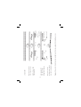
1
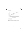
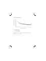
GE Healthcare
Amersham
Megaprime™ DNA
Labelling Systems
Product Booklet
Codes:
RPN1604
RPN1605
RPN1606
RPN1607
Page finder
1. Legal
3
2. Handling
2.1. Safety warnings and precautions
2.2. Storage and stability
2.3. Quality control
4
4
4
4
3. System components
3.1. Megaprime DNA labelling systems
6
8
4. Introduction
9
5. Megaprime DNA labelling protocols
5.1. Standard megaprime protocol
5.2. New megaprime protocol
5.3. Use of alternative reaction conditions
11
11
15
20
6. Appendices
6.1. Appendix I. Labelling of DNA fragments in low melting
point agarose
6.2. Appendix II. Monitoring the reaction and calculating
the specific activity of the labelled DNA
6.3. Appendix III. Removal of unincorporated nucleotides
6.4. Appendix IV. Additional equipment and reagents
27
27
28
32
34
7. Troubleshooting guide
35
8. References
38
9. Related products
39
2
1. Legal
GE and GE monogram are trademarks of General Electric Company.
Amersham, Megaprime, Hybond, Hyperfilm, Hypercassette,
Hyperscreen, Sensitize, Sephadex and SepRate are trademarks of GE
Healthcare companies.
© 2006 General Electric Company – All rights reserved.
General Electric Company reserves the right, subject to any
regulatory and contractual approval if required, to make changes in
specifications and features shown herein, or discontinue the product
described at any time without notice or obligation.
Contact your GE Representative for the most current information
and a copy of the terms and conditions
http://www.gehealthcare.com/lifesciences
GE Healthcare UK Limited.
Amersham Place, Little Chalfont,
Buckinghamshire, HP7 9NA UK
3
2. Handling
2.1. Safety warnings
and precautions
safety glasses and gloves.
Care should be taken to avoid
contact with skin or eyes. In
the case of contact with skin
or eyes, wash immediately
with water. See material safety
data sheet(s) and/or safety
statement(s) for specific advice.
Warning: For research use
only. Not recommended
or intended for diagnosis
of disease in humans or
animals. Do not use internally
or externally in humans or
animals.
2.2. Storage and
stability
Caution: For use with
radioactive material.
This product is to be used with
radioactive material. Please
follow the manufacturer’s
instructions relating to the
handling, use, storage and
disposal of such material.
Upon receipt of these
systems components should
be stored at -15°C to -30°C.
The components are stable
for at least 3 months when
stored under recommended
conditions.
All chemicals should be
considered as potentially
hazardous. We therefore
recommend that this product is
handled only by those persons
who have been trained in
laboratory techniques and
that it is used in accordance
with the principles of good
laboratory practice. Wear
suitable protective clothing
such as laboratory overalls,
2.3. Quality control
The Megaprime DNA labelling
systems are tested by our
quality control group to ensure
an incorporation rate greater
than 55% after 10 minutes at
37°C.
The performance of RPN
1604/1605 is tested with
the standard DNA provided
4
using 17 pmol/25 ng DNA of
[α–32P] labelled nucleotides,
specific activity 3000 Ci/mmol
(codes PB 10204-7) and
RPN 1606/1607 are tested
using 17 pmol/25 ng DNA of
[α–32P]dCTP, 3000 Ci/mmol
(code PB 10205). Incorporations
greater than 55% are achieved
after 10 minutes incubation
at 37°C, as assayed by thinlayer chromatography on PEI
cellulose in 1.25 M KH2PO4.
PH3.4.
In addition components of the
kits are checked for identity by
HPLC and the DNA solutions
for concentration by UV
spectrophotometry.
5
3. System components
Magaprime DNA
labelling
RPN1604
RPN1605
RPN1606
RPN1607
Primer solution:
Random nonamer
primers in an
aqueous solution
150 µl
300 µl
150 µl
300 µl
Labelling buffer;
dATP, dGTP and
dTTP in Tris/HCl
pH7.5,
2-mercaptoethanol
and MgCl2
–
–
300 µl
600 µl
(a) dATP
(b) cCTP
(c) dGTP
(d) dTTP
in Tris/HCl pH8.0,
0.5 mM EDTA
120 µl
120 µl
120 µl
120 µl
240 µl
240 µl
240 µl
240 µl
–
–
–
–
–
–
–
–
Reaction buffer:
A 10x concentrated
buffer containing
Tris/HCl pH7.5,
2-mercaptoethanol
and MgCl2
150 µl
300 µl
–
–
Nucleotide solutions
6
Magaprime DNA
labelling
RPN1604
RPN1605
RPN1606
RPN1607
Enzyme solution;
60 µl
1 unit/µl DNA
polymerase 1 Klenow
fragment (cloned in
100 mM potassium
phosphate pH6.5,
10 mM 2-mercaptoethanol and
50% glycerol
120 µl
60 µl
120 µl
Standard DNA
solution; 5 ng/µl
Hind III digested
lambda DNA in
10 mM Tris/HCl
pH 8.0, 1 mM
EDTA
25 µl
50 µl
25 µl
50 µl
Carrier DNA
solution; 500ng/ml
sonicated herring
sperm DNA in
10 mM Tris/HCl
pH 8.0, 1 mM
EDTA
1.25 ml
2.5 ml
1.25 ml
2.5 ml
7
3.1. Megaprime DNA labelling systems
30 standard labelling reactions –
for use with any radioactive nucleotide
RPN 1604
60 standard labelling reactions –
for use with any radioactive nucleotide
RPN 1605
30 standard labelling reactions –
for use with radioactively labelled dCTP
RPN 1606
60 standard labelling reactions –
for use with radioactively labelled dCTP
RPN 1607
8
4. Introduction
Feinbereg and Vogelstein (1,2) introduced the use of random
sequence hexancleotides to prime DNA synthesis on denatured
template DNA at numerous sites along its length. The primertemplate complex is a substrate for the ‘Klenow’ fragment of DNA
polymerase 1. By substituting a radiolabelled nucleotide for a nonradioactive equivalent in the reaction mixture newly synthesized
DNA is made radioactive (see Figure 1). The absence of the 5’–3’
exonuclease activity associated with DNA polymerase 1 ensures
that labelled nucleotides incorporated by the polymerase are not
subsequently removed as monophosphates. Very small amount of
input DNA can be labelled, enabling very high specific activity DNA
probes to be produced with relatively small quantities of added
nucleotides. These radioactive labelled fragments can then be used
as sensitive hybridization probes for a wide range of filter based
applications (3-6).
Previous protocols for the random primer labelling of DNA have
required reaction times of at least 30 minutes. GE Healthcare’s
Magaprime DNA labelling system allows the labelling of template
DNA to the same high specific activity but at a greatly accelerated
rate. Probes of specific activity 1.9x10⁹ dpm/µg can be produced with
the majority of DNA substrates, using the standard protocol, after
10 minutes incubation at 37°C. This rapid labelling is achieved by
the use of nonamer primers rather than the conventional hexamers
(Figure 1). Nonamers allow for more efficient priming from the
template DNA at 37°C, resulting in fast and efficient labelling of the
DNA. A new alternative protocol has further reduced the variability
in labelling which can occur with DNA template from a variety
of sources. Both the standard Megaprime protocol and the new
protocol are given as options in this booklet. The labelling of DNA in
low melting point agarose takes only 15–30 minutes in contrast to
conventional systems where overnight incubation are necessary.
9
10
Labelled dNTP
‘Klenow’ polymerase
Unabelled dNTPs
Random sequence
monamers
Figure 1. Preparation of labelled probes using GE Healthcare’s megaprime DNA labelling systems.
Denature to release
labelled probe and add
directly to hybridization
Add labelled dNTP
and ‘Klenow’ DNA
polymerase. Incubate
Add Multiprime DNA
reaction buffer
Denature in presence
of monamer primers
Linear dsDNA
5. Megaprime DNA labelling protocols
The Megaprime systems allow DNA from a variety of sources
to be labelled in vitro to high specific activity with 32P and other
radionuclides. The specific activity of the probes generated by these
systems will vary according to the specific activity of the labelled
dNTP used.
The standard Megaprime protocol is presented, together with a new
protocol which reduces the variation in labelling efficiency that can
occur with DNA template from a variety of sources.
The protocols given here are for use with 17 pmol[α–32P]dNTP,
specific activity 3000 Ci/mmol. For alternative reaction conditions
refer to page 20.
DNA prepared by standard minilysate methods may be used in
either protocol. DNA solutions which are too dilute to be used
directly should be concentrated by ethanol precipitation followed by
redissolution in an appropriate volume of water or 10 mM Tris/HCl,
pH 8.0, 1 mM EDTA. DNA in restriction enzyme buffers may be added
directly to the reaction. The reaction can also be performed with
DNA in agarose gel slices (see note 3 and Appendix 1).
5.1. Standard Megaprime protocol
Protocol
Notes
1. Dissolve the DNA to be
labelled to a concentration
of 2.5–25 ng/µl in either
distilled water of 10 mM
Tris/HCl, pH8.0, 1 mM EDTA
(TE buffer).
1. If desired, the labelling
efficiency of a DNA sample
can be compared with that
of the standard DNA
supplied with the kit. In
this case 5 µl of standard
DNA should be used.
11
Protocol
Notes
2. Place the required tubes from
the Megaprime system, with
the exception of the enzyme,
at room temperature to
thaw. Leave the enzyme at
-15°C to -30°C until required,
and return immediately after
use.
3. Place 25 ng of template DNA
into a microcentrifuge tube
and to it add 5 µl of primers
and the appropriate volume
of water to give a total
volume of 50 µl in the final
Megaprime reaction.
Denature by heating to
95–100°C for 5 minutes
in a boiling water bath.
3. When labelling DNA in low
melting point agarose, first
place the tube containing the
stock DNA in a boiling water
bath for 30 seconds to melt
the agarose before removing
the required volume. The
volume of low melting point
agarose DNA should not
exceed 25 µl in a 50 µl reaction.
4. Spin briefly in a microcentrifuge
to bring the contents to the
bottom of the tube.
5. Keeping the tube at room
temperature, add the
nucleotides and reaction
buffer (RPN 1604/5) or the
labelling buffer (RPN 1606/7)
followed by the radiolabelled
dNTP(s) and enzyme as
follows:
5. The reaction volume may be
scaled up or down if more or
less than 25 ng of DNA is to
be labelled.
12
Protocol
Component
Notes
RPN1604/5
Labelling
RPN1606/7
10 µl
buffer
Unlabelled
4 µl of each
dNTPs
omitting
–
those to be
used as
label
Reaction
5 µl
–
buffer
Radiolabelled
(dNTP)
5 µl
5 µl (dCTP)
Enzyme
2 µl
2 µl
6. Mix gently by pipetting up
and down and cap the tube.
Spin for a few seconds in a
microcentrifuge to bring the
contents to the bottom of the
tube.
6. Avoid vigorous mixing of the
reaction mixture as this can
cause severe loss of enzyme
activity.
7. Incubate at 37°C for 10
minutes
7. Purified DNA can be labelled
to high specific activity in
10 minutes at 37°C but, if
desired, can be labelled for
up to 1 hour at this
temperature. When labelling
DNA in low melting point
agarose, longer incubation
of 15–30 minutes at 37°C are
required for optimum
labelling. Longer incubation
13
Protocol
Notes
7. Incubate at 37°C for
10 minutes continued.
7. Continued.
times (up to 60 minutes)
are required when nucleotide
analogues (e.g. [35S]dNTPαS)
are used.
8. Stop the reaction by the
addition of 5 µl of 0.2 M EDTA.
For use in a hybridization,
denature the labelled DNA
by heating to 95–100°C for
5 minutes, then chill on ice.
8. Labelled probe can be stored
at -15°C to -30°C in a non
frost-free freezer. Prolonged
storage of 32P-labelled
probes can lead to substantial
probe degradation(7). High
specific activity probes
should be stored for no
longer than 3 days. Although
probe purification is not
usually necessary for most
membrane applications,
the removal of unicorporated
nucleotide is sometimes
useful to reduce background
in filter hybridizations
for probes >109 dpm/µg or
when the reaction yields an
incorporation of less than 50%.
This procedure is described
in Appendix III. Calculation
of probe specific activity
is described in Appendix II.
Extensive experimentation
with Rapid-hyb buffer
(RPN1635/6) has shown that
probe purification, even
14
Protocol
Notes
8. Stop the reaction by the
addition of 5 µl of 0.2 M EDTA.
For use in a hybridization,
denature the labelled DNA
by heating to 95–100°C for
5 minutes, then chill on ice
continued.
8. Continued
under the conditions given
above is not required with
the isotopes 32P and 33P.
Purification of 35S labelled
probes is however required
to reduce filter background.
5.2. New Megaprime protocol
Protocol
Notes
1. Dilute the DNA to a
concentration of 5 ng/µl in
either distilled water or
10 mM TE buffer.
1. DNA solutions at
concentrations in the range
5–25 ng/µl can be used if desired.
However the denaturing
volume (step 3) should not be
less than 10 µl to maximize
the efficiency of primer
annealing. The labelling
efficiency of a DNA sample
can be compared with that
of the standard DNA supplied
with the kit. In this case 5 µl
of standard DNA should be
used.
2. Place the required tubes
from the Megaprime system
with the exception of the
enzyme at room temperature
to thaw. Leave the enzyme
at -15°C to -30°C until
required, and return
immediately after use.
15
Protocol
Notes
3. Place 25 ng (5 µl) of
template DNA into a clean
microcentrifuge tube and to
it add 5 µl of primers.
Denature by heating to
95–100°C for 5 minutes in a
boiling water bath.
3. If the volume of DNA and
primers is less than 10 µl
make up to this volume with
water. When labelling DNA
in low melting point agarose
first place the tube
containing the stock DNA
in a boiling water bath for
30 seconds to melt the
agarose before removing
the required volume. The
volume of low melting point
agarose DNA should not
exceed 25 µl in a 50 µl
reaction.
4. Spin briefly in a
microcentrifuge to bring the
contents to the bottom of
the tube.
5. Keeping the tube at
room temperature add the
nucleotides and 10x
reaction buffer (RPN 1604/5)
or the labelling buffer (RPN
1606/7), water and enzyme:Component
Labelling
buffer
RPN1604/5
Unlabelled
dNTPs
4 µl of each –
omitting
5. The enzyme can be added
directly to the reaction mix
or pipetted on to the side of
the microcentrifuge tube
and “washed” down with the
water.
RPN1606/7
10 µl
those to be
used as label
16
Protocol
Reaction
buffer
Notes
5 µl
–
Enzyme
2 µl
2 µl
Water*
as appropriate
for a final
reaction
volume of
50 µl*
* When calculating this volume
remember to allow for the
volume of radioactive
nucleotide to be added.
6. Cap the tube and spin for a
few seconds in a
microcentrifuge to bring the
contents to the bottom of the
tube.
7. Add the radiolabelled dNTP,
for example 5µl [α–32P]dNTP,
specific activity 3000 Ci/mmol.
Mix by gently pipetting up and
down. Spin for a few seconds
in a microcentrifuge to bring
the contents to the bottom of
the tube.
7. Avoid vigorous mixing of the
reaction mixture as this can
cause severe loss of enzyme
activity.
8. Incubate at 37°C for
10 minutes.
8. Purified DNA can be labelled
to high specific activity in
10 minutes at 37°C but, if
desired can be labelled for
up to 1 hour at this
temperature.
17
Protocol
Notes
8. Incubate at 37°C for
10 minutes continued.
8. Continued
When labelling DNA in low
melting point agarose,
longer incubation of 15–30
minutes at 37°C are
required for optimum
labelling. Longer incubation
times (up to 60 minutes)
are required when nucleotide
analogues (e.g. [35S]dNTP(S)
are used.
9. Stop the reaction by the
addition of 5 µl of 0.2 M
EDTA. For use in a
hybridization, denature the
labelled DNA by heating to
95–100°C for 5 minutes, then
chill on ice.
9. Labelled probe can be stored
at -15°C to -30°C in a non
frost-free freezer. Prolonged
storage of 32P-labelled
probes can lead to substantial
probe degradation(7). High
specific activity probes
should be stored for no
longer than 3 days. Although
probe purification is not
usually necessary for most
membrane applications the
removal of unincorporated
nucleotide is sometimes
useful to reduce background
in filter hybridizations
for probes >109 dpm/µg or
when the reaction yields an
incorporation of less
than 50%. This procedure is
18
Protocol
Notes
9. Stop the reaction by the
addition of 5 µl of 0.2 M
EDTA. For use in a
hybridization, denature the
labelled DNA by heating to
95-100°C for 5 minutes, then
chill on ice continued.
9. Continued
described in Appendix III.
Calculation of probe specific
activity is described in
Appendix II.
Extensive experimentation
with Rapid-hyb buffer
(RPN1635/6) has shown that
probe purification, even
under the conditions given
above is not required with
the isotopes 32P and 32P.
Purification of 32S labelled
probes is however required
to reduce filter background.
19
% added label incorporated into DNA
90
80
70
60
50
40
30
20
10
0
10
20
30
40
50
60
Length of incubation in minutes
Figure 2. Time course of incorporation of [α–32P]dCTP (17 pmoles) in
a Megaprime reaction at 37°C. The DNA used was the standard DNA
supplied with the system.
5.3. Use of alternative reaction conditions
a. Use of more than one labelled [α–32P]dNTP.
Table 1 lists the results of a selection of standard reactions, using
a variety of input labels under optimum conditions. Figure 3 gives
more complete information on their use in Megaprime reactions.
Reactions were carried out at 37°C for 5 minutes.
b. Use of alternative specific activity [α–32P]dNTPs.
When using [α–32P]dNTPs of specific activity <3000 Ci/mmol the
incubation time should be extended to 1 hour at 37°C.
20
c. Use of [32P]dNTPαS.
When using 32S-labelled radionucleotides the incubation time should
be extended to 1 hour at 37°C.
d. Labelling at room temperature.
If desired, labelling reactions can be carried out at room
temperature. Maximum incorporation occurs after an incubation
time of 45–60 minutes. A decline in incorporation can be observed if
reactions are left overnight.
e. Factors affecting the labelled DNA.
1. Specific activity
Figure 3a should be used to ascertain the number and quantity of
labelled dNTP’s required in order to prepare a probe of the desired
specific activity.
2. Efficiency
Figure 3b indicates the efficiency of the chosen reaction
conditions, and thus permits a balance of specific activity and
economy.
3. Probe length
Figure 3c gives a measure of mean probe lengths obtained under
standard conditions. Probe lengths were measured by denaturing
agarose gel electrophoresis followed by autoradiography with
reference to molecular weight standards.
Probe length can be affected by the concentration of DNA, primer
and nucleotide, the size of the template DNA and also radiolysis
of the labelled probe. The data in the figure was obtained using
linearized plasmid DNA, 4.5 Kb in length under the standard
labelling conditions.
It is recommended that not less than 10 pmol and not more
than 125 pmol of any labelled dNTP is used in the reaction and
combinations shown offer optimum balance of stability, specific
activity and economy.
21
5.3 x 10⁹
1.9 x 10⁹
Quantity of each
dNTP required
32
} 3.4 x 10
Product
code
200 20
17
Formulation
(see note b)
5
17
17
Specific activity
(see note a)
7.4
50
5
5
Compounds
PB 10205
1.85
50
50
dpm/ug
PB 10475
1.85
Specific
activity of
probe
(see notes
c,d and e)
1
PB 10204
1.85
17
Ci/mmol
1
PB 10205
pmol
~3000
1
PB 10204
17
17
5
~6000
1
5
5
µl
~111
TBq/mmol
~3000
1
50
50
50
~222
~3000
1.85
1.85
µCi
~111
~3000
PB 10206
PB 10205
1.85
~111
1
1
MBq
[α–32P]dATP
~111
~3000
~3000
[α–32P]dCTP
[α–32P]dCTP
~111
3.7 x 10⁹
9
[α–32P]dATP
~111
}
[α–32P]dGTP
[α–32P]dCTP
a. At the specific activity reference date of the labelled nucleotide.
b. Formulation code 1 = 370 MBq/ml, 10 mCi/ml in stabilized aqueous solution.
c. The probe specific activities were calculated using observed incorporation levels which are similar to
those found in figure 3b.
d. It is important to note that the specific activity of probes made from different amounts of labelled
dNTP cannot be calculated on a proportional basis, because net DNA synthesis occurs.
e. Brackets enclose nucleotides used in combination.
22
Specific activity of the labelled
product (dpm/µg) x 109
a) Specific activity
5
(iii)
4
(ii)
3
(i)
2
1
0
10
20
30
40
50
60
70
80
90 100
Total input label (pmols)
i) One labelled dNTP
ii) Two labelled dNTP
iii) Three labelled dNTP
Figure 3. The use of [α–32P]dNTPs in the Megaprime DNA labelling
system (see notes on page 26).
23
Percentage of added label
b) Incorporation efficiency
100
80
60
(i)
(ii)
(iii)
40
20
0
10
20
30
40
50
60
70
80
90 100
Total input label (pmols)
i) One labelled dNTP
ii) Two labelled dNTP
iii) Three labelled dNTP
Figure 3. The use of [α–32P]dNTPs in the Megaprime DNA labelling
system (see notes on page 26).
24
Mean probe length (bases)
c) Probe length
100
80
60
(i)
(ii)
(iii)
40
20
0
10
20
30
40
50
60
70
80
90 100
Total input label (pmols)
i) One labelled dNTP
ii) Two labelled dNTP
iii) Three labelled dNTP
Figure 3. The use of [α–32P]dNTPs in the Megaprime DNA labelling
system (see below).
Notes to figure 3
a. The results shown are the means of a number of experiments in
which different nucleotides and combinations of nucleotides
were used. Observed results may deviate ± 10% from those
shown.
b) As the number of different labelled nucleotides is increased, at a
given level of total input label, the net synthesis of DNA is reduced.
Although the overall incorporation efficiency is reduced the
labelled product is of a higher specific activity.
25
c. The data was generated using the standard labelling protocols.
If dNTPs <3000 Ci/mmol are to be used, then the desired probe
specific activity must be multiplied by a conversion factor, before
determining the amount of input label.
For a single labelled dNTP:Total input label (pmols) =
3000 Ci/mmol
x required probe
specific activity of
specific activity
dNTP to be used
For more than one labelled dNTP the mean specific activity of the
labelled dNTP to be used should be inserted in the above
calculation.
Having determined the required number of pmols of input label
with reference to figure 3a, the required volume of each labelled
dNTP can be calculated. Note that the figures give the total
amount of input label required. If more than one labelled dNTP is
to be used, this figure should be divided by the number of
labelled dNTPs to be used to give the required number of pmols
of each labelled dNTP.
Volume of each labelled dNTP required in µl =
pmol of dNTP required x specific activity of dNTP (Ci/mmol) x 10–3
radioactive concentration of dNTP (mCi/ml)
26
6. Appendices
6.1. Appendix I. Labelling of DNA fragments in
low melting point agarose
The DNA samples produced by the following protocol have been
found to be labelled to approximately the same extent as purified
DNA. 15–20 minutes at 37°C is optimum for labelling. The standard
labelling protocol may be found to be more appropriate for labelling
DNA in agarose as the volume of DNA to be added using the new
protocol is limited to 5 µl, requiring a relatively high initial DNA
concentration.
Protocol
Notes
1. Fractionate restriction
endonuclease digested DNA
in a suitable low melting
point agarose gel containing
0.5 µg/ml ethidium bromide.
Estimate the DNA content
of the band by reference to
a set of standards of known
concentration on another
track. 250 ng should allow
25 ng to be used in the
standard labelling protocol
without further concentration
1. A low melting point agarose
of high purity for example
SepRate-LMP is
recommended for maximum
labelling efficiency.
2. Excise the desired band
cleanly, with the minimum
of excess agarose and
transfer to a pre-weighed
1.5 ml microcentrifuge tube.
2. It is recommended that
the exposure to UV light
is minimized, as prolonged
exposure can damage the
DNA.
27
Protocol
Notes
3. Add water to a ratio of 3 ml
per gram of gel and place
in a boiling water bath for
5 minutes to melt the gel and
denature the DNA.
3. If the DNA is not to be used
immediately divide the boiled
samples into suitably sized
aliquots and store at -15°C to
-30°C in a non frost-free
freezer.
4. If the DNA is to be used
immediately remove the
appropriate volume
containing 25 ng, add to
the primers as indicated in
the labelling protocol (page
11, step 3). The volume of
DNA should not exceed 25 µl
for the standard labelling
protocol.
4. When using DNA which has
been previously boiled and
then stored at -15°C to -30°C,
first place the tube in a
boiling water bath for
30 seconds to melt the
agarose, before removing the
required volume containing
25 ng. Do not reboil DNA
aliquots more than twice.
5. Incubate the labelling
reaction for 15–20 minutes
at 37°C.
6.2. Appendix II. Monitoring the reaction and
calculating the specific activity of the labelled
DNA
A. Adsorption to DE81 paper
Monitoring of the progress of the labelling reaction and
measurement of probe specific activity can be achieved by
determining the proportion of the radionucleotide incorporated
during the Megaprime reaction.
28
Protocol
Notes
1. Remove a 1 or 2 µl aliquot of
the reaction mixture to a
clean microcentrifuge tube
containing 20 µl of water
or 10 mM Tris/HCl pH.8.0.
1 mM EDTA buffer. Mix well
by pipetting up and down.
2. Spot, in quadruplicate,
5 µl aliquots of this dilution
on to Whatman DE81
chromatography paper
squares (minimum size
1 x 1 cm), placed on a nonabsorbent backing. These
squares may be marked
with a pencil for identification
if required.
3. Take two of the filters and
dry under a heat lamp.
10–15 minutes should be
adequate.
4. In aqueous solution DE81
paper becomes fragile and
care should be taken when
handling. In order to stabilize
the paper the squares are
rinsed in ethanol.
4. Wash the remaining two
filters twice for 5 minutes
each, at room temperature
in excess 2xSSC (30 mM Na3
citrate, 300 mM NaCl pH7.0)
using gentle agitation. Rinse
briefly in distilled water and
then once with ethanol for
5 minutes. Then dry the
filters under a heat lamp.
29
Protocol
Notes
5. Place the squares in separate
vials with at least 5 ml of
scintillation fluid and count.
5. Determination of the
proportion of the 32P labelled
nucleotide incorporated may
be achieved using Cerenkov
counting if desired in this
case drying the filter is not
necessary.
6. Efficiency of counting will
vary, but the percentage
incorporation can be used
to calculate probe specific
activity. Unlike the nick
translation labelling reaction,
Megaprime labelling leads
to net DNA synthesis, and
so the total amount of DNA
at the end of the reaction
must be calculated.
6. The mean value of the
counts on the washed filter
represents the proportion
of the radionucleotide
incorporated into the DNA
probe, while the mean of the
unwashed filters represents
the total amount of
radioactivity in the reaction
mix, such that;
% incorporation = mean counts on washed filters x 100
mean counts on unwashed filters
Total amount of DNA (A) ng =
Total number of µCi added x 13.2* x % incorporation + 25
Number of radioactive dNTPs added x average specific
activity of dNTPs added
This assumes a 25% content
of any one dNTP in the newly
synthesized DNA, and
25 ng of template DNA.
*13.2 equals four times the
average molecular weight of
the four dNTPs divided by 100.
30
Protocol
Notes
6. Continued.
The amount of radioactivity
incorporated during the
reaction (B) in dpm.
B = total number of µCi added x
2.2x10⁴ x % incorporation
Thus the specific activity of the
labelled DNA is
specific activity = B x 103 dpm per µg
specific activity = A
B. Precipitation with trichloroacetic acid
Plastic or siliconized glass tubes must be used to avoid adsorption
of DNA.
1. Dilute an appropriate aliquot of the reaction mixture as described
in section A1.
2. Transfer 1–10 µl of diluted reaction mixture to two duplicate tubes
containing 200 µl water or 0.2M EDTA and 50 µl carrier DNA
solution. Mix well. Use this mixture (less any set aside in step 3) for
the TCA precipitation described in step 4 below.
3. Set aside an appropriate aliquot from each tube in step 2 for the
determination of total input radioactivity.
4. To the diluted samples from step 2, add 2 ml ice-cold 10%
trichloroacetic acid (TCA) solution, vortex, and allow to stand in an
ice-bath for 10–15 minutes. The labelled and carrier DNA will
co-precipitate. Note that TCA is corrosive, and care should be
taken in its handling.
5. Collect the precipitated DNA by vacuum filtration on a glass fibre
or nitrocellulose filter disc.
31
6. Wash the filter discs six times with 2 ml 10% TCA solution and
dry the filter discs thoroughly, for example using an infra-red
lamp. Avoid overheating and possible charring of the discs.
7. Count the dried filter discs by liquid scintillation or Cerenkov (32P)
and count with the samples set aside in step 3.
8. Determine % incorporation and probe specific activity as in
section A6.
6.3. Appendix III. Removal of unincorporated
nucleotides
Removal of unincorporated nucleotides is sometimes desirable to
reduce background produced by the probe during hybridization. It is
considered important to remove these free nucleotides particularly if
the radioactive probe is to be kept for several days before use or the
incorporation is less than 50%. If 32P or 33P-labelled probes are to
be used in combination with GE Healthcare’s new Rapid-hyb buffer
(RPN1635/6), purification is not required unless the probe is to be
used more than 24 hours after preparation. Probes can be purified
by Sephadex chromatography or selective precipitation (8,9).
A. Sephadex™G-50 spin columns
Probe reaction are passed through columns packed with Sephadex
G-50, which retains the free nucleotides within the column matrix.
A number of pre-packed columns are commercially available.
However columns may also be prepared as indicated below:
1. Equilibrate Sephadex G-50 in TE buffer either overnight or at 65°C
for 1–2 hours.
2. Plug a 1.0 ml syringe with a piece of siliconized glass wool.
3. Fill the syringe with the equilibrated Sephadex. Place in a 15 ml
conical tube, in which a decapped 1.5 ml microcentrifuge tube
has been inserted. Centrifuge at 1600 g for 5 minutes. Remove
32
any liquid from the microcentrifuge tube. Refill with Sephadex
and centrifuge as before. Continue until the column is packed to a
volume of 1 ml.
™ Sephadex is a trademark of GE Healthcare
4. Add a volume of TE buffer equal to the reaction volume, to the
top of the column and centrifuge, as in step 3. A minimum of 50 µl
should be applied to the column.
5. Repeat once more to ensure fractions of the correct size are
collected from the column.
6. Place the column in a clean 15 ml conical tube containing a
decapped 1.5 ml microcentrifuge tube.
7. Apply the DNA sample to the column. Centrifuge as before. The
purified probe is collected in the microcentrifuge tube.
B. Selective precipitation of labelled DNA
The following protocol leads to precipitation of DNA greater than
about 20 nucleotides in length with unicorporated nucleotides
remaining in solution. Recovery of the labelled DNA by this method
varies according to the DNA concentration and size, and may be as
low as 50%.
1. Add one volume of 4 M ammonium acetate, pH4.5 to the nick
translation reaction, and mix gently by pipetting up and down.
2. Add four volumes of ethanol, mix by inversion. Chill the mixture for
15 minutes in a dry-ice ethanol bath or place at -70°C for at least
30 minutes.
3. Thaw the mixture if necessary by placing at 37°C for 2 minutes.
4. Spin in a microcentrifuge for 15 minutes. Carefully aspirate and
dispose of supernatant in a suitable manner.
5. Wash the pellet once in 0.5 ml of 0.67 M ammonium acetate,
pH 4.5, 67% ethanol at room temperature by gentle inversion,
centrifugation and aspiration.
33
6. Wash the pellet once in 90% ethanol, in the same manner. Dry the
pellet.
7. Finally redissolve the DNA pellet in TE buffer for use as a probe
and for storage.
6.4. Appendix IV. Additional equipment and
reagents
TE buffer (10 mM Tris/HCl, pH 8.0, 1 mM EDTA)
0.2 M EDTA solution
Adjustable pipettes for example Pipetman™
Sterile pipette tips
Waterbaths at 37°C and 100°C
Polypropylene microcentrifuge tubes
Microcentrifuge
Gloves
Radiation safety equipment
DE81 ion-exchange chromatography paper (Whatman)
Trichloroacetic acid (TCA) solution: 10% (w/v) TCA in water
Filter discs; glass fibre or nitrocellulose
Plastic or siliconized glass tubes, capacity ~5 ml
Filtration apparatus
2x SSC (30 mM Na3 citrate, 300 mM NaCl, pH 7.0)
™ Pipetman is a registered trademark of Gilson
34
7. Troubleshooting guide
If poor results are obtained, the following guide may help to
determine the cause of the problem.
Problem
Possible cause
Remedy
1. Low signal
1. Incomplete
denaturation of
template DNA
1. Ensure denaturation
protocol is followed.
2. Low probe
concentration
2. Accurately measure
the concentration of
template DNA used in
the labelling reactions.
Check recovery of
probe if purification
is performed to
remove unincorporated
nucleotide.
3. Low probe specific 3. If the specific activity
of the labelled DNA is
activity
lower than expected,
a labelling reaction
should be carried out
using a sample of the
control DNA supplied
with the system.
If this proceeds
satisfactorily, check
the concentration
and purity of your
DNA.
35
Problem
2. Non-specific
background
over whole of
filter
Possible cause
Remedy
4. Loss of dNTP
during
evaporation
4. If the dNTP solution
has been evaporated
to dryness prior to
use, handling losses
may have occurred.
Check this loss has
not occurred during
lyophilization of the
solvent, during
transfer of the
reconstituted
dNTP solution or
by adsorption of the
dNTP onto the walls
of the tube. If
necessary the
reconstituted dNTP
solution may be
counted and an
adjustment made
before setting up the
labelling reaction.
1. Presence of
unincorporated
label
1. Unincorporated
nucleotides can give
high backgrounds.
Remove by Sephadex
G-50 spin columns
or ethanol
precipitation (see
page 32 for protocol)
36
Problem
Possible cause
Remedy
2. Concentrated
2. It is suggested
probe has contacted
that up to 1.0 ml
membrane
of the buffer used
directly during
for prehybridization
probe addition
is withdrawn for
mixing with the probe.
The mixture should
then be added back
to the hybridization
container in an area
away from the filter.
3. Probe concentration 3. Ensure measurement
of template DNA
is too high
concentration is
accurate
4. Probe not denatured
37
4. Non-denatured
double-stranded
probes often
give high backgrounds.
8. References
1. FEINBERG, A.P. and VOGELSTEIN, B., Anal. Biochem., 132, pp.6-13,
1983.
2. FEINBERG, A.P. and VOGELSTEIN, B., Addendum Anal. Biochem.,
137, pp.266-267, 1984.
3. SOUTHERN, E.M., J.Mol.Biol., 98, pp.503-517, 1975.
4. THOMAS, P.S., Proc. Natl. Acad. Sci. USA., 77, pp.5201-5205, 1980.
5. MEINKOTH, J. and WAHL, G., Anal, Biochem., 138, pp. 267-284,
1984.
6. GRUNSTEIN M. and HOGNESS, D.S., Proc. Natl. Acad. Sci. USA., 72,
pp. 3961-3965, 1975.
7. HODGSON, C.P., FISK, R.Z. and WILLET, L.B., Biotechniques, 6,
pp.208-211.
8. SAMBROOK, J. FRITSCH, E.F. and MANIATIS, T., Molecular Cloning, a
laboratory manual (second edition), Cold Spring Harbour
Laboratory, 1989.
9. MUNDY, C.R., CUNNINGHAM, M.W. and READ, C.A., Essential
Molecular Biology; A Practical Approach Vol 2 (T.A. Brown, ed)
Oxford University Press, Oxford, 1991. pp.57-109.
38
9. Related Products
Labelling systems
Nick translation kits
3’-end labelling kit
5’ end labelling kit
RNA labelling system
(paired promoter SP6/T7 system)
N5000/5500
N4020
RPN 1509
RPN 3100
Hybridization buffers
Rapid-hyb buffer
Hybridization buffer tablets
RPN 1635/6
RPN 131
Hybridization membranes
Hybond™ - Range of nylon and nitrocellulose
blotting membranes
Autoradiography products
Hyperfilm™ - high performance autoradiography films
Hypercassettes™ and Hyperscreens™ - available from stock
Safety Products
Radiation safety products for safe handling and storage of 32P/33P/
35S and 125I, liquid scintillation products
Agarose
SepRate™ - range of highly purified agarose for a range of DNA
fragment sizes and users
Labelled dNTPs
See Table 2
39
Table 2. Labelled dNTPs and analogues available from GE Healthcare
Compound
[α–32P]dATP
Specific Activity
Formulation
TBq/mmol
Ci/mmol
(see key)
~220
~110
~6000
~3000
~30
~15
~800
~400
~220
~110
~6000
~3000
~30
~15
~6000
~400
~220
~3000
~30
~15
~800
~400
~110
~300
~30
~15
~800
~400
[α–32P] dATP
37–110
[35S]dATPαS
>37
~22
~15
[α–32P]dCTP
[α–32P]dGTP
[α–32P]dTTP
Product
code
1
1
2
1
1
2
PB 1074
PB 10204
PB 204
PB 10384
PB 10164
PB 164
1
2
1
1
2
PB 10475
PB 10205
PB 205
PB 10385
PB 10165
PB 165
1
2
1
1
2
PB 10206
PB 206
PB 10386
PB 10166
PB 166
1
2
1
1
2
PB 1027
PB 207
PB 10387
PB 10167
PB 167
1000–3000
1
BF 1001
>1000
~600
~400
1
1
1
SJ 1304
SJ 304
SJ 264
40
Compound
Specific Activity
TBq/mmol Ci/mmol
Formulation
(see key)
Product
code
[35S]dCTPαS
>37
~22
~15
>1000
~600
~400
1
1
1
SJ1305
SJ 305
SJ 265
[35S]dGTPαS
~22
~600
1
SJ 306
[35S]dTTPαS
~22
~600
1
SJ 307
0.37–1.1
10–30
[8–3H]dATP
[1’,2’,2,8–3H]dATP
1.83–3.7 50–100
[1’,2’,5–3H]dCTP
1.85–3.14 50–85
[5–3H]dCTP
0.55–1.1
15–30
[8–3H]dGTP
0.185–0.740 5–20
[1’2,(–3H]dGTP
0.9–1.85
25–50
[methyl, 1’,2’–3H]TTP
3.3–4.8 90–130
[methyl–3H]TTP
40–60
30
[125I]dCTP
>55
>1500
2
2
2
2
2
2
2
2
2
3
Formulation codes:
1) = 370 MBq/ml, 10 mCi/ml in stabilized solution
2) = 37 MBq/ml, 1 mCi/ml in 50% aqueous ethanol
3) = 185 MBq/ml, 5 mCi/ml in 50% aqueous ethanol
See GE Healthcare Products catalogue for further details.
41
TRK 347
TRK 633
TRK 625
TRK 352
TRK 350
TRK 627
TRK 576
TRK 424
TRK 354
IM 5103
GE Healthcare
regional office
contact numbers:
France
Tel: 01 69 35 67 00
Fax: 01 69 41 98 77
Portugal
Tel: 21 417 7035
Fax: 21 417 3184
Asia Pacific
Tel: +85 65 62751830
Fax: +85 65 62751829
Germany
Tel: 0800 9080 711
Fax: 0800 9080 712
Russia, C.I.S. & N.I.S
Tel: +7 495 956 5177
Fax: +7 495 956 5176
Australasia
Tel: + 61 2 8820 8299
Fax: +61 2 8820 8200
Greater China
Tel: +852 2100 6300
Fax: +852 2100 6338
Spain
Tel: 902 11 72 65
Fax: 935 94 49 65
GE Healthcare UK Limited
Amersham Place
Little Chalfont
Buckinghamshire
HP7 9NA
UK
Austria
Tel: 01/57606-1613
Fax: 01/57606-1614
Italy
Tel: 02 26001 320
Fax: 02 26001 399
Sweden
Tel: 018 612 1900
Fax: 018 612 1910
Belgium
Tel: 0800 73 890
Fax: 02 416 8206
Japan
Tel: +81 3 5331 9336
Fax: +81 3 5331 9370
Switzerland
Tel: 0848 8028 10
Fax: 0848 8028 11
GE Healthcare Bio-Sciences
Corp
800 Centennial Avenue
P.O. Box 1327
Piscataway
NJ 08855-1327
USA
Canada
Tel: 1 800 463 5800
Fax: 1 800 567 1008
Korea
Tel: 82 2 6201 3700
Fax: 82 2 6201 3803
UK
Tel: 0800 515 313
Fax: 0800 616 927
Central, East, & South
East Europe
Tel: +43 1 972 720
Fax: +43 1 972 722 750
Latin America
Tel: +55 11 3933 7300
Fax: + 55 11 3933 7304
USA
Tel: +1 800 526 3593
Fax: +1 877 295 8102
GE Healthcare offices:
GE Healthcare Bio-Sciences AB
Björkgatan 30 751 84
Uppsala
Sweden
GE Healthcare Europe GmbH
Munzinger Strasse 5 D-79111
Freiburg
Germany
GE Healthcare Bio-Sciences KK
Sanken Bldg. 3-25-1
Hyakunincho Shinjuku-ku
Tokyo 169-0073
Japan
Denmark
Tel: 45 70 25 24 50
Fax: 45 45 16 2424
Eire
Tel: 1 800 709992
Fax: +44 1494 542010
Finland & Baltics
Tel: +358 9 512 3940
Fax: +358 9 512 39439
Middle East & Africa
Tel: +30 210 96 00 687
Fax: +30 210 96 00 693
Netherlands
Tel: 0800-82 82 82 1
Fax: 0800-82 82 82 4
Norway
Tel: +47 815 65 777
Fax: +47 815 65 666
http://www.gehealthcare.com/lifesciences
GE Healthcare UK Limited
Amersham Place, Little Chalfont, Buckinghamshire, HP7 9NA
UK
imagination at work
RPN1604PL Rev B 2006