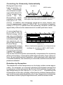
1
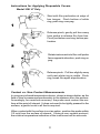
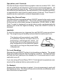
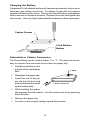
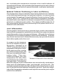
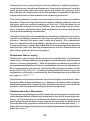
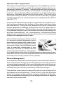
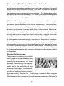
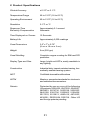
EXERGEN
Infrared Thermographic Scanner
DermaTemp 1001
USER’S MANUAL
AND REFERENCE
BOOK
Unparalleled Accuracy
. . .at the Speed of Light
Table of Contents
I. The Instruments............................................................ 1
The Instruments’ Features................................................. 2
Optional Disposable Covers............................................... 2
Instructions for Applying Disposable Covers...................... 3
Contact vs. Non-Contact Measurements............................3
Operation and Controls...................................................... 4
ON/OFF.............................................................................. 4
To Lock Reading................................................................. 4
To Restart........................................................................... 4
Operating Modes................................................................ 5
Non-Contact Scanning....................................................... 5
Changing the Battery.......................................................... 6
Fahrenheit or Celsius Conversion...................................... 6
Care and Maintenance........................................................7
Self Diagnostics..................................................................7
Customer Service............................................................... 8
II. Body Surface Temperature.......................................... 9
History and Introduction......................................................9
Body Surface Temperature................................................. 10
Infrared Thermometry......................................................... 11
The DermaTemp Infrared Thermographic Scanner............13
Method Impedimenta.......................................................... 13
Ambient Effect on Body Surface Temperature................... 14
Solving the Problems..........................................................14
Emissivity............................................................................15
Alice’s Quest for Emissivity................................................ 17
Correcting for Emissivity Automatically...............................18
Detection by Exception....................................................... 18
III. Clinical Applications....................................................20
Regional Blocks..................................................................20
Epidural Catheter Positioning............................................. 21
Joint Inflammation..............................................................21
Digital Perfusion Assessment............................................ 22
Reconstructive Surgery.......................................................22
Lower Back Pain.................................................................23
Diabetic Foot Screening..................................................... 23
Peripheral Nerve Injury........................................................24
Cerebrovascular Disorders.................................................24
Neonatal Skin Temperature................................................ 25
Wound Management.......................................................... 25
Thermal Assessment of Skin Diseases and Allergy........... 26
Skin Temperature in Prognosis of the Critically Ill...............26
Temperature Gradients in Detection of Shock.................... 27
Raynaud’s Syndrome..........................................................27
Other Areas or Applications of Interest...............................28
IV. References.................................................................. 29
V. Product Specifications................................................ 31
I. The Instruments
The DermaTemp is a high precision hand-held infrared thermographic
scanner designed to detect the subtle skin temperature variations caused
by underlying perfusion variations.
These instruments feature a patented automatic emissivity compensation system for absolute accuracy regardless of skin type or color, and
provide an instant temperature measurement on any surface location
on the human body without the need for tissue contact.
In those applications where tissue contact is desirable or cross-contamination is an issue, the use of disposable wraps or sheaths allows
even moist or wet tissue to be measured with precision accuracy.
The models include:
DT-1001
the standard model
DT-1001 LT
has a conveniently angled stainless steel probe,
and can be used with or without disposable probe
wraps
DT-1001 LN
has a longer probe than the DT-1001, and can
be used with or without a disposable sheath. The
sheath encases the entire instrument.
DT-1001 RS
has a remote stainless steel sensor attached to
the instrument by cable, convenient for those especially hard to reach areas.
All instruments can be cleaned with any hospital approved disinfectant,
including bleach, and can be gas or plasma sterilized.
The DermaTemp is recommended for use in such areas as plastic and
vascular surgery, anesthesiology, pain management, rheumatology,
neurology, oncology, and wound management.
1
The Instruments Feature:
•
•
•
•
•
•
•
•
Full range resolution to 0.1°F/C
SCAN, MAX and/or MIN modes of operation, model specific
Fahrenheit/Celsius conversion
A 10-second display lock
An audible beeper to signal functional or conditional changes
Hermetically sealed sensing system to withstand gas and plasma
sterilization, and cleaning with any hospital approved disinfectant
including bleach and alcohol.
Pencil-like stainless steel sensor on the RS version.
Optional disposable cover usage:
- Complete encasement with disposable sheaths for the LN
version.
- Full probe covering with disposable wraps for the LT version.
Optional Disposable Covers
The use of disposable covers with the DT-1001 LT is optional, depending on the requirements of the application. Recommended guidelines
are as follows:
Use With Disposable Cover
For absolute accuracy, minimizing the effects of emissivity and
evaporative cooling, contact with the measurement site is recommended. Accordingly, when direct contact is employed, use of a
disposable probe cover is recommended.
When touching moist tissue, use of a disposable cover is required
specifically to avoid a lower temperature from the effects of evaporative cooling, and to protect against the risk of cross contamination.
Use Without Disposable Cover
If the measurement site is dry, direct contact can confidently be
made without the use of a disposable cover. When the site is dry
and the precise temperature is not a prerequisite, the measurement can be made without even contacting the skin. The probe
can be cleaned with any hospital approved disinfectant, even bleach
solution.
2
Instructions for Applying Disposable Covers
Model 1001 LT Only
Start with film perforation at edge of
box tongue. Pinch bottom of white
ring, push ring over peg.
1
Stop at Perforation
Release pinch, gently pull box away
from probe to release film from box.
Pinch just below next ring, before perforation.
2
Rotate instrument into film until probe
faces opposite direction, push ring on
peg.
3
Release pinch. Pull box slightly away
until next white ring is visible. Pinch
ring, break film apart at perforation.
4
Contact vs. Non-Contact Measurements
In using any infrared temperature device, closer is always better, as the
field of view increases proportionately to the distance from the surface.
Accordingly, for maximum accuracy the probe must contact the surface at the point of interest. It does not need to be tightly pressed to the
surface; a gentle touch is all that is required.
When contact with the surface is not an option, position the probe within
1/2 inch from the surface of interest. If using a non-contact protocol,
the relative temperature indication of the instrument will be accurate.
3
Operation and Controls
The DermaTemp infrared thermographic scanner models 1001, 1001
LN, LT and RS are all identical in performance and specifications. All
are maximized for ease of use. The remote sensor on the RS version
can either be left attached to the instrument for one-handed operation,
or separated for use in hard-to-reach areas of interest. The LN and LT
models can be used with or without disposable covers
Using the DermaTemp
The DermaTemp is equipped with an ON/OFF power button and a mode
selector switch, model specific. The mode selector switch allows you
to choose one of the three modes of operation, SCAN, MAX, or MIN.
The LT model is designed in a peak select mode, and automatically
selects and locks the highest reading when the ON/OFF button is
released.
ON/OFF:
To turn the instrument on, depress the red ON/OFF power push button. The single beep will audibly indicate that the instrument is on.
The display will momentarily read 8888,
an indication that the microprocessor is
performing a self-diagnostic check. After the test, the unit will measure and display temperature in the selected operating mode for as long as the power button
is depressed.
To Lock Reading:
Release the red ON/OFF button to lock reading on display. The single
beep will audibly indicate that the display is locked. The DermaTemp
will hold the last reading on the display for 10 seconds before it automatically turns off.
If you are using a DermaTemp 1001 LT, the highest temperature measured will be retained before automatic turn off.
To Restart:
Depress the button anytime to restart. It is not necessary to wait until
the display is clear. The DermaTemp automatically recalibrates each
time the button is depressed.
4
Operating Modes (Model Specific)
•
SCAN: In the SCAN mode, the target’s instantaneous temperature is continuously displayed and updated 10 times per second
for as long as you keep the button depressed. After the power
button is released, the display will lock on the last temperature
measured and hold that reading for 10 seconds.
•
MAX: In the MAX (peak hold) mode, the display will lock on the
highest temperature measured for as long as you hold the power
button down. Each time a new peak temperature is measured or
repeated, an audible beep will sound. After you release the power
button, the display will lock on the maximum recorded temperature and hold that reading for 10 seconds.
•
MIN: In the MIN (valley hold) mode, the display will lock on the
lowest temperature measured as long as the power button is depressed. Each time a new low temperature is measured, a beep
will sound. After the power button is released, the display will lock
on the minimum recorded temperature and hold that reading for
10 seconds.
Non-Contact Scanning
For situations where even light contact is contraindicated, bring the
instrument nose as close to the measuring site as safely possible, keeping the following in mind:
The instrument’s field-of-view, also referred to as the distance-tospot ratio, is 1:1. A 1:1 field-of-view means that the sensor sees
a circular area with a diameter equal to the distance between the
sensor and the target area.
For example, at a distance of 2 inches (5 cm), the sensor sees a 2
inch (5 cm) diameter spot. The minimum spot size is approximately 1/4 inch (6 mm) when touching.
The DermaTemp averages the temperature of everything in its
field-of-view.
A small hot spot may get lost in a large viewing area. The closer
you hold the instrument to a surface, the sharper its target resolution.
5
Changing the Battery
A standard 9-Volt alkaline battery will require replacement only once or
twice per year under normal use. To replace, loosen the four captive
screws and remove the cover. Disconnect the old battery and replace
with a new one in the same location. Replace the cover and tighten the
four screws. Use only high quality alkaline batteries or their equivalent.
Captive Screws
9-Volt Alkaline
Battery
Fahrenheit or Celsius Conversion
The DermaTemp can be used in either °F or °C. The only tool necessary to convert from one scale to the other is a paper clip.
•
•
•
Find the small hole on the
left side of the red display
filter.
2. Push
Straighten the paper clip.
Insert the end of the paper clip into the hole and
push to activate the small
switch underneath.
1. Push and hold
•
While holding the paper
clip pressed into the switch, turn the instrument on by pressing
the red button.
•
•
Remove the paper clip.
To return to the original setting, repeat the process.
6
Care and Maintenance
Handling
Your DermaTemp is designed and built to industrial durability standards in order to provide long and trouble-free service. However,
it is also a high precision optical instrument, and should be accorded the same degree of care in handling as you would provide
other precision optical instruments, such as cameras or otoscopes.
Calibration
Factory calibration data is installed via a computer through an optical link with the microprocessor. The instrument automatically
self-calibrates each time it is turned on using this data, and will
never require recalibration. If readings are not correct, the instrument should be returned for repair.
Cleaning
The DermaTemp can be gas or plasma sterilized, or wiped down
with any hospital approved disinfectant, even bleach. With normal use, the only maintenance required is to keep the lens on the
end of the probe clean. It is made of special mirror-like, infraredtransmitting material called Germanium.
Dirt, greasy films or moisture on the lens will interfere with the
passage of infrared heat and affect the accuracy of the instrument. If necessary, clean the lens with an alcohol prep or a cotton
swab dipped in alcohol. Periodic cleaning is a good practice.
Self Diagnostics
Continuous Single Beeping
The high performance DermaTemp continuously monitors its ability to produce accurate temperature readings. If either the target’s
temperature or the unit’s ambient temperature exceeds the operational limits, the beeper will sound once per second and the LED
display will default to a display message.
When the target temperature is outside of the instrument’s operating range, the unit will display either HI or LO and will beep continuously at one beep per second. When the instrument’s own
temperature is outside operating limits for ambient temperature,
the display will show either HI A or LO A, and will beep continuously at one beep per second.
7
Continuous Double Beeping
The battery voltage is also monitored. A low battery is indicated
by a continuous double beep per second. Temperatures will continue to be displayed as long as accuracy can be assured. If the
battery drops below 5.7 volts, it is considered “dead” and the display defaults to
(——).
Customer Service
If repair is required:
•
•
•
•
Contact Exergen for a Return Materials Authorization Number (RMA).
Mark the RMA number on the outside of your package and
packing slips.
Include a description of the fault if possible.
Send the instrument freight/postage prepaid to
Exergen Corporation
51 Water Street
Watertown, MA 02172
Attention: RMA_______
•
The instrument will be returned freight/postage prepaid.
Questions:
Should you have any clinical or technical questions, please contact a customer service representative in the medical division at
Exergen Corporation. They may be contacted either by phone
(617-923-9900), fax (617-923-9911) or email to
[email protected].
8
II. Body Surface Temperature
History and Introduction
As early as 2800 BC, the Egyptians, using the scanning sensitivity of
the fingers over the surface of the body, recognized that the body produces heat, and that heat increases with disease. Further recognizing
the distinction between local inflammation and fever, the Egyptians set
the foundation for monitoring body surface temperature as a separate
and distinct diagnostic methodology from the monitoring of core body
temperature.
But the ancient diagnostic technique
of feeling for heat is highly subjective, and only as sensitive as the
hand of the feeler. The test of temperature is relative to the detector.
A cold hand will indicate a warm Typical 19th Century Thermometer
body surface that a warm hand will
indicate as cold. Certainly, the hand
of an experienced physician laid upon the skin could provide much useful information about the temperature of the patient and the course of
an illness, but eventually a more objective assessment was possible
with the introduction of the clinical thermometer developed during the
last century.
One of the earliest references to actually quantifying body surface temperature as a clinical diagnostic was in 1864 during the Civil War. Dr.
Jackson Chambliss, a surgeon in the Confederate Army, used a thermometer to diagnose a traumatic femoral aneurysm by showing that
surface temperature was decreased distally in the affected leg. 1
In more recent times, the measurement of the surface temperature of
the human body has not been routinely undertaken in many clinical environments - not because the measurement lacks clinical significance,
but because it has been difficult to
acquire. Conventional mercury or
electronic thermometers have generally been ineffective for surface
temperature measurements for three reasons: 1) they are difficult to
properly attach to the body surface, 2) they require a significant amount
of time for the sensor portion of the device to equilibrate to the body
9
surface temperature and 3) they are prone to low readings because it is
not always evident that the surface thermal connection is adequate.
Body Surface Temperature
Heat signatures vary considerably over the
surface of the human body, and physicians
have long appreciated the relationship between heat and disease. In fact as early as
400 BC, Hippocrates wrote “In whatever
part of the body excess of heat or cold is
felt, the disease is there to be discovered.”1
Undoubtedly the earliest use of clinical thermography, Hippocrates found when he covered his patient’s body with wet clay, the mud
dried quicker on the diseased area, thus presenting a crude but dramatic demonstration
of the heat signatures.
Thermographic scan of
It is impossible to define the surface temthe patient with clay on
perature by any single normal value, since it
is the result of a thermal balance between his body. (Dorex, Inc. CA)
energy supplied from the core via perfusion
and energy lost to the environment via radiation, conduction, convection, and evaporation. All objects, whether animate or inanimate, homeothermic or poikilothermic, radiate electromagnetic energy (radiation) to the surroundings at a rate dependent on its temperature. In
accordance with a basic law of physics, this invisible radiation is constantly emitted, absorbed, and re-emitted by everything in our surroundings so that thermal equilibrium can be maintained. A simple example:
left in normal room temperature, a cup of hot coffee quickly cools and a
glass of iced tea quickly warms to the temperature of the room.
If the human eye had the optical power to see the emitted radiation,
which has all the same properties as a beam of light, but differing in
wavelength, all mankind would have an incandescent glow. Because
the temperature over the surface of the human body changes at a rapid
rate in response either to its external environment or to its internal control mechanism, the incandescence would be quite bright over some
areas and quite dark over others. This variability of the temperature
pattern gives question as to its significance, and yet it is a remarkable
indication of the underlying body physiology.
All biological tissue generates energy in proportion to the metabolic activity occurring within the cells. About 80% of the energy developed by
10
the human body is converted into heat, with the balance converted into
external work or into tissue growth. The circulatory system, in addition
to circulating blood for its metabolic characteristics also distributes heat,
thus replacing the heat energy lost to the environment, as well as nourishing the tissue. The resultant increase in heat energy delivered by the
blood causes the temperature to rise until the heat energy lost to the
environment again balances with the heat delivered.
It has long been recognized that where there is injury or infection, there
is inflammation, but injury or infection of itself does not create heat
energy. When there is trauma, whether an injury or abnormal stimulation caused by a physical, chemical, or biologic agent, a pathologic
process of reactions occurs in the blood vessels and adjacent tissues
in response to the perturbation. The natural defense mechanism triggered immediately increases the flow of blood to the area of concern,
causing the temperature to rise in proportion to the increase in blood
flow. However, the maximum temperature can be no higher than that
of the core arterial supply to the trauma tissue.
Consider as a simple analogy, the action of washing your hands in a
sink. If the water from the hot faucet were to be trickling in a small
stream, it is likely it would feel only lukewarm. However, if you were to
open that tap full force, the rushing water would feel quite hot. But, no
matter how intense the rush, the water could never be hotter than the
water from its source of heat, the furnace.
The ancient diagnostic technique of feeling for heat over the body is a
longtime indicator of inflammation. While localized temperature elevations may be felt merely by the touch of the hand, the technique is
highly subjective, and not sufficiently sensitive to detect the subtle
temperature rises indicative of increased cellular or metabolic activity.
With the introduction of infrared techniques, accurate surface temperature patterns are immediately quantifiable and any changes easily detected. It is this knowledge that enables us to study any disease process resulting in a change in heat generation or thermal properties of
the tissue.
Infrared Thermometry
Temperature is a fundamental property of all matter related to its energy content, and can be described by a numeric value expressed on a
scale of temperature. A human’s touch produces an instinctive sense
of hot or cold to judge the relative temperature of two objects. However, as a practical matter, clinicians must have a temperature scale
that is independent of the observer, by which unknown temperatures
11
can be evaluated. With a proper temperature scale, measurements
taken at different times or places can be compared. Without a thermometer, it would be impossible to measure the temperature of a human with respect to a fixed scale of reference. Remember, the human
test of temperature is relative to the detector. A cold hand will indicate
a warm body surface that a warm hand will indicate as cold.
Numerous techniques and devices are employed in the measurement
of temperature. Many of these techniques, such as the use of glass
mercury thermometers or electronic display devices using thermocouples or thermistors, are generally understood and as a result well
accepted in clinical medical practice. All three of the devices have one
very important characteristic: they measure their own temperature, not
the temperature of the object being measured, except in an indirect
way. In order to make an accurate temperature determination using
one of these measurement techniques, it is necessary for the device to
have intimate contact with the subject for sufficient time to raise the
temperature of the thermometer to the same, or close to the same,
temperature as that of the subject. Thermal contact thermometers require too much time to equilibrate, are sensitive to variations in contact
pressure resulting in changes in the thermal resistance between the
skin and the temperature detector, and tend to have too great a variation from reading to reading. If these devices are not properly located,
properly attached, or left in place for enough time to equilibrate, they all
will give incorrect readings.
The infrared method is fundamentally different from the other methods
in that there is no temperature device to heat. Like an eye, the infrared
instrument simply looks at the heat radiation naturally emitted from the
body surface. Since there is nothing to heat, the measurement can be
made very fast, orders of magnitude faster than the probe devices.
Historically, most of the published clinical data on body surface temperature measurements are based on the use of infrared thermography. Infrared thermography has long been recognized as a reliable,
highly technical diagnostic tool, and refers to the process of recording
and interpreting variations in temperature of the surface of the skin in
color or shades of gray. The clinical information is contained in the
relative temperature profiles. The technique is effective, but the equipment is complex and expensive.
Decades ago, the common image of a computer was that of an enormous, very expensive piece of equipment, something requiring an environmentally controlled room and complex installation. Today’s computers have been reduced to hand held units. Infrared thermography
12
was not a lot different: large and expensive, requiring environmentally
controlled rooms, trained technicians, and exotic gases. Today’s advanced technology makes it possible to put the power of infrared thermography in the palm of your hand, at a fraction of the cost of all previous techniques. While there are a variety of infrared thermometers available, only one is designed specifically to meet the stringent clinical requirements, the DermaTemp Infrared Thermographic Scanner.
The DermaTemp Infrared Thermographic Scanner
The DermaTemp is a high precision handheld infrared thermographic scanner designed to detect the subtle skin temperature variations caused by underlying perfusion variations. These instruments instantly measure temperature on any surface location on the human body without
the need for tissue contact.
The Dermatemp is highly recommended
for use in plastic and vascular
surgery,anesthesiology, pain management, rheumatology, neurology, oncology,
and wound management. Other applications follow this section.
DermaTemp DT 1001 and
DT 1001-RS
Infrared thermometry is fast, stable, repeatable, and is relatively insensitive to user technique. Skin temperature measurements with infrared
thermometry are attractive because they are objective, low cost, and
cause absolutely no trauma or discomfort to the patient. The versatility
of the products allows for absolute temperature measurement, surface
scanning, and comparative methods of temperature differential.
Method Impedimenta
Despite the tremendous benefits of using infrared technology for clinical applications, there are several impediments which should cause
pause, such as variable skin characteristics, wet skin, and environmental influences. Since the process of measuring temperature by viewing the infrared radiation of the surface is significantly faster than the
other techniques mentioned earlier, the user needs to be aware of several important considerations. The surface temperature of the human
body is sensitive to the external environment and can vary by several
degrees in a short period of time. Drafts will lower the surface temperature. A cold room environment will lower the surface temperature.
Any surface moisture will lower the surface temperature. Exercise will
raise the surface temperature due to increased perfusion as a ther
13
moregulatory response. Exposure to the sun or any other warm surface will raise the surface temperature. The user needs to be aware of
these concepts and not be surprised in the event the temperature readings are not as expected.
Ambient Effect on Body Surface Temperature
The cardinal rule of interpretation of skin temperature is that the same
environment will produce the same temperature if perfusion is the same.
If the environment is the same and the temperature is different, then
perfusion must be different. But body surface temperature can be
significantly influenced by the temperature of the surrounding environment as evidenced in the table.
Effect of Ambient Temperature on Skin1
Ambient
4°C
23°C
27°C
Hand
8.9°C
26.9°C
33.2°C
Forehead
13.7°C
29.2°C
33.2°C
Therefore, absolute temperature readings must be interpreted in relation to the environment, and the practitioner should be careful to protect
the patient from drafts or exposure to large cold surfaces, to position
the extremities to minimize pooling, and to allow time for the surface
temperature to equilibrate to its environment.
The distribution of the temperature on the body surface is generally
bilaterally symmetric. This symmetry can form the basis for clinical
interpretation of the surface temperature data. The temperature data
from the normal or reference area can also be used to adjust for the
circadian variations and for variations in the temperature environment.
In general, it is the relative readings between the body surface temperatures that is of interest.
Solving the Problems
The DermaTemp is the result of many years of active
scientific research in both the technology and clinical
requirements. The patented reflective cup on the probe
of the DermaTemp provides accuracy heretofore unavailable for clinical use. The instrument is completely
unaffected by conditions prohibiting the use of other
infrared devices. Because of its unique design, the
classical problems in producing accurate temperatures
have been solved.
14
Reflective cup
on probe tip
Emissivity
An important concept needed to understand how temperature is measured using infrared radiation is the one of emissivity. Emissivity is a
surface property which determines just how well an object’s temperature can be measured by an infrared device. Emissivity (along with
background thermal radiation) is the primary source of errors in infrared temperature measurement. Emissivity can be more easily understood if it is realized that infrared has similar properties to visible light.
Simply stated, emissivity is the opposite of reflectivity. A perfect mirror
has a reflectivity of unity and an emissivity
of zero. A perfect black body has an emissivity of unity and a reflectivity of zero. In
actuality, all real bodies (including human
ones) have an emissivity between these two
limits.
It is not possible to accurately measure the
surface temperature of any body with an
emissivity of less than 1.0 without making
Emissivity = 0.1
a correction for this source of error. HuReflectivity = 0.9
man skin is near but not equal to 1.0 and, if
1.0
not accounted for, can introduce errors in
the order of one to two degrees. The cuplike mirror used in the nosepiece of the DermaTemp scanner removes
this source of error by trapping all of the radiation from the skin surface
and in effect causing the skin surface to act like a black body with an
emissivity of 1.0.
Poor Emitter
Blackbody
Emissivity =1.0
Reflectivity = 0.0
1.0
Mirrors figure prominently in the discussion of heat radiation and emissivity.
Since heat and light radiation behave the
same way, we can use what we see with
our eyes as examples of what the
DermaTemp sees. When you look in the
mirror, you see only reflections, nothing
of the mirror itself. If the mirror is perfect, it has 100% reflectivity. Because it
reflects everything, it emits nothing. For
this condition, the emissivity is zero.
If we consider an imperfect mirror, the eye then sees mostly reflection,
but also some of the imperfections on the mirror surface. If, for ex
15
ample, we saw 90% of the mirror as a perfect reflector and 10% as
imperfections, 90% of the mirror would reflect; the remaining 10% would
emit. Therefore, the emissivity equals 0.1.
Consider for a moment the exact opposite of
a perfect mirror, which is a perfect emitter.
The eye looks at a perfect emitter and sees
no reflection at all, only the emitting surface.
Since 100% of the surface emits, and 0%
reflects, the emissivity equals 1.0. This type
of object is called a blackbody.
And finally, consider a good emitter. The eye
sees a small amount of reflection interspersed
Emissivity = 0.9
with the large amount emitting. If, for exReflectivity = 0.1
ample, 10% of the surface did not emit, and
1.0
instead reflected, then we would have 10%
reflecting and the remaining 90% emitting.
Therefore, the emissivity equals 0.9. Accordingly, we can state the
following rule of emissivity: The emissivity of the surface is simply
the percentage of the surface that emits. The remaining percentage of the surface reflects.
Good Emitter
16
Alice’s Quest for Emissivity
Is it possible to see a mirror?
When the mirror is looked at, all other objects in the
room are seen.
Is it invisible?
No, if it were, the wall would show behind it.
So how can it be seen?
If crayon spots are painted on the mirror, then the
mirror can be seen.
Of course, it can only be seen where there are spots.
Everywhere else still reflects.
Thus, light is emitted from the spots
and reflected from the non-spots.
(Full reprint available from Exergen)
17
Correcting for Emissivity Automatically
Biological tissue has
No AECS
AECS active
high emissivity, i.e.
~0.95. Accordingly,
the reflected component will be about 5%
of the energy measured
by
the
DermaTemp, which When AECS is active, ambient radiation is excluded
and replaced by reflections of emitted radiation.
translates to an absolute error of ~1°F
(0.5°C). In addition, skin emissivity varies due to color, texture, etc.
over the approximate range of 0.92 to 0.98. An uncertainty of approximately ±1°F (0.5°C) results from this emissivity variation, which can
appreciably influence the assessment of a subtle perfusion issue.
A more significant er- deg F
deg C
ror is due to the re- 110
4
16
27
38
43
flected energy, which 106
Conventional Infrared
can vary considerably 102
DermaTemp
38
if the ambient radiation
98
includes sunlight, radi94
ant warmers, etc. To
90
32
solve this problem the
0
20
40
60
80
100
120
Ambient
T
(F)
DermaTemp
is
equipped with a unique
Effect of ambient temperature on infrared device
patented feature called
readings for a surface at 38ºC (100ºF) with
Automatic Emissivity
emissivity 0.9.
Compensation System
(AECS). The reflective
cup on the end of the probe automatically compensates for emissivity
when it is touching, or brought to within approximately 1mm of the surface. By excluding ambient radiation, and replacing it with reflections
of emitted radiation, the emissivity is corrected, and the accurate temperature indicated
Detection by Exception
The distribution of the temperature on the body surface varies appreciably. For example, on a normal individual, the highest average skin
temperature is the forehead at 34.5°C (±0.73°C) and the lowest average temperature is the toes at 27.1°C (±2.72°C).1 Considering the
temperature of the skin is highly influenced by ambient temperature,
one could wonder what diagnostic role, if any, temperature would play.
The answer is that it plays a significant role, and the reason is the
18
bilateral symmetry. Skin temperature differences from one side of the
body compared to the other are not only extremely small, but also very
stable, and unaffected by the age of the patient. Data show differences
between sides at the forehead to be 0.12°C at the forehead, and 0.25°
at the lumbar region of the back. This symmetry forms the foundation
for clinical interpretation of the varying surface temperature data.
In general, it is the relative readings between the body surface temperatures that are of interest. Hence, the general principle is all detection is by exception. Accordingly, the temperature data from the normal or reference area can then be used to adjust for the circadian variations and for variations in the ambient temperature.
The change in body surface temperature with compromised blood flow
is profound. A recent study was undertaken to mimic both partial and
complete occlusion of blood flow to an extremity. The results indicate
changes in skin surface temperature of an extremity reflect blood flow
interruption or alteration in blood flow to that extremity.
A baseline for systolic blood pressure was determined for each subject
and the manometer cuff inflated to three levels, 30 mmHg above systolic, 25 mmHg below systolic and 50 mmHg below systolic, with temperature readings taken on the inside wrist at 15 second intervals. Even
at the lowest cuff pressure, there is a clear indication at the end of three
minutes of the surface temperature change due to the lowered tissue
perfusion caused by the reduction in arterial blood flow. The data also
indicate the time between occlusion or partial occlusion and a measurable temperature drop is very short, well under one minute.
The surface temperature readings of the human body tend
to be quite close between the
bilaterally symmetric surfaces
of region because of perfusion
symmetry, but vary by several
degrees on different body locations because of perfusion
differences. Both the hands
and the feet can be substantially colder than the rest of the
body surface due to vasomotor constriction of arteriovenous shunts as a thermoregulatory response.
Effect of blood flow on body surface
temperature
19
A striking example of perfusion effects can be demonstrated by compromise of circulation to the arm. A complete or partial occlusion of the
artery in the upper arm will result in an immediate drop in hand temperature, and detectable in less than 30 seconds from the time of occlusion. The rapid response and the simplicity of infrared measurements make the technique effective in the hospital environment.
III. Clinical Applications
The following is a brief synopsis of a number of clinical applications for
surface temperature measurements. These subjects are not covered
in sufficient detail to be used for clinical protocols and are intended to
be general indications for the use of infrared temperature measurements for clinical purposes. Because of the sensitivity of surface
temperatures to the environment, it is important that certain precautions be followed in making surface temperature measurements. They
are:
1.
2.
3.
Provide for adequate equilibration time in the room environment at
which the measurements will take place.
Protect the patient from drafts and exposure to cold surfaces (windows in winter).
Consider the use of a skin surface marker to ensure the measurement sites are repeatable.
Regional Blocks
The effectiveness of regional
blocks can be monitored using the
change in surface temperature
due to sympathetic vasodilation of
the tissue in the blocked area,
eliminating the subjective pin prick
assessment method. Depending
on the type and location of the
block, one can expect to see a
temperature increase in the order
of 1 to 1.5°C on the skin surface
of the blocked area in 10 to 30
minutes after the injection of the
blockade drug.
Using the DermaTemp to verify the
geography of the block
In a recent study on sympathetic blockade, Chamberlain et al (1986)1
measured the dynamic pattern of skin changes during spinal anesthe
20
sia, concluding skin temperature increase to be a useful indicator of
sympathetic blockade, demonstrating that temperature elevation always
preceded the upper limits of sensory blockade, and had a similar pattern of onset.
Epidural Catheter Positioning in Labor and Delivery
Foot temperature has successfully been demonstrated as an indicator
in the functional positioning of an epidural catheter. In a recent study
conducted at Georgetown University Medical Center involving 70 parturients, Shin et al1 confirmed the associated temperature changes
provided better and objective evidence compared to the sensory pinprick test or subjective pain scales. The rapid and differential rise of
foot temperature allowed early positioning of the patient with the unblocked cooler side down.
Joint Inflammation
Thermographic techniques have generally been used to demonstrate
that surface temperature variations are an effective means to assess
joint inflammation due to trauma and disease. Although the technique
is effective it is not readily available in most clinical situations. In almost
any clinical environment, infrared thermometry can provide the same
basic data rapidly and at low cost.
In a paper on skin temperature as an indicator of joint inflammation, Guadagni et al
(1974)2 describe the surface
temperature elevation over arthritic joints and the correlation of this measurement with
the more conventional inflammatory index. They concluded averaged joint skin
temperature not only offers
Evidence of connective tissue disease
quantitative but as reliable and
reproducible information
about the degree of joint inflammation as conventionally used parameters such as inflammatory index, grip strength, and joint size. Recorded temperature data provides an objective means for the evaluation of the joint and its treatment modality over time. Both the magnitude of the temperature elevation and its profile across the joint may be
used in the evaluation.
21
Digital Perfusion Assessment
Levinsohn et al (1991)1 demonstrated that the infrared method of assessing perfusion was as reliable as Doppler methods, but far less
expensive, much faster, and easier to use.
A:
B:
C:
D:
E:
Venous congestion was induced
by placing a 28 mm wide cuff
on the proximal phalanx of the
long finger and then inflating the
cuff to 5 mm Hg above resting
diastolic pressure. With the aid
of a nitrogen pressure regulator, cuff pressure was maintained for 60 minutes and assessment of digital perfusion
was performed at 10 minute intervals using:
Laser Doppler Flowmetry
Pulse Oximetry
Skin Surface Fluorescence
Skin Surface Temperature Measurement via a DermaTemp
(Levinsohn et al 1991).
Evalutaion of methods of
detecting perfusion impairment
Reconstructive Surgery
Despite satisfactory technical replantation, patients may develop vascular perfusion problems postoperatively, which
lead to marginally perfused tissue or to
failure.
Because any significant
change in perfusion is reflected as a
change in body surface temperature,
temperature measurement is an effective method of monitoring the ongoing
viability of replants and flaps1 .
Stirrat et al (1978) study on the
effect of temperature on digital
A study by Stirrat et al (1978)2 on the
replantation
effect of temperature monitoring in digital replantation demonstrated a decline
in perfusion may be recognized earlier via temperature monitoring and
improvement gained by clinical measures before the need for reoperation
occurs. The objective temperature measurements allow a nurse or
nurses aide to follow condition, especially where skin color cannot be
followed easily, e.g. dark-skinned patients or with severely trauma
22
tized or ecchymotic digits, calling the physician for significant changes.
The technique is atraumatic, and avoids patient anxiety which produces
unwanted peripheral vasoconstriction. Temperature monitoring is also
inexpensive and readily available.
Lower Back Pain
Lower back pain is one of the most common complaints of patients
seeing a physician. Many complaints originate from work related accidents and contribute to a tremendously large number of hours lost from
work. A study of 800 patients presenting with lumbar complaints and
radicular asymptomatology
by Weinstein et al3 compared the relative value of five
diagnostic modalities and
confirmed the accuracy of
temperature as a method of
confirming the presence or
absence of root syndrome in
low back pathology to be well
above the 90th percentile.
Barkan demonstrated that lumbar radiculopathy can be detected by
temperature measurement with accuracy equal to CT Scan or myelogram.4 These studies support the findings of many other similar studies,5 ,6 ,7 ,8 ,9 and clearly support the use of temperature measurement
as a non-invasive technique without radiation, capable of reducing the
number of invasive and uncomfortable myelograms and expensive CT
scans of the lumbar spine.
Diabetic Foot Screening
Pedal infection is the most common cause of hospital admissions for
diabetic patients in the United States and Great Britan1 ,2 ,3 , with more
than 50% of the 125,000 amputations
performed in the United States each
year directly attributable to their disease.4 The American Diabetes Association estimates the costs of treating lower extremity amputations approaches $10 billion annually, but interestingly, data from the Centers for
Disease Control demonstrate up to
85% of diabetic foot and leg amputations can be prevented.
23
Temperature is an early indicator of foot problems in diabetic patients5 .
Long before any clinical manifestations, heat can be detected, and the
more sensitive the detection instrument, the earlier the warning.. As a
key indicator of complications from the disease, temperature has been
incorporated into routine diabetic foot screening protocols.6
Two foot problems of major concern are foot ulcers and neuropathic
fractures. Because of peripheral neuropathy, diabetic patients may not
feel pain, and can continue walking on the foot. If the problem is not
identified and treated in a timely fashion, they are at high risk for ulceration, infection, and deformities, with amputation of a lower limb always
a real and devastating complication.
Using the DermaTemp for temperature monitoring in diabetic foot screening can immediately determine the thermal geography of the area of
concern, identify hot spots, and locate cool areas. As a diagnostic
tool, it is objective and quantifiable. Because it is relatively insensitive
to user technique, many physicians have recommended their patients
monitor their own foot and leg temperatures with the DermaTemp as
part of their patient’s self-care program.
Peripheral Nerve Injury
Temperature monitoring can be used in the quantification of peripheral
nerve injury, differentiating among organic nerve damage, psychogenic
factors, or even malingering.7 Skin temperature is altered in the field of
an impaired peripheral nerve due to sympathetic vasomotor disturbance.
Skin temperature in a normal individual differs between sides of the
body only 0.24 ± 0.073°C. In patients with peripheral nerve injury, the
temperature of the skin innervated by the damaged nerve deviates an
average of 1.55°C.8 ,9 ,10
Temperature monitoring has been found to be highly successful in identifying the difficult pain problems e.g., diabetic or ischemic radiculopathy,
facial pain syndrome, carpal tunnel, whiplash injuries of neck and upper back, and the phantom limb pain seen in amputees.
Cerebrovascular Disorders
Temperature monitoring is a useful method for screening for cerebrovascular disease before subjecting the patient to the risk of invasive procedures. In the evaluation of extracranial carotid complex, temperature
monitoring demonstrates a high degree of sensitivity in detection of
hemodynamically significant stenosis of the internal carotid artery.11 12
Early detection allows the physician to institute appropriate therapy before
a stroke occurs.13
24
Neonatal Skin Temperature
The goal of neonatal thermal management is to establish an environment of thermoneutrality in which the metabolic heat production requirement is minimal. Perlstein14 indicates that both the core and surface temperature of the neonate are required to quantify the rate of
heat loss. The greater the difference between core and surface temperatures, the greater the heat loss from the infant. (This holds only if
vasomotor activity is absent, as is the case for a neonate.) A typical
surface temperature for minimum heat loss is indicated as 36.0-36.5°C
(96.8-97.7°F).
Conventional thermal sensor systems are sensitive to the thermal contact resistance between the surface of the patient and the surface
mounted device. A large thermal resistance will result in inaccurate
surface temperature readings, tending to be on the low side of the actual surface temperature. This technique requires time for the sensor
to equilibrate and great care in the surface mounting methodology for
accurate measurements. As a consequence, conventional surface
detectors are usually used to monitor one location on the neonate and
multiple site readings are rarely taken.
Infrared thermometry provides a method
for accurate surface temperature measurements on multiple skin surface locations. The infrared technology has a short
one-second time interval between readings, is essentially independent of user
technique, and has no variable thermal
contact resistance problem. The capability of rapid and accurate multi-surface temperature measurements provides the clinician a new and expanded method for the assessment of
heat loss from the body surface of the neonate.
Wound Management
Increased skin temperature has long been associated with infection,
thus measuring the changes in skin temperature in the area of incision
or trauma when compared to the surrounding tissue provide the necessary quantifiable information for early recognition of such infections,
well before the process has caused any visible skin changes.
Temperature measurement is especially useful for early diagnosis of
postoperative wound infections1 , those at the IV site, and decubitus
ulcers, for example, and provides for routine quantification of the infection and subsequent monitoring of the healing process in an objective
manner by the clinical staff.
25
Thermal Assessment of Skin Diseases and Allergy
Temperature monitoring provides an objective assessment of skin diseases2 as well as allergy and vasomotor tests3 since most of the skin
diseases, or the percutaneous injection of pharmacodynamic substances
used for testing, generate significant changes in the thermal pattern of
the skin.
Skin Temperature in Prognosis of the Critically Ill
Skin temperature has been the subject of several studies monitoring
blood flow in the critically ill.
Data from these studies indicate increases in the temperature of skin, especially
the big toe, were accompanied by improvement in the
clinical status of the patient,
and significantly greater
survival. Boycks and Weil4
concluded toe temperature
provided the best correlation
with cardiac index and progToe temperature vs. cardiac index
nosis of survival compared
(Boycks et al)
to arm, finger, thigh, or rectal temperatures.
Kholoussy et al (1980)5 demonstrated attainment of normal rectal-toe temperature gradient consistently coincided with hemodynamic stabilization of the patient
as indicated by other simultaneously measured parameters
and by the clinical condition. In
all the patients that died, rectaltoe temperature gradient gradually and progressively increased
as the patient’s condition became
terminal.
Toe temperature as a prognosis
(Boycks et al)
Monitoring central peripheral temperature gradient was determined can
accurately reflect the state of peripheral circulation, though may be limited by peripheral vascular disease, central hypothermia, and the use
of vasoactive drugs.
26
Temperature Gradients in Detection of Shock
Temperature monitoring of the gradient between forehead and sole temperatures has been demonstrated to provide early detection of masked
symptoms during and after surgery. The effect of treatment and the
prognosis for the patient are predictable according to the trends of the
two temperatures as divergent or convergent. The dissociation when
the two temperature are more than 7°C apart from each other suggests
that the hemodynamical condition is worse than in the convergence
when they remain within 2°C.1
The blood flow in finger skin is known to be very susceptible to sympathetic nervous activity. Palm tissue temperature varies more with the
emotional stress than does sole tissue temperature. Assuming forehead and abdominal readings correspond to core temperature,2 and
sole and palm readings to shell temperature, the hemodynamical condition in convergence is usually better than in dissociation. If dissociation is observed in a post-op patient, the hemodynamical parameters
have to be checked. When the arterial systolic pressure is less than 90
mmHg and the urine output less than 1ml/min/mg, a state of shock can
be diagnosed based on the dissociation (difference >7°C).
A chilling sensation or shivering is common in dissociation, however,
the symptoms can be overlooked in the patient just after surgery because an intubated patient cannot complain of a chilling sensation, and
shivering does not occur in patients whose muscles are flaccid owing
to residual pharmacological effects of anesthesia. Monitoring of the
patient’s body surface temperature allows for early detection of shock
in postoperative patients with minimum discomfort and maximum safety
to the patient.
Raynaud’s Syndrome
Temperature monitoring of patients
with Raynaud’s Syndrome provides
a useful, non-invasive method of
quantifying temperature and heat
patterns in determining the underlying pathogenesis of Raynaud’s attacks, and in the evaluation of any
subsequent therapy. Temperature
Evidence of Raynaud’s Syndrome
monitoring may also be useful diagnostic tool in differentiating primary
from secondary Raynaud’s. Preliminary research data suggest
Raynaud’s may be a common denominator in certain sleep disorders.
Many patients with connective tissue diseases present with Raynaud’s
27
phenomenon, particularly those with scleroderma and progressive systemic sclerosis where it is the first symptom in 90% of cases, and may
precede other manifestations by many years.3 ,4
Other Areas or Applications of Interest
•
•
•
•
•
•
•
•
•
•
•
•
•
Bone Fractures
Diabetic Neuropathy
Oncology
Stress Fractures
Breast Cancer Screening
Diseases of Scrotum and/
or Testicles
Orthopedic Surgery
Trigger Points
Burn Injury
Hansen’s Disease
Pagets Disease
Tumor Screening
Carpal Tunnel Syndrome
•
•
•
•
•
•
•
•
•
•
•
•
28
Headache Clinic
Pain Management
Varicocele Detection
Cerebral Vascular Disease
Joint Trauma
Peripheral Nerve Injury
Vascular Obstruction
Nerve Root Compression
Soft Tissue Injuries
Dentistry
Neuromuscular Injury
Sports Medicine
IV. References
Chambliss J. Case of traumatic femoral aneurism (sic) treated by digital compressionligation afterwards of the external iliac artery. Confederate States Med Surg J, 1:9799,1864.
1
Coar T. The Aphorisms of Hippocrates with a Translation into Latin and English 88 (AJ
Valpy, London 1822).
2
3
Robertson T. Clinical Temperature Measurement - Survey. CEC/Bell & Howell.
Uematsu S, Thermographic imaging of cutaneous sensory segment in patients with
peripheral nerve injury. J Neurosurg, Vol 62, 717-720, May 1985.
4
Chamberlain DP, Chamberlain BDL. Changes in the skin temperature of the trunk and
their relationship to sympathetic blockade during spinal anesthesia. Anesthesiology
65:139-143, 1986.
5
6
Shin Y, Pearson L, Burnett M. Anesthesiology V77,No 3A,Sep 1992.
Guadagni DN, Dreith F, Smyth CJ, Bartholomew BA. Skin temperature as an indicator of
joint inflammation, ISA BM 74321 (105-110), 1974.
7
Levinsohn G, Gordon L, Sessler DI: Comparison of four objective methods of monitoring
digital venous congestion; J Hand Surgery, Vol 16, No 6, 1056-1062, Nov 1991.
8
Bloomenstein RB, Viability prediction in pedicle flaps by infrared thermograpy: Plast.
Reconstr. Surg. 421:452-461, 1968.
9
Sirrat CR, Seaber AV, Urbaniak JR, Bright DS. Temperature monitoring in digital
replantation. J of Hand Surg, Am Soc Surg of the Hand, 1978.
10
Weinstein SA, Weinstein G. Thermography, EMG, CT Scan, Myelography and Surgery in
800 Patients: Georgetown University Medical Center, 14th Ann Meeting, Am Acad of
Thermology.
11
Barkan I, Thermography: A useful adjunct to differential diagnosis: lumbar radiculopathy
versus plexopathy in 10 cases. Georgetown University Medical Center, 14th Ann Meeting,
Am Acad of Thermology
12
Albert SM, Glickman M, Kallish M: Thermography in orthopedics, Ann NY Academy of
Science 121, 157-170, 1964.
13
Heinz ER, Goldberg HI, Taveras JM: Experiences with thermography in neurologic
patients. Annual NY academy of Science 121:177-189, 1964.
14
Raskin M, Martinez-Lopez M, Sheldon JJ: Lumbar thermography in discogenic disease.
Radiology:119:149-152, 1976.
15
Tischauer IR: The objective corroboration of back pain through thermography. J Occup
Med:19;727-731, 1977.
16
Ching C, Wexler CE: Peripheral thermographic manifestations of lumbar disc disease.
Appl Rad:100:53-58, 1978.
17
Levin ME: Pathophysiology of diabetic foot lesions. In Davidson JK (ed): Clinical
Diabetes Mellitus: A Problem-Oriented Approach, p504. Theime Medical, NY, 1991.
18
Gibbons G, Eliopoulos GM. Infection of the diabetic foot. In: Kozak GP, Hoar CS,
Rowbotham JL, (eds). Management of Diabetic Foot problems. 97-102, WB Saunders,
1984.
19
Pliskin MA, Todd WF, Edelson GW. Presentations of Diabetic Feet. Arch Fam Med,
3:273-279, 1994.
20
29
Most RS, Sinnock P. The epidemiology of lower extremity amputations in diabetic
individuals. Diabetes Care, 6:87-91, 1983.
21
Bergtholdt HT. Thermography on insensitive limbs: Medical Thermography, Theory and
Clinical Applications 69-79, ed Uematsu S, Brentwood Publishing Co., Los Angeles, 1976.
22
Dorgan MB, Birke JA, Moretto JA, Patout CA, Rehm BD: Performing foot screening for
diabetic patients. AJN 32-37, Nov 1995.
23
Uematsu S: Thermographic imaging of cutaneous sensory segment in patients with
peripheral nerve injury. J Neurosurg 62:716-720, 1985.
24
Rasmussen TB, Freedman H: Treatment of causalgia: analysis of 100 cases. J
Neurosurg 3:165-173, 1946.
25
Uematsu S, Shendler N, Hungerford D, et al: Thermography and electromyography in the
differential diagnosis of chronic pain syndromes and reflex sympathetic dystrophy.
26
Wexler CE, Small RB: Thermographic demonstration of a sensory nerve deficit. J Neurol
Orthoped Surg 3:73-75, 1981.
27
Ackerman RH, Noninvasive diagnosis of carotid disease in the era of digital subtraction
angiography; Neurol. Clin:1;70-85, 1983.
28
Abernathy M, Nichols R, Robinson C, Brandt M. Noninvasive testing for carotid stenosis:
Thermography’s place in the diagnostic profile. Thermology;1;61-66, 1985.
29
Abernathy M, Chang L, et al. Cerebrovascular thermograhy: technique and quality
control. Am Acad of Thermology Ann Mtg. Georgetown University Medical Center, 1985.
30
Perlstein P: Future directions for device design and infant management. Medical
Instrumentation 21:1;36-41;Feb, 1987.
31
Robicsck F, et al. The value of thermography in the early diagnosis of postoperative
sternal wound infections. Thorac. Cardiovasc. Surg. 32, 260-65, 1984.
32
Warshaw TG, Lopez F: Thermoregulatory function in skin: an aspect of psoriasis. Acta
Thermographica 5:22, 1980.
33
Stuttgen G: Thermographic evaluation of the benign diseases and reactive changes of
the skin: Biomedical Thermology, ed Gautherie M, Albert E. 397-411, Alan R Liss, Inc., NY,
1982.
34
Boycks E, Weil MH. Toe temperature as an indication of blood flow in the critically ill.
Biology and Medicine, Ch 190, 2073-2078.
35
Kholoussy AM, Sufian S, Pavlides C, Matsumoto T: Central peripheral temperature: its
value and limitations in the management of critically ill surgical patients. Am J of Surgery,
Vol 140:609-612, Nov, 1980.
36
Tsuji T: Patient monitoring during and after open heart surgery by an improved deep body
thermometer. Medical Progress Through Technology 12, 25-38, Martinus Nijhoff
Publishers, Boston, 1987.
37
Benzinger TH. Heat regulation; Homeostasis of central temperature in man. Physiol Rev
49:671-759, 1969.
38
Basset LW, Gold RH, Clements PJ, Furst D. Hand thermography in normal subjects and
scleroderma, Acta Thermographica:5:19-22, 1980.
39
Haberman JD, Ehrlich GE, Levenson C: Thermography in rheumatic diseases. Arch.
Phys. Med and Rehab 49:187-191, 1968.
40
30
V. Product Specifications
Clinical Accuracy
± 0.2°F or 0.1°C
Temperature Range
65 to 110°F (18 to 43°C)
Operating Environment
60 to 110°F (16 to 43°C)
Resolution
0.1°F or °C
Response Time
Emissivity Compensation
Approximately 0.1 second
Automatic
Time Displayed on Screen
10 Seconds
Battery Life
Approximately 5,000 readings
Case Dimensions
3.5" x 7" x 0.75"
(9 cm x 18 cm x 2 cm)
Weight
9 oz (255 gm)
Case Shielding
Complete copper coating for EMI and RFI
protection
Display Type and Size
Large, bright red LED’s, easily readable in
any lighting
Construction
Industrial duty, impact resistant casing, hermetically sealed sensing system
NIST
Certifiable traceable calibrations
ASTM
Meets or exceeds standards for electronic
and radiation thermometers.
Patents
Protected by one or more of the following
US patents: 6056435, 6047205, 6045257,
5893833, 5874736, 5653238, 5628323,
5445158, 5381796, 5325863, 5199436,
5017019, 5012813, 4993419, 4874253,
4636091, RE035554, D03708. Other US
and foreign patents pending.
31
Five Year Warranty
Exergen Corporation warrants each new Exergen DermaTemp (except battery) against defects in materials or workmanship for a
period of five years from the date of purchase, and agrees to repair
or replace any defective product without charge.
IMPORTANT: This warranty does not cover damage resulting from
accident, misuse or abuse, lack of reasonable care, the affixing of
any attachment no provided with the product or loss of parts or
subjecting the product to any but the specified battery.* Use of
unauthorized replacement parts will void this warranty.
Exergen Corporation will not pay for warranty service performed
by a non-authorized repair service and will not reimburse the customer for damage resulting from warranty service performed by a
non-authorized repair service. No responsibility is assumed for
any special, incidental or consequential damages.
In order to obtain warranty service, simply call Exergen Corporation Customer Service, 617-923-9900, for an Return Material Authorization number (RMA). Then send the product, postage or
shipping prepaid, to Exergen in accordance with the instructions
given with the RMA number. It is suggested that for your protection, you ship the product, insurance prepaid. Damage occurring
during shipment is not covered by this warranty.
NOTE: No other warranty, written or verbal, is authorized by
Exergen Corporation. This warranty gives you specific legal rights
and you may also have other rights which vary from state to state.
Some states do not allow the exclusion or limitation of incidental or
consequential damages, so the above exclusion and limitations
may not apply to you.
EXERGEN
Straight From the Heart°
EXERGEN CORPORATION . 51 WATER STREET . WATERTOWN, MA, 02472
PHONE: 617.923.9900 . FAX: 617.923.9911
WWW.EXERGEN.COM
P/N: 818511 Rev 1