1
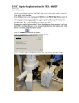
JEOL JSM-6060LV SCANNING ELECTRON MICROSCOPE
Insert Nickname Here
Operating
Instructions
J E O L
J S M - 6 0 6 0 L V
S C A N N I N G
E L E C T R O N
M I C R O S C O P E
Table of Contents
1
Safety
2
5
INTRODUCTION
3
BACKG ROUND
Background Information
4
References
4
3
SAMPL ES
Sample Holders
7
Sample Preparation
7
4
OPERA TION
Instrument Startup
8
Sample Loading
8
Getting an Image
9
Moving Around
9
Image Scanning
10
Zooming In (or Out)
10
Other Toolbar Buttons
10
System Shutdown
10
1
PHOTO GRAPHY
Image Options
11
Lower Resolution (but quick) Photos
11
Higher Resolution (slower) Photos
12
J E O L
J S M - 6 0 6 0 L V
S C A N N I N G
E L E C T R O N
Introduction
M I C R O S C O P E
1
T
he JEOL JSM-6060LV is a state-of-the-art scanning electron microscope that features
a low vacuum for observation of non-conductive specimens, a fully automated electron
gun, a backscattered electron detector for atomic number contrast imaging, fully
integrated digital control, motorized x-y stage, and a NORAN System 6 elemental
analysis system (see separate operating instructions for the NORAN system). Best of all, the
JEOL JSM-6060LV scanning electron microscope is user-friendly and easy to operate.
Safety
The scanning electron microscope is a relatively safe instrument.. You can do much more
damage to it than it can do to you. When the electron beam is turned on, some x-rays are
produced as a result of electron beam interaction with the sample, but these x-rays are of relatively
low energy and do not escape the sample chamber. The instrument also produces some radio
frequency energy.
2
J E O L
J S M - 6 0 6 0 L V
S C A N N I N G
E L E C T R O N
Background
M I C R O S C O P E
2
The scanning electron microscope (SEM) is one of the most versatile instruments for the
examination and analysis of the microstructural characteristics of solids. Although the SEM and
optical microscope share the same primary function – making microstructural features and objects
visible to the human eye – the scanning electron microscope offers some distinct advantages over
the optical microscope. The SEM uses electrons rather than visible light waves (200 – 750 nm
wavelength) for imaging, which allows for observation of relatively large sample features at low
magnifications or very fine details (high resolution) at high magnifications. The SEM also offers a
large depth of field that provides good focus over rough specimen surfaces. The large depth of
focus provides a three-dimensional appearance of the specimen in a SEM compared to the nearly
planar or two-dimensional imaging found in optical microscopes. In addition, many attachments
are available for scanning electron microscopes, including x-ray spectrometers for chemical
composition analysis, backscattered electron detectors for atomic number contrast, transmitted
electron detectors, hot and cold stages for microscopic observation of high or low temperature
phenomena, tensile testing stages for observation of deformation and fracture, and special stages
for analysis of semiconductor devices.
Disadvantages of the
scanning electron
microscope include
relatively high initial,
operational, and
maintenance costs, a high
vacuum operating
atmosphere that is
unsuitable for some
specimens, and difficulty in
preparing certain types of
specimens. Figure 1
schematically illustrates
image formation in the
optical microscope and the
scanning electron
microscope.
Figure 1. Basic image formation in an optical microscope and a
scanning electron microscope.
3
J E O L
J S M - 6 0 6 0 L V
S C A N N I N G
E L E C T R O N
M I C R O S C O P E
The basic components of a SEM are the vacuum system, electron gun, lens system, electron
detector, imaging system, and the electronics associated with these components. The vacuum
system is necessary to minimize the interference of air particles with the electron beam and to
prevent rapid oxidation of the tungsten filament. The vacuum system consists of roughing
pumps, an oil diffusion pump, and various vacuum fittings, valves and seals that provide a
working pressure in the SEM of 10-6 to 10-4 Torr (10-4 to 10-2 Pa). Our SEM has a vacuum
system that may be switched to low vacuum mode (close to atmospheric pressure). The electron
gun in our JEOL SEM contains a tungsten filament that is heated with a filament power supply
and maintained at a high negative voltage (typically 10-20 kV) during operation. When the
tungsten filament is heated, electrons are emitted from the tip and accelerated to ground by the
10-20 kV potential between the filament
and the anode. Figure 2 shows the typical
configuration of an electron gun in a
SEM. After electrons are emitted from
the gun and accelerated down the SEM
column in an electron beam, they are
controlled and directed to the specimen
by a series of electromagnetic lenses and
apertures.
When the electrons in the electron beam
hit the specimen, a number of electronspecimen interactions may occur. Some
of these interactions include elastic
scattering of electrons, secondary electron
emission (emission of loosely bound
electrons of the conduction band),
ionization of inner shell electrons
Figure 2. Schematic illustration of SEM electron
(produces x-rays and Auger electrons),
and excitation of phonons (causes heating of the specimen). If a sample is thin enough, some
electrons will be transmitted all the way through the sample. Different materials and sample
geometries will produce different amounts or different types of secondary electrons, backscattered
electrons, Auger electrons, transmitted electrons, and x-rays, and all of these interactions may be
used for imaging or analysis of the sample. The most common type of imaging in a SEM is
secondary electron imaging (SEI), which involves the use of a secondary electron detector. The
secondary electron detector collects secondary electrons and some backscattered electrons that are
emitted from the specimen surface, amplifies the detected signal, and converts the electron signal
into a video signal that is sent to the monitor.
References
•
•
JEOL JSM-35CF Scanning Microscope Instruction Manual
J. I. Goldstein, et al., Scanning Electron Microscopy and X-Ray Microanalysis, Plenum Press,
New York (1981).
4
J E O L
J S M - 6 0 6 0 L V
S C A N N I N G
E L E C T R O N
M I C R O S C O P E
Samples
T
3
he JEOL JSM-6060LV in standard high vacuum operating mode can handle a variety
of sample types. The low vacuum mode enables the instrument to handle nonconductive samples, including organic materials (polymers, etc.), ceramics and glasses,
and biological samples.
Sample Holders
Please use clean, powder-free gloves when handling the sample holders and loading your samples
into the instrument.
Two different sample holders are available. The smaller sample holder is designed to take 10 mm
diameter x 10 mm tall cylindrical sample mounts or other small specimens that fit into the same
volume. We have a large number of aluminum cylindrical sample mounts for use with the smaller
sample holder. The larger sample holder is approximately 32 mm in diameter x 10 mm tall, and is
designed for creative attachment of larger specimens. It’s best if you can fit your samples onto
one of the standard sample holders, but if not, other arrangements may be made (talk to
Professor Stolk).
Other types of sample holders are available. See Professor Stolk for information on ordering
sample holders.
Sample Preparation – Conductive Materials
1. Small samples that fit securely onto one of the standard sample holders are best. Use a
precision saw to section larger materials to an appropriate size, if possible.
2. Remove excess lint, dust, moisture, and other debris from your specimen, unless you want to
examine those things.
3. Use conductive carbon tape, carbon tabs, carbon paint, or copper tape to attach your sample
to a sample holder or cylindrical sample mount. If you use carbon paint, please wait for the
paint to dry (or use a heat gun or hair dryer) before you load the sample into the instrument.
4. A sample height approximately even with the top surface of the sample holder is
recommended. With tall samples, you risk contacting one of the detectors or other sensitive
interior components and causing major problems (and repair bills!).
Sample Preparation – Non-conductive Materials
1. Small samples that fit securely onto one of the standard sample holders are best. Use a
precision saw to section larger materials to an appropriate size, if possible.
5
J E O L
J S M - 6 0 6 0 L V
S C A N N I N G
E L E C T R O N
M I C R O S C O P E
2. Remove excess lint, dust, moisture, and other debris from your specimen, unless you want to
examine those things.
3. Use the sputter coater to create a conductive gold coating on the surface of your sample. See
separate operating instructions for the sputter coater.
4. After coating your sample, use conductive carbon tape, carbon tabs, carbon paint, or copper
tape to attach your sample to a sample holder or cylindrical sample mount. If you use carbon
paint, please wait for the paint to dry (or use a heat gun or hair dryer) before you load the
sample into the instrument.
5. A sample height approximately even with the top surface of the sample holder is
recommended. With tall samples, you risk contacting one of the detectors or other sensitive
interior components and causing major problems (and repair bills!).
6
J E O L
J S M - 6 0 6 0 L V
S C A N N I N G
E L E C T R O N
Instrument Operation
T
M I C R O S C O P E
4
his section will walk you through the basic operation of the scanning electron
microscope. You will follow a series of tasks that includes
• Powering up the instrument,
• Loading your sample,
• Evacuating the chamber,
• Turning on the electron gun, and
• Gazing at some wonderful high magnification images.
Instrument Startup
1. If the instrument is already turned on, skip to the next section on Sample Loading. If the
instrument is off, continue to Step 2.
2. Turn on the water chiller. The on/off switch is on the front panel. The temperature should
read between 65 and 69 degrees F.
3. Turn on the SEM by turning and releasing the key on the front panel of the instrument (it’s
just like starting a car).
4. Turn on the Dell computer on the left and hit the ENTER key at the log on screen (no
password necessary).
Sample Loading
1. If the chamber is evacuated (EVAC button on the front of the instrument is lit green), press
2.
3.
4.
5.
and hold the VENT button until it lights up yellow. This will vent the chamber.
Wait for the VENT button to stop flashing (about 40 seconds).
Open the chamber by grabbing the door assembly at the side depressions and carefully sliding
the door assembly outwards. You should be able to see the brass, circular platform of the
sample stage when you slide open the door.
Slide your sample holder with sample attached onto the copper-colored dovetail on the stage.
CAUTION: Adjust the sample height so that the top of the sample is well below the
position of the interior chamber components (detectors, lenses, etc.). Use the z-axis control
(the adjustment on the door assembly with the knob that is pointing straight up and the
micrometer with horizontal divisions) for height adjustments. Steer clear of anything inside
the chamber.
Bumping interior components can cause thousands of dollars in damage to the
instrument. Know your sample height, and know approximately how much clearance you
have around your sample, particularly along the z-axis.
7
J E O L
J S M - 6 0 6 0 L V
S C A N N I N G
E L E C T R O N
M I C R O S C O P E
6. Close the chamber door by gently pushing it forward until it stops.
7. Press and hold the EVAC button (on the front of the instrument) until it lights up green.
8. WAIT for evacuation of the chamber (about 1 minute). While you’re waiting, go to the next
section – Software.
Getting an Image
1. Double click on the SEM Main Menu icon to open the SEM software.
IMPORTANT NOTE: Nearly all of the basic SEM controls may be accessed by clicking on
the standard toolbars buttons and the standard display items (buttons above the image, numbers
at the bottom of the window, etc.).
2. When the chamber is properly evacuated, the HT button at the top left will be blue and read
3.
4.
5.
6.
7.
8.
Ready. If the chamber is vented or still evacuating, the HT button will be grayed out and
read Wait.
Click on the blue HT-Ready button to turn on the electron beam. The button should turn
green and read HT-ON.
Adjust magnification to the lowest possible value (usually between 10X and 20X).
Click on the blue AF-Focus button on the toolbar or the AUTO FOCUS button on the
keypad. This should bring your specimen into focus. If it doesn’t, try turning on the
COARSE button and adjusting the focus knob on the keypad.
Click on the blue ACB to automatically adjust the contrast and brightness.
Click on the blue AS-Stigma button for automatic astigmatism correction.
You should have a focused image at this point. If so, congratulations…you’re an electron
microscopist. If not, call your instructor or teaching assistant for help.
IMPORTANT SETTINGS
o Accelerating Voltage. Check the accelerating voltage value shown at the bottom of the
window (Acc. Volt). If you’re happy with your voltage, then continue being happy. If you’d
like to change the accelerating voltage, click once on the voltage number, then double click on
the desired voltage. A setting of 15 kV works well for most specimens, but feel free
experimenting with other accelerating voltages.
o Spot Size. A spot size between 40 and 60 works well for most specimens, but feel free
experimenting with the spot size value. You’ll need to adjust the focus, contrast, and
brightness after you change the spot size value.
Moving Around
1. The motorized x and y axes are amazing! If you want to move your specimen, simply double-
click on the area of interest to re-center the image on this spot, or click and drag the image to
move to a spot of interest. It’s kind of like Mapquest.
2. Rotation. Turn the small knob at the bottom of the chamber door assembly (between the x
and y axis knobs) to rotate the stage.
8
J E O L
J S M - 6 0 6 0 L V
S C A N N I N G
E L E C T R O N
M I C R O S C O P E
3. Tilt. Turn the knob at the lower right side of the chamber door assembly to tilt your
specimen. BE CAREFUL when adjusting the sample tilt – you don’t want to run your
sample into anything inside the column.
4. Height. Sample height may be changed by manually adjusting the y-axis position knob at the
top of the door assembly. BE CAREFUL when adjusting the sample height – you don’t
want to run your sample into anything inside the column.
5. The FINE SHIFT joystick on the keypad is great for small position adjustments when
you’re working at high magnification.
6. To view your location relative to the entire sample, click on the blue and white Stage button
toward the left end of the toolbar. This opens a schematic image of the sample holder with
your location marked. If the sample holder you’re using doesn’t appear, select the correct
holder under the Holder menu.
Image Scanning
•
Green Toolbar Buttons. The image scan rate may be changed to a number of different
settings using the green buttons on the toolbar. Play with the scan rate buttons to see what
they do. Scan 2 is a TV-like mode. Scan 3 is a slower scan with higher resolution.
Zooming In (or Out)
•
To change magnification, use the MAGNIFICATION knob on the keypad or the blue
Mag- and Mag+ buttons on the toolbar.
Other Toolbar Buttons
•
•
•
The Gun button on toolbar will display the electron gun settings. The automatic settings
usually work great, so don’t mess with these unless you know what you’re doing.
The Recipe button on the toolbar automatically adjusts the instrument settings according to
preset values that work well with various materials. If you’re not a creative e-beam cook, try
these recipes, if you like.
The Sample button on the toolbar provides control of the chamber VENT and EVAC
(same as the buttons on the front of the instrument).
System Shutdown
1.
2.
3.
4.
5.
Click on the HT-On toolbar button to turn off the electron gun.
Click on the “EXIT” toolbar button to exit the software.
Vent the chamber.
Remove your specimen.
Close the chamber door and evacuate the chamber. Always leave the chamber under vacuum
to prevent dust and debris from collecting on the stage and detectors.
6. Please leave the entire system (instrument, computer, and water chiller) running. Talk to
Professor Jon Stolk or Professor Debbie Chachra if you need to or would like to shut down
the system.
9
J E O L
J S M - 6 0 6 0 L V
S C A N N I N G
E L E C T R O N
M I C R O S C O P E
Photography
A
5
cquiring an image from the SEM is relatively easy, but there are quite a few options
for live image scanning and corresponding image resolution. This section outlines
several options for acquiring digital images of different resolutions, and it provides a
few general tips for good SEM photos.
Image Options
The most commonly used options for digital images are outlined in the following table.
Scanning
Mode
Scan3
Scan3
Scan3
Scan3
Scan4
Scan4
•
•
•
•
Scan Speed,
seconds
10 (default)
10 (default)
20
20
80 (default)
80 (default)
Image
Resolution
640x480
640x480
1280x960
1280x960
1280x960
1280x960
Image
Format
jpg
tif
jpg
tif
jpg
tif
File Size
100k
300k
200k
1,200k
200k
1,200k
The Scanning Mode is adjusted via the green toolbar buttons. Scan3 and Scan4 are the
only modes recommended for photography. Images taken on the Scan4 mode are less grainy
than those acquired in Scan3 mode.
The Scan Speed is adjusted in the Scan…Scan Speed section of the Setup…Fundamental
Setup pull down menu. The default scan time settings are 10s (640x480) for Scan3 and 80s
(1280x960) for Scan4.
Image Format is selected in the pull down box after the image is frozen and the Save button
is pressed.
Image Size is a function of both scan speed and image format. JPG images are relatively
small, and TIF images are relatively large. JPG images generally work fine for most users.
Lower Resolution (but quick) Photos
1. Press the green Scan2 button on the toolbar.
2. Adjust the focus, contrast, and brightness (or use the automatic features). It is good practice
to focus the image at a higher magnification, then back down to the desired photo
magnification.
3. Freeze the image by pressing the green Scan3 toolbar button followed by the green Freeze
toolbar button.
10
J E O L
J S M - 6 0 6 0 L V
S C A N N I N G
E L E C T R O N
M I C R O S C O P E
4. Once the image is frozen, click the Save button above the image, select a save directory, type
a filename, select a file format, and click Save.
Higher Resolution (slower) Photos
1. Press the green Scan2 button on the toolbar.
2. Carefully adjust the focus and stigmation settings at a higher magnification. It is good practice
to focus the image at a higher magnification, then back down to the desired photo
magnification
3. Adjust the contrast, brightness (or use the automatic features).
4. Freeze the image by pressing the green Scan4 toolbar button. After about 80 seconds of high
resolution image acquisition, the image will freeze.
5. click the Save button above the image, select a save directory, type a filename, select a file
format, and click Save.
11