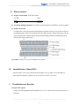
1
Non-radioactive EMSA-STAT3 Kit Non-Radioactive EMSA-STAT3 Kit User’s Manual VER. 3.11 This user’s manual is suitable for the production of TFDET003, Related Products Catalog Number Description Detections TFDET001 Non-radioactive NFκB EMSA 50X TFDET002 Non-radioactive STAT 1 EMSA 50X TFDET003 Non-radioactive STAT 3 EMSA 50X TFDET004 Non-radioactive STAT 5 EMSA 50X TFDET005 Non-radioactive AP-1 EMSA 50X TFDET006 Non-radioactive HIF-1 EMSA 50X Viagene Biotech Inc. 3802 Spectrum Blvd., Suite 128B Tampa, Florida 33612 ◙ Tel: (813) 902-2209 ◙ Fax: (813) 903-0649, ◙ E-mail: [email protected] For technical support: Tel: (813) 902.2209/205.9601 ◙ E-mail: [email protected] -1- Non-radioactive EMSA-STAT3 Kit TABLE OF CONTENTS A. Introduction ………………………..………………. 3 B. Kit components …………………..………………… 4 C. Additional materials required ……..……………….. 4 D. Binding reaction …………………………………… 5 E. Gel preparation …………………………………….. 6 F. Electrophoresis ……………………………………. 6 G. Transfer …………………………………………… 7 H. Immobilization of bound DNA …………………. 7 I. Chemiluminescent reaction ………………………. 7 J. Chemiluminescent imaging ………………………. 9 K. Troubleshooting …………………………………… 9 L. References …………………………………………. 10 M. Notes ………………………………………………. 11 N. Probe Sequences ………………………………….. 11 Viagene EMSA kits are intended for research purpose only. 2007© Viagene Biotech, Inc. All rights reserved. For technical support: Tel: (813) 902.2209/205.9601 ◙ E-mail: [email protected] -2- Non-radioactive EMSA-STAT3 Kit A. Introduction The Electrophoretic Mobility Shift Assay (EMSA) is a powerful tool for evaluating DNA-protein or RNA-protein interactions, which often referred to as gel shift or gel retardation. The assay is based on the principle that when subjected to electrophoresis, free DNA will migrate differently from a DNA/protein or RNA/protein complexes. The activated transcription factors that bind with DNA or RNA in nucleus can be detected with EMSA. Viagene’s non-radioactive EMSA-STAT3 kit is based on high sensitivity of the chemiluminescent technology. It is different from the one on the market in the design of the probe and optimizing procedure. Comparing with radioactive detection of STAT3 activation, this non-radioactive EMSA Kit offers the sensitivity and speed of radioactive assays without the hazards, waste and probe instability problems. We pre-tested this kits intensively and set up the optimum condition for detecting STAT3 activationeach. This kit is ready to use and guaranteed to work. The protocol is very easy to use. This kit comes with all necessary components for performing 50 DNA/STAT3 binding reactions and assays. If one runs 11 samples and 1 positive & 1 negative controls in a gel, all 4 binding-membranes would be used out, but, if one runs 13 samples and 1 positive & 1 negative controls, only 3 binding-membranes would be used. For a mini gel (e.g. BioRad), 15 samples can be loaded into one gel. Viagene EMSA kits are intended for research purpose only. For technical support: Tel: (813) 902.2209/205.9601 ◙ E-mail: [email protected] -3- Non-radioactive EMSA-STAT3 Kit B. Kit components (stored as indicated on labels) (1) The EMSA standard Kits include the follows: o n 10X Binding Buffer (4 C) o o Poly (dI: dC) (-20 C) o p Biotin-Labeled Probe (-20 C) o q Cold Oligonucleotide (-20 C) o r Streptavidin-HRP (4 C) o s 6X Loading Buffer (4 C) o 2X Blocking Buffer (4 C) o 5X Washing Buffer (4 C) o 2X Equilibration Solution (4 C) Binding Membrane (R/T) User’s Manual o Lightgen®-CL, Buffer A (4 C) o Lightgen®-CL, Buffer B (4 C) 1 Vial 1 Vial 1 Vial 1 Vial 1 Vial 1 Vial 1 Bottle 1 Bottle 1 Bottle 4 Sheets 1 Set 1 Bottle 1 Bottle (2) EMSA-Controls:(Not included in standard EMSA) o Positive Nuclear extracts (-80 C) o Negative Nuclear extracts (-80 C) 1 Vial* 1 Vial* * Since many customers have already had positive and/or negative controls, the standard kits from Viagene DO NOT include nuclear extracts (controls). However, there are 6 positive and 6 negative controls for the kits of NF-ΚB, STAT 1, STAT3, STAT5, AP-1 and HIF-1, respectively, which can be purchased separately from the kit. These controls will be shipped on dry ice. C. Additional materials required y Mini-polyacrylamide gel electrophoresis apparatus and related chemicals and buffers. y Electrophoretic transfer apparatus and buffers. y X-ray films or a CCD imager (e.g. Viagene Cool Imager™ Catalog # IMGR003). y Sample storage apparatus such as refrigerators and ultra-low freezers. y Orbital Shaker, vials and tubes y UV Crosslink apparatus or vacuum oven For technical support: Tel: (813) 902.2209/205.9601 ◙ E-mail: [email protected] -4- Non-radioactive EMSA-STAT3 Kit D. Binding Reaction (1) Binding Reaction: 10X binding buffer n 1.5 µl Poly (dI: dC) o 1.0 µl Nuclear extracts* X µl dH2O X µl Total 15.0 µl Mix well and let it sit at room temperature (R/T) for 20 minutes. Biotin-probe p 0.5 µl Total 15.5 µl Allow mixture to react at R/T for at least 20 minutes. * The total 1-4 µg of nuclear proteins are required for non-radioactive EMSA, and the protein concentration of the nuclear extracts should not be less than 1 µg/µl for best results. (2) Competing Reaction: 10X binding buffern Poly (dI: dC) o Nuclear extracts* 1.5 µl 1.0 µl X µl 3.0 µl Cold oligonucleotides q X µl dH2O Mix well and let it sit for 15 minutes at R/T Biotin-probe p Total 0.5 µl 15.0 µl Allow mixture to react for at least 20 minutes at R/T * Each reaction needs 2-5 µg nuclear proteins; therefore, the volume of nuclear extracts and dH2O should be adjusted to meet the final volume of 15µl. For technical support: Tel: (813) 902.2209/205.9601 ◙ E-mail: [email protected] -5- Non-radioactive EMSA-STAT3 Kit E. Gel preparation (1) Prepare and make 6.5% mini Gels: 10X TBE 40% Acrylamide/Bis 50% Glycerol dH2O TEMED 10% AP Total 1.0 ml 3.3 ml 1.0 ml 14.8 ml 20 µl 300 µl 20.0 ml 20 ml is enough to make 2 mini gels (90 X 70 X 1.5 mm) (2) Prepare cooled 0.5X TBE: 10X TBE dH2O Total 60 ml 1140 ml 1200 ml (3) Pre-running : Pre-run the gel for at least 30 min at 120V in cooled 0.5X TBE on ice. Then flush each well with 0.5% TBE before loading samples. F. Electrophoresis: (1) Prepare samples: Binding reaction from step 4 15.0 µl 6X loading buffer s Total 3.0 µl 18.0 µl Mix well and keep at R/T for 5 minutes. Then centrifuge for 3 minute at 140000 rpm. (2) Sample loading: Load all the supernatant (all 18.0 µl) into each well. (3) Electrophoresis: Run the gel on ice at 180V for 80 minutes. For technical support: Tel: (813) 902.2209/205.9601 ◙ E-mail: [email protected] -6- Non-radioactive EMSA-STAT3 Kit G. Electro-transfer: (1) Prepare transfer buffer: 0.5X TBE 1200 ml. 10× TBE ddH2O Total 60 ml 1140 ml 1200 ml (2) Presoak the binding membrane: Pre-soak the nylon membrane in 0.5X TBE for at least 10 minutes. (3) Prepare electro-blot: Carefully remove one glass from the gel and mark the orientation of the gel. Cover with one sheet of dry Whatman paper (3mm). The gel should stick onto Whatman paper. Gently lift the paper with gel away from glass plate. Sandwich the gel with presoaked binding-membrane and Whatman papers following the picture shown below. (4) Transfer: Transfer in 0.5× TBE at 500mA for 30 minutes. H. Immobilization of bound DNA After the transfer, remove the nylon-membrane and place it in a UV linker to cross link DNA by o following the manufacturer’s guidance or in a vacuum oven at 80 C for 2 hours. I. Chemiluminescent Reaction: (1) Prepare the reagent: Gently warm Blocking Buffer and Wash Buffer in a 37-50°C water bath until all the precipitates are dissolved. For technical support: Tel: (813) 902.2209/205.9601 ◙ E-mail: [email protected] -7- Non-radioactive EMSA-STAT3 Kit (2) Block the Binding-membrane: 1) Prepare 1 X blocking buffer: 2) 2 X Blocking buffer 6 ml dH2O 6 ml Total 12 ml Block the membrane with 12 ml of 1X blocking buffer for 15 minutes on a shaker at R/T. (3) Streptavidin-HRP binding reaction: Add 10 μl Streptavidin-HRPr solutions into the blocking buffer after step (2) treatment at R/T for 30 minutes on a shaker. (4) Wash the membrane: 1) Prepare 1 x washing solution: Wash buffer (stocking) 8 ml dH2O 32 ml Total 40 ml 2) Wash the membrane(s) 4 times with 10 ml of 1X washing solution, 5 minutes for each wash. (5) Equilibrate the membrane: 1) Prepare 1 X Equilibration buffer: 2 X Equilibration buffer dH2O Total 6 ml 6 ml 12 ml 2) After washing, equilibrate the membrane with 12 ml of 1X Equilibration buffer at R/T for 5 minutes on shaker. (6) Chemiluminescent detection: 1) Prepare the Chemiluminescent substrate solution: Solution A Solution B Total 1.0 ml 1.0 ml 2.0 ml 2.0 ml is enough to cover a mini-gel membrane (9 x 7 cm). 2) Add 2.0 ml of chemiluminescent substrates onto the membrane. 3) Depending on the detection technique, proceed with one of the following procedures. For technical support: Tel: (813) 902.2209/205.9601 ◙ E-mail: [email protected] -8- Non-radioactive EMSA-STAT3 Kit J. Chemiluminescent imaging: (1) Detecting with Chemiluminescence Imager: An imager for capturing chemiluminescent picture should have very high The system of Chemiluminescent-imager is high sensitivity to pick up weak signals and has the capacity to compute thousands of pictures in minutes. Cool Imager™ by Viagene (Catalog IMGR002) is well designed for detecting chemiluminescent images of Western immunoblotting, DNA/RNA hybridization or non-radioactive EMSA. In the manipulation of the Cool Imager™, you do not need to remove the chemiluminescent substrate from the membrane for imaging. The pictures will appear on the screen in 3-5 minutes. Please refer to the operation manual of Cool Imager™ for chemiluminescent imaging. (2) Detecting with exposure and develop X-ray film: When using X-ray film to capture a chemiluminescent image of blots, one must have a darkroom, film developer, filming chemicals and X-film, etc. After the chemiluminescent reaction (usually 1-2 minutes), discard the chemiluminescent substrate and wrap the blots with a piece of transparent paper or membrane. Place a sheet of X-ray film on the surface of the transparent paper. Exposure time may vary depending on the strength of chemiluminescent signaling, and several X-ray films with different exposure time may need to attain the desired pictures. Develop the films according to the manufacturer’s instructions of the X-ray film developer. K. Troubleshooting: Problem Cause Recommendation Weak or no imaging Poor transfer. Use electroblotting for best results. Exposure time is too short. Increase time of exposure Not enough biotin probe is used. Increase probe used The image is on the other side of the blot. Revert the blot and try again The Extract degraded Try using protease inhibitors For technical support: Tel: (813) 902.2209/205.9601 ◙ E-mail: [email protected] -9- Non-radioactive EMSA-STAT3 Kit High background Membrane is dry. Particulates in blocking or wash buffer Keep membrane moist during detection. Gently warm until no particulate remain. Exposure time is too long Shorten exposure time No shift observed. The image is too dark. Not enough extract. Use more extract. Quality of extract is not good or the extract degraded Try using protease inhibitors or high quality extract. Over-exposed Shorten the exposure time. The chemiluminescent is too strong. Dilute the chemiluminescent substrate for 1-5 multiple. Too much biotin probe is used. Use higher dilution of biotin-probe L. References: 1. Crothers, D.M. (1998) Nature 325:464-5. 2. Garner, M.M. & Revzin, A. (1986) Trends in Biochemical Sciences 11:395-6. 3. Hendrcikson, W. (1985) Bio Techniques 3:198 –207. 4. Dignam, J.D., Lebovitz, R.M., and Roeder, R.G. (1983) Nucleic Acids Research 11:1475 –1489. 5. Bannister, A. and Kouzarides, T. (1992). Basic peptides enhance protein-DNA interaction in vitro. Nucl. Acids Res. 20:3523. 6. Kironmai, K.M., et al. (1998). DNA-binding activities of Hop1 protein, a synaptonemal complex component from Saccharomyces cerevisiae. Mol. Cell Biol. 18:1424-35. 7. Liu R.Y., Fan C., Garcia R., Jove R., Zuckerman K.S. (1999). Constitutive activation of the JAK2/STAT5 signal transduction pathway correlates with growth factor independence of megakaryocytic leukemic cell lines. Blood. 93:2369-79. 8. Liu R.Y., Fan C., Olashaw N.E., Wang X., Zuckerman K.S. (1999). Tumor necrosis factor-alpha-induced proliferation of human Mo7e leukemic cells occurs via activation of nuclear factor kappaB transcription factor. J Biol Chem. 274:13877-85 For technical support: Tel: (813) 902.2209/205.9601 ◙ E-mail: [email protected] - 10 - Non-radioactive EMSA-STAT3 Kit M. Note: 1. Upon receipt, check the package and the kit components immediately. If problems arise, please contact Viagene within 24 hours. 2. Before opening vials, spin down the components in the vials. 3. The kit can be stored for 12 months at the condition indicated on the labels. 4. When kits are stored at low temperature, white precipitates may be observed. Warm up the bottles in a water bath to dissolve the precipitate before use. 5. Follow this instruction strictly to obtain the best result. 6. Follow the laboratory regulation when handling Acrylamide/Bis solution. N. Probe Sequences: The core sequence of the probes used in Viagene’s EMSA kits. Probes Sequence NF-kB 5'-AGT TGA GGG GAC TTT CCC AGG C STAT1 5’-CAT GTT ATG CAT ATT CCT GTA AGT G STAT3 5’-GAT CCT TCT GGG AAT TCC TAG ATC STAT5 5’-AGA TTT CTA GGA ATT CAA TCC AP1 HIF-1 5’-CGC TTG ATG ACT CAG CCG GAA 5'-TCT GTA CGT GAC CAC ACT CAC CTC-3' For technical support: Tel: (813) 902.2209/205.9601 ◙ E-mail: [email protected] - 11 -