1
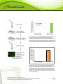
EPIGENTEK Complete Solutions for Epigenetics EpiQuik™ MeDIP Ultra Kit Base Catalog # P-1052 PLEASE READ THIS ENTIRE USER GUIDE BEFORE USE Uses: The EpiQuik™ MeDIP Ultra Kit is suitable for selective enrichment of DNA fragments containing 5-methylcytosine. The highly sensitive and specific format can use DNA isolated from various species. The methylated DNA that is enriched with this kit can be used for various downstream applications including qualitative and quantitative PCR (MeDIP-PCR), microarray (MeDIP-chip) and especially sequencing (MeDIP-seq). Starting Material and Input Amount of DNA: The starting material should be good quality purified DNA. The amount of DNA for each reaction can be 50 ng (approximately 10,000 cells) to 500 ng. For an optimal reaction, the input DNA amount should be 100-200 ng per well. DNA Shearing: Genomic DNA should be sheared by sonication before starting MeDIP. The sheared DNA fragments should range in size from 100-600 base pairs. For the best results and your convenience, we recommend using the EpiSonic™ Multi-Functional Bioprocessor 1100, which allows for simultaneous sonication of multiple samples in sealed vials to achieve consistent and desired DNA size range (100-600 base pairs). Internal Control: Negative control (non-immune IgG) and specificity are provided in this kit. The specificity controls are 200 base pair DNA fragments containing 44 cytosine residues which are methylated (mDNA) or unmethylated (unDNA). The kit also includes control PCR primers that can be used for verifying the enrichment efficiency and specificity of control DNA. Antibody: The 5-methylcytosine antibody used in this kit is highly specific against methylated DNA fragments, both single and double stranded, and is not cross-reactive to hydroxymethylated and unmethylated DNA fragments. This antibody can capture >50% of DNA fragments containing as few as two 5-mCs and enriches all DNA fragments containing four or more 5-mCs. Methylated DNA yield: The yielded methylated DNA is about 4 ng for 100 ng input DNA (4%), which is consistent with the expected percentage (4-5%) at which the highest sensitivity and specificity for enriched methylated DNA has been demonstrated by bisulfite sequencing. Precautions: To avoid cross-contamination, carefully pipette the sample or solution into the strip wells. Use aerosol-barrier pipette tips and always change pipette tips between liquid transfers. Wear gloves throughout the entire procedure. In case of contact between gloves and sample, change gloves immediately. 110 Bi County Blvd. Ste. 122, Farmingdale, NY 11735 Tel: 1-877-374-4368 ■ Fax: 1-718-484-3956 ■ E-mail: [email protected] ■ Web: www.epigentek.com © Epigentek Group Inc. All rights reserved. Products are for research use only. Page 1 Printed 2014-03-03 P-1052 EPIGENTEK Complete Solutions for Epigenetics KIT CONTENTS Component 24 Reactions Cat. #P-1052-24 48 Reactions Cat. #P-1052-48 Storage Upon Receipt WB (Wash Buffer) 15 ml 30 ml 4°C MeDIP Buffer 4 ml 8 ml RT DRB (DNA Release Buffer) 14 ml 28 ml RT DBS (DNA Binding Solution) 7 ml 14 ml RT BS (Blocker Solution)* 200 µl 400 µl 4°C DEB (DNA Elution Buffer) 1 ml 2 ml RT Non-Immune IgG * 10 µl 20 µl 4°C 5-mC Antibody * 25 µl 50 µl 4°C Proteinase K (10 mg/ml)* 28 µl 56 µl 4°C Control unDNA (200 ng/ml)* 5 µl 10 µl -20°C Control mDNA (200 ng/ml)* 5 µl 10 µl -20°C Control Primer-Forward (20 µM)* 5 µl 10 µl 4°C Control Primer-Reverse (20 µM)* 5 µl 10 µl 4°C Spin Column 30 50 RT Collection Tube 30 50 RT 8-Well Assay Strips (With Frame) 3 strips 6 strips 4°C 8-Well Strip Caps 3 strips 6 strips RT Adhesive 8-Well Strip Film 3 6 RT User Guide 1 1 RT * Spin the solution down to the bottom prior to use. SHIPPING & STORAGE The kit is shipped in two parts: the first part at ambient room temperature and the second part on frozen ice packs at 4°C. Upon receipt: (1) Store Control unDNA and Control mDNA at -20°C. Store WB, BS, Non-Immune IgG, 5-mC Antibody, Proteinase K, Control Primer-Forward, Control Primer-Reverse, and 8-Well Assay Strips at 4°C away from light; (2) Store remaining components at room temperature away from light. All components of the kit are stable for 6 months from the date of shipment, when stored properly. Note: Check if WB (Wash Buffer) contains salt precipitates before use. If so, briefly warm at room temperature or 37°C and shake the buffer until salts are re-dissolved. 110 Bi County Blvd. Ste. 122, Farmingdale, NY 11735 Tel: 1-877-374-4368 ■ Fax: 1-718-484-3956 ■ E-mail: [email protected] ■ Web: www.epigentek.com © Epigentek Group Inc. All rights reserved. Products are for research use only. Page 2 Printed 2014-03-03 P-1052 EPIGENTEK Complete Solutions for Epigenetics MATERIALS REQUIRED BUT NOT SUPPLIED Variable temperature waterbath or incubator oven Thermalcycler with 48- or 96-well block Sonication device Orbital shaker Adjustable pipette and multiple-channel pipette Aerosol resistant pipette tips Parafilm M 0.2 ml or 0.5 ml PCR tubes 1X TE buffer, pH 8.0 90% Ethanol GENERAL PRODUCT INFORMATION Quality Control: Each lot of the EpiQuik™ MeDIP Ultra Kit is tested against predetermined specifications to ensure consistent product quality. Epigentek guarantees the performance of all products in the manner described in our product instructions. Product Warranty: If this product does not meet your expectations, simply contact our technical support unit or your regional distributor. We also encourage you to contact us if you have any suggestions about product performance or new applications and techniques. Safety: Suitable lab coat, disposable gloves, and proper eye protection are required when working with this product. Product Updates: Epigentek reserves the right to change or modify any product to enhance its performance and design. The information in this User Guide is subject to change at any time without notice. Thus, only use the User Guide that was supplied with the kit when using that kit. Usage Limitation: The EpiQuik™ MeDIP Ultra Kit is for research use only and is not intended for diagnostic or therapeutic applications. Intellectual Property: The EpiQuik™ MeDIP Ultra Kit and the method of processing contain proprietary technologies by Epigentek. A BRIEF OVERVIEW Core mechanisms for epigenetic alteration of genomic DNA are hypermethylation of CpG islands in specific genes and global DNA hypomethylation. Region-specific DNA methylation plays an important role in the repression of gene transcription and is mainly found in 5’-CpG-3’dinucleotides within promoters or in the first exon of genes. Global DNA hypomethylation is likely caused by methyl110 Bi County Blvd. Ste. 122, Farmingdale, NY 11735 Tel: 1-877-374-4368 ■ Fax: 1-718-484-3956 ■ E-mail: [email protected] ■ Web: www.epigentek.com © Epigentek Group Inc. All rights reserved. Products are for research use only. Page 3 Printed 2014-03-03 P-1052 EPIGENTEK Complete Solutions for Epigenetics deficiency due to a variety of environmental influences. It has been demonstrated that alterations in DNA methylation are associated with many diseases, especially cancer. Highly specific isolation of methylated DNA combined with next generation sequencing for genomewide methylation analysis should provide an advantage for convenient and comprehensive identification of methylation status of normal and diseased cells, such as cancer cells [1]. Such analysis requires the isolated methylated DNA to contain minimal background in order to achieve high specificity (>98%) for reliably identifying true methylated regions. The major method for enriching methylated DNA used for genome-wide methylation profiling is methylated DNA immunoprecipitation (MeDIP) [2]. However, currently used MeDIP methods, represented by most commercially available kits, have significant weaknesses including highly non-specific enrichment (amount of enriched DNA is >75% of the amount of input DNA) [3], time consuming, labor intensive, and has low throughput. Thus, for effectively and specifically capturing methylated DNA used for next generation sequencing analysis, an ideal MeDIP method requires maximum sensitivity with minimal background levels. Epigentek’s EpiQuik™ MeDIP Ultra Kit is designed to achieve these goals by maximizing sensitivity and minimizing non-specific background signals. The EpiQuik™ MeDIP Ultra Kit has the following advantages and features: Extremely fast and convenient protocol with a total procedure time (from input sample to ready-to-use methylated DNA) of less than 3 hours, which includes a minimal handling time of less than 20 minutes. Optimized buffers and protocol allow minimal background by overcoming the weaknesses that cause non-specific enrichment. A highly specific 5-mC monoclonal antibody included in the kit can strongly bind both single and double stranded DNA fragments containing 2 or more 5-mCs which enables highly sensitive enrichment of methylated DNA with >99% specificity. Flexible 96 strip-well microplate format makes the assay very easy to handle: manual method with one reaction at a time or high throughput method with 24-48 reactions at a time. Spin columns and collection tubes conveniently included for a DNA purification step. Low DNA input requirement as low as 50 ng (10,000 cells) per reaction. High reproducibility using pre-optimized MeDIP conditions. Compatible with various downstream analysis workflows including MeDIP-PCR and MeDIPchip, and specifically for MeDIP-Seq. References 1. Ruike Y et al: BMC Genomics, 11: 137, 2010. 2. Weber M et al: Nature Genetics, 37: 853-862, 2005. 3. Brebi-Mieville P et al: Epigenetics, 7: 106-112, 2012. PRINCIPLE & PROCEDURE The EpiQuik™ MeDIP Ultra Kit contains all reagents required for carrying out a successful MeDIP procedure using DNA isolated from mammalian cells or tissues. This kit includes a methylated DNA (mDNA) control and an unmethylated DNA (unDNA) control, a negative control non-immune IgG, and control primers that can be used with the control DNA to demonstrate the enrichment efficacy and specificity for methylated DNA. The positive control DNA containing 5-mC can be immunoprecipitated by the 5-mC antibody but not by the non-immune IgG. In this MeDIP, immunoprecipitation of 5-mCenriched DNA fragments is processed in a microplate under optimized reaction conditions, which enables MeDIP to be completed within 3 hours with high efficiency. Immunoprecipitated methylated DNA is then cleaned, released, and eluted. Eluted DNA can be used for various downstream applications including PCR (MeDIP-PCR) and microarray (MeDIP-chip), and is especially suitable for MeDIP-seq. 110 Bi County Blvd. Ste. 122, Farmingdale, NY 11735 Tel: 1-877-374-4368 ■ Fax: 1-718-484-3956 ■ E-mail: [email protected] ■ Web: www.epigentek.com © Epigentek Group Inc. All rights reserved. Products are for research use only. Page 4 Printed 2014-03-03 P-1052 EPIGENTEK Complete Solutions for Epigenetics Fig 1. Schematic procedure of the EpiQuik™ MeDIP Ultra Kit Relative Fold Enrichment Fig. 2. Selective enrichment of methylated DNA with the EpiQuik™ MeDIP Ultra Kit | 50 pg of unmethylated or methylated DNA control were each spiked into fragmented human genomic DNA (100 ng). MeDIP was processed with the included 5-mC monoclonal antibody and non-immune IgG included in the kit. Eluted DNA was analyzed by real time PCR with the control primers included in the kit to detect the presence of spiked control DNA. Fold-enrichment represents the amount of recovered control DNA and was calculated based on the real time PCR Ct value. 450 400 350 300 250 200 150 100 50 0 Non-immune IgG 5-mC Ab Fig. 3. Sensitive detection of gene-specific methylation by MeDIP-qPCR | Fully methylated HeLa DNA (500 ng) was fragmented to 100-500 bps using an EpiSonic™ 1100. The fragmented DNA was used for methylated DNA enrichment with the EpiQuik™ MeDIP Kit. Eluted DNA was analyzed by real time PCR with primers specific for MLH1 sequences in the promoter regions. Results show the specificity of the 5-mC antibody and low background for non-immune IgG. Foldenrichment represents the amount of recovered DNA and was calculated based on the real time PCR Ct value. 110 Bi County Blvd. Ste. 122, Farmingdale, NY 11735 Tel: 1-877-374-4368 ■ Fax: 1-718-484-3956 ■ E-mail: [email protected] ■ Web: www.epigentek.com © Epigentek Group Inc. All rights reserved. Products are for research use only. Page 5 Printed 2014-03-03 P-1052 EPIGENTEK Complete Solutions for Epigenetics PROTOCOL For the best results, please read the protocol in its entirety prior to starting your experiment. Starting Materials Input DNA Amount: DNA amount can range from 50 ng to 500 ng per reaction. An optimal amount is 100 ng per reaction. DNA Isolation: You can use your method of choice for DNA isolation. Epigentek offers a series of genomic DNA isolation kits for your convenience. DNA Storage: Isolated genomic DNA can be stored at 4°C (short term) or –20°C (long term) until use. 1. Shearing of Genomic DNA For the best results, DNA should be fragmented by a suitable sonication method: Waterbath Sonication: For ideal results, sonicate DNA with the EpiSonic™ Multi-Functional Bioprocessor 1100 (Epigentek, Cat. #EQC-1100). Use 30 µl of DNA solution (from 100 ng up to 3 µg) per 0.2 ml PCR tube or per PCR plate well. Shear for 60 duty cycles under cooling condition, 30 seconds ON, 30 seconds OFF, at 120130 target Watts. For more detailed information of use, please see the User Manual for the EpiSonic™ Multi-Functional Bioprocessor 1100. If using other waterbath-based sonicators, please follow the supplier’s instruction. Probe-based Sonication: Use 300 µl of DNA solution per 1.5 ml microcentrifuge tube. As an example, sonication can be carried out with a microtip attached to a Branson 450 sonifier, set to 25% power output. Sonicate 3-4 pulses of 10-15 seconds each, followed by 30-40 seconds rest on ice between each pulse. The conditions of DNA shearing should be optimized based on the sonication instrument. Note: When probe-based sonication is carried out, shearing effect may be reduced if foam is formed in the DNA sample solution. Under this condition, discontinue sonication and centrifuge the sample at 4˚C at 12,000 rpm for 3 min to remove the air bubbles. The isolated DNA can also be sheared with various enzyme-based methods. Optimization of the shearing conditions, such as enzyme concentration and incubation time, is needed in order to use enzyme-based methods. The DNA solution can now be used immediately or stored at –80°C after aliquoting appropriately until further use. Avoid multiple freeze/thaw cycles. Note: The size of sonicated DNA can be verified before starting the immunoprecipitation step. Use 10 µl (>500 ng) for DNA fragment size analysis along with a DNA marker on a 1-2% agarose gel; Stain with ethidium bromide or a suitable fluorescent dye for DNA and visualize it under ultraviolet light. The length of sheared DNA should be between 100-600 bps with peak size of 250 bps. 110 Bi County Blvd. Ste. 122, Farmingdale, NY 11735 Tel: 1-877-374-4368 ■ Fax: 1-718-484-3956 ■ E-mail: [email protected] ■ Web: www.epigentek.com © Epigentek Group Inc. All rights reserved. Products are for research use only. Page 6 Printed 2014-03-03 P-1052 EPIGENTEK Complete Solutions for Epigenetics 2. Preparation of MeDIP Reaction a. Dilute the Control mDNA and unDNA to 50 ng/ml (50 pg/µl) by adding 1 µl of Control mDNA or Control unDNA to 3 µl of MeDIP Buffer. Dilute your sample DNA with MeDIP Buffer to 10 µg/ml (10 ng/µl). Setup the MeDIP reactions by adding the appropriate reagents to each corresponding 0.5 ml vial according to the following chart: Reagent Sample vial Negative Control vial for Sample Control mDNA vial Control unDNA vial MeDIP Buffer 84 µl 84 µl 93 µl 93 µl Sample DNA (10 ng/µl) 10 µl 10 µl 0 0 Control DNA (50 pg/µl) 0 0 1 µl 1 µl BS (Blocker Solution) 5 µl 5 µl 5 µl 5 µl 5-mC Ab 1 µl 0 1 µl 1 Non-Immune IgG 0 1 µl 0 0 b. Cap vials and incubate at room temperature for 60 min on an end-to-end rotator or a rolling shaker for 1 hour. c. Predetermine the number of strip wells required for your experiment. Carefully remove un-needed strip-wells from the plate frame and place them back in the bag (seal the bag tightly and store at 4°C). d. Transfer the reaction solution from each vial to the strip-wells. Carefully seal the wells with Adhesive Covering Film. Incubate at room temperature for 60 minutes on an orbital shaker (100 rpm). e. Peel away the Adhesive Covering Film carefully to avoid contamination between each well. 3. Washing of the Reaction Wells a. Carefully remove and discard the solution by pipetting it out of each well. b. Wash each well three times with 200 µl of WB Wash Buffer each time. This can be done by simply pipetting WB into the wells and then removing it from the wells. c. Wash each well once with 200 µl of 1X TE buffer. 4. Release and Purification of DNA a. For each sample, prepare DRB-PK Solution by adding 1 µl of Proteinase K to 39 µl of DRB. Mix. b. Add 40 µl of the DRB-PK Solution to each well. Cover with Strip Caps and incubate at 60°C for 20 min. c. Quickly transfer the DNA solution from each well to a 0.2 ml strip PCR tube. Cap the PCR tubes. d. Incubate the PCR tubes containing DNA solution at 95°C for 5 min in a thermalcycler. e. Place the PCR tubes at room temperature. If liquid is collected on the inside of the caps, briefly spin the liquid down to the bottom. The eluted DNA can now be directly used for PCR. Otherwise, continue to Step 4f for further purification for use in MeDIP sequencing. 110 Bi County Blvd. Ste. 122, Farmingdale, NY 11735 Tel: 1-877-374-4368 ■ Fax: 1-718-484-3956 ■ E-mail: [email protected] ■ Web: www.epigentek.com © Epigentek Group Inc. All rights reserved. Products are for research use only. Page 7 Printed 2014-03-03 P-1052 EPIGENTEK Complete Solutions for Epigenetics f. Place a Spin Column into a 2 ml Collection Tube. Add 250 µl of DBS DNA Binding Solution to each sample and transfer the solution to a column. Centrifuge at 12,000 rpm for 30 sec. g. Add 200 µl of 90% ethanol to the Spin Column; centrifuge for 30 secs at 12,000 rpm. Remove the Spin Column from the Collection Tube and discard the flow-through. h. Place Spin Column back into the Collection Tube. Add 200 µl of 90% ethanol to the column. Centrifuge at 12,000 rpm for 30 sec. i. Remove the Spin Column and discard the flow-through. Place the Spin Column back into the Collection Tube and wash the Spin Column again with 200 µl of 90% ethanol at 12,000 rpm for 1 min. j. Place Spin Column in a 1.5 ml tube. Elute DNA by adding 20 µl DEB DNA Elution Buffer directly to the filter of the Spin Column and centrifuge at 12,000 rpm for 30 sec. Purified DNA is now ready for PCR, MeDIP-chip, and MeDIP-seq use, or for storage at –20°C. Note: For real time PCR analysis, we recommend the use of 1 µl of eluted DNA in a 20 µl PCR reaction. For end point PCR, the number of PCR cycles may need to be optimized for better PCR results. In general, the amplification difference between “normal IgG control” and “5-mC enriched DNA” may vary from 3 to 8 cycles, depending on experimental conditions. The amplification difference between “mDNA control” and “unDNA control” is generally >7 cycles (Specificity >99%). For MeDIP-Seq, if sequencing is on MiSeq, HiSeq 1000, HiSeq 2000, HiSeq 2500, HiscanSQ, or Genome Analyzer, ChIP DNA library can be constructed using an Illumina ChIP-seq DNA Sample Prep Kit. For sequencing on other sequencing platforms, appropriate library preparation methods that are compatible with these platforms should be selected accordingly. In general, at least 10 ng of MeDIPed DNA are required for methylated DNA library construction. We recommend pooling the DNA solution from several wells with the MeDIP reaction from the same sample to gain 10 ng or higher DNA concentrations. We also recommend performing qPCR to verify the quality of the MeDIPed DNA. TROUBLESHOOTING Problem Possible Cause Suggestion Little or no PCR products generated from samples Poor DNA quality due to insufficient cell amounts, extraction, or degradation. To obtain the best results, the amount of DNA per MeDIP should be between 50-500 ng with 260/280 ratio >1.6. Inappropriate DNA fragmenting conditions. DNA fragment size should be between 100600 bp. Oversized DNA fragments may reduce targeted DNA capturing via antibody and undersized DNA fragments may decrease PCR efficiency. 110 Bi County Blvd. Ste. 122, Farmingdale, NY 11735 Tel: 1-877-374-4368 ■ Fax: 1-718-484-3956 ■ E-mail: [email protected] ■ Web: www.epigentek.com © Epigentek Group Inc. All rights reserved. Products are for research use only. Page 8 Printed 2014-03-03 P-1052 EPIGENTEK Complete Solutions for Epigenetics No difference in signal intensity between negative control and positive control Incorrect temperature and/or insufficient time during DNA release. Ensure the incubation time and temperature described in the protocol is followed correctly. Improper PCR program settings. Ensure PCR program settings are properly programmed. Inappropriate PCR reaction solution. If using a homemade PCR reaction solution, check if each component is correctly mixed. If using a PCR Kit, check if it is suitable for your PCR. Inappropriate primers. Confirm the species specificity of your primers. Primers should be designed to cover a short sequence region (70-150 bp) for more efficient and exact amplification of target DNA regions. Improper sample storage. DNA samples should be stored at –20°C (36 months). Insufficient washing of wells. Check if washing recommendations at each step is performed according to the protocol. If the signal intensity in the negative control is still high, washing stringency can be increased in the following ways: 1. Increase wash time at each wash step: after adding WB, leave it in the wells for 2-3 min before removing it. 2. Add an additional one or two wash steps: The volume of WB is sufficient for at least two extra washes for each sample. No difference in signal intensity between Control mDNA and Control unDNA Too many PCR cycles. Plateau phase of amplification caused by excessive number of PCR cycles in endpoint PCR may mask the difference in signal intensity between negative control and positive control. Decreasing the number of PCR cycles (ex: 32-35 cycles) to keep amplification at exponential phase will reduce high background in endpoint PCR and allow differences in amplification to be seen. Real time PCR is another alternative in such cases. Amount of the controls added into the PCR reaction is too high. Make sure that the final concentration of both Control mDNA and Control unDNA is < 0.01 pg/20 µl of PCR reaction solution. Insufficient washing of wells Check if washing recommendations at each step is performed according to the protocol. If the signal intensity in the negative control is still high, washing stringency can be increased in the following ways: 110 Bi County Blvd. Ste. 122, Farmingdale, NY 11735 Tel: 1-877-374-4368 ■ Fax: 1-718-484-3956 ■ E-mail: [email protected] ■ Web: www.epigentek.com © Epigentek Group Inc. All rights reserved. Products are for research use only. Page 9 Printed 2014-03-03 P-1052 EPIGENTEK Complete Solutions for Epigenetics 1. Increase wash time at each wash step: after adding WB, leave it in the wells for 2-3 min before removing it. 2. Add an additional one or two wash steps: The volume of WB is sufficient for at least two extra washes for each sample. APPENDIX Real Time PCR Primer Design Primers designed should meet the criteria for real time PCR. For example, the covered sequence region should be 50-150 bp in length. G/C stretches at 3’ ends of primers should be avoided. PCR Reaction Real time PCR can be performed using your own proven method. For your convenience and the best results, Epigentek offers the EpiQuik™ Quantitative PCR Fast Kit (Cat #P-1029) which is optimized for fast qPCR reactions. As an example, the protocol is presented below: Prepare the PCR Reactions Thaw all reaction components including master mix, DNA/RNA free water, primer solution and DNA template. Mix well by vortexing briefly. Keep components on ice while in use, and return to –20˚C immediately following use. Add components into each well according to the following: Component Size (:l) Final Concentration Component Size (µl) Final Concentration Methylamp Master Mix (2X) 10 µl 1X Forward Primer 1 µl 0.4-0.5 µM Reverse Primer 1 µl 0.4-0.5 µM DNA Template 1-2 µl 50 pg-0.1 µg DNA/RNA-free H2O Total Volume 6-7 µl 20 µl For the negative control, use DNA/RNA-free water instead of DNA template. Program the PCR Reactions Place the reaction plate in the instrument and set the PCR conditions as follow: Cycle Step Temp Time Cycle Activation 95°C 2 min 1 Denature 95°C 5 min Cycling 95°C 55°C 72°C 10 sec 10 sec 10 sec 40 Final Extension 72°C 1 min 1 110 Bi County Blvd. Ste. 122, Farmingdale, NY 11735 Tel: 1-877-374-4368 ■ Fax: 1-718-484-3956 ■ E-mail: [email protected] ■ Web: www.epigentek.com © Epigentek Group Inc. All rights reserved. Products are for research use only. Page 10 Printed 2014-03-03 P-1052 EPIGENTEK Complete Solutions for Epigenetics Fold Enrichment Calculation Fold enrichment (FE) can be calculated by simply using a ratio of amplification efficiency of the MeDIP sample over that of non-immune IgG. FE % = 2(IgG CT – Sample CT) x 100% For example, if the CT for IgG is 38 and the Ct for the sample is 34, then… FE % = 2(38 – 34) x 100% = 1600% RELATED PRODUCTS Methylated DNA Immunoprecipitation P-1015 Methylamp™ Methylated DNA Capture Kit P-2019 EpiQuik™ Methylated DNA Immunoprecipitation Kit P-2020 EpiQuik™ Tissue Methylated DNA Immunoprecipitation Kit Global DNA Methylation Analysis P-1034 MethylFlash™ Methylated DNA Quantification Kit (Colorimetric) P-1035 MethylFlash™ Methylated DNA Quantification Kit (Fluorometric) Sonication Instruments EQC-1100 EpiSonic™ Multi-Functional Bioprocessor 1100 PCR Analysis P-1029 EpiQuik™ Quantitative PCR Fast Kit 110 Bi County Blvd. Ste. 122, Farmingdale, NY 11735 Tel: 1-877-374-4368 ■ Fax: 1-718-484-3956 ■ E-mail: [email protected] ■ Web: www.epigentek.com © Epigentek Group Inc. All rights reserved. Products are for research use only. Page 11 Printed 2014-03-03 P-1052