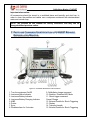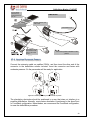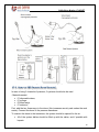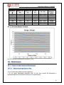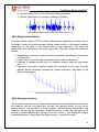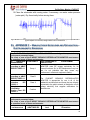
1
Defibrillator Monitor E-HEART User Manual Defibrillator Monitor E-HEART Manufacturer: US Defib Medical Technologies LLC Address : 7831 NW 72nd Avenue, Medley - Miami Zip Code: 33166 Phone: +1 305 8877552 / +1 305 8877541 Legal Representative: Amanda Coelho Rodrigues Felix OBELIS s.a AV. DE TERVUEREN 34 , BTE 44, BRUSSELS CITY – BELGIUM Defibrillator Monitor E-HEART FOREWORD Congratulations for acquiring a US DEFIBequipment. This product incorporates up to date technology. We are sure you will be very satisfied with E-HEART BIPHASIC DEFIBRILLATOR MONITOR. READ ALL OF THE OPERATION INSTRUCTIONS BEFORE operating E-HEART BIPHASIC DEFIBRILLATOR MONITOR. This User Manual contains all of the necessary information for a complete interaction with the equipment, from information concerning operation to necessary care for better conservation of E-HEART BIPHASIC DEFIBRILLATOR MONITOR. This equipment should only be used by a well-trained professional to provide advanced life support. After you finish reading the entire User Manual, keep it protected so you can check it at any moment. A future reference could be necessary for new users. The permanent consultation of this manual is a requirement to obtain better equipment performance, correct operation, and more safety for operator and patient. This manual also contains information related to technical support and Warranty Certificate. Read carefully the instructions on pages 7-9 of this manual. 2 Defibrillator Monitor E-HEART Summary 1. Warnings _________________________________________________________________________________ 6 2. Symbols and Abbreviations ___________________________________________________________________ 9 3. Measurement Units _________________________________________________________________________ 9 4. Acronyms Used in this User Manual ___________________________________________________________ 10 5. Description of E-HEART Biphasic Defibrillator Monitor and Its Components ___________________________ 11 5.1 Presentation ______________________________________________________________________________ 11 6. Overview* _______________________________________________________________________________ 12 6.1 Parts List and Included Accessories _____________________________________________________________ 0 6.2 Optional Accessories ________________________________________________________________________ 0 6.3 Optional Accessories List _____________________________________________________________________ 1 7. Parts and Commands Identification of E-HEART Biphasic Defibrillator Monitor ________________________ 11 8. E-HEART Biphasic Defibrillator Monitor Characteristics __________________________________________ 14 8.1 ECG Characteristics _______________________________________________________________________ 16 8.2 Patient analysis system – AED Mode (optional) __________________________________________________ 16 8.3 Thermal Printer Characteristics ______________________________________________________________ 16 8.4 Pulse Oximetry Characteristics _______________________________________________________________ 17 8.5 Pacemaker Technical Specification and Characteristics ____________________________________________ 17 8.5.1 Pacemaker Characteristics ______________________________________________________________ 18 8.5.2 Stimulation System Specification _________________________________________________________ 19 8.6 Non-Invasive Blood Pressure (NIBP) __________________________________________________________ 19 8.7 Invasive Blood Pressure Module (IBP) _________________________________________________________ 20 8.7.1 Functional characteristics of the IBP ______________________________________________________ 20 8.8 Capnography Module (EtCO2) _______________________________________________________________ 20 8.8.1 Functional characteristics of the Capnography ______________________________________________ 21 8.9 ST Segment Analysis Characteristics ___________________________________________________________ 21 9. Some Cardiac Arrhythmias Characteristics ______________________________________________________ 24 10. Impedance Indicator _______________________________________________________________________ 26 11. Equipment Installation ______________________________________________________________________ 27 11.1 Equipment Unpacking and Adaptation _______________________________________________________ 27 11.2 Power Supply / Battery ___________________________________________________________________ 28 11.2.1 Lithium-Polymer (Li-PO) _______________________________________________________________ 28 11.3 Digital Status of Battery Load _____________________________________________________________ 29 11.4 Transport Conditions ____________________________________________________________________ 29 12. Equipment Installation and Handling Recommendations ___________________________________________ 29 12.1 Safety and Protection ____________________________________________________________________ 30 13. Using E-HEART Biphasic Defibrillator Monitor in Strong Electromagnetic Fields _______________________ 30 14. Defibrillator Monitor Operation in High Frequency Environment ____________________________________ 30 15. Care When Applying Defibrillation / Cardioversion _______________________________________________ 31 16. Operation Mode ___________________________________________________________________________ 31 16.1 Equipment Configuration _________________________________________________________________ 31 16.1.1 Menu Screen _________________________________________________________________________ 32 16.1.2 Setup Screen _________________________________________________________________________ 32 16.1.3 ECG Setup Menu (Electrocardiogram) ____________________________________________________ 14 16.1.4 Pacemaker Setup Menu ________________________________________________________________ 14 16.1.5 SPO2 Setup Menu _____________________________________________________________________ 15 16.1.6 Printer Setup Menu ____________________________________________________________________ 15 16.1.7 NIBP Setup Menu _____________________________________________________________________ 15 16.1.8 Drugs Setup Menu ____________________________________________________________________ 16 16.1.9 IBP Setup Menu ______________________________________________________________________ 16 16.1.10 Configuration menu of the CAP (Capnography) ___________________________________________ 14 16.1.11 Ventilation Setup Menu _______________________________________________________________ 14 16.1.12 Other Functions ____________________________________________________________________ 15 16.1.12.1 Pacemaker (optional) ________________________________________________________________ 15 3 Defibrillator Monitor E-HEART 16.1.12.2 Printer (optional) ___________________________________________________________________ 15 16.1.12.3 NIBP (optional)_____________________________________________________________________ 15 16.1.12.4 SYNC. ON _________________________________________________________________________ 15 16.1.12.5 Cancel load ________________________________________________________________________ 15 16.1.12.6 2 MIN ____________________________________________________________________________ 15 16.1.12.7 Selection / Menu ____________________________________________________________________ 15 16.1.12.8 Load / charge ______________________________________________________________________ 16 16.1.12.9 Shock Triggering____________________________________________________________________ 16 16.1.12.10 AED Mode ______________________________________________________________________ 16 17. Adapting Cables and Accessories _____________________________________________________________ 16 17.1 ECG Module ___________________________________________________________________________ 16 17.2 SPO2 Oximetry Sensor ___________________________________________________________________ 17 17.3 Adapting The Nibp Cuff __________________________________________________________________ 18 17.4 Adapting Pacemaker Paddles ______________________________________________________________ 20 17.5 Pacemaker Use Instructions _______________________________________________________________ 21 17.6 Adapting The Defibrillator Monitor Paddles __________________________________________________ 24 17.7 Inserting Thermal Paper in Printer _________________________________________________________ 25 17.7.1 Instructions to Place Thermal Paper In the TR-50 or SP-48 Printer ______________________________ 25 17.8 Using Capnography and Sensors ___________________________________________________________ 14 17.9 Using the IBP (Invasive Blood Pressure) _____________________________________________________ 14 17.10 Internal Battery _______________________________________________________________________ 16 18. Foundations ______________________________________________________________________________ 16 18.1 Defibrillation Concept ___________________________________________________________________ 16 18.1.1 Defibrillation Importance _______________________________________________________________ 16 18.2 The Cardioversion ______________________________________________________________________ 17 18.3 External Transthoracic Cardioversion / Defibrillation (Indirect) on adults __________________________ 17 19. E-HEART Biphasic Defibrillator Monitor Classification ___________________________________________ 18 19.1 General Classification ___________________________________________________________________ 18 19.2 Detector of Cardiac Rhythm – AED Mode (Optional) ___________________________________________ 18 19.2.1 Recording Methods (for AED Mode) ______________________________________________________ 19 19.2.2 Rhythm source (for AED Mode) __________________________________________________________ 19 19.2.3 Rhythm Selection Criteria (for AED Mode) _________________________________________________ 19 20. Note Methods _____________________________________________________________________________ 19 20.1 Detector performance results ______________________________________________________________ 19 21. Applied Technology ________________________________________________________________________ 20 21.1 Comparative: Monophasic X Biphasic _______________________________________________________ 20 21.2 Truncated Exponential Dual Phase Waveform _________________________________________________ 14 21.3 Variation according to Patient Thoracic Impedance ____________________________________________ 14 21.4 Variation of delivered energy and duration of defibrillation phases performed with Truncate Dual Phase Waveform ___________________________________________________________________________________ 15 21.5 Delivered Energy X Charge _______________________________________________________________ 16 22. Maintenance ______________________________________________________________________________ 16 22.1 Corrective and Preventive Maintenance______________________________________________________ 16 22.1.1 Precautions and Special Care ___________________________________________________________ 16 22.1.1.1 Preventive Inspections and Cleaning ____________________________________________________ 17 22.1.2 Preventive Inspections _________________________________________________________________ 17 22.1.3 Cleaning ____________________________________________________________________________ 17 22.1.4 Preventive maintenance ________________________________________________________________ 18 23. Additional information ______________________________________________________________________ 18 24. Troubleshooting ___________________________________________________________________________ 18 25. Precautions and Special Care ________________________________________________________________ 21 25.1 Equipment and Accessories Cleaning and Disinfection __________________________________________ 21 25.2 Storage and Transport ___________________________________________________________________ 22 25.3 Cables and Accessories Handling __________________________________________________________ 22 25.4 Power Supply and Grounding______________________________________________________________ 23 26. Physiological Effects _______________________________________________________________________ 23 4 Defibrillator Monitor E-HEART 26.1 ECG Module ___________________________________________________________________________ 24 26.2 Non Invasive Blood Pressure (NIBP) ________________________________________________________ 24 26.3 Oximetry Module _______________________________________________________________________ 24 26.4 Defibrillation Module ____________________________________________________________________ 24 26.5 Invasive Blood Pressure Module ___________________________________________________________ 25 26.6 Capnography Module ____________________________________________________________________ 25 27. Adverse Effects ____________________________________________________________________________ 25 28. Technical Specifications _____________________________________________________________________ 26 28.1 Pacemaker Technical Specifications (PM) ____________________________________________________ 27 28.2 Capnography Technical Specifications (EtCO2) _______________________________________________ 27 28.3 Invasive Blood Pressure Technical Specifications (IBP) _________________________________________ 28 28.4 ECG Technical Specifications _____________________________________________________________ 28 28.5 Non Invasive Blood Pressure Technical Specifications (NIBP) ____________________________________ 29 28.6 Oximetry Technical Specifications __________________________________________________________ 29 28.7 Disposable Accessories Manufacturers ______________________________________________________ 30 28.8 Permanent Accessories Manufacturers ______________________________________________________ 30 29. APPENDIX A – Fuse Replacement ____________________________________________________________ 31 29.1 Fuse change procedure ___________________________________________________________________ 31 30. APPENDIX B – ECG Trace Instability and Noises ________________________________________________ 14 30.1 The most common ECG Interferences _______________________________________________________ 15 30.2 AC Power Supply Interference _____________________________________________________________ 15 30.3 Muscle Artifacts ________________________________________________________________________ 15 30.4 Baseline displacements ___________________________________________________________________ 16 30.5 Movement artifacts ______________________________________________________________________ 16 31. APPENDIX C – Manufacturer Guidelines and Declaration – Electromagnetic Emissions _________________ 18 32. Technical Assistance _______________________________________________________________________ 21 33. Version Control ___________________________________________________________________________ 24 5 Defibrillator Monitor E-HEART 1. WARNINGS WARNING! The E-HEART BIPHASIC DEFIBRILLATOR MONITOR was designed for cardioversion and clinical monitoring applications with guaranteed operation when used correctly, in an appropriate medical place and by qualified personal. WARNING! The operator should proceed to check the equipment conditions and its accessories (regular tests) as well as its operation before use. WARNING! The operator should have knowledge and be aware of all possible collateral effects that can be caused during use of E-HEART BIPHASIC DEFIBRILLATOR MONITOR. WARNING! The E-HEART BIPHASIC DEFIBRILLATOR MONITOR is restricted to one patient at a time and NON FREQUENT USE. WARNING! Do not touch the patient, the bed (stretcher), the equipment or any other accessory connected to the patient and/or DEFIBRILLATOR MONITOR during the electrical discharge (shock). WARNING! When installing the equipment, make sure it is in a place with enough space for ventilation (10cm of upper side, 15cm at rear side and 10cm at sides) and far from heat radiation. WARNING! Equipment and devices connected to E-HEART BIPHASIC DEFIBRILLATOR MONITOR (different equipment connected to same patient), should be connected to perform equal potential among them, to obtain perfect potential equalization, thus preventing damage to them, otherwise the system equipment-patient grounding may be compromised. WARNING! There is risk of electric shock if the equipment case is open. There are no internal fuses to be replaced by the user. All service type or future upgrades of this equipment and its 6 Defibrillator Monitor E-HEART parts can only be performed by trained personnel and authorized by US Defib Medical Technologies LLC. WARNING! There is risk of explosion if this equipment is used in presence of flammable agents, like anesthetic gases, fuels, among others. WARNING! When the E-HEART BIPHASIC DEFIBRILLATOR MONITOR is used simultaneously with an electric scalpel, the orientations about equipment operation in the presence of high frequency devices indicated in this manual must be observed. WARNING! The E-HEART BIPHASIC DEFIBRILLATOR MONITOR equipment is designed for public network connection, and it does not suffer any interference or electromagnetical disturbances in its modules operation – according to NBR IEC 60601-1-2 / CISPR 11 recommendations – Electromagnetic disturbance characteristics in industrial, scientific and medic equipment radiofrequency measurement limits and methods (ISM). WARNING! To prevent fire or shock risks, avoid operating or fitting the DEFIBRILLATOR MONITOR near a water source; also avoid spilling any liquid product on the case. WARNING! The protection against the discharge effects of a cardiac defibrillator is present in the modules inside the equipment. The sensors and cables don’t have additional protection against a cardiac defibrillator discharge effects or when used simultaneously with an equipment that operates in high frequency. WARNING! The materials categorized as disposable should not be reused or even submitted to cleaning process and sterilization. The disposable materials should be discarded in appropriate places according to special procedures for hospital waste. WARNING! In general, the EQUIPMENT and ACCESSORIES Parts of E-HEART BIPHASIC DEFIBRILLATOR MONITOR, designed to be in physical contact with biological tissues, cells and corporeal fluids are tested according to ISO 10993-1 guidelines and principles, which deals exclusively with the test of biocompatibility of the applied parts. 7 Defibrillator Monitor E-HEART WARNING! If it is necessary to replace any part of equipment, except the disposable materials, the manufacturer or authorized network should be contacted to supply the material and to perform the substitution. WARNING! There is risk of environment pollution associated to the use of accessories and consumption materials at the end of its lifecycle. The accessories and consumption materials should be discarded in hospital waste according to environmental laws. The intern batteries should be returned to the manufacturer after substitution due to defect or end of lifespan. WARNING! All of the material replacement should be performed according to the specifications included in this manual. US DEFIB only guarantees the equipment perfect operation if the orientations are properly observed. WARNING! In special cases, if necessary, US DEFIB will keep available, with agreement, all of the technical material like circuit diagrams, material list, technical information, components list, calibration and benchmarking instructions or whatever is necessary, so the qualified technical personnel can perform repairs in the repairable parts determined by the manufacturer. The maintenance authorization should be formally expressed by US DEFIB MEDICAL TECHONLOGIES LLC. 8 Defibrillator Monitor E-HEART 2. SYMBOLS AND ABBREVIATIONS Terminal or potential equalization point Dangerous Electric Voltage Check attached documents BF type Equipment applied part with patient isolation CF type Equipment with defibrillator protection This side up: indicates the correct position in which the box should be transported Fragile: indicates that the package should be transported and handed carefully Keep dry: indicates the package should be kept in dry place Number 5: indicates maximum pilling of five units Indicates medical device and, therefore, special treatment Indicates composition with recyclable raw material Symbol for marking of electrical and electronic devices in accordance to the Directive 2002/96/EC. The device, accessories and packaging must be disposed properly at the end of its use. Please follow local ordinances or regulations for disposal. 3. MEASUREMENT UNITS Symbols Unit m, cm, mm h, m, s, mseg kg, g °F, °C mmHg, hPa hz, rpm, bpm, ppm V, mV m/s, mm/s, bps, Length Time Mass Temperature Pressure Frequency Voltage Speed Description Meter, centimeter, millimeter Hour, minute, second, millisecond Kilogram, gram Fahrenheit degrees, Celsius Degrees Mercury Millimeters, hectopascal Hertz, breeding per minute, beat per minute, pulses per minute Volts, millivolts Meter per second, millimeter per second, beat per second, liters per 9 Defibrillator Monitor E-HEART l/m J 3 3 m , mm Impedance Energy Volume minute Ohms Joules Cubic meters, cubic millimeters 4. ACRONYMS USED IN THIS USER MANUAL CLAS: Cardiology Life Advanced Support; AHA: American Heart Association; BLS: Life Basic Support; IDM: Implantable Defibrillator Monitor ECG: Electrocardiogram; VF: Ventricular Fibrillation; Hb: Hemoglobin (cHb: Hemoglobin concentration); HbO2: Oxihemoglobin (cHbO2: oxihemoglobin concentration); PRI: Printer INCOR: Heart Institute; LDE: Light Diode Emissor LCD: Liquid Crystal Display; PM: Pacemaker SAN: Sinu-atrial Node; ABP: Arterial Pressure CPA: Cardio-respiratory Arrest; NIBP: Non-Invasive Blood Pressure; IBP: Invasive Blood Pressure DBI: Defibrillator CPR: Cardiopulmonary Resuscitation; BCS: Brazilian Cardiology Society; SPO2: Oxygen Saturation; VT: Ventricular Tachycardia; UTI: Intensive Care Unit; VOO: Pacemaker Asynchronous Mode; VVI: Pacemaker on Demand Mode. 10 Defibrillator Monitor E-HEART 5. DESCRIPTION OF E-HEART BIPHASIC DEFIBRILLATOR MONITOR AND ITS COMPONENTS 5.1 PRESENTATION The E-HEART BIPHASIC DEFIBRILLATOR MONITOR is a portable and dual phase equipment (truncated dual phase pulse). It can be used in any position in ground hospital environment, in the surgical bed and benches rescue units, by air or land, providing better handling in advanced life support. This equipment was designed to apply electric stimulus to the heart in cases that the cardioversion and/or defibrillation are indicated. This revolutionary defibrillation technology requires less energy than that used by conventional single phase DEFIBRILLATOR MONITOR, producing a better performance. This equipment is indicated for adult and pediatric use and it integrates several functions like Defibrillation, ECG Monitoring, Non-invasive Blood Pressure – NIBP (optional) and Invasive Blood Pressure – IBP (optional), Oxygen Blood Saturation Scouting (SPO2), Capnography monitoring (optional) and an external Pacemaker with non-invasive multi programmable module (asynchronous and by demand and switching for emergency modes, also presents a Thermal Printer - optional). The ECG can be captured by monitoring electrodes and by the external defibrillation paddles, allowing, event memorization (Compact Flash). The DEFIBRILLATOR MONITOR has a high-resolution and high-contrast liquid crystal display (LCD) that allows perfect visualization in different angles, with the exclusive Lap Top (check page 14, figure 2) for cardiac monitoring. It also has a microprocessor for heart activity analysis, which takes approximately 10 seconds. ECG analysis (ventricular fibrillation, ventricular tachycardia) and, optionally, it can be supplied with the software for ST Segment and Arrhythmias analysis and software for ventilation / intubation mode, Drugs Calculation. Easy to use, E-HEART BIPHASIC DEFIBRILLATOR MONITOR offers, through AED mode, the possibility to be used by trained staff and under medical supervision. It is highly safe and presents minimum risks for accidents for patient and operator. In AED mode (optional), it provides a voice and text command to instruct the rescuer during the CPR sequence. The use of E-HEART BIPHASIC DEFIBRILLATOR MONITOR increases human survival rates in a cardio-respiratory arrest. The E-HEART BIPHASIC DEFIBRILLATOR MONITOR could be configured and adapted according to your necessity and be delivered in the following versions: 11 Defibrillator Monitor E-HEART ECG/PRI: DEFIBRILLATOR MONITOR of ECG, Defibrillator and 01 channel Thermal Printer and optionally 02 or 03 channels; ECG/PM: DEFIBRILLATOR MONITOR with ECG, defibrillator and external pacemaker, transthoracic, non-invasive, multi programmable. ECG/SPO2/NIBP: DEFIBRILLATOR MONITOR with ECG, Defibrillator, Pulse Oximetry and Arterial Pressure Scouting; ECG/SPO2/NIBP/PRI: DEFIBRILLATOR MONITOR with ECG, Defibrillator, Pulse Oximetry, Arterial Pressure Scouting and single channel thermal printer. ECG / AED MODE / DRUG CALCULATION / IBP / CAPNOGRAPHY Defibrillator monitor with AED (voice and text command), drug calculation, Invasive Blood Pressure (IBP), Capnography and Telemetry. NOTE: The External Transthoracic Pacemaker, AED MODE, Drug Calculation Software, software for ventilation / intubation mode, capnography and Invasive Blood Pressure module can be incorporated into the versions of E-HEART BIPHASIC DEFIBRILLATOR MONITOR. Any option can be assembled, at the discretion of the specific needs of each client, and it does not change the characteristics of the product purpose. 6. OVERVIEW* Figure 1 - E-HEART BIPHASIC DEFIBRILLATOR MONITOR Front View 12 Defibrillator Monitor E-HEART LAP TOP 45º Figure 2 - Side view with the LAP TOP (45° opening) 6.1 PARTS LIST AND INCLUDED ACCESSORIES 01 conductive gel tube 100 ml; 01 Interchangeable and permanent external electrodes pair (Shock Paddles) (adult/infantile); 01 5-way patient cable; 05 Disposable electrodes; 01 Adult NIBP Cuff (only on the versions with NIBP); 01 IBP kit (only on the versions with IBP) 01 CO2 kit (Capnography) (only on the versions with Capnography) 01 SpO2 Sensor (configuration with SpO2); 01 3-pole power supply cable; 01 Grounding equalization cable; 02 Replacement fuses; 01 User manual; 01 Set of disposable electrodes (only on the versions with AED mode); 01 Set of disposable electrodes (only on the versions with pacemaker); 01 Roll of printer thermal paper (only on the versions with thermal printer); Warranty Certificate. NOTE: The user must observe the parameter modules that have been previously configured in his equipment. 6.2 OPTIONAL ACCESSORIES Transport case destined for transport and also safer storage of E-HEART BIPHASIC DEFIBRILLATOR MONITOR; Connection cable for external battery, used in ambulances, aircrafts or occasions where electrical energy is not available for long periods of time; Defibrillator Monitor E-HEART Cables, sensors, paddles (external and internal electrodes) adult, infantile or neonatal; Mobile trolley; Equipment support. 6.3 OPTIONAL ACCESSORIES LIST Figure 3 - Connection cable for external battery (permanent use accessory - Exclusive) Figure 4 - Conductive gel for ECG (disposable content) Figure 5 - Thermal sensitive paper (disposable) Figure 6 - Adult and Child Shock Paddles for external defibrillation and cardioversion (permanent material Exclusive) Figure 7 - Adult Shock Paddles for internal defibrillation and cardioversion (permanent use material - Exclusive) Figure 8 - Child Shock Paddles for internal defibrillation and cardioversion (permanent use material - Exclusive) 1 Defibrillator Monitor E-HEART Figure 9 - Adult oximetry sensor (permanent use accessory - Exclusive) Figure 11 - Ear Oximetry sensor (permanent use accessory Exclusive) Figure 10 - Pediatric / Neonatal Oximetry Sensor (permanent use accessory - Exclusive Figure 12 - Capnography Sensors (permanent use accessory - Exclusive) Figure 13 - Permanent ECG electrodes (permanent use accessory - Exclusive) Figure 14 - ECG electrodes (disposable content) Figure 15 - 3-way Patient Cable(permanent use accessory Exclusive) Figure 16 - 5-way Patient Cable (permanent use accessory - Exclusive) 2 Defibrillator Monitor E-HEART Figure 17 - Adult Adhesive Electrode Paddles – AED and Pacemaker (disposable) Figure 19 – Adult NIBP Cuff (permanent use accessory – Exclusive) Figure 18 - 3-pole Power Cable (permanent use accessory - Exclusive) Figure 20 - Potential equalization cable (permanent use accessory - Exclusive) 1 Defibrillator Monitor E-HEART Important observation: All accessories should be stored in a ventilated place and humidity and dust free. In order to clean the paddles and cables use a compress moistened with demineralized water and neutral soap. NOTE: The pictures on this manual are merely illustrative, and they can be changed without previous notice. 7. PARTS AND COMMANDS IDENTIFICATION OF E-HEART BIPHASIC DEFIBRILLATOR MONITOR Figure 21 - Commands identification of the Front Panel 1. Turn the equipment On/Off 2. Luminous Alarm Indicator 3. Luminous Battery Indicator 4. Luminous Battery Charging Indicator 5. NIBP 6. Printer 7. Sync. 8. Disables alarm for 2 minutes 9. Defibrillator charge command 10. Enables / Disables AED Mode 11. Shock trigger command Cancel Charge 12. External Paddle for Shock Triggering 13. Display 14. Transport handle 15. External Paddle for Shock Triggering Defibrillator Monitor E-HEART 16. Pacemaker on/off switch 17. Select between Synchronous Asynchronous Mode 18. Pacemaker emergency mode and 19. Disables Pacemaker Beep 20. Disables Pulse 21. Navigation Button (Browser) NOTE: The pictures contained in this manual are merely illustrative and they can be changed without prior notice. Figure 22 - Module Identification 1- ECG Connector 2- External Shock Paddles Connector 3- Adhesive Paddles Connector for AED module 4- Adhesive Paddles Connector for MP module 5- NIBP Connector 6- SPO2 Connector 1 Defibrillator Monitor E-HEART Figure 23 - Rear Panel Identification 1- Power Grid Connector 2- Fuse Port 3- Ambulance Inlet Connector 8. E-HEART BIPHASIC DEFIBRILLATOR MONITOR CHARACTERISTICS Truncated dual phase exponential waveform, with charge of 1 to 200 Joules or optional of 1 to 360 Joules, with operating instructions on the panel of E-HEART BIPHASIC DEFIBRILLATOR MONITOR, or any other energy configuration desired by the user. With the equipment powered by electric grid or even powered by the new battery (and fully charged) its charge time for maximum energy of up to 5 seconds for 200 joules or up to 6 seconds for 360 joules. Adaptable to any patient including intelligent safety system that limits the charge for internal use and pediatric/neonatal use; Patient thoracic impedance analysis, increased defibrillation efficiency and reduced cardiac injuries risk; Automatic internal discharge after 30 seconds if there is no trigger, or manually through the key; 14 Defibrillator Monitor E-HEART Clock, date and shock counter. Clear phase identification: charging, ready, discharging and disarming. Use of reusable electrodes (permanent adult / pediatric / interchangeable paddles), that needs activation of two simultaneous controls, one in each paddle; Performs self-test when switched on; Rechargeable internal battery with manageable charger, with alarms and status indicator for several levels: low level, charging (medium and high) and full charge, with indication in approximately 6 levels on the display. Capacity to perform up to 220 discharges with full load (new battery fully charged). External reserve batteries with internal charger (optional); Electroluminescent liquid crystal display, that shows signal of ECG, SPO2 (optional), Capnography and invasive blood pressure (optional), Non-invasive blood pressure (optional), derivation, cardiac frequency, beep indicator, battery status, alarms, pacemaker pulse, programming parameters, indicating the selected energy for triggering. Event memory including curve, date and hour (optional), of approximately 250 MB, that corresponds to over 100 hours of continuous recording. Indicates mode and charge value on the screen; Informs when the capacitor is discharging: “CANCELED”; Charge automatic adjust; Allows continuous ECG registration in memory, critical events and performed procedures; Enables communication with microcomputer, through connection, for memory data visualization (Optional); Allows posterior reading of ECG tracing through the hardware and/or software (Optional); Language: Portuguese (possibility to change language to English or Spanish (optional). It performs synchronized triggering with QRS complex, with energy time of <20ms when in SYNCHRONIZED MODE. Maximum time for signal stabilization: 5 seconds after ideal connection of sensor on patient; Full system of audible and visual alarms with the possibility of programming maximum and minimum values, including, besides the physiological alarms for loose electrode, asystole, tachycardia, bradycardia, fibrillation; When E-HEART BIPHASIC DEFIBRILLATOR MONITOR is configured in automatic mode, the charge energy follows a trigger sequence of 150J, 200J and 200J; Pacemaker pulse detection and rejection. Impedance detection in the 25 Ohm to 500 Ohm range for the triggering; Software for drug calculation (optional); Software for Ventilation / Intubation Mode (optional); ST Segment and Arrhythmias analysis (optional); Telemetry Software (optional); 15 Defibrillator Monitor E-HEART Memory data card of 250 MB that corresponds to over 100 hours of continuous recording; Charge level selection by the “APEX” Paddle key and charging by the “STERNUM” paddle key and triggering by pressing both paddles keys simultaneously. 8.1 ECG CHARACTERISTICS 3 derivations (DI, DII, DIII), for 3-way ECG cable; 12 derivations (DI, DII, DIII, aVL, aVR, aVF and V1 to 6) for 5-way ECG cable; Capture of ECG signal through defibrillation Pads, adhesive Pacemaker transthoracic Pads, through reusable defibrillator Pads and/or through ECG patient cable Cardiac frequency: Any frequency reading frequency from 10 to 300 rpm, with numerical presentation; Protection against defibrillation and cardioversion; Filter rejects band of 35Hz and 60Hz, pass low frequency: 120Hz and pass high frequency: 0,5Hz; QRS detector; Pacemaker detection and rejection; Synchronized beep with QRS; Display Beep indicator; Cardiac Frequency Indicator on display (bpm); Pacemaker Indicator on display; Speed control for curve tracing; ECG channel gain control; ST Segment and Arrhythmias analysis. 8.2 PATIENT ANALYSIS SYSTEM – AED MODE (OPTIONAL) ECG evaluation automatic system that detect QRS complex and identifies automatically malignant arrhythmia (ventricular tachycardia and ventricular fibrillation) that requires defibrillation; Synchronism with R wave in case of QRS complex presence (when on “synchronized mode”):; Pacemaker detector; Impedance measurement to adjust phases 1 and 2 of dual phase wave and it does not allow triggering with open or short-circuited paddles. It offers voice and text command to instruct the rescuer during the CPR sequence. 8.3 THERMAL PRINTER CHARACTERISTICS High-resolution thermal printer with automatic and manual registration of one channel, with optional of two or three channels, with the possibility of ECG registration with diagnostic-quality with manual or automatic triggering after 16 Defibrillator Monitor E-HEART defibrillation with annotation of date and time, heart rate, derivation, amplitude of the ECG, etc. It allows manual records, regardless of the defibrillation, by the paddles. This record is made on thermo-sensitive paper of 48 mm (width) x 30 m (length) for GSI printer and 48 mm (width) x 20 m (length) for TR-50 or SP-48 printers. Print speed of 12,5-25-50 mm / sec. 8.4 PULSE OXIMETRY CHARACTERISTICS Pulse oximetry, with plethismography curve and saturation indication of numeric oxygen in percentile; plethismography wave amplitude adjusted automatically on screen; complete alarms system and audio and visual indication of SPO 2 level, through tone of pulse signal; the alarm volumes and pulse audio indicator are adjusted independently; adjustable audiovisual alarms: low and high SPO 2 and low and high cardiac frequency (bradycardia and tachycardia); pulse alarm not detected; low perfusion; disconnected sensor; key for silent alarm for 02 minutes; good behavior in low perfusions, The frequency detected by the equipment is between 30 and 250 ppm with a 3% precision. The pulse oximetry is used in situations where the oxygen saturation (SPO 2) is essential, in anesthesia, during surgeries and post-surgery, patients under intensive care treatment, in ambulances or at home. It has been proven efficient, with a sample range from 70 to 100% with accuracy up to 3%. The scouted saturation accuracy is undetermined when it is between 0% and 69%. The oxygen saturation, SPO2, is defined by the concentration rate of two main forms of blood hemoglobin, the arterial hemoglobin or oxihemoglobin (HbO2) and the concentration of HbO2 + Hb (unsaturated hemoglobin), ie, (cHbO2+cHb). The oxygen saturation is expressed in percentage and calculated by the formulae below: Biolight and Nellcor Technology. SPO2 = 8.5 cHbO2 X 100% cHbO2 + cHb PACEMAKER TECHNICAL SPECIFICATION AND CHARACTERISTICS The external pacemaker was designed to stimulate the heart in cases of rhythm disturbance and flaw to conduct internally electric pulse. It is used in cardiac surgeries as emergency cardiac pacemaker. Some indicated transthoracic applications of pacemaker are: 17 Defibrillator Monitor E-HEART Treatment of symptomatic bradycardia or bradyasystole during emergency During and after cardiac surgery To ease the insertion of a transvenous stimulator electrode. 8.5.1 PACEMAKER CHARACTERISTICS External pacemaker, transthoracic, non-invasive, multi-programmable; in modes on Demand , assynchorouns (fixed) and Emergency. It is composed by: One control unit based on micro controller with serial data transmission, and one QRS detection circuit. One high voltage source and stimulation pulse generator with amplitude and pulse width enough to successfully perform a noninvasive transitory stimulation, which requires stimulation rate between 30 and 200 bpm. (Other frequencies may be optionally configured according to the user choice) Note: The operator can control the stimulation process by means of E-HEART BIPHASIC DEFIBRILLATOR MONITOR keyboard. The stimulation pulse application can be visualized through a panel LED. 3 pacemaker operation modes are possible: VOO: In this operation mode, the system stimulates the patient continuously, according to frequency parameters, amplitude and width configured in the pacemaker menu. VVI: In this mode, the system only stimulates when it detects a cardiac frequency lower than the configured menu value, remaining the stimulus until the patient natural cardiac frequency returns to a value equal or greater to that configured to avoid inhibition of abnormal “T” waves or extra systole. The pacemaker has refractory period of approximately 250ms. Emergency: Regardless of the chosen mode, when the EMERGENCY key is pressed, the pacemaker switches to the VOO mode, configured in 100 mA, 70 ppm and 20 ms. OBSERVATION: In VOO and VVI modes the pacemaker will be stimulated and transmitting information to display (amplitude, width, frequency and mode). Stimulation current: Without connected charge: 200 mA; Off: 0 mA Power supply: 12V ECG capture through own adhesive Pads. Stimulation output: Adhesive electrodes (PADs) Frequency: It is possible to adjust the stimulation frequency between 30 and 200 ppm. Amplitude: The amplitude value could be adjusted between 5 and 200 mA. Width: It is possible to select pulse widths between 5 and 50 ms. 18 Defibrillator Monitor E-HEART 8.5.2 STIMULATION SYSTEM SPECIFICATION Stimulation frequency: 30 ppm to 200 ppm in steps of 1 ppm Pulse amplitude: 0 mA to 200 mA in steps of 1 mA Pulse width: 0 ms to 50 ms in steps of 1 ms. Stimulation exit: disposable pads ECG capture by the disposable pads Emergency: VOO 70 ppm - 100 mA 20 ms Protection against defibrillation discharge: Up to 400 joules Other specifications may be configured as chosen by the user. Observation: The specifications above can be altered according to the user’s needs. ATTENTION! The pacemaker operation in the VOO mode is asynchronous. If the patient presents proper cardiac pace the pacemaker may induce ventricular fibrillation, if the pacemaker pulse is regularly applied to the ascendant portion of the T wave. In case of a bradycardia support, it must be assured that the stimulation frequency is higher than the patient’s own pace, and that the detection is trustable. In the VVI mode, the fixation region of the pacemaker electrodes must be verified, once that it is external and presents negative voltage, the stimulation may produce polarizations that change the common mode voltage, compromising the normal detection of the heartbeat. This equipment can only be operated by authorized technical staff. The non-invasive pacemaker is suitable for use in pre hospital or hospital environments, when urgent cardiac pacemaker use is necessary. 8.6 NON-INVASIVE BLOOD PRESSURE (NIBP) This item was elaborated to make possible a simplified understanding of NIBP channel basic functions. That channel provides diastolic, average and systolic arterial pressure. The automatic measurement time is configured by operator. The Non-Invasive Blood Pressure module (NIBP) is protected against discharges of cardiac defibrillator, and it does not need specific precaution concerning the equipment. In NIBP use with cuff, which has no metallic wires, therefore it does not cause any interference when used together with other High frequency equipment. Measurement by oscillometer in adult, pediatric, infantile and neonatal patients; Manual and automatic operation mode; Measurements of systolic, diastolic and average arterial pressure; Configurable interval to inflate the cuff; 19 Defibrillator Monitor E-HEART Automatic zero before each measurement; Alarm of minimum, average and maximum pressure; PAR Technology. 8.7 INVASIVE BLOOD PRESSURE MODULE (IBP) The invasive blood pressure module (IBP) presents on the screen the systolic, diastolic and average pressures (mmHg). The E-HEART BIPHASIC DEFIBRILLATOR MONITOR is normally supplied with the pressure cable to connect the reusable transducers. In the connection of the transducer to the cable, observe the narrows that are present on the two pieces that indicate the correct fitting position. All of the IBP system should be fulfilled with saline, if possible with heparin. Make sure that there are no bubbles, especially in the tube that goes to the patient. Do not use latex in the circuit. The transducer must be assembled around the midline of the patient in order to achieve trustable pressure measures. 8.7.1 FUNCTIONAL CHARACTERISTICS OF THE IBP The trace is shown continuously on the screen; Option to select the blood pressure types AP, AO, LV, AE, PVC, AD, RV, PAP, PCP, PIC, Right, Left Esf; Maximum, average and minimum blood pressures shown continuously on the screen (mmHg); Manual control of gain of in several ranges, with baseline variation; Possibility of curve superposing; Alarm for the maximum and minimum blood pressures; Option of continuous recording of the P.I.C. with aid of a thermal printer, with special algorithms for alpha, beta and “C” wave visualization. 8.8 CAPNOGRAPHY MODULE (ETCO2) The Capnography module uses the Sidestream and Mainstream sensors miniaturized, with optional procedure of auto calibration that dismisses the use of specific gases for periodic calibration. It presents to the user the following parameters: Exhaled CO2 curve, showed continuously on the screen; Value of the exhaled CO2; Minimum of inhaled CO2; Respiratory frequency value. After connecting the sensor, it is necessary to wait for approximately 1 minute in order to set up to be ready for the measurements. After this while, a light must be observed on the sensor indicating its habilitation. Once connected to the respirator tube, we’ll 20 Defibrillator Monitor E-HEART have the patient information. The Capnography sensor should be on the adaptor to avoid the condensation, if that occurs, interfere on the reading measurement. 8.8.1 FUNCTIONAL CHARACTERISTICS OF THE CAPNOGRAPHY Sensors Sidestream and Mainstream type; Exhaled CO2 curve showed continuously on the screen; Auto calibration optional procedure that dismisses the specific gases use for periodic calibration; Exhaled CO2 value, minimum inhaled CO2 and respiratory frequency value showed continuously on the screen; Miniaturized sensor with auto calibration; Side stream option, Mainstream or both; Disposable water filter; Disposable nasal line; Disposable intubated line; Disposable tube adaptor; Respironics technology. 8.9 ST SEGMENT ANALYSIS CHARACTERISTICS The first step in order to make the analysis of the ST segment is to scan the signal for 10 seconds at a rate of 500 samples per second. Eight of the derivations are of direct acquisition (I, II and V1 to V6). The remaining four derivations (III, aVR, aVL and aVF) are derived via Einthoven's law as follows: III II I ( I II ) aVR 2 (II ) aVL I 2 (I ) aVF II 2 We strongly recommend that you filter the signal in order to reject noise and achieve better results. The result of these steps is the digital ECG. After the acquisition, the program measures the ECG as the second phase of the interpretation process. The measurements may be detailed in five steps: 1. QRS Detection: This step is very important, because if it is performed incorrectly, the next steps will be wrong. An auxiliary function is computed for QRS detection, based on eight independent derivations. The complexes are classified as normal or abnormal in order to achieve a 21 Defibrillator Monitor E-HEART 2. 3. 4. 5. normal QRS standard from derivation to derivation. Besides, the RR interval is measured and the heartbeat is computed. Identification of the T wave end: This point is very important because it identifies the end of the cardiac cycle and it is used to measure the QT interval. P wave study: The program looks for the P waves in all segments T-Q (the end of the T wave to the beginning of the next QRS complex) to determine if the duration of the PR interval is varying. Beginnings and endings: These points are identified for each wave in order to measure its length and find its peaks. Measure: For each wave amplitude and duration each interval derivation is measured. Also, the deviation of the ST segment such as other parameters is measured. The result of the measurement process is the following: Duration of normal QRS complex. Duration of PR interval. Length of the QT interval. Heart rate (beats per minute). Duration of PR interval. Length of the P, Q, R and R' waves. Amplitude of the P, P ', Q, S, R' and T waves. Amplitude at the beginning, middle and end of the ST segment. Intrinsic deflection (time from start of the QRS complex to the peak of R wave) Projection of the electric axis in the frontal framing (P wave, RS complex and T wave vectors). The ventricular gradient is also measured. The derivation of the median cardiac cycle is also stored as it is useful for printed reports. The last step is the evaluation of medical reports from the ECG measurements made. The ST segment analysis has a number of advantages that must be mentioned: Considerable time saving of the cardiology professionals devoted to ECG interpretation in hospitals that offer a large number of these examinations. Stability and uniformity in the ECG interpretation and uniformity in interpreting ECG. Human fatigue or work pressure can cause specialists not to interpret ECGs maintaining the same needed uniformity. The EQUIPMENT always applies the same algorithm and the same rules for ECG interpretation, thus providing more stable findings in a timely manner. The possibility to store all information relating to a patient allows you to get the same examination report several times without any need to repeat the ECG. This information is a valuable component to an ECG database in research applications. 22 Defibrillator Monitor E-HEART All medical criteria used in this ST segment analysis fluctuate from a mere recommendation or alerting about the ECG results until a complete diagnosis of a specific change. That's why these criteria have varying degrees of specificity and may include phrases such as "NOT NECESSARILY PATHOLOGICAL", "CONSISTENT WITH", “PROBABLE…”, “CONSIDERING …", when there isn’t absolute certainty about the specific pathology. In these cases, the physician should determine whether the given measures and other complementary factors are conclusive or not. The EQUIPMENT evaluates all medical criteria taking into account all measurements previously made and determines, in his conclusions, which criteria are unique and which eliminate others due to their greater diagnostic accuracy. These criteria were grouped as follows: Changes in heart rhythm; Changes in the electrical axis; Left or right ventricular hypertrophy; Intraventricular blockade; Left branch blockade; Changes in the ST segment; Changes in T wave; Heart attack; Other cases. This type of diagnosis must not be considered a substitute, in any way, for diagnosis by cardiologists, because they simply are not. They should be seen as an efficient tool that assists the physician who specializes in its diagnosis because they are highly efficient in the classification of normality, and they have high sensitivity for the detection of pathological cases. This relieves the physician from reviewing normal cases and it can be used as a guide for the classification of pathological cases. When the electrocardiographic signs are ambiguous or highly complex, the final diagnosis is left to the physician. The following is a list of medical criteria: WARNING! The interpretive ST segment analysis report, using the E-HEART BIPHASIC DEFIBRILLATOR MONITOR is one of the valuable tools that helps the physician to interpret ECG efficiently, but only if, combined with a detailed patient history and medical exams. All computerized ECG system is unable to analyze the ECG waveform as the human eye-brain system. The physician should re-read and correct the automatic ECG interpretation report. WARNING! The ACC / AHA recommended the computerized interpretation of ECGs to doctors. 23 Defibrillator Monitor E-HEART "Several studies have examined the accuracy of computerized ECG interpretation programs and suggested that computer analysis cannot replace the physician’s ECGs interpretation. A systematic study of the computerized ECG interpretation performed in 1991 showed that computer programs were, on average, 6.6 % less accurate than the cardiologist in identifying ventricular hypertrophy, myocardial infarction (MI). Rhythm disorders were not assessed in that investigation, and informal experience suggests that computer interpretation has a higher error rate in the analysis of the rhythm than in the diagnosis of MI and hypertrophy. A Japanese study reported that the latest false-positive rate and false-negative was 18 times higher for computer interpretation than trainee doctors in more important ECG diagnosis. However, ECGs computerized interpretation may be useful in the accurate calculation heartbeat, conductive intervals and axis, as long as there is manual review. Thus, despite computerized ECG interpretations that may have useful auxiliary value, and cannot replace the interpretation of experienced electrocardiograms professionals and should not be used to make clinical decisions”."11) 9. SOME CARDIAC ARRHYTHMIAS CHARACTERISTICS The arrhythmia’s symptoms are quite variable, and they may be silent (that does not present symptoms). They can be diagnosed by a physician during a cardiologic exam (pulse exam and auscultation of the heart with a specific device). The most common symptom is palpitation. Fainting can also occur (quick and spontaneous recovery with no motor abnormalities), dizziness, shortness of breath, discomfort, feeling of heaviness in the chest, weakness, fatigue, chest pain, among others. Symptoms that indicate gravity are mental confusion, low blood pressure, chest pain and fainting. If any of these symptoms occur, it is necessary to perform EMERGENCY medical treatment to prevent the patient’s death. Cardiac arrhythmias can be classified in various ways, depending on frequency, formation mechanism, place of origin, etc. We will present some terms that are more general, common in everyday life. Regarding frequency, arrhythmias may be classified as: • Bradycardia: it occurs when the heart beats less than 60 times per minute. In some people, it can be a normal finding, such as athletes. Various types of bradycardia are known, each one with its own peculiar characteristics. Cardiac pacemakers are used in the treatment of this type of arrhythmia. 1 ) A report of the ACC/AHA/ACP-ASIM Task Force on Clinical Competence (ACC/AHA Committee to Develop a Clinical Competence Statement on Electrocardiography and Ambulatory Electrocardiography), ACC/AHA Competence Statement, 2001;104:3169-3178. 24 Defibrillator Monitor E-HEART Types of Bradycardia There are three basic types of bradycardia, depending on where the heart's electrical system blockage occurs. When blockage occurs in the sinus node, which is the heart's natural pacemaker, it is called the sinus node dysfunction. Besides, the blockade of the electrical impulse may occur in the atrioventricular node or the right or left branch of the heart's electrical system. The important thing is that all of these types of blocks can lead to reduction of heart rate and cause symptoms such as dizziness and fainting. Depending on the type of block, and of the symptoms shown, it may require a pacemaker implant. • Tachycardia: it occurs when the heart beats 100 times per minute. It usually occurs during physical activity, emotional stress, in the presence of anemia and other diseases. There are several types, some extremely serious. Types of Tachycardia • Atrial Tachycardia: it is a rapid heart rhythm that originates in the atrium. • Atrial Flutter: it is an arrhythmia caused by electric circuits of low conduction that originates in the atrium and promotes a rapid and regular rhythm of the heart. • Nodal reentrant tachycardia (NRT): it is an extra electrical pathway, near the atrioventricular node, which causes the electrical impulse to move in a circle and pass through areas that have been previously passed, making the heart beat at a frequency way above normal. • Tachycardia by an accessory pathway or Wolff-Parkinson-White Syndrome: it is an extra electrical pathway that exists from birth and connects the atriums to the ventricles, causing the electrical impulse to reach the ventricle faster. • Atrial Fibrillation: it is an extra electrical impulses originated in the atriums that trigger rapid irregular and disorganized heartbeats. • Extra-Ventricular Systole: it is an extra electrical impulse originated in the ventricle that promotes beat ahead of time. • Ventricular Tachycardia: it is an electrical impulse originated in the ventricles that promotes a rapid potentially life threatening pace. Generally, it is a medical emergency. • Ventricular Fibrillation: it is a fast, disorganized and erratic rhythm, which does not produce ventricular contraction that causes sudden death and requires immediate cardiopulmonary resuscitation and defibrillation (electrical shock). As the place of origin, these arrhythmias are classified as: 25 Defibrillator Monitor E-HEART • Atrial: as we know, the heart consists of four cameras (or divisions), two atriums and two ventricles. The normal stimulus for the heartbeat is generated in the right atrium. In some arrhythmias, these stimuli are generated in excess or in smaller numbers, by the structure that normally generates them, in others, the stimulus appears elsewhere in the atrium, leading to the occurrence of atrial arrhythmias. Figure 24 - Atrial Arrhythmia Electrocardiogram • Junctional: these arrhythmias occur at the junction between the atriums and ventricles. • Ventricular: it arises within the ventricles, some with great potential to lead to death. Figure 25 - Ventricular Arrhythmia Electrocardiogram 10. IMPEDANCE INDICATOR The instrument provides a visual indicator related to the total transthoracic impedance between the defibrillation paddles. The impedance indicator is used to evaluate: Proper placing of the shock PADDLES on the patient; The quality and integrity of the shock PADDLES; The shock PADDLES contact to the patient's skin; The proper connection of the shock PADDLES to the equipment; It provides a rapid assessment of the patient impedance. 26 Defibrillator Monitor E-HEART ATTENTION: The impedance indicator is only shown in the display when using the ECG reading via shock PADDLES. The impedance index is divided into 4 (four) sections, where the ideal operating section is Section 2 (impedance range from 30 [] to 150 []). Section 1 2 3 4 Section 1 Impedance Range [] 20 < IMP 300 30 < IMP 150 150 < IMP 180 180 < IMP Contact Description GOOD GREAT REGULAR BAD Table 3 Section 2 Presented Color Blue Green Yellow Red Section 3 Section 4 Figure 26 - Impedance Indicator 11. EQUIPMENT INSTALLATION 11.1 EQUIPMENT UNPACKING AND ADAPTATION Remove the equipment from package; Remove the plastic protection; Adapt on adequate and easy access place; Install the equipment far from other equipment that generates strong magnetic fields, like radiological devices , and others; Make sure that the installation place has an adequate ventilation and is within the pressure and temperature range indicated in this manual (page 68); 27 Defibrillator Monitor E-HEART This equipment was designed to operate in environments without inflammable anesthetic or cleaning agents. Do not operate it in presence of inflammable gases in general. 11.2 POWER SUPPLY / BATTERY 11.2.1 LITHIUM-POLYMER (LI-PO) Rechargeable Lithium-Polymer (Li-PO) with specific manageable battery charger internal to the equipment. For a long lifespan, keep the charger connected to the equipment and the power grid. With a capacity of up to 150 shocks at 200 J and up to 6 hours of monitoring when the battery is fully charged (new battery fully charged). This device has an internal intelligent battery charging control circuit. Remembering that this battery (LI-PO) should have special attention, as described below: WARNING! Do not use any battery charger other than the one recommended by US DEFIB MEDICAL TECHONLOGIES LLC; Do not short-circuit the battery; Charge in a well-ventilated location; Do not discharge the battery completely; Do not compress or disassemble it; Risk of burns, fire and explosion, if the recommendations above are not followed. 1) Optionally with Lithium-Iron Battery (LI-FE); Lithium-Iron (LI-IR) rechargeable battery with a proper manageable charger internal to the equipment. For a prolonged lifespan keep the charger connected to the equipment and the power grid. With a capacity of 6 hours of monitoring (battery fully charged) or at least 160 shocks at 360 J or 220 shocks at 200 J, when the battery is fully charged (new battery fully charged). This device has an internal intelligent circuit control of battery charging. 2) Optionally with sealed lead acid battery; 3) Optionally with external battery for ambulances, aircraft: In occasions when there is no power grid available by periods of long use, don’t use the network cable when the power supply cable for ambulances is used for ambulance; 28 Defibrillator Monitor E-HEART 4) Optionally with external batteries (spare) with its own charger with a maximum charge time of approximately 4 hours: External batteries can be delivered in different charging capacity versions that varies between 2 and 15 hours of monitoring or 50 to 200 consecutive shocks respectively, with a proper charger; External Batteries are easily replaced (battery interchangeable). WARNING! The E-HEART BIPHASIC DEFIBRILLATOR MONITOR can remain connected to the power grid 24 hours a day to keep the battery fully charged and it is also able to operate normally. 11.3 DIGITAL STATUS OF BATTERY LOAD In the panel of equipment is an LED indicator such as below: Connected to the power grid: Battery status Loading: Battery status , Discharged: ; ; . 11.4 TRANSPORT CONDITIONS Ambient temperature range from -40º to +70º C Relative humidity range from 10% to 100% Atmospheric pressure range from 700 hPa to 1060 hPa (525mmHg to 795mmHg) 12. EQUIPMENT INSTALLATION AND HANDLING RECOMMENDATIONS Avoid turning off the equipment if connected to a patient; remove the cable from electrodes before. If the patient is connected to E-HEART BIPHASIC DEFIBRILLATOR MONITOR, with floating isolation (not connected to the power grid grounding), connected to any other device that cannot have the same isolation type, the patient could touch the conductive parts and cancel the equipment protection effect; The connection of E-HEART BIPHASIC DEFIBRILLATOR MONITOR with any other equipment is only allowed when it is not prejudicial to patient, operator and ambient. If the additional equipment specifications does not inform about connection effects, seek for the manufacturer or expert. 29 Defibrillator Monitor E-HEART The E-HEART BIPHASIC DEFIBRILLATOR MONITOR should only be operated by qualified personnel. It is the hospital administration responsibility to make available proper and accessible operation instructions. Do not shock with short circuit paddles, because the trigger device could be damaged. 12.1 SAFETY AND PROTECTION a) Patient The capacitor is charged just before trigger and the charge voltage is connected to electrodes only at shock moment. The trigger command is only enabled if capacitor is charged with selected voltage and inside trigger time (30 s). Outside this period or during capacitor charging and/or is detected any anomaly in the relay operation that controls the capacitor discharge is disconnected, causing capacitor discharge. b) Operator Internal battery to isolate the equipment from external electric mains. Internal manageable battery charger with external power supply and isolation between power grid, patient and operator. c) Aircrafts Low-level radiation of electromagnetic fields. High immunity for external electromagnetic fields and transients. High mechanical resistance for vibration. 13. USING E-HEART BIPHASIC DEFIBRILLATOR MONITOR IN STRONG ELECTROMAGNETIC FIELDS Subways, helicopters and train stations could interfere in DEFIBRILLATOR MONITOR, when it is on the automatic external mode, because they are composed of intense electromagnetic fields in which were observed high alterations in the sensibility and specificity. Do not operate the equipment near cell phones, wet surfaces, high voltage lines or places near strong electromagnetic fields. 14. DEFIBRILLATOR MONITOR OPERATION IN HIGH FREQUENCY ENVIRONMENT An extreme care should be taken during surgeries that use equipment operating in high frequency, especially in patients with pacemaker. Besides the risk of 30 Defibrillator Monitor E-HEART pacemaker damages, the electric cauterization currents could cause fibrillation in the patient. Always keep a defibrillator monitor nearby. Respect the minimum distance of 15 cm between ECG electrodes and the electric scalpel or defibrillator, if are used at same time. In case of doubt, disconnect the ECG cable. This equipment may cause radio interference or may interrupt the operation of near equipment. It may be necessary to take migratory actions, like re-orientation or relocation of the defibrillator monitor or the location shielding. 15. CARE WHEN APPLYING DEFIBRILLATION / CARDIOVERSION Do not place the paddles directly on ECG electrodes. Some care should be taken with patients on pacemaker, in order to avoid damages to device and to patient himself: The applied energy should be the lowest possible; Keep an external pacemaker nearby; Check pacemaker after defibrillation. Keep distance between the generator and the patient pacemaker and the defibrillator monitor paddles. IMPORTANT: The protection against the defibrillator discharge effects is located in the intern modules of the equipment; The cable, electrodes and accessories don’t have protection against burn outs caused by the use of high frequency equipment. 16. OPERATION MODE 16.1 EQUIPMENT CONFIGURATION When you press the navigation button of E-HEART BIPHASIC DEFIBRILLATOR MONITOR the Configuration Menu modules will appear on the display. A cursor with an arrow shape (>) appears at left of the items of this menu indicating that this is the selected item one. By rotating the navigation button clockwise or anticlockwise, the cursor moves to a new menu item according to rotation sense. To configure the desired module, position the cursor on this module and press the navigation key. After choosing the module to be configured, a new menu is presented on the display with configuration items of the selected module. Select the item of the chosen module to be configured proceeding with same manner described previously. When you select the item, notice that it will blink indicating that it 31 Defibrillator Monitor E-HEART is ready to be altered. Rotate the navigation key to alter the item values, increasing or decreasing them. After choosing the desired value, press the navigation button to hold it. To exit the menu, place the cursor on the item Exit with the browser button. NOTE: In the modules Configuration Menu only the ones that are installed in the EHEART BIPHASIC DEFIBRILLATOR MONITOR (configuration options) will appear. It is possible to select the charge to be released in case of treatment(1 to 200 joules or 1 to 360 Joules according to configuration) by rotating the navigation key without pressing it. 16.1.1 MENU SCREEN Settings Exit Setup ECG Pacemaker SPO2 Printer 16.1.2 NIBP IBP Ventilation Drugs Capnography SETUP SCREEN Settings Exit Sync. ON Auto-Charge Paddles Selection Alarm Volume BPM Volume Key Volume Beep Key Date Time Language 1. Exit - Returns to the previous menu; 2. Alarm Vol. - Set the alarm volume, mute = 001, max = 009; 3. BPM Volume - Sets the beeper volume BPM = 001 mute, 009 = max; 4. Volume key - Sets the volume of the keyboard beep, mute = 001, max = 004; 5. Beep key - Enables (YES) or disables (NO) the beep sound on the keyboard; 6. Language - PTG = Portuguese, English = ENG, SPA = Spanish; 7. Time - Changes the Time; 8. Sync. ON – Enables (YES) or disables (NO) the synchronism with the QRS signal of the ECG sign; 9. Auto-Charge - Enables (YES) or disables (NO) the defibrillator charge automatic sequence; 32 Defibrillator Monitor E-HEART 10. Paddles Selection - Enables (YES) or disables (NO) the paddles button command; 11. Date – Adjust Day/ Month/Year 16.1.3 ECG SETUP MENU (ELECTROCARDIOGRAM) ECG > Exit Derivation Filter 60Hz Filter 35Hz Tachycardia Bradycardia Speed Gain Beep Alarm 1. Exit – Returns to previous Menu; 2. Derivation – Defines the electrocardiogram derivation to be presented on display (CAL = calibration, D1, D2, D3, AVR, AVL, AVF, V); 3. Filter 60Hz - Enable (yes) or disable (no) 60 Hertz filter; 4. Filter 35Hz – Enable (yes) or disable (no) 35 Hertz filter; 5. Tachycardia – Defines the bpm value for alarm operation in tachycardia (100 – 220); 6. Bradycardia – Defines bpm value for alarm operation in bradycardia (30 – 60); 7. Speed – Select the sweeping speed of ECG for 12.5, 25 or 50mm/s; 8. Gain – Select the ECG amplitude for N/2, 1N or 2N; 9. Beep - Enable (yes) or disable (no) the synchronism beep with QRS complex of ECG signal; 10. Alarm – Enable (yes) or disable (no) any ECG alarm; 16.1.4 PACEMAKER SETUP MENU Pacemaker > Exit Mode Width Amplitude Frequency Beep Pulse 1. Exit – Returns to previous Menu; 2. Mode – Select Pacemaker operation mode (PM) of the following modes: VOO: The PM send pulses according to configured parameters regardless of any ECG signal detected in patient; VVI: The PM sends pulses according to configured parameters only if the patient detected signal is outside these parameters range. 3. Width – Defines the pulse width from 5 to 50 ms; 4. Amplitude - Defines the pulse amplitude from 5 to 200 ms; 5. Frequency - Defines the pulse frequency from 30 to 300 ppm (pulse per minute); 6. Beep – Enable (yes) or disable (no) pulse beep; 7. Pulse – Enable (yes) or disable (no) the PM pulse sending. 14 Defibrillator Monitor E-HEART 8. Alarm - Enables (YES) or disables (NO) any alarm. 16.1.5 SPO2 SETUP MENU SPO2 > Exit Max Sat Min Sat Max PPM Min PPM Gain Beep Alarm 1. Exit – Returns to previous Menu; 2. Max Sat – Defines the minimum saturation for alarm operation from 40 to 100%; 3. Min Sat – Defines the minimum saturation for alarm operation from 40 to 100%; 4. Max PPM – Defines the pulsation maximum frequency for alarm operation from 40 to 240 ppm; 5. Min PPM – Defines the pulsation minimum frequency for alarm operation from 30 to 120 ppm; 6. Gain – Select the SPO2 amplitude for N/2, 1N or 2N; 7. Beep – Enable (yes) or disable (no) the pulses beep; 8. Alarm – Enable (yes) or disable (no) SPO2 alarm. 16.1.6 PRINTER SETUP MENU Printer > Exit Automatic Report 1. Exit – Returns to previous Menu; 2. Automatic – Enable or disable Automatic Printing when blade detects ECG; 3. Report – Enable (yes) or disable (no) the report Printing; 16.1.7 NIBP SETUP MENU NIBP > Exit Patient Mode Automatic Systolic 1. 2. 3. 4. Average Diastolic Start/Stop Alarm Exit – Returns to previous Menu; Patient – Select patient: Adult or Child; Mode – Select measurement mode: Manual or Automatic; Automatic: defines the measurement time interval when the automatic mode is selected; 15 Defibrillator Monitor E-HEART 5. Systolic – Configures the systolic pressure for alarm triggering; (from 30 to 300 mmHg) 6. Average – Configures the average pressure for alarm triggering; (from 30 to 300 mmHg) 7. Diastolic – Configures the diastolic pressure for alarm triggering; (from 30 to 300 mmHg) 8. Start/Stop – Enable (yes) or disable (no) NIBP module; 9. Alarm – Enable (yes) or disable (no) NIBP alarm. 16.1.8 Drugs DRUGS SETUP MENU Exit Adrenaline Amiodarone Atropine Sodium Bicarb. Calcium Dofetilide Lidocaine Mexiletine Noraadrenaline Potassium Procainamide Sotalol Verapamil 1. Exit - Returns to the previous menu; 2. Procainamide - Selects the level of the injected drug; 3. Lidocaine - Selects the level of the injected drug; 4. Amiodarone - Selects the level of the injected drug; 5. Dofetilide - Selects the level of the injected drug; 6. Sotalol - Selects the level of the injected drug; 7. Verapanil - Selects the level of the injected drug; 8. Drugs 8 - and others used in CPR; 9. Drugs 9 - and others used in CPR. 16.1.9 IBP SETUP MENU IBP > Exit Maximum Average Minimum Patient 1. 2. 3. 4. 5. 6. Blood Pressure Gain Speed Alarm Calibrate Exit – Returns to the previous menu Maximum - Allows adjusting the alarm range. Average - Allows adjusting the alarm range. Minimum - Allows adjusting the alarm range. Patient – Select patient: adult or child. Blood Pressure – When activated we have access to the following pressure types: PVC, AD, VD, PAP, PCP, AE, VE, AO, PA, PIC, P1, P2, P3, P4. 16 Defibrillator Monitor E-HEART 7. Gain – The available gains are from 0,5N to 2N. 8. Speed – Allows to vary the scanning speed of the screen to 12,5 mm/s, 25 mm/s and 50 mm/s. 9. Alarm – Enables (YES) or disables (NO) the IBP alarm. 10. Calibrate – When activated it calibrates the pressure channel with the air, measuring subsequently the desired pressure safely. 16.1.10 CONFIGURATION MENU OF THE CAP (CAPNOGRAPHY) CAP > Exit EtCO2 Resp Insp Apnea Speed Curve Gain Patient Line width Alarm Exit – Returns to the previous menu EtCO2 - Allows adjusting the high and low alarm range. Resp - Allows adjusting the high and low alarm range. Insp - Allows adjusting the alarm range. Apnea - Allows adjusting the alarm range. Speed – Allows to vary the scanning speed of the screen to 12,5 mm/s, 25 mm/s and 50 mm/s. 7. Curve – Allows varying between full line or just the line. 8. Gain – The available gains are from 0,5N to 2N. 9. Patient – Select patient: adult or child 10. Line width – the available widths are 1px to 3px. 11. Alarm - Enables (YES) or disables (NO) the Capnography alarm. 1. 2. 3. 4. 5. 6. 16.1.11 VENTILATION SETUP MENU VENTILATION Exit Ventilation Intubation Peripheral AV. Central AV. 1. Exit - Returns to the previous menu; 2. Ventilation - Allows you to set the ventilation mode: Spontaneous, Assisted, Controlled; 3. Intubation – Allows you to set between YES or NO and the method of intubation: oral, nasal, tracheostomy; 4. Peripheral AV. – Allows you to set between YES or NO; 5. Central AV. - Allows you to set between: IJV, VSC, OUT, NO; 14 Defibrillator Monitor E-HEART 16.1.12 OTHER FUNCTIONS Besides the accessible items on the Configuration Menu, there are other accessible functions on the E-HEART BIPHASIC DEFIBRILLATOR MONITOR panel: 16.1.12.1 PACEMAKER (OPTIONAL) A) Turns on / off – Enables or disables the pacemaker; B) MODE (Sync, or Assync.) – Changes between the VOO and VVI modes; C) Inhibits beep – Enables or disables the synchronized beep with the pacemaker pulses; D) Inhibits pulse– Enables or disables the pacemaker pulses deflagration; E) EMERGENCY – Changes the pacemaker configuration for the ones from the Emergency Mode (VOO, 70 rpm, 150 mA, 40 ms). 16.1.12.2 PRINTER (OPTIONAL) When the key is pressed for the first time the ECG printing gets started, when it is pressed again, the procedure is interrupted. 16.1.12.3 NIBP (OPTIONAL) When the key is pressed for the first time the Arterial Pressure scouting gets started, when it is pressed again, the procedure is interrupted. 16.1.12.4 SYNC. ON Enables or disables the electric discharge with the peak of the R wave. When the equipment is turned on, the synchronism is off, when activated, a “Sync, ON” message will appear on the display (right below the heart frequency indication) and its led will appear on. 16.1.12.5 CANCEL LOAD This key should be activated when the deflagration of the electric discharge is no longer desired, in order to discharge the capacitor. 16.1.12.6 2 MIN It inhibits the sound alarms for 2 minutes. 16.1.12.7 SELECTION / MENU It allows adjusting the load level that is going to be used in the next trigger. 15 Defibrillator Monitor E-HEART 16.1.12.8 LOAD / CHARGE It charges the capacitor preparing the equipment for the next trigger. If the equipment is not configured for command by the paddles buttons, the capacitor will only be charged if this key is pressed. 16.1.12.9 SHOCK TRIGGERING When activated, it performs the treatment. The treatment can also occur when both of the blade buttons are activated, simultaneously. NOTE: If the equipment is configured for command through the paddle buttons, there will be the following functions: 1. Sternum Loads the capacitor; 2. APEX Select the Load; The equipment allows you to perform the selection of load level by pressing the APEX key and also charges the capacitor by pressing the STERNUM key and triggers by its own shock paddles by pressing, simultaneously, both the keys of the paddles. 16.1.12.10 AED MODE When the AED mode is activated, the equipment performs the functions of an AED automatically, to exit the AED mode, you just have to press the AED key again on the control panel. 17. ADAPTING CABLES AND ACCESSORIES 17.1 ECG MODULE Connect the patient cable to the defibrillator monitor observing the correct position through 5 ways patient cable tags. Insert the connector until the end so it is firm. The other end will be positioned on the patient chest as described below. Follow the drawing indicated positions as in figure 31, using the color in the correct place for each wire. Blue = V (precordial), represents the six precordial. Green = LL (left leg), represents the chest lower left side. Yellow = LA (left arm), represents the chest upper left side. Red = RA (right arm), represents the chest upper right side. Black = RL (right leg), represents the chest lower right side 16 Defibrillator Monitor E-HEART RA LA 1 V RL 2 3 4 5 6 LL Figure 27 - Patient electrodes positioning Use water and neutral soap to clean the reusable electrodes. After dried, disinfect using a moistened compress with ethylic alcohol at 70%. Do not use steel sponges on metallic parts, because the thin silver layer could be removed and make it useless. To clean and disinfect patient cable, use a compress moistened with demineralized water and neutral soap, and other compress moistened with isopropyl alcohol, respectively. Do not use abrasive products, because the cable could become dry and brittle. Do not store the patient cable twisted, because it tends to follow this format and consequently break the internal wires and damage it. Just put it on the table with bends corresponding approximately to 1/3 of cable. For disposable electrodes, after its use, they should be discarded in appropriate places following special procedures of hospital waste. 17.2 SPO2 OXIMETRY SENSOR Figure 28 - Oximetry sensor positioning. Connect the oximetry sensor observing the correct position and in a way that the connector is inserted until the end. Put the sensor on the patient finger as indicated in the above figure. Some care should be taken in order to obtain the correct reading: Remove enamel and dummy nails, because they can block the sensor light blocking the correct reading. 17 Defibrillator Monitor E-HEART Do not use adhesive to fix sensor, like plaster, for instance. The sensor is a very fragile device. Avoid dropping or leaving it on floor. For reusable sensors, after its use, clean the cable and sensor with a cloth moistened with demineralized water and neutral soap. Disinfect with an isopropyl alcohol moistened compress. 17.3 ADAPTING THE NIBP CUFF This topic was elaborated to the ones that are operating the equipment for the first time. It allows a very simplified basic functions training of NIBP channel. This channel provides diastolic, average and systolic arterial pressure. The Non-invasive Blood Pressure Module (NIBP) has discharge protection against cardiac defibrillator, and specific precaution concerning the equipment is not necessary. It was detected damages to the Ulnae nerve during the use of blood pressure automatic cycle in some researches. A special attention is required not to block the patient’s blood circulation: Do not leave the cuff over the elbow Ulnae nerve path; Select a measurement interval that regulates the adequate venous drain during cuff deflection; Check periodically the member that support the cuff to discover "Venous Stasis"; Avoid compression or restriction of pressure pipes, this can cause the equipment mal-function. Connect the air hose to the cuff and the socket marked with NIBP and place the cuff on the patient's arm as shown below. Make sure that the following marking “ф” on the cuff is placed on the arm’s femoral artery and air hose must be positioned below the cuff, to ensure that the hose does not become entangled / enmeshed after exiting the cuff. The white line on the cuff should be within the range of "", otherwise you will need to replace it with a more appropriate cuff (smaller or larger). The cuff should be placed on the same level as the heart in order to avoid false readings caused by the effects of hydrostatic of the blood column between heart and cuff. If the position of the cuff is higher than the level of the heart, the reading of the ABP measured tends to be lower and if the position of the cuff is lower than the level of the heart, blood pressure reading tends to be far greater. 18 Defibrillator Monitor E-HEART WARNING! The accuracy of the ABP depends on the adequacy of the cuff. Select the size of the cuff according to the size of the patient's arm. The cuff width should be 40% of arm circumference or 2 / 3 of the arm length. WARNING! ▪ You should not perform measurements of NIBP in patients in any condition in which skin is damaged or with the possibility to be damaged. ▪ For a patient with thrombosis, it is important to determine whether or not the measurement of blood pressure should be performed automatically. The determination should be based on clinical evaluation. ▪ Prolonged measurement of non-invasive blood pressure in automatic mode may be associated with content, ischemia and neuropathy in the limbs that are using the cuff. When monitoring a patient, examine the ends of the members frequently, if it presents normal color and temperature and sensitivity. If any abnormality is observed, stop the blood pressure measurements. WARNING! Do not use cuffs and/or pipes with water inside, with risk to damage the equipment. If equipment liquid infiltration occurs, turn it off from the electric power grid, store it and call a technician to check the equipment. 19 Defibrillator Monitor E-HEART Arrows indicate maximum and minimum space for cuff use This side towards patient NIBP Module Position reference, this side inward Cuff extension 2 m Guide line The guideline should limit indicated inside space by two white arrows - Cuff correct position indication on the patient 17.4 ADAPTING PACEMAKER PADDLES Connect the extension cable on paddles (PADs), and then insert the other end of the connector to the defibrillator monitor terminal. Insert the connector and fasten with moderate pressure. So the pacemaker will be ready for application. LL Frente Trás RA LL LL Frente Trás Figure 29 - Paddles pacemaker position variants on patient. The stimulation electrodes should be positioned in a way that does not interfere in a possible defibrillation. Normally, non-invasive stimulation is performed in the Apex/Front or Front/Back configuration. Nevertheless, we recommend the Front/Back configuration, to ease the defibrillation procedure. 20 Defibrillator Monitor E-HEART 17.5 PACEMAKER USE INSTRUCTIONS When you turn the equipment on, the pacemaker module begins a self-test routine. In this routine, it checks high voltage circuit and output pulses parameter (Amplitude, Frequency and Width). The pulse Pacemaker generator of E-HEART BIPHASIC DEFIBRILLATOR MONITOR can be used for non-invasive transthoracic stimulation application in a frequency range from 30ppm to 200ppm. WARNING! NON-INVASIVE Pacemaker – In the VOO mode, the pacemaker can induce ventricular fibrillation, if stimulation pulse is applied regularly on ascending portion of patient T wave. A – E-HEART BIPHASIC DEFIBRILLATOR MONITOR Pacemaker General Description The external multi-configurable pacemaker offers non-invasive stimulation. It could be used during cardiac surgeries, as cardiac stimulator in emergencies. The E-HEART BIPHASIC DEFIBRILLATOR MONITOR Pacemaker is composed by a control unit based on micro controller with intelligent data transmission capacity to the defibrillator monitor central CPU and one proper circuit for QRS detection and impedance detection circuit, one high voltage power supply and one pulse generator with amplitude, frequency and pulse width enough to execute stimulation in both Pacemaker modes through the membrane keyboard. The equipment produces a sound warning that identifies in audible form the stimulation pulses. B – Non-invasive Stimulation As a non-invasive transthoracic stimulation pulse, the Pacemaker of E-HEART BIPHASIC DEFIBRILLATOR MONITOR delivers to patient stimulus that varies from 30 to 200 pulses per minute, in asynchronous mode. You can program the frequency, amplitude and pulse width with the purpose to get reliable stimulation with minimum delivered energy, to minimize patient disturbance. The Pacemaker stimulator of E-HEART BIPHASIC DEFIBRILLATOR MONITOR was designed to help during emergencies. By pressing the emergency key, you can turn on the VOO stimulation with the following emergency parameters: Frequency = 70 ppm (pulses per minute); Amplitude = 100 mA (milliamps); Pulse width = 20 ms (milliseconds). 21 Defibrillator Monitor E-HEART C – Applications: The non-invasive Pacemaker of E-HEART BIPHASIC DEFIBRILLATOR MONITOR is suitable for pre-hospital and hospital environment. In general, it is extremely important when fast cardiac stimulation is required. Some transthoracic applications when continuity is indicated are: Symptomatic bradycardia treatment during emergency. During and after cardiac surgery. To ease the implantation intravenous stimulator electrode. D – Stimulator Operation WARNING! The described procedure is recommended for support stimulation in bradysystole patient (missing intrinsic rhythm). In case of bradycardia support, should assure that the stimulation frequency is higher than the patient own rhythm and the patient QRS capture is reliable. There is risk to induce ventricular fibrillation if the stimulation pulse happens during T wave ascending period. In order to get reliable QRS capture, the operator has to alter the amplitude and pulse width to lower levels, targeting: The reduction of the energy delivered to the patient, to prolong the equipment battery duration. If the patient is conscious, seek parameter values that cause less inconvenience to him/her. E – Operation Modes The Pacemaker of E-HEART BIPHASIC DEFIBRILLATOR MONITOR features two operation modes: 1. VOO; 2. VVI. In the VOO and VVI modes the Pacemaker will be stimulating and transmitting information to the operator through the display. In the VOO mode the pacemaker stimulates the patient continuously. In the VVI mode, stimulation will only be triggered when the patient natural frequency is below the selected by the operator. 22 Defibrillator Monitor E-HEART F – Mode Selection You can the operation between “Synchronous” and “Asynchronous” mode by pressing the MODE key. G – Parameter Configuration The parameters are configured inside the pacemaker menu. Press the Browser menu to enter main menu, rotate the Browser until the Pacemaker menu, and press it again. Navigate until the desired parameter, press the button again, turn it to alter and select the Navigator to confirm. H – Emergency Regardless of the operation mode selected for the E-HEART BIPHASIC DEFIBRILLATOR MONITOR Pacemaker, when you press the EMERGENCY key the pacemaker changes to VOO mode, and takes on the following parameters: 100mA, 20ms, and 70ppm. SWITCH ON AND OFF PACEMAKER: In order to turn the pacemaker on or off you should press the following button (check page 40, item 3.5): I – Pacemaker Specifications Stimulation configurable parameters: Frequency, Amplitude and Pulse width; Current: stimulation without any charge connected: 100mA, off 3mA; Power supply: 12V. Stimulation system specifications: Stimulation frequency: 30ppm to 200ppm in steps of 1ppm; Pulse amplitude: 0mA to 200 mA in steps of 1mA; Pulse width: 0ms to 50ms in steps of 1ms; Emergency: VOO 70ppm 100mA 20ms; Defibrillation protection: 400 joules. Location of stimulation electrodes: The stimulation electrodes should be positioned in a way that does not interfere with a possible defibrillation. Normally the non-invasive stimulation is performed in the Apex/Front and Front/Back configuration. Nevertheless, we recommend the Front/Back configuration, to ease the defibrillation procedure, if necessary. 23 Defibrillator Monitor E-HEART After the connection of both units with interface cable and switching on the pulse generator, you should press the EMERGENCY key to select the more suitable stimulation. In this configuration, the Front Electrode (negative pole) is located on the V3 derivation and the Electrode Back (positive pole) on the left scapula near the spinal column. J – Stimulation Electrodes Application The steps to apply the stimulation electrodes (pacing pads) are indicated next: Remove or loosen the patient cloths. Clean and dry the skin area with a dry cloth. Check the expiration date of the stimulation "Pads". Attach them separately following the manufacturer instructions, that normally consists in removing the protective cover and attaching them separately, pressing them only over the adhesive zones. If the electrode does not adhere adequately, discard it and repeat the previous steps with a new pair. Insert the self-fixing connector indicated on the electrodes cable end to the correspondent extension cable connector of E-HEART BIHASIC DEFIBRILLATOR MONITOR In case of doubt, always follow the instructions indicated by the stimulation electrodes manufacturer. 17.6 ADAPTING THE DEFIBRILLATOR MONITOR PADDLES At trigger moment conductive gel Nothemomento do disparo as pás should on thecom paddles, should devembeestar gel that condutor, be firmly positioned (as on the figure) firmemente posicionadas (conforme and theepaddles trigger keys pressed figura) as teclas de disparo das pás simultaneously. apertadas simultaneamente. STERNUM APEX Figure 30 - Shock Paddles Positioning at trigger moment. Connect the external adhesives paddles (electrodes), observing correct position; after the correct paddles connection, to the equipment and to the patient, the ON, CHARGE and TREATMENT key should be pressed to release the electric discharge to the patient. 24 Defibrillator Monitor E-HEART Connect the adhesive electrodes (AED mode), observing the correct position. After the correct connection of the electrodes, both to equipment and to the patient you must press the ON/OFF, AED and TREATMENT keys to release the electrical charge to the patient. Important Observations: Do not apply shock with short-circuited paddles, because it could damage the trigger device. Always transport equipment with the paddles protected against dropping. There is as adaptor to use disposable paddles with the BIPHASIC E-HEART DEFIBRILLATOR MONITOR. 17.7 INSERTING THERMAL PAPER IN PRINTER Use appropriate thermo sensitive paper that is easily found in shops for hospitalmedical and surgical equipment or directly with US DEFIB MEDICAL TECHONLOGIES LLC, so a sharp printing can be warranted. It should be observed that the thermal paper presents a great variation concerning sensibility and abrasion, therefore, it is possible to find difference in tracing tones from one manufacturer to another or different batch. To introduce the paper in printer, push the printer lever. When the printer lid releases, insert the paper roll, with the printed millimeter part upward. Place both roll sides (holes) until the side clamps fit inside (depending on the model). After the roll placement, leave some paper spare outside of the printer, and place the lid on horizontal position. Pull the paper outward the printer and position it centralized related to paper passage slot. Push the printer lid so it locks. Then, it is ready to use (see next illustration). 17.7.1 INSTRUCTIONS TO PLACE THERMAL PAPER IN THE TR-50 OR SP-48 PRINTER Figure 31 2 - Move the lid until it is positioned at 70 °; 3 - Enter the print platen in the loop and print side up; 4 - Drag out the paper centering; 5 - Lift the printer cover in the opposite direction ensuring that it doesn’t lock; 1 - Press the cover latch; 25 Defibrillator Monitor E-HEART Figure 32 6 - Set the print paper again so that it centralizes with the printer; 7 - After the correct positioning of the paper, press the printer cover until it locks. After locking the lid, the equipment will be ready for use. If the paper does not move properly during printing, repeat the procedure; Figure 33 17.8 USING CAPNOGRAPHY AND SENSORS The E-HEART BIPHASIC DEFIBRILLATOR MONITOR can use either the nasal line or intubated for the capnography. To use the capnography in intubated patients, adaptors should be used as shown in pictures below. The capnography can be damaged because of the reuse of the water filter. Follow the accessories use instructions. The filter should be replaced at every patient according to the manufacturer instructions. 14 Defibrillator Monitor E-HEART Figure 34 - Mean Stream Assembling Design 17.9 USING THE IBP (INVASIVE BLOOD PRESSURE) In case of using 2 channels of pressure, 2 systems should also be used. - Material needed: 01 disposable dome; 01 syringe; 02 three-ways; 01 extensor; First, add the two three-ways to the dome (this increases security and makes the work easier). Connect the dome to the pressure transducer. To connect the dome to the transducer, the system should be opened for the air. All of the system bellow should be filled up with the saline, and if possible with heparin. 14 Defibrillator Monitor E-HEART Make sure that there are no bubbles, especially in the tube that connects to patient. Do not use any latex in the circuit. The transducer should be placed around the midline of the patient, if not, the values of pressure will not be accurate. The path of the IBP system should be inspected observing the existence of air bubbles in these ways. If there are air bubbles, they should be eliminated, by letting the saline flow all the way until we can verify that there are no such bubbles in the IBP system. For IBP Calibration, we have: NOTE: Before calibration, check the correct assembly of the blood pressure measurement system. Select the channel of the blood pressure and close the 3-way so that the transducer is closed for the patient; Open the 3-way for the air and hit ZERO; After the ZERO (O) appears, the equipment will be able to measure the pressure. Now, just close the 3-way; In order to measure the pressure of the RV (right ventricle) and LV (left ventricle), the pressure to be measured must correspond to the pressure type displayed in the equipment, since that measurements don’t have an average pressure value; The tolerance for calibration is within the interval of 100 mm/Hg. Figure 35 - IBP Assembling Design IMPORTANT: The whole IBP system should be filled with saline, if possible with heparin. Make sure that there are no bubbles, especially in the tube that connects to patient. Do not use any 15 Defibrillator Monitor E-HEART latex in the circuit. The transducer should be placed around the middle line of the patient in order to obtain accurate values of pressure. 17.10 INTERNAL BATTERY The E-HEART BIPHASIC DEFIBRILLATOR MONITOR has a rechargeable internal battery, which allows operation without power grid cable. The battery charging is automatic when you connect the power cable to electric power grid (127 and 220 V), regardless if equipment is switched on in upper panel. User should proceed to check or replace the internal battery through US DEFIB when observing degradation in the useful charge time. IMPORTANT: The DEFIBRILLATOR MONITOR battery should be returned to manufacturer after a substitution motivated by defect or end of useful life. WARNING! The E-HEART BIPHASIC DEFIBRILLATOR MONITOR should remain connected to the power grid 24 hours a day in order to keep the battery fully charged, the equipment is also able to operate normally when connected to the POWER SUPPLY. 18. FOUNDATIONS 18.1 DEFIBRILLATION CONCEPT The Defibrillation is the emergency procedure that consists in the application of an electric current shock non-synchronized on the patient chest (external defibrillation) or directly on the cardiac muscle (internal defibrillation) with the objective to revert the Ventricular Fibrillation or Ventricular Tachycardia without pulsation. It should be differentiated from Cardioversion that consists in an elective or emergency procedure that needs synchronization and is classically indicated in instable tachycardia cases or with medical criteria. 18.1.1 DEFIBRILLATION IMPORTANCE The Early Defibrillation it is a link of the Survival Chain. It allows a complete myocardium depolarization, enabling, the cardiac rhythm regulator centers to recover the control of cardiac electrical activity. The defibrillation is the only effective treatment for Ventricular Fibrillation (VF) – the most serious arrhythmia – characterized by irregular wave’s presence, in amplitude and frequency, defining the chaotic cardiac rhythm. In case of VF it is necessary to perform the early defibrillation, because the chance for well succeed treatment in these cases decreases quickly as time goes by – about 7 (seven) to 10 (ten) percent at each minute 16 Defibrillator Monitor E-HEART 18.2 THE CARDIOVERSION The Cardioversion is the other electric therapy modality in order to treat certain cardiac arrhythmia. Different from defibrillation, the cardioversion is performed by applying a synchronized electric discharge with ventricular depolarization. The synchronization is obtained with the detection of QRS complex. When you choose the synchronized shock (SYNC), every time that the QRS complex is detected by the defibrillator monitor, it provides a visual and sound signal. Remember that, in certain situations, there is a mechanism to inhibit the energy exit, the signals captured by ECG are difficult to detect, for instance, when there was a wide and short R wave. When the defibrillator monitor is charged in the synchronized mode its discharge only happens if there is R wave, and the patient impedance is within the range from 25Ω to 500 Ω and the paddles buttons are simultaneously operated. It is necessary to be careful not to apply the charge in asynchronous mode during the vulnerable period, because in this case a ventricular fibrillation (VF) could be induced. Recommendations about necessary Energy Levels for arrhythmia treatment (according to Guideline 2010 of AHA for Truncate Dual Phase Technology): 18.3 EXTERNAL TRANSTHORACIC CARDIOVERSION / DEFIBRILLATION (INDIRECT) ON ADULTS Atrial Fibrillation - 100J to 120J; Atrial “Flutter” - 50J; Supraventricular paroxysmal tachycardia - 100J; Ventricular tachycardia monomorphic - 100J Transthoracic External Defibrillation (Indirect) in adults: First Defibrillation: 150 J; Second Defibrillation: 150 to 200 J; Third and subsequent Defibrillation: 200 J. Transthoracic External Defibrillation (Indirect) in children: First Defibrillation: 2 J/Kg; Subsequent Defibrillation: 2 to 4 J/Kg; Internal Defibrillation (Direct) in children: First Defibrillation: to use the lowest energy level available, with the unit about in 2J; Subsequent Defibrillation: 3 to 10 J; Table 4 17 Defibrillator Monitor E-HEART 19. E-HEART BIPHASIC DEFIBRILLATOR MONITOR CLASSIFICATION 19.1 GENERAL CLASSIFICATION Class I Equipment, CF and BF Type ; Equipment internally energized; Equipment should not be used near flammable agents, like nitrous oxide; Intermittent Operation Mode Equipment. Equipment not destined for frequent use. The protection rating CF or BF type is according to the module in the equipment and is indicated near the connector of this module. 19.2 DETECTOR OF CARDIAC RHYTHM – AED MODE (OPTIONAL) The E-HEART BIPHASIC DEFIBRILLATOR MONITOR is prepared to recognized and indicate cardiac rhythms defibrillation of ventricular tachycardia (TV) of several frequencies and QRS width, and rhythms of ventricular fibrillation (VF) of several amplitudes, AUTOMATICALLY, remaining for the operator to connect the paddles on the patient chest. The rhythm detection system of the E-HEART BIPHASIC DEFIBRILLATOR MONITOR analyses the patient ECG and informs if the defibrillator has detected a rhythm that needs to be submitted to shock and contrariwise. The system allows a person, without the training of the ECG rhythm, to use defibrillation measures for ventricular fibrillation and ventricular tachycardia in victims without pulse. The rhythm detection system of the E-HEART BIPHASIC DEFIBRILLATOR MONITOR: Detects contact of the electrode; Analyses automatically the ECG; Provides orientation for the operator during the therapy of defibrillation; It offers voice and text command to instruct the rescuer during the CPR sequence. The transthoracic impedance of the patient will be measured by the defibrillation electrodes. If the impedance of the base line is greater than the maximum limit value, the E-HEART BIPHASIC DEFIBRILLATOR MONITOR will determine if the electrodes don’t have the adequate contact with the patient or haven’t been correctly connected to the E-HEART BIPHASIC DEFIBRILLATOR MONITOR. Consequently, the ECG analysis and the release of the defibrillation shocks will be interrupted. The text message on the display will instruct the user to replace the electrodes at the patient’s chest, if the electrodes contact is not enough. Optionally, in the AED Mode, for Pediatric use, the charge is limited to ¼ of the energy for adults, automatically. When the PEDIATRIC PADDLE is inserted, the system 18 Defibrillator Monitor E-HEART automatically limits the energy in the proportion of the sequence of the 1st, 2 nd, and 3rd shocks, respectively. 19.2.1 RECORDING METHODS (FOR AED MODE) The possible arrhythmias for TV and VF defibrillation in the equipment, eliminating the necessity for operator configuration, result significant time gain in the treatment. 19.2.2 RHYTHM SOURCE (FOR AED MODE) Through the Defibrillator Analyzer equipment, model QA-40M, of METRON Company, cardiac rhythms likely to defibrillation are simulated, such as TV and VF, the natural rhythms, in several widths and frequencies. 19.2.3 RHYTHM SELECTION CRITERIA (FOR AED MODE) The selected rhythms are the well-known classic indication for defibrillation, such as ventricular fibrillation and ventricular tachycardia. 20. NOTE METHODS The E-HEART BIPHASIC DEFIBRILLATOR MONITOR is equipped with an electroluminescent liquid crystal display, where the emergency attending procedures and ECG tracing are plotted, allowing cardiac rhythms graph registration. 20.1 DETECTOR PERFORMANCE RESULTS Rhythm Ventricular tachycardia Ventricular fibrillation Classification A/(A+B) A/(A+B) True Positive (A): Rhythm correct classification possible to be defibrillated. True Negative (B): Organized or in perfusion or asystole rhythm that was classified incorrectly as a possible rhythm to be defibrillated. False Positive (C): It is a VT or VF associated with cardiac stop arrest that was classified incorrectly as not possible to be defibrillated. False Negative (D): Correct classification of all rhythms, in which a shock is not indicated. 19 Defibrillator Monitor E-HEART 21. APPLIED TECHNOLOGY 21.1 COMPARATIVE: MONOPHASIC X BIPHASIC The defibrillation pulse waves are classified into monophasic and biphasic. In the monophasic type, the polarity is constant: the biphasic type, the current is conducted in one direction, the polarity is reversed in the opposite direction after a short pause, the waves are also classified according to the form, and they can be truncated or damper sinusoidal, as shown below: Figure 36 - Pulse wave types: monophasic and biphasic Biphasic technology is safer and more effective to eliminate VF compared with Monophasic Technology as comparative table below. Biphasic Technology Increased survival rate in the reversal of arrhythmia (96% success in the first shock) Less energy required (150 to 200J) Monophasic Technology Decreased survival rate in the reversal of arrhythmia (59% success in the first shock) More energy required (360J) Less time required to return to normal heart rate More time required to return to normal heart rate Minor occurrence of side effects (minor burn injury) Greater effectiveness when used in cases of Prolonged Ventricular Fibrillation Greater occurrence of side effects (greater burn injury) Minor effectiveness when used in cases of Prolonged Ventricular Fibrillation Table 6 20 Defibrillator Monitor E-HEART Figure 44 – Energy x Time 21.2 TRUNCATED EXPONENTIAL DUAL PHASE WAVEFORM Figure 37 - Truncated Dual Phase Waveform 21.3 VARIATION ACCORDING TO PATIENT THORACIC IMPEDANCE IMPEDANCE = 25 Ohms = 30 Ohms = 40 Ohms = 50 Ohms ≥ 60 Ohms A (PHASE 01) B (PHASE 02) 5 ms 3.3 ms 6 ms 4 ms 8 ms 5.3 ms 10 ms 6.7 ms 12 ms 8 ms The phase B corresponds to 2/3 of phase A Maximum width (A+B): 20 ms Dead-time (C): 0.5 ms 14 Defibrillator Monitor E-HEART Figure 38 - Variation of waveform according to patient Impedance 21.4 VARIATION OF DELIVERED ENERGY AND DURATION OF DEFIBRILLATION PHASES PERFORMED WITH TRUNCATE DUAL PHASE WAVEFORM Table 8 Capacitor Charge 1237 Volts (150 Joules) ms Phase 2 – B ms A+B ms %A – %B Delivered Energy in Joules 5,0 10,0 12,0 12,0 12,0 12,0 12,0 3,3 6,7 8,0 8,0 8,0 8,0 8,0 8,3 16,7 20,0 20,0 20,0 20,0 20,0 60% – 40% 60% – 40% 60% – 40% 60% – 40% 60% – 40% 60% – 40% 60% – 40% 149,9 147,4 140,1 130,5 120,7 111,5 103,2 Impedance Phase 1 – A 25 50 75 100 125 150 175 Table 9 Capacitor Charge 1428 Volts (200 Joules) ms Phase 2 – B ms A+B ms %A – %B Delivered Energy in Joules 5,0 10,0 12,0 12,0 12,0 12,0 12,0 3,3 6,7 8,0 8,0 8,0 8,0 8,0 8,3 16,7 20,0 20,0 20,0 20,0 20,0 60% – 40% 60% – 40% 60% – 40% 60% – 40% 60% – 40% 60% – 40% 60% – 40% 199,94 196,62 186,84 174,02 160,92 148,70 137,69 Impedance Phase 1 – A 25 50 75 100 125 150 175 Table 10 15 Defibrillator Monitor E-HEART Capacitor Charge 1428 Volts (240 Joules) ms Phase 2 – B ms A+B ms %A – %B Delivered Energy in Joules 5,0 10,0 12,0 12,0 12,0 12,0 12,0 3,3 6,7 8,0 8,0 8,0 8,0 8,0 8,3 16,7 20,0 20,0 20,0 20,0 20,0 60% – 40% 60% – 40% 60% – 40% 60% – 40% 60% – 40% 60% – 40% 60% – 40% 239,96 236,00 224,30 208,95 193,40 178,73 165,37 Impedance Phase 1 – A 25 50 75 100 125 150 175 21.5 DELIVERED ENERGY X CHARGE OBS: All of the data is subjected to a tolerance of ± 15% 22. MAINTENANCE 22.1 CORRECTIVE AND PREVENTIVE MAINTENANCE 22.1.1 PRECAUTIONS AND SPECIAL CARE • Do not place any material on the equipment; • Do not reuse disposable materials, after its use they should be discarded in appropriate places as special procedures for hospital waste; 16 Defibrillator Monitor E-HEART • We recommend keeping some auxiliary materials such as surgical scissors, disposable razor to remove hair of the chest and disposable gloves, if needed. 22.1.1.1 PREVENTIVE INSPECTIONS AND CLEANING For longer lifespan of the E-HEART BIPHASIC DEFIBRILLATOR MONITOR and its accessories we recommend that the Inspections and Preventive Cleaning are performed regularly, as in the chart below. Applied Verification Preventive Inspections Cleaning Frequency Half yearly Weekly Table 12 For each process, make sure that the equipment is switched off and its electrodes disconnected, thus avoiding the risk of shock. This process should be performed following the criteria below: 22.1.2 PREVENTIVE INSPECTIONS We recommend that an inspection is performed every six months in the E-HEART BIPHASIC DEFIBRILLATOR MONITOR and its accessories regardless of whether the equipment was used or not, following the instructions below: • Check the validity / expiry date of the (disposable shock paddles) and the accessories functional status. If some of these accessories are near expiration or already expired or in bad conditions of use, we ask you to purchase a new material only by the manufacturer US DEFIBDO NORDESTE LTD. or any representative; • Check the maintenance of equipment and its accessories, if there is any irregularity in the equipment it needs to be sent to the manufacturer for maintenance and in the case of the accessories, it should be bought new material only by the manufacturer; • Perform the triggering test at the terminals of the equipment, following the instructions already described in the manual, if there is any irregularity, send it to the manufacturer or any authorized service center. 22.1.3 CLEANING We recommend a cleaning to be performed every three months in the E-HEART BIPHASIC DEFIBRILLATOR MONITOR and its accessories, following the instructions below: • Use a cloth lightly moistened with a solution of 70% alcohol, and perform the cleaning of equipment and its accessories; • Do not spill any liquid on the equipment and / or its accessories; 17 Defibrillator Monitor E-HEART • Do not immerse the equipment and its accessories in any liquid to perform the cleaning; • To perform these cleanups, the labels contained in the equipment should not be removed. 22.1.4 PREVENTIVE MAINTENANCE The corrective and/or preventive maintenance of the E-HEART BIPHASIC DEFIBRILLATOR MONITOR must only be performed exclusively by US DEFIBDO NORDESTE LTDA. or any representative, which frequency of this maintenance is up to the customer in accordance to the table below: Maintenance Frequency Every 3 months Half-yearly Annually Indication Advised Recommended Mandatory Table 13 E-HEART BIPHASIC DEFIBRILLATOR MONITOR requires no periodic calibration because it is calibrated at the factory according to technical specifications, without the need of new calibrations. 23. ADDITIONAL INFORMATION The E-HEART BIPHASIC DEFIBRILLATOR MONITOR is programmed with several security systems for failure detection, following the adequate hardware and software procedure. In order to assure the quality and reliability of the E-HEART BIPHASIC DEFIBRILLATOR MONITOR, US DEFIB Medical Technologies LLC. Relates the procedures to assure security informing the DANGER and RISK according to the Norm NBR, IEC 60601-1-4: 2003 – general security prescriptions – Collateral Norm: Electromedical Programmable Systems, reducing the probability of systematic failure. For better clarifying, doubts or request for Technical Assistance, please contact US DEFIB MEDICAL TECHONLOGIES LLC: US DEFIBMEDICAL TECHNOLOGIES LLC. www.usdefib.com or: [email protected] 24. TROUBLESHOOTING The user should always verify the conditions of the equipment. This section has the purpose of solving functionality problems of the E-HEART BIPHASIC DEFIBRILLATOR MONITOR. The solutions suggested here involve common procedures that are easy for 18 Defibrillator Monitor E-HEART the User to solve. These procedures do not involve the opening of the main cabinet AT ALL, of the modules or the permanent accessories. If the procedures here described do not solve the problems, the User should collect the equipment and contact the Technical Assistance of US DEFIB. Among the items that should be observed are: The conditions of the cabinet (if it is in one piece or presents any cracks or dirty); The battery conditions (if it is charged or not); Are all the accessories required for the use present? (adults or pediatric electrodes, patient cables, oximetry sensor, among others) Are the accessories in good conditions? PROBLEM The E-HEART DEFIBRILLATOR MONITOR turn on. The supply cable is in perfect but still the E-HEART DEFIBRILLATOR MONITOR turn on. RECOMMENDED ACTION BIPHASIC Check the tripolar supply cable, making does not sure that it is correctly connected to the electricity. conditions, Check the security fuse (located at the BIPHASIC back part): After disconnecting the does not equipment from the electricity, open the fuse case and remove the fuse that is inside. Observe if the internal wire of the fuse is broken. If it is, replace the component for another of the same model. If you can’t observe this wire, place a new fuse in order to eliminate this possibility (fuse model for replacement: F 3A 20AG) See Appendix A Instability of the parameter curves The main causes of the trace instability are: bad sensor and electrodes connection in the patient and the lack of grounding. Therefore, if that occurs, check if the connections on the patients are perfect and if the equipment is correctly grounded. Check for leak in the IBP cuff and the status of the cables and connectors of the other sensors. Instability and noises in the ECG** trace The majority of the cases of signal instability and excess of noises in the ECG trace is caused by the following factors: Use of damaged or inappropriate electrodes; Inappropriate fixation of the electrodes in the patients; Insufficient grounding of the 19 Defibrillator Monitor E-HEART equipment; Absence of conductor gel; Table 14 **NOTE: For better information, check Appendix B ATTENTION: If the recommended actions are not enough to correct the problem, contact the Authorized Technical Assistance by US Defib Medical Techonlogies LLC. IMPORTANT: Every time that you withdraw or put the fuse in, do it with the equipment off. Errors Code in the NIBP Module When the equipment detects some error related to the NIBP module, it will show a message on the display that should be observed. It can be one of the following: MESSAGE SHOWN ON ERROR DESCRIPTION THE DISPLAY The module filled up for over 30 seconds Insufficient pressure The pressure is not high enough to produce result. Wrist pressure is smaller <10mmHg or >250mmHg than 10mmHg (adult mode). Wrist pressure is smaller 05mmHg or >150mmHg than 5mmHg (neonatal mode). Excess movement Excess movement Irregular measure Irregular measure The pulse measurement Pulse without rhythm could not be performed. It measured for over 90 The measure exceeded seconds (60 seconds for 90s neonatal). Over 100 pulses without +100 neutral pulses any result were observed. High blood pressure High blood pressure Weak pulse Weak pulse Wrong cuff Wrong cuff RECOMMENDED ACTION Do not repeat the action, check the connection tube and cuff. Check the placement of the cuff. Check the placement of the cuff. Check the placement of the cuff. Try to calm the patient down. Check waveform Check the placement of the cuff. Only if the patient is adult, repeat the measurement; do not measure again if neonatal. Check the device configuration. Keep the patient in observation. Check the replacement of the cuff and repeat the operation Review the cuff connection Table 15 20 Defibrillator Monitor E-HEART Capnography Messages MESSAGE SHOWN ON MESSAGE THE DISPLAY DESCRIPTION Initializing … Time spent by the capnography module to begin the measuring Calibrating … While the sensor is calibrating Check the flow of the entry line! or Check the flow of the outline! (Occlusion) Hit the reset option These last messages appear when there is some dirt or fold on the tube that impedes the air passage. RECOMMENDED ACTION None Wait approximately 1 minute for the end of the calibration Check the conditions of the tube and if necessary replace the filter. Finally, reset the capnography. Table 16 25. PRECAUTIONS AND SPECIAL CARE 25.1 EQUIPMENT AND ACCESSORIES CLEANING AND DISINFECTION Do not put serum, needles or objects in general on the equipment. The case cleaning and disinfection should be done with a cloth lightly moistened with demineralized water and neutral liquid soap and other soft cloth moistened with demineralized water with 2% de hypochlorite. Do not use cleaning agents with abrasives, organic solvents, chloride, alcohol or hydrocarbon solvents. To prevent scratches on the panel exhibition screen (display), wipe carefully with a dry flannel or in case of dirty, a cloth lightly moistened in water, and remove dust or dirty particles. The labels existent on equipment are important, and for this reason cannot be removed. The cleaning and disinfection of permanent cables should be done at every equipment use. This cleaning is performed with a cloth lightly moistened with demineralized water and neutral liquid soap. After it dries, disinfect it using a moistened compress with ethylic alcohol at 70%. For disposable electrodes and accessories, after use, they should be discarded in appropriate places according to special procedures for hospital waste. For the capnography sensor cleaning, after the use, use a cloth moistened in demineralized water with a small quantity of neutral liquid soap and for disinfection, use gauze moistened in isopropyl alcohol. The pipes, water filter (side stream water-trap), mainstream sensor adaptor and miscellaneous are considered disposable, they must not be reused and should be disposed in hospital waste according to each hospital procedure. 21 Defibrillator Monitor E-HEART To clean the NIBP and hose, use a mild solution of soap and water and do not immersed it in chemicals, detergent and water. Accidental entry of fluid in the tubing or bladder can damage the module and equipment. 25.2 STORAGE AND TRANSPORT Once you acquire the equipment, make sure that it doesn’t have any sign of damage. Store all package materials, because they can be useful in case of posterior transport. o Environment temperature range from -40º to 70° C o Relative humidity range from 10% to 100% o Atmospheric pressure range from 500 hPa to 1060 hPa (525mmHg to 795mmHg) Observation: US DEFIB Medical Technologies LLC, does not warrant and is not responsible for any damage that occurs in the equipment that has been transported or stored in other package – it should only and exclusively be transported in its original package. 25.3 CABLES AND ACCESSORIES HANDLING Before putting the equipment in contact with the patient, the operator should check if it is in normal operation condition. Observe regularly the expiry date and package integrity of the transthoracic electrodes. Only use accessories, consumable articles and other listed in this manual. US DEFIB does not warrant the equipment good operation with the use of unknown accessories, besides, US Defib is not be responsible for equipment operation flaws or possible damages provoked by the unknown accessories. The capnography module could be damaged due to water filter reuse. Follow the accessories use instructions supplies by its manufacturer. The water filter must be replaced after each patient and / or according to the manufacturer use instructions. WARNING! In general, the EQUIPMENT Parts and ACCESSORIES of E-HEART BIPHASIC DEFIBRILLATOR MONITOR, destined to be in contact with biological tissues, cells or corporeal fluids are tested and analyzed according to the guidelines and principles of ISO 10993-1, which treats exclusively the bio-compatible of applied parts. US Defib warrants all permanent and disposable materials in contact with the patient do not cause any damage type or prejudicial physiological effect, as long as: the described procedures in this manual are respected; it is installed in appropriate medical place; it is used with correct accessories; it is operated by qualified people and that follow all precautions described in this User Manual. The disposable electrodes are Single Use, therefore should not be re-sterilized. Do not use disposable electrodes if its package is damaged. 22 Defibrillator Monitor E-HEART There is risk for patient skin burns when applying defibrillation. Check the Operation Mode and other instructions on this manual. 25.4 POWER SUPPLY AND GROUNDING When a medical device is connected to the power grid, it may be observed the possibility of a current leakage from some point of its structure to the patient. When that occurs, a current may circulate between the equipment and the patient body that is eventually connected to it. The human body identifies a 1 mA current (on average) as a sensibility threshold. The currents with a superior value tend to cause muscle contractions or even burns and ventricular fibrillation. Currents bellow 1 mA become imperceptible under the point of view of a shock, but they can become lethal – generating a cardiac arrest or ventricular fibrillation –provided that a current flow in the heart of the order of 20 microamperes. It is extremely important not to let the conductive part of the electrodes, transducers, connectors and the own patient to be in contact with other conductive parts of the equipment, including the “ground wire”. The safe isolation of the patient can only be assured if the cables and electrodes are correctly used by the operator. The cable for potential equalization should be used when other equipment are used in the same patient or operator. This way, it is guaranteed that all of the metallic cases are under the same potential. For this, connect the terminal (banana plug) on the E-HEART BIPHASIC DEFIBRILLATOR MONITOR and the other extremity (alligator plug) on another equipment. That should be connected to a third equipment and so on. In case of cable reposition, it should be the same model as the original. The patient safe isolation can only be guaranteed if cables, electrodes, transducers, are listed in the manual. The adequate grounding is required as a safety measure for the patient. We recommend, for the installation of the E-HEART BIPHASIC DEFIBRILLATOR MONITOR, compliance with the requirements of the standard NBR 13534 – Electrical Installations on Health Assistance Establishments – Requirements for safety, published by ABNT in November, 1995. In the lack of power supply the E-HEART BIPHASIC DEFIBRILLATOR MONITOR starts to operate through its internal battery (with duration from 30 minutes to 3 hours, depending on the equipment configuration). When the power supply is normalized, the equipment itself will switch to the option of energy supply by the power grid, and the battery will recharge automatically. Even with the interruption of energy and right after its normalization, the equipment will not lose the programmed settings, preventing that the user would be required to set them again. 26. PHYSIOLOGICAL EFFECTS In general, the E-HEART BIPHASIC DEFIBRILLATOR MONITOR doesn’t offer any damage or causes any physiological effects, provided that it is installed to function in an 23 Defibrillator Monitor E-HEART appropriate medical location, which it is used with the correct accessories, and that it is operated by qualified staff and that all of the precautions listed in the user’s manual are followed. We accentuate some basic procedures of special care: 26.1 ECG MODULE The appropriate gel should be placed on the electrodes as indicated in this manual only at the moment of the use on the patient; If the electrode is pre gel, do not forget to check the validity date; Use permanent or disposable electrodes of good quality; All of these procedures must be followed independently of the patient (Adult or Pediatric / Neonatal); Other standard procedures should be followed too, already mentioned in the E-HEART BIPHASIC DEFIBRILLATOR MONITOR Description and its components, item: “Presentation” of this manual (see page 11); 26.2 NON INVASIVE BLOOD PRESSURE (NIBP) You must use the appropriate cuff for each type of patient (Adult, teenager, pediatric and neonatal) and install it properly. Certify of the correct equipment configuration so that its use is in accordance with the patient, in order to make the pressure compatible with it and avoid, this way, the circulation interruption. 26.3 OXIMETRY MODULE A sensor composed of LEDs and light sensors is placed on the patient finger (adult or pediatric). The sensor position must be changed every 4 hours to avoid possible skin burns, bruises or lesions to the skin; Neonatal and pediatric patients require special care, using another type of sensor, model on Y format; its position must be changed every 2 hours, to avoid possible skin burn, bruises or lesions to the skin. This application on a neonatal patient is performed by fixating an adhesive tape that cannot be used excessively to avoid lesions to the skin or incorrect readings; Other standard procedures listed in this manual (see pages 19 and 31) should also be followed. 26.4 DEFIBRILLATION MODULE It is necessary to be careful not to discharge the defibrillator during the vulnerable period, because in that case a ventricular fibrillation could be induced; A special care should be taken regarding the different use conditions of the equipment: Defibrillation or Cardioversion. 24 Defibrillator Monitor E-HEART To use the equipment as a defibrillator, if the synchronism function is on, it will not perform the shock in the cases of ventricular fibrillation – “VF” – or Asystole (even when the paddles contacts are activated), because the applied charge part is waiting the information of the R wave presence, that is not identifiable (because the ECG is not on or because the R wave does not exist). In this situation, the operator activates the paddle keys, but the equipment does not trigger. This can make the user think that the equipment has a defect, but, actually, the equipment only triggers when there is no sign of the R wave or when the person turns off the synchronism by pressing the synchronism key on the DEFIBRILLATOR MONITOR control panel. In an opposite situation, if the objective is the cardioversion (synchronized discharge with the R wave) and the equipment is configured for defibrillation, when the buttons of the paddle trigger are pressed, the discharge will occur immediately, regardless of the presence of the R wave. Consequently, in the randomness of the trigger, the shock can occur during the vulnerable period and cause ventricular fibrillation. 26.5 INVASIVE BLOOD PRESSURE MODULE The reusable transducers cable should be verified to check if they are in perfect condition of use, because they are sealed and sterilized to avoid patient contamination. 26.6 CAPNOGRAPHY MODULE The adaptors should be verified both in the side stream system and in the mainstream system to check if they are clean, sterilized, in perfect condition to avoid a possible bacterial contamination. 27. ADVERSE EFFECTS The US DEFIB, manufacturer of hospital-medical equipment, requests the users, to report possible defects or occurrences of any undesirable event, with the purpose to warrant the equipment quality. Therefore, any flaw or mal-function, contact the nearest Authorized Technical Support or directly with the sale consultant on our web site: www.usdefib.com or Phone: +1 305 887 7552/ 7541 Important Observations: Do not apply shock with short-circuited paddles in, because the trigger device could be damaged. For the functionality test of the defibrillation trigger at the triggering terminals, there must be an interval between the discharges and charges of at least 30 seconds; Always transport the equipment with Pads inside its transport case. 25 Defibrillator Monitor E-HEART It is recommended maintain the patient totally immobile during ECG analysis, with the purpose to avoid reading errors. 28. TECHNICAL SPECIFICATIONS NBR IEC 60601-1, NBR IEC 60601-1-1, NBR IEC 60601-1-2, NBR IEC 60601-14, NBR IEC 60601-2-4, NBR IEC According to harmonized technical 60601-2-27, NBR IEC 60601-2-30, NBR standards IEC 60601-2-49, MDD 93/42/EEC, EN ISO 14971:2007, EN ISO 13485:2003 and others. Protection type against electrical shock Class I Protection rating against electrical shock Applicable of each CF and BF module Closed equipment with splash protection IPX4 and projection of water Equipment is not adequate to use in Use safety rating in presence of presence of inflammable air mixture, O2 inflammable anesthetic mixture and NO2 Operation mode Continuous operation Energization Equipment energized internally Printing format 1 channel Automatic and Manual Cardiac frequency 10 – 300 bpm Input impedance < 10 M with filter: 0,5 – 35 Hz Frequency response without filter: 0,5 – 100 Hz Filters AC: 60 – 50 Hz - Muscular: 35 Hz Gain 5 – 10 – 20 mm/mV Printing speed (of the Ecg trace) 12.5mm/s, 25 mm/s, 50mm/s Printing type Thermal Printer of High Resolution Paper type Thermal Paper 48 mm (width) x 30m (length) Paper dimensions 48 mm (width) x 20m (length) Electroluminescent Color Display, 7 in Liquid Crystal Display - LCD 45º Opening angle. AC Power supply 127/220 VAC – automatic – 50/60 Hz Internal power supply DC (internal 12 VDC 2300 mAh – Lithium-Polimer battery) (LI-PO) - Rechargeable Internal power supply DC (internal 12 VDC 2400 mAh – Lithium-Iron (LI-IR) battery - optional) - Rechargeable 2 to 15 hours of monitoring or 50 to 200 External DC Power Supply (reserve) consecutive shocks respectively AC Current 220V – 2,5 / 127V – 5 A maximum Pads output voltage 256 - 1570 VDC Pads output current at 50 ohms 50 A maximum 26 Defibrillator Monitor E-HEART Charging time Case Discharge time Discharge time with synchronism Operation Temperature Operation Humidity Dimensions Weight Operation Atmospheric Pressure ≤5s for 200 J ≤6s for 360 J High impact with electric isolation < 240 ms < 20 ms 0ºC to + 50ºC 30% to 75% 235 x 260 x 245 mm Approximately 6.5 Kg or 14,3 pounds 700 Pa to 1060 Pa (525 mmHg 795 mmHg) Table 17 28.1 PACEMAKER TECHNICAL SPECIFICATIONS (PM) Pacemaker Modes Protection against defibrillation HF Filter Output pulse current Output pulse frequency Pulse width Power supply Degree of protection against electric shock VVI, VOO, beep option Internal suppressor diode, 400 joules Filter for high frequency interference 0 to 200 mA Stable in steps of 1mA 30 to 200 ppm, adjustable in steps of 1ppm 0ms to 50ms adjustable in steps of 1ms Internal CF Type Applied Part defibrillator proof Table 18 28.2 CAPNOGRAPHY TECHNICAL SPECIFICATIONS (ETCO2) Parameter reading method Parameters Unit CO2 Concentration reading range Respiratory frequency reading range Stable condition Compensation Protection level against electric shock ALARMS Type Silent alarm Characteristics Side stream and Mainstream EtCO2, CO2 Inhalation, Respiratory frequency % 0 to 50 mmHg 0 to 35 RPM Graphic line and numerical values 0 to 99 mmHg with ± 3 seconds. N2O, O2, and Deflurane Applied part CF type defibrillator proof Manual for maximum and minimum limits of respiratory frequency, EtCO2, stable condition and CO2 inhalation. Sound alarm disabled for 2 minutes Disables audio, adjust tone and 27 Defibrillator Monitor E-HEART Limits volume, alarm delay. CO2 Inhalation: 0 to 10 mmHg Respiratory frequency: 0 to 35 RPM EtCO2: 0 to 50 mmHg 28.3 INVASIVE BLOOD PRESSURE TECHNICAL SPECIFICATIONS (IBP) Reading range Tolerance Frequency response Isolation Zero adjust Transformation standard Digital pressure display Protection level against electric shock Limit values ALARMS Silent alarm Retard - 50 to 300 mmHg ± 2% 0 to 40 Hz ± 8000V, 360J defibrillator ± 50 mmHg 5µ V/V/mmHg Systolic, Diastolic, average value CF type applied type defibrillation proof 0 to 300 mmHg Manual to maximum and minimum value Sonorous alarm disabled for 120s 5 to 75 seconds Table 20 28.4 ECG TECHNICAL SPECIFICATIONS Input impedance Frequency response Filters Gains Beating reading range Tolerance Input Output Offset (potential) Current leak Defibrillation protection Baseline recovery Systolic indicator (QRS) Calibration sign Protection level against electric shocks ALARMS Limits Adjust > 10 Mohms Monitor: 0,5 to 25 Hz Diagnosis: 0,05 to 100 Hz Notch: 60 – 50 Hz Muscular: Low-pass: 35 Hz 5 – 10 – 20 mm/Vm 0 to 300 rpm ± 6 rpm 3 or 5 way electrodes cable Analogical ECG sign 1V/mVpp ± 300 mV < 10 uA Maximum of 360J 6 seconds after defibrillation Audible beep 1 mVpp ± 3 % CF type applied part defibrillation proof 25 to 220 BPM Manual; maximum and minimum limits 28 Defibrillator Monitor E-HEART Silent alarm Retard Sound beep disabled for 120 seconds 0 to 7 seconds Table 21 28.5 NON INVASIVE BLOOD PRESSURE TECHNICAL SPECIFICATIONS (NIBP) Reading range Operation mode Time programming Protection level against electric shock Reading range: Adult systolic Adult Diastolic Average Adult Neonatal systolic Neonatal diastolic Average Neonatal Maximum blood pressure: Adult Neonatal ALARMS Type Retard Oscilometric Manual / automatic 1 to 60 minutes CF type applied type defibrillation proof 40 to 300 mmHg 40 to 300 mmHg 40 to 300 mmHg 20 to 150 mmHg 20 to 150 mmHg 20 to 150 mmHg 300 mmHG 150 MMhg Manual; maximum and minimum limits 0 to 7 seconds Table 22 28.6 OXIMETRY TECHNICAL SPECIFICATIONS Pulse reading range Tolerance Resolution SpO2 reading range Tolerance Protection level against electric shock ALARMS Type Limits Silent alarm Retard 20 to 250 RPM ± 3 rpm 1 bpm 40 to 100 % 70 to 100% ± 2 digits, finger clip 70 to 100% ± 4 digits, ear clip 70 to 95% ± 3 digits, neonatal Under 70%, undefined for all of the sensors BF type applied type defibrillation proof Manual; maximum and minimum limits 40 to 100% Sonorous alarm disabled for 120s 0 to 7 seconds Table 23 29 Defibrillator Monitor E-HEART 28.7 DISPOSABLE ACCESSORIES MANUFACTURERS Accessory / Module ECG ECG Conductive Gel Register in Ministry of Health od Brazil: 80184040004 ECG Disposable Electrode Register in Ministry of Health od Brazil: 10002070152 Thermal Sensitive Paper Register in Ministry of Health od Brazil: Not required Manufacturer/Model Suprimed Industry and Commerce Ltd. Gel In Shape 3M do Brasil Adult Electrode Model Child/Neonatal Electrode Model Adult/Pediatric Electrode Model Daru 48mm x 16mm x 30m thermal paper Other Pacemaker Paddles set (PADs) AED Mode Paddles Set (PADs) – exclusive use IBP - Complete kit Child Nasal Circuit Adult intubated line Adult air adapter LDS air adapter Side stream filter (Water strap) Nasal line FIAB SpA F7959, F7959P FIAB SpA F7959, F7959P MEDEX MX9604A RESPIRONICS RESPIRONICS RESPIRONICS RESPIRONICS RESPIRONICS RESPIRONICS Table 24 28.8 PERMANENT ACCESSORIES MANUFACTURERS Accessory / Module ECG 5 ways patient cable (exclusive use) 3 ways patient cable (exclusive use) Permanent Electrodes SPO2 SPO2 Adult Sensor SPO2 Neonatal Sensor SPO2 Y Pediatric Sensor Others 3-pole Power cable (MAINS CABLE) Ambulance cable Neonatal External Paddles Adult / Pediatric External Paddles Extension cable for PADs Mainstream CO2 sensor Invasive Blood Pressure Sensor Blood Pressure Transducer Clamp Support (Sergeant) Manufacturer/Model BIO-LIGHT or GE DINAMAP BIO-LIGHT or GE DINAMAP POLIMED / AG802 BIO-LIGHT or NELLCOR BIO-LIGHT or NELLCOR BIO-LIGHT or NELLCOR ITALCABOS / Italflex US Defib– nº AM–001–MP US Defib– nº AC–0030 US Defib– nº AC–0011 US Defib– nº AC–0015–EX RESPIRONICS MEDEX – n° MX960 MEDEX – n° MX960 MEDEX – n° MX260 30 Defibrillator Monitor E-HEART NIBP Cuff Transducer support BAUMANOMETER MEDEX – n° MX262 Table 25 29. APPENDIX A – FUSE REPLACEMENT 29.1 FUSE CHANGE PROCEDURE For the fuse replacement proceed the following steps: 1 – Check if the equipment is on. If it is, turn it off by pressing the ON/OFF key on the frontal panel. 2 – Remove the power cable from the plug and from the equipment; 3 – With the help of a screwdriver, remove the cover of the fuse compartment, as shown in figure A.1 and figure A.2 below: 1.a) Introduce the screwdriver in the fillister of the fuse compartment and press it (moderately) until it reaches the limit. 1.b) Still pressing the screwdriver, turn it approximately 45° on the counterclockwise until it reaches the limit. Now, just remove the screwdriver and the set will be released for the fuse removal. Check if the fuse is broken (observing a thin wire in its interior), figure A.2. If it is, replace it with the reserve or with another from the same type and characteristic). 1.c) To replace the fuse in the fuse compartment, proceed the opposite way, changing only the screwdriver turn (figure A.1c) on the opposite way. Figure A.1 Figure A.2 Procedure for fuses replacement WARNING! Always when withdrawing or replacing the fuse, do it with the equipment off. 31 Defibrillator Monitor E-HEART 30. APPENDIX B – ECG TRACE INSTABILITY AND NOISES When you realize degradation on the output sign, as frequent saturations (sign loss), noise presence juxtaposed to the ECG (even with the filters activation) and wave morphology deformities, check carefully the following items: 1. Status of the electrodes connection cable. Observe the existence of cracks or breaks over the cable that must be homogenous in all its extension. 2. Integrity of cable extremities and junctions, close to the connector, to the connection box and the electrodes. These points are more susceptible to handling and, therefore, more susceptible to breaks. 3. If verified a possible damage to the connection cable, it should be tested by specialized staff and, if necessary, replaced. 4. Status of the clip type electrodes and precordial, observing, especially, the metallic part that stays in contact with the patient skin. There should not be any evidence of oxidation or dirt. 5. Status of the disposable electrodes that should be of good quality and used just once. 6. Type of conductor gel used on electrodes that should be proper for ECG. Other gel types, as ultra-sound gel and /or for other aims, are not indicated, because they can, not only introduce noises and make the exam unviable, but also cause the early wearing off of the electrodes. 7. Preparation of the patient skin before fixating the electrodes. The excess of skin oil, along with the layer of dead epithelial cells that naturally accumulates on the epidermis, increases the impedance of the electrode-patient interface, causing the degradation of the cardiac sign and introducing noises of several sources on the ECG. Proceed the preparations on the electrodes fixation location according to the usual clinic practice (hair cleaning and shaving, if necessary). 8. Grounding of the power supply plug where the E-HEART BIPHASIC DEFIBRILLATOR MONITOR is installed. Observe the recommendations about power supply and grounding described in this manual (see page 50) 9. Proximity to external interference sources (radio-frequency generators and power lines), if that occurs, move them away. 10. Equipment filters adjustment. 11. For additional support, don’t hesitate to contact US DEFIB MEDICAL TECHONLOGIES LLC. 14 Defibrillator Monitor E-HEART 30.1 THE MOST COMMON ECG INTERFERENCES The ECG sing registered in normal conditions, without noise contamination is shown on figure B1. If the ECG acquisition conditions are not appropriate, four main types of interference may occur: (1) AC Power supply interference; (2) Muscular artifacts (“muscular tremors”); (3) Baseline displacement (drift); and (4) Movement artifacts. Figure B1: Noiseless Electrocardiogram 30.2 AC POWER SUPPLY INTERFERENCE The power supply induces a specific frequency interference (50 or 60 Hz), which juxtaposes to the ECG sign, as shown in picture B2. The main causes of contamination by the AC network may be related as follows: Presence of magnetic fields next to the equipment and electrode cables, as X – ray, electrical transmission lines, reactors for fluorescent lamps and so on; Insufficient connection to the grounding; Electrode cable of the patient and supply cable crossing; Break or disruption of the electrode cable. In this case, the interference is of a high amplitude and appears exclusively on the derivation related to the damaged cable; Loose or worn out electrode, lack of conductor gel or insufficient preparation of the patient skin. These conditions increase the impedance of the electrode-skin interface and deregulate the sign impedance read by the equipment, compromising the rejection effect of common mode of the input amplifiers. In these cases, the trace normally appears saturated. Figure B2: ECG with 60 Hz interference on the AC Power Supply 30.3 MUSCLE ARTIFACTS The muscular activity appears juxtaposed to the ECG as irregular and inconstant waves, as the trace exemplified on figure B3. The main causes are listed below: 15 Defibrillator Monitor E-HEART Unquiet patient, due to cold or discomfort during the exam; Specific pathologies (for example: Parkinson’s Disease). Figure B3: ECG contaminated with muscular artifacts (“muscle tremors”) 30.4 BASELINE DISPLACEMENTS This trace disturb causes an ECG baseline displacement regarding the central zero of the graphic (center of the printing paper), taking a while to return to the normal condition (depending on the order of the internal filters of the equipment). The trace can momentarily raise difficulties to the exam (figure B4). The main causes are related as follows: Inappropriate connection of the electrode to the patient, with little gel or using worn out electrodes; Fixation tape of the electrodes poorly positioned or without adherence; Presence of strange particles (dirt, for example) between electrode and patient skin; Rupture in the junction between patient and electrode. In this case, normally appears abrupt oscillations between the graphic extremities, with delay on the return to the baseline. Figure B4: ECG with oscillation on the baseline (drift) 30.5 MOVEMENT ARTIFACTS The movement artifacts have its origin on the interface of contact between the electrode, the conductor gel and the patient skin. Actually, the electrode works, not only as an electric sensor, but performs a more complex electrochemical transduction, the frequency scouted by the equipment is placed between 0 and 300 ppm with a 3% accuracy; transforming the ionic activity of the skin surface – that reflects the internal electrical generators, among them the cardiac activity – in electrical current. 16 Defibrillator Monitor E-HEART When it is fixed to the patient body, through a layer of conductor gel, the electrode establishes conditions of chemical balance in this interface, generating a double potential layer called half-cell potential. The input amplifier realizes this potential as a constant tension level and it does not interfere on the ECG measuring. However, when the electrode is moved, the interface balance is momentarily altered, so it is necessary to achieve a new condition of balance. This transient disturbance produces an artifact of electrical movement (Figure B5), which may be of the order of several times the biometrical sign to be measured. Still, this type of noise is predominantly of a low frequency, spectrally juxtaposing to the ECG and making impossible its elimination through simple filtering. The correct application of the conductor gel between electrode and patient skin and electrode utilization of Ag-AgCl type reduce substantially the movement artifacts generation, stabilizing the electrode-gel-skin interface. The appropriate preparation of the location of skin contact with the electrode also contributes to obtaining a more defined ECG sign. The superficial layer of the skin (corner extract) is composed of dead epithelial cells, besides having a fat pellicle, presenting high impedance characteristics. After cleaning and abrasion of the location – for example, using gauze moistened with alcohol – the impedance of skin contact may be reduced from 200KOhms to something around 5KOhms in 90% of the patients. Some practices may help minimize the movement artifacts on the ECG: 1. Always use electrodes in perfect condition, preferentially of Ag-AgCl. 2. The electrodes of all derivations must be made from the same material, to minimize the resultant DC potential and impede the amplifier saturation. 3. Clean the skin with alcohol to remove the oil and layer of dead cells. 4. Use gel or conductive paste with a Cl basis, specific for ECG exams; never use other kind of gels (for example, gel for ultra-sound exam). 5. Apply the gel only under the contact area of the electrode. 6. Never apply abrasive or conductive paste on the injured skin. 7. If it is necessary to remove the hair excess, perform the trimming and not the shaving of the area. 8. Use the proper adhesive tape (micro-pore or patch) on the back of the electrodes and fixate it to the place of contact with the skin, certifying that there is a light pressure of the electrode against the skin. 9. When the connection is well done, if the electrodes are moved, you should observe a little momentary artifact, with a quick restitution of the trace to normal. 10. In long registers, the conductor gel tends to dry, modifying the interface characteristics; in these cases (for example, registers of verge of bed) proceed the periodic replacement of the electrodes on the patient, preferentially in a place slightly different from the previous. 11. Clean the skin after the exam, applying gauze moistened with neutral soap for complete removal of the conductor gel. 17 Defibrillator Monitor E-HEART 12. Clean the electrodes with running water. If necessary, use water under pressure (water pick). Dry them totally before storing them. Figure B5: ECG with contamination by movement artifacts: In [A] and [B] the detection of cardiac sign is impossible and in [C] the amplifier even saturates, taking a while to return to the baseline. 31. APPENDIX C – MANUFACTURER GUIDELINES AND DECLARATION – ELECTROMAGNETIC EMISSIONS The E-HEART BIPHASIC DEFIBRILLATOR MONITOR was designed to operate in any environment presented below. The client or user of the E-HEART BIPHASIC DEFIBRILLATOR MONITOR must assure its operation in one of those environments. RF EMISSIONS CONFORMITY ELECTROMAGNETIC ENVIRONMENT MEASUREMENT ORIENTATION RF emissions The E-HEART BIPHASIC DEFIBRILLATOR according to ABNT MONITOR uses RF energy exclusively for its NBR IEC CISPR 11 Group 1 internal functions. So, its RF emissions are very low and it’s not probable that they cause any interference in electronic equipment nearby. RF emissions according to ABNT Class B NBR IEC CISPR 11 The E-HEART BIPHASIC DEFIBRILLATOR Harmonic MONITOR is appropriate for use in all of the emissions IEC Class A residential environments and those that are directly 61000-3-3 connected to the public network of distribution of low voltage electricity that supplies edifications for Emissions due to domestic use. Scintillation Conform Tension Float IEC 61000-3-3 Table C1 The E-HEART BIPHASIC DEFIBRILLATOR MONITOR was designed to operate in any environment presented below. The client or user of the E-HEART BIPHASIC DEFIBRILLATOR MONITOR must assure its operation in one of those environments. Interference Essay level of Conformity Electromagnetic Environment resistance test ABNT NBR IEC level Orientation 18 Defibrillator Monitor E-HEART 60601 Static Electricity Discharge (SED) ± 6kV per contact according to IEC ± 8kV by air 61000-4-2 Conform Fast transient electric disturbances / triggers according to IEC 61000-4-4 Conform ± 2kV in the supply lines ± 1kV in the input / output lines The floors must be made of wood or cement, and should have ceramic tiles. If the floor is made of synthetic material, the relative humidity must be of at least 30%. Electricity supply quality should correspond to the voltage supplied in a typical commercial environment or hospital. ± 1kV differential Over voltages mode according to IEC Conform ± 2kV common 61000-4-5 mode < 5% Ut (> 95% of voltage The quality of the supplied voltage drop in Ut) for 0,5 quality should correspond to the cycle voltage supplied in a typical 40% Ut commercial environment or Voltage drops, (60% of voltage hospital. If the user of the Ebrief interruptions drop in Ut) for 5 HEART BIPHASIC and floatation on cycles DEFIBRILLATOR MONITOR Conform the supplied 70% Ut requires continuous operation voltage according (30% of voltage even when there are interruptions to IEC 61000-4-11 on the electricity supply, the Edrop in Ut) for 25 HEART BIPHASIC cycles DEFIBRILLATOR MONITOR < 5% Ut ( >95% of voltage should receive energy without drop in Ut) for 5 interruptions or with a battery. seconds Magnetic field in Magnetic fields in the supply the supplied frequency must be in levels that frequency (50/60 3 A/m Conform are characteristic of a typical Hz) according to location on a hospital environment IEC 61000-4-8 or commercial typical. Note: Ut is the AC supply voltage before applying the essay level. Table C2 The E-HEART BIPHASIC DEFIBRILLATOR MONITOR was designed to operate in any environment presented below. The client or user of the E-HEART BIPHASIC DEFIBRILLATOR MONITOR must assure its operation in one of those environments. Interference Essay level of Conformity Electromagnetic Environment – resistance test ABNT NBR IEC level Orientation 60601 Conducted RF IEC 61000-4-6 3 Vrms 150 kHz to 80 Mhz [V1]V Conform Portable and mobile RF communication equipment should only be used next to any part of E19 Defibrillator Monitor E-HEART Radiated IEC 61000-4-3 3 V/m 80 Mhz to 2,5 Ghz [E1] V/m Conform HEART BIPHASIC DEFIBRILLATOR MONITOR, including cables, with a separation distance smaller than the recommended. This safe distance will be calculated from the equation that is applicable to the transmitter frequency. Recommended Separation Distance: d=[3,5/V1]√P d=[3,5/V1]√P 80 MHz to 800 Mhz d=[7/E1] √P 800 MHz to 2,5 GHz where P is the maximum nominal potency of the transmitter output in watts (w), according to the transmitter manufacturer, and d is the recommended separation distance in meters (m). It is recommended that The field intensity established by the RF transmitter, as determined through an electromagnetical inspection on then location, ª is smaller than the conformity level in each frequency range.° It may occur interference around the equipment marked with the following symbol. Note 1 In 80 Mhz and 800 MHz, it is applied a higher frequency range. Note 2 These guidelines may not be applicable in all of the situations. The electromagnetic propagation is affected by structures, objects and people absorption and reflection. ª The field intensities established by the fixed transmitters, such as radio base, telephone (wireless cell phone) and mobile terrestrial radios, amateur radio, AM and FM radio transmissions and TV transmission may not be theoretically predicted with accuracy. To evaluate the electromagnetical environment due to RF fixed transmitters, it is recommended an electromagnetical inspection on the location. If the field intensity measured on the location that the E-HEART BIPHASIC DEFIBRILLATOR MONITOR is used exceeds the conformity level used above, the E-HEART BIPHASIC DEFIBRILLATOR MONITOR should be observed to check if the operation is normal. If an abnormal performance is observed, additional procedures may be necessary, such as reorientation or replacement of the E-HEART BIPHASIC DEFIBRILLATOR MONITOR. ° Above the frequency range of 150 KHz to 80 MHz, the field intensity should be smaller than [V1] V/m Table C3 20 Defibrillator Monitor E-HEART 32. TECHNICAL ASSISTANCE Permanent Technical Assistance Mr. Owner, The US Defib Medical Technologies LLC has available a large list of representative and technical support. In order to be able to offer a personalized service, we ask you to send a registering form. It seeks to update our databank to better address authorized technical support service for each region, training and others. For complaints, doubts, suggestions and technical support, contact with our CAS (Customer Assistance Service) bellow: Manufacturer: US Defib Medical Techonlogies LLC Address : 7831 NW 72nd Avenue, Medley - Miami Zip Code: 33166 Phone: +1 305 8877552 / +1 305 8877541 Legal Representative: Amanda Coelho Rodrigues Felix OBELIS s.a AV. DE TERVUEREN 34 , BTE 44, BRUSSELS CITY – BELGIUM 21 Defibrillator Monitor E-HEART CUSTOMERS TEMPLATE FORMULÁRIO PARA REGISTERING CADASTRAMENTO DE CLIENTES Serial Number EQUIPAMENT DESCRIPTION DESFIBRILLADOR MONITOR E-HEART CUSTOMER NAME ADDRESS: CITY : STATE: PHONE: FAX: TECHNICAL SUPPORT Attention: Mr. Owner Please fill in the blanks above in order to update our databank for questioning and make sure you send us the registration form in order to get a customized service 22 Defibrillator Monitor E-HEART US DEFIB MEDICAL TECHNOLOGIES LLC WARRANTY CERTIFICATE The US DEFIB MEDICAL TECHNOLOGIES assures Legal Warranty against any manufacturing defect applicable to the following conditions: 1- The valid warranty period lasts 12 (twelve) months from the emission of the sales receipt related to the purchase date of the equipment by the customer with obligatory model identification, serial number and equipment characteristics. 2- US DEFIB does not grant any warranty for equipment that is not accompanied by the customer sales receipt. 2.1 - Prescribed conditions of this warranty - Any defect found during installation and (or) product use, the customer should contact US DEFIB MEDICAL immediately, that will contact the authorized representatives listed on this manual. They, on the other hand, can only perform any intervention with FORMAL AUTHORIZATION, following the validity terms of this authorization. - The manufacturer is responsible for the replacement of parts and products that present abnormalities proved to be manufacturing defects, besides the involved labor in this process. - Sensors, cables in general and accessories required for the correct product operation are warranted, against manufacturing defects , by a legal term of 90 (ninety) days from the product purchase date indicated in the customer sales receipt. 2.2 The warranty will be cancelled when: l. 1 – The equipment’s serial number is removed or changed in any way. 2 – The equipment was installed or used in a way different than the one indicated in the USER MANUAL. 3 – The equipment was used with cables, sensors, accessories or consumable materials not indicated by US DEFIB or outside normal use conditions, as expiry date or period or use. 4 - The customer will lose his warranty right during the mentioned period of 12(twelve) months if the equipment: a – had a maintenance or repair performed by a non-authorized professional, according to the list provided by the manufacturer. b – was used in a way different than the one described in the user manual. c – suffer any damages caused by accidents and natural phenomenon. 5 – The manufacturer will not be responsible for expenses related to installations, damaged products or accessories due to transport accident, handling, scratches, kneading, non-operation or flaws caused by electric energy supply shortage. In places where there isn’t a US DEFIB authorized technical support, the expenses of device transport or authorized technician displacement to the place where the equipment is, it will be charged at the service petitioner Customer. Serial Nº 23 Defibrillator Monitor E-HEART 33. VERSION CONTROL USER MANUAL PROJECT NAME: E-HEART BIPHASICO DEFIBRILLATOR MONITOR CODENOME ENGª: CBI400 VERSION CONTROL Ver 1.0 Date Nov, 06, 2011 Author Luara Delfin Description First Issue 24