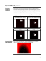
1
ProtoArray® Protein Microarray PPI
Kits for V5 Epitope-Tagged Proteins
For detecting protein-protein interactions (PPI) using a
human or yeast protein microarray and a V5 epitope-tagged
protein
Catalog nos. PA013, PA0121013, and PAH0524013
Version D
18 October 2006
25-0785
Corporate Headquarters
Invitrogen Corporation
1600 Faraday Avenue
Carlsbad, CA 92008
T: 1 760 603 7200
F: 1 760 602 6500
E: [email protected]
For country-specific contact information visit our web site at www.invitrogen.com
User Manual
ii
Table of Contents
Table of Contents ................................................................................................................................................. iii
Kit Contents and Storage .................................................................................................................................... iv
Accessory Products............................................................................................................................................. vii
Introduction ................................................................................................................... 1
Overview.................................................................................................................................................................1
Description of Kit Components ...........................................................................................................................4
ProtoArray® Human Protein Microarray ...........................................................................................................6
ProtoArray® Yeast Proteome Microarray ...........................................................................................................9
ProtoArray® Control Protein Microarray .........................................................................................................11
Experimental Overview ......................................................................................................................................14
Methods ....................................................................................................................... 17
Preparing the Protein Probe ...............................................................................................................................17
Probing the ProtoArray® Control Protein Microarray....................................................................................18
Probing the ProtoArray® Human or Yeast Microarrays.................................................................................24
Scanning Arrays...................................................................................................................................................27
Data Acquisition and Analysis ..........................................................................................................................30
Expected Results ..................................................................................................................................................34
Troubleshooting ...................................................................................................................................................38
Appendix...................................................................................................................... 41
Technical Support ................................................................................................................................................41
Product Qualification ..........................................................................................................................................42
Purchaser Notification ........................................................................................................................................43
References .............................................................................................................................................................45
iii
Kit Contents and Storage
Types of Kits
This manual is supplied with the following kits.
Product
Catalog no.
ProtoArray® Human Protein Microarray PPI Complete Kit v4.0 PAH0524013
for V5 epitope-tagged proteins
ProtoArray® Yeast Proteome Microarray PPI Complete Kit v1.1 PA0121013
for V5 epitope-tagged proteins
ProtoArray® Protein-Protein Interaction Application Kit
for V5 epitope-tagged proteins
Kit Components
PA013
The ProtoArray® PPI Kits for Epitope-Tagged Proteins include the following
components. For a detailed description of the contents of each component, see
pages v-vi.
Note: Catalog nos. PAH0524013 and PA0121013 include two ProtoArray® Human Protein
or Yeast Proteome Microarrays, as appropriate and one ProtoArray® Control Protein
Microarray.
Component
Catalog no.
PAH0524013 PA0121013
®
ProtoArray Human Protein Microarray nc v4.0
√
√
®
ProtoArray Yeast Proteome Microarray nc v1.1
ProtoArray Control Protein Microarray nc v4.0
√
√
Array Control Protein
√
√
Anti-V5-Alexa Fluor® 647 Antibody
√
√
√
®
√
√
√
®
√
√
√
®
ProtoArray PPI Buffer Module A
ProtoArray PPI Buffer Module B
Shipping and
Storage
PA013
The components included in the ProtoArray® PPI Kits for Epitope-Tagged
Proteins are shipped as detailed below. Upon receipt, store as indicated.
All kit components are stable for 12 months when stored properly.
Shipping
Storage
®
Components
Blue ice
-20°C
®
Blue ice
-20°C
®
ProtoArray Control Protein Microarray nc v4.0
Blue ice
-20°C
Array Control Protein
Dry ice
-20°C
ProtoArray Human Protein Microarray nc v4.0
ProtoArray Yeast Proteome Microarray nc v1.1
®
Anti-V5-Alexa Fluor 647 Antibody
Blue ice
4°C
®
Dry ice
-20°C
®
Blue ice
4°C
ProtoArray PPI Buffers Module A
ProtoArray PPI Buffers Module B
Continued on next page
iv
Kit Contents and Storage, Continued
ProtoArray®
Human or Yeast
Microarrays
Each ProtoArray® Microarray PPI Complete Kit contains mailers with the
following ProtoArray® Microarrays:
•
Human Kit (Catalog no. PAH0524013): Contains two ProtoArray® Human
Protein Microarrays nc v4.0
•
Yeast Kit (Catalog no. PA0121013): Contains two ProtoArray® Yeast
Proteome Microarrays nc v1.1
Store the microarrays at -20°C.
For details on array specifications, see pages 6-11.
ProtoArray®
Control Reagents
Each ProtoArray® Microarray PPI Complete Kit includes the following control
reagents. Store the microarray and Array Control Protein at -20°C.
Item
®
Composition
Amount
ProtoArray Control Protein
Microarray nc v4.0
--
1 array
Array Control Protein (V5-tagged,
biotinylated yeast Calmodulin
kinase)
0.5 mg/ml in phosphatebuffered saline (PBS), pH 7.4
40 µl
For details on array specifications, see page 11. For information about the Array
Control Protein, see page 19.
Continued on next page
v
Kit Contents and Storage, Continued
Anti-V5-Alexa
Fluor® 647
Antibody
The ProtoArray® Microarray PPI Complete Kits and the ProtoArray® ProteinProtein Interaction Application Kit each contain 1 tube of Anti-V5-Alexa Fluor®
647 Antibody with the following specifications:
•
Concentration: 0.5 mg/ml in 0.1 M NaCl, 0.1 M sodium phosphate, pH 7.5
with 2 mM sodium azide
•
Amount supplied: 80 µl
Store at 4°C. Protect the Anti-V5-Alexa Fluor® 647 Antibody from exposure to
light. Avoid repeated freezing and thawing of the antibody as this may result in
loss of activity.
ProtoArray® PPI
Buffer Module A
The ProtoArray® PPI Buffer Module A includes the following reagents.
Store at -20°C.
Note: The amount of reagents supplied is sufficient to perform 4 microarray screening
experiments.
Item
ProtoArray® PPI
Buffer Module B
Composition
Amount
Bovine Serum Albumin (BSA)
30% BSA in 0.85% NaCl
30 ml
DTT
1 M DTT in deionized water
400 µl
The ProtoArray® PPI Buffer Module B includes the following reagents.
Store at 4°C.
Note: The amount of reagents supplied is sufficient to perform 4 microarray screening
experiments.
Item
Amount
ProtoArray® Blocking Buffer (10X) 10X PBS, pH 7.4
1% Tween 20
12 ml
ProtoArray® Probe Buffer (5X)
5X PBS, pH 7.4
0.25% Triton X-100
25% Glycerol
175 ml
MgCl2
1 M MgCl2 in deionized water
4 ml
HybriSlip Cover Slip
60 mm x 22 mm, RNase-free
5 cover
slips per
pack
Array Chambers
--
2
™
vi
Composition
Accessory Products
Additional
Products
The table below lists additional products available separately from Invitrogen.
For more information about these products, refer to our web site
(www.invitrogen.com) or call Technical Support (page 41).
Product
Quantity
Catalog no.
ProtoArray® Reagents
ProtoArray® Human Protein Microarray nc v4.0
1 array
PAH052401
ProtoArray® Yeast Proteome Microarray nc v1.1
1 array
PA012101
®
1 array
PA1007
®
1 kit
PA014
ProtoArray Control Protein Microarray nc v4.0
ProtoArray Protein-Protein Interaction Buffer Modules
®
ProtoArray Human Protein Microarray PPI Complete Kit v4.0 1 kit
for biotinylated proteins
PAH0524011
ProtoArray® Yeast Proteome Microarray PPI Complete Kit v1.1 1 kit
for biotinylated proteins
PA0121011
ProtoArray® Protein-Protein Interaction Application Kit
for biotinylated proteins
1 kit
PA011
ProtoArray® Mini-Biotinylation Kit
1 kit
AL-01
®
ProtoArray Human Protein Microarray KSI Complete Kit v4.0 1 kit
for kinase substrate identification
PAH0524065
ProtoArray® Yeast Proteome Microarray KSI Complete Kit v1.1 1 kit
for kinase substrate identification
PA0121065
ProtoArray® Kinase Substrate Identification Application Kit
1 kit
PA015
®
1 array
PAH052406
®
1 array
PA012106
®
1 array
PA1002
®
1 kit
PA016
ProtoArray Human Protein Microarray mg v4.0
ProtoArray Yeast Proteome Microarray mg v1.1
ProtoArray Control Protein Microarray mg v4.0
ProtoArray Immune Response Biomarker Profiling
Application Kit
Continued on next page
vii
Accessory Products, Continued
Product
Quantity
Catalog no.
Reagents
Anti-V5 Antibody
50 µl
R960-25
Anti-V5-HRP Antibody
50 µl
R961-25
Anti-V5-AP Antibody
50 µl
R962-25
®
1 kit
WB7103
®
WesternBreeze Chemiluminescent Kit-Anti-Mouse
1 kit
WB7104
Nitrocellulose (0.45 µm)
20 membrane/filter
paper sandwiches
LC2001
1 kit
PQ10002-01
1 kit
PQ10001-01
WesternBreeze Chromogenic Kit-Anti-Mouse
ProQuest™ Two-Hybrid System
™
®
ProQuest Two-Hybrid System with Gateway Technology
Pre-Cast Gels and
Pre-made Buffers
A variety of pre-cast gels including NuPAGE® Novex® Pre-cast Gels and premade buffers for gel electrophoresis are available from Invitrogen. For details
on these products, visit our website at www.invitrogen.com or contact
Technical Support 41).
Vectors
A large selection of vectors is available from Invitrogen to express your protein
of interest with an N- or C-terminal V5-tag. For more details on the vectors
available, visit www.invitrogen.com or call Technical Support (page 41).
viii
Introduction
Overview
Introduction
The ProtoArray® Human Protein and Yeast Proteome Microarray PPI (proteinprotein interaction) Kits for Epitope-Tagged Proteins allow rapid and efficient
detection of human or yeast protein-protein interactions using a V5 epitopetagged protein probe of interest. The ProtoArray® Human Protein Microarray nc
contains thousands of purified human proteins, while the ProtoArray® Yeast
Proteome Microarray nc contains > 4000 purified yeast proteins from
Saccharomyces cerevisiae. In both cases, the proteins are printed in duplicate on a
nitrocellulose (nc)-coated glass slide. See the next page for an overview of the
system.
ProtoArray®
Microarray PPI
Applications
The ProtoArray® Human Protein and Yeast Proteome Microarrays allow you to:
V5 Epitope Tag
•
Detect novel protein-protein interactions
•
Validate previously observed protein-protein interactions
•
Confirm positive interactions using the identified interacting protein on the
array as a probe in reciprocal experiments
•
Test various experimental conditions for your protein-protein interactions
The V5 epitope tag is a 14 amino acid (GKPIPNPLLGLDST) epitope derived
from the P and V proteins of the paramyxovirus, SV5 (Southern et al., 1991). In
this application, you will express the V5 epitope tag as a fusion to your protein
of interest, and use the tag to detect your protein with a fluorescently-labeled
Anti-V5 Antibody.
Note: Anti-V5 Antibodies are also available from Invitrogen (page vii) for use in
Western analysis to detect recombinant proteins containing the V5 epitope.
Continued on next page
1
Overview, Continued
System Overview
To use the ProtoArray® Human Protein or Yeast Proteome Microarray PPI Kits,
you will:
•
Express your protein of interest as a fusion protein containing an N- or
C-terminal V5 epitope tag, and purify the fusion protein using a method of
choice.
•
Use the supplied Array Control Protein to probe the ProtoArray® Control
Protein Microarray nc to verify probing conditions and the background
level.
•
Probe the ProtoArray® Human Protein or Yeast Proteome Microarray nc,
with the V5-tagged protein probe to detect protein-protein interactions.
The ProtoArray® detection protocol includes instructions to block the array,
probe the array with your protein probe, wash to minimize non-specific
interactions, detect interactions using the Anti-V5-Alexa Fluor® 647 Antibody,
wash to remove unbound antibody, dry, scan the array to view results, acquire
the array image, and analyze data (see figure below). For a detailed
experimental workflow, see page 16.
00027734
Block (1 hr), add
protein probe with
V5 tag (90 min)
Advantages
Wash (10 min), add
®
Anti-V5-Alexa Fluor 647
Antibody for
detection (30 min)
Wash, dry, and
scan (1 hr)
Using the ProtoArray® Human Protein or Yeast Proteome Microarray PPI Kits
to detect protein-protein interactions offers the following advantages:
•
Provides a simple, rapid, and efficient method to identify protein
interactions within a day
•
Includes qualified buffers and detection reagents for probing, eliminating
the need to prepare reagents
•
Allows screening of your protein of interest against thousands of human
proteins or yeast proteins
•
Provides sensitive, stable, fluorescence detection using the Alexa Fluor® 647
dye
•
Built-in controls are printed on each array to control for background and
detection
•
Arrays are compatible with most commercially available fluorescence
microarray scanners
Continued on next page
2
Overview, Continued
The reagents and protocols included with the ProtoArray® Human Protein or
Yeast Proteome Microarray PPI Kits support probing microarrays with a
protein probe containing an N- or C-terminal V5 tag.
Our studies have shown that proteins with other epitope tags such as FLAG,
myc, and HA (hemagglutinin) can be used as probes against the ProtoArray®
Human Protein or Yeast Proteome Microarray nc using an appropriate labeled
antibody.
To detect the protein-protein interaction, use a labeled antibody specific for the
tag. The antibody can be directly labeled with a fluorescent dye such as Alexa
Fluor® 647 or labeled with a secondary antibody conjugated to a fluorescent dye
such as Alexa Fluor® 647. Make sure that direct labeling of the antibody does
not affect antibody activity.
•
Since most of the human and yeast proteins printed on the microarray
contain a GST (Glutathione-S-Transferase) fusion tag and some proteins
also contain polyhistidine (6x) tag, do not use an anti-GST antibody or antipolyhistidine antibody for detecting interactions on a ProtoArray® Human
or Yeast Protein Microarray nc. We strongly recommend that you probe the
ProtoArray® Human or Yeast Protein Microarray nc with only your
detection reagent to detect signals resulting due to interactions between the
detection reagent and proteins printed on the array.
•
The ProtoArray® Microarrays are not compatible for use with Alexa Fluor®
555 or Cy3™ dyes. Use of these fluorescent dyes results in high background
on the array as the nitrocellulose surface has high intrinsic fluorescence at
the wavelength used to visualize Alexa Fluor® 555 or Cy3™ dyes.
Important
Purpose of the
Manual
This manual provides the following information:
•
An overview of the ProtoArray® Human Protein, Yeast Proteome, and
Control Protein Microarrays
•
Instructions to probe the ProtoArray® Microarray with your protein probe
•
Guidelines to perform data analysis
•
Expected Results and Troubleshooting
3
Description of Kit Components
Components of
the ProtoArray®
PPI Kits
ProtoArray®
Human Protein
and Yeast
Proteome
Microarrays
The ProtoArray® Human Protein or Yeast Proteome Microarray PPI Complete
Kits for Epitope-Tagged Proteins include the following major components:
•
The ProtoArray® Human Protein or Yeast Proteome Microarray, a highdensity protein microarray that allows you to screen your protein of interest
(protein probe) against thousands of human proteins or the Saccharomyces
cerevisiae proteome, respectively
•
The ProtoArray® Control Protein Microarray and the Array Control Protein
for verification of the probing conditions and background levels
•
The ProtoArray® PPI Buffer Modules A and B contain pre-made, qualified
reagents for performing the blocking and washing steps during probing
•
The Anti-V5-Alexa® Fluor 647 Antibody for detection
The ProtoArray® Human Protein and Yeast Proteome Microarrays are highdensity protein microarrays containing human or S. cerevisiae proteins,
respectively. The ProtoArray® technology is based on the yeast protein
microarray technology developed by Zhu et al., 2001 to detect molecular
interactions with proteins.
Each human or S. cerevisiae open reading frame (ORF) is expressed as an Nterminal GST (Glutathione-S-Transferase) fusion protein, purified, and printed in
duplicate on a nitrocellulose-coated glass slide. The use of nitrocellulose as a
surface to print the arrays ensures maximum utility for protein assays since the
nitrocellulose surface is known to be compatible with a variety of protein
functions (Espejo et al., 2002; Kukar et al., 2002; Michaud et al., 2003). The
nitrocellulose coating is thin and does not interfere with scanning of the array.
Each ProtoArray® Microarray PPI Complete Kit includes two microarrays to
allow you to assay for protein interactions using different experimental
conditions or two distinct proteins. Using a labeled protein probe, you can screen
against the human or S. cerevisiae proteins within a day to identify protein-protein
interactions.
For array specifications and more details on how the human and yeast proteins
are prepared, see pages 6-9.
ProtoArray®
Control Protein
Microarray
The ProtoArray® Control Protein Microarray nc contains human and yeast
protein interactors and various controls printed on a nitrocellulose-coated glass
slide. Use the Control Microarray to validate the assay prior to probing the
ProtoArray® Human Protein or Yeast Proteome Microarray nc. One control array
is included in each kit; probe with the Array Control Protein supplied in the kit
(V5-tagged,biotinylated yeast calmodulin kinase) to validate assay conditions and
demonstrate a known protein-protein interaction between yeast calmodulin
kinase (Array Control Protein) and yeast calmodulin (Cmd1p-Ybr109C) on the
array.
For specifications and more details on the ProtoArray® Control Protein
Microarray, see page 11.
Continued on next page
4
Description of Kit Components, Continued
ProtoArray® PPI
Buffers Module
The ProtoArray® PPI Buffers Module A and B include qualified reagents used in
the blocking, washing, and detection steps during probing of the ProtoArray®
Microarrays. The pre-made buffers provide consistent results and eliminate the
time required to prepare reagents.
ProtoArray® PPI Buffers Module B includes HybriSlip™ cover slips that hold a
small reagent volume to minimize the amount of valuable probe used and
prevent evaporation of reagents. Array Chambers are also included in the module
for washing the microarrays.
Alexa Fluor® 647
Detection
The high sensitivity, low background, signal stability, and commercial availability
of fluorescence microarray scanners make fluorescence detection the preferred
method for detecting protein-protein interactions on microarrays.
The ProtoArray® Human Protein and Yeast Proteome Microarray PPI Kits include
the Anti-V5-Alexa Fluor® 647 Antibody (i.e. Mouse Anti-V5 Antibody conjugated
to Alexa Fluor® 647) for detection of the V5-tagged protein probe. The Alexa
Fluor® 647 fluorophore is brighter and more stable than other commercially
available dyes such as Cy™ Dyes and is more sensitive for detecting interactions
on protein arrays. We have demonstrated that detection with Alexa Fluor® 647
produces approximately 2-fold higher signal/background ratios than Cy5™
detection.
ProtoArray® PPI
Application Kit
The ProtoArray® Protein-Protein Interaction Application Kit includes
ProtoArray® PPI Buffer Modules A and B, and the Anti-V5-Alexa Fluor® 647
Antibody only. You will need to obtain a ProtoArray® Human Protein or Yeast
Proteome Microarray nc separately from Invitrogen before performing a
microarray screening experiment.
ProtoArray®
Central Portal
The ProtoArray® Central Portal at www.invitrogen.com/protoarray provides a
web-based user interface to access ProtoArray® specific information including
online tools, applications, and other resources. You will also use the portal to
retrieve ProtoArray® Lot Specific information (see page 30), which is required for
analysis of the array data and identification of statistically significant interactions.
ProtoArray®
Prospector
The ProtoArray® Prospector software quickly analyzes the microarray data
acquired from the image acquisition software and easily identifies significant
hits, saving you time and effort. In addition, the software has features that
allow you to modify the analysis method and compare data obtained from
different microarrays.
The ProtoArray® Prospector software and manual are available free-of-charge
to ProtoArray® users, and are accessible online at the ProtoArray® Central Web
Portal. To download the ProtoArray® Prospector software and manual, go to
www.invitrogen.com/protoarray, and click on Online Tools tab.
5
ProtoArray® Human Protein Microarray
Introduction
The ProtoArray® Human Protein Microarray nc is a high-density protein
microarray containing thousands of human proteins. Each human open reading
frame (ORF) is expressed as an N-terminal GST fusion protein, purified, and
printed in duplicate on a nitrocellulose-coated glass slide. This section provides
details about the human protein microarray including array specifications and
preparation of proteins.
Note: The ProtoArray® Human Protein Microarray PPI Complete Kit includes
2 ProtoArray® Human Protein Microarrays.
Human Protein
Microarray
Specifications
The specifications for the ProtoArray® Human Protein Microarray nc are listed
below.
Dimensions:
1 inch x 3 inch (25 mm x 75 mm)
Material:
Glass slide coated with nitrocellulose membrane
Membrane Size:
20 mm x 60 mm
Membrane Properties:
Thickness: 15-20 µm; Pore Size: 0.2 µm
Each microarray has a barcode for tracking samples. The barcode is also used to
retrieve array specific information from the ProtoArray® Central Portal (see
page 30).
Array
Specifications
The array specifications for the ProtoArray® Human Protein Microarray nc are
listed below. The proteins on the microarray are printed in 48 subarrays and are
equally spaced in vertical and horizontal directions.
For details on the subarray layout, and human protein and control spots on the
ProtoArray® Human Protein Microarray nc, go to the ProtoArray® Central Portal
at www.invitrogen.com/protoarray.
Total Subarrays:
48 (4 columns x 12 rows)
Subarray Size:
4400 µm x 4400 µm
Subarray Dimensions:
20 rows x 20 columns
Median Spot Diameter:
~150 µm
Spot Center to Center Spacing:
220 µm
Distance Between Subarrays:
100 µm
Replicates per Sample:
2
Total human protein spots on v4.0
~8000*
®
*Refer to ProtoArray Central Portal for exact number of human proteins printed on the
microarray.
Continued on next page
6
ProtoArray® Human Protein Microarray, Continued
Array Content
The majority of human protein collection is derived from the human Ultimate™
ORF (open reading frame) Clone Collection available from Invitrogen (see
http://orf.invitrogen.com for more information). Each Ultimate™ ORF Clone is
full insert sequenced and is guaranteed to match the corresponding GenBank®
amino acid sequence.
Some of the human proteins printed on the array represent the human protein
kinase collection derived from full insert sequenced clones but are not
Ultimate™ ORF Clones. Some of the kinases from the kinase collection have
been cloned as catalytic domains rather than full-length proteins. About
250 proteins printed on the array are derived from the purified protein kinase
collection available from Invitrogen. Approximately 25 proteins, peptides, and
nucleic acids that have been demonstrated to be antigens in a variety of
autoimmune diseases are also printed on the array.
For accession number and amino acid sequence for each protein as well as
information on peptides and nucleic acids printed on the array, download the
Protein Information File from www.invitrogen.com/protoarray as described on
page 30.
Expression and
Purification of
Human Proteins
Almost all clones used to generate the human protein collection are entry clones
consisting of a human ORF cloned into a Gateway® entry vector. Each entry
clone is subjected to a LR recombination reaction with a Gateway® destination
vector to generate an expression clone. The expression clone is then used to
express the protein (as an N-terminus GST-fusion protein in some clones) using
the Bac-to-Bac® Baculovirus Expression System available from Invitrogen. For
more information on the Bac-to-Bac® Baculovirus Expression System, visit
www.invitrogen.com.
The LR reaction mix obtained after performing the LR reaction is transformed
into competent DH10Bac™ E. coli to generate a recombinant bacmid. The high
molecular weight recombinant bacmid DNA is isolated and transfected into Sf9
insect cells to generate a recombinant baculovirus that is used for preliminary
expression experiments. After the baculoviral stock is amplified, the high-titer
stock is used to infect Sf9 insect cells for expression of the recombinant protein
of interest.
After verifying that each clone expresses a protein of the expected molecular
weight by western blotting, the proteins are expressed and purified using highthroughput procedures. The expressed proteins are purified by affinity
chromatography under conditions optimized to obtain maximal protein
integrity, function, and activity.
Approximately 5000 human proteins printed on the ProtoArray® Human
Protein Microarray nc v4.0 were also present on the previous version of the
product, ProtoArray® Human Protein Microarray nc v3.0. However, not all
proteins printed on ProtoArray® Human Protein Microarray nc v3.0 are also
printed on ProtoArray® Human Protein Microarray nc v4.0. To obtain a list of
ProtoArray® Microarray nc v3.0 human proteins that are not printed on
ProtoArray® Microarray nc v4.0, contact Technical Support (page 41).
Continued on next page
7
ProtoArray® Human Protein Microarray, Continued
Controls
Various proteins and controls are printed on each ProtoArray® Human Protein
Microarray to allow you to verify background and detection conditions during
probing. For details, see page 12.
Printing the
Human
ProtoArray®
The purified human proteins are printed on nitrocellulose-coated slides in a
dust-free, temperature, and humidity controlled environment to maintain
consistent quality of the microarrays. The arrays are printed using an
automated process on an arrayer that is extensively calibrated and tested for
printing ProtoArray® Microarrays.
Maintaining
Stringent Quality
Control
ProtoArray® Human Protein Microarrays are produced using rigorous
production and quality control procedures with an integrated data
management system to ensure consistent results with every array and
maximize inter-and intra-lot reproducibility.
Pre-Printing Quality Control
Prior to production, the arrayer and supporting components are tested and
adjusted to production specifications. To maintain protein stability and
function, arrays are printed at 6ºC under controlled environmental conditions.
Post-Printing Quality Control
After production each microarray is visually inspected for obvious defects that
could interfere with the experimental results. To control for the quality of the
printing process, several microarrays from each lot are probed with an anti-GST
antibody. Since the proteins contain a GST fusion tag, probing the microarrays
with an anti-GST antibody allows identification of irregular spot morphology
or missing spots. The arrays are functionally qualified by probing control
proteins to detect the appropriate protein-protein interactions.
For detailed product qualification, see page 42.
Detecting
Reciprocal
Interactions
ProtoArray® Human Protein Microarrays are ideal for detecting reciprocal
interactions since the microarrays are manufactured under highly controlled
conditions to ensure maximum protein function.
Once you have identified a positive interaction using the ProtoArray® Human
Protein Microarray nc, use the identified interacting protein from the array as a
probe for probing another human microarray.
For example, perform an initial probing with calmodulin as a probe with a
ProtoArray® Human Protein Microarray nc to detect the interacting protein,
calmodulin kinase. Then perform the reciprocal interaction with another
human microarray using calmodulin kinase as the probe to detect the
interacting protein, calmodulin. The ability to observe reciprocal interactions
indicates that the proteins maintain a proper folded state on the array.
8
ProtoArray® Yeast Proteome Microarray
Introduction
The ProtoArray® Yeast Proteome Microarray nc is a high-density protein
microarray containing the majority of proteins from S. cerevisiae. Each S. cerevisiae
open reading frame (ORF) is expressed as an N-terminal GST-6xHis fusion
protein, purified, and printed in duplicate on a nitrocellulose-coated glass slide.
This section provides details about the yeast proteome microarray including
array specifications and preparation of proteins.
Note: The ProtoArray® Yeast Proteome Microarray PPI Complete Kit includes
2 ProtoArray® Yeast Proteome Microarrays.
Yeast Proteome
Microarray
Specifications
The specifications for the ProtoArray® Yeast Proteome Microarray nc are listed
below.
Dimensions:
1 inch x 3 inch (25 mm x 75 mm)
Material:
Glass slide coated with nitrocellulose membrane
Membrane Size:
20 mm x 60 mm
Membrane Properties:
Thickness: 15-20 µm; Pore Size: 0.2 µm
Each microarray has a barcode for tracking samples. The barcode is also used to
retrieve array specific information from the ProtoArray® Central Portal (see
page 30).
Array
Specifications
The array specifications for the ProtoArray® Yeast Proteome Microarray nc are
listed below.
The proteins on the microarray are printed in 48 subarrays and are equally
spaced in vertical and horizontal directions.
For details on the subarray layout, and yeast protein and control spots on the
ProtoArray® Yeast Proteome Microarray nc, go to the ProtoArray® Central Portal
at www.invitrogen.com/protoarray.
Note: The subarray layout and controls are different in ProtoArray® Yeast Proteome
Microarray nc v1.1 as compared to the previously available ProtoArray® Yeast Proteome
Microarray nc v1.0.
Total Subarrays:
48 (4 columns x 12 rows)
Subarray Size:
4400 µm x 4400 µm
Subarray Dimensions:
16 rows x 20 columns
Median Spot Diameter:
~150 µm
Spot Center to Center Spacing:
220 µm
Distance Between Subarrays:
100 µm
Replicates per Sample:
2
Total Yeast Proteins Spots on v1.1:
>4000*
®
*Refer to ProtoArray Central Portal for exact number of human proteins printed on the
microarray.
Continued on next page
9
ProtoArray® Yeast Proteome Microarray, Continued
Preparing Yeast
Proteins
The yeast proteome collection is derived from the S. cerevisiae clone collection of
5800 yeast ORFs (Zhu et al., 2001). Each S. cerevisiae open reading frame (ORF) is
expressed as an N-terminal GST-6xHis fusion protein in the yeast expression
vector pEG-KG (Mitchell et al., 1993). The identity of each clone is verified using
5’-end sequencing and the expression of GST-tagged fusion protein by each
clone is confirmed with Western immunodetection using an anti-GST antibody.
The proteins are then expressed and purified using high-throughput
procedures.
Briefly yeast stocks are initiated in growth media, protein expression is induced
with galactose, and cell lysates prepared. The proteins are purified using
glutathione affinity chromatography, eluted, and purified proteins are used to
spot the proteome microarray.
Printing the Yeast
ProtoArray®
The purified yeast proteins are printed on nitrocellulose-coated slides in a dustfree, temperature, and humidity controlled environment to maintain consistent
quality of microarrays. The arrays are printed using an automated process on
an arrayer that is extensively calibrated and tested for printing ProtoArray®
Microarrays.
Controls
Various proteins and controls are printed on each ProtoArray® Yeast Proteome
Microarray to allow you to verify background and detection conditions during
probing. For details, see page 12.
Detecting
Reciprocal
Interactions
The ProtoArray® Yeast Proteome PPI Microarrays are ideal for detecting
reciprocal interactions since the microarrays are manufactured under highly
controlled conditions to ensure maximum protein function.
Once you have identified a positive interaction using the ProtoArray® Yeast
Proteome Microarray nc, use the identified interacting protein from the array as
a probe for to probe another ProtoArray® Yeast Proteome Microarray nc to
confirm the reciprocal interaction. For examples of reciprocal interactions, see
page 37.
Maintaining
Stringent Quality
Control
10
The ProtoArray® Yeast Proteome Microarrays are produced using the same
rigorous production and pre-printing and post-printing quality control
procedures used to produce the human protein microarrays (see page 8). For
detailed product qualification, see page 42.
ProtoArray® Control Protein Microarray
Introduction
The ProtoArray® Control Protein Microarray nc contains protein interactors and
various controls printed on a nitrocellulose-coated glass slide. The Control
Protein Microarrays allow you to validate probing procedures prior to probing
the ProtoArray® Human Protein or Yeast Proteome Microarray nc.
Details about the ProtoArray® Control Protein Microarray are described in this
section.
Control
Microarray
Specifications
The specifications for the ProtoArray® Control Protein Microarray are listed
below.
Dimensions:
1 inch x 3 inch (25 mm x 75 mm)
Material:
Glass slide coated with nitrocellulose
membrane
Membrane Size:
20 mm x 60 mm
Membrane Properties:
Thickness: 15-20 µm; Pore Size: 0.2 µm
Each microarray has a barcode for tracking samples. The barcode is also used to
retrieve array specific information from the ProtoArray® Central portal (see
page 30).
Control Array
Specifications
The control array specifications are listed below.
The proteins on the microarray are printed in 48 subarrays and are equally
spaced in vertical and horizontal directions.
For details on the subarray layout and control protein spots, go to the
ProtoArray® Central portal at www.invitrogen.com/protoarray.
Total Subarrays:
48 (4 columns x 12 rows)
Subarray Size:
4400 µm x 4400 µm
Subarray Dimensions:
8 rows x 20 columns
Median Spot Diameter:
~150 µm
Spot Center to Center Spacing:
220 µm
Distance Between Subarrays:
100 µm
Replicates per Sample:
2
Continued on next page
11
ProtoArray® Control Protein Microarray, Continued
Controls Printed
on Each
ProtoArray®
Microarray
Various proteins and controls are printed on each ProtoArray® Human Protein,
Yeast Proteome, and Control Protein Microarray to allow you to verify
reagents, background, and detection conditions used during probing. The table
below lists the controls printed on each ProtoArray® Microarray.
Protein
Function
Control Spots required for PPI Data Analysis
Alexa Fluor® Antibody
(Rabbit anti-mouse IgG Antibody
labeled with Alexa Fluor® 647,
Alexa Fluor® 555, and Alexa
Fluor® 488)
Serves as a positive control for fluorescence scanning and for
orientation of the microarray image.
Bovine Serum Albumin (BSA)
A negative control for non-specific protein interactions.
Biotinylated Anti-mouse
Antibody
A positive control for interaction with streptavidin-labeled
detection reagent.
V5 Control Protein (biotinylated,
V5-tagged control protein)
A positive control for detection with the Anti-V5-Alexa Fluor®
647 Antibody.
Human IgG Protein Gradient
A positive control for the immune response serum profiling
application. Interacts with Alexa Fluor® 647 goat anti-human IgG.
Anti-Human IgG Antibody
Gradient (goat anti-human IgG)
A positive control for the immune response serum profiling
application. Interacts with serum IgG antibodies which are then
bound by Alexa Fluor® 647 goat anti-human IgG.
Yeast calmodulin (Cmd1p)
A positive control for protein-protein interaction application
and interacts with the Array Control Protein.
GST Protein Gradient
Serves as a negative control and signals are used by ProtoArray®
Prospector software for background and statistical significance
calculations.
Control Spots NOT required for PPI Data Analysis
12
Fiduciary Kinases
Kinases autophosphorylate and produce fiduciary (marker)
signals which are used for orientation of the microarray image;
also serves as a positive control for the radiolabel and assay
conditions.
Control Kinase Substrate
A substrate for the Control Kinase used to verify assay
conditions. The Control Kinase phosphorylates the Control
Kinase Substrate.
CAMK2A (Calcium/calmodulindependent protein kinase II
alpha)
A human protein kinase that is used as a positive control for the
small molecule profiling application.
ProtoArray® Control Protein Microarray, Continued
The yeast calmodulin protein (Cmd1p; expressed as described on page 10) is
printed on each microarray. When probing the ProtoArray® Control Protein
Microarray nc with the Array Control Protein (i.e. V5-tagged, yeast calmodulin
kinase), these proteins interact. This interaction can be used to verify the reagents
and procedures used to probe the human and yeast microarrays.
Maintaining
Stringent Quality
Control
The ProtoArray® Control Protein Microarrays are produced using the same
rigorous production and pre-printing and post-printing quality control
procedures used to produce the ProtoArray® Human Protein and Yeast Proteome
Microarrays (see page 8). In addition, the control arrays are functionally qualified
by probing the arrays with the Array Control Protein (V5-tagged, yeast
calmodulin kinase) to detect the appropriate interaction with calmodulin. For
detailed product qualification, see page 42.
13
Experimental Overview
Experimental
Timeline
The recommended experimental timeline is outlined below. A detailed
experimental workflow is shown on the next page.
Purify Protein Probe
Confirm presence of V5-tag
with Western blot or ELISA
Day 1
Probe Control Array with
Array Control Protein
Scan Array and
Analyze Results
Probe Human or Yeast Array
Day 2
Scan Array and
Analyze Results
Continued on next page
14
Experimental Overview, Continued
Control Array
Experimental
Workflow
The experimental workflow for probing the ProtoArray® Control Protein
Microarray nc with the Array Control Protein is shown below.
Array Control
Protein
Control Array
Observe Alexa Fluor®
Ab and Control
Protein Signals
No
See
Troubleshooting
Yes
Probe Human
or Yeast Array
Continued on next page
15
Experimental Overview, Continued
Protein Array
Experimental
Workflow
The experimental workflow for probing the ProtoArray® Human Protein or Yeast
Proteome Microarray nc with your V5-tagged probe is shown below.
Purify
Protein
Protein
Probe with
V5 tag
Quantify
Protein
Probe
Protein
Array
Do I have
enough
protein
Observe
Alexa Fluor® Ab
and control
signals
No
Yes
No
Confirm
presence
of V5 tag
Yes
See
Troubleshooting
Protein has
a V5 tag
Yes
Observe
Protein
Interactions
No
No
Probe another
protein array
with higher
V5-tagged probe
concentration
16
Yes
Probe another
protein array
with different
probe concentration
or another V5tagged protein
probe
Methods
Preparing the Protein Probe
Introduction
Before using the ProtoArray® Human Protein or Yeast Proteome Microarray
PPI Kit, you will need your purified protein of interest to probe the microarray.
The protein of interest must contain a V5 epitope tag at the N- or C-terminus of
the protein (see below). You may purify proteins using any method of choice.
You can use proteins purified from E. coli, yeast cells, or higher eukaryotes to
probe the ProtoArray® Microarray.
The amount of protein and quality of protein required for probing are
described below.
Generating V5Tagged Protein
Probe
To generate your protein probe with a V5 tag, you need to express your protein
of interest as a fusion protein in an expression vector containing a V5 tag at the
N-or C-terminus of the protein.
A large variety of vectors is available from Invitrogen for expression of your
protein of interest with an N- or C-terminal V5-tag. For more information about
the vectors available, visit www.invitrogen.com or call Technical Support (see
page 41).
Protein Amount
and Quality
After you have expressed your V5-tagged fusion protein, follow the guidelines
below to purify and prepare the protein probe.
•
Purify the protein using native conditions.
•
Proteins must be > 90% pure as determined by Coomassie® staining.
•
Verify the presence of the V5 tag using Western detection with an Anti-V5
Antibody (see page vii).
Note: To ensure that the V5 tag is accessible under the native conditions used for
probing microarrays, perform ELISA of your V5-tagged protein probe using an
Anti-V5 Antibody (page vii).
•
Check the functionality of the protein probe using a method of choice.
•
Make sure the protein probe is soluble and active in buffers used for
probing the microarray (see recipe on page 20).
•
You need at least 6 µg of your purified, V5-tagged protein probe at a
recommended protein concentration of 50 µg/ml to probe each
ProtoArray® Microarray.
17
Probing the ProtoArray® Control Protein Microarray
Introduction
The ProtoArray® Control Protein Microarray nc allows you to verify probing
conditions. Probe the ProtoArray® Control Protein Microarray nc prior to
probing the ProtoArray® Human Protein or Yeast Proteome Microarrays.
Instructions are provided in this section to probe the ProtoArray® Control
Protein Microarray with the Array Control Protein supplied with the kit.
ProtoArray® PPI
Buffers Modules
The ProtoArray® PPI Buffers Module A and B supplied with the complete kits
include qualified reagents for blocking, washing, and detection during the
microarray probing procedure. The pre-made buffers provide consistent results
and eliminate the time required to prepare reagents.
ProtoArray® PPI Buffers Module B also includes HybriSlip™ cover slips that hold
a small reagent volume to minimize the amount of valuable probe used and
prevent evaporation of reagents. Array Chambers are also included in the
module for washing the microarrays.
ProtoArray®
Application Kit
The ProtoArray® Protein-Protein Interaction Application Kit includes
ProtoArray® PPI Buffer Modules A and B and the Alexa Fluor® detection reagent.
The use of the application kit provides consistent results and eliminates the time
required to prepare reagents.
The ProtoArray® PPI Buffer Modules A and B include qualified reagents for
blocking, washing, and probing during the microarray probing procedure (see
above).
Before using the ProtoArray® Application Kit, you need to purchase a
ProtoArray® Human, Yeast, or Control Protein Microarray nc.
Materials Needed
Important
•
ProtoArray® Control Protein Microarray nc (included in the complete kits
only or available separately)
•
ProtoArray® PPI Buffers Module A and B (included with the kit)
•
Anti-V5-Alexa Fluor® 647 Antibody (included with the kit; keep on ice in dark
until immediately before use)
•
Array Control Protein in Probing Buffer (included in the complete kits only;
see next page)
•
Sterile 50 ml conical tube
•
Ice bucket
•
Deionized water
•
Optional: Microarray slide holder and centrifuge equipped with a plate holder
The ProtoArray® Control Protein Microarray nc can only be used once. Do not
reuse the microarray or reprobe the same microarray with another probe.
Continued on next page
18
Probing the ProtoArray® Control Protein Microarray, Continued
Experimental
Outline
Important
Guidelines
Array Control
Protein
1.
Block the ProtoArray® Control Protein Microarray nc.
2.
Probe with the Array Control Protein (V5-tagged calmodulin kinase).
3.
Perform detection with the Anti-V5-Alexa Fluor® 647 Antibody.
4.
Dry the array for scanning.
5.
Scan the array to obtain an array image and analyze results.
Since proteins are sensitive to various environmental factors, each array is
produced in an environment-controlled facility to ensure protein integrity and
maintain consistency. To obtain the best results and avoid any damage to the
array or array proteins, always handle the ProtoArray® Microarrays with care
using the following guidelines:
•
Always wear clean gloves while handling microarrays
•
Do not touch the surface of the array to avoid any damage to the array
surface resulting in uneven or high background
•
Maintain the array and reagents at 2-8°C during the experiment
•
To prevent condensation on the array that may reduce protein activity or
alter spot morphology, allow the mailer containing the array to equilibrate
at 4°C for at least 15 minutes prior to removing the array from the mailer
and immersing the array immediately in blocking solution equilibrated at
4°C
•
Perform array experiments at a clean location to avoid dust or
contamination and filter solutions if needed (particles invisible to eyes can
produce high background signals and cause irregular spot morphology)
•
Avoid drying of the array during the experiment and ensure the array is
completely covered with the appropriate reagent during all steps of the
protocol
•
Always dry the array prior to scanning and scan the array on the same day
at the end of the experiment
•
Do not dry the array using compressed air or commercial aerosol sprays
•
Avoid exposing the array to light after probing with Anti-V5-Alexa Fluor®
647 antibody
The Array Control Protein (included in the complete kits) is an N-terminal,
V5-tagged, biotinylated yeast calmodulin kinase (Cmk1p), and interacts with
calmodulin (Cmd1p) printed on the ProtoArray® Control Protein Microarray nc.
If you see an interaction between the Array Control Protein and calmodulin, this
indicates that the probing procedure is performed correctly.
Continued on next page
19
Probing the ProtoArray® Control Protein Microarray, Continued
Preparing Buffers
Prepare the following buffers fresh prior to use. The recipe below provides
sufficient buffer to probe 1 microarray.
PBST Blocking Buffer
1X PBS
1% BSA
0.1% Tween 20
1.
Use reagents provided in the kit to prepare 30 ml PBST Blocking Buffer as
follows:
ProtoArray® Blocking Buffer (10X)
30% BSA
Deionized water
2.
3 ml
1 ml
to 30 ml
Mix well (do not vortex) and store on ice until use.
Probing Buffer
1X PBS
5 mM MgCl2
0.5 mM DTT
0.05% Triton X-100
5% Glycerol
1% BSA
1.
Use reagents provided in the kit to prepare 180 ml Probing Buffer as
follows:
ProtoArray® Probe Buffer (5X)
1 M DTT
1 M MgCl2
30% BSA
Deionized water
2.
36 ml
90 µl
0.9 ml
6 ml
to 180 ml
Mix well (do not vortex) and store on ice until use.
After preparing PBST Blocking Buffer and Probing Buffer, immediately return
the remaining 5X ProtoArray® Probe Buffer, 10X ProtoArray® Blocking Buffer,
and 1 M MgCl2 to 4°C, and the 30% BSA and 1 M DTT to -20°C.
Preparing the
Probe
Mix 1.2 µl Array Control Protein (included in the complete kits) with 120 µl of
Probing Buffer. Mix well (do not vortex) and store on ice until use.
Continued on next page
20
Probing the ProtoArray® Control Protein Microarray, Continued
Before Starting
Blocking Step
•
Before starting the probing procedure, make sure you have all items on hand
especially buffers (previous page), probes in Probing Buffer (previous page),
Array Chambers (included in the kit), and HybriSlips™ (included in the kit).
•
Make sure the buffers are cold. Store buffers on ice until use. Place the Array
Chambers on ice to chill the chamber until use.
•
Review Important Guidelines on page 19 prior to starting the probing
procedure.
Instructions for blocking the control microarray are described below:
1.
Remove the mailer containing the ProtoArray® Control Protein Microarray nc
from storage at -20°C and immediately place the mailer at 4°C.
2.
Allow the mailer containing the array to equilibrate at 4°C for at least
15 minutes prior to performing the blocking step.
3.
Perform blocking in the mailer. Ensure that the microarray is placed properly
in the chamber with the printed (white) side facing up. Add 30 ml PBST
Blocking Buffer (page 20) to the chamber containing the array.
Note: You can block 2 arrays simultaneously in the mailer using 30 ml PBST Blocking
Buffer.
4.
Incubate for 1 hour in the cold room with gentle shaking (~50 rpm).
5.
Decant the PBST Blocking Buffer. Drain excess buffer by inverting the mailer
on paper towels for a few seconds. Remove the array from the mailer. Tap
one edge of the array gently on a laboratory wipe for a few seconds to drain
any buffer without allowing the array to dry. Place the array on a clean, flat
surface with the printed side of the array facing up.
6.
Proceed immediately to Probing the Array, next page.
Continued on next page
21
Probing the ProtoArray® Control Protein Microarray, Continued
Probing Control
Array
1.
Pipet 120 µl of the Array Control Protein (5 µg/ml) prepared in Probing Buffer
(page 20) on top of the array without touching the array surface. The liquid
quickly spreads over the nitrocellulose membrane.
2.
Carefully lift the HybriSlip™ cover slip from the support liner with forceps and
lay the clear side of HybriSlip™ on the array to cover the membrane area
without trapping any air-bubbles. The HybriSlip™ is designed to exactly cover
the membrane area. Gently adjust the HybriSlip™ to remove any air bubbles.
3.
Insert the array with HybriSlip™ into a separate 50 ml conical tube with the
printed side of the array facing up. Cap the conical tube.
4.
Place the conical tube on a flat surface such that the printed side of the array is
facing up and the tube is as level as possible. You can tape the conical tube on
the flat surface to avoid any accidental disturbances. Incubate the array in the
tube for 1.5 h at 2-8°C without shaking.
5.
Remove the array from the conical tube and insert diagonally (see Note below)
into the Array Chamber kept on ice.
Note: The microarray with HybriSlip™ does not fit on the rails of the chamber. You
must insert the microarray diagonally into the chamber.
6.
Using a sterile pipette, add 25 ml Probing Buffer (page 20) to the chamber wall
while keeping the chamber on ice. Avoid pipetting buffer directly onto the
array surface. Gently move the array in the chamber to dislodge the
HybriSlip™. Do not remove the HybriSlip™ with the forceps if the HybriSlip™ is
not dislodged from the array.
7.
Using forceps, carefully remove the HybriSlip™ without touching the array
surface. Discard the HybriSlip™. Reposition the array on the chamber rails.
8.
Incubate the array in Probing Buffer for ~1 minute on ice. Decant the Probing
Buffer. Invert chamber on paper towels to drain excess buffer.
9.
Repeat Step 8 two more times, using 25 ml Probing Buffer each time.
10. While the array is incubating, prepare Anti-V5-Alexa Fluor® 647 Antibody
solution (260 ng/ml) by mixing 16 µl Anti-V5-Alexa Fluor® 647 Antibody
(included with the kit) with 25 ml Probing Buffer.
11. After the third wash with Probing Buffer (Step 9), decant the buffer. Invert
chamber on paper towels for a few seconds to drain excess buffer. Add 25 ml
Anti-V5-Alexa Fluor® 647 Antibody solution from Step 10 to the chamber.
12. Incubate the chamber for 30 minutes on ice in the dark (cover the ice bucket).
Decant the buffer. Invert the chamber on paper towels for a few seconds to
drain excess buffer.
13. Slowly add 25 ml Probing Buffer onto the chamber wall while keeping the
chamber on ice. Avoid pipetting buffer directly onto the array surface.
14. Incubate the array in Probing Buffer for ~1 minute on ice. Decant the buffer.
Drain excess buffer by inverting chamber on paper towels for a few seconds.
15. Repeat Steps 13-14 two more times, using 25 ml Probing Buffer each time.
16. Proceed to Drying the Array, next page.
Continued on next page
22
Probing the ProtoArray® Control Protein Microarray, Continued
Drying the Array
Data Analysis
1.
Remove the array from the chamber at the end of the probing procedure. Tap
one edge of the array gently on a laboratory wipe for a few seconds to drain any
buffer.
2.
Place the array in a slide holder (or a sterile 50 ml conical tube, if you do not
have a slide holder) in a vertical orientation. Ensure the array is properly placed
and is secure in the holder to prevent any damage to the array during
centrifugation.
3.
Centrifuge the array in the slide holder or 50 ml conical tube at 800 x g for
3-5 minutes in a centrifuge (equipped with a plate rotor, if you are using the
slide holder) at room temperature.
4.
Place the array in a slide box and keep the box with the lid open in the dark for
30-60 minutes at room temperature to dry the array. Make sure that the array is
completely dry; there should be no translucent areas.
5.
Scan the array using a fluorescence microarray scanner (see page 27 for details).
After scanning and saving an image of the array, analyze results to identify
positive interactors. For more details, see page 30.
1.
To acquire data from the scanned image, use the barcode on the array to
download the .GAL file from ProtoArray® Central as described on page 30.
2.
Use the .GAL file and suitable microarray data acquisition software to
acquire pixel intensity values for all features on the control array.
3.
Analyze data using the guidelines on page 32 to determine significant
signals with the Array Control Protein.
Note: The expected results obtained after probing a Control Array are shown on
page 34. For troubleshooting, see page 38.
4.
Cleaning the
Chamber
After confirming the appropriate interactions on the Control Array, proceed
to Probing the ProtoArray® Human or Yeast Microarrays, next page.
At the end of probing experiments, clean the Array Chamber properly and
rinse with sterile water before re-using the chamber.
23
Probing the ProtoArray® Human or Yeast Microarrays
Introduction
After using the ProtoArray® Control Protein Microarray nc to verify probing
conditions and the background, proceed to probing the ProtoArray® Human
Protein or Yeast Proteome Microarray nc using your protein probe. Follow the
guidelines provided in this section.
Materials Needed
•
ProtoArray® Human Protein Microarray nc or ProtoArrray® Yeast Proteome
Microarray nc (included in the complete kit)
Note: If you have purchased the ProtoArray® Protein-Protein Interaction Application
Kit, you also need to purchase the ProtoArray® Human Protein or Yeast Proteome
Microarray separately.
Important
Experimental
Outline
Important
Guidelines
•
ProtoArray® PPI Buffers Module A and B (included in the kit)
•
Your V5-tagged protein probe in Probing Buffer (see next page)
•
Anti-V5-Alexa Fluor® 647 conjugate (keep on ice in the dark until immediately
before use)
•
Sterile 50 ml conical tube
•
Ice bucket
•
Deionized water
•
Optional: Microarray slide holder and centrifuge equipped with a plate holder
Each ProtoArray® Human Protein or Yeast Proteome Microarray nc can only be
used once. Do not re-use the array or reprobe the same array with another probe.
1.
Block the ProtoArray® Human Protein or Yeast Proteome Microarray nc.
2.
Probe with your V5-tagged protein probe.
3.
Detect with Anti-V5-Alexa Fluor® 647 Antibody.
4.
Dry the array for scanning.
5.
Scan the array to obtain an array image and analyze results.
Follow the important guidelines on page 19 to obtain the best results with the
arrays.
Continued on next page
24
Probing the ProtoArray® Human or Yeast Microarrays,
Continued
Probes for
Proteome Arrays
The ProtoArray® Human Protein or Yeast Proteome Microarray PPI Complete Kit
contains 2 human or yeast arrays, respectively, and can be probed using different
probing options as described below. Choose the option that best fits your needs.
The recommended starting protein probe concentration for probing each array
is 50 µg/ml.
Probing Options
•
You can probe both arrays simultaneously, probing one array with your
protein probe and the second array with no protein probe (negative control).
The negative control allows you to determine which signals are specific to
your probe.
OR
•
You can probe one array with your protein probe of interest at 50 µg/ml and
the second array with the Array Control Protein supplied with the complete
kit (positive control). The results from the positive control help you to
determine signals specific to your probe.
OR
•
You can probe one array with an initial probe concentration (e.g. 50 µg/ml). If
the initial signal is strong with low background, confirm the initial results
with the second array using the same experimental conditions. If the initial
results indicate weak signal and unacceptable signal-to-noise ratio, probe the
second array with a different probe concentration as described in the table
below:
Probe first array….
And….
Then Probe Second Array….
With 50 µg/ml probe
Weak signal
With 500 µg/ml probe
With 50 µg/ml probe
High background
With 5 µg/ml probe
Note: To identify protein-protein interactions specific to your protein probe, we
recommend probing a second array with another V5-tagged protein probe or the Array
Control Protein (see above). Probing using options 1 or 2 allows you to determine the
probe-specific interactions and helps you identify non-specific interactions.
Preparing Buffers
Prepare PBST Blocking Buffer and Probing Buffer as described on page 20.
Preparing Probes
You need 120 µl of your V5-tagged protein probe for probing one protein
microarray. Dilute the probe to 50 µg/ml in Probing Buffer. Mix well (do not
vortex) and store on ice until use.
Continued on next page
25
Probing the ProtoArray® Human or Yeast Microarrays,
Continued
Before Starting
Probing Arrays
•
Before starting the probing procedure, make sure you have all items on hand
especially buffers (see previous page), probes in Probing Buffer (see
previous page), Array Chambers (included in the kit), and HybriSlips™
(included in the kit).
•
Make sure the buffers are cold. Store buffers on ice until use. Place the Array
Chamber on ice to chill the chamber until use.
•
Review Important Guidelines on page 19 prior to starting the probing
procedure.
The options for probing the array are described on the previous page. Choose
the option that best fits your needs.
1.
Probe the ProtoArray® Human Protein or Yeast Proteome Microarray nc
using the procedure described on page 21.
2.
Dry the array as described on page 23.
3.
Scan the arrays as described on the next page and analyze results (page 30).
Examples of expected results obtained after probing the ProtoArray® Human
Protein or Yeast Proteome Microarrays are shown on pages 35 and 36,
respectively.
If you obtain weak signals or high background, see Troubleshooting, page 38.
26
Scanning Arrays
Introduction
Once you have probed the ProtoArray® with your V5-tagged protein, scan the
microarray using a fluorescence microarray scanner.
Materials Needed
You need a fluorescence microarray scanner to scan the ProtoArray® Human or
Yeast Microarray nc. To acquire ProtoArray® data from the image, you also
need an appropriate microarray data acquisition software.
The recommended microarray data acquisition software for analysis is
GenePix® Pro (Molecular Devices Corporation) or ScanArray® Software
(PerkinElmer, Inc.).
The scanner specifications are listed below and recommended scanners are
listed on the next page.
Experimental
Outline
Scanner
Specifications
Array Compatibility
Detection
1.
Insert array into the fluorescence microarray scanner.
2.
Adjust scanner settings.
3.
Preview the microarray and adjust settings, if needed.
4.
Scan the microarray.
5.
Align grid over spots and use image analysis software to align features.
6.
Export and analyze results.
The fluorescence microarray scanner specifications required to image the
ProtoArray® Human Protein or Yeast Proteome Microarray nc are listed in the
table below.
Size
Standard 1" x 3" or 25 mm x 75 mm microscope slides
Thickness
1 mm
Light and Detector
Orientation
Facing array
Scanned Area
22 mm x 73 mm
Focus
Auto focus or adjustable (+200 µm)
Excitation
635 nm or equivalent
Detection limit
0.1 fluor/µm2
Resolution
<10 µm
Dynamic Range
>3 orders of magnitude
Output
16-bit TIFF
27
Scanning Arrays, Continued
Recommended
Scanners
The following scanners are compatible for scanning ProtoArray® Human Protein
or Yeast Proteome Microarrays:
•
GenePix® 4000A (Molecular Devices Corporation)
•
GenePix® 4000B (Molecular Devices Corporation)
•
GenePix® Professional 4200A (Molecular Devices Corporation)
•
GenePix® Personal 4100A (Molecular Devices Corporation)
•
ScanArray® Lite (PerkinElmer, Inc.)
•
ScanArray® Express (PerkinElmer, Inc.)
•
ScanArray® Express HT (PerkinElmer, Inc.)
•
LS Series Laser Scanner (Tecan Group AG)
The following scanners may be compatible with ProtoArray® Human Protein or
Yeast Proteome Microarrays:
•
AlphaArray® Reader (Alpha Innotech Corporation)
•
arrayWoRx®e 4-Color Biochip Reader (Applied Precision, LLC)
•
SpotLight™ (TeleChem International, Inc.)
The following scanners are not compatible with ProtoArray® Human Protein or
Yeast Proteome Microarrays:
•
GeneChip® Scanner 3000 (Affymetrix, Inc.)
•
DNA Microarray Scanner (Agilent Technologies, Inc.)
Unlike most DNA microarrays, you will scan the ProtoArray® Human Protein
or Yeast Proteome Microarray nc using only one color.
Continued on next page
28
Scanning Arrays, Continued
Scanning
Procedure
A brief procedure for scanning the ProtoArray® Human Protein or Yeast
Proteome Microarrays with a fluorescence microarray scanner at 635 nm is
described below.
For details on using a specific scanner, refer to the manual supplied with the
scanner.
The scanning time for each array is ~7-8 minutes.
1.
Start the appropriate array acquisition and analysis software on the
computer connected to the fluorescence microarray scanner.
2.
Open the microarray enclosure on the scanner.
3.
Place the ProtoArray® Human Protein or Yeast Proteome Microarray in the
holder such that the nitrocellulose-coated side of the array faces the laser
source and the barcode on the array is closest to the outside of the
instrument.
4.
Close the microarray enclosure on the scanner.
5.
Set the following settings to image the microarray:
6.
•
Wavelength: 635 nm
•
PMT Gain: 600
•
Laser Power: 100%
•
Pixel Size: 10 µm
•
Lines to Average: 1.0
•
Focus Position: 0 µm
Perform a preview to quickly scan the microarray. Adjust the PMT Gain, if
needed.
Note: The image should have very few saturated spots (white). Adjust settings such
that the Alexa Fluor® Ab spots are at or near the pixel saturation.
7.
Select the area of the array to scan in detail (include the barcode in the area
for record) and then scan the array to provide a high-resolution image.
8.
After acquiring the image, save the image to a suitable location as “multiimage TIFF” file. Be sure the barcode is included in the name of the image.
Note: Examples of image scans of control, human, and yeast arrays are shown on
pages 34-37.
9.
Open the microarray enclosure and remove the microarray from the holder.
10. Proceed to download lot specific information available on the ProtoArray®
Central Portal, next page.
To orient the results obtained from the .GAL file and ProtoArray® Prospector with
the array image, position the microarray image such that the barcode is at the
bottom of the image. In this orientation, the top left corner of the microarray image
is Block 1.
29
Data Acquisition and Analysis
Introduction
After scanning and saving an image of the array, download the protein array lot
specific information (including the .GAL file) from the ProtoArray® Central
Portal. Use the lot specific information to acquire and analyze the data to identify
protein-protein interactions.
Note: To familiarize yourself with the array and subarray layout, you may also download
a file showing the subarray layout from ProtoArray® Central. To access the file, go to
www.invitrogen.com/protoarray and click on Online Tools.
Important
GAL File
While downloading the lot specific information files, ensure that you are
downloading files that are associated with your specific barcode on the array.
Since lot specific information files are updated frequently based on recently
available sequence or protein information, make sure that you download the
latest version of the lot specific information files.
The .GAL (GenePix® Array List) files describe the location and identity of all
spots on the Human, Yeast, and Control microarrays and are used with the
microarray data acquisition software to generate files that contain pixel intensity
information for feature/spot and non-features of the slide.
The .GAL files are available for downloading from the ProtoArray® Lot Specific
Information available on ProtoArray® Central, see below.
Note: The .GAL files are text files that contain the data in a format specified by GenePix®
Pro Microarray data acquisition software. If you are using any other microarray data
acquisition software, you can use data from the .GAL files to generate files that are
compatible with your microarray data acquisition software.
ProtoArray®
Central
The ProtoArray® Central Portal provides a web-based user interface to retrieve
ProtoArray® Lot Specific information. This information (.GAL file) is required for
acquiring the array data.
If the scanner computer is connected to the Internet, then click on the link below to
connect to the portal. If the scanner computer is not connected to the internet,
download the array-specific information as described below to portable media and
then download the information onto the scanner computer.
1.
Go to www.invitrogen.com/protoarray and click on the Online Tools tab.
2.
Click on the link to ProtoArray® Lot Specific Information.
3.
Enter the array barcode in the Input Barcode Number(s) box. Click on the
Search button.
Continued on next page
30
Data Acquisition and Analysis, Continued
ProtoArray®
Central, continued
4.
For each input barcode, the following files are displayed:
.GAL file (LotNumber.gal):
This file is essential for data acquisition by the software and defines spot
locations and identities of all protein spots on the array. The file also includes
the “equivalent solution protein concentration” in nM for use during data
analysis.
Protein Information File (LotNumber_info.txt):
This file contains a listing and description of human proteins on the array.
Protein Sequence File (LotNumber_seq.txt):
This tab-delimited text file lists the GenBank® accession number, Ultimate™ ORF
Clone ID number (if available), FASTA header, and amino acid sequence of the
ORF for each array protein.
Control Data File (LotNumber_control.txt):
This file contains a description of control spots on the array.
Protein Application File (LotNumber_application.PAI):
ProtoArray® Prospector uses the Protein Application Files for data analysis.
Different PAI files are designed for different applications. For example,
ProtoArray® Prospector uses the file HA10756 PPI.PAI to analyze data from PPI
experiments performed on array from lot HA10756.
Slide Information File (LotNumber_slide.txt):
This file contains a listing of all barcodes associated with a specific lot of arrays.
5.
Download the files listed above for human or yeast array-specific information
from a specific lot. Use these files to interpret your results with the ProtoArray®
Human Protein or Yeast Proteome Microarray nc as described below.
Note: The file size for some files such as the Protein Sequence File may be> 1 MB.
6.
Start the microarray data acquisition software on the computer and open the
saved image (.tiff) from Step 8, page 29.
7.
To acquire data from ProtoArray® experiments:
•
For GenePix® Pro Software, download the .GAL files from ProtoArray®
Central for control or protein arrays, which defines the array grid
required by the microarray data acquisition software.
•
For other microarray data acquisition software, use data from the .GAL
files from ProtoArray® Central for control or protein arrays to generate
files that are compatible with your microarray data acquisition software
to define the array grid.
Scroll through the image to ensure that the grid is in the proper location for
each subarray. Adjust the subarray grid, if needed.
Continued on next page
31
Data Acquisition and Analysis, Continued
ProtoArray®
Central, continued
8.
After the grid is properly adjusted and all of the features are aligned,
save/export the results as a .GPR (GenePix® Results) file for data analysis
using ProtoArray® Prospector (next page). The results contain the pixel
intensity information for each spot/feature on the array and information on
additional parameters depending on the type of software used for data
acquisition.
Alternatively, save/export the results with an .xls extension or rename the .tab
or .gpr file using the .xls extension for data analysis using Microsoft® Excel.
Analyzing Data
After data acquisition, analyze the data to identify protein interactions.
Visual identification of interactions can be performed after initial identification
of significant interactions is done using the data analysis guidelines listed
below.
We recommend using the ProtoArray® Prospector software available from
Invitrogen for data analysis. This software allows rapid data analysis without
the need to perform any manual calculations. For more information, see below.
Performing the data analysis by importing the data file into Microsoft® Excel or
an equivalent spreadsheet program to identify potential substrates is not
recommended. This approach requires a certain degree of expertise with
statistics and Excel or another spreadsheet program.
Data Analysis
Using ProtoArray®
Prospector
The ProtoArray® Prospector software quickly analyzes the data acquired from
the image acquisition software and easily identifies statistically significant
interactors, saving you time and effort. In addition, the software has features
that allow you to modify the analysis method and compare data obtained from
different slides.
The ProtoArray® Prospector software and manual are available free-of-charge to
ProtoArray® users. To download the ProtoArray® Prospector software or
manual, go to www.invitrogen.com/protoarray, and click on the Online Tools
tab. Install the Basic version of ProtoArray® Prospector for data analysis.
The ProtoArray® Prospector software currently accepts the output files (.GPR)
generated by the GenePix® Pro microarray data acquisition software, and
analyzes the data using specified algorithms to generate a list of human
proteins showing significant interactions with the protein probe. If .GPR files
are not available, consult the ProtoArray® Prospector manual for guidelines to
format a results file that is compatible for import into ProtoArray® Prospector.
ProtoArray®
Prospector
Results
After data analysis, ProtoArray® Prospector presents a summary of the analyzed
data in a table format (see ProtoArray® Prospector manual for details).
The proteins that score as positive in the experiment are proteins that satisfy the
basic program options.
Based on the Z-score and available protein sequence information, we recommend
validating the protein-protein interaction by ProtoArray® Technology or other
methods as described on the next page.
Continued on next page
32
Data Acquisition and Analysis, Continued
The Next Step
After identifying a positive interaction on the ProtoArray® Human Protein or
Yeast Proteome Microarray nc, you may validate the protein-protein interaction
using the ProtoArray® Technology or other methods.
Using the ProtoArray® Technology, validate the protein-protein interactions by
performing experiments with additional arrays to ensure:
•
Reproducibility: Probe the ProtoArray® Human Protein or Yeast Proteome
Microarray nc using a similar or a different probe concentration to observe
similar interactions.
•
Specificity: Probe a ProtoArray® Human Protein or Yeast Proteome
Microarray nc with different V5-tagged proteins to identify interactions
specific to your protein probe of interest and also identify any non-specific
interactions.
•
Reciprocal Interactions: Determine reciprocal interactions as described on
page 10 using a purified protein probe. For an example, see page 37.
Other methods for validating protein-protein interactions include:
Accessing Clones
•
Yeast Two-Hybrid Systems (page vii)
•
Co-immunoprecipitation
•
Gel-shift assay
Since the majority of human proteins printed on the array are derived from the
Ultimate™ ORF Clone Collection or purified proteins (protein kinases) available
from Invitrogen, it is very easy to order the clone or purified protein
corresponding to the protein identified on the array and validate the
interaction.
Visit www.invitrogen.com/clones to access our clone collections. Each
Ultimate™ ORF Clone is full insert-sequenced and guaranteed to match the
corresponding GenBank® amino acid sequence. Contact Technical Support
(page 41) to order the purified protein kinases printed on the array.
Note: The yeast proteins printed on the array are derived from the Snyder collection
(Zhu et al., 2001). For information on obtaining the yeast clone corresponding to the
potential protein identified on the array, contact Technical Support (page 41).
33
Expected Results
Control Array
Probing Results
Results obtained after probing the ProtoArray® Control Protein Microarray nc v4.0
with the Array Control Protein (BioEase™-V5-tagged calmodulin kinase) are shown.
Image showing calmodulin (Cmd1p) signal when probed
with the Array Control Protein
Array image
Boxed Area shown in detail
Alexa Fluor® Ab
Cmd1p
V5
Control
Alexa Fluor®
Ab
•
Alexa Fluor® Ab signal
This is an antibody labeled with Alexa Fluor® 647. The fluorescent antibody
signals indicate that the array has been properly scanned, and are used as
reference spots to orient the microarray and help assign spot identities.
•
Cmd1p signal
The Array Control Protein (V5-tagged calmodulin kinase) binds to the
calmodulin printed on the array. The signal is used to verify the probing
procedure.
•
V5 Control signal
The Anti-V5-Alexa Fluor® 647 Antibody binds to a control protein (V5 Control)
containing an N-terminal V5 tag printed on the microarray. The signals indicate
that the antibody is functional and probing is performed properly. The signal is
also used to check the background.
Continued on next page
34
Expected Results, Continued
Human
ProtoArray® v4.0
Probing Results
The results obtained after probing the ProtoArray® Human Protein Microarray
nc v4.0 with 5 µg/ml of Array Control Protein (i.e. BioEase™-V5-tagged yeast
calmodulin kinase) is shown below.
Image showing Array Control Protein (probe) interacting with
Cmd1p on the array
Array Image
Boxed Area shown in detail
Alexa Fluor®
Ab
Cmd1p
Alexa
Fluor®
Ab
V5 Control
Alexa
Fluor®
Ab
Continued on next page
35
Expected Results, Continued
Yeast ProtoArray®
Probing Results
The results obtained after probing the ProtoArray® Yeast Proteome Microarray
nc v1.1 with 5 µg/ml of the Array Control Protein probe are shown below.
Image showing Array Control Protein (probe) interacting with
calmodulin (Cmd1p) on the array
Array Image
Boxed Area shown in detail
Alexa Fluor®
Ab
Cmd1p
Alexa
Fluor®
Ab
V5 Control
Alexa
Fluor®
Ab
Note: The column of interactions observed on the top left corner for the array is due to
calmodulin printed on the microarrays as control spots. The interaction of the yeast
proteome calmodulin with the calmodulin kinase probe is shown in detail.
Continued on next page
36
Expected Results, Continued
Examples of
Reciprocal
Interactions
Demonstration of reciprocal interactions provides more confidence that the
interactions observed most likely result from a direct protein-protein interaction
between the labeled protein probe and the array protein. Examples of reciprocal
interactions observed after probing the ProtoArray® Yeast Proteome Microarray
nc v1.0 are shown below. Reciprocal interactions have also been demonstrated
with the ProtoArray® Human Protein Microarray (results not shown).
Note: Calmodulin and calmodulin kinase are involved in signal transduction, and Mog1p
and Gsp1p are nuclear proteins involved in nuclear protein transport.
V5-Calmodulin (probe) interacting
with calmodulin kinase (Cmk1p)
V5-Calmodulin kinase (probe)
interacting with calmodulin (Cmd1p)
Cmk1p
Cmd1p
V5-Mog1p (probe) interacting with
Gsp1p
V5-Gsp1p (probe) interacting with
Mog1p
Mog1p
Gsp1p
Example Showing
High Background
In this example, the ProtoArray® Control Protein Microarray nc was dried
during the probing procedure, producing high background.
37
Troubleshooting
Introduction
The table below provides some solutions to possible problems you may
encounter when using the ProtoArray® Human Protein or Yeast Proteome
Microarray PPI Complete Kit.
Problem
Cause
Solution
Poor or incomplete transfer
Monitor the transfer of pre-stained protein
standard bands to determine the transfer efficiency.
Insufficient exposure time
Increase the exposure time.
V5 tag not present or
cleaved
Confirm the presence of the V5 tag by sequence
analysis and ensure the tag is cloned in frame.
Perform all purification steps at 4°C . Use protease
inhibitors to prevent proteolytic cleavage of the tag.
Protein Probe
No signal after
Western detection
with Anti-V5
Antibody
Control Array Results
No signal
Incorrect scanning or
imaging
Be sure to scan the array at 635 nm or equivalent
and place the array in the slide holder such that the
proteins on the array are facing the laser source.
If scanning is performed correctly, the spots
corresponding to the Alexa Fluor®-labeled antibody
will be visible.
Use the recommended settings (page 29) to obtain a
good image.
Weak or no signal
with Array Control
Protein (V5-tagged
calmodulin kinase
interacting with
calmodulin)
Incorrect probing procedure Follow the recommended protocol for probing. Be
sure all incubations are performed at 4°C. Prepare
the PBST Blocking Buffer and Probing Buffer fresh
as described on page 20.
Do not allow the array to dry during the probing
procedure.
Avoid prolonged exposure of the Anti-V5-Alexa
Fluor® 647 Antibody to light.
High background
Incorrect scanning or
imaging
See above.
Improper blocking
Prepare the PBST Blocking Buffer fresh as
described on page 20. Do not use the ProtoArray®
Blocking Buffer included in the kit without the
addition of BSA.
Improper washing
For the best results, perform the recommended
washing steps. Prepare the Probing Buffer fresh as
described on page 20.
Array dried during probing
Do not allow the array to dry during probing.
Array not dried properly
before scanning
Dry the array as described on page 23 before
scanning.
Continued on next page
38
Troubleshooting, Continued
Problem
Cause
Solution
Control Array Results, continued
Uneven
background
Uneven blocking or
washing
During the blocking or washing steps, ensure the
array is completely immersed in PBST Blocking
solution or Probing Buffer and use at least 30 ml
buffer in the Array Chamber to cover the array
completely with buffer.
Improper washing
To obtain the best results, perform the recommended
washing steps. Prepare the Probing Buffer fresh as
described on page 20.
Portions of array have dried
Do not allow the array to dry during probing.
Improper array handling
Always wear gloves and avoid touching the surface
of the array with gloved hands or forceps. Take care
while inserting the array into the Array Chamber to
avoid scratching the array surface.
Protein probe not applied
properly
Apply the probe solution and HybriSlip™ to the
array as described in the manual. To avoid drying of
the membrane, make sure the HybriSlip™ covers the
array membrane area and adjust, if needed.
Probe or detection reagents
contain precipitates
Centrifuge the probe or detection reagents to remove
precipitates prior to probing the array.
Human Protein or Yeast Proteome Array Results
Weak or no signal V5 tag not present or not
with protein
accessible
probe
Confirm the presence of V5 tag by Western
detection. Ensure the tag is accessible under native
conditions by performing an ELISA.
Low probe concentration
Perform probing with higher probe concentration or
increase the incubation time.
Incorrect scanning or
imaging
Scan the array at 635 nm or equivalent and place the
array in the slide holder such that the proteins on the
array are facing the laser source.
Use the recommended settings (page 29) to obtain a
good image.
Interaction conditions too
stringent
Decrease the number of washes. Perform probing
and washing in the absence of or in lower
concentration of detergent or salts.
Incorrect probing procedure Follow the recommended protocol for probing. Be
sure all incubations are performed at 4°C. Prepare
the PBST Blocking Buffer and Probing Buffer fresh
as described on page 20.
Do not allow the array to dry during the probing
procedure.
Avoid prolonged exposure of the Anti-V5-Alexa
Fluor® 647 Antibody to light.
Continued on next page
39
Troubleshooting, Continued
Problem
Cause
Solution
Human Protein or Yeast Proteome Array Results, continued
High background
Uneven background
40
Improper blocking
Prepare the PBST Blocking Buffer fresh as
described on page 20. Do not use the
ProtoArray® Blocking Buffer included in the kit
without the addition of BSA.
Improper washing
To obtain the best results, perform the
recommended washing steps. Prepare the
Probing Buffer fresh as described on page 20.
Array dried during probing
Do not allow the array to dry during probing.
Array not dried properly
before scanning
Dry the array as described on page 23 before
scanning.
High probe concentration
Decrease the probe concentration to 5 µg/ml or
decrease the incubation time.
Anti-V5-Alexa® Fluor 647
Ab cross-reactivity
Probe a proteome array using only the Anti-V5Alexa® Fluor 647 Ab without the protein probe
to detect cross-reactivity with the Ab only.
Used Alexa Fluor® 555 or
Cy3™ dyes for detection
Always use Alexa Fluor® 647 or Cy5™ dyes for
detection as the nitrocellulose surface has high
intrinsic fluorescence at the wavelength used to
visualize Alexa Fluor® 555 or Cy3™ dyes.
See previous page for
details
See previous page for details.
Appendix
Technical Support
Web Resources
Contact Us
Visit the Invitrogen Web site at www.invitrogen.com for:
•
Technical resources, including manuals, vector maps and sequences,
application notes, MSDSs, FAQs, formulations, citations, handbooks, etc.
•
Complete technical support contact information
•
Access to the Invitrogen Online Catalog
•
Additional product information and special offers
For more information or technical assistance, call, write, fax, or email. Additional
international offices are listed on our Web page (www.invitrogen.com).
Corporate Headquarters:
Invitrogen Corporation
1600 Faraday Avenue
Carlsbad, CA 92008 USA
Tel: 1 760 603 7200
Tel (Toll Free): 1 800 955 6288
Fax: 1 760 602 6500
E-mail: [email protected]
Japanese Headquarters:
Invitrogen Japan
LOOP-X Bldg. 6F
3-9-15, Kaigan
Minato-ku, Tokyo 108-0022
Tel: 81 3 5730 6509
Fax: 81 3 5730 6519
E-mail: [email protected]
European Headquarters:
Invitrogen Ltd
Inchinnan Business Park
3 Fountain Drive
Paisley PA4 9RF, UK
Tel: +44 (0) 141 814 6100
Tech Fax: +44 (0) 141 814 6117
E-mail: [email protected]
MSDS
MSDSs (Material Safety Data Sheets) are available on our website at
www.invitrogen.com/msds.
Limited Warranty
Invitrogen is committed to providing our customers with high-quality goods and services.
Our goal is to ensure that every customer is 100% satisfied with our products and our
service. If you should have any questions or concerns about an Invitrogen product or
service, contact our Technical Support Representatives.
Invitrogen warrants that all of its products will perform according to specifications stated
on the certificate of analysis. The company will replace, free of charge, any product that
does not meet those specifications. This warranty limits Invitrogen Corporation’s liability
only to the cost of the product. No warranty is granted for products beyond their listed
expiration date. No warranty is applicable unless all product components are stored in
accordance with instructions. Invitrogen reserves the right to select the method(s) used to
analyze a product unless Invitrogen agrees to a specified method in writing prior to
acceptance of the order. Invitrogen makes every effort to ensure the accuracy of its
publications, but realizes that the occasional typographical or other error is inevitable.
Therefore Invitrogen makes no warranty of any kind regarding the contents of any
publications or documentation. If you discover an error in any of our publications, please
report it to our Technical Support Representatives. Invitrogen assumes no responsibility
or liability for any special, incidental, indirect or consequential loss or damage
whatsoever. The above limited warranty is sole and exclusive. No other warranty is
made, whether expressed or implied, including any warranty of merchantability or
fitness for a particular purpose.
41
Product Qualification
Introduction
The components supplied in the ProtoArray® Human Protein and Yeast
Proteome Microarray PPI Complete Kits are qualified as described below.
ProtoArray®
Human, Yeast,
and Control
Microarrays
The ProtoArray® Human Protein, Yeast Proteome, and Control Microarrays are
visually examined for obvious defects. The quality of the printing process is
verified by probing several arrays from each lot with an anti-GST antibody. The
scanned image of the array must show a uniform spotting pattern. The arrays
are also functionally qualified by probing with the Array Control Protein
(V5-tagged calmodulin kinase) probe to ensure that the appropriate interactions
and controls are detected.
ProtoArray® PPI
Buffers Module
Buffers
The 10X Blocking Buffer and 5X Probe Buffer are diluted to 1X with deionized
water and subjected to pH and conductivity measurements. The pH and
conductivity for each buffer must be within the specified range.
Array Control Protein
The Array Control Protein (V5-tagged calmodulin kinase) is qualified by
performing a Western detection with Anti-V5 antibody, and must show that the
protein contains a V5 tag. The protein concentration of the Array Control
Protein must be within the specified range.
Anti-V5-Alexa
Fluor® 647
Antibody
The spectra for Anti-V5-Alexa Fluor® 647 antibody must indicate an absorption
maxima 654 + 5 nm and emission maxima of 669 + 5 nm. The degree of labeling
is verified and must contain 3-5 moles of Alexa Fluor® 647 dye per mole of
protein.
The Anti-V5 Antibody is qualified by performing Western detection using
25 ng of a V5-tagged fusion protein and must demonstrate a detectable signal.
42
Purchaser Notification
Introduction
Use of the ProtoArray® Human Protein or Yeast Proteome Microarray PPI
Complete Kit for Epitope-Tagged Proteins is covered under the licenses
described below.
Limited Use Label
License No. 5:
Invitrogen
Technology
The purchase of this product conveys to the buyer the non-transferable right to
use the purchased amount of the product and components of the product in
research conducted by the buyer (whether the buyer is an academic or for-profit
entity). The buyer cannot sell or otherwise transfer (a) this product (b) its
components or (c) materials made using this product or its components to a
third party or otherwise use this product or its components or materials made
using this product or its components for Commercial Purposes. The buyer may
transfer information or materials made through the use of this product to a
scientific collaborator, provided that such transfer is not for any Commercial
Purpose, and that such collaborator agrees in writing (a) not to transfer such
materials to any third party, and (b) to use such transferred materials and/or
information solely for research and not for Commercial Purposes. Commercial
Purposes means any activity by a party for consideration and may include, but
is not limited to: (1) use of the product or its components in manufacturing; (2)
use of the product or its components to provide a service, information, or data;
(3) use of the product or its components for therapeutic, diagnostic or
prophylactic purposes; or (4) resale of the product or its components, whether
or not such product or its components are resold for use in research. Invitrogen
Corporation will not assert a claim against the buyer of infringement of patents
owned or controlled by Invitrogen Corporation which cover this product based
upon the manufacture, use or sale of a therapeutic, clinical diagnostic, vaccine
or prophylactic product developed in research by the buyer in which this
product or its components was employed, provided that neither this product
nor any of its components was used in the manufacture of such product. If the
purchaser is not willing to accept the limitations of this limited use statement,
Invitrogen is willing to accept return of the product with a full refund. For
information on purchasing a license to this product for purposes other than
research, contact Licensing Department, Invitrogen Corporation, 1600 Faraday
Avenue, Carlsbad, California 92008. Phone (760) 603-7200. Fax (760) 602-6500.
Email: [email protected]
Limited Use Label
License
No. 125: GST
This product is the subject of WIPO patent WO8809372 and foreign equivalents to
be used for scientific investigation and research and for no other purpose
whatsoever. Licenses for commercial use of the above mentioned patents must be
negotiated directly with Amrad Corporation, 576 Swan Street, Richmond, Victoria
Australia 3121, Phone: 61 3 9208 4000.
Continued on next page
43
Purchaser Notification, Continued
Limited Use Label
License No. 221:
Microarrays of
Biological
Samples
This product may be covered by one or more of U.S Patent numbers 5,807,522,
and 6,110,426 licensed exclusively to Incyte Corporation. The purchase of this
product conveys to the buyer (whether employed in academia, government,
not-for-profit entity or a for-profit entity) the limited, non-exclusive, nontransferable right (without the right to resell, repackage, or further sublicense)
under these patents to use this product for research and development purposes.
No other license is granted to the buyer whether expressly, by implication, by
estoppel or otherwise. In particular, the purchase of this product does not
include or carry any right or license to use, develop, or otherwise exploit this
product commercially. This product is sold pursuant to authorization from
Incyte Corporation and Incyte Corporation reserves all other rights under these
patents. For information on purchasing a license for purposes other than
research and development, please contact Incyte Corporation, Corporate
Licensing, Route 141 and Henry Clay Boulevard, Wilmington, DE 19880.
Phone 302-498-6825, Fax 302-498-2707.
Limited Use Label
License No. 295:
Polypeptides
Expressed in
Yeast
This product is the subject of one or more of U.S. Patent Nos. 5,618,676,
5,854,018, 5,856,123, 5,919,651 and foreign equivalents. Rights to use this
product are limited to research use only. Rights are available from Washington
Research Foundation to practice under the above-referenced patents for any use
by contacting Washington Research Foundation, 2815 Eastlake Avenue East,
Suite 300, Seattle, Washington 98102. Tel: 206-336-5600. Fax: 206-336-5615.
44
References
Espejo, A., Cote, J., Bednarek, A., Richard, S., and Bedford, M. T. (2002) A Protein-Domain Microarray
Identifies Novel Protein-Protein Interactions. Biochem J 367, 697-702
Kukar, T., Eckenrode, S., Gu, Y., Lian, W., Megginson, M., She, J. X., and Wu, D. (2002) Protein
Microarrays to Detect Protein-Protein Interactions Using Red and Green Fluorescent Proteins.
Anal Biochem 306, 50-54
Michaud, G. A., Salcius, M., Zhou, F., Bangham, R., Bonin, J., Guo, H., Snyder, M., Predki, P., and
Schweitzer, B. (2003) Analyzing Antibody Specificity With Whole Proteome Microarrays. Nature
Biotechnol 21, 1509-1512
Mitchell, D., Marshall, T., and Deschenes, R. (1993) Vectors for the Inducible Overexpression of
Glutathione S-Transferase Fusion Proteins in Yeast. Yeast 9, 715-722
Southern, J. A., Young, D. F., Heaney, F., Baumgartner, W., and Randall, R. E. (1991) Identification of an
Epitope on the P and V Proteins of Simian Virus 5 That Distinguishes Between Two Isolates with
Different Biological Characteristics. J. Gen. Virol. 72, 1551-1557
Zhu, H., Bilgin, M., Bangham, R., Hall, D., Casamayor, A., Bertone, P., Lan, N., Jansen, R., Bidlingmaier,
S., Houfek, T., Mitchell, T., Miller, P., Dean, R. A., Gerstein, M., and Snyder, M. (2001) Global
Analysis of Protein Activities Using Proteome Chips. Science 293, 2101-2105
©2004-2006 Invitrogen Corporation. All rights reserved.
For research use only. Not intended for any animal or human therapeutic or diagnostic use.
Trademarks referenced herein are registered trademarks or trademarks of Invitrogen Corporation. Any registration or
trademark symbols used herein denote the registration status of trademarks in the United States. Trademarks may or
may not be registered in other countries.
Cy5™ is a trademark of Amersham Biosciences.
GenePix® is a registered trademark of Molecular Devices Corporation.
ScanArray® is a registered trademark of PerkinElmer, Inc.
AlphaArray® is a registered trademark of Alpha Innotech Corporation.
arrayWoRx®e is a registered trademark of Applied Precision, LLC.
SpotLight™ is a trademark of TeleChem International, Inc.
GeneChip® is a registered trademark of Affymetrix, Inc.
Microsoft® is a registered trademark of Microsoft Corp.
Coomassie® is registered trademark of Imperial Chemical Industries, PLC.
45