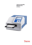
1
Photonics
Model 400 Series Spectrophotometer
User's Manual
P/N 1656 Rev. D
©1994, 1995, 1996, 1997, 1998, 2000, 2005
S.I. Photonics, Inc.
Tucson, Arizona
Contents
Table of Contents
1.
Introduction to UV-Vis Spectroscopy..........................................................1
2.
Installation and Setup....................................................................................2
2.1 Quick Installation Instructions.................................................5
2.2 Hardware....................................................................................7
2.3 Computer Requirements........................................................13
2.4 Lamp Adjustment....................................................................14
3.
Software Overview......................................................................................20
3.1 Introduction.............................................................................20
3.2 System Overview....................................................................21
3.3 Drop Down Menus..................................................................28
3.4 Saving Files.............................................................................33
3.5 Exporting Data........................................................................37
4.
Applications................................................................................................40
4.1 Peak Find.................................................................................40
4.2 Quantification..........................................................................47
4.3 Timed Acquisition...................................................................56
4.4 Kinetics....................................................................................62
5.
Troubleshooting..........................................................................................63
6.
Index.............................................................................................................68
Introduction
Introduction
The S.I. Photonics 400 Series Spectrophotometers are laboratory instruments
that utilize the latest solid state CCD array detector technology. The optical
system is specifically designed to be used with fiber optic probes for sampling. A
variety of probes and probe configurations are available for different sample
requirements.
The instrument consists of tungsten and deuterium (model 420 and 440) light
sources, optical shutters for the light sources, a spectrograph with integrated
CCD array detector, and the detector control electronics.
The tungsten source is part of the main power supply printed circuit board. The
lamp is a miniature quartz-halogen type. The lamp output is optically regulated
using a photodiode detector and feedback circuit. The lamp power supply also
features over-current protection during lamp start-up and operation. The
tungsten lamp is used for the 350 to 980 nm wavelength range when the
deuterium lamp is not used and from 460 to 980 nm when the deuterium lamp is
used.
The deuterium lamp is a high output and long life unit capable of 4000 hours of
operation. The lamp bulb is pre-aligned to a flange, which makes changing the
lamp a simple operation. The lamp power supply and ignition functions are
performed under software control so that the lamp is turned on only when
needed for acquiring data in the 190 to 460 nm range.
The spectrograph is a sealed, all-reflective optics system that uses a single
mirror for collimating and focusing, and a 325-line/mm diffraction grating blazed
at 300 nm. An integrated, fixed order-sorting filter is used to eliminate second
order diffraction. The spectrograph optics, CCD array detector, and detector
electronics are housed in an insulated, temperature regulated enclosure to
ensure high baseline stability.
A digital signal processor is used in the 400 series instrument to execute the
commands issued by the host computer. This very high speed 24-bit processor
directly generates the timing states necessary for CCD readout, sums the data
from multiple CCD reads, and controls other devices in the spectrophotometer
such as optical shutters and deuterium lamp power relays. The processor has
an integrated serial communications interface that is used to transfer command,
status, and data information between the spectrophotometer and the host
computer.
This User's Manual covers the installation, use, and maintenance of the S.I.
Photonics 400 Series Spectrophotometers. It is valid for models 410, 430
(tungsten lamp source only) and 420, 440 (tungsten and deuterium lamp
sources).
1
Introduction
Items Included in 400 Series Systems
Basic items shipped with your instrument are listed below, see the packing slip
for a detailed list.
1)
Spectrophotometer
2)
Fiber optic sampling accessory
3)
AC power cord
4)
6' long DB9M to DB9F RS-232 cable
5)
Software installation CD including manual and tutorial
6)
Trimpot adjustment tool
7)
Hex wrenches (2)
8)
Spare tungsten bulb
Verify the contents of the shipping container and notify S.I. Photonics if any items
are missing. We can be reached at (520) 293-6911 or via the internet at
www.si-photonics.com
2
Introduction
Technical Assistance, Service, and Spare Parts
If it is necessary to contact S.I. Photonics for support, please have ready the
instrument model number, serial number or optics serial number and control
software revision number. The About Spectrometer window accessed from the
Help menu contains much of this information. Print out a copy and keep on file
for future reference. The model and serial numbers can be located on labels
affixed to the inside of the instrument base. The software revision number is
printed on the distribution CD label and can also be found in the View menu of
the main front panel under VI info.
Figure 1-1: About Spectrophotometer Window
Replacement parts for the 400 Series Spectrophotometers are:
Part Number
1991
Description
Lamp, Tungsten, Quartz
1103
Lamp, Deuterium
The tungsten lamp is part number Thorn M29 (product no. 72170T) distributed by
Microlamp, Ft. Lauderdale, Florida and may also be purchased directly from
them. It is recommended to have a spare tungsten lamp.
For technical support and to order supplies and replacement parts contact the
Service Department at (520) 293-6911 or via email at [email protected].
3
Introduction
Safety Message
CAUTION!
It is necessary to remove the cover of the 400
Series Spectrophotometer for some procedures
described in this manual. In some cases, power
to the instrument must be applied while
adjustments are made. There are two regions of
the instrument power supplies that are shielded
and marked where dangerous voltages exist.
Avoid touching any components behind these
shields. If the shields are removed for any
reason, re-install them before power is applied
to the instrument.
4
Quick Installation and Setup
Quick Installation Instructions
This section is for those who either enjoy the challenge of setting up a new
instrument without reading the manual, or do not have the patience to read a
manual thoroughly before plunging in. If you are this type of person, and you
have experience installing scientific instruments and Windows software, read this
section to get up and running quickly. Then, when the excitement has worn off or
you get stuck, go back and thoroughly read the manual. For those persons that
are more cautious, read the manual Sections 2.2 through 2.4, then follow the
instructions to get the instrument operational before continuing with the detailed
software description. A tutorial video CD is also available that covers various
topics including instrument setup and software features.
Before you plug in AC power to the instrument, remove the cover by loosening
(not removing) the four screws on the ends. If you are using 220VAC power, the
power selection switch (located near the main power switch on the main power
supply board) must be switched and the fuse value changed.
If equipped with a probe, install the probe holder by sliding it onto the probe
stand and install the fiber optic probe next. Make sure that the ends of the fiber
connectors are clean (wipe with alcohol and lens paper), and that the SMA
connectors are plugged into the correct locations. A nylon-tipped setscrew is
used to hold the connector ferrules in the sleeves. Tighten the setscrews using
the supplied hex (Allen) wrench. Replace the cover on the spectrometer and
attach the AC power cord. Connect the RS-232 port on the spectrometer to an
unused port on the host computer. An RS-232 to USB converter accessory is
available for systems lacking an available serial port.
When the Control Software CD is loaded Windows should automatically begin
the installation process. If the install does not start automatically, execute the
SETUP.EXE on the CD-ROM and follow the directions.
Once installation is complete, start the SI400 application and turn on the
spectrophotometer power. The tungsten lamp should immediately light, but the
deuterium lamp (Model 440 and Model 420 instruments) will not light until
commanded by software.
Click the OK box to initialize the instrument. If the instrument is not
communicating with the host computer, an error message will pop up. To correct
the error, pull down the Operate menu from the main screen and click on the
Configure option.
Select the COM port that you are using on the computer. Leave the baud rate
set to 38,400 as the spectrometer is shipped with this baud rate. Choose printer
type, Monochrome or Color. Click the Save button to keep any changes, and
then select from the Operate menu the Initialize item.
5
Quick Installation and Setup
At this point, if everything has gone just right and the computer and
spectrophotometer are communicating, you should hear a faint click-clack from
the tungsten lamp shutter confirming communication. Also, the Blank button in
the upper right corner of the screen will change from a light gray inactive mode to
a red active mode. The Setup file is automatically read from the hard drive and
the operating parameters of the spectrometer will be set. The parameters
include the wavelength range and the lamp crossover wavelength. The
wavelength scale on the main spectrum display will reflect the wavelength range.
At this point, the wavelength range and lamp crossover wavelength can be
changed if desired by selecting the Wavelength Range item, which is located in
the Operate menu. If you have a Model 440 or Model 420 instrument and
choose a wavelength that is below the lamp crossover wavelength, you will be
asked to turn on the deuterium lamp.
If you get error messages instead at this point, follow the advice of the error
messages.
Once the spectrophotometer is operating, immerse the probe in water, and start
pushing buttons. The Blank takes a blank spectrum, and Sample takes a
sample spectrum and records the results on the screen. Most of the buttons are
self-explanatory in the usual Windows fashion. The Quantitative Item in the
Operate menu, loads the quantitative analysis feature. The Timed Acquisition
item in the Operate menu opens a new screen where entire spectra, discrete
wavelengths and kinetics can be acquired at set time intervals. The Multi
Spectra Display item can be accessed from the View Menu allowing up to 10
spectra to be viewed and manipulated.
When you are ready for more detail, read Sections 3.1 through 4.4. This way
you will be able to use the software to its fullest capabilities, and change lamps
and adjust lamp intensity when the need arises.
6
Hardware Setup
Setup of the 400 Series Spectrophotometer
The 400 Series Spectrophotometer includes a fiber optic probe that is not
connected during shipment. It is necessary to connect the probe in order to
conduct the initial system tests. The probe stand and sliding probe holder is
designed to conveniently hold the probe when it is not in use, or when sampling
the contents of small containers. It is not essential for the operation of the
spectrophotometer and may be removed if desired.
Removing the Cover
The spectrophotometer cover must be removed to install the probe. Before
starting, make sure that the AC power cable is disconnected where it enters the
instrument. Next, locate the four cover screws, two on each end of the
instrument. Using a Phillips screwdriver, loosen the four screws approximately 5
turns, or 1/8". The screws should not be removed. Next, lift the cover straight
up until it clears the internal components, and
set it aside.
BAUD rate table
BAUD rate
jumpers
RS-232 connector
Fuse holder
AC power connector
Power switch
Spectrograph
Voltage selection
switch
External lamp connector
Cooling
fan
D2 power supply
(only models
420/440)
External shutter connector
W lamp housing
D2 lamp
housing
Input
Figure 2-1: Main compartment of the 440 Series Spectrophotometer
7
Hardware Setup
Inside the Spectrophotometer
Inside the spectrophotometer is a compact spectrograph (which is thermally
insulated for temperature stability), a digital electronic controller card, and the
main and optional deuterium lamp power supplies as shown in Figure 2.1.
Initial Inspection
With the cover removed, inspect the inside of the spectrophotometer for any
parts that may have vibrated loose during transit.
AC Voltage Selection
The spectrophotometer is configured to operate from 100 to 120VAC mains
when delivered from the factory. If the instrument is to be operated from 220VAC
mains, the AC voltage selection switch must be changed. This switch is located
beneath a guard near the power switch. The switch is accessible through a hole
in the guard. A small flat blade screwdriver is used to change the switch setting.
Installation of Probe Holder
To install the probe holder on the probe stand rod, squeeze the flat metal spring
stop slightly and slide it first over the rod end with the black plastic surface on
top.
Installation of Fiber Optic Sampling Accessories
All sampling accessories for the 400 Series Spectrophotometer are fiber optically
coupled to the instrument. Each accessory will have either two (UV only or
Visible only) or three (UV/Visible) fiber optic connectors. The standard probe
supplied with the 400 Series instrument is a fiber optic dip probe. Also available
with the instrument is the Universal Fiber Optic Sampling Bench, which can be
fitted with a cuvette holder or optical filter holder, or a fiber optic reflectance
probe.
The 400 Series instruments are designed to be used with probes terminated with
standard SMA type connectors, such as the standard fixed path length dip
probes. The connectors are not attached to the instrument in the conventional
manner of tightening the retaining nut, but rather the connector ferrule is clamped
in place using a nylon-tipped setscrew. This approach was taken to ensure a
more mechanically stable connection. It is important to use only the supplied
nylon tipped setscrews to clamp the connector ferrules. Standard setscrews will
damage the ferrule, and possibly break the fiber inside the connector.
Fiber optic probes are fragile in some operations. Use caution with the probe as
damaged probes cannot be easily repaired; replacement is usually the only
alternative. The fiber optics in the probe are fragile and can be broken if
8
Hardware Setup
mishandled by placing excessive tension stress, by exerting too small of a bend
radius on the cable sheath, or by kinking or crushing the sheath. Never swing
the probe from the fiber optic sheath, or pull on the probe when attached to the
spectrophotometer such that the sheath is stretched tight.
Fiber Optic Probe Installation
The fiber optics at the connectors must be clean, free of dust and especially
fingerprints (to avoid UV absorption) before they are installed. Use lens tissue
moistened with isopropanol to clean if necessary.
To install any of the sampling accessories, locate the small hex (Allen) wrenches
provided with the instrument. Remove the plugs in the spectrometer where the
fibers attach, and remove the caps from the ends of the fiber optic ferrules. Take
care not to scratch or damage the ends of the fiber optics when the protective
caps are removed. Insert the W source fiber into the W source bushing until it
stops and secure it with the setscrew. Install the Input fiber (and for Model
420/440 instruments, the D2 source fiber) in the same manner.
RS-232 Communications Link
The communications channel between the host computer and the 400 Series
Spectrophotometer is an RS-232 serial link. Information passed over this link
includes commands to operate light shutters, initiate the CCD array detector
readout, turn on or off the optional deuterium lamp, etc. The link is also used to
pass the light intensity as a function of wavelength information used to determine
the optical transmission or absorbance of a sample. One important task for the
communications link is to initialize the spectrophotometer by passing a software
program to its microprocessor. This task must be accomplished before the
spectrophotometer can recognize any commands or perform any functions.
Configuring the Communications Link
In order for the communications link to pass information, it must be properly
configured involving both the physical cabling of the host computer COM port to
the spectrophotometer, and ensuring that the two pieces of equipment are
communicating at the same data rate, or baud rate. All other communications
parameters, such as number of data bits, parity bits, etc. are permanently
configured in the spectrophotometer, and are configured in the host computer by
the SI400 application without the need for user intervention.
RS-232 Cable
The communications cable must be a high-quality, shielded cable intended for
RS-232 use, such as the one supplied with the instrument. This is especially
critical when higher baud rates (19,200 and higher) are used. The cable supplied
with the 400 series instruments is a 6' DB9 male to DB9 female, wired straight
9
Hardware Setup
through (pin 1 to pin 1, pin 2 to pin 2, etc.) which can be connected to a DB9
male COM port connector on the host computer. A DB25 female to DB9 male
adapter is also supplied to allow connection to a DB25 male serial port on the
host computer. It is possible to use a longer cable; however it may be necessary
to use a lower baud rate to ensure reliable communications.
Connect one end of the cable to the spectrophotometer and the other to any
unused COM port on the host computer configured as COM1, COM2, COM3, or
COM4. If all COM ports on the computer are in use, it may be necessary to add
a COM port connector or interface card. It is important that the COM port to be
used is not sharing an interrupt. Refer to your computer equipment supplier for
information.
Baud Rate Selection
It is advantageous to use as high a baud rate for the communications link that
reliably works as spectral data will be transferred from the spectrophotometer
and be displayed or analyzed faster. Under some conditions it may be
necessary to decrease the baud rate to ensure reliable communications with
some host computers.
The COM ports for most modern PC compatible computers are capable of
reliable operation at baud rates of at least 19,200 if the cable length is kept short.
If it is necessary to use a cable of approximately 15' or greater between the host
computer and the spectrophotometer, or if an older PC computer that can not
support 19,200 baud is used, it will be necessary to decrease the baud rate to
9,600. If the host computer is capable of communicating at 38,400 baud or
higher, and only the 6' cable supplied with the instrument is used, it may be
advantageous to increase the baud rate.
The baud rate for the 400 Series Spectrophotometers is set to 38,400 at the
factory. With a 16550 UART serial port, baud rates up to 57,600 are possible
To change the baud rate, it is necessary to remove the cover of the
spectrophotometer (Refer to Section 2.2, Removing the Cover) and change the
baud rate jumpers. The jumpers are on the controller board near the RS-232
connector. There are 3 pairs of pins emerging from a small block labeled JP1,
JP2, and JP3. A plastic plug containing a wire fits down over a pair of pins
electrically connecting them (Closed or 1) or is removed or pushed down over
only one pin (Open or 0). Select the desired baud rate by positioning the
jumpers as shown in the Table 1 below. This table is also printed on the
controller board near the spectrograph.
10
Hardware Setup
JP3
JP2
JP1
BAUD Rate
0
0
0
9600
0
0
1
19200
0
1
0
38400
0
1
1
57600
1
0
0
115200
1
0
1
230400
1
1
0
460800
1
1
1
921600
Table I: Baud Rate Jumper Table
Figure 2-2: Configuration Drop Down Menu
11
Hardware Setup
Spectrograph Warm-up
At this point, the spectrophotometer cover is ready to be replaced. Follow the
cover removal procedure in reverse order and then connect the AC power cord
supplied to the instrument and to a standard 110VAC wall outlet.
The spectrograph in the 400 series instruments is temperature regulated at
approximately 35ºC to improve baseline stability.
A warm-up time of
approximately 30 minutes is required for the temperature of all optical elements
to equilibrate. During this time, the baseline will drift. If careful measurements
are to be made, or if long periods between blank and sample acquisition are
anticipated, temperature equilibration and warm-up is essential.
12
PC Requirements
Computer Requirements
The host computer using the SI400 Series application software controls the 400
Series Spectrophotometer. This software system operates in the Windows 95 or
later environment and conforms to normal Windows operating conventions
whenever they apply. Users unfamiliar with the Windows environment should
refer to the "Getting Started with Microsoft Windows" and "Microsoft Windows
User's Guide" that is part of the Windows distribution package before proceeding.
Host Computer Hardware and Software Requirements
The SI400 control application runs under Windows 95, 98, NT, 2000 and XP
operating systems. Minimum hardware requirements include:
1) a Pentium 90 CPU
2) a hard disk drive with a minimum of 6 MB free space
3) a CD-ROM Drive
4) a two-button mouse (or a three button mouse in two-button mode)
5) 32 MB RAM
6) one RS-232 serial port equipped with a 16550 buffered UART configured as
COM1, COM2, COM3, or COM4 using the standard Windows addresses
and interrupt vectors. The port can have either a DB9 or DB25 male
connector.
**Alternatively, an RS-232 to USB conversion cable is available for PC
configurations utilizing USB input.
A VGA monitor with a minimum 800 X 600 resolution is required.
Throughout this manual, a basic knowledge of the Windows environment and its
conventions is assumed. If necessary, refer to the Windows User's Manual for
guidance.
Installing the SI400 Control Software
The SI400 Control Software is distributed in compressed form on a CD-ROM.
The files must be installed onto your hard disk drive using and Setup program on
the CD-ROM from Windows. Disks can be made from the CD-ROM – Call S.I.
Photonics for instructions. To install, insert the CD-ROM into your drive. The
install set-up window should open automatically, if not, go to the Start Menu and
click on Run. Type in CD-ROM drive designation (i.e. D: ) A window will appear
Click on SET UP. Press the Install button and follow the prompts to install.
13
Installation and Setup
Lamp Adjustments
The tungsten source used in the 400 Series instruments is a 10 watt quartzhalogen lamp with approximately 3100ºK filament temperature. Its average life
expectancy is over 200 hours (longer life, lower output lamps are available; call
S.I. Photonics for information). The deuterium source is a high-intensity, prealigned lamp with an average life of 4,000 hours. Both lamps need to be
periodically replaced as normal instrument maintenance. The tungsten source is
replaced only when it fails. The deuterium source is replaced whenever the
estimated operating hours exceeds its lifetime, or when the lamp intensity and
stability are unacceptable.
Use care in replacing both tungsten and deuterium lamps as the lamp envelopes
are fragile and can break during handling. Also, both lamps and their lamp
housings become extremely hot during operation. Allow them to cool before
disassembling. Finally, the deuterium lamp outputs ultraviolet radiation that is
harmful to eyes and skin. Observe the precautions on the replacement lamp,
and protect the eyes with plastic safety glasses designed to absorb UV light if it
is necessary to view the lamp during operation.
Adjusting Lamp Intensities
Any time the tungsten or deuterium lamps are replaced or adjusted, or the fiber
optic probe is changed on the spectrophotometer, it may be necessary to adjust
the lamp intensities such that the minimum detector integration time is used, and
the CCD detector does not become overexposed. This is accomplished by lamp
intensity adjustment screws that prevent a portion of the light from the lamps
from reaching the fiber optic probe input.
During all lamp intensity tests and adjustments, the fiber optic probe used with
the spectrophotometer should be installed and sampling a "blank". If a dip probe
is used, it should be immersed in a blank solution. If a reflectance probe is being
used, it should be pointed towards a white reflectance standard or shiny
aluminized surface. The reflectance probe rarely needs the light reduced.
To adjust the lamp intensity, the AC power must be disconnected and the
spectrophotometer cover must be removed. Next, the main power must be
reconnected while the cover is removed. Care must be taken to not touch any
electrical components under or near the guards on the main power supply
or the deuterium lamp power supply as high voltages exist in these
regions. Also, avoid dropping tools or other metal objects that could cause
electrical shorts in the spectrophotometer electronics.
14
Installation and Setup
Switch on the spectrometer power and initialize. There is a lamp adjust utility
that is accessed from the Configure option from Operate menu. Once the
spectrometer is initialized, two additional buttons on the Configure screen
become active for adjusting the deuterium and tungsten lamps. When pressed, a
lamp adjustment window appears displaying a
bar scale that monitors the intensity of the
particular lamp. The bar is blue when the
intensity is too low, green when it is in an
acceptable range, and red when the intensity is
too high. Locate the appropriate lamp intensity
adjustment screw and adjust the lamp so that
the bar is closest to the top of the bar scale
while it is still green. At this point, the lamp
intensity should be adjusted such that the
detector is never over ranged, yet a minimum
acquisition time is required.
Deuterium lamp intensity
The same Allen wrench used to lock the fiber
optic probe ferrules in the spectrometer is used
for the intensity adjustment screw.
The Figure 2-3: Lamp Adjustment
adjustment screw is self-locking, and is
somewhat more difficult to turn than the probe setscrews. Turn the screw in or
out to adjust the intensity.
Lamp intensity
adjustment screw
Fiber optic clamp screw
Figure 2-4: Deuterium lamp intensity adjust
15
Installation and Setup
Tungsten lamp intensity
The tungsten lamp intensity is adjusted by rotating a flag into the path of the lamp
output. The exposure is very sensitive to the flag rotation. The hex head
adjustment screw will rotate the flag; a correct size hex wrench was supplied with
the instrument. The flag should have enough rotating resistance to stay in place.
Intensity adjustment screw
Adjustment flag tension
screw (no adjustment
necessary)
Figure 2-5: Tungsten Lamp Adjustment Flag and Screw
Tungsten Lamp Replacement
Touching the replacement lamp envelope with the hands during installation can
cause premature failure. Replacement lamps are shipped with a plastic or paper
cover over the envelope to protect them from being touched. Do not remove this
cover until the lamp is installed in the socket, and ready for installation in the
lamp housing. If the lamp envelope is accidentally touched, clean it with
absorbent lens tissue moistened with isopropyl alcohol.
16
Installation and Setup
The AC power must be disconnected and the spectrometer cover must be
removed. The tungsten lamp housing on the main power supply circuit board, is
located to the left of the main power supply transformer. The tungsten lamp is
retained in the housing with a spring-loaded clamp. Grasp the clamp at the hook
in the clamp stem between the two springs and lift straight up to release.
Lamp socket (shown
partially removed)
Clamp (shown lifted)
Lamp Housing
Fiber optic
locking setscrew
Lamp current indicator
Lamp current
adjustment trimpot
Shutter/photodiode mount
Figure 2-6: Tungsten Lamp Housing Location
Grasp the lamp socket leads and pull the lamp socket from the housing. Pull the
lamp from the socket and discard.
Install the replacement lamp in the socket, remove the protective cover, and then
set the lamp into place in the housing while again pulling up on the clamp.
Before releasing the clamp, make sure that the lamp is properly seated in the
housing such that the bulb is not damaged. Gently release the clamp to secure
17
Installation and Setup
Adjusting Tungsten Lamp Current
In the event that the tungsten lamp may need adjustment or is removed and reinstalled for any reason, the lamp current set point must be adjusted. This is
performed by adjusting a trimpot until a LED indicates the proper lamp current.
The lamp current is adjusted with the spectrometer cover removed, and power
applied. Make sure that the tungsten lamp is illuminated and the deuterium lamp
is off before proceeding. Locate the lamp current adjustment trimpot and the
lamp current indicator LED. Use a trimpot adjustment tool to turn the trimpot
clockwise until the LED is illuminated. Next, turn the trimpot counterclockwise
until the LED just becomes fully extinguished. When the lamp current indicator is
not illuminated, the lamp current is being regulated at the correct level by an
optical feedback circuit.
This trimpot is NOT meant for adjusting the lamp intensity. Before placing the
unit in service, if necessary adjust the lamp intensity.
Deuterium Lamp Replacement
The AC power must be disconnected and the spectrometer cover must be
removed. Locate the deuterium lamp electrical connector near the right side of
the lamp power supply transformer. There is a latch on the right side. Press the
top of the latch towards the connector body to release it, then pull straight up on
the connector to detach it from the printed circuit board.
The deuterium lamp is held in its housing by two screws located on top of the
lamp mount. Use a small, flat blade screwdriver to remove these two screws.
The lamp can now be lifted from the spectrometer and disposed. Install a new
lamp by reversing the procedure above. No optical alignment is necessary.
If necessary before placing the unit in service, adjust the lamp intensity.
18
Installation and Setup
Deuterium lamp
electrical connector
Connector Latch
Lamp mount
screws
Deuterium lamp
electrical socket
Lamp intensity
adjustment screw
Fiber optic clamp screw
Figure 2-7: Deuterium Lamp Housing and Electrical Connector
Lamp Warm Up Times
Both tungsten and deuterium lamps require a warm up period of approximately 1
hour before they reach maximum stability, but for routine use, 30 minutes may be
sufficient. The lamp warm up time is independent of the time necessary for the
spectrograph temperature to stabilize. This is especially important to remember
if the deuterium lamp is not turned on when the instrument is first powered up. If
either lamp has not had sufficient warm up time, baseline drift will be observed.
19
Software Overview
Software Overview
Introduction
The SI400 Control Software uses the Virtual Instrument (VI) concept where the
various buttons, knobs, dials, displays, and controls that you would expect to find
on an instrument, along with pull down menus, have been replaced by their
graphical equivalents that appear on the computer screen. The instrument
hardware no longer has physical controls, but the combination of computer
screen, keyboard, and mouse has replaced their function in ways that can be
easily optimized for specific tasks. The SI400 applications have different front
panels for general data acquisition, quantitative data acquisition, and for
acquiring data at fixed time intervals, each optimized for the specific task.
During installation a program group was created and can be accessed in
Windows 95/98/NT/2000 and XP from the Programs item on the Start menu. To
start the application, SI 400 program group icon, then double click on the SI400
application icon. If you do not select to have a program group created, execute
the SI400.exe program using the file manager.
When the SI400 application is started, it presents the main VI front panel that
appears (see Figure 3-1 Main front panel) to have an assortment of buttons and
other digital controls and drop down menus, along with a graphical display of
optical absorbance, transmission or reflectance. The buttons are "pressed" by
using the mouse to point to the button, and pressing the left mouse button. If a
button is active, it will react when pressed so that it looks like it was pressed, and
that some activity is being performed.
There are times when a button cannot become active until some other function is
first performed. For example, the Blank button is inactive until the instrument is
initialized. The Sample button is inactive until a blank has been acquired. When
a button is inactive, it is a light gray, semitransparent color.
20
Software Overview
System Overview: Main Panel
In this section the various features of the 400 Series Control Software are
demonstrated. The figure below shows a graphical representation of the front
panel with each feature identified. At this point it is advisable to acquire both a
blank and a sample spectrum so that you can evaluate each feature as they are
presented.
Drop Down Menus
Cursor Position
Indicators
Cursor Step
Scrolling
Graphical Display
Preferences
Cursor difference
Indicator
Measurement Unit
Sample Acquire
Blank Acquire
Graphical Display
Zoom Restore
Y Axis
Auto Scale
Reference
Spectrum
Display
Save File Name
Peak
Smoothing
Comment Entry
Fields
Initiate Peak
Find
Deuterium Status
Current Date
Current Time Last Baseline
Acquired
Discrete Wavelength
Information Table
Figure 3-1: SI400 Control Software Main Panel
21
Update Cursor
Information to
Table
Software Overview
Measurement Unit
Allows the user to toggle between measurement
modes including Absorbance, Transmittance,
Reflection and Intensity. Since the raw data is stored
in memory, the units can be mathematically
adjusted after the acquisition.
Sample
Once a blank has been acquired the sample button
becomes active and sample measurements can be
acquired. A full spectrum acquisition is
automatically presented in seconds.
Blank
Allows for the establishment of a baseline for
subsequent sample measurements.
Lock Blank
Locks the blank button so that the current baseline
cannot be accidentally overwritten.
Restore Full View
Restores current graphical display to the original
aspect ratio as demonstrated when the sample was
acquired. This feature is used to return to normal
view if a zoom feature or different scaling was used.
Y Scale Restore
Restores display to the original Y-scale displayed
when the sample was acquired. This feature is
useful if a zoom or different scaling were used.
Reference Spectrum
Allows for two spectra to be viewed and one is
designated as a reference. Deviations between
samples can be ascertained using this feature.
Peak Smoothing
Smoothes displayed data using a Savitzky-Golay
quadratic-cubic algorithm with an adjustable number of
points. This effectively increases the signal to noise
ratio of spectral data at the expense of reduced spectral
resolution. Data saved to disk files is not saved in
smoothed form, but any file read from disk can be
smoothed. The smoothing algorithm is described in J.
Anal. Chem., 1964, 36, p. 1627.
UV Lamp
Turns on or off the deuterium lamp. The lamp power
supply requires approximately 20 seconds to ignite the
lamp discharge. The lamp should be allowed to warm
up for at least 20 minutes to reach maximum stability.
Baseline drift will be observed if the lamp has not had
time to stabilize.
22
Software Overview
Peak Find
Opens a new front panel for automated peak and
valley analysis. Push the Close button to return to
the main front panel. Function Key F4
Blue Cursor
Adds an entry into the wavelength column of the
Data Table at the current position of the blue
cursor.
Red Cursor
Adds an entry into the wavelength column of the
Data Table at the current position of the red cursor.
Update Table
Updates the absorbance, % transmission, %
reflectance or intensity fields in the Data Table.
Used when wavelengths are manually entered in
the Data Table rather than using the blue cursor/red
cursor buttons.
Clear Table
Clears the Data Table.
λ Table
The wavelength table is useful when referencing
specific or multiple wavelengths for analysis.
Users can manually type the desired wavelength
into the table and depress the Update Table
button to retrieve the information.
Alternatively, users can drag the blue and red
cursors to the desired location and depress the
Blue Cursor and Red Cursor buttons to update
the table with those specific coordinates.
23
Software Overview
File Name
Indicates the file name to which the current spectrum will be saved.
Comment
This field allows users to enter specific information and experimental conditions
about a sample.
Operator
Allows for the entry of a user name indicating the name of the analyst.
Blank
Indicates the date and time when the last blank spectrum was acquired.
Info Table
Allows users to enter specific information about a spectrum.
Date & Time Displays the date and time the current spectrum was acquired.
24
Software Overview
Graphical Display
Displays the current spectrum on the monitor. X-axis consists of
wavelengths while the Y-axis indicates the measurement mode. Users
can select either a blue or red cursor for assistance in identifying specific
spectral features. Zooming can be accomplished by directly editing the
max/min labels on the axes’ of the graph. Clicking on the max or min
label on either axis and editing it to the desired value does this. The tic
marks will automatically be adjusted.
A right mouse click allows for changing of display views.
Cursor Indictor
The cursor position indicator is located just above the graphical display
and identifies the wavelength and intensity. Users can choose either blue
or red cursor mapping. The small square indicator changes from white to
black indicating which cursor is currently enabled. An icon consisting of
the cursor intersection within a small white box is a control that allows
changing various cursor display attributes such as a cursor or no cursor.
Clicking on this will bring up a menu of selections. An icon that appears
as a padlock to the right of the attribute control is a control that forces the
cursor to be constrained to the displayed data points, or free to move
anywhere on the display.
The four-diamond triangle located immediately to the right of the cursor
position indicator allows for single step incremental scrolling of the
cursor currently selected as indicated (↑) by the darkened box.
Depressing the right mouse button allows users to hide the cursor
indicator.
25
Software Overview
Graphic Control
Axis Lock
The graphic Control allows for
modifying the appearance of the
displayed spectrum.
The X and Y buttons on the left side of the Graphic Control
permit locking of either axis. When selected, the X an Y axis will
no longer autoscale with successive spectrum acquisitions but
instead will remain fixed at the established values. If autoscaling
is turned on, the display scale will be adjusted to the data
whenever the display is updated (such as when a new spectrum is
acquired). When autoscaling is turned off, the display scale will
remain constant during data acquisitions.
Display Units X.XX and Y.YY buttons allow users to select the desired display
units, significant figures, etc.
Zoom
The zoom button allows users to select the between a variety of
zoom options.
Zoom Region
User selects a defined zoom region
Zoom Wavelength Region
User defines the wavelength
region to be zoomed
Zoom Y-Axis Region
User selects the defined y-axis
region to be zoomed
Zoom Restore
Returns graphical display to it original size
Zoom In Stepwise
Permits stepwise zooming in of a selected
region
Zoom Out Stepwise
Permits stepwise zoom out of selected
region
26
Software Overview
Panning Allows users to pan around the Graphical Display
Cursor Select
Allows users to select either the blue or the
red cursor
Delta X
Displays the wavelength difference between the
two cursors.
Delta Y
Displays the intensity unit difference between the
two cursors.
27
Software Overview
Drop Down Menus
This section summarizes the features for functions accessible from the drop down menus.
File Menu
Open
Loads a previously saved spectrum and
dataset. Function Key F6 or Ctrl+O
Save
Saves the current spectrum and dataset.
Function Key F7 or Ctrl+S
Save As
Saves the current spectrum and data set
as either a text or an Excel file format.
Auto Save
Automatically saves each successive
spectrum and dataset to a selected file
in the *.SIS format.
Auto Save As
Automatically saves each successive
spectrum and dataset as either the default *.SIS format or as a
*.TXT format.
Export
Exports a single file or group of files that have been saved on the
disk to an Excel or delimited ASCII text spreadsheet file. Select
the Export function from the Files menu and a window will
open (Figure 3-6 Directory Selection) asking the user to select
the directory or folder containing data files to be exported.
Function Key Ctrl+E
Set File Filter
Limits the sort criteria when retrieving files.
Save Setup
Saves the current settings of the program to be recalled when the
spectrometer is initialized. The settings saved include the
wavelength range, lamp crossover wavelength, the table of
wavelengths to be monitored, current disk paths for data,
precision setting, smoothing on/off and points, and the grid
on/off.
Print Graph
& Info
Outputs the displayed spectrum, comments section and dataset to
the selected Windows output device. Function Key Ctrl+P
Print Only
Outputs the displayed spectrum to the selected Windows output
device.
Graph
Printer Setup
Recalls the Windows printer setup dialog routine.
Exit
Closes the application.
28
Software Overview
Edit Menu
Cut
Cuts the selected information and copies
it to the clipboard.
Copy
Copies the selected information to the
clipboard.
Paste
Pastes the clipboard contents into
application.
Clear
Deletes the current entry.
Preferences
Allows for customization of input/output path selection, PC
memory allocation performance, display options and colors,
printing margins, time and date formats.
29
Software Overview
Operate
Set Precision
Allows users to select the desired
level of precision and acquisition
speed. Available options are low,
medium and high precision.
Wavelength
Range
Allows users to select the desired
wavelength range and source lamp
crossover wavelength.
Key Function Ctrl + W
Quantitative
Opens the quantitative analysis
feature. Function Key Ctrl + Q
Timed
Acquisition
Opens the timed Acquisition and
kinetics features. Function Key Ctrl+T
Initialize
Established communication with spectrophotometer by utilizing
information input through the Configure option. This dialog is
automatically executed during instrument power on after the SI
400 Software Application has been selected.
30
Software Overview
View
Multiple Spectra
Opens the Multiple
Spectrum Display
feature allowing for the
view of up to 10 spectra
at once. from the View
menu opens a new front
panel to view up to 10
spectrum at once.
Auto Add to
Multiple Spectra
automatically places the newly acquired spectra into the
Multiple Spectrum Display feature.
Function Key Ctrl+M
Restore Full View
Displays original spectrum to full scale. Function Key F3 or
Ctrl + F
Grid
Allows user to add or remove the grid lines in the spectrum.
Function Key Ctrl+G
31
Software Overview
Help
Show Help
Provides a description of
each feature currently
selected by moving the
cursor over the selected
item.
An on-line help feature is available to explain the purpose
and functions of the various controls and indicators on all
front panels except the previously described controls
associated with the graphical displays. A help window is
opened by selecting "show help" item from the Help item on
the menu bar or with the key combination Ctrl + H. The
help window will contain a description of the control or
indicator that the mouse cursor is currently over. To view a
description of a control or indicator, simply position the
mouse cursor over the item of interest.
About
Spectrometer
Provides system information including software version,
spectrograph calibration coefficients and controller hardware.
32
Saving Files
Saving Files
Save
The most common way to save a current data file is to use the Save or Save As
options from the File menu. Selecting the Save option will result in a typical
Windows dialog box allowing users to select a specific file location for saving
files.
Figure 3-2 : Saving Data Files
Current data files acquired using the Main Panel or Quantitative feature will be
saved with the default *.SIS file extension. This ensures data security as these
files can only be accessed via the 400 Series software and cannot be altered.
The displayed spectra, comments and data table contained on the screen will be
saved to the designated folder. Data files can be retrieved using the Open menu
option.
The hotkey option for saving data files is accessed using Function Key F7 or
Ctrl+S.
Data files acquired using the timed acquisition feature will be saved as a *.SID
file format which cannot be altered as well.
In addition, files having the *.SID file extension can only be accessed via the
timed acquisition menu.
33
Saving Files
Save As
Selecting the Save As option permits users to choose between text or Excel file
formats for saving data files. Selecting either the Text or the Excel format will
result in a different Windows dialog box allowing users to select either the *.TXT
or *.XLS file extension formats. These files cannot be opened in the SI400
application.
Figure 3-3: Saving Files in Text or Excel
Data files that have been saved using either the *TXT or *.XLS file formats can
be readily imported into various spreadsheet programs such as Excel and
Quattro Pro as well as text recognition programs. The *.TXT file extension is a
comma delimited format.
It should be noted that data files containing the *.TXT or *.XLS are editable and
therefore the data content is not secure from manipulation or tampering.
34
Saving Files
Auto Save
The Autosave feature allows users to automatically save each successive
spectrum and dataset to a selected file in the *.SIS format. Selecting the
Autosave option will result in a new dialog box prompting the user to input a file
name and select the appropriate folder for successive files to be saved to.
Figure 3-4: Auto Save Path Settings
Depressing the Select Folder button will result in another dialog box allowing the
user to select the appropriate folder and location. Once the appropriate folder
has been selected the user can chose the Select Current Directory button and
all subsequent data files will be saved to the appropriate file location.
Figure 3-5: Selecting File Folder
35
Saving Files
Auto Save As
The Auto Save As option allows users to automatically save a data file as the
default *.SIS format or can be selected to save as *.TXT file extension.
36
Exporting Data
Exporting Data
Export
Selecting the Export option from the File menu exports a single file or a group of
files that have been saved on the disk to an Excel or delimited ASCII text
spreadsheet file. A new pop-up window will open prompting the user to select
individual data files or a folder containing a group of files to be exported. In the
active window choose individual files using the Shift key and right clicking the
mouse button for individual files.
To select an entire file for exporting, choose the Select New Folder button to
designate the folder containing the data to be exported.
Figure 3-6 : The file list contains the files for export
37
Exporting Data
If the Select New Folder option was chosen, a new dialog window will appear
prompting the user to select the desired folder. Once the desired folder has been
highlighted, the Select Cur Dir button located at the lower right of the dialog
window should be selected. This will return the user to the previous screen
indicated in Figure 3-6. The selected files will be indicated in the File List dialog
box.
Figure 3-7: Selecting File Folder
Once the appropriate data files are indicated in the File List dialog box, a new
window opens and listing the selected files and allows the user to choose the
type of data to export, the type of file format, and whether to smooth the data.
Figure 3-8: Selecting File Format
38
Exporting Data
The final window in the export dialog prompts the user to select the appropriate
file format and destination. The exported data must have the same wavelength
range since the first column contains the wavelength values and each successive
column contains data for each selected file.
Figure 3-9: Choosing Files to Save
At this point the user can complete the export process by pressing the Save
button, or Cancel the operation.
39
Applications
Peak Find
The Peak Find feature is opened by pressing the button on the Main front panel.
This feature is used to analyze the peaks and valleys of a spectrum and display
the associated wavelengths and intensities. These values are also displayed in
the Wavelength Table. A spectrum must have been taken or opened in the Main
front panel before utilizing the Peak Find feature.
Figure 4-1: Peak Find front panel
The graphical display on the Peak Find front panel is used to present
absorbance, transmission, reflectance or intensity as a function of wavelength.
When the window is opened, the default representation will show the peaks of
the spectrum. To view the valleys of the spectrum, select the box in the lower
right corner, and choose the valley option.
40
Applications
Peak Find Front Panel Controls
Close
Closes the Peak Find front
panel and opens the Main front
panel
Film Thickness
Calculates the thickness of
substrate in micrometers (um).
The user must type in the
refractive index of film being
tested. This calculation is found
under the peaks find table in the
“No Calculation” menu.
Find Peaks
Calculates the highest point in
each peak, or lowest point in a
valley, marks the spectrum and
updates table up to 60 points.
(Function Key F5)
Peak Width
Used in the calculation
determining the peaks.
Peaks/Valleys
Toggle switch to change the
display of peaks to valleys.
Smoothing
Smoothes the data that is
displayed by a Savitzky-Golay
quadratic-cubic algorithm with an
adjustable number of points.
This effectively increases the
signal to noise ratio of spectral
data at the expense of reduced
spectral resolution. Data saved
to disk files is not saved in
smoothed form, but any file read
from disk can be smoothed. The
smoothing algorithm is described
in J. Anal. Chem., 1964, 36, p.
1627.
Points
Varies the number of points in
the smoothing algorithm. As the
number of points is changed, the
current spectrum displayed is
dynamically updated with the
new smoothing parameters.
41
of
Applications
On/Off
Turns smoothing on or off.
Only the currently displayed
spectrum is affected.
Once
smoothing has been turned on
any
subsequently
acquired
spectra, or data files read from
disk
will
be
automatically
smoothed.
Threshold
The threshold cursor allows the
manipulation of the peak find
function to only show the peaks
above the threshold cursor
level. In valley analysis, only the
troughs below the threshold
cursor are displayed. The cursor
movement is performed the
same as the other two cursors.
The default color for the
threshold cursor is purple and is
represented as a horizontal line
in the graphical display.
Y Scale
Adjusts the y-axis to fit the
spectrum to fill the full display.
Cursor Status
Indicator
Displays the current cursor
position with respect to
wavelength and intensity. The
cursors are color coded for
clarity.
42
Threshold
Multiple Spectra Display
Multiple Spectra Overlay
The Multiple Spectra Display is accessed through the View menu on the Main
front panel and is used to see and manipulate up to 10 spectra at a time. To be
displayed in this window, the spectra must have been acquired in the Main front
panel or as saved disk files.
Figure 4-2: Multi Spectra Display front panel
The spectra can be manually opened in the Multi Spectra Display window from
saved files or they can be automatically transferred over by selecting the Auto
Add to Multiple Spectra option in the Main front panel. This feature is on by
default. As each spectrum is acquired and transferred to the Multi Spectra
Display panel, the spectrum shift down the list. When more than 9 spectrum are
opened or transferred, the spectrum are removed from the bottom. The tenth
spectrum in the table is a reference cell and will not automatically be removed.
The spectrum in the Multi Spectra Display front panel can be manipulated in
simple mathematic functions such as adding, subtracting and multiplying. The
arithmetic can be performed on the spectra with two spectra or a spectrum and a
constant. The newly formed spectrum will transfer up to the number one
spectrum cell in the table.
43
Multiple Spectra Display
Graphical Display
The graphical display on the Multi Spectra Display front panel is used to present
absorbance, transmission, reflectance or sample or blank intensity as a function
of wavelength.
The spectra displayed can be turned on and off by selecting the on/off button
next to each spectrum.
Controls
The following is an alphabetical listing of Multi Spectra Display controls
Blue Cursor - Adds an entry into the wavelength column of the data table at the
current position of the blue cursor.
Calculate – Performs the mathematical operation between the two selected
spectra or spectra and a constant value.
Clear Spectrum – Clears all or individual spectra from the spectra list.
Clear Table - Clears the data table.
Close – Closes the Multi Spectra Display panel and re-opens the Main front
panel.
Export Files - From the File menu exports a single file or a group of files that
have been saved on the disk to an Excel spreadsheet file or a
delimited ASCII text spreadsheet.
File Name Filter - limits the sort criteria when retrieving files from the disk.
Grid – From the View menu allows user to add or remove the grid lines in the
spectra display.
Info – Opens a new window to edit and review File Name and Comment section
ON/OFF – Adds or removes the spectra from the display window
44
Multiple Spectra Display
Figure 4-3: Spectra Info Box
Open File - Opens a data file created with the Save File menu item. The
spectrum in the data file, along with all other file information, is displayed.
Open Multiple Files -Opens multiple data files saved on disk. The
spectrum in the data file, along with all other file
information, is displayed.
Select a Folder – Opens new window to selects a folder from
which to open files. Click on Select Cur Dir to finish
operation and return to Open Multiple Files window.
Select Files – Click on individual files or use the shift key for
multiple selections up to 9 spectra. To select a
contiguous series of file names, shift + click and drag.
Click OK to finish.
Print Graph - Outputs spectrum only with legend
Restore Full View – Rescales the image to the original settings.
Save – Select the spectrum to be saved and edit comment section in the
comment window. Click on Close and a save file window opens. Type
in file name and click on Save.
Set Reference – Selects which spectra in the table is to be used as a reference
and transfers all data of the chosen spectra to the reference
spectrum destination. To transfer a spectrum into the
reference cell, the spectrum must be turned ON to come up in
selection choices.
45
Multiple Spectra Display
Smoothing - Smoothes the data that is displayed and that used for quantitative
data analysis by a Savitzky-Golay quadratic-cubic algorithm with an
adjustable number of points. This effectively increases the signal to
noise ratio of spectral data at the expense of reduced spectral
resolution. Data saved to disk files is not saved in smoothed form,
but any file read from disk can be smoothed. The smoothing
algorithm is described in J. Anal. Chem., 1964, 36, p. 1627.
On/Off - Turns smoothing on or off. Only the currently displayed spectrum
is affected.
Once smoothing has been turned on any
subsequently acquired spectra, or data files read from disk will be
automatically smoothed.
Points - Varies the number of points in the smoothing algorithm. As the
number of points is changed, the current spectrum displayed is
dynamically updated with the new smoothing parameters.
Update Table - Updates the absorbance, % transmission, % reflectance or
intensity fields in the data table. Used when wavelengths are
manually entered in the data table rather than using the blue
cursor/red cursor buttons.
View All – From the view menu, turns all spectra in the table to the ON position,
displaying all spectra.
Y Scale – Adjusts the y-axis to fit the spectrum to fill the full display
46
Quantitative Analysis
Quantitative Analysis
Quantitative Analysis Front Panel
The Quantitative Analysis feature is accessed via the Operate menu on the
Main Panel. Before opening the Quantitative Analysis front panel, it is best to
take a blank spectrum and a sample spectrum of a standard solution to facilitate
the choosing of analysis wavelengths.
Controls are available to acquire data from standards and unknowns, fit
standards data to a calibration curve, and determine concentrations of unknowns
based on the calibration curve data. The Quantitative Analysis front panel has
a graphical display for analytical data and a calibration curve, a section for
controls and data related to analytical standards and a section for controls and
data related to unknowns.
Figure 4-4: Quantitative Front Panel
47
Quantitative Analysis
Standards and Unknowns Tables
The standard number and concentrations for each standard are entered in the
Standards Table.
The absorbance for the standards at the analytical
wavelengths and the confirmation wavelengths will automatically be entered into
the rest of the table when the Acquire Standard button is pressed.
Description for the unknowns is entered in the Unknowns table. Concentration
and absorbance results will be calculated and inserted when the Acquire
Unknown button is pressed.
Graphical Display
The Graphical Display on the Quantitative Analysis front panel is used to
present absorbance as a function of concentration information. Data from the
analytical wavelength, or confirmation wavelengths, can be viewed directly from
the graphical display as well as the tables.
The Graphical Display contains many of the control features as indicated on the
Main Panel for your convenience in viewing your data.
Comment and File Name Dialog Boxes
The upper right corner of the Quantitative Analysis front panel contains many of
the same user dialog entries as indicated on the Main Panel. These entries allow
the user to specify comments, user name and file name.
48
Quantitative Analysis
Quantitative Analysis Procedures
Once the Quantitative window is open, the procedure for performing quantitative
analyses is as follows:
Select the Operate menu option and choose Selected Wavelengths. The
Quantitative Wavelengths Selection menu will allow for the selection of the
wavelengths to be used for analysis.
Figure 4-5: Quantitative Wavelengths Selection Menu
The Analytical Wavelength is usually the wavelength of maximum absorbance
for the sample.
The Reference Wavelength is usually a point of minimum absorbance for the
sample. When a Reference Wavelength is selected true background correction
is available. The absorbance at this wavelength is automatically subtracted from
the absorbance at the Analytical Wavelength during the computations in the
Quantitative Analysis front panel. This is a substantial feature not available on
scanning instruments.
The Confirmation wavelengths are typically used for tests of purity or chemical
interference in a sample. The wavelength is usually chosen in a region of
appreciable absorbance, but removed from the Analytical Wavelength.
The selection of analytical, reference, and confirmation wavelengths may be
made by typing in the value into the appropriate box. An alternate method is to
position a cursor at the desired location on the spectrum in the graphical display.
Select Analytical, Reference, Confirmation 1, or Confirmation 2 on the
Wavelength Selection control box and press the Blue Cursor or Red Cursor
49
Quantitative Analysis
button depending on which cursor is being used. The wavelength of the cursor is
entered into the selected item. A spectrum of an analytical standard must be
acquired in the Main front panel to use the graphical input approach.
Each type of wavelength must be turned On in order to be incorporated into the
quantitative calculations.
1. There is an option to acquire a new blank spectrum if it is deemed necessary.
Press the Unlock Blank button to make the Blank button active and proceed
with taking a new blank. After the blank is acquired, the Blank button is
automatically locked to lessen the chance of it inadvertently being pressed.
2. Enter the concentrations of the standards into the Standards Table. If there
are more than ten standards, a scroll bar will appear on the right side of the
table to allow the table to be scrolled down.
3. Next, acquire the spectra for the standards. When the Acquire button for
standards is pressed, the data will be put into the table for the standard
number indicated in the Standard Number control. This control defaults to 1
and will automatically increment to the next standard number. This control
can also be used to manually set the next standard number to be acquired.
4. When all the standards have been acquired, select the Calibrate item in the
Operate menu and the calibration curve (linear least squares fit) for the data
will be calculated and displayed on the Graphical Display. At this point, the
fit results can be viewed by pressing the View Fit Results option in the
Operate menu.
5. Now you are ready to measure the concentration of unknown samples.
Prepare the first unknown sample to be measured. A description can be
typed into the unknown table in the Name column. The Unknown Number
control selects the unknown number in the Unknowns Table for which data
will be collected. This control is initially set to 1 and will automatically set to
increment to the next number after a new spectrum is acquired. Data is
acquired by pressing the unknown Acquire button and the results will appear
in the Unknowns Table. There are scrollbars on this table for both vertical
and horizontal movement. The horizontal scrollbar allows viewing of the
results for the Confirmation Wavelengths.
50
Quantitative Analysis
Quantitative Front Panel Controls
Acquire
Two Acquire buttons are present, one in the
standards section and the other in the
unknowns section. These buttons acquire a
spectrum for the data tables they are adjacent
to. The standard and unknown numbers are
chosen before the acquisition, then data is
stored in the corresponding line in the table.
For the standards Acquire button, the known
concentration must be entered before taking
the spectrum.
NOTE: Analytical, reference and confirmation
wavelengths must be set before the button is
active. See Wavelength Selection.
Function Key Acquire Standards F1
Function Key Acquire Unknowns F6
Blank
Acquires and stores spectrum of blank sample
for calculation of absorbance and %
transmission relative to sample spectrum. Data
is stored until the wavelength range is changed
or a new blank is acquired.
Select Wavelengths
Calibrate
Located in the Operate menu,
this option opens a new front
panel that displays a graph of a
spectrum for the selection of
analytical,
reference
and
confirmation wavelengths for the
quantitative analysis front panel.
Found in the Operate menu calculates the best fit function to
standards data using linear regression.
51
Quantitative Analysis
View Fit Results
Displays statistical data from linear regression for
Analytical and Confirmation wavelengths and is found in
the Operate menu.
Clear Standards
Clears all information entered into the Standards Table
and accessed from the Operate menu.
Clear Unknowns
Clears all information entered into the Unknowns Table
and accessed from the Operate
menu.
Close
Closes the Quantitative front panel and opens
the Main front panel. Data in the Quantitative
front panel will not be lost.
Concentration units
Molarity
Data units displayed as molarity (mol/L).
PPM
Data units displayed as parts per million
(ppm or mg/L).
Format
Sets
the
display
preferences
for
concentration values. The units may be
decimal, scientific or engineering. Number
of decimal places displayed is also set
here. Function Key F11
Spectrum
Found under the View menu displays a spectrum at full screen size
of the most recent spectra acquired.
Standard number
Changes the line number where data
is inserted in the Standards Table
when Acquire button is pressed.
This number will automatically
increment by one after each sample.
52
Quantitative Analysis
Unknown number
Changes the line number where data is
inserted in the Unknowns Table when
Acquire button is pressed. This number
will automatically increment by one after
each sample.
Unk. Conf.
Used to set the acceptable percent
difference between analytical and
confirmation concentration.
A
warning will be displayed if
unknown concentration is out of
tolerance.
Unlock Blank
This button will enable/disable the Blank
button. This prevents accidental acquisition
of a new blank.
53
Quantitative Analysis
Quantitative Wavelengths Front Panel Controls
Analytical Wavelength
Is a control where the analytical
wavelength can be directly
entered from the keyboard.
There is also an on/off control to
enable or disable the use of the
analytical wavelength in the
quantitative front panel.
Blue Cursor
Enters the wavelength into the selected
control box (see Wavelength selection) at
current position of the blue cursor.
Close
Closes the Analytical Wavelengths window
and brings back the Quantitative screen.
Confirmation Wavelengths
A control where
the confirmation wavelengths can
be directly entered from the
keyboard. There is also an on/off
control to enable or disable the use
of confirmation wavelengths in the
quantitative front panel.
54
Quantitative Analysis
Red Cursor
Enters the wavelength into the selected
control box (see Wavelength selection) at
current position of the red cursor.
Reference Wavelength
Wavelength Selection
Is a control where the reference
wavelength can be directly
entered from the keyboard.
There is also an on/off control to
enable or disable the use of a
reference wavelength in the
quantitative front panel.
Determines which wavelength
type will be recorded when the
Red Cursor or Blue Cursor
button is pushed. Selections
are:
Analytical
Reference
Confirmation 1
Confirmation
55
Timed Acquisition
Timed Acquisition
The Timed Acquisition front panel is accessed by going through the Operate
menu of the Main front panel and is used to acquire spectral information at fixed
time intervals. The wavelength range over which spectra will be acquired must
have been previously set using the wavelength range item on the Main front
panel’s Operate menu.
The Timed Acquisition front panel has a graphical display similar to the one on
the Main front panel. An acquisition mode selector allows the selection of full
spectrum acquisition, or discrete wavelengths.
Figure 4-6: Timed Acquisition, Full Spectrum Mode
In full spectrum mode, the spectra are automatically
saved either as text files or in SI400 format as the user
selects. The naming and path for saving the spectra is
specified in the acquisition setup covered below.
Opening and printing a spectrum taken with timed
acquisition is performed in the Main front panel.
56
Timed Acquisition
Figure 4-7: Timed Acquisition Setup Full Spectrum Mode
Multiple spectrum files are saved using a data series name, followed by an
incremented number, followed by the .SIS extension. Example: Data Series
TEST.SIS. The data series name is entered in the Data Series dialog box while
the Start at # control box selects the beginning number in the series.
The Select Folder button opens a dialog box to choose the folder where the
spectra will be saved. Once a folder is selected, click on Select Cur Dir button
to return back to the set-up box.
Figure 4-8: Set path dialog box
57
Timed Acquisition
Figure 4-9: Time Acquisition, Discrete Wavelengths Mode
In discrete wavelength mode, a table appears displaying
the wavelengths to be monitored, along with a color
assignment for that wavelength as it is plotted on the
graphical display. Data acquired at discrete wavelengths
can be viewed graphically as changes in absorbance or
transmission as a function of time. As many as ten discrete
wavelengths can be simultaneously monitored.
Figure 4-10: Discrete Wavelengths with Manual Save Selected
58
Timed Acquisition
In the discrete wavelength mode, the parameters to be chosen are the time
interval between acquisitions, number of acquisitions, and the wavelengths to be
acquired. Up to ten wavelengths can be chosen. Any wavelength set to zero will
be ignored.
Figure 4-11: Discrete Wavelength Setup with Continuous Saving Selected
In this mode, only the values at the chosen wavelengths are saved. If the
Saving Mode is set to Manual, no Select Folder button is present, as the data
for the discrete wavelength acquisition will be saved manually at the end of the
run with the Save to File button in the Timed Acquisition panel. When set to
Continuous as indicated above, the Select Folder button is present and the file
path is set in a dialog box. In this mode, the data is saved to the same file after
each spectrum is taken. This is useful when doing long time acquisition so the
data is not lost if there is a computer problem or power failure. Files saved as
discrete can only be opened in the Time Acquisition window.
An assortment of controls for saving, opening, and printing data files similar to
that found in the discussion of the Main front panel appear on the Timed
Acquisition front panel. Files from data acquired in the discrete wavelength
mode are incompatible with those acquired using the full spectrum mode, and
can only be viewed from the Timed Acquisition front panel. There is an
additional command on the discrete wavelengths mode panel for kinetics that is
described below.
59
Timed Acquisition
Timed Acquisition Front Panel Controls
The following is a listing of the Timed Acquisition front panel controls.
Close
Closes the Timed Acquisition front panel and
opens the Main front panel.
File Menu
Open
Retrieves a data file from the disk
that was previously saved.
Save
Saves the displayed data and
comments shown on screen to
disk. Data is retrieved with the
Open command. The file format is
compatible
with
the
SI400
application only.
Save as Text
From the File menu saves data as an ASCII tab delineated
file for export to spreadsheets and data manipulation
programs.
Save as Excel
From the File menu saves data as an Excel spreadsheet file
for export later to perform data manipulations
Set File Filter
Limits sort criteria when retrieving files and is accessed from
the File menu.
Print Graph
Outputs data in the graph, comments section, and
wavelengths displayed on the screen to selected output
device.
60
Timed Acquisition
Printer Setup
Opens a dialog box where a number of setup parameters
are selected. The dialog box changes depending upon
which mode (full spectrum or discrete wavelengths) is
chosen. In full spectrum mode, the dialog box allows
selection of the time interval between acquisitions, number
of acquisitions, and the file information, and specific
wavelengths of interest. The full spectrum is saved and if
any wavelengths are entered into the Wavelength table,
these wavelengths are also saved with the spectrum. When
the spectrum is opened in the Main front panel, the values at
those wavelengths will be displayed in the wavelength table
on the Main front panel.
Time Remaining
Time Until Next Acq.
Shows the amount of time
remaining to complete the
data acquisition.
This indicator displays
the time remaining until
the next acquisition will
take place.
Samples Taken This indicator shows the
number of data acquisitions
completed. This number is
updated as acquisitions are
made.
Start
Pressing this button starts acquisition for either full
spectrum or discrete wavelength mode.
61
Timed Acquisition
Kinetics
Selected from the Operate menu, the Kinetics front panel allows the viewing of
discrete wavelength data as a kinetic plot of raw data or a first order plot with rate
constants for each wavelength measured. There is also an option to limit the plot
display and rate calculations between two cursors.
Figure 4-12: Kinetics front panel
62
Troubleshooting
Troubleshooting
Error Messages
If initialization is not successful, one of three error messages will appear in dialog
boxes. The first possible error message is:
Error 37 occurred In Com Port Init.VI:
The selected COM port either does not exist or is in use by another
application. Press the CONFIGURE button to select another COM
port. Refer to user's manual for more information.
Possible reasons:
LabVIEW: Device not found.
The SI400 control application could not find the COM port you specified during
configuration. The COM port may physically exist or not, but it could not be
"found" through the Windows operating system. If another Windows application
is open which may be using the COM port, close the application. If you are sure
that the COM port physically exists, make sure that Windows knows of its
presence. For Windows 95 and higher, a good way to start troubleshooting this
problem is to open the System application in the Control Panel and look under
Device Manager, Ports, for the COM ports that are available.
The second error message that can occur is:
Error -1201 Occurred
Spectrometer Initialization.
Series 400 spectrophotometer is not responding to initialization
command. Verify that spectrophotometer power is switched on, and
that the communications cable is connected.
This will arise if the communications port is found on the host computer end, but
there is no response from the 400 Series Spectrophotometer when information is
sent to it. Make sure that the RS-232 cable is plugged into the spectrometer, and
to the port that you specified during configuration. Also, make sure that the
communications cable has no broken conductors. The 9 pin D male to 9 pin D
female connector cable supplied with the 400 series instruments is wired straight
through with pin 1 connecting to pin 1, pin 2 to pin 2, etc.
63
Troubleshooting
A third message that can appear is:
Error -1202 Occurred
Spectrometer Init.VI.
An error has occurred while transmitting the initialization file to the
series 400 spectrometer. A likely cause is a mismatch in the
communications data rate (baud rate) settings. The computer port
baud rate must match the spectrometer baud rate. The computer
baud rate is changed using the CONFIGURE button on the main
program screen. Refer to user's manual for information about the
spectrometer baud rate.
If the COM port baud rate is correct, check the integrity of the
communications cable.
This error indicates that while there was a response from the spectrophotometer,
it was not the expected one. In addition to checking baud rates, make sure that
the COM port selection is correct. If the SI400 application is trying to
communicate with the spectrophotometer on a serial port that another device
(such as a mouse or modem) is connected, this error can result. It is also
necessary that the COM port being used is not sharing an interrupt (IRQ) with
another COM port or modem.
This error can also occur if the cable between the host computer and the
spectrophotometer is too long, or is being used in an electrically noisy
environment, or is not properly shielded. If it is necessary to use a long cable, try
to reduce the baud rate to 9,600.
Sometimes it is necessary to set the serial transmit buffer to Low in Windows 95.
This is performed in the System Properties window launched from the Control
Panel. Select the Device Manager tab; expand the Ports (COM & LPT) item;
double click on the COM port being used to display its properties. Next select
the Port Settings tab and then press the Advanced button to open the
Advanced Port Settings window. Here there are two controls, one for Receive
Buffer and one for Transmit Buffer. The Receive Buffer is normally set near High
and should be left this way. The Transmit buffer needs to be set to Low for the
most reliable communications.
64
Troubleshooting
Unreliable Communications
Unreliable communications can result from a number of sources, including the
computers RS-232 port, cable, and software drivers. One of the primary sources
of problems is slow computer response time that is related to how many tasks
the Windows task scheduler has in its queue. If the system can not respond to
the serial communications port sufficiently fast, characters may be occasionally
and unpredictably dropped, resulting in errors during the spectrophotometer
initialization.
The use of a buffered serial interface port equipped with 16550 UARTs is
required, as it will collect characters internally while waiting for service from the
computer's processor. The older 8250 type UARTs has no internal buffer, and
will not work reliably. The type of UART used in a serial port can be determined
using the DOS 6.22 "MSD" command.
The use of 16550 UART equipped serial ports and high quality RS-232 cables
will result in reliable communications in nearly every situation. If communications
problems persist, contact Spectral Instrument's service department for assistance
with other solutions.
It has been found that on some notebook computers that it is necessary to turn
off the power management to obtain reliable communications. This is due to the
overhead added to the system by the power management causing some
communications to be lost.
Screen Size
The programs front panels are designed for use with at least 800 x 600 resolution
on the Windows display. If the display is set to 640 x 480, parts of the front panel
will be off the screen and it is necessary to change the resolution of the display.
See the Windows manual or Help screens for the procedure to change the
display resolution.
Lamp Intensity Warning Messages
Figure 5-1 shows what a normal Blank intensity spectrum looks like. This
spectrum will vary from one spectrometer to another and depending on where
the crossover wavelength for the UV and visible lamps is set. Here the crossover
is at 460 nm and is not optimized.
65
Troubleshooting
Figure 5-1: Normal Blank intensity spectrum
High Lamp Intensity
The SI400 application will alert the user if the tungsten lamp intensity is too high
when a blank is acquired by giving the warning message:
The Tungsten source intensity is too HIGH
and must be adjusted. See the manual for
instructions.
A similar message will result from high deuterium lamp intensity. The user may
choose to either cancel the operation or continue by pressing the appropriate
buttons in the dialog box.
If either warning message occurs, turning the intensity adjustment screw
clockwise should reduce the appropriate lamp intensity (see Section 2.4).
Low Lamp Intensity
If the light intensity reaching the detector from the tungsten lamp is very low, the
warning message:
The Tungsten source intensity is too LOW
and may need to be adjusted or the lamp
is out. See the manual for instructions.
will appear. A similar message results from a low deuterium lamp intensity.
Again, the user may cancel or continue by pressing the appropriate button.
66
Troubleshooting
A low intensity means that the spectrophotometer has determined that even by
increasing the readout integration time to a maximum (10 seconds for both
tungsten and deuterium sources using "normal" precision), the intensity of light
reaching the detector is so low that an unacceptable signal to noise may result.
Light may not reach the detector due to:
1) A lamp is not functioning.
2) The fiber optic probe is not installed, not sampling a blank, or very
inefficient or defective.
3) Lamp intensity adjustment screw is blocking too much light.
Check each item above in the order listed.
described in Section 2.4.
67
Adjust the lamp intensity as
Index
Index
A
I
Auto Save · 28, 35, 36
Autoscaling (axis lock) · 26
Initializing Spectrophotometer · 30
K
B
Kinetics · 61
Baud Rate Selection · 12
L
C
Lamps
Adjusting Lamp Intensities · 14
Adjusting Tungsten Lamp Current · 18
Deuterium Lamp Intensity · 15
Deuterium Lamp Replacement · 18
Tungsten Lamp Intensity · 16
Tungsten Lamp Replacement · 16
COM Ports · 10, 62
Configure · 15, 30
D
Linear regression · 51
Deuterium · See lamps
M
E
Multiple Spectra Display · 43
Errors
Error -1201 · 62
Error -1202 · 63
Error 37 · 62
P
Export Files · 44
External Lamp Connector · 7
External Shutter Connector · 7
Peak Find · 23, 40, 41
Printing · 28, 45, 60
F
Q
Fiber Optic Sampling Accessories · 8
Function Keys · 23, 28, 30, 31
Quantitative Analysis · 30, 47, 49
Quantitative Wavelengths Selection ·
49
G
R
Graphical Displays and Associated
Controls · 25
RS-232 Communications · 9
68
Index
S
Save As Excel or Text file · 60
Save Setup · 28
Save to File · 33
Smoothing · 22, 41, 46
Software Installation · 5-6
Specifications · 3
T
Technical Assistance · 3
Timed Acquisition · 30, 56
Troubleshooting · 63
Tungsten · See Lamps
V
Voltage Selection · 8
W
Wavelength Range · 30
Z
Zoom Controls · 26
69