1
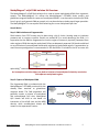
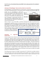
MethylMagnet® mCpG DNA Isolation Kit User Manual Version O 9-21-15 Catalog # MM101 Table of Contents Kit Components and Storage ………………………………………………………….. 2 MethylMagnet® Work Flow …………………………………………………………… 3 About MethylMagnet® Proteins……………………………………………………… 4 MethylMagnet® mCpG DNA Isolation Kit Overview………………………… 5 Advantages of MethylMagnet®………………………………………………….….. 6 MethylMagnet® Protocol ……………………………………………………..….…… 7 Control Reactions …………………………………………………………………………… 9 Recommendations for processing FFPE samples...............................11 Troubleshooting Guide……………………………………………………….……...... 12 Kit Components and Storage Component (One tube) (One tube) (One bottle) (One tube) (Two bottles) (One bottle) (One tube) (One tube) 10 x AluI buffer DNA dilution buffer Wash Buffer 1 2x Binding Buffer Wash Buffer 2 TE Buffer GST-MBD magnetic beads Bead Fraction buffer Control HeLa DNA 100 ng, MseI cut, 2ng/µl Positive Control Primers, 10 µM each Negative Control Primers, 20 µM each User Manual (One tube) (One tube) (One tube) Volume 0.5 ml 1 ml 3 ml 0.75 ml 24 ml 12 ml 200 µl 1.2 ml 50 µl Storage -20°C -20°C -20˚C -20˚C -20˚C -20˚C -20˚C -20˚C -20˚C 15 µl 15 µl 1 -20˚C -20˚C Kits are shipped on ‘dry ice’. All components should be stored at -20˚C. Beads frozen on arrival can be stored for greater than 6 months at -80°C, but once thawed should be stored at -20°C. Kit components are stable at freezer temperatures between -14°C and -22°C. Beads should be stored in an insulated container if the freezer has a defrost cycle. www.ribomed.com MethylMagnet®-FFPE mCpG DNA Isolation Kit User Manual Version O 2 Please read entire manual before starting. Reagents are included for 30 samples. This can be 30 samples of the user’s DNA or 25 samples of the user’s DNA and 5 controls with HeLa DNA or any combination thereof. User-supplied Materials/Equipment Microcentrifuge tubes (1.7 ml polypropylene) and microcentrifuge Filter pipette tips Sample mixing equipment (e.g. Eppendorf Thermomixer, shaker or tube rotator) Thermocycler and PCR components for control reactions DNAse-free ultrapure water Rare-earth magnet or magnetic rack (e.g. RiboMed Cat #MR22-01) Caution: Rare earth magnets can be extremely powerful. Care should be taken when handling them. Keep magnetized parts away from instruments that may be damaged by high magnetic fields. MethylMagnet® Work Flow Purify FFPE DNA Cut DNA with AluI Determine amplifiable DNA concentration Dilute DNA samples into Binding Buffer Remove bead storage buffer Incubate beads with DNA sample 1 hr Pull-down beads and recover supernatant fraction Wash beads and suspend in Bead Fraction Buffer Analyze Supernatant and Beads fractions with CAP kit www.ribomed.com MethylMagnet®-FFPE mCpG DNA Isolation Kit User Manual Version O 3 About MethylMagnet® Proteins The MethylMagnet® mCpG DNA isolation Figure 1 kit utilizes RiboMed’s GST-MBD protein (MethylMagnet®). MethylMagnet® proteins are versatile tools for the study of CpG methylation in DNA. These proteins contain the methyl binding domain (MBD) of the mouse MBD2 protein fused to the glutathione-S-transferase protein (GST) from S japonicum. The two domains are separated by a linker containing a thrombin cleavage site (Figure 1A). The MBD from the MBD2b protein was chosen because MBD2b has the highest affinity among the known methyl CpG binding proteins for Me-CpG sites and the lowest cross reactivity with unmethylated CpGs. Additionally, there are no sequence context effects on MBD2 CpG recognition, as there are for MeCP2, which requires a run of A-Ts near a CpG site, therefore a greater number of mCpG sites will be recognized by MethylMagnet®. In addition to its high specificity for methylated DNA, MethylMagnet® proteins have several unique features that make them useful tools in the study of DNA methylation. The GST allows the fusion protein or its complexes with methylated DNA to be isolated on glutathione agarose, magnetic beads or plates and eluted in native form with glutathione. The GST group further allows the eluted protein-DNA complexes to be immobilized to beads or microtiter plates with GST antibodies. Alternatively, the GST group can be used for visualization of the DNA-protein complexes by using labeled GST antibodies or GST antibodies and labeled secondary antibodies, or other detection methods for GST. The GST group contains surface cysteine groups (Figure 1B) that can be chemically modified to add other reporters or affinity tags, such as biotin, fluorescein or other functional groups. MethylMagnet® has high affinity for fully methylated CpG sites MethylMagnet® proteins Figure 2 A. have high affinity and specificity for binding to double-stranded DNA containing 5-Methyl-CpG groups. They have much higher affinity for DNA methylated on both strands, versus hemimethylated (not shown) or unmethylated DNA (Figure 2). Purified MethylMagnet® protein (Cat# MM101) was incubated with a 550 bp p16 amplicon that was either fully methylated or unmethylated or with no DNA (Figure 2A). The biotinylated DNA target was immobilized to magnetic beads. The MethylMagnet® protein was incubated with the immobilized DNA for 1 hour. After washing, bound MethylMagnet® was detected with an anti-GST antibody-horseradish peroxidase conjugate (Figure 2B). The HRP reaction was performed for 5 min. The filled and unfilled bars represent duplicate measurements. www.ribomed.com MethylMagnet®-FFPE mCpG DNA Isolation Kit User Manual Version O 4 MethylMagnet® mCpG DNA Isolation Kit Overview The MethylMagnet® mCpG DNA Isolation Kit is used to isolate methylated dsDNA from a genomic sample. The MethylMagnet® kit utilizes the MethylMagnet® GST-MBD fusion protein and glutathione magnetic beads for capture of methylated dsDNA. It can be used to isolate mCpG DNA from 1 ng to 1 µg of genomic DNA per sample, or it can be scaled up to isolate much larger quantities. The MethylMagnet® kit can capture DNA containing 6 or more methylated CpG sites. How it Works Step 1: DNA Isolation and Fragmentation DNA isolation from FFPE tissues may be done using a kit of choice. Heating steps to inactivate protease and to remove crosslinks should not exceed 80°C to avoid denaturing the DNA. It is imperative that the DNA be fragmented so that the region of interest is physically separated from other regions of DNA that may be methylated. Failure to adequately separate CpG islands could lead to co-purification of unmethylated islands with neighboring methylated regions. Fragmentation by restriction enzyme digestion is recommended. A 10x AluI buffer is included in the kit to ensure the optimal Mg2+ concentration in the DNA binding reaction. An incomplete restriction digest can lead to isolation of unmethylated islands by association with adjacent methylated regions. Step 2: Capture of Methylated DNA The fragmented DNA is incubated with the MethylMagnet® GST-MBD protein which has already been attached to glutathione magnetic beads. The DNA population will generally contain a mixture of methylated and unmethylated CpG sites. Methylated CpG islands will bind to the beads via interaction of the mCpG sites and the MBD domain, while unmethylated islands will remain in the supernatant fraction. www.ribomed.com MethylMagnet®-FFPE mCpG DNA Isolation Kit User Manual Version O 5 Step 3: Recovery of unmethylated Supernatant DNA Fraction and preparation of the methylated Bead Fraction Advantages of MethylMagnet®: High sensitivity without loss of specificity The MethylMagnet® mCpG DNA Isolation Kits can be used with 1 ng of input DNA up to 1 µg of DNA (not shown). Shown below (Figure 3), HeLa genomic DNA or artificially methylated HeLa DNA were purified with the MethylMagnet® mCpG DNA Isolation Kit and eluted in 20 µl of glutathione buffer. Figure 3 2 µl samples (1/10th of input) were tested for p16 DNA by PCR (40 cycles) and analyzed by gel electrophoresis and ethidium bromide staining. 2 ng input corresponds to approximately 300 cells, so methylated P16 from 200 pg of genomic DNA, or 30 cells is detected here. P16 was only detected with artificially methylated DNA. Shown in Figure 4, 1 ng of HeLa DNA or artificially methylated HeLa DNA was purified with the MethylMagnet® mCpG DNA Isolation Kit. DNA was eluted in 50 µl and 2 µl samples were used for amplification of P16. This corresponds to detection from 6 cells, or 40 pg of DNA and 12 copies of P16. The gel was stained with SYBR® Green and imaged with a Typhoon fluorescence reader. MethylMagnet® mCpG DNA Isolation Kits are very specific for methylated DNA. When unmethylated DNA is processed, nothing is captured and detected in the eluted (bound) fraction, even with 40 cycles of PCR. When methylated DNA Figure 4 is captured, nothing is detected in the supernatant (unbound) fraction. Preparation Genomic DNA of Fragmented Isolation and purification of DNA from cells or tissues may be done using any of several commercially available kits. Cleavage of DNA to produce the largest CpG island fragments can be most easily accomplished with the use of 4-base-recognition restriction endonucleases that are biased to A-T rich sequences (e.g. MseI, TTAA, and Tsp509I, AATT). Four base cutters that include G and/or C can be used to fragment within CpG islands to isolate important sub-regions. The choice of restriction endonuclease should be based on the user’s analysis of the target CpG island. The kit includes a 10x AluI buffer with a final Mg2+ concentration of 5 mM allowing restricted DNA to be added to the binding buffer without a clean-up step. The completeness of digestion can be tested by performing PCR with a primer pair flanking the restriction site verses a primer pair that does not have an intervening site. A negative control with undigested DNA should also be performed. Upon completion of the restriction digestion and heat inactivation of the enzyme, the DNA can be added directly to the binding buffer for capture. The binding reaction requires a final MgCl2 concentration of 2.5 mM which is provided entirely by the restriction digest. Most commercially available restriction buffers have a 1x MgCl2 concentration of 10 mM. DNA samples cut with these buffers should be diluted to a MgCl2 concentration of 5 mM www.ribomed.com MethylMagnet®-FFPE mCpG DNA Isolation Kit User Manual Version O 6 with 20 mM Tris pH 7.6, 50 mM NaCl. If the DNA is fragmented by sonication than the binding reaction should be supplemented to 2.5 mM MgCl2. MethylMagnet® mCpG DNA Isolation Kit Protocol Approximate time: 1.5 hours Thaw the reagents and keep them at room temperature. It is not necessary to hold the reagents or beads on ice during the procedure. Invert buffer tubes and mix bottles upon thawing to ensure homogeneous mixtures. Separate mixing instructions for beads are given in step 2. 1. Preparation of DNA samples in Binding Buffer Mix the following for a single DNA sample: 20 μl 2x Binding Buffer 20 μl fragmented DNA (For controls add 20 l of HeLa DNA) If less than 10 μl of DNA is used, add the volume difference in DNA dilution buffer If a DNA volume greater than 20 μl is used, add 1 volume of Binding Buffer per volume of DNA. 2. Preparation of GST-MBD beads a. Uniformly suspend the bead stock by gently flicking the tube. The viscosity of the bead suspension is great enough that protein denaturation by foaming is not a concern. DO NOT VORTEX THE BEADS. To prevent loss of beads, we do not recommend pipetting to resuspend the beads when they are in storage buffer. b. Transfer beads to a 1.7 ml microcentrifuge tube. A 5 μl bead volume is sufficient for sample sizes ranging from 1 ng to 300 ng. c. Dilute the beads with 100 μl of Wash Buffer 1. d. Pull-down the beads with the magnet and discard the wash buffer. e. Resuspend the beads in the DNA samples (usually 40 μl). Resuspend by gentle pipetting. 3. DNA binding reaction Incubate the beads at room temperature (18-23C) for 1 hour. The beads should be mixed during the incubation (e.g. with an Eppendorf Thermomixer set at 1,000 rpm). A 1.7 ml microcentrifuge tube is optimal if the samples are mixed with a horizontal rotary motion. Smaller tubes can be used if the samples are mixed end-over-end (8 rpm). Be aware that with end-over-end mixing, some of the beads may get caught at the interface between the tube wall and the lid. If this happens, spin the samples in a microcentrifuge for a few seconds. Do not go above 500 rcf (≈2700 rpm). www.ribomed.com MethylMagnet®-FFPE mCpG DNA Isolation Kit User Manual Version O 7 4. Removal of unbound (unmethylated) DNA fragments. a. Spin the samples in a microcentrifuge for a few seconds if droplets have collected on the walls of the tubes. Do not go above 500 rcf. The beads will settle to the bottom of the tubes but will rapidly form pellets on the sides of the tubes in response to the magnet. b. Place the tubes in the magnetic separation rack and remove the supernatant fraction containing the unbound, unmethylated DNA. It should take only a few seconds for the beads to form a pellet. c. Wash the beads with 0.4 ml of Wash Buffer 2. Rinse the tube walls while initially adding the buffer and completely suspend the beads. d. Incubate the beads for 5 min with mixing at room temperature and 1000 rpm. Perform one additional wash and incubation with Wash Buffer 2. e. Wash the beads one time with 0.4 ml of TE buffer. No incubation is needed for this step. 5. Elution of methylated DNA with heat treatment The eluted fraction can be isolated in TE buffer with heat if the kit is used simply to purify methylated DNA. However, if quantitative comparisons are to be made between the eluted fraction and the supernatant or input DNA preparation, then we recommend elution by heating the beads in Bead Fraction buffer in a volume equal to the original input volume. a. Incubate 10 minutes at 65˚C with mixing. b. Pull down the beads and save the supernatant fraction containing the eluted DNA. 6. Bead processing for FFPE DNA Methylated FFPE DNA fragments are inefficiently eluted from the beads with heat treatment. In this case simply suspend the washed beads in a volume of Bead Fraction buffer equal to the original DNA input volume. The beads can be subjected to PCR without further processing. 6. Alternative methods to recover methylated DNA Although we recommend elution with heat treatment, other elution methods may be used. Glutathione treatment (not recommended for downstream applications like qPCR with SYBR® Green or bisulfite treatment). Note: Glutathione elution buffer can lose potency with extended storage due to oxidation of the glutathione. Glutathione elution buffer is 60 mM reduced glutathione, 10 mM TrisCl, pH 8.0, 0.05% (v/v) Tween 20. If glutathione elution buffer is to be made for short term storage (1 month or less), then a concentration of 20 mM glutathione is sufficient. Glutathione elution buffer should be stored at -20°C. www.ribomed.com MethylMagnet®-FFPE mCpG DNA Isolation Kit User Manual Version O 8 a. Suspend the beads in elution buffer and incubate 10 min with mixing b. Pull down the beads with the magnet and remove the supernatant fraction containing the eluted, methylated DNA. DNA can be used in PCR and gel electrophoresis or abscription without further purification. Glutathione eluted DNA cannot be used directly in qPCR with SYBR® Green or for bisulfite treatment. Thrombin digestion The MethylMagnet® GST-MBD protein has a Thrombin cleavage site linking the GST and MBD domains. This site can be cleaved with Bovine Thrombin (BioPharm Laboratories, 2 units/50 l sample) in a buffer containing 10 mM TrisCl pH 8, 150 mM NaCl. Calcium chloride can be omitted from the cleavage buffer to avoid possible interference with downstream processing. The cleavage reaction can be performed at room temperature (22 to 25 °C) for 20 minutes. Thrombin digestion can release DNA containing the bound MBD domain. Bovine Thrombin does not interfere with PCR, and targets can be successfully amplified if the samples are used immediately without inactivation of the protease. Nuclease contamination of Thrombin preparations is usually not characterized, so the samples should be treated with heat (65 °C for 20 min) or EDTA (10 mM) to inactivate potential contaminating nuclease activities before long term storage. We are currently evaluating the stability of Thrombin-treated eluted fractions. Downstream Analysis Once the methylated DNA fraction has been eluted, analysis can be carried out using several methods. For PCR or qPCR, follow the manufacturer’s recommendations for amplification and detection. We recommend using hot start PCR. Control Reactions Overview A PCR primer pair is included for use as a positive control to confirm that the kit can discriminate between methylated and unmethylated DNAs. The primer set is specific for the imprinted gene SNRPN. Amplification of MethylMagnet® processed HeLa DNA with this primer pair will produce a 230 bp amplicon in both the supernatant fraction of the binding reaction (the unbound, unmethylated copy) and the DNA fraction eluted from the GST-MBD beads (the methylated copy). Figure 5A shows the results of control amplifications with this primer set for 20 ng of HeLa DNA processed with the MethylMagnet® mCpG DNA Isolation Kit and eluted in 50 µl of glutathione buffer. A second primer pair for the COX2 CpG island is included as a negative control for use with HeLa DNA. Amplification of MethylMagnet® fractions should produce a 442 bp amplicon only in the supernatant fraction (Figure 5B). Positive Control The positive control primer pair should not be used as a universal internal control. While the methylation pattern of the imprinted gene SNRPN is expected to be consistent over normal human www.ribomed.com MethylMagnet®-FFPE mCpG DNA Isolation Kit User Manual Version O 9 DNA samples, deviations from a 1:1 ratio of methylated to unmethylated copies are possible for tumor DNA samples. The negative control should not be used on samples other than HeLa DNA because the methylation pattern for this marker is expected to vary among different DNA sources. Protocol A total of 110 ng (2 ng/µl) of fragmented HeLa DNA is included in the kit, together with primers for 5 control samples plus 1 no-DNA control for each sample. Control PCR reactions should be done after processing 20 ng of fragmented HeLa DNA with the MethylMagnet® kit, as described above, and eluting in 50 µl of TE elution buffer and heat. PCR reactions should be done on both the supernatant fraction and the first elution fraction. A protocol using a hot start Taq DNA polymerase is shown in Table 1. Table 1. Taq Hot Start PCR (Volumes for a single reaction) Component Volume per 20 µl Final Concentration 10x PCR buffer 2.0 µl 1x Ultrapure water 10.48 µl dNTPs (10 mM, 2.5 mM each) 1.6 µl 800 µM (200 µM each) Control Primer mix 1.0 µl 0.5 µM,1 M each primer2 DMSO 1.2 µl 6% v/v MgCl2 (25 mM) 2 mM 1.6 l 1 MethylMagnet® DNA fraction 2.0 µl Hot start Taq DNA polymerase 5 U/μl 0.12 µl 0.5 U/reaction 1 Unbound supernatant fraction (unmethylated) or bound/eluted fraction (methylated). Replace with 2 l of Bead Fraction buffer for the no-DNA control. Suspended beads containing FFPE DNA should be added directly to the PCR buffer. The suspended bead volume should be 2 µl plus the bead solids volume. The optimal Mg2+ concentrations are determined by the user for different primer pairs. The DNA samples will contribute 0.25 mM Mg2+ to the PCR reaction. The µl volume of bead solids per microliter of bead fraction is: (A*0.3)/B A = The bead input volume (µl) used in the binding reaction. B = Total binding reaction volume (µl). 2The positive control primer final concentrations are 0.5 M each and the negative control primer final concentrations are 1 M each. PCR cycling conditions – 30 cycles 1. 95C, 2 min 2. 65C, 30 sec 3. 72C 30 sec 4. Repeat steps 1 to 4 2 times 5. 95C, 30 sec 6. 65C, 30 sec 7. 72C 30 sec 8. Repeat steps 5 to 7 26 times 9. Hold 4°C Steps 1-4 of the PCR program are intended to maximize the amplification efficiency of heavily methylated fragments. After 3 cycles with the extended denaturation time the majority of the target will be unmethylated. The annealing temperatures and the total cycle number are shown www.ribomed.com MethylMagnet®-FFPE mCpG DNA Isolation Kit User Manual Version O 10 for illustration only. The user should determine the optimal annealing temperature and the appropriate number of cycles for endpoint PCR for a particular target. A S E1 E2 E3 B S E1 S: Unbound Supernatant E1-E3: Elutions 1-3 Figure 5. Amplification of fractionated fragmented HeLa DNA with the positive control primer pair. MethylMagnet® fractions (2 µl) from an input of 20 ng of HeLa DNA were subjected to PCR with positive control primers that target the imprinted gene SNRPN (A), and the negative control primers that target the unmethylated COX2 CpG island (B). S is the fraction containing the unmethylated DNA. E1-E3 represent 3 serial elutions of the methylated DNA target from the beads. Virtually all of the methylated DNA is recovered in the first elution. Stringency The stringency of the binding reaction and the bead wash is controlled by the NaCl concentration. The binding and wash buffers supplied with the kit contain NaCl at a final concentration of 160 mM. This stringency level will allow capture of fragments with a minimum of 6-8 closely spaced methylated CpGs. Recommendations for Processing FFPE samples Many commercial kits for FFPE DNA purification specify incubations at 90°C to reverse protein crosslinks and to denature Proteinase K. These conditions will likely denature the undamaged amplifiable fraction making it unsuitable for fractionation by the MBD protein. We recommend that heat treatments during FFPE DNA purification not exceed 80°C. FFPE DNA samples contain a majority of damaged DNA that could affect the efficiency of binding of the undamaged fraction to the magnetic beads if the binding capacity is exceeded. The concentration of amplifiable should be determined by PCR verses a titration of an undamaged DNA. This information will aid in determining the appropriate cycle number in end-point PCR. The total DNA concentration should be determined to ensure that an adequate bead volume is used. A bead volume of 5 µl is adequate for total DNA amounts as high as 300 ng. A bead volume of 10 µl should be used for total DNA amounts between 300 ng and 1 µg. www.ribomed.com MethylMagnet®-FFPE mCpG DNA Isolation Kit User Manual Version O 11 Table 2. Troubleshooting Problem No PCR product in any of the fractions Unmethylated DNA in the elution fraction Methylated DNA in the supernatant Signal appears only in the supernatant fractions of all samples High background in the bead fraction of the negative control. Cause Primers flank a restriction site Beads not washed thoroughly Binding capacity of the beads has been exceeded Binding capacity of the beads is reduced because of improper storage or handling Binding capacity of the beads is reduced because of non-optimal buffer conditions Failure of the beads to bind methylated DNA Failure to release methylated DNA from the beads with glutathione buffer. Incomplete washing Remedy Be sure that the region you want to amplify does not contain any restriction sites for the enzyme you are using. Be sure to follow all wash instructions so that all unmethylated DNA is removed before eluting methylated DNA Be sure to use less than or equal to 300 ng of DNA per 5 μl of beads. If processing more DNA, increase the volume of beads used. Use methylated DNA to confirm decreased binding capacity. Increase volume of beads. Use only the reagents supplied with the MethylMagnet® kit. Reagents can be ordered at our website (www.ribomed.com). Check kit expiration date. Ensure that the beads are thoroughly mixed during the binding reaction Incubate the beads at 65C for 10 min in TE buffer or Bead Fraction buffer and amplify the eluted fraction. Ensure that the walls of the tubes are rinsed when applying Wash Buffer 2 Briefly spin the binding reactions at 500 rcf before washing to collect droplets from the tube walls. NOTICE TO PURCHASER: This product is for Research Use Only and may not be resold. Not for use in diagnostic or therapeutic procedures. www.ribomed.com MethylMagnet®-FFPE mCpG DNA Isolation Kit User Manual Version O 12 DISCLAIMER OF LICENSE: This product can be used in conjunction with the polymerase chain reaction (PCR) covered by patents owned by F. Hoffmann-La Roche Ltd. No license under these patents to use the PCR Process is conveyed expressly or by implication to the purchaser by the purchase of this product. A license to use the PCR Process for certain research and development activities accompanies the purchase of certain Roche, Applied Biosystems or other licensed suppliers' reagents when used in conjunction with an authorized thermal cycler, or is available from Applied Biosystems. Further information on purchasing licenses to practice the PCR Process may be obtained by contacting the Director of Licensing at Applied Biosystems, 850 Lincoln Centre Dr., Foster City, California 94404, USA. LIMITED WARRANTY: RiboMed warrants that its products will perform according to specifications stated in product manuals and certificates of analysis in effect at the time of sale. RiboMed’s sole obligation and the customer’s sole remedy is limited to replacement of any product that does not meet those specifications. There is no obligation to replace Products as the result of (i) accident, disaster or event of force majeure, (ii) misuse, fault or negligence of or by Buyer, (iii) use of the Products in a manner for which they were not designed, or (iv) improper storage and handling of the Products. No other warranties, express or implied, are granted, including without limitation, implied warranties of merchantability, fitness for any particular purpose, or non infringement. Information presented herein is accurate and reliable to the best of our knowledge, but is not guaranteed to be so. Nothing herein is to be construed as recommending any practice in violation of any patent or in violation of any law or regulation. It is the user's responsibility to determine the suitability of any material and/or procedure for a specific purpose and to adopt such safety precautions as may be necessary. MethylMagnet® protein is the subject of a pending patent. www.ribomed.com MethylMagnet®-FFPE mCpG DNA Isolation Kit User Manual Version O 13