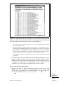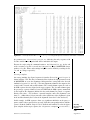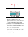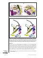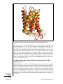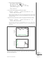
1
An Introduction to Modeling Structure
from Sequence
UNIT 5.1
There are literally millions of protein sequences in the various sequence databases, but
there are only a few tens of thousands of protein structures in the Protein Data Bank.
The rate of growth of new sequences is a steeply rising exponential curve; that of new
structures, if exponential at all, is much shallower. There is no possibility that the number
of structures will ever approach, much less equal, the number of sequences. So what is
the point of initiatives such as Structural Genomics? What sense does it make to be the
tortoise in a race in which the hare has already won?
The underlying premise behind all attempts to determine a large number of diverse
structures is that the total number of protein domain folds is much smaller, by many
orders of magnitude, than the total number of sequences; in other words, that many
sequences adopt essentially the same fold. If the fold of a protein could be recognized
from sequence information alone, then a complete database of all possible folds would
allow the structure corresponding to any sequence to be modeled, to at least some level
of accuracy.
How reasonable is this assumption? It depends first of all on the reality of the limited
universe of domain folds. (For the purpose of this discussion, the term “domain” means
any part of the structure of a protein that is sufficiently compact so as to give the
impression that it could fold stably without the rest of the protein. Although there are
various mathematical/topological definitions of a domain, most domains are like Supreme
Court Justice Potter Stewart’s 1964 explanation of pornography: we may not know how
to define it, but we usually know it when we see it.) The best evidence that this universe
is indeed limited is the diminishing number of new folds found every year despite the
sharp increase in new structures (Hou et al., 2005). Simple application of Fisher statistics
to this frequency distribution gives a crude estimate of the total number of folds. A recent
attempt at cataloging estimates this number to be around 4000, of which nearly half
(1700) are already known (Sadreyev and Grishin, 2006).
Therefore, there is reason to assume that the total number of folds will be known
eventually and that it will indeed be many orders of magnitude less than the number
of sequences. The problem of assigning a fold for every sequence now reduces to two
steps: identifying the fold that corresponds to a given sequence, and deriving the best
possible atomic model for that the structure of that sequence given knowledge of its
domain fold(s).
That doesn’t sound so difficult, but in practice it has proven to be a formidable challenge.
Both steps are far from straightforward in all but the simplest cases, and both represent
very active areas of investigation. It is these steps that are the subjects of the protocols in
this chapter.
We begin with a discussion of what would seem to be the easiest situation: homology
modeling of a protein structure from a sequence that displays significant identity to one
adopting a known fold. This is the subject of UNIT 5.6 by Andrej Sali and colleagues, who
have made some of the most important contributions to homology modeling. They discuss
every aspect of the procedure, from fold assignment to alignment of the target with the
template to model construction and validation. They emphasize that even very similar
sequences may have regions of structure that diverge significantly (principally loops).
Modeling
Structure from
Sequence
Contributed by Gregory A. Petsko
Current Protocols in Bioinformatics (2006) 5.1.1-5.1.3
C 2006 by John Wiley & Sons, Inc.
Copyright 5.1.1
Supplement 15
They show how multiple sequence alignments and the use of a family of templates can
improve the accuracy of such regions. They also explain how to decide what size grain of
salt should be used in taking the results of a homology model as factual. Their program,
MODELLER, is one of the most widely used tools for homology model construction,
and they describe in detail how to use it.
A different approach to model construction is discussed in the unit by Umeyama and
Iwadate. Their program FAMS (UNIT 5.2) uses a simulated annealing algorithm to “refine”
the model so as to improve the accuracy, particularly of the soft variables (torsion angles).
Since this program is fully automated, it has some appeal for less sophisticated users
who may not be willing or able to try different strategies to obtain a suitable model.
I have always believed that, although integral membrane protein structures are the most
difficult type to determine experimentally, they ought to be among the easiest to model.
In general, their topologies are much simpler than those of soluble proteins: for example, mixed α-helical and β-sheet domains in the membrane are essentially unknown.
Membrane-spanning domains tend to be either bundles of α-helices or barrels of antiparallel β strands, both of which are relatively easy to recognize in amino acid sequences.
Although the available database of membrane protein structures is still quite limited,
enough patterns have already begun to emerge to give confidence that this type of modeling will eventually become common. Considering that over half of all known drugs
target integral membrane proteins, mostly G-protein coupled receptors and ion channels,
it is also likely that such modeling will have considerable practical importance.
In the third unit in this chapter, a collaborative team from the Hebrew University in
Jerusalem and the Lawrence Berkeley Laboratory in California describes a tool for
predicting the structures of simple α-helical bundle membrane proteins (UNIT 5.3). By
running a global molecular dynamics search of configuration space, the protocol generates
a set of candidate structures. The best one is selected from among these using the silent
amino acid substitutions in the protein family as a stringent test for robustness. It seems
likely that this procedure is just the tip of the proverbial iceberg for membrane protein
prediction.
Homology modeling demands that the model be inspected, not only by computer program
but also by eye. For this and numerous other reasons, the ability to display and manipulate
the three-dimensional structures of proteins has passed from the province of a select few
into the routine toolkit of almost every biologist. Among the many public software
packages available for this purpose, RasMol (UNIT 5.4) is one of the oldest, most versatile,
and easiest to use. In UNIT 5.4, David Goodsell gives an overview of its capabilities and
then describes a number of useful protocols that should not only familiarize readers with
RasMol, but also enable them to carry out many of the most common procedures.
An Introduction
to Modeling
Structure from
Sequence
New units in this chapter address two other important issues in structure modeling. One
of the most frequently asked questions about any “new” protein structure is: does it
resemble any previously known fold? This is not just an academic matter. Increasingly,
protein structures are being determined for gene products of unknown function, not only
because of the structural genomics initiatives but also because genetics often leads to
the identification of a sequence as being an important contributor to, for example, a
human disease, but there is no information from sequence comparisons about what the
biochemical or biological function(s) of the gene product might be. The hope is that,
since structure changes much more slowly than sequence, similarity to a structure of
known function might provide a valuable clue.
I am not completely sanguine about this belief. On the one hand, there are some impressive
examples of its success (Kim et al., 2004). On the other hand, it is clear that the coupling
5.1.2
Supplement 15
Current Protocols in Bioinformatics
between overall fold and biochemical function is often quite loose, especially for some
protein superfamilies (Hegyi and Gerstein, 2001). Nevertheless, comparing a protein’s
fold with those already known is an important and sometimes powerful method. Liisa
Holm, whose program DALI (UNIT 5.5) is the most widely used tool for this purpose,
describes in her unit in this chapter how that tool should be employed. As the pace of
structure determination increases, DALI will be in the vanguard not only for comparison
of structures but also for assembling the database of fold libraries and assessing fold
divergence.
The growth of structure determination has turned most biochemists and biologists into
consumers of structural information. Genomics is accelerating this trend. As the demand
for such information continues to outstrip the supply, all aspects of structure modeling
assume increasing importance. For those who have yet to try their hand at such endeavors,
the encouraging news is that the tools are getting easier to use as well as more accurate.
Dip into the protocols in this chapter and see!
LITERATURE CITED
Hegyi, H. and Gerstein, M. 2001. Annotation transfer for genomics: Measuring functional divergence in
multi-domain proteins. Genome Res. 11:1632-1640.
Hou, J., Jun, S.R., Zhang, C., and Kim, S.H. 2005. Global mapping of the protein structure space and
application in structure-based inference of protein function. Proc. Natl. Acad. Sci. U.S.A. 102:36513656.
Kim, Y., Yakunin, A.F., Kuznetsova, E., Xu. X., Pennycooke, M., Gu, J., Cheung, F., Proudfoot, M.,
Arrowsmith, C.H., Joachimiak, A., Edwards, A.M., and Christendat, D. 2004. Structure- and functionbased characterization of a new phosphoglycolate phosphatase from Thermoplasma acidophilum. J. Biol.
Chem. 279:517-526.
Sadreyev, R.I. and Grishin, N.V. 2006. Exploring dynamics of protein structure determination and homologybased prediction to estimate the number of superfamilies and folds. BMC Struct. Biol. 6:6.
Contributed by Gregory A. Petsko
Brandeis University
Waltham, Massachusetts
Modeling
Structure from
Sequence
5.1.3
Current Protocols in Bioinformatics
Supplement 15
FAMS and FAMSBASE for Protein Structure
UNIT 5.2
The computer program FAMS (Full Automatic Modeling System; Ogata et al., 2000;
Iwadate et al. 2001) performs homology modeling of protein structures by means of
an algorithm consisting of database searches and simulated annealing. FAMS produces a model in which the torsion angles of the backbone and sidechains are highly
accurate.
An overview of the processes for obtaining a protein model via FAMS is shown in
Figure 5.2.1. This unit describes a procedure for searching FAMSBASE (Yamaguchi
et al., 2003), the database of structural models calculated by FAMS (see Basic
Protocol).
CHECKING FAMSBASE FOR A PROTEIN MODEL
When a 3-D structural model is required for a particular protein, one should first check
whether or not the protein is already modeled. FAMSBASE is a relational database of
comparative protein structure models for the entire genomes of 41 species, as presented in the GTOP (Genomes TO Protein structures and functions) database at
http://spock.genes.nig.ac.jp/~gtop-old/gtop.html. The models in that database were
all calculated using FAMS. FAMSBASE provides versatile search and query functions, including searching by name of ORF (open reading frame), ORF annotation,
Protein Data Bank (PDB) ID, and sequence similarity. FAMSBASE is available online
at http://famsbase.bio.nagoya-u.ac.jp/famsbase/. The present percentage of ORFs
with 3-D protein models in FAMSBASE is 42%; therefore, requested protein models
are currently available in approximately half of all cases.
BASIC
PROTOCOL
Necessary Resources
Hardware
Any computer with an Internet connection
Software
Web browser (Internet Explorer v. 5.0 or later or Netscape v. 4.7 or later for
Windows; Internet Explorer v. 4.5 or later for Macintosh)
1. Log in to FAMSBASE as follows.
a. Go to the URL of FAMSBASE, http://famsbase.bio.nagoya-u.ac.jp/famsbase/.
Figure 5.2.2 shows the login page of FAMSBASE.
b. Enter a login name and password. If accessing the database for the first time, obtain
a login name and a password by clicking the link labeled “For the first user.”
Alternatively, click on the “Public Login” hyperlink.
After logging in, one arrives at the FAMSBASE search page. Figure 5.2.3 shows the upper
part of the search page; Figure 5.2.4 shows the lower part. “Public Login” only provides
sufficient access to determine whether or not a model exists in FAMSBASE. Individuals
who select “Public Login” cannot view structures.
2. Specify search criteria.
a. Species. The upper part of the search page (Fig. 5.2.3; Section 1) lists 41 species
whose genome ORFs have been determined. The check boxes on the left-hand
side of the query form allow the user to specify which species should be included
Contributed by Hideaki Umeyama and Mitsuo Iwadate
Current Protocols in Bioinformatics (2003) 5.2.1-5.2.16
Copyright © 2003 by John Wiley & Sons, Inc.
Modeling
Structure from
Sequence
5.2.1
Supplement 4
in the search. It is possible to select multiple species. Figure 5.2.5 shows an
example in which Escherichia coli is selected.
Mo re d eta ils on the 41 species are describ ed in th e GTOP h omepage,
http://spock.genes.nig.ac.jp/~gtop-old/org.html, which contains the results not only of
PSI-BLAST but also of FASTA and normal BLAST, among others (Pearson and Lipman,
1988; Altschul et al., 1990).
b. The lower part of the search page provides the following text boxes and radio
buttons for searching: (2) Search for ORFs by Gene (ORF) Name; (3) Search for
ORFs by PDB ID of Reference Protein; (4) Search for ORFs by Motif Name; and
(5) Search for ORFs by FAMS Results.
The gene name used in the Search for ORFs by Gene (ORF) Name text box is based on the
gene names used in the GTOP Web site mentioned above. The motif name used in the Search
for ORFs by Motif Name text box is based on the PROSITE motifs, http://us.expasy.org/prosite/. The FAMS results used in Search for ORFs by FAMS Results means
whether or not the model exists in the database. As an example, Figure 5.2.6 shows a query
for Gene Name, “abc.”
Once the search criteria have been entered, click the Search button at the top of the
Web page.
c. Alternatively, there are two additional text boxes in the lower part of the search
page: Search for ORFs by Hetero Atom of Reference Protein and Search for ORFs
by Amino Acid Sequence. After entering the corresponding information in the text
box(es), click the Search button (for Search for ORFs by Hetero Atom of
Reference Protein) or the Submit Query button (for Search for ORFs by Amino
Acid Sequence).
In the Search for ORFs by Hetero Atom of Reference Protein text box, the Hetero Atom
refers to the HETATM line in PDB format. An amino acid sequence search using FASTA
(UNIT 3.9) is performed by the Search for ORFs by Amino Acid Sequence text box (Fig.
5.2.7). Users can search by several criteria at once, but the Amino Acid Sequence search
is exclusive.
Select a model
3. Examine the model list that appears (Fig. 5.2.8) with annotations of ORFs, model
lengths (number of amino acid residues), and identity percentages of amino acid
sequence alignments (with experimentally known structure).
4. Select one line in the model list by clicking on a template ID (in the PSIBlast column
in Fig. 5.2.8) from the model list, which will then bring up the amino acid alignment
view page (Fig. 5.2.9). Display the selected model structure by clicking on the View
Target button.
Both the model and the template will be displayed simultaneously (Fig. 5.2.10) by clicking
the Superimpose button when using an appropriate model viewer, e.g., RasMol,
http://www.umass.edu/microbio/rasmol/. The model file (not containing the template) can
also be downloaded by clicking on the View Target button (Fig. 5.2.11).
GUIDELINES FOR UNDERSTANDING RESULTS
FAMS and
FAMSBASE for
Protein Structure
Once the required model has been obtained, whether from FAMSBASE or from
FAMS, one may wonder about its accuracy. Generally, if the query sequence and the
amino acid sequence of the experimentally known structure shar a high percent
identity, this strongly supports the accuracy of the model structure. Quantitatively, if
the percentage is >30%, the RMSD (root mean square distance) values are within ∼4
5.2.2
Supplement 4
Current Protocols in Bioinformatics
Å (over the Cα backbone) of the true structure. Note that in low-homology cases,
regions of locally high homology exist that may contain important information in a
model. In cases of low percent identity (<30%), statistically half of all models whose
alignment E-values are low enough (<10−3) will have a small enough RMSD (within
4 Å), to be considered accurate models. The E-value guarantees the length of the
model. In the case of alignments of low-enough E-value, the reliable region is
sufficiently large in comparison to the entire ORF region. After a few years, the
number of high-identity-percentage models will increase, and, at that time, the
homology-modeling method will produce more accurate protein structures.
COMMENTARY
Background Information
The authors of this unit developed a computer program, FAMS (Full Automatic Modeling System) to build model structures based
on reference structures solved using X-ray
diffraction, NMR, or other experimental
methods, as well as amino acid sequence
alignment between a target and its reference
structure. FAMSBASE is a relational database of comparative protein structure models
in GTOP (Genomes TO Protein structures
and functions) alignment, calculated by
FAMS. Both GTOP and FAMSBASE are projects of the Japanese government.
The basic FAMS algorithm consists of a
database search and simulated annealing. The
first step obtains the Cα coordinates, the second step, the backbone, the third step, side
chains, and the last step, all atoms.
The effectiveness of the software was
highlighted by its performance in the CAFASP2 and CAFASP3 competitions (Fischer
et al., 2001), especially in terms of side-chain
accuracy, with good performance in regard to
the backbone as well. CAFASP (Critical Assessment of Fully Automated Structure Prediction) is a competition for determining the
best software of this kind. Another competition, CASP (Critical Assessment of Techniques for Protein Structure Prediction) determines the best researcher in this area.
CASP experiments were started in 1994 as
CASP1, and continued biennially through to
2002 as CASP5. CAFASP experiments were
started at the same time as CASP3, beginning
with CAFASP1, and hence CAFASP3 was
running in 2002. Results from the comparative modeling section of CASP5 suggested
that fully automated building procedures
were less accurate than procedures with human intervention (Iwadate et al., 2001). Human intervention worked effectively on
CASP5 and the assessments have highlighted
the algorithmic improvement of sequence
alignments. However, fully-automated procedures are essential, and, indeed, have been
used for large-scale genome modeling. CAFASP3 assessments did not judge human intervention, but only software performance.
The use of typical alignment software
such as FASTA (UNIT 3.9), BLAST (UNITS 3.3 &
3.4), or PSI-BLAST to determine which modeling software demonstrates the best performance is very important, and the results
are of interest not only to computational biologists but also to biologists at the laboratory bench.
Suggestions for Further Analysis
It is currently not possible to access the
FAMS server. However, the authors expect
that in the future, researchers will be able to
submit novel sequences directly to FAMS in
order to obtain structure predictions (see Fig.
5.2.12 for the FAMS Web page).
Literature Cited
Altschul, S.F., Gish, W., Miller, W., Myers, E.W.,
and Lipman, D.J. 1990. Basic local alignment
search tool. J. Mol. Biol. 215:403-410.
Fischer, D., Elofsson, A., Rychlewski, L., Pazos,
F., Valencia, A., Rost, B., Ortiz, A.R., and
Dunbrack, R.L., Jr. 2001. CAFASP2: The second critical assessment of fully automated
structure prediction methods. P ro t e i n s
45:171-183.
Iwadate, M., Ebisawa, K., and Umeyama, H.
2001. Comparative modeling of CAFASP2
competition. Chem-Bio. Informatics J. 1:136148.
Ogata, K. and Umeyama, H. 2000. An automatic
homology modeling method consisting of database searches and simulated annealing. J.
Mol. Graph. Model. 18:258-272, 305-256.
Pearson, W.R. and Lipman, D.J. 1988. Improved
tools for biological sequence comparison.
Proc. Natl. Acad. Sci. U.S.A. 85:2444-2448.
Modeling
Structure from
Sequence
5.2.3
Current Protocols in Bioinformatics
Supplement 4
Yamaguchi, A., Iwadate, M., Suzuki, E.-I., Yura,
K., Kawakita, S., Umeyama, H., and Go, M.
2003. Enlarged FAMSBASE: Protein 3D
structure models of genome sequences for 41
species. Nucleic Acids Res. 31:1-6.
http://spock.genes.nig.ac.jp/~genome/
gtop.html
Internet Resources
Contributed by Hideaki Umeyama and
Mitsuo Iwadate
Kitasato University
Tokyo, Japan
http://physchem.pharm.kitasato-u.ac.jp/FAMS/
FAMS Web site.
GTOP Web site.
http://famsbase.bio.nagoya-u.ac.jp/famsbase/
FAMSBASE Web site.
Figures 5.2.1-5.2.12 appear on the following pages.
FAMS and
FAMSBASE for
Protein Structure
5.2.4
Supplement 4
Current Protocols in Bioinformatics
protein sequence
check FAMSBASE
at
http://famsbase.bio.nagoya-u.ac.jp/famsbase/
3-D structure found
good structure?
yes
protein
structure
3-D structure not found
no
model a protein structure
at
http://physchem.pharm.kitasato-u.ac.jp/FAMS/
good structure?
yes
protein
structure
no
report to developer of FAMS
at
[email protected]
Figure 5.2.1 Flowchart of modeling by FAMS, from sequence to structure. Basic Protocol outlines
the searching of FAMSBASE.
Modeling
Structure from
Sequence
5.2.5
Current Protocols in Bioinformatics
Supplement 4
Figure 5.2.2 The login page of FAMSBASE. As stated on the page, one must first obtain an ID
and password from an administrator of FAMSBASE. If time is a factor or one just wishes to check
the contents of the database, click on the “Public login” link to go to the search page.
FAMS and
FAMSBASE for
Protein Structure
5.2.6
Supplement 4
Current Protocols in Bioinformatics
Figure 5.2.3 The upper part of the search page of FAMSBASE. 41 species whose genome ORFs
have been determined are listed with check boxes on the left-hand side. More details of the 41
species are described in http://spock.genes.nig.ac.jp/~gtop-old/org.html.
Modeling
Structure from
Sequence
5.2.7
Current Protocols in Bioinformatics
Supplement 4
Figure 5.2.4 The lower part of the search page of FAMSBASE. Text boxes and radio buttons for
searching the database are provided.
FAMS and
FAMSBASE for
Protein Structure
5.2.8
Supplement 4
Current Protocols in Bioinformatics
Figure 5.2.5 If a particular species is of interest, one may click the check boxes to the left of the
species names. In this figure, Escherichia coli is selected.
Modeling
Structure from
Sequence
5.2.9
Current Protocols in Bioinformatics
Supplement 4
Figure 5.2.6 To search the database using an ORF or protein name, input the name directly into
the text box. As an example, an ORF named “abc” has been input.
FAMS and
FAMSBASE for
Protein Structure
5.2.10
Supplement 4
Current Protocols in Bioinformatics
Figure 5.2.7 If an amino acid sequence is of interest, input the sequence in the large text box as
shown here.
Modeling
Structure from
Sequence
5.2.11
Current Protocols in Bioinformatics
Supplement 4
Figure 5.2.8 A model list with annotations, model lengths (number of amino acids), and identity
percentages of amino acid sequence alignments with experimentally known structure. To obtain a
particular model, select one line by clicking on a template ID (shown in the PSIBlast column in this
figure).
FAMS and
FAMSBASE for
Protein Structure
5.2.12
Supplement 4
Current Protocols in Bioinformatics
Figure 5.2.9 The amino acid alignment view page. To display the selected model, click the View
Target button. Both the model and the template will be displayed by clicking the Superimpose button.
Modeling
Structure from
Sequence
5.2.13
Current Protocols in Bioinformatics
Supplement 4
Figure 5.2.10 A Superimpose view using RasMol. The model is in blue and the template is in
green. This black-and-white facsimile of the figure is intended only as a placeholder; for full-color
version of figure go to http://www.interscience.wiley.com/c_p/colorfigures.htm.
FAMS and
FAMSBASE for
Protein Structure
5.2.14
Supplement 4
Current Protocols in Bioinformatics
Figure 5.2.11 The model viewed after clicking the View Target button. This black-and-white
facsimile of the figure is intended only as a placeholder; for full-color version of figure go to
http://www.interscience.wiley.com/c_p/colorfigures.htm.
Modeling
Structure from
Sequence
5.2.15
Current Protocols in Bioinformatics
Supplement 4
Figure 5.2.12 The FAMS Web page. The server status is displayed in the upper right-hand corner.
FAMS and
FAMSBASE for
Protein Structure
5.2.16
Supplement 4
Current Protocols in Bioinformatics
Modeling Membrane Proteins Utilizing
Information from Silent Amino Acid
Substitutions
UNIT 5.3
Transmembrane α-helical bundles represent a simple topology that can be described by a
relatively small number (n) of parameters: (1) helix tilt, (2) rotational position, and (3) register
(Fig. 5.3.1). Thus for any hetero-oligomer, 3 × n parameters are needed to describe the overall
structure, while for any symmetrical homo-oligomer only 2 parameters are generally sufficient to describe the structure: helix tilt (β) and rotational pitch angle (φ).
Due to the reduced number of degrees of freedom, it is possible to exhaustively search each
of the above parameters computationally in a procedure for which the name Global Molecular
Dynamics Search (GMDS) has been coined (Adams et al., 1995). GMDS has been automated
by a comprehensive series of task files and modules, written by Paul D. Adams, called CHI
(CNS searching of Helix Interactions; Adams et al., 1995), to be used in the general
computational structural biology software suite CNS (Crystallography and NMR System).
Depending on the parameters used, CHI routinely yields several candidate structures with a
characteristic tilt and rotational pitch angle. Selection amongst the different candidate
structures can be done using variety of procedures, such as the fitting of each structure to
some experimental data (e.g., mutagenesis; Lemmon et al., 1992b; Treutlein et al., 1992;
Arkin et al., 1994).
In this unit, a different procedure is described for the selection the correct structure from a
list of plausible competing structures based on silent amino acid substitutions (see Basic
Protocol). This procedure makes use of homology data in an objective manner to select the
correct model, and, in principle, can be applied as a screening procedure whenever more than
one model exists.
βj
φi
φj
βi
rj
ri
Ωij
Figure 5.3.1 In a bundle with n transmembrane α-helices (helices i and j in this case), 3n
parameters can be used to describe the general structure, assuming rigid helices: (1) the inclination
of the helices with respect to the bundle axis, βi, related to the commonly used crossing angle Ω;
(2) the rotational angle about the helix director, φi, which defines which side of helix i is facing towards
the bundle core; and (3) the helix register, ri, which defines the relative vertical position of the helix.
Modeling
Structure from
Sequence
Contributed by Uzi Kochva, Hadas Leonov, Paul D. Adams, and Isaiah T. Arkin
5.3.1
Current Protocols in Bioinformatics (2003) 5.3.1-5.3.15
Copyright © 2003 by John Wiley & Sons, Inc.
Supplement 4
BASIC
PROTOCOL
SELECTING A CORRECT PROTEIN STRUCTURE USING CHI
CHI is a series of user-friendly task files and modules written by Adams (1995) to be used
in the general software suite CNS (Crystallography and NMR System; Brünger et al.,
1998). CHI constructs multiple bundles of helices, each differing from the other by the
rotation of the helices about their axes, as well as the bundle handedness. These are then
used as starting positions for molecular dynamics simulations and energy minimization
protocols. The output structures from these simulations are compared and grouped into
clusters that contain similar structures. An average of the structures forming a cluster
represents a model with characteristic interhelical interactions and helix tilt. The “Silent
Amino Acid Substitution Protocol” performs the above simulations on close sequence
variants that are likely to share the same structure, followed by a comparison of the clusters
from the different variants, in an attempt to find a common cluster into which all these
variants fold.
In the protocol it will be assumed that the user is using a generic Unix system employing
the csh or tcsh shell. The commands are entered at a terminal with the %> command
prompt. Text files are edited using a text editor. Those who are unfamiliar with the Unix
environment should refer to APPENDIX 1C & APPENDIX 1D.
Necessary Resources
Hardware
Hardware requirements are defined by those that are officially supported by
CNSsolve, i.e., one of the following computers:
SGI (R4000 and later) running IRIX 4.0.5 or later
HP (PA Risc) running HP-UX 9.05 or later
DEC Alpha running OSF1/Digital Unix/Tru64 Unix
PC (i386, i486, i586, or i686) running Linux or Windows 98 or NT or higher
Additionally, CNSsolve also provides unsupported installations for other systems:
Convex running ConvexOS:
Cray (J90, YMP, C90, T90) running Unicos
Cray T3E (single CPU) running Unicosmk
IBM RS/6000 running AIX
Sun running SunOS
Unix systems with g77/gcc (EGCS-1.1)
Windows 98 or NT (or higher) systems with g77/gcc (EGCS-1.1)
A Macintosh OS X port is also available (contact the authors for details;
[email protected])
Modeling
Membrane
Proteins
Software
CNSsolve: available free of charge for academic users at http://cns.csb.yale.edu
CHI: available from Paul D. Adams ([email protected])
Perl: Perl is a component of nearly all standard Unix distributions. It is available
free of charge at www.perl.org. Install according to the instructions on the Web
page.
Three Perl scripts: (1) ak cluster.pl, (2) compare_rmsd.pl, and (3) to
gly.pl (available from the authors; [email protected])
A CNSsolve input script, cns.inp (available from the authors; [email protected].
ac.il)
A standard text editor, e.g., jot, notepad, or nedit)
A Web browser
Software to perform multiple sequence alignment (e.g., ClustalX, ClustalW, or
Pileup from the GCG Wisconsin package)
5.3.2
Supplement 4
Current Protocols in Bioinformatics
Install software and set up environment
1. Install CNSsolve as follows (more detailed installation instructions can be found on
the CNSsolve Web page, http://cns.csb.yale.edu):
a. Uncompress and extract the CNSsolve tar archive in /usr/local/:
%> tar -xzf cns_solve_1.1_basic_inputs.tar.gz
b. Assuming that the above file was uncompressed in /usr/local/ there is now
a new directory:
/usr/local/cns_solve_1.1/
c. Using any text editor, edit the file:
/usr/local/cns_solve_1.1/cns_solve_env
by changing only one line, as follows (assuming that CNSsolve is located in
/usr/local/cns_solve_1.1/):
setenv CNS_SOLVE /usr/local/cns_solve_1.1
d. In order to compile the program, in the CNSsolve directory that was created in
substep 1b (/usr/local/cns_solve_1.1/), type:
%> make install
This process may take several minutes depending on the computer platform, at the end of
which there is a new executable program called cns.
2. Install CHI as follows:
a. Uncompress and extract the CHI tar archive:
%> tar -xzf chi.tar.gz
b. Assuming that the above file was uncompressed in /usr/local/, there is now
a new directory:
/usr/local/chi/
c. Using a text editor edit the file:
/usr/local/chi/chi_env
by changing only one line, as follows (assuming that CHI is located in /usr/
local/chi):
setenv CHI_ROOT /usr/local/chi
d. In order to compile the program, in the src directory of CHI (/usr/local/
chi/src/) type:
%> make
3. Place the three Perl scripts (ak_cluster.pl, compare_rmsd.pl, and
to_gly.pl) in /usr/local/chi/bin/.
4. Place the file cns.inp in /usr/local/chi/bin/.
Modeling
Structure from
Sequence
5.3.3
Current Protocols in Bioinformatics
Supplement 4
5. In order for the system to recognize both CNS and CHI, which have been recently
compiled, edit the .cshrc file (APPENDIX 1C) to include the following two lines:
source /usr/local/chi/chi_env
source /usr/local/cns_solve 1.1/cns_solve env
Define the sequences for the GMDS
There are two considerations that one must take into account. The first is the identity of
the transmembrane segments to be simulated. The transmembrane α-helices must therefore be delineated from the rest of the protein. The second is, what are the homologous
sequences to one’s protein of interest?
6. Determine the transmembranal amino acids range, either by prior knowledge or by
using programs predicting transmembranal domains: e.g., via the interactive programs
TMHMM (http://www.cbs.dtu.dk/services/TMHMM/) or PSIPRED (http://bioinf.cs.
ucl.ac.uk/psipred/).
7. Search protein databases (e.g., NCBI, PDB, or GeneBank, all accessible from the
NCBI home page: http://www.ncbi.nlm.nih.gov) for homologous sequences using the
transmembrane segments determined above.
The minimal identity between the sequences should be kept very high, in order to ensure
that all changes are indeed “silent.” The authors typically use sequences that are at least
75% identical.
8. Perform multiple sequence alignment (MSA) of the desired homologous sequences
using MSA programs—e.g., ClustalX, ClustalW (UNIT 2.3), or PileUp (UNIT 3.6) from
the GCG Wisconsin package.
No gaps should be allowed, i.e., the length of all homologous sequences should be identical.
The results of the MSA will make it possible to select the exact sequences from the homologous
proteins that correspond to the transmembrane domains of the protein of interest.
Set up an appropriate directory structure
Since GMDS produces a large number of files, it is best to work in an orderly and
organized fashion. The authors therefore recommend the following directory setup.
9. Create a directory that will contain all the subdirectories and files used in the
GMDS (it will be assumed that this directory is directly under the home directory,
e.g., ~/MyProtein). Create a specific subdirectory in that directory for each
variant (e.g., ~/MyProtein/variantA, ~/MyProtein/variantB, . . . ~/
MyProtein/variantN).
Prepare the instructions file chi_param
In order to run the GMDS using CHI, all that is needed is a single instructions file called
chi_param, which, as its name suggests, contains the parameters needed for a CHI run.
chi_param can be generated by a Web server in the CHI site (Fig. 5.3.2;
http://www.csb.yale.edu/userguides/datamanip/chi/html/chi.html). One can obtain the file
simply by contacting the authors and editing it manually with any text editor. chi_param
contains exhaustive comments making the editing of the file self-explanatory.
To create a new parameter file from scratch
10a. In the CHI main menu on the left-hand side of the CHI home page (Fig. 5.3.2), click
on “Create setup.”
11a. In the first “Create setup” screen that appears (Fig. 5.3.3), type the desired molecule name.
Modeling
Membrane
Proteins
For convenience the name of the molecule should be identical to the subdirectory name
(e.g., variantA).
5.3.4
Supplement 4
Current Protocols in Bioinformatics
Figure 5.3.2 CHI main page.
Figure 5.3.3 CHI “Create setup” first screen.
12a. Type the number of helices and choose the proper option between homo-oligomer
false or true.
13a. Click “Edit sequence.” A new editing screen will appear (Fig. 5.3.4).
14a. Type the first residue number, then enter the sequence in one-letter amino acid
format (see APPENDIX 1A).
Note that the residue number is only important for the proper indexing of the sequence,
and does not mean that the input sequence will be considered from that position.
15a. Choose the orientation of the helix.
If “true” was chosen for for homo-oligomer on the previous screen (step 12a), than one
may choose either “up” or “down,” as this option only describes the relative orientation
between helices in a hetero-oligomer.
Clicking “View file” will allow one to view the chi_param that was created.
Choosing Edit file (see following) will allow one to edit all of the parameters in the
chi_param file.
Modeling
Structure from
Sequence
5.3.5
Current Protocols in Bioinformatics
Supplement 4
Figure 5.3.4 CHI “Create setup” Edit Sequence screen.
Figure 5.3.5 CHI “Edit setup” first screen for editing an existing parameters file.
To edit a parameter file that already exists
10b. In the CHI main menu on the left-hand side of the CHI home page (Fig. 5.3.2), click
on “Edit setup.” In the first “Edit setup” screen that appears (Fig. 5.3.5), enter the
full path and the name of the chi_param file, or click the Browse button, navigate
to its location, and select it.
11b. Click on “Edit file.” Note the molecule structure parameters on the new screen that
appears (Fig. 5.3.6):
Name of molecule
Number of helices
homo-oligomer, true/false.
12b. If one has chosen to simulate a hetero-oligomer, set the next parameters for each
helix individually (otherwise they should only be set once):
Modeling
Membrane
Proteins
Sequence
Residue number at start of sequence
Initial rotation offset around helix axis (the starting rotation angle about the
helix axis relative to some arbitrary starting position, angle φ in Figure
5.3.1, default is 0.0°)
continued
5.3.6
Supplement 4
Current Protocols in Bioinformatics
Figure 5.3.6 CHI “Edit file” screen with structure parameters.
Direction of helix, up/down.
Initial translational offset for helix along the z axis (default is 0.0 Å).
13b. Set search parameters (Fig. 5.3.6):
i. Extent of the search: full or symmetric.
In a symmetric search, all of the helices rotate about their axis concomitantly (ωi = ωj) due
to the symmetry assumption in homo-oligomers (Arkin, 2002). In a full search all rotation
combinations are examined (ωi ≠ ωj ). The default for a homo-oligomeric complex is a
“symmetric search” while a “full search” is the default when analyzing hetero-oligomers.
A full search will obviously take much longer than a symmetric search due to the larger
number of structures generated (see below).
ii. Search left-handed crossing angles, true/false (default is “True”).
iii. Search right handed crossing angles true/false (default is “True”).
iv. Type of molecular dynamics to use, torsion/cartesian (default is “torsion”).
The reader is referred to Rice and Brünger (1994) in order to evaluate which type of
molecular dynamics to choose.
Modeling
Structure from
Sequence
5.3.7
Current Protocols in Bioinformatics
Supplement 4
v. Number of trials per structure, i.e., number of searches to perform using different
initial random velocities for each structure (default is 4).
If one has chosen to simulate a hetero-oligomer, the next parameters should be set for each
helix individually; otherwise they should only be set once:
vi. Rotation start (default is 0°).
vii. Rotation finish (default is 360°).
viii. Rotational step size (increment step default is 45°; the authors suggest setting it
to 10° for a symmetric search).
14b. Set other restraints (it is not necessary to use these parameters in the Silent Amino
Acid Substitution Protocol):
Electrostatic effects:
Value of the dielectric constant (for a membrane matrix enter 2.0; for a vacuum matrix enter 1.0)
Initial rotation and tilt:
Distance between centers of neighboring helices (default is 10.4 Å)
Left-hand crossing angle (default is 25°)
Right-hand crossing angle (default is −25°)
Clustering parameters:
Cutoff for root mean square difference between two structures (indicates
structure similarity of two structures; default is 1 Å; a larger number
would result in finding more clusters that are not as well grouped)
Minimum number of structures which define a cluster (default is 10).
15b. Click the “Save updated file” at the bottom of the screen, which will download a
new, updated chi_param file into the local computer. Save it to the correct
directory (e.g., ~/MyProtein/variantA/).
Run the GMDS search
16. Change to the correct working directory (e.g., ~/MyProtein/variantA) with
the following command:
%> cd ~/MyProtein/variantA/
All commands should be issued form this directory unless noted otherwise.
17. In order to create the starting structure run:
%> chi_create -verbose
This is a fast process (taking a few minutes). The output files will be:
~/MyProtein/variantA/variantA.psf
~/MyProtein/variantA/variantA.pdb
~/MyProtein/variantA/chi_create.log
~/MyProtein/variantA/results/create.out.
All of these files are accessory files that CHI uses.One might want to search for an error
in the log file by issuing the following command:
%> grep -i err chi_create.log | more
Modeling
Membrane
Proteins
5.3.8
Supplement 4
Current Protocols in Bioinformatics
18. Run the searching algorithm, to create all the structures, using the following command:
%> chi_search -verbose
The number of structures is:
φend − φstart
× handedness × trials =
increment
360, − 0,
× 2 × 4 = 288
10
This process is time-consuming (typically many hours). As an example, simulating a bundle
of 5 helices, each composed of 28 amino acids, takes ~20 to 30 min per structure, on a
DEC Alpha 433 AU (a relatively slow machine nowadays).
This will produce the following output:
~/MyProtein/variantA/results/search.out
which contains the results of the simulation (energy and orientational parameters for each
structure) and the pdb for each structure simulated.
The names of the pdb files are as follows:
~/MyProtein/variantA/results/left_i_j.pdb
where i is the initial angle of rotation and j is the trial number.
Right-handed structures will be designated similarly:
~/MyProtein/variantA/results/right_i_j.pdb
One may wish to check for errors during the run by screening the log file with the following
command:
%> grep -i err chi_search.log | more
If no errors are found, it is best to delete the log files since they can be very large (several
megabytes).
19. To calculate the Cα RMSD between all of the structures, type:
%> chi_rmsd -verbose
This process is relatively time-consuming (roughly 0.1 sec per comparison, i.e., 93 min for
288 structures). The output is a single file:
~/MyProtein/variantA/results/rmsd.out.
This file contains a list of structures and the Cα RMSDs between them. Note that the file
only lists those structure that are lower than the RMSD threshold plus 1 Å.
Note that when the number of structures increases to a certain point, it is the RMSD
calculation that consumes the largest amount of CPU time. This is because the time
required for molecular dynamics simulations scales linearly with the number of structures
generated (576 structures take only twice the amount of time as 288 structures), whereas
the RMSD calculation scales with the square of the number of structures (comparison of
576 structures takes 4 times longer than 288 structures). The chi_rmsd script is therefore
best suited to cases where the number of structures is approximately 2000 or less. If
interested in simulating a larger system, one can contact the authors ([email protected])
for alternative scripts to chi_rmsd. These scripts reduce the computational cost by not
calculating the RMSD between two clusters if their orientational parameters differ markedly.
Modeling
Structure from
Sequence
5.3.9
Current Protocols in Bioinformatics
Supplement 4
20. In order to search for clusters to which structures have converged, run the following
command:
%> perl /usr/local/chi/bin/ak_cluster.pl
This file is different from the clustering file in the CHI package (chi_cluster) in that
all structures that it places in a cluster are similar to one another. In chi_cluster, all
structures are similar to at least one structure, but not necessarily to all of them. This step
is very fast (taking a few seconds).
21. Using any text editor, view the output file, ~/MyProtein/variantA/
results/cluster.out, to see how many clusters were obtained.
The authors recommend creating at least 10 to 15 clusters for each variant in order to find
a “complete set” for all the variants (see below). This can be achieved by empirically
changing the clustering parameters in the file ~/MyProtein/variantA/
chi_param, either using a text editor or through the CHI Web interface (see above).
There are two methods for increasing the number of clusters: (1) relaxing the RMSD
threshold (i.e., increasing it) and (2) decreasing the required number of structure per
cluster. Both methods should be tried.
22. In order to calculate an average, representative structure for each cluster, issue the
following command:
%> chi_average
This process is moderately time consuming (taking a few hours). The output is both a file
depicting the results of the program:
~/MyProtein/variantA/results/average.out
which includes the orientational parameters and energy of each cluster average, and the
structure for each cluster average:
~/MyProtein/variantA/results/clusterN.pdb
where N is the number of the cluster.
Find a “complete set”
23. Repeat GMDS (steps 16 to 22) for all the variants.
Remember that each variant’s search should be undertaken in its specific subdirectory, e.g.:
~/MyProtein/variantB/, ~/MyProtein/variantC/)
The following steps are in preparation for comparing clusters of different variants and are
are not part of the “standard” CHI package. The process starts with creating a virtual GLY
variant. Selecting the right cluster, which is the one that exists in all the variants, depends
on comparing the RMSD between all the structures obtained in the previous steps.
Comparing RMSD for all the atoms of every two variants is impossible, due to the fact that
they differ at one or more of their amino acids. However, one may avoid this problem by
comparing only the RMSD of their backbones. Therefore, a virtual variant, whose sequence
is composed only of glycine should be created.
24. Create a new subdirectory named GLY in the upper directory, using, e.g., the
command:
~/MyProtein/GLY/
Modeling
Membrane
Proteins
25. Create a new chi_param file (also see steps 10a to 15a and 10b to 15b) in which
the molecule name is GLY and all the amino acids are glycine (Fig. 5.3.7). Leave all
other parameters exactly as they are in all the other variants’ parameter files (including
the length of the sequence). Save that file in ~/MyProtein/GLY/ directory.
5.3.10
Supplement 4
Current Protocols in Bioinformatics
Figure 5.3.7 Creating a glycine parameter file.
26. Change directory:
%> cd ~/MyProtein/GLY/
and run:
%> chi_create
This will create the files ~/MyProtein/GLY/GLY.pdb and ~/MyProtein/GLY/
GLY.psf. See step 24 annotation for explanation.
27. Change directory to the parent directory:
%> cd ~/MyProtein/
and copy the following files:
%> cp ~/MyProtein/GLY/GLY.p* ~/MyProtein/
28. Create a variants list, i.e., edit a text file named list (not list.txt) that will
contain the names of all the variants subdirectories.
Each line should contain only a single variant. An example of the content of such a file with
three variants is listed below:
variantA
variantB
variantC
Save the list file to the upper directory (i.e., ~/MyProtein/list).
29. Copy and paste the following file:
%> cp /usr/local/chi/bin/cns.inp ~/MyProtein/
30. Check that the parent directory, ~/MyProtein/ contains the appropriate files by
issuing the following command:
%> ls ~/MyProtein/
A typical directory listing with three homologs should be:
continued
Modeling
Structure from
Sequence
5.3.11
Current Protocols in Bioinformatics
Supplement 4
variantA/
variantB/
variantC/
GLY/
GLY.pdb
GLY.psf
cns.inp
list
31. Compare all the cluster averages from each homolog, obtained by GMDS by their
Cα RMSD. Look for the cluster that exists in all variants with a minimal RMSD
between every pair of variants. Issue all of the following commands from the parent
directory, ~/MyProtein/. To be sure that one is in the right directory, issue the
following command:
%> cd ~/MyProtein/
Run the following command to compare the different homologs:
%> perl /usr/local/chi/bin/compare_rmsd.pl N
where N is a number that signifies the RMSD threshold in Å.
There are several output files:
~/MyProtein/compare_rmsd.out: This file contains the list of clusters that are
found in all of the homologs. If more than one structure is found, try to enforce a stricter
threshold by reducing the number.
~/MyProtein/cns_rmsd.result: This file lists all pairwise RMSD results.
~/MyProtein/rmsd_calculation_list: This is an accessory file to be used by
CNSsolve.
~/MyProtein/log: This is the CNSsolve log file.
As stated above, view the file compare_rmsd.out in order to decide whether to repeat
the previous step with a different threshold or not.
32. Repeat the above steps until a single cluster is identified that is found in all variants.
GUIDELINES FOR UNDERSTANDING RESULTS
The procedure outlined in the Basic Protocol is a relatively simple one that involves two
steps: (1) generate possible structures for each of the variants and (2) check if there is one
structure that persists in all of the different variants. There are few key points to which
one should pay close attention, and these are outlined below.
How Well Do the Individual Variants Cluster?
The clustering parameters (i.e., the RMSD threshold and the minimal number of structure
per cluster) are chosen arbitrarily. They will obviously change the outcome of the
simulation in that they will change the number of possible structures from each variant.
The authors have tended not to extend the RMSD threshold beyond 1.25 Å, and the number
of structures per cluster is not lowered beyond 7.
How Well Does a Single Structure Persist in All of the Variants?
Modeling
Membrane
Proteins
As stated in the above section, it is difficult to place an upper boundary on the RMSD
threshold, stating unequivocally that above a certain limit the structures are no longer the
same. One should keep in mind however that RMSD is obviously not a linear repre-
5.3.12
Supplement 4
Current Protocols in Bioinformatics
sentation of similarity. In other words, when the RMSD between two structures is 1 Å
instead of 2 Å, they have not become twice as similar. Empirically, the authors tend not
to raise the RMSD threshold beyond 1.5 Å.
COMMENTARY
Background Information
Structural studies have so far shown that
membrane proteins fold into one of only two
topologies: β-barrels or α-helical bundles.
Since α-helical membrane proteins are far more
abundant, as well as pharmaceutically more
important, the following discussion will be restricted to this family.
Predicting membrane protein structure is of
significant importance because, despite the
pharmaceutical importance that they possess,
out of nearly 20,000 protein structures solved
using crystallographic or NMR methods, only
a few dozen are membrane proteins. This paucity of experimentally solved structures is striking, considering that according to a recent census of genomes, 20% to 30% of all genes are
predicted to encode membrane proteins
(Stevens and Arkin, 2000).
Knowledge-based homology methods that
rely on structural information are difficult to
implement for membrane proteins, simply because of the lack of solved structures. On the
other hand, other modeling methods are relatively easy to implement for membrane proteins, compared to water-soluble proteins, due
to the overall simplicity of membrane proteins,
in particular those formed from α-helical bundles. Furthermore, assignment of the different
helices in an α-helical bundle (the more abundant and pharmaceutically important family) is
relatively straightforward. Thus, it can be concluded that while the structures of α-helical
membrane proteins are the most difficult to
determine experimentally, fortunately they are
the easiest to predict computationally.
Despite the apparent ease with which it is
possible to simulate membrane proteins using
molecular dynamics, there is one issue that has
can potentially present difficulty: the presence
or absence of a lipid bilayer. In the simulations
of membrane proteins using molecular dynamics in CHI, no lipids or solvent molecules are
employed, because of the prohibitive computational cost. However it is possible to argue that
the most important stabilizing force in any
oligomeric bundle will be the interaction between the helices themselves (Torres et al.,
2001). Thus, there is some justification in the
simulation procedure described here, although
the lack of a lipid environment should always
be borne in mind.
Critical Parameters and
Troubleshooting
The underlining premise of GMDS is that it
is possible to exhaustively search the configuration space of a transmembrane helical bundle
and come up with several candidate structures.
One of these structures is presumed to be that
which is found in nature.
The underlining premise of silent substitution modeling is that silent substitutions do not
disrupt the native structure, but may destabilize
non-native structures. Thus, it is possible to
select the correct structure among several candidate structures using silent substitution modeling by looking for a model that is present in
all of the homologs.
When will this procedure fail? There are
several possible situations in which this may
occur: (1) where no single structure is found to
be in all of the homologs; (2) where more than
one structure is found in all of the homologs;
and (3) where the structure that is found in all
of the sequences is not the native one. Below,
the potential causes of these failures are analyzed and the ways to avoid them are suggested.
No structure is found
There can be two simple reasons for the
failure to find a structure that persists in all of
the homologs.
GMDS was not able to identify the native
structure in at least one homolog and perhaps
in all of them. The authors have found from
experience that this may happen when the tilt
of the helices is relatively large, as is the case
in the Influenza A M2 H+ channel (Kukol et al.,
1999). This problem may be overcome by increasing the crossing angle used for the right
and left-handed searches from 25° by editing
the chi_param file. Other options that may
be pursued are to increase the number of trials
and to reduce the rotational increment (again
in chi_param). Obviously both of these
changes will be reflected in increased computational time.
Some of the mutations are not silent. In other
words, some of the homologs do not adopt the
Modeling
Structure from
Sequence
5.3.13
Current Protocols in Bioinformatics
Supplement 4
same structure (Torres et al., 2002a). Here the
authors suggest an increase in the similarity
threshold of the sequences used in the simulation, i.e., sequences that are closer to the target
protein have a better chance of adopting the
same structure.
More than one structure is found
In this instance, it is possible that the “filtering capabilities” of the silent mutant were not
sufficient. The recommendation is simply to
use more sequences, by potentially lowering
the identity threshold.
The structure found is incorrect
In all of the cases in which the authors have
used the combination of GMDS and silent
amino acids substitution modeling, it produced
the correct structure as verified using other
experimental methods (Kukol et al., 2002; Torres et al., 2002b; Torres et al., 2002c). However,
this may not always be the case. Identifying
such a situation is difficult and requires the
application of potentially time-consuming experimental methodologies (see Suggestions for
Further Analysis).
Suggestions for Further Analysis
It is obvious that the best way to analyze the
results of any modeling exercise is by experimentation. There are several methods that can
be applied; however most experiments, short of
directly solving the structure, are better suited
to refuting models, rather then confirming
them. The reason is that, typically, more than
one model can be consistent with the experimental results.
Modeling
Membrane
Proteins
Mutagenesis
Mutagenesis has been used in several instances to determine which residues are essential for oligomerization of particular transmembrane helices. This is possible only when an
oligomerization assay exists, as with glycophorin A, which remains dimeric in SDS-PAGE
(Lemmon et al., 1992a). In that series of experiments, several residues were identified that
were shown to line one side of a helix projection
(Lemmon et al., 1992b; Lemmon et al., 1994).
A solution NMR study in detergent micelles
has shown those residues to be intimately involved in the helix-helix interface (MacKenzie
et al., 1997). Mutagenesis has also been performed for phospholamban, which also remains a pentamer in SDS-PAGE (Arkin et al.,
1994). In this instance, however, more than one
model was consistent with the mutagenesis
results and only a a direct structural method was
able to resolve this ambiguity (Torres et al.,
2000).
Literature Cited
Adams, P.D., Arkin, I.T., Engelman, D.M., and
Brünger, A.T. 1995. Computational searching
and mutagenesis suggest a structure for the pentameric transmembrane domain of phospholamban. Nat. Struct. Biol. 2:154-162.
Arkin, I.T. 2002. Structural aspects of oligomerization taking place between the transmembrane
alpha-helices of bitopic membrane proteins. Biochim. Biophys. Acta 1565:347-363.
Arkin, I.T., Adams, P.D., MacKenzie, K.R., Lemmon, M.A., Brünger, A.T., and Engelman, D.M.
1994. Structural organization of the pentameric
transmembrane alpha-helices of phospholamban, a cardiac ion channel. EMBO J. 13:47574764.
Brünger, A.T., Adams, P.D., Clore, G.M., DeLano,
W.L., Gros, P., Grosse-Kunstleve, R.W., Jiang,
J.S., Kuszewski, J., Nilges, M., Pannu, N.S.,
Read, R.J., Rice, L.M., Simonson, T., and Warren, G.L. 1998. Crystallography & NMR system:
A new software suite for macromolecular structure determination. Acta Crystallogr. D Biol.
Crystallogr. 54:905-921.
Kukol, A., Adams, P.D., Rice, L.M., Brünger, A.T.,
and Arkin, T.I. 1999. Experimentally based orientational refinement of membrane protein models: A structure for the Influenza A M2 H+ channel. J. Mol. Biol. 286:951-962.
Kukol, A., Torres, J., and Arkin, I.T. 2002. A structure for the trimeric MHC class II-associated
invariant chain transmembrane domain. J. Mol.
Biol. 320:1109-1117.
Lemmon, M.A., Flanagan, J.M., Hunt, J.F., Adair,
B.D., Bormann, B.J., Dempsey, C.E., and Engelman, D.M. 1992a. Glycophorin A dimerization
is driven by specific interactions between transmembrane alpha-helices. J. Bio l. Ch em.
267:7683-7689.
Lemmon, M.A., Flanagan, J.M., Treutlein, H.R.,
Zhang, J., and Engelman, D.M. 1992b. Sequence
specificity in the dimerization of transmembrane
alpha-helices. Biochemistry 31:12719-12725.
Lemmon, M.A., Treutlein, H.R., Adams, P.D.,
Brünger, A.T., and Engelman, D.M. 1994. A
dimerization motif for transmembrane alphahelices. Nat. Struct. Biol. 1:157-163.
MacKenzie, K.R., Prestegard, J.H., and Engelman,
D.M. 1997. A transmembrane helix dimer:
Structure and implications. Science 276:131133.
Rice, L.M. and Brünger, A.T. 1994. Torsion angle
dynamics: Reduced variable conformational
sampling enhances crystallographic structure refinement. Proteins 19:277-290.
Stevens, T.J. and Arkin, I.T. 2000. Do more complex
organisms have a greater proportion of membrane proteins in their genomes? Proteins
39:417-420.
5.3.14
Supplement 4
Current Protocols in Bioinformatics
Torres, J., Adams, P.D., and Arkin, I.T. 2000. Use of
a new label, 13C=18O, in the determination of a
structural model of phospholamban in a lipid
bilayer: Spatial restraints resolve the ambiguity
arising from interpretations of mutagenesis data.
J. Mol. Biol. 300:677-685.
Torres, J., Kukol, A., and Arkin, I.T. 2001. Mapping
the energy surface of transmembrane helix-helix
interactions. Biophys. J. 81:2681-2692.
Torres, J., Briggs, J.A., and Arkin, I.T. 2002a. Contribution of energy values to the analysis of
global searching molecular dynamics simulations of transmembrane helical bundles. Biophys. J. 82:3063-3071.
Torres, J., Briggs, J.A., and Arkin, I.T. 2002b. Convergence of experimental, computational and
evolutionary approaches predicts the presence of
a tetrameric form for CD3-zeta. J. Mol. Biol.
316:375-384.
Torres, J., Briggs, J.A., and Arkin, I.T. 2002c. Multiple site-specific infrared dichroism of CD3zeta, a transmembrane helix bundle. J. Mol. Biol.
316:365-374.
Treutlein, H.R., Lemmon, M.A., Engelman, D.M.,
and Brünger, A.T. 1992. The glycophorin A
transmembrane domain dimer: Sequence-specific propensity for a right-handed supercoil of
helices. Biochemistry 31:12726-12732.
Key References
Arkin et al., 1994. See above.
In this article, global searching molecular dynamics
simulation is used to find a model for phospholamban.
Adams et al., 1995. See above.
Here, the theory of global searching molecular dynamics simulation is presented in detail.
Briggs, J.A.G., Torres, J., Kukol, A., and Arkin, I.T.
2001. A new method to model membrane protein
structure based on silent amino-acid substitutions. Proteins Struct. Funct. Genet. 44:370-375.
In this article, silent substitution modeling is introduced for the first time.
Torres et al., 2002a. See above.
In this paper, results of global searching molecular
dynamics simulations are analyzed in terms of energy, thereby enabling the user to further select
among candidate models.
Torres et al., 2002b. See above.
In this work, silent substitution modeling is employed to derive a structure of the TCR CD3ζ transmembrane helical bundle, shown to coincide with
that obtained experimentally.
Contributed by Uzi Kochva, Hadas Leonov,
and Isaiah T. Arkin
The Hebrew University
Jerusalem, Israel
Paul D. Adams
Lawrence Berkeley Laboratory
Berkeley, California
Modeling
Structure from
Sequence
5.3.15
Current Protocols in Bioinformatics
Supplement 4
Representing Structural Information with
RasMol
UNIT 5.4
Thousands of atomic structures of proteins, nucleic acids, and other biomolecules are
available for use in research and education. Many effective tools are available for the
display of these structures. These tools run on popular computer hardware, and they
provide a standard set of options for representation of the molecule. This unit will
describe the use of a common program, RasMol, for the display of molecular structures.
RasMol is simple to get started and provides a wide range of options as one explores
a molecular structure. Many of the principles of selection and display used in RasMol
will then be directly applicable when moving to other molecular graphics programs for
specific applications.
The unit will begin with the basics of obtaining coordinates and displaying them in RasMol (Basic Protocol 1). Next, the advantages and limitations of different representations
will be discussed (Alternate Protocol). A common pitfall encountered in the display
of atomic coordinates, obtaining the proper biological unit, will be presented (Basic
Protocol 2). Finally, some ideas for customizing a molecular graphics session will be
presented (Basic Protocol 3).
USING RasMol TO DISPLAY A PROTEIN STRUCTURE
In this protocol, the coordinates of hemoglobin will be downloaded from the Protein Data
Bank and the structure displayed in RasMol using a few basic representations. RasMol is
an open-source program designed for the display of biological molecules. The program
reads molecular coordinates from a file and interactively displays the molecule in a variety
of representations. RasMol is an excellent place to start when learning about molecular
graphics, since the program has a number of useful options available in convenient
pull-down menus. Then, as further functionality is needed for specific applications, the
Command Line interface allows additional selection and representation options.
BASIC
PROTOCOL 1
Necessary Resources
Hardware
RasMol runs on a variety of computer hardware, including personal computers.
Software
Operating system: RasMol runs under Microsoft Windows and Apple Macintosh
OS 7.0 or higher (including Mac OS X). It may also be run on workstations
under Unix, Linux, or VMS.
RasMol: Binary versions of RasMol are available on the WWW at: http://www.
bernstein-plus-sons.com/software/rasmol/. Downloading and installation
instructions are given in Support Protocol 1.
Files
Coordinate files are read in a variety of formats, including PDB, Mol2, CHARMm,
and mmCIF. The program deals gracefully with a number of variations of these
files, including files containing coordinates for multiple conformers or multiple
models. In this example, coordinates for hemoglobin (2HHB.pdb) obtained
from the Protein Data Bank (PDB; UNIT 1.9) are used; instructions for
downloading the PDB coordinate file are given in Support Protocol 2.
Contributed by David S. Goodsell
Current Protocols in Bioinformatics (2005) 5.4.1-5.4.23
C 2005 by John Wiley & Sons, Inc.
Copyright Modeling
Structure from
Sequence
5.4.1
Supplement 11
Display hemoglobin with RasMol
1. Download and install RasMol on the local machine (Support Protocol 1).
2. Download the PDB coordinate file 2HHB.pdb from the PDB (UNIT 1.9) as described
in Support Protocol 2.
3a. On Unix and Linux machines: Type rasmol 2HHB.pdb at the prompt. This will
start RasMol and load the coordinates from the file 2HHB.pdb.
3b. On personal computers: Double-click on the RasMol icon. This will launch
RasMol. Next, select Open from the File menu to load the coordinates from the
file 2HHB.pdb. Finally, go to the Window menu and select the Command Line
window. This will open a new window that contains the Command Line interface.
With the Mac OS X operating system one can also simply drag the icon for the
desired PDB file on to the icon for RasMol and the program will automatically open,
load the coordinates, and display the default wireframe representation.
At this point, the screen should look like Figure 5.4.1, with a graphics window that shows
the hemoglobin structure and a Command Line window. The molecule may be rotated
by holding down the mouse button in the graphics window and dragging the cursor in
different directions. Other transformations, such as scaling the image to different sizes
and translating the molecule to different locations in the screen, are accessible through
different buttons (if using a three-button mouse) and through combinations of holding the
mouse button and depressing the Shift or Control keys (if using a one-button mouse).
4. The pull-down menus in the graphics window make it possible to change the representations used to display the molecule, as well as to change some common parameters
used to create the display.
a. In the Display menu, several common representations of the structure may be
chosen, as described more fully in the Alternate Protocol.
Representing
Structural
Information with
RasMol
Figure 5.4.1 RasMol running on the computer display. The viewer window is at upper left, behind
the Command Line window at lower right.
5.4.2
Supplement 11
Current Protocols in Bioinformatics
b. Using options in the Colours menu, the structure may be colored using traditional
atomic colors or several other schemes that highlight different characteristics.
c. In the Options menu, slab mode may be used to cut away the nearest portions of
the molecule, and specular highlights and shadows may be toggled on and off.
d. The Settings menu makes it possible to choose an action that will be performed
when clicking on a portion of the molecule, e.g., measuring distances between
atoms.
e. Finally, the Export menu makes it possible to save images from the graphics
window.
5. The Command Line window (labeled “Terminal” in Fig. 5.4.1) allows direct control
of all of the commands available in RasMol. A few of the most common commands
will be used in this unit. The user manual contains instructions for using a variety of
other specialized commands once the basics are mastered.
Take some time to explore the options available in the pull-down menus and to become
familiar with manipulating the molecule. When ready to move on to the next step, quit the
program by typing, in the Command Line window:
RasMol> quit
The remainder of this protocol will discuss a few useful representations and provide a
few tips to solve common problems. The Command Line window will be used to change
representations and colors, allowing more control than that available through the pulldown menus.
Representations and their uses
Three basic types of representations are commonly used to display biological molecules.
Each has its own strengths and weaknesses, and each is designed for a specific use.
6. Wireframe diagrams: The default representation in RasMol is a wireframe diagram.
Each line represents a covalent bond between atoms. This representation is ideal for
examination of the atomic details of the structure. However, wireframe representations tend to be very complicated. This is acceptable when examining the structure
interactively, but wireframe representations are generally too crowded for printed
images. The following describes how to create a wireframe diagram.
a. Restart RasMol using the 2hhb coordinates.
b. In the Command Line window, type the following series of commands:
RasMol> select HEM
This selects the heme group.
RasMol> wireframe 150
This represents the heme with a thick wireframe; values go from 1 (thin) to 500 (thick).
RasMol> select iron
This selects the iron ion.
RasMol> cpk 150
This represents the iron as a sphere. The command cpk, which represents atoms as
spheres, refers to the plastic Corey-Pauling-Koltun models used for building small organic
molecules, which were the first models that used a spacefilling representation. The units
used by RasMol are integers that correspond to 1/250th of an Angstrom (Å).
The display should look like Figure 5.4.2. The protein is displayed with a wireframe,
colored by the atom type, and thicker bonds are used to make the heme group more
apparent.
Modeling
Structure from
Sequence
5.4.3
Current Protocols in Bioinformatics
Supplement 11
Figure 5.4.2
spheres.
Hemoglobin with the heme groups in thick bonds and the iron ions shown as small
c. Rotate the display and notice the following. (1) Individual amino acids may be
identified from their shape and chemical composition. For instance, look for
aromatic amino acids while rotating the structure. (2) The overall conformation
of the backbone is difficult to comprehend. Wireframe images often look like a
tangle of atoms, not a folded chain. (3) Zoom the molecule to higher magnification
and notice that the wireframe works best on close-up pictures, which focus on a
few details.
7. Spacefilling diagrams: Spacefilling representations show the size and shape of the
entire molecule. Each atom is represented by a sphere that represents the optimal
contact distance between nonbonded atoms. The following describes how to create
a spacefilling diagram.
a. In the Command Line window, type the following series of commands:
RasMol> select all
This selects all atoms.
RasMol> wireframe off
This turns off the wireframe.
RasMol> select protein or ligand
This selects only the protein atoms and the ligand (heme) atoms.
RasMol> cpk
Representing
Structural
Information with
RasMol
This displays atoms as spheres, using the default radius for the spheres.
5.4.4
Supplement 11
Current Protocols in Bioinformatics
Figure 5.4.3 Spacefilling (cpk) representation of hemoglobin with each chain colored differently.
For the color version of this figure go to http://www.currentprotocols.com.
RasMol> color chain
This colors each chain a different color.
The display should look like Figure 5.4.3. Now, the entire protein is displayed as spacefilling spheres for each atom. The four individual polypeptide chains that make up the
hemoglobin tetramer are each given a different color. The logical operation used in the
selection command is a typical Boolean OR, so the command "select protein or ligand"
will select all atoms in the protein and all atoms in the ligand. Similarly, the command
"select protein and ligand" will select no atoms, since there are no common atoms that are
in both the set of protein atoms and the set of ligand atoms. Selection of an appropriate
set of atoms is probably the most difficult, and the most useful, aspect of RasMol usage.
b. Rotate the display and notice the following. (1) Spacefilling representations show
the bulk of the protein. Notice the way the different subunits interdigitate, and the
way the heme slots into a form-fitting groove. (2) Many people find it difficult
to identify individual amino acids in spacefilling representations, even if they are
colored by atom type.
8. Backbone and Ribbon Diagrams: Two schematic representations are commonly used
to display the topology of a protein chain. In a backbone representation, cylinders
are drawn between successive alpha carbon positions. In a ribbon diagram, a helical
ribbon is used to display alpha helices, a large flat arrow is used to display beta
sheets, and smooth tubes are used to display other portions of the chain. Ribbon
diagrams are excellent for presentation of protein folding, and are currently the most
common representation used in journal publications. The following describes how
to create backbone and ribbon diagrams.
Modeling
Structure from
Sequence
5.4.5
Current Protocols in Bioinformatics
Supplement 11
Figure 5.4.4 Backbone representation of the hemoglobin protein chains, with the hemes still
shown as spacefilling spheres. For the color version of this figure go to http://www.currentprotocols.
com.
a. In the Command Line window, type:
RasMol> select protein
This selects the protein.
RasMol> cpk off
This turns off the spheres (the spheres for the heme remain on).
RasMol> backbone 100
This draws a tube along the backbone.
The display should look like Figure 5.4.4.
b. Rotate the display and notice the following. (1) Backbone representations show
the folding of the protein chain, making it easy to recognize the many alpha helices
in this globin fold. (2) Backbone representations typically under-represent the size
of the protein, and ignore the dense packing of atoms in the structure. Explore
this by flipping the spacefilling representation on and off by typing cpk and then
cpk off in the Command Line window. (3) The position of each alpha carbon
is retained in the diagram, so it is possible to identify the location of each amino
acid.
c. Next, in the Command Line window, type the following commands:
RasMol> backbone off
Representing
Structural
Information with
RasMol
This turns off the protein backbone.
5.4.6
Supplement 11
Current Protocols in Bioinformatics
Figure 5.4.5 Ribbon diagram (cartoon) of the hemoglobin protein chains, with the hemes as
spacefilling spheres. For the color version of this figure go to http://www.currentprotocols.com.
RasMol> cartoon
This turns on the ribbon diagram.
The display should look like Figure 5.4.5.
d. Rotate the display and notice the following. (1) Ribbon diagrams make it easy to
identify secondary structural elements, such as the alpha helices in hemoglobin.
(2) Visual cues to amino acid positions are lost in the smooth ribbon, unless the
ribbon is colored to show the types of amino acids.
9. When finished, type the following in the Command Line window to exit the program:
RasMol> quit
DOWNLOADING AND INSTALLING RasMol ON A LOCAL COMPUTER
This protocol describes how to download and install RasMol on a local computer. Executable versions of RasMol are available on the WWW, so this is relatively straightforward.
SUPPORT
PROTOCOL 1
Necessary Resources
Hardware
RasMol runs on a variety of computer hardware, including personal computers.
Modeling
Structure from
Sequence
5.4.7
Current Protocols in Bioinformatics
Supplement 11
Software
Operating system: RasMol runs under Microsoft Windows and Apple Macintosh
OS 7.0 or higher (including Mac OS X). It may also be run on workstations
under Unix, Linux, or VMS.
Browser: An Internet browser is required
1. Point the browser to http://www.bernstein-plus-sons.com/software/rasmol/.
2. Click on the appropriate version at the top of the page to download the executable
file.
On personal computers, the program will appear as a RasMol icon. On Linux machines,
the program will appear as a file with a name like rasmol 8BIT or rasmol-32BIT.
3. On workstations, ensure that the permission is set correctly for an executable file,
for instance, with the command:
chmod a+x rasmol-32BIT
SUPPORT
PROTOCOL 2
DOWNLOADING COORDINATES FROM THE PROTEIN DATA BANK
The Protein Data Bank (UNIT 1.9) is the primary repository of protein structure data. It is
designed for easy searching and downloading. This protocol describes how to download
the coordinates of hemoglobin.
Necessary Resources
Hardware
The Protein Data Bank on a variety of computer hardware, including personal
computers
Software
An Internet browser is required.
1. On the main PDB WWW page (http://www.pdb.org), type 2hhb in the Search the
Archive box, then hit the Search button.
This will load the Structure Explorer page for the structure.
2. Click on the Download/Display File link on the left side.
3. Click on the link for “complete with coordinates” in the “PDB” and “TEXT” format.
4. Click the “Save full entry to disk” button. This will download the file 2HHB.pdb to
the local computer.
Coordinates for thousands of other biomolecules at the Protein Data Bank may be accessed in a similar way. On the main PDB WWW page, one may use the Search the Archive
box to search the database using the names of molecules, authors, molecule types, and a
variety of different searching options.
ALTERNATE
PROTOCOL
Representing
Structural
Information with
RasMol
TWO USEFUL VIEWS IN RasMol
This protocol includes two quick methods for creating RasMol images that fill specific needs. The first method provides a fast overview of the structure, making it
possible to see the major structural features when exploring a new protein. The second method makes it possible to pinpoint key amino acids within a complex protein
structure.
5.4.8
Supplement 11
Current Protocols in Bioinformatics
Necessary Resources
Hardware
RasMol runs on a variety of computer hardware, including personal computers.
Software
Operating system: RasMol runs under Microsoft Windows and Apple Macintosh
OS 7.0 or higher (including Mac OS X). It may also be run on workstations
under Unix, Linux, or VMS.
RasMol: Binary versions of RasMol are available on the WWW at:
http://www.bernstein-plus-sons.com/software/rasmol/. Downloading and
installation instructions are given in Support Protocol 1.
Files
Coordinate files are read in a variety of formats, including PDB, Mol2, CHARMm,
and mmCIF. The program deals gracefully with a number of variations of these
files, including files containing coordinates for multiple conformers or multiple
models. In this example, coordinates for hemoglobin (2HHB.pdb) obtained
from the Protein Data Bank (PDB; UNIT 1.9) are used; instructions for
downloading the PDB coordinate file are given in Support Protocol 2.
An overview representation
This representation is useful for the first look at a protein, to provide a quick understanding
of the overall shape, the number of chains and how they are folded, and the location of any
ligands or prosthetic groups. This representation is also commonly used in publications
to give an overall summary of the structure of the protein. This overview representation
will display the protein chains as backbones (or ribbons, if preferred), with different
colors on each chain. The ligands are drawn with spacefilling spheres to make them easy
to find.
1. Restart RasMol with the 2hhb coordinate set (see Basic Protocol 1).
This will give the wireframe representation.
2. In the Command Line window, type the following series of commands:
RasMol> wireframe off
This turns off the default representation.
RasMol> select ligand
This selects just the ligand.
RasMol> cpk
This displays the ligand with spheres.
RasMol> select protein
This selects just the protein.
RasMol> backbone 100
This displays the protein with a thick backbone.
RasMol> color chain
This colors each chain a different color.
The display should look like Figure 5.4.6.
Modeling
Structure from
Sequence
5.4.9
Current Protocols in Bioinformatics
Supplement 11
Figure 5.4.6 A quick overview representation of hemoglobin. For the color version of this figure
go to http://www.currentprotocols.com.
3. Rotate the display and note that this representation quickly makes it possible to see
that: (1) hemoglobin is composed of four similar chains with lots of alpha helices
and (2) there are four hemes that are sandwiched between alpha helices.
Finding key residues
When looking for a particular amino acid, it is possible to examine a wireframe representation. This tends to be rather confusing, however, and it may be difficult to find the
desired amino acid among the many surrounding ones. By using a simple combination of
selection and representation commands, this process may be facilitated. The following
example shows an easy way to find the histidine residues in hemoglobin that interact
with the iron ions, without the need to go to the literature to find the residue number.
4. From the overview representation presented above, type, in the Command Line
window:
RasMol> select his
This selects all histidines.
RasMol> cpk
This draws the histidines with spheres.
Representing
Structural
Information with
RasMol
The display should look like Figure 5.4.7.
5.4.10
Supplement 11
Current Protocols in Bioinformatics
Figure 5.4.7 All histidines in hemoglobin are shown with spacefilling spheres. For the color
version of this figure go to http://www.currentprotocols.com.
5. At this point it is fairly simple to zoom in on one of the hemes, as in Figure 5.4.8,
and click on one of the histidine atoms to find the residue number. This is a bit tricky
with hemoglobin, since it has histidines on both sides of the heme. However, by
looking closely, it is possible to see that one histidine is coordinated directly to the
iron. In this case, the view is centered on HIS92 in chain D.
6. Clean up the picture by typing the following series of commands in the Command
Line window:
RasMol> cpk off
This turns off the spheres on all the histidines.
RasMol> select HIS92:D
This selects just histidine 92 in chain D.
RasMol> wireframe 100
This draws a thick wireframe on this histidine.
RasMol> color cpk
This colors the histidine by atom type.
This should give a display like the one in Figure 5.4.9.
Modeling
Structure from
Sequence
5.4.11
Current Protocols in Bioinformatics
Supplement 11
Figure 5.4.8 Zooming in on one heme group, it is easy to locate histidines on either side of the
iron ion. The one on the right is histidine 92, which coordinates with the iron ion. For the color
version of this figure go to http://www.currentprotocols.com.
Representing
Structural
Information with
RasMol
Figure 5.4.9 Histidine 92 is displayed with a thick wireframe representation, colored by atom
type. For the color version of this figure go to http://www.currentprotocols.com.
5.4.12
Supplement 11
Current Protocols in Bioinformatics
7. Notice that this display is a bit messy because the backbone atoms are included for
the histidine. This can be cleaned up with:
RasMol> wireframe off
This turns off the wireframe on histidine 92.
RasMol> select HIS92:D and (sidechain or alpha)
This selects only the sidechain atoms and the alpha carbon atom in histidine 92.
RasMol> wireframe 100
This draws the histidine 92 sidechain with a thick wireframe.
VIEWING THE APPROPRIATE BIOLOGICAL UNIT
Coordinate files from the PDB are full of surprises. This is sometimes a source of delight, but often a source of frustration. A major challenge when examining a structure
is determining whether it includes an appropriate biological unit. The biological unit is
defined as the physiologically relevant state of the molecule, such as a complex of four
chains in hemoglobin or an entire icosahedral structure in a viral capsid. Unfortunately,
the coordinate sets obtained from the PDB, since they are subject to the methodology used in the structure determination, do not always include exactly one biological
unit. The challenge is to generate a file that includes coordinates for the entire biological unit. This protocol describes how to view the appropriate biological unit using
RasMol.
BASIC
PROTOCOL 2
Necessary Resources
Hardware
RasMol runs on a variety of computer hardware, including personal computers.
Software
Operating system: RasMol runs under Microsoft Windows and Apple Macintosh
OS 7.0 or higher (including Mac OS X). It may also be run on workstations
under Unix, Linux, or VMS.
RasMol: Binary versions of RasMol are available on the WWW at:
http://www.bernstein-plus-sons.com/software/rasmol/. Downloading and
installation instructions are given in Support Protocol 1.
Files
Coordinate files are read in a variety of formats, including PDB, Mol2, CHARMm,
and mmCIF. The program deals gracefully with a number of variations of these
files, including files containing coordinates for multiple conformers or multiple
models. In this example, coordinates for hemoglobin (2HHB.pdb) obtained
from the Protein Data Bank (PDB; UNIT 1.9) are used; instructions for
downloading the PDB coordinate file are given in Support Protocol 2.
Modeling
Structure from
Sequence
5.4.13
Current Protocols in Bioinformatics
Supplement 11
1. Three different problems with biological units may be encountered as one goes to
the Protein Data Bank for coordinates. First, the coordinate file may include only
a portion of the physiologically active complex. The examples in this unit have
been using the deoxygenated form of hemoglobin so far, which as four protein
chains. However, an overview picture of PDB entry 1hho, the oxygenated form of
hemoglobin, will look like Figure 5.4.10.
2. Notice that there are only two chains in the file, even though it is known that
hemoglobin is active with four chains. This is due to the details of the crystallographic
experiment, where the two halves of the protein are crystallographically identical
in the structure, so the researchers only report coordinates for half. Fortunately, the
need for appropriate biological units has become clear, and the PDB has a facility
for downloading coordinate sets with the presumed biological unit. These may be
found at the bottom of the Download/Display File page for the structure, as shown
in Figure 5.4.11.
Figure 5.4.10 Overview representation of the coordinate file for oxyhemoglobin in PDB entry
1hho. For the color version of this figure go to http://www.currentprotocols.com.
Representing
Structural
Information with
RasMol
5.4.14
Supplement 11
Current Protocols in Bioinformatics
Figure 5.4.11 The Download/Display File page for oxyhemoglobin at the PDB. The link at the
bottom of the page allows access to coordinates of the biological unit.
3. The opposite problem also occurs in other structure files. In these cases, there are
multiple biological units in the coordinate file, again due to the details of symmetry
and packing of molecules in the crystal. For instance, PDB entry 1hbs includes eight
chains, forming two complete hemoglobin tetramers, as shown in Figure 5.4.12. In
this case, however, the multiple structure is interesting, since it shows the presumed
stacking of this sickle-cell hemoglobin. To show only the biological unit—i.e., the
tetramer—the chain identifiers can be used to blank out one of the hemoglobin
tetramers. Alternatively, it is often easiest to edit the coordinates directly, using a
text editor to remove the unwanted chains.
4. Another problem occurs when looking for proteins that are large or flexible. In these
cases, the researchers may have trimmed off flexible portions or cut the protein into
pieces for individual study. The example shown in Figure 5.4.13 is ATP synthase,
which has been solved in different parts. These two pieces were taken from PDB
entries 1c17 and 1e79. There is no quick solution to this problem, unfortunately.
Careful study of the published reports is necessary to ensure that the functionally
relevant portion of the molecule is being displayed.
Modeling
Structure from
Sequence
5.4.15
Current Protocols in Bioinformatics
Supplement 11
Figure 5.4.12 Overview representation of sickle-cell hemoglobin from PDB entry 1hbs. For the
color version of this figure go to http://www.currentprotocols.com.
Representing
Structural
Information with
RasMol
Figure 5.4.13 ATP synthase in a spacefilling representation. For the color version of this figure
go to http://www.currentprotocols.com.
5.4.16
Supplement 11
Current Protocols in Bioinformatics
CUSTOMIZING A RasMol SESSION
When beginning to use a new molecular graphics program, it is common practice to use
the default parameters during the learning process. However, these default settings are
only guidelines, and many simple modifications can improve the utility of the program for
different applications. The important thing is to understand the goal of the representation
when beginning. For instance, one type of display is needed to understand the effect of
a point mutation in hemoglobin, and a different display is needed to show the allosteric
changes between oxy and deoxy forms. Much of the artistry, and the fun, of molecular
graphics begins when displays are customized for specific applications, as described in
this protocol.
BASIC
PROTOCOL 3
To develop sophisticated displays, it is useful to use the scripting function of RasMol. This
makes it possible to type all of the commands in a separate text file and then read them
into RasMol. The command is RasMol> script file.txt, where file.txt is
the name of the script file.
Necessary Resources
Hardware
RasMol runs on a variety of computer hardware, including personal computers.
Software
Operating system: RasMol runs under Microsoft Windows and Apple Macintosh
OS 7.0 or higher (including Mac OS X). It may also be run on workstations
under Unix, Linux, or VMS.
RasMol: Binary versions of RasMol are available on the WWW at:
http://www.bernstein-plus-sons.com/software/rasmol/. Downloading and
installation instructions are given in Support Protocol 1.
Files
Coordinate files are read in a variety of formats, including PDB, Mol2, CHARMm,
and mmCIF. The program deals gracefully with a number of variations of these
files, including files containing coordinates for multiple conformers or multiple
models. In this example, coordinates for hemoglobin (2HHB.pdb) obtained
from the Protein Data Bank (PDB; UNIT 1.9) are used; instructions for
downloading the PDB coordinate file are given in Support Protocol 2.
Color management
RasMol recognizes a number of common colors with commands such as color red
or color blue. These tend to be saturated colors, however, which rapidly become
confusing in complex pictures. For instance, the pictures of hemoglobin shown in the
figures illustrating the previous protocols use the default chain colors, which are all
bright primary and secondary colors. Saturated colors compete with each other on the
screen and often confuse the perception of the relative depth of different portions of
the molecule. It is possible to use custom colors to design a picture that minimizes
these artifacts and focuses more attention on the functional details. Pastel colors are
often easier to read, and they do not compete with each other in the display. RasMol
does not contain a graphical color browser, but it does allow the user to design custom
colors.
1. Restart RasMol with the file 2hhb, and in the Command Line window type the
following series of commands:
Modeling
Structure from
Sequence
5.4.17
Current Protocols in Bioinformatics
Supplement 11
RasMol> select protein or ligand
RasMol> cpk
RasMol> select ∗ :A
This selects all atoms in chain A.
RasMol> color [100,100,255]
This colors the chain light blue.
RasMol> select
∗ :C
This selects chain C.
RasMol> color [100,150,255]
This colors the chain blue-green.
RasMol> select
∗ :B
This selects chain B.
RasMol> color [100,255,100]
This colors the chain green.
RasMol> select
∗ :D
This selects chain D.
RasMol> color [50,255,150]
This colors the chain aqua.
RasMol> select ligand
This selects the heme groups.
RasMol> color [255,100,100]
This colors the hemes pink.
The display should look like Figure 5.4.14. Notice how the color differences are still
apparent, but they do not distract from the inter-relationship of the subunits within the
entire structure.
2. To get an impression of the limitations of saturated colors, now type:
RasMol> select ligand
RasMol> color red
3. Notice how the saturated red causes confusion between the heme group and the
surrounding protein chain. The impression of the heme being buried in a pocket is
not as clear. However, if the goal is to focus all attention on the hemes, this bright
red might be the best choice.
4. Use of the color command takes some practice in order to come up with the desired
color. The values in the brackets are the intensity of red, green, and blue, with ranges
from 0 to 255. The easiest way to start is to begin with a saturated color, and then
modify it to give the desired color. In most cases, it will take a few experiments to
get the proper color. Here is an example when looking for a peach color. First type:
Representing
Structural
Information with
RasMol
RasMol> select ligand
5.4.18
Supplement 11
Current Protocols in Bioinformatics
Figure 5.4.14 An alternate coloring scheme for hemoglobin. For the color version of this figure
go to http://www.currentprotocols.com.
then modify it using the following steps:
a. Start with saturated red:
RasMol> color [255,0,0]
b. Try a little more green to get bright orange:
RasMol> color [255,100,0]
c. Now raise everything by 50 to get a lighter color (except red, because it is already
at the maximum of 255):
RasMol> color [255,150,50]
d. Raise by 50 more to get the pastel peach:
RasMol> color [255,200,100]
Combinations of representations
The best picture for most applications will be composed of a number of different representations. For instance, the overview representation shown above uses backbones
for the proteins and spacefilling representations for the ligands. The backbones are
simple, showing at a glance the whole structure and the relationships between the protein chains. The ligands, however, are small, so the bulky spacefilling representation is
used to make sure that they stand out in a complex structure. Most molecular graphics programs give considerable flexibility in the modification of these representations.
For instance, it is possible to vary the diameter of the cylinders used in wireframe
Modeling
Structure from
Sequence
5.4.19
Current Protocols in Bioinformatics
Supplement 11
representations and add small balls at the atom positions, to help distinguish different
parts of the structure. One way to improve the clarity of a given picture is to stick to a
common representation for each part of the structure. The example shown in Figure 5.4.9
is an example. The backbone representation used for the protein and the wireframe used
for the histidine have a similar look, so the viewer automatically treats them as part of
the same structure, even though the coloring scheme is different between the backbone
and the sidechain. The heme is shown in spacefilling, so it is distinguished as a different
molecule.
5. To see how this works, restart RasMol with the file 2hhb, and type:
RasMol> select all
RasMol> wireframe off
This turns off the default representation.
RasMol> select protein
RasMol> backbone 100
This uses a thick protein backbone.
RasMol> color [100,150,255]
This colors the protein backbone blue-green.
RasMol> select ligand
RasMol> cpk
This uses spheres for the heme.
Representing
Structural
Information with
RasMol
Figure 5.4.15 A close-up image of the histidine-iron interaction in hemoglobin. For the color
version of this figure go to http://www.currentprotocols.com.
5.4.20
Supplement 11
Current Protocols in Bioinformatics
Figure 5.4.16 An alternate close-up image of the histidine-iron interaction in hemoglobin. For
the color version of this figure go to http://www.currentprotocols.com.
RasMol> select HIS92:D and (sidechain or alpha)
RasMol> wireframe 100
This uses wireframe for the histidine.
RasMol> color cpk
This colors the histidine by atom type.
6. Rotate and scale the display to find a satisfactory view of the interaction, like that in
Figure 5.4.15.
7. Type the following command:
RasMol> cpk
This will use a spacefilling representation for the histidine, as in Figure 5.4.16. Notice
that the picture is more confusing now, and it is difficult to tell if the histidine is part of
the protein or part of the heme. By mixing different representations, one always runs the
risk of creating this type of confusion.
GUIDELINES FOR UNDERSTANDING THE RESULTS
To create effective molecular graphics requires a combination of scientific background
and aesthetic judgement. When approaching a new project, it is first necessary to define
what needs to be shown, and then develop a representation that clearly shows it. Two
guidelines will assist in this process.
Modeling
Structure from
Sequence
5.4.21
Current Protocols in Bioinformatics
Supplement 11
Define the Medium and the Audience
Before sitting down at the computer, it is important to understand the goals of the
graphics session. For instance, at the beginning of a project the goal may be to display an
entirely new structure and do some exploration. Alternatively, the goal may be to create
a figure for journal publication that shows the specifics of binding of a ligand within
an enzyme active site. These two goals will each require entirely different approaches
to the subject, and may be best served with two entirely different molecular graphics
programs.
Parameters to define when beginning a project include:
The medium of presentation. Interactive display will allow the use of very complex
representations, whereas print media require simpler representations that will be comprehensible in a still image.
The audience. Images created for molecular biologists typically can be far more
complex than images created for the lay audience or for researchers in other fields.
Researchers are often willing to spend more time with an image to ferret out all of the
details.
Set Achievable Goals
When designing a representation for a given goal, it is important to set achievable goals.
It is rarely possible to show many concepts in a single figure. Instead, it is often best to
pick one concept and create a representation that best serves that goal. For instance, the
overview representation given above is only good for one purpose: to give an overview
of the protein fold and the location of ligand-binding sites. If the details of the ligandbinding site were added, perhaps by adding all of the sidechains that interact with the
ligand, the representation would suffer. The binding site would become too complex and
would distract from the global features of the protein, and the details of the active site
would be so small that they would not be comprehensible. The better approach is to
split this into two figures: an overview to show the context and a close-up to show the
details.
COMMENTARY
Background Information
Representing
Structural
Information with
RasMol
A decade ago, molecular graphics was the
domain of experts in computer graphics, but
today a wide variety of molecular graphics programs are available, allowing researchers, students, and educators to create their own molecular illustrations. Since molecules are themselves smaller than the wavelength of light, a
metaphor must be employed to create a model
that captures some properties of the molecule
in visual form. Several of these metaphors have
had lasting success: bond diagrams to show the
covalent geometry of the molecule, spacefilling diagrams to show the shape and form of
the molecule, and backbone representations to
show the topology and folding of a macromolecular chain. Most molecular graphics programs allow the user to create an image of a
molecule using a combination of these rep-
resentations, making it possible to tailor the
image to one’s own application.
Critical Parameters and
Troubleshooting
Most computer graphics programs contain
hundreds of user-controlled parameters for selecting and displaying different portions of
molecules. These programs also provide default values for these parameters, so that an
initial image may be generated rapidly. These
defaults should provide a guide, but not a limitation, to the creative process.
Default parameters are chosen by the programmer for a good reason: they are the best
guess for what the user will most often need.
In many cases, they will also define a representation that corresponds to what a viewer
will expect to see. For instance, most programs
5.4.22
Supplement 11
Current Protocols in Bioinformatics
provide a familiar atomic coloring scheme, using black/gray for carbon, red for oxygen, blue
for nitrogen, and so on. Before changing this
default coloring scheme, it is worth thinking
about how the picture will be viewed. Many
of the defaults provided by graphics programs
are designed to create familiar images with
chemical features that are recognizable at a
glance. For instance, if the color of all of the
oxygens is changed to yellow, most viewers
will automatically assume that they are sulfurs, potentially causing confusion. The radii
of spacefilling representations are another example of defaults that should be respected,
since they are designed to show a particular
physical characteristic of the molecule.
That being said, default parameters are only
guidelines, and should be modified to suit the
current goal. Color, in particular, is a powerful
tool for directing attention to key features, and
default parameters are rarely able to draw attention to exactly the feature that needs to be
highlighted. The width of cylinders in backbone and bond diagrams provide another effective avenue for customizing a representation.
niques. It is also highly instructive to browse
through a few issues of Science, Nature, or
Structure, and look for figures that are particularly effective. This is a good way to preview
the capabilities of different programs before
investing the necessary time to master them.
But most important, have fun and explore the
many possibilities while developing an individual graphical style.
Literature Cited
Goodsell, D.S. 2003. Looking at molecules: An essay on art and science. ChemBioChem 4:12931298.
Goodsell, D.S. 2005. Visual methods from atoms to
cells. Structure 13:347-354.
Olson, A.J. and Goodsell, D.S. 1992a. Macromolecular graphics. Curr. Opin. Struct. Biol. 2:193201.
Olson, A.J. and Goodsell, D.S. 1992b. Visualizing
biological molecules. Sci. Am. 267:76-81.
Richardson, J.S. 1992. Looking at proteins: Representations, folding, packing, and design. Biophys. J. 63:1186-1209.
Internet Resources
http://www.rcsb.org/pdb
Suggestions for Further Analysis
Fortunately, when beginning to explore
the capabilities and possibilities of molecular
graphics, there is a rich tradition to build upon.
As with other artistic techniques, a good way
to choose an approach for a particular application is by example. A number of reviews
are available (Goodsell, 2003, 2005; Olson
and Goodsell, 1992a,b; Richardson, 1992) to
provide an overview of approaches and tech-
Web site for the Protein Data Bank (PDB).
http://www.rcsb.org/pdb/software-list.html
Contains links to many molecular graphics programs and provides access to macromolecular coordinates.
Contributed by David S. Goodsell
The Scripps Research Institute
La Jolla, California
Modeling
Structure from
Sequence
5.4.23
Current Protocols in Bioinformatics
Supplement 11
Using Dali for Structural Comparison
of Proteins
UNIT 5.5
Dali (distance matrix alignment) is a tool for both pairwise structure comparison and
structure database searching. It is equipped with a Web interface to easily view the
results, multiple alignments, and three-dimensional (3D) superimpositions of structures.
The method is fully automated and very sensitively identifies common structural cores
and structural resemblances. Dali uses 3D Cartesian coordinates of Cα atoms of each
protein in order to calculate residue-residue distance matrices. A similarity score for
these sets is defined as a weighted sum of equivalent intramolecular distances, resulting
in a scored list of all important structural alignments. This method allows for any length
of gaps in the sequence (i.e., insertions or deletions) and detects similarities involving
geometrical distortions.
Dali is easily accessible through Web servers, and Table 5.5.1 outlines the relationships of
Dali resources. The DaliLite server can be used to compare two known structures to each
other and visualize their superimposition (Basic Protocol 1). This server requires two sets
of atomic coordinates in PDB format as input. The comparison is usually quite fast, and
results should be returned after about one minute. A search against all known structures
takes much longer and can be performed using the DALI Server (Basic Protocol 2). This
server is routinely used by protein crystallographers to compare a newly solved structure
to known structures in the database in order to detect possible evolutionary relationships.
The structure neighbors of proteins already in the PDB (Protein Data Bank) can be found
in the Dali database. Its Web interface allows browsing of the hierarchical classification
of protein structures based on all-against-all comparisons of known structures (Basic
Protocol 3: Dali database).
Table 5.5.1 Overview of Dali Resources and Their Relations
DaliLite
Dali server
Dali database
ADDA database
Input
Two (lists of) PDB
structures
One PDB structure
All PDB structures
All known protein
sequences
Steps
Pairwise structure
comparison
Database search using
cascaded algorithms
Remove redundancy
Remove redundancy
All-against-all structure
comparison
All-against-all
sequence comparison
Domain decomposition
Domain decomposition
Clustering
Clustering
Output
Structure neighbors of
query
Structure neighbors of
query
Protein fold classification
Protein family
classification
Protocol
Basic Protocol 1
Alternate Protocols 1
and 2
Support Protocol
Basic Protocol 2
Basic Protocol 3
Linked to Dali database
Modeling
Structure from
Sequence
Contributed by Liisa Holm, Sakari Kääriäinen, Dariusz Plewczynski, and Chris Wilton
Current Protocols in Bioinformatics (2006) 5.5.1-5.5.24
C 2006 by John Wiley & Sons, Inc.
Copyright 5.5.1
Supplement 14
When it is necessary to query many structures, it may be convenient to download the
DaliLite stand-alone program package. This package uses the same comparison algorithms as the Dali Web servers but can be run locally on Linux-based computers (see
Alternate Protocol 1, Alternate Protocol 2, and the Support Protocol).
BASIC
PROTOCOL 1
USING THE INTERACTIVE DaliLite SERVER FOR PAIRWISE
COMPARISONS
This interactive Web server provides a quick, convenient means for checking the structural
alignment of two known protein structures and for visualizing their structural superimposition. Only the PDB identifiers of the structures are required. It is also possible to upload
user-specific structures. A fast server can be accessed at http://www.ebi.ac.uk/DaliLite.
Necessary Resources
Hardware
Computer connected to the Internet
Software
Internet browser (e.g., Internet Explorer, http://www.microsoft.com; Netscape,
http://browser.netscape.com; or Firefox, http://www.mozilla.org/firefox)
RasMol (UNIT 5.4; downloadable from http://www.bernstein-plus-sons.com/
software/rasmol/) or other PDB viewer
Files
User-specific PDB files, optional
1. Go to http://www.ebi.ac.uk/DaliLite. The submission page for pairwise comparison
of protein structures is shown in Figure 5.5.1.
Using Dali for
Structural
Comparison of
Proteins
Figure 5.5.1
Submission page of the DaliLite server.
5.5.2
Supplement 14
Current Protocols in Bioinformatics
2. Input First and Second Structures in the submission page (Fig. 5.5.1) as PDB entry
codes (for known structures) or upload user-specific coordinate files in PDB (UNIT 1.9)
format. For example, to compare the structures of 1qku (estrogen nuclear receptor,
ligand binding domain) and 1k4w (orphan nuclear receptor, ROR beta ligand-binding
domain), enter the PDB identifiers in the “PDB entry code” boxes as shown in
Figure 5.5.1, or enter the .pdb filenames in the lower row of input boxes in the
Figure 5.5.1.
Searches for the PDB entry codes of known structures for a query protein can be performed
using Entrez at NCBI (http://www.ncbi.nlm.nih.gov), SRS (http://srs.ebi.ac.uk), and other
similar database cross-linking resources.
For a structure file containing a number of different chains, a specific chain can be selected
in the submission page. If no chain is specified, structural comparisons will be performed
on every chain in the structure file, and the return of results will take much longer.
Size limits for the comparison are between 30 and 1000 amino acid residues per chain.
3. Click on the Run DaliLite button. The summary page for the results of a structure
comparison appears; the top part of the page is shown Figure 5.5.2.
The page (Fig. 5.5.2) includes the following information:
Z-Score: The Z-Score is a measure of quality of the alignment—the higher, the better. As
a general rule, Z-scores above 8 yield very good structural superimpositions, Z-scores
between 2 and 8 indicate topological similarities, and Z-scores below 2 are not significant.
Aligned Residues: The number of aligned residues is the number of structurally equivalent
residue pairs.
RMSD: The root-mean-square deviation (RMSD) is a measure of the average deviation in
distance between aligned alpha carbons in structural superimposition. Long alignments,
◦
e.g., over 100 aligned residues with RMSD below 3 A, indicate similar folds.
Sequence identity: It is generally assumed that if sequences of two chains share over 40%
identity, then they are unambiguously homologous and structurally very similar. However,
distantly related proteins may share very low sequence identity but still be structurally
similar.
For each chain in the query structure, a table is presented showing significant hits against
each chain of the subject structure. Note that the first structure is named “mol1”, the
second structure is named “mol2”, chain A of the first structure is “mol1A”, and so on.
Suboptimal alignments are reported; the highest scoring alignment per any pair of chains
is highlighted by light blue background.
4. To access information in the table for Results of Structure Comparison about structural alignments (including secondary structure information) between the indicated
chains, click the “click here” link under the Structural Alignment category to generate
the alignment shown in Figure 5.5.3.
5. To generate a coordinates file of the superimposed Cα traces for the indicated chains,
viewable in RasMol (UNIT 5.4) or other PDB structure viewers, click the CA 1.pdb
link under Superimposed C-alpha Traces. In the example, the Cα trace shown in
Figure 5.5.4 is generated.
Only the Cα coordinates are transmitted; therefore use the backbone display in RasMol!
Note that in the coordinates sent to RasMol the first structure chain (from mol1) is renamed
Q, and the second structure chain (from mol2) is renamed S.
6. To view the full superimposition, either open both files under the heading “PDB Files:
mol2 is rotated/translated to mol1 position” in the PDB viewer, or concatenate the
two files and view the resulting file. The second option preserves ligands that might
have been co-crystallized with the protein as well as showing quaternary structure
interactions.
Modeling
Structure from
Sequence
5.5.3
Current Protocols in Bioinformatics
Supplement 14
Using Dali for
Structural
Comparison of
Proteins
Figure 5.5.2
Results summary page of the DaliLite server.
Figure 5.5.3
Structural alignment by the DaliLite server.
5.5.4
Supplement 14
Current Protocols in Bioinformatics
Figure 5.5.4 Superimposition of the two protein chains in RasMol (stereo view) obtained by
clicking on the “Superimposed C-alpha traces” link on view shown in Figure 5.5.2. The query
structure (mol1) is blue, and the second structure (mol2) is red. For the color version of this figure
go to http://www.currentprotocols.com.
In the example in Figure 5.5.2 the link to the first structure file (unchanged) is called
mol1 original.pdb. The second structure file with all ATOM coordinates of the
indicated chain rotated/translated to match the first structure is called mol2 1.pdb.
Note that only the indicated chains are superimposed (e.g., mol1A with mol2B). However,
since any other chains will still be contained in the structure files, it may be desirable to
remove unwanted chains using a text editor before viewing the structures.
7. To view the files for Rotation/translation matrices for each alignment, Listing of
structurally equivalent residue ranges, and View the log (indicating all the steps
taken by the DaliLite application), click on the hyperlinks under the “Additional
data” header.
These files are included for completeness but are not important to most users.
8. Check the data under the Inputs header at the bottom of the results page for a
summary of the two inputs, including header information and a report of the chains
found within each structure file.
If these data are not as expected, it is apparent that file upload (rather than the program
itself) may have failed for one reason or another.
Modeling
Structure from
Sequence
5.5.5
Current Protocols in Bioinformatics
Supplement 14
BASIC
PROTOCOL 2
SEARCHING FOR STRUCTURAL NEIGHBORS USING THE Dali E-MAIL
SERVER
The Dali server is an easy-to-use network service for comparing protein structures. It
is routinely used by structural biologists to compare a newly solved structure against
previously known structures. In favorable cases, comparing 3D structures may reveal
biologically interesting similarities that are not detectable by comparing sequences.
Submitting the coordinates of a query protein structure to Dali compares them to those in
the Protein Data Bank, and a multiple alignment of structural neighbors is e-mailed back.
Structural neighbors of a protein already in the Protein Data Bank can be found in the
Dali database (Basic Protocol 3). The Dali server (http://www.ebi.ac.uk/dali) is hosted
by the European Bioinformatics Institute (EBI). Structure submission can be made either
interactively or by e-mail. E-mail submission may be more convenient for larger sets of
queries.
Necessary Resources
Hardware
Computer connected to the Internet
Software
Internet browser (e.g., Internet Explorer, http://www.microsoft.com; Netscape,
http://browser.netscape.com; or Firefox, http://www.mozilla.org/firefox)
E-mail account
Files
Atomic coordinates of protein structure in PDB format
To submit coordinates interactively
1a. Go to http://www.ebi.ac.uk/Interactive.html. The submission page is shown in
Figure 5.5.5
2a. Click on the “3D structure x PDB database” link below “Database search” to access
the “Database search form” shown in Figure 5.5.6. Type in the e-mail address to
which results are to be sent, ignore the password box, and upload the coordinate file.
Click on the “Submit query” button.
The results will be sent to the e-mail address provided on the submission page. Type
carefully.
To submit coordinates by e-mail
1b. Send an e-mail message containing the PDB coordinates in plain text to
[email protected].
The submission will fail unless the message is plain text. Encoded messages (e.g., MIME
or BinHex) are rejected by the server.
2b. An e-mail with the results may be expected within a few days of submission. In case
of longer delays, notify [email protected].
Using Dali for
Structural
Comparison of
Proteins
The comparison is carried out against a representative subset of PDB structures. The set
is constructed so that the sequence identity between any two chains in the set should be
less than 25%. Proteins with higher sequence identity usually have very similar folds. A
typical summary of structural neighbors is shown in Figure 5.5.10. See Basic Protocol 3
for a description of this.
3. Use the DaliLite server for pairwise comparison (Basic Protocol 1) to visualize
interesting pairs of structures.
5.5.6
Supplement 14
Current Protocols in Bioinformatics
Figure 5.5.5
Interactive submission menu of the Dali server.
Figure 5.5.6
Submission page proper of the Dali server.
Modeling
Structure from
Sequence
5.5.7
Current Protocols in Bioinformatics
Supplement 14
BASIC
PROTOCOL 3
USING THE Dali DATABASE TO INVESTIGATE FAMILIAL RELATIONS
AMONG THE UNIVERSE OF PROTEIN FOLDS
The Dali database is based on exhaustive all-against-all 3D structure comparison of
protein structures currently in the Protein Data Bank (PDB). The classification and
alignments are automatically maintained and continuously updated using the Dali search
engine. The database currently contains 10,562 representative structures (May 2006).
This protocol describes how to search for familial relationships among the known set of
protein folds.
Necessary Resources
Hardware
Computer connected to the Internet
Software
Internet browser (e.g., Internet Explorer, http://www.microsoft.com; Netscape,
http://browser.netscape.com; or Firefox, http://www.mozilla.org/firefox)
RasMol (UNIT 5.4; downloadable from http://www.bernstein-plus-sons.com/
software/rasmol) or other PDB viewer
Using Dali for
Structural
Comparison of
Proteins
Figure 5.5.7 Home page of the Dali database. The user has typed estradiol receptor in
the query box.
5.5.8
Supplement 14
Current Protocols in Bioinformatics
Figure 5.5.8
The result of the query for “estradiol receptor” structures.
Browse the Dali database
1. Go to the Dali database at http://www.bioinfo.biocenter.helsinki.fi/dali/start. The
home page is shown in Figure 5.5.7.
The set of representative structures is called PDB90, and it contains all polypeptide chains
from the PDB with less than 90% sequence identity to each other. The representative
structures are decomposed into 14,020 domains. Hierarchical clustering reveals 3,107
fold types. Fold types are defined as clusters of structural neighbors in fold space with
average pairwise Dali Z-scores above 2. The threshold has been chosen empirically and
groups together structures that have topological similarity. Higher Z-scores correspond to
structures that agree more closely in architectural detail. The Fold Index lists all chains in
PDB90 ordered by structural similarity. The order is that of a dendrogram derived in the
hierarchical clustering. Fold types are indexed. A “heavier” branch with more members
is listed above a branch with fewer members. Domains that are structural neighbors are
found next to each other. Fold types with similar structural motifs are also found next to
each other.
2. Enter into the fold classification from the FOLD INDEX or enter a PDB identifier
or text term (protein name or keyword) that occurs in the COMPND records of the
PDB entries into the text box under Search for PDB Identifier or Protein (Fig. 5.5.7).
More sophisticated queries should be performed using specialized search engines such
as Entrez at NCBI (http://www.ncbi.nlm.nih.gov) or SRS (http://srs.ebi.ac.uk).
3. For example, type estradiol receptor into the text box. Figure 5.5.8 shows
the result for this query.
The leftmost column shows that there are two PDB entries for estradiol receptor, namely
1qkt and 1qku. The latter has three chains named A, B and C. The second column indicates
that the chain 1qkuA is representative of all the chains in the PDB90 set, which retains
a style representative for clusters of very similar proteins. The third column shows that
1qkuA belongs to domain fold class 1060. Fold class indices are not stable, i.e., they may
change between updates of the Dali database.
4. Click on a link in the Fold column to show a section of the Fold Index. All members
of the fold class can be seen here at a glance (Fig. 5.5.9).
Domains in the Fold Index are annotated by the sequence family to which they belong.
Sequence families are defined in the ADDA database (Heger and Holm, 2003) based
on shared sequence motifs. ADDA unifies many structural neighbors with little overall
Modeling
Structure from
Sequence
5.5.9
Current Protocols in Bioinformatics
Supplement 14
Figure 5.5.9 A large number of nuclear receptors belonging to the same fold class as estradiol
receptor. Where a sequence-structure-domain mapping is available, they have all been classified
into the same ADDA domain family (numbered 1060).
sequence similarity in terms of percent identity. As can be seen from Figure 5.5.9, the
nuclear receptors are unified by ADDA into one family.
ADDA family indices are not stable; that is, they may change between releases of the
ADDA database.
5. Go back to the previous page (Fig. 5.5.8) and click on the “interact” link to see
details about the structural neighbors of each domain. The list of neighbors of
estradiol receptor is shown in Figure 5.5.10.
The hits are ranked by Z-Score with best hits at the top of the table. As a general rule, a
Z-score above 20 means the two structures are definitely homologous, between 8 and 20
means the two are probably homologous, between 2 and 8 is a grey area, and below 2
is not significant. When structural similarity is due to homology, the proteins often have
related biochemical functions, e.g., in Figure 5.5.10 the top hits are all nuclear receptors.
Other listed parameters in Figure 5.5.10 are as follows: %ide (percentage amino acid
identity in aligned positions); rmsd (root-mean-square deviation of Cα atoms in superimposition; lali (number of structurally equivalent positions); and lseq2 (length of
the structural neighbor).
Using Dali for
Structural
Comparison of
Proteins
6. To display structural alignments between estradiol receptor and its neighbors as
one-dimensional alignments or in three-dimensional superimposition, select a few
structures by clicking on check boxes on the left. Then click on the Structure Alignment button, which results in a multiple structure alignment page (Fig. 5.5.11) similar
to a sequence alignment. Secondary structure definitions are shown below the amino
acid sequences.
5.5.10
Supplement 14
Current Protocols in Bioinformatics
Figure 5.5.10 Clicking on the “interact” link in Figure 5.5.8 or 5.5.9 leads to the list of structural
neighbors of estradiol receptor. Hits 1-34 are members of the same fold class comprising nuclear
receptors. Hits further down the list have a much lower Z-score than the nuclear receptors and
represent biologically noninteresting hits that match in a helical bundle motif.
Typically secondary structure assignments agree very well even though sequence identity
may be low (see Fig. 5.5.10).
The Structure/Sequence Alignment button, shown in Figure 5.5.10, augments the structural alignment by additionally displaying related sequences, which are detected by PSIBlast and stored in the ADDA database (Heger and Holm, 2003). This view is useful for
checking sequence patterns that are conserved across distantly related protein families.
Conserved functional sites are a strong hint at common evolutionary origins.
In the alignment, residues are colored if the frequency of the amino acid type in the column
is above 50%.
7. Go back to the previous page and click on the 3D Superimposition button to view the
superimposed Cα traces of the selected structures in 3D using RasMol or another
PDB viewer. The 3D Superimposition button launches a RasMol script if the browser
is appropriately configured. Use the “PDB format” button to download the Cα
coordinates of selected neighbors superimposed onto the query structure.
Make external links to the Dali database
8. External sites may be linked directly to the query engine of the Dali
database. To make a link from a PDB identifier to the database, use
the call http://www.bioinfo.biocenter.helsinki.fi/daliquery?search term, where the
search term is a PDB identifier (e.g., 2kau or 2kauC).
Modeling
Structure from
Sequence
5.5.11
Current Protocols in Bioinformatics
Supplement 14
Figure 5.5.11 Multiple structure-alignment of estradiol receptor and selected structural neighbors. Notation: threestate secondary structure definitions by DSSP (reduced to H=helix, E=sheet, L=coil) are shown below the amino acid
sequences. For the color version of this figure go to http://www.currentprotocols.com.
Download data from the Dali database
9. For noninteractive use, comprehensive computer-readable database-dumps are provided for large-scale studies. These are accessed from the link to Downloads from the
home page of the Dali database (http://www.bioinfo.biocenter.helsinki.fi/dali/start).
ALTERNATE
PROTOCOL 1
COMPARING TWO STRUCTURES USING THE STAND-ALONE VERSION
OF DaliLite
This simple protocol is the command-line version of that performed online by the DaliLite
server for pairwise structure comparison (Basic Protocol 1). The inputs are two protein
structures in PDB format. The output is a set of HTML files, which should be viewed
from a browser. Rough timings are from a few seconds up to tens of seconds per pairwise
comparison.
Necessary Resources
Hardware
Computer that operates the Linux operating system (e.g., Sun, Alpha, Silicon
Graphics, PC)
Software
DaliLite program (see Support Protocol)
Perl interpreter (Perl v. 5.0 or higher; http://www.perl.org)
Internet browser (e.g., Internet Explorer, http://www.microsoft.com; Netscape,
http://browser.netscape.com; or Firefox, http://www.mozilla.org/firefox)
Files
Using Dali for
Structural
Comparison of
Proteins
Two protein structures in PDB format files
1. Download and install DaliLite as described in the Support Protocol.
5.5.12
Supplement 14
Current Protocols in Bioinformatics
2. The option to run DaliLite is DaliLite –pairwise <pdbfile1> <pdbfile2>, where
the arguments <pdbfile1> <pdbfile2> should be replaced by the PDB file names
entered as user input after the Linux prompt as in the example below.
Linux-prompt> perl DaliLite -pairwise /pdb/1wsy.brk
/pdb/2kau.brk > log
Linux-prompt> netscape index.html
3. The program computes the structural alignments for all chains in pdbfile1 against
all chains in pdbfile2 and creates a set of HTML pages linked from the top page
“index.html”. The first structure is called “mol1” and the second, “mol2”. All data
are stored in the current work directory, overwriting any previous results generated
using this option. The output is identical to that from Basic Protocol 1 (Figs. 5.5.2
through 5.5.4).
COMPARING LARGE SETS OF STRUCTURES USING THE STAND-ALONE
VERSION OF DaliLite
ALTERNATE
PROTOCOL 2
This is a more advanced protocol that allows the systematic comparison of large sets of
structures using the stand-alone version of DaliLite. It performs the structural comparisons between all pairs of two user-provided lists of structures. The results are stored in
an internal alignment format which can be processed by computer programs for further
statistical analysis. There is an option to reformat the results as “human-readable” output.
Necessary Resources
Hardware
Computer that operates the Linux operating system (Sun, Alpha, Silicon Graphics,
PC)
Software
DaliLite program (see Support Protocol)
Perl interpreter (Perl v. 5.0 or higher, http://www.perl.org)
Internet browser (e.g., Internet Explorer, http://www.microsoft.com; Netscape,
http://browser.netscape.com; or Firefox, http://www.mozilla.org/firefox)
Files
Protein structures in PDB format
1. Download and install DaliLite as described in the Support Protocol.
Prepare structures
2. Prepare all structures that one wants to compare using the -readbrk option,
supplying a unique identifier for the structure as the second argument as follows.
Linux-prompt> perl DaliLite -readbrk <pdbfile> <pdbid>
The identifier must be in PDB style, i.e., four characters long, as shown in the
examples below.
DaliLite -readbrk 3ubp.brk 3ubp
DaliLite -readbrk /data/pdb/3ubp.brk 3ubp
DaliLite -readbrk /data/pdb/pdb3ubp.ent 3ubp
These structural data are stored in a DAT subdirectory under the DaliLite home directory.
Modeling
Structure from
Sequence
5.5.13
Current Protocols in Bioinformatics
Supplement 14
3. The program automatically generates a data file for each chain in the PDB entry. In
the above examples, 3ubpA.dat, 3ubpB.dat, and 3ubpC.dat are created in
the DAT subdirectory. The system uses the DSSP program by Kabsch and Sander
(included in the DaliLite distribution package) to parse the information out of the
PDB file.
DSSP requires that the complete backbone (N, Cα, C, O atoms) is present or it will skip
the residue. The MaxSprout server (http://www.ebi.ac.uk/maxsprout) can be used to build
full coordinates from a Cα trace.
4. The DAT file includes information about the Cα coordinates, primary structure,
secondary structure elements (from DSSP, Kabsch and Sander, 1983), and putative
folding pathway of the protein (from PUU, Holm and Sander, 1994). The first line
of a properly formed DAT file is shown in Figure 5.5.12.
If reading of the coordinates fails for any reason, only zeros will appear on the first line
of the DAT file.
Generate structural alignments
5. There are options for pairwise, one-against-many, and many-against-many comparisons. The structures are specified using the unique identifiers, introduced in step 2
when reading in PDB structures using the --readbrk option.
Pairwise alignments of two structures are generated using exhaustive search (Parsi
method). If the query structure has few secondary structure elements, the program automatically switches to the Soap method. Monte Carlo optimization is used for refinement
(see Table 5.5.2).
6. DaliLite has three main options for alignment. The simplest is pairwise alignment
(-align option) which takes two chain identifiers as argument, for example:
Linux-prompt> perl DaliLite --align 3ubpC 1gkpA
The arguments are the unique identifier with the chain identifier appended. Alignment
data is automatically output to alignment files: <code>.dccp.
7. An optimal and a number of suboptimal structural alignments are reported for each
pair of structures. Similarities with a Z-score below zero are omitted from the output.
The format is shown and explained in Figure 5.5.13.
Figure 5.5.12
Format of the DAT file.
Using Dali for
Structural
Comparison of
Proteins
5.5.14
Supplement 14
Current Protocols in Bioinformatics
Table 5.5.2 Program Modules of the Dali Suite
Program
Purpose
Reference
DSSP
Parse PDB entry; define secondary structure
elements
Kabsch and Sander (1983)
PUU
Derive a tree of compact substructures to guide
alignment
Holm and Sander (1994)
Wolf
Very fast filter to identify obvious similarities
Holm and Sander (1995)
Soap
Align structures with little secondary structure
Falicov and Cohen (1996)
Parsi
Sensitive branch-and-bound alignment algorithm
Holm and Sander (1996)
Dalicon
Refine all alignments generated by the above
Holm and Sander (1993)
methods (with different objective functions) using a
Monte Carlo algorithm that maximizes the Dali
score
Figure 5.5.13
Format of the DCCP file.
8. Prepare a list of chain identifiers in a file to perform a pairwise comparison of the
query to each structure in the list. For example, the list file “mylist” may have the
following contents.
1bf6A
1j79A
1a4mA
1k70A
3ubpC
9. To compare 3ubpC against each entry in the list file, enter the following user input
after the Linux prompt.
Linux-prompt> perl DaliLite --list 3ubpC mylist
10. For all-against-all comparison enter the following user input after the Linux prompt.
Linux-prompt> perl DaliLite --AllAll mylist
Modeling
Structure from
Sequence
5.5.15
Current Protocols in Bioinformatics
Supplement 14
The database search option (-search) uses the same shortcuts as the Dali server.
Note that using this option is dependent on an up-to-date list of representative
structures and the complete database of precomputed structural alignments. This
database resides in the DCCP/ subdirectory. Updates of the database are available
for download. Click the Downloads link on the home page of the Dali database
http://www.bioinfo.biocenter.helsinki.fi/dali/start.
11. Convert the alignment file (files with the extension .dccp in DaliLite’s internal
format) to a readable format using the --format option.). The arguments to the
--format option are the identifier of the query structure, the alignment datafile, a
listfile of valid identifiers, and the name of the output file illustrated in the following
command.
Linux-prompt> perl DaliLite -format 3ubpC 3ubpC.dccp
representatives.list 3ubpC.html
Only comparisons to structures listed in the listfile will be output.
12. The output file is in HTML format. It contains the list of structural neighbors and
links to the structural alignments similar to Figure 5.5.2.
13. To construct a similarity matrix of a large set of proteins, extract the DCCP lines
from the alignment data files (*.dccp).
The similarity matrix can be used as input data for hierarchical clustering.
Note that several alternative alignments may be reported by protein pair.
SUPPORT
PROTOCOL
DOWNLOADING AND INSTALLING THE DaliLite STAND-ALONE
PROGRAM
DaliLite is a stand-alone program package that can help researchers compare large numbers of protein structures for specialized projects efficiently and locally. The DaliLite
distribution package contains a self-contained package of scripts and programs written in
Perl and Fortran 77. It has been tested on the Linux operating systems (RedHat distribution, version 6.0; http://www.redhat.com) and on Cygwin, a Linux-like environment for
Microsoft Windows (http://cygwin.com). The program code is distributed to academic
users. Commercial use is prohibited.
Necessary Resources
Hardware
Computer that operates the Linux operating system (e.g., Sun, Alpha, Silicon
Graphics, PC)
Software
Fortran 77 compiler (http://www.gnu.org/software/fortran/fortran.html)
Perl interpreter (Perl v. 5.0 or higher http://www.perl.org)
Cygwin (http://cygwin.com), optional
1. Download the academic license agreement from http://www.bioinfo.biocenter.
helsinki.fi/dali lite/downloads, and print, sign, and fax it to the address indicated.
2. Download the DaliLite program package by clicking on the link at the top of the
above Web page.
Using Dali for
Structural
Comparison of
Proteins
The current distribution version (as of this writing) is 2.4.2.
Complete instructions for compilation and installation are available in the INSTALL
file included in the DaliLite distribution, as well as instructions for where to obtain the
necessary software resources. Test examples are included in the distribution package.
5.5.16
Supplement 14
Current Protocols in Bioinformatics
3a. To unpack the distribution package using Linux: Enter the following user input after
the Linux prompt.
Linux-prompt> tar -zxvf DaliLite 2.4.2.tar.gz
Linux-prompt> cd ./DaliLite 2.4.2/Bin
3b. To unpack the distribution package using Cygwin: Enter the following user input
after the Linux prompt.
Linux-prompt> mv -f Makefile cygwin Makefile
4. Use a text editor to set proper HOMEDIR and ESCAPED HOMEDIR in Makefile
by typing the following commands.
Linux-prompt> make clean
Linux-prompt> make install
Linux-prompt> make test
Linux-prompt> cd ../
Linux-prompt>./DaliLite -help
Note that the maximum acceptable length of the HOMEDIR path is 70 characters.
GUIDELINES FOR UNDERSTANDING RESULTS
As in sequence analysis, the goal of structural database searching is usually to identify
homologous proteins that might provide clues to the function of the query protein.
Homology means descent from a common ancestor. One can infer homology from
sequence or structural similarities that are so strong they would not be expected to have
arisen by chance. The structural neighbors reported by Dali (Basic Protocol 2) are ranked
in order of decreasing structural similarity (Z-score). Basic Protocol 3 allows browsing a
precomputed clustering of all structures into groups with similar folds. The clustering is
hierarchical, so that the most similar structures are found near the tips of the “fold tree,”
and more general similarities of fold types are found nearer the root. The organization of
fold space is based on Z-scores.
The Z-Score is the most important measure of quality of the structural alignment. Homologous proteins cluster at the top of the ranked list, but the boundary between homologous
and unrelated proteins varies from one family to another. As a general rule, a Z-score
above 20 means the two structures are definitely homologous, between 8 and 20 means
the two are probably homologous, between 2 and 8 is a grey area, and a Z-Score below
2 is not significant.
The size of the proteins influences Z-scores: small structures will tend to have small
Z-Scores, whereas a medium Z-Score for very large structures need not imply a biologically interesting relationship. Fold type also has an effect: α/β proteins also usually
have higher Z-scores than all-β proteins. For example, TIM barrel proteins have about
sixteen secondary structure elements in a similar (βα) 8-barrel topology and are unified
at Z-scores above 10. In contrast, two small avian polypeptides (PDB codes 1ppt and
1bba) contain only one helix and a proline-rich loop and get a Z-score around 4. In
view of the Z-score, it is much more improbable to observe sixteen helices and strands
arranged in a similar fold than to find a similar arrangement of just a helix and a loop.
Modeling
Structure from
Sequence
5.5.17
Current Protocols in Bioinformatics
Supplement 14
Homologous proteins often share significant functional similarities. An attempt should
be made to place the query structure in the context of a fold similarity dendrogram as in
Figure 5.5.6 before transferring function. There is always a best hit. Reciprocal nearest
neighbors suggest more similar functions than if the query protein joins a whole branch
of functionally diverse proteins. For example, in the receptor dendrogram (Fig. 5.5.6),
sex hormone receptors form one subcluster while the orphan receptor is about equidistant
from all the other receptors.
RMSD is a measure of the average deviation in distance between aligned alpha carbons. For sequences sharing 50% identity, this should be around 1.0. Dali maximizes a
geometrical similarity score, which is defined in terms of similarities of intramolecular
distances and is thus not primarily aiming to generate alignments with low RMSD. The
RMSD and number of equivalent residues (NE) are reported because they are traditional
measures. Note that an alignment is considered better if it has both a smaller RMSD and
a larger NE. If both RMSD and NE are smaller or both are larger, it is not possible to
establish an order between the alignments.
It is generally assumed that if two sequences share over 40% identity, then they are
unambiguously homologous. However, two distantly related proteins may share very
low sequence identity but still be homologous, and conversely, two sequences may
locally share as much as 30% identity but be unrelated. Therefore, the percentage of
sequence identity is only a guide.
In lieu of numbers, it is often informative to inspect using RasMol or another graphics program, whether the structurally equivalent regions form a continuous, compact structural core. If there are many known structures in a superfamily, secondary
structure elements will line up consistently in the multiple structure alignment views
(Fig. 5.5.11). Check especially for the conservation of known active site residues. Conservation profiles can be studied in multiple sequence alignments of protein families
in sequence classification databases such as the Automatic Data Decompostion Algorithm (ADDA) at http://www.bioinfo.biocenter.helsinki.fi/sqgraph/pairsdb or PFAM
(http://www.sanger.ac.uk/Pfam). Enzyme superfamilies have sharp signatures but binding domains can have very little sequence similarity. Without a sequence signature, it is
harder to establish homology.
COMMENTARY
Background Information
Using Dali for
Structural
Comparison of
Proteins
The rapidly growing number of known tertiary structures makes protein structure comparison important. In the center of biological
interest are evolutionary relationships inferred
from quantifiable similarities between proteins. Sequence similarity searches are able
to detect evolutionary relationships down to a
sequence identity of about 25%. Below this
level of sequence identity starts the “twilight
zone” of similarity. Comparing structures can
help to extend the validity of an evolutionary relationship between proteins through this
zone. This is because the structure of proteins
is much better preserved during evolution than
the sequence (Chothia and Lesk, 1986). By
searching structural databases, molecular biologists can gain a considerable amount of
information about connections between pro-
tein families that are unseen using sequence
alone. The prediction of protein function based
on structure aims at the unification of protein families into larger sets (superfamilies).
Functionally divergent families classified into
the same superfamily typically exploit a conserved mechanical or biochemical mechanism
that has been adapted to different cellular
processes and substrates (Holm and Sander,
1996). Inferring complex conserved properties
is the basic reason for providing the systematic
structure-structure comparison and classification of available proteins.
Improved methods of protein engineering,
crystallography and NMR spectroscopy have
led to a surge of new protein structures deposited in the Protein Data Bank (PDB). At the
end of 2004, the PDB contained over 28,000
protein structures, and the structural genomics
5.5.18
Supplement 14
Current Protocols in Bioinformatics
initiative aims to provide a structure for each
major protein family within a decade. This
wealth of data needs to be organized and correlated using automated methods. Nearly all
proteins have structural similarities to other
proteins. General similarities arise from principles of physics and chemistry that limit the
number of ways in which a polypeptide chain
can fold into a compact globule. Evolutionary
relationships result in surprising similarities
(which are even stronger than similarity due to
convergence caused by physical principles).
Because structure tends to diverge more conservatively than sequence during evolution,
structure alignment is a more powerful method
than pairwise sequence alignment for detecting homology and aligning the sequences of
distantly related proteins. In favorable cases,
comparing 3D structures may reveal biologically interesting similarities that are not detectable by comparing sequences and may help
to infer functional properties of hypothetical
proteins.
Automatic methods enable exhaustive allagainst-all structure comparisons. As a result,
each structure in the PDB can be represented
as a node in a graph where similar structures
are neighbors of each other and structurally
unrelated proteins are not neighbors. Clustering the graph at different levels of granularity removes redundancy and aids navigation in
protein space. At long range, the overall distribution of folds is dominated by secondary
structure composition (e.g., all alpha or alternating alpha/beta). At intermediate range,
clusters are related by shape similarity that
does not necessarily reflect similarity of biological function (for example, globins and
colicin A). At close range, clusters represent
protein families related through strong functional constraints (for example, hemoglobin
and myoglobin). Evolutionary relationships
can be recovered by searching for continuous
neighborhoods (Dietmann and Holm, 2001).
In order to identify natural groupings of
any set of objects, one needs a measure of
distance or similarity. Structure comparison
programs derive a structural alignment, which
maximizes similarity or minimizes distance.
The alignment defines a one-to-one correspondence of amino acid residues (sequence positions) in two proteins. This is analogous to
sequence alignment except that the notion of
similarity or dissimilarity is much more complex between three-dimensional objects than
between linear strings. For example, the conformation of a point mutant usually differs
from the wild-type protein only locally and
only by a few tenths of an angstrom. Much
larger deviations are commonly observed in
pairs of homologous proteins, and with increasing sequence dissimilarity small shifts in
the relative orientations of secondary structure elements accumulate and reach several
angstroms and tens of degrees. At the largest
evolutionary distances, only the topology of
the fold or folding motif is conserved. (Topology here means the relative location of helices
and strands and the loop connections between
them.) Deviations can be even larger and qualitatively different when structural similarity is
the result of convergent rather than divergent
evolution. In particular, convergent evolution
may result in similar 3D folds that differ in the
topology of loop connections. The modular
architecture of proteins presents another complication. Large proteins can be decomposed
into semiautonomous, globular folding units
called domains. Domains are often evolutionarily mobile modules and may carry specific
biological functions. Because a common domain may be surrounded by completely unrelated domains, most structure comparison
methods search for local similarities.
Given a measure of similarity or distance,
the algorithmic problem is to find the set of
corresponding points in two structures that optimize this target function. Just as there is much
latitude in the formulation of the structure
comparison problem, many different types of
optimization algorithms have been employed.
Similarity measures of the sum-of-pairs form
and subgraph isomorphism formulations of
the structure comparison problem belong to
the NP-complete class of problems and one
has to resort to heuristics for practical algorithms. Heuristic approaches do not aim for
provably correct solutions, gaining computational performance at the potential cost of accuracy or precision. Many programs use a hierarchical approach, where promising seeds
for alignment are identified using local criteria based on dynamic programming, distance difference matrices, maximal common
subgraph detection, fragment matching, geometric hashing, unit vector comparison, or
local geometry matching (reviewed by Sierk
and Kleywegt, 2004). The initial set of correspondences is then optimized globally using
methods such as double dynamic programming, Monte Carlo algorithms or simulated
annealing, a genetic algorithm, or combinatorial searching. Recently, it has been proved
that brute force, exhaustive scanning of the six
degrees of freedom from rotations and translations in rigid-body superimposition leads to
Modeling
Structure from
Sequence
5.5.19
Current Protocols in Bioinformatics
Supplement 14
a polynomial-time approximation algorithm
for the problem of determining the maximum
number of Cα atom pairs that can be superimposed within a given RMSD at a given error.
However, this solution is too computationally
demanding for practical application (Kolodny
and Linial, 2004).
The Dali method is based on a sensitive measure of geometrical similarities defined as a weighted sum of similarities of intramolecular distances (see the appendix for
details). Three-dimensional shape is described
with a matrix of all intramolecular distances
between the Cα atoms. Such a distance matrix is independent of coordinate frame but
contains more than enough information to reconstruct the 3D coordinates, except for overall chirality, by distance geometry methods.
Imagine sliding a (transparent) distance matrix on top of another one. Depending on the
register of the two matrices, similar substructures will stand out as submatrices with similar
patterns. Structurally equivalent regions can
be filtered out with a fixed cutoff on acceptable differences of intramolecular distances or,
as the authors prefer, with a continuous function defined in terms of relative distance deviations. The common structure is revealed when
two distance matrices are brought into register
by keeping only rows or columns corresponding to the structurally equivalent residues
(Fig. 5.5.14).
The Dali program has a modular architecture, where the structure alignment/database
searching problem is approached by a cascade
of algorithms. The Dali package consists of
many Fortran programs and Perl5 scripts. The
program flow is controlled by a Perl wrap-
Using Dali for
Structural
Comparison of
Proteins
per script that calls other programs as needed.
Each program implements pairwise structure
comparisons using different algorithms. References for these programs are given in Table 5.5.2. The goal of a database search is to
find all structures that are significantly similar to the query. A conceptual map of fold
space is determined by the precomputed allagainst-all structural alignments between all
representative structures. Based on this map,
the database search by the Dali server tries
shortcuts to quickly place the query structure
in a “known” location of fold space. If a strong
match is found to one database structure, then
the search can be restricted to the precomputed neighborhood of this structure. Fast but
approximate methods can quickly find obvious structural resemblances. Slower but most
sensitive algorithms need then only be applied
to a smaller set of candidates. DaliLite has
the core algorithmic functionality of the Dali
server. The DaliLite programs perform systematic pairwise comparisons without shortcuts and can therefore be run independently of
database updates.
Applications
The exponential growth in the number of
newly solved protein structures makes correlating and classifying the data an important
task. Dali is now used routinely by crystallographers worldwide to screen the database of
known structures for similarity to newly determined structures. The application of Dali
to newly released structures led to a string of
discoveries of unexpected distant evolutionary
relationships. For example, a remarkably diverse set of distant relatives of urease were
Figure 5.5.14 Distance matrix representations. Unaligned: Distance matrix representation of
two different proteins, one in the upper and the other in the lower triangle. Aligned: Structural
alignment identifies a one-to-one correspondence between a subset of residues. The respective
submatrices of the distance matrix display similar contact patterns.
5.5.20
Supplement 14
Current Protocols in Bioinformatics
identified based on structural and sequence
analysis (Holm and Sander, 1997); several
blind fold predictions have since been verified
by experimental structure determination.
Comparison to other techniques
Dali was ranked at the top among seven
protein structure comparison methods and two
sequence comparison programs that were evaluated on their ability to detect either protein
homologues or domains with the same topology (fold) as defined by the CATH structure
database (Novotny et al., 2004).
Critical Parameters
The Dali program has been run successfully
with default parameters since its inception
(Holm and Sander, 1993). The results usually
agree quite well with human experts’ assessments. For example, the dendrogram of structural similarities by Dali has similar topology
to the SCOP hierarchical classification based
on visual analysis and biological knowledge
(Dietmann and Holm, 2001).
While the authors strongly advise against
changing parameter values from their default
values, a description of the numerical parameters that go into the algorithms is given in the
appendix.
Troubleshooting
Similarity not reported
The Dali system reports only similarities
above an empirically chosen threshold of Z =
2. This captures most cases of topological
similarity of globular domains. However, in
some fold types structural similarities between
parts of globular domains also score above this
threshold.
Known similarity not reported
The Dali server currently reports similarities only to PDB25 representatives. The
purpose of using PDB25 is to suppress the
redundancy of output due to multiple structure determinations of mutants or of the same
protein in slightly differing conditions. Thus,
a particular PDB entry, known to be structurally similar to the query, might appear
to be missing from the output list only because the representative structure is a different PDB entry. The Dali database reports
similarities between PDB90 representatives.
The PDB90 representatives for any PDB entry
can be found by using the search functionality on the homepage of the Dali database
(http://www.bioinfo.biocenter.helsinki.fi/dali).
Empty result
The Dali database includes all peptide
chains from the PDB, except Cα-only entries
and chains that are shorter than thirty residues.
DaliLite requires that the backbone atoms (N,
Cα, C, O) must be complete. The user can
build a complete backbone model from the Cα
trace using the MaxSprout Server. The Dali
server runs MaxSprout automatically, if only
a Cα trace is submitted. The submission to
the Dali server will fail unless the message is
plain text, as encoded messages (e.g., MIME
or BinHex) are rejected by the server.
Complex comparison
Each chain is compared separately. For example, similarities to structural units made up
of a dimer of two different chains (e.g., A
and B) will not be detected. There is a way
around this limitation, which requires manual
editing of the PDB entry by the user: renumber
the residues in a sequential order and give all
chains the same chain identifier.
Multidomain proteins
It is advisable to break a multidomain query
structure into its constituent domains, because
the Dali server is designed to report all matches
only to the first-found structural neighborhood. That is, if the query protein has one
common domain that is found by the fast filters, the search termination criteria are satisfied without a more unique domain in the same
query being tested systematically.
Which Z-score threshold implies homology?
This varies for each protein family
(Dietmann and Holm, 2001). The topology of
the fold dendrogram (hierarchical clustering
of domains based on structure similarity) represents evolutionary relationships fairly faithfully, so that homologous structures are found
collected in one branch of the tree. However,
the borders of the homologous families might
be found at Z-scores around 4 (helix-turn-helix
DNA-binding domains) or around 14 (TIM
barrels).
Technical failures
The Dali server at the EBI is running automatically with minimal human administrative effort. The assumption that the fold space
graph is complete is critical to exhaustive
database searching but can sometimes be violated for the following reasons: unpredictable
failure of the database update (blackouts, computer crashes, network failures, over-running
disk space, etc.), failure to process the PDB
entry (for example, chains longer than 1000
Modeling
Structure from
Sequence
5.5.21
Current Protocols in Bioinformatics
Supplement 14
residues are not handled well), or program
bugs. Please report unexpected behavior to
[email protected].
Key References
Holm and Sander. 1993. See above.
The original Dali reference.
Holm and Sander, 1996. See above.
Literature Cited
Chothia, C. and Lesk, A.M. 1986. The relation between the divergence of sequence and structure
in proteins. EMBO J. 5:823-826.
Dietmann, S. and Holm, L. 2001. Identification
of homology in protein structure classification.
Nat. Struct. Biol. 8:953-957.
Falicov, A. and Cohen, F.E. 1996. A surface of minimum area metric for the structural comparison
of proteins. J. Mol. Biol. 258:871-892.
Heger, A. and Holm, L. 2003. Exhaustive enumeration of protein domain families. J. Mol. Biol.
328:749-767.
Holm, L. and Sander, C. 1993. Protein structure
comparison by alignment of distance matrices.
J. Mol. Biol. 233:123-138.
Holm, L. and Sander, C. 1994. Parser for protein
folding units. Proteins 19:256-268.
Holm, L. and Sander, C. 1995. 3-D lookup: Fast
protein structure database searches at 90% reliability, pp. 179-187. In Proceedings of the International Conference on Intelligent Systems for
Molecular Biology. AAAI Press, Menlo Park,
Calif.
Holm, L. and Sander, C. 1996. Mapping the protein
universe. Science 273:595-602.
Reviews structure comparison methodology, key results, and implications.
Holm, L. and Park, J. 2000. DaliLite workbench
for protein structure comparison. Bioinformatics
16:566-567.
The main DaliLite reference, which should be cited
in any publication using DaliLite results.
Internet Resources
http://www.ebi.ac.uk/DaliLite
The interactive DaliLite server for comparing two
structures to each other and visualizing the structural superimposition.
http://www.ebi.ac.uk/dali
The Dali e-mail server for comparing a new structure against the database of known structures.
http://www.bioinfo.biocenter.helsinki.fi/dali
The Dali database for browsing structural and sequence neighbors of proteins.
http://www.bioinfo.biocenter.helsinki.fi/sqgraph/
pairsdb
The ADDA classification assigns every residue of
known protein sequences into a domain family and
interactively visualizes the sequence neighbors of
any query protein in a multiple alignment.
Holm, L. and Sander, C. 1997. An evolutionary
treasure: Unification of a broad set of amidohydrolases related to urease. Proteins 28:72-82.
http://srs.ebi.ac.uk
Kabsch, W. and Sander, C. 1983. Dictionary of
protein secondary structure: Pattern recognition
of hydrogen-bonded and geometrical features.
Biopolymers 22:2577-2637.
SRS at EBI and Entrez at NCBI are comprehensive search engines that cross-reference the PDB
identifier of a protein to many other databases.
Kolodny, R. and Linial, N. 2004. Approximate protein structural alignment in polynomial time.
Proc. Natl. Acad. Sci. U.S.A. 101:12201-12206.
Contributed by Liisa Holm, Sakari
Kääriäinen, and Chris Wilton
Institute of Biotechnology
University of Helsinki
Helsinki, Finland
Novotny, M., Madsen, D., and Kleywegt, G.J. 2004.
Evaluation of protein fold comparison servers.
Proteins 54:260-270.
Sierk, M.L. and Kleywegt, G.J. 2004. Deja vu all
over again: Finding and analyzing protein structure similarities. Structure 12:2103-2111.
http://www.ncbi.nlm.nih.gov
Dariusz Plewczynski
Interdisciplinary Centre for Mathematical
and Computation Modeling
University of Warsaw
Warsaw, Poland
Using Dali for
Structural
Comparison of
Proteins
5.5.22
Supplement 14
Current Protocols in Bioinformatics
APPENDIX
Objective Function
The objective function of the Dali algorithm and the normalization of structural similarity
scores to obtain the Z-score are described below.
Consider two proteins labeled A and B. The match of two substructures is evaluated
using an additive similarity score S of the form:
Equation 5.5.1
where i and j label residues, L is the number of matched pairs (the size of each substructure), and ϕ is a similarity measure based on some pairwise relationship, in this case, on
the Cα-Cα distances dijA , dijB . Unmatched residues do not contribute to the overall score.
For a given functional form of ϕ(i,j), the largest value of S corresponds to the optimal
set of residue equivalences.
Structural similarity algorithms, in this case, search for the largest common substructure between two proteins, but one needs to define a similarity measure that balances
two contradictory requirements: maximizing the number of equivalenced residues and
minimizing structural deviations. The use of relative rather than absolute deviations of
equivalent distances is tolerant to the cumulative effect of gradual geometrical distortions.
In Dali, the residue-pair score ϕ has the form of the equation:
Equation 5.5.2
where dij∗ is the average of dijA , dijB , θ is the similarity threshold, and w is an envelope
function. Dali uses the value of θ equal to 0.2. Since pairs in the long distance range are
abundant but less discriminative, their contribution is weighted down by the envelope
◦
function w(r) = exp(– r2 /α2 ), where α = 20 A, calibrated on the size of a typical domain. Alignments generated using the similarity measure of Equation 5.5.2 are reported,
imposing the constraint of strictly sequential alignment. The resulting raw Dali score
describing the structural similarity is given by:
Equation 5.5.3
where values of constants in the equation are explicitly inserted. The core is defined as
a set of equivalences between residues in A and B proteins, which is analogous to a
sequence alignment.
For a random pairwise comparison the expected Dali score (Equation 5.5.3) increases
with the number of residues in the compared proteins. In order to describe the statistical
significance of a pairwise comparison score S(A,B) the Dali server uses the Z-score
Modeling
Structure from
Sequence
5.5.23
Current Protocols in Bioinformatics
Supplement 14
defined as
Equation 5.5.4
where the denominator is an estimation of the average standard deviation of scores for
various lengths of protein chains. The approximate experimental relation between the
mean score (m) and the average length (with L < 400)
Equation 5.5.5
of two proteins is given by:
Equation 5.5.6
The Z-score is computed for every possible pair of domains, and the highest value is
reported as the Z-score of the protein pair.
Possible domains are determined by the PUU algorithm (parser for Protein Unfolding
Units). The algorithm recursively cuts a structure into smaller compact substructures at
the weakest interface. A number of postprocessing rules were introduced to supplement
numerical criteria. The whole procedure is fully described in the original publication
(Holm and Sander, 1995).
Program Parameters
The following parameters are set at the top of the main Perl script. The default values,
as used by the Dali server, are indicated. These parameters mainly affect the pruning of
search space in the database search.
$MINLEN=30 Structures with fewer residues are excluded from comparison. Dali was
designed to detect similarities at the level of globular domain folding patterns that involve
several secondary structure elements. It is not designed to compare conformations of short
peptides.
$MINSSE=2 The Wolf and Parsi methods reduce the complexity of the structural comparison by representing structures (partly) as secondary structure elements. If there are
fewer than $MINSSE secondary structure elements in the protein, then the Soap method
is used.
$cut0=20.0; $cut1=4.0; $cut2=2.0 The database search by the Dali server uses
a set of rules to prune search space after a strong similarity has been found. If a similarity
has been found that is above a Z-score equal to $cut0, then the search is stopped
completely because the query is structurally almost identical to the best hit. If similarities
have been found with Z-scores above $cut1, then the search list is restricted to the first
neighbor shells of all hits. If the best Z-score lies between $cut1 and $cut2, then the
search list is restricted to the second neighbor shells of all hits.
Using Dali for
Structural
Comparison of
Proteins
$nbest=1 This parameter controls the number of hits in output. All hits with a Z-score
above 2, or at least $nbest hits, will be reported.
5.5.24
Supplement 14
Current Protocols in Bioinformatics
Comparative Protein Structure Modeling
Using Modeller
UNIT 5.6
Functional characterization of a protein sequence is one of the most frequent problems in
biology. This task is usually facilitated by an accurate three-dimensional (3-D) structure of
the studied protein. In the absence of an experimentally determined structure, comparative
or homology modeling often provides a useful 3-D model for a protein that is related
to at least one known protein structure (Marti-Renom et al., 2000; Fiser, 2004; Misura
and Baker, 2005; Petrey and Honig, 2005; Misura et al., 2006). Comparative modeling
predicts the 3-D structure of a given protein sequence (target) based primarily on its
alignment to one or more proteins of known structure (templates).
Comparative modeling consists of four main steps (Marti-Renom et al., 2000; Figure
5.6.1): (i) fold assignment, which identifies similarity between the target and at least one
Figure 5.6.1 Steps in comparative protein structure modeling. See text for details. For the color version of
this figure go to http://www.currentprotocols.com.
Modeling
Structure from
Sequence
Contributed by Narayanan Eswar, Ben Webb, Marc A. Marti-Renom, M.S. Madhusudhan, David
Eramian, Min-yi Shen, Ursula Pieper, and Andrej Sali
5.6.1
Current Protocols in Bioinformatics (2006) 5.6.1-5.6.30
C 2006 by John Wiley & Sons, Inc.
Copyright Supplement 15
Table 5.6.1 Programs and Web Servers Useful in Comparative Protein Structure Modeling
Name
World Wide Web address
Databases
BALIBASE (Thompson et al., 1999)
http://bips.u-strasbg.fr/en/Products/Databases/BAliBASE/
CATH (Pearl et al., 2005)
http://www.biochem.ucl.ac.uk/bsm/cath/
DBALI (Marti-Renom et al., 2001)
http://www.salilab.org/dbali
GENBANK (Benson et al., 2005)
http://www.ncbi.nlm.nih.gov/Genbank/
GENECENSUS (Lin et al., 2002)
http://bioinfo.mbb.yale.edu/genome/
MODBASE (Pieper et al., 2004)
http://www.salilab.org/modbase/
PDB (UNIT 1.9; Deshpande et al., 2005)
http://www.rcsb.org/pdb/
PFAM (UNIT 2.5; Bateman et al., 2004)
http://www.sanger.ac.uk/Software/Pfam/
SCOP (Andreeva et al., 2004)
http://scop.mrc-lmb.cam.ac.uk/scop/
SWISSPROT (Boeckmann et al., 2003)
http://www.expasy.org
UNIPROT (Bairoch et al., 2005)
http://www.uniprot.org
Template search
123D (Alexandrov et al., 1996)
http://123d.ncifcrf.gov/
3D PSSM (Kelley et al., 2000)
http://www.sbg.bio.ic.ac.uk/∼3dpssm
BLAST (UNIT 3.4; Altschul et al., 1997)
http://www.ncbi.nlm.nih.gov/BLAST/
DALI (UNIT 5.5; Dietmann et al., 2001)
http://www2.ebi.ac.uk/dali/
FASTA (UNIT 3.9; Pearson, 2000)
http://www.ebi.ac.uk/fasta33/
FFAS03 (Jaroszewski et al., 2005)
http://ffas.ljcrf.edu/
PREDICTPROTEIN (Rost and Liu, 2003)
http://cubic.bioc.columbia.edu/predictprotein/
PROSPECTOR (Skolnick and Kihara, 2001)
http://www.bioinformatics.buffalo.edu/
new buffalo/services/threading.html
PSIPRED (McGuffin et al., 2000)
http://bioinf.cs.ucl.ac.uk/psipred/
RAPTOR (Xu et al., 2003)
http://genome.math.uwaterloo.ca/∼raptor/
SUPERFAMILY (Gough et al., 2001)
http://supfam.mrc-lmb.cam.ac.uk/SUPERFAMILY/
SAM-T02 (Karplus et al., 2003)
http://www.soe.ucsc.edu/research/compbio/HMM-apps/
SP3 (Zhou and Zhou, 2005)
http://phyyz4.med.buffalo.edu/
SPARKS2 (Zhou and Zhou, 2004)
http://phyyz4.med.buffalo.edu/
THREADER (Jones et al., 1992)
http://bioinf.cs.ucl.ac.uk/threader/threader.html
UCLA-DOE FOLD SERVER (Mallick et al.,
2002)
http://fold.doe-mbi.ucla.edu
Target-template alignment
BCM SERVERF (Worley et al., 1998)
http://searchlauncher.bcm.tmc.edu
BLOCK MAKERF (UNIT 2.2; Henikoff et al.,
2000)
http://blocks.fhcrc.org/
CLUSTALW (UNIT 2.3; Thompson et al., 1994)
http://www2.ebi.ac.uk/clustalw/
COMPASS (Sadreyev and Grishin, 2003)
ftp://iole.swmed.edu/pub/compass/
continued
Comparative
Protein Structure
Modeling Using
Modeller
5.6.2
Supplement 15
Current Protocols in Bioinformatics
Table 5.6.1 Programs and Web Servers Useful in Comparative Protein Structure Modeling, continued
Name
World Wide Web address
Target-template alignment (continued)
FUGUE (Shi et al., 2001)
http://www-cryst.bioc.cam.ac.uk/fugue
MULTALIN (Corpet, 1988)
http://prodes.toulouse.inra.fr/multalin/
MUSCLE (UNIT 6.9; Edgar, 2004)
http://www.drive5.com/muscle
SALIGN (Eswar et al., 2003)
http://www.salilab.org/modeller
SEA (Ye et al., 2003)
http://ffas.ljcrf.edu/sea/
TCOFFEE (UNIT 3.8; Notredame et al., 2000)
http://www.ch.embnet.org/software/TCoffee.html
USC SEQALN (Smith and Waterman, 1981)
http://www-hto.usc.edu/software/seqaln
Modeling
3D-JIGSAW (Bates et al., 2001)
http://www.bmm.icnet.uk/servers/3djigsaw/
COMPOSER (Sutcliffe et al., 1987a)
http://www.tripos.com
CONGEN (Bruccoleri and Karplus, 1990)
http://www.congenomics.com/
ICM (Abagyan and Totrov, 1994)
http://www.molsoft.com
JACKAL (Petrey et al., 2003)
http://trantor.bioc.columbia.edu/programs/jackal/
DISCOVERY STUDIO
http://www.accelrys.com
MODELLER (Sali and Blundell, 1993)
http://www.salilab.org/modeller/
SYBYL
http://www.tripos.com
SCWRL (Canutescu et al., 2003)
http://dunbrack.fccc.edu/SCWRL3.php
SNPWEB (Eswar et al., 2003)
http://salilab.org/snpweb
SWISS-MODEL (Schwede et al., 2003)
http://www.expasy.org/swissmod
WHAT IF (Vriend, 1990)
http://www.cmbi.kun.nl/whatif/
Prediction of model errors
ANOLEA (Melo and Feytmans, 1998)
http://protein.bio.puc.cl/cardex/servers/
AQUA (Laskowski et al., 1996)
http://urchin.bmrb.wisc.edu/∼jurgen/aqua/
BIOTECH (Laskowski et al., 1998)
http://biotech.embl-heidelberg.de:8400
ERRAT (Colovos and Yeates, 1993)
http://www.doe-mbi.ucla.edu/Services/ERRAT/
PROCHECK (Laskowski et al., 1993)
http://www.biochem.ucl.ac.uk/∼roman/procheck/procheck.html
PROSAII (Sippl, 1993)
http://www.came.sbg.ac.at
PROVE (Pontius et al., 1996)
http://www.ucmb.ulb.ac.be/UCMB/PROVE
SQUID (Oldfield, 1992)
http://www.ysbl.york.ac.uk/∼oldfield/squid/
VERIFY3D (Luthy et al., 1992)
http://www.doe-mbi.ucla.edu/Services/Verify 3D/
WHATCHECK (Hooft et al., 1996)
http://www.cmbi.kun.nl/gv/whatcheck/
Methods evaluation
CAFASP (Fischer et al., 2001)
http://cafasp.bioinfo.pl
CASP (Moult et al., 2003)
http://predictioncenter.llnl.gov
CASA (Kahsay et al., 2002)
http://capb.dbi.udel.edu/casa
EVA (Koh et al., 2003)
http://cubic.bioc.columbia.edu/eva/
LIVEBENCH (Bujnicki et al., 2001)
http://bioinfo.pl/LiveBench/
Modeling
Structure from
Sequence
5.6.3
Current Protocols in Bioinformatics
Supplement 15
known template structure; (ii) alignment of the target sequence and the template(s);
(iii) building a model based on the alignment with the chosen template(s); and (iv)
predicting model errors.
There are several computer programs and Web servers that automate the comparative
modeling process (Table 5.6.1). The accuracy of the models calculated by many of
these servers is evaluated by EVA-CM (Eyrich et al., 2001), LiveBench (Bujnicki et al.,
2001), and the biannual CASP (Critical Assessment of Techniques for Proteins Structure
Prediction; Moult, 2005; Moult et al., 2005) and CAFASP (Critical Assessment of Fully
Automated Structure Prediction) experiments (Rychlewski and Fischer, 2005; Fischer,
2006).
While automation makes comparative modeling accessible to both experts and nonspecialists, manual intervention is generally still needed to maximize the accuracy of the
models in the difficult cases. A number of resources useful in comparative modeling are
listed in Table 5.6.1.
This unit describes how to calculate comparative models using the program MODELLER
(Basic Protocol). The Basic Protocol goes on to discuss all four steps of comparative
modeling (Figure 5.6.1), frequently observed errors, and some applications. The Support
Protocol describes how to download and install MODELLER.
BASIC
PROTOCOL
MODELING LACTATE DEHYDROGENASE FROM TRICHOMONAS
VAGINALIS (TvLDH) BASED ON A SINGLE TEMPLATE USING MODELLER
MODELLER is a computer program for comparative protein structure modeling (Sali
and Blundell, 1993; Fiser et al., 2000). In the simplest case, the input is an alignment
of a sequence to be modeled with the template structures, the atomic coordinates of the
templates, and a simple script file. MODELLER then automatically calculates a model
containing all non-hydrogen atoms, within minutes on a Pentium processor and with no
user intervention. Apart from model building, MODELLER can perform additional auxiliary tasks, including fold assignment (Eswar, 2005), alignment of two protein sequences
or their profiles (Marti-Renom et al., 2004), multiple alignment of protein sequences
and/or structures (Madhusudhan et al., 2006), calculation of phylogenetic trees, and
de novo modeling of loops in protein structures (Fiser et al., 2000).
NOTE: Further help for all the described commands and parameters may be obtained
from the MODELLER Web site (see Internet Resources).
Necessary Resources
Hardware
A computer running RedHat Linux (PC, Opteron, EM64T/Xeon64, or Itanium
2 systems) or other version of Linux/Unix (x86/x86 64/IA64 Linux, Sun, SGI,
Alpha, AIX), Apple Mac OSX (PowerPC), or Microsoft Windows 98/2000/XP
Software
The MODELLER 8v2 program, downloaded and installed from
http://salilab.org/modeller/download installation.html (see Support Protocol)
Files
Comparative
Protein Structure
Modeling Using
Modeller
All files required to complete this protocol can be downloaded from
http://salilab.org/modeller/tutorial/basic-example.tar.gz (Unix/Linux) or
http://salilab.org/modeller/tutorial/basic-example.zip (Windows)
5.6.4
Supplement 15
Current Protocols in Bioinformatics
Figure 5.6.2
File TvLDH.ali. Sequence file in PIR format.
Background to TvLDH
A novel gene for lactate dehydrogenase (LDH) was identified from the genomic sequence
of Trichomonas vaginalis (TvLDH). The corresponding protein had higher sequence similarity to the malate dehydrogenase of the same species (TvMDH) than to any other LDH.
The authors hypothesized that TvLDH arose from TvMDH by convergent evolution relatively recently (Wu et al., 1999). Comparative models were constructed for TvLDH and
TvMDH to study the sequences in a structural context and to suggest site-directed mutagenesis experiments to elucidate changes in enzymatic specificity in this apparent case
of convergent evolution. The native and mutated enzymes were subsequently expressed
and their activities compared (Wu et al., 1999).
Searching structures related to TvLDH
Conversion of sequence to PIR file format
It is first necessary to convert the target TvLDH sequence into a format that is readable
by MODELLER (file TvLDH.ali; Fig. 5.6.2). MODELLER uses the PIR format to
read and write sequences and alignments. The first line of the PIR-formatted sequence
consists of >P1; followed by the identifier of the sequence. In this example, the sequence
is identified by the code TvLDH. The second line, consisting of ten fields separated by
colons, usually contains details about the structure, if any. In the case of sequences with
no structural information, only two of these fields are used: the first field should be
sequence (indicating that the file contains a sequence without a known structure) and
the second should contain the model file name (TvLDH in this case). The rest of the file
contains the sequence of TvLDH, with an asterisk (*) marking its end. The standard
uppercase single-letter amino acid codes are used to represent the sequence.
Searching for suitable template structures
A search for potentially related sequences of known structure can be performed using the profile.build() command of MODELLER (file build profile.py).
The command uses the local dynamic programming algorithm to identify related sequences (Smith and Waterman, 1981; Eswar, 2005). In the simplest case, the command
takes as input the target sequence and a database of sequences of known structure (file
pdb 95.pir) and returns a set of statistically significant alignments. The input script
file for the command is shown in Figure 5.6.3.
The script, build profile.py, does the following:
1. Initializes the “environment” for this modeling run by creating a new environ
object (called env here). Almost all MODELLER scripts require this step, as the
new object is needed to build most other useful objects.
2. Creates a new sequence db object, calling it sdb, which is used to contain large
databases of protein sequences.
Modeling
Structure from
Sequence
5.6.5
Current Protocols in Bioinformatics
Supplement 15
Figure 5.6.3 File build profile.py. Input script file that searches for templates against a database of nonredundant PDB sequences.
3. Reads a file, in text format, containing nonredundant PDB sequences, into the sdb
database. The sequences can be found in the file pdb 95.pir. This file is also
in the PIR format. Each sequence in this file is representative of a group of PDB
sequences that share 95% or more sequence identity to each other and have less than
30 residues or 30% sequence length difference.
4. Writes a binary machine-independent file containing all sequences read in the previous step.
5. Reads the binary format file back in for faster execution.
6. Creates a new “alignment” object (aln), reads the target sequence TvLDH from the
file TvLDH.ali, and converts it to a profile object (prf). Profiles contain similar
information to alignments, but are more compact and better for sequence database
searching.
7. prf.build() searches the sequence database (sdb) with the target profile (prf).
Matches from the sequence database are added to the profile.
8. prf.write() writes a new profile containing the target sequence and its homologs
into the specified output file (file build profile.prf; Fig. 5.6.4). The equivalent
information is also written out in standard alignment format.
Comparative
Protein Structure
Modeling Using
Modeller
The profile.build() command has many options (see Internet Resources for
MODELLER Web site). In this example, rr file is set to use the BLOSUM62 similarity matrix (file blosum62.sim.mat provided in the MODELLER distribution).
Accordingly, the parameters matrix offset and gap penalties 1d are set to
the appropriate values for the BLOSUM62 matrix. For this example, only one search
iteration is run, by setting the parameter n prof iterations equal to 1. Thus, there
is no need to check the profile for deviation (check profile set to False). Finally,
5.6.6
Supplement 15
Current Protocols in Bioinformatics
Figure 5.6.4
An excerpt from the file build profile.prf. The aligned sequences have been removed for convenience.
the parameter max aln evalue is set to 0.01, indicating that only sequences with
E-values smaller than or equal to 0.01 will be included in the output.
Execute the script using the command mod8v2 build profile.py. At the end
of the execution, a log file is created (build profile.log). MODELLER always
produces a log file. Errors and warnings in log files can be found by searching for the
E> and W> strings, respectively.
Selecting a template
An extract (omitting the aligned sequences) from the file build profile.prf is
shown in Figure 5.6.4. The first six commented lines indicate the input parameters used
in MODELLER to create the alignments. Subsequent lines correspond to the detected
similarities by profile.build(). The most important columns in the output are the
second, tenth, eleventh, and twelfth columns. The second column reports the code of
the PDB sequence that was aligned to the target sequence. The eleventh column reports
the percentage sequence identities between TvLDH and the PDB sequence normalized
by the length of the alignment (indicated in the tenth column). In general, a sequence
identity value above ∼25% indicates a potential template, unless the alignment is too
short (i.e., <100 residues). A better measure of the significance of the alignment is given
in the twelfth column by the E-value of the alignment (lower the E-value the better).
In this example, six PDB sequences show very significant similarities to the query sequence, with E-values equal to 0. As expected, all the hits correspond to malate dehydrogenases (1bdm:A, 5mdh:A, 1b8p:A, 1civ:A, 7mdh:A, and 1smk:A). To select the appropriate template for the target sequence, the alignment.compare structures()
Modeling
Structure from
Sequence
5.6.7
Current Protocols in Bioinformatics
Supplement 15
Figure 5.6.5
Script file compare.py.
command will first be used to assess the sequence and structure similarity between the
six possible templates (file compare.py; Fig. 5.6.5).
In compare.py, the alignment object aln is created and MODELLER is instructed
to read into it the protein sequences and information about their PDB files. By default,
all sequences from the provided file are read in, but in this case, the user should restrict it to the selected six templates by specifying their align codes. The command
malign()calculates their multiple sequence alignment, which is subsequently used as
a starting point for creating a multiple structure alignment by malign3d(). Based
on this structural alignment, the compare structures() command calculates the
RMS and DRMS deviations between atomic positions and distances, differences between
the main-chain and side-chain dihedral angles, percentage sequence identities, and several other measures. Finally, the id table() command writes a file (family.mat)
with pairwise sequence distances that can be used as input to the dendrogram()
command (or the clustering programs in the PHYLIP package; Felsenstein, 1989).
dendrogram() calculates a clustering tree from the input matrix of pairwise distances, which helps visualizing differences among the template candidates. Excerpts
from the log file (compare.log) are shown in Figure 5.6.6.
The objective of this step is to select the most appropriate single template structure
from all the possible templates. The dendrogram in Figure 5.6.6 shows that 1civ:A and
7mdh:A are almost identical, both in terms of sequence and structure. However, 7mdh:A
◦
◦
has a better crystallographic resolution than 1civ:A (2.4 A versus 2.8 A). From the
second group of similar structures (5mdh:A, 1bdm:A, and 1b8p:A), 1bdm:A has the best
◦
resolution (1.8 A). 1smk:A is most structurally divergent among the possible templates.
However, it is also the one with the lowest sequence identity (34%) to the target sequence
(build profile.prf). 1bdm:A is finally picked over 7mdh:A as the final template
because of its higher overall sequence identity to the target sequence (45%).
Comparative
Protein Structure
Modeling Using
Modeller
Aligning TvLDH with the template
One way to align the sequence of TvLDH with the structure of 1bdm:A is to use
the align2d() command in MODELLER (Madhusudhan et al., 2006). Although
align2d() is based on a dynamic programming algorithm (Needleman and Wunsch,
1970), it is different from standard sequence-sequence alignment methods because it takes
into account structural information from the template when constructing an alignment.
This task is achieved through a variable gap penalty function that tends to place gaps in
solvent-exposed and curved regions, outside secondary structure segments, and between
two positions that are close in space. In the current example, the target-template similarity
is so high that almost any alignment method with reasonable parameters will result in
the same alignment.
5.6.8
Supplement 15
Current Protocols in Bioinformatics
Figure 5.6.6
Excerpts from the log file compare.log.
Figure 5.6.7
structure.
The script file align2d.py, used to align the target sequence against the template
The MODELLER script shown in Figure 5.6.7 aligns the TvLDH sequence in file
TvLDH.ali with the 1bdm:A structure in the PDB file 1bdm.pdb (file align2d.py).
In the first line of the script, an empty alignment object aln, and a new model object mdl,
into which the chain A of the 1bmd structure is read, are created. append model()
transfers the PDB sequence of this model to aln and assigns it the name of 1bdmA
(align codes). The TvLDH sequence, from file TvLDH.ali, is then added to aln
using append(). The align2d() command aligns the two sequences and the alignment is written out in two formats, PIR (TvLDH-1bdmA.ali) and PAP (TvLDH1bdmA.pap). The PIR format is used by MODELLER in the subsequent model-building
stage, while the PAP alignment format is easier to inspect visually. In the PAP format,
all identical positions are marked with a * (file TvLDH-1bdmA.pap; Fig. 5.6.8). Due
to the high target-template similarity, there are only a few gaps in the alignment.
Modeling
Structure from
Sequence
5.6.9
Current Protocols in Bioinformatics
Supplement 15
Figure 5.6.8 The alignment between sequences TvLDH and 1bdmA, in the MODELLER PAP format. File TvLDH1bmdA.pap.
Figure 5.6.9
Script file, model-single.py, that generates five models.
Model building
Once a target-template alignment is constructed, MODELLER calculates a 3-D model
of the target completely automatically, using its automodel class. The script in Figure
5.6.9 will generate five different models of TvLDH based on the 1bdm:A template
structure and the alignment in file TvLDH-1bdmA.ali (file model-single.py).
Comparative
Protein Structure
Modeling Using
Modeller
5.6.10
Supplement 15
The first line (Fig. 5.6.9) loads the automodel class and prepares it for use. An
automodel object is then created and called “a,” and parameters are set to guide the
model-building procedure. alnfile names the file that contains the target-template
alignment in the PIR format. knowns defines the known template structure(s) in
alnfile (TvLDH-1bdmA.ali) and sequence defines the code of the target sequence. starting model and ending model define the number of models that
are calculated (their indices will run from 1 to 5). The last line in the file calls the
make method that actually calculates the models. The most important output files are
model-single.log, which reports warnings, errors and other useful information
including the input restraints used for modeling that remain violated in the final model,
and TvLDH.B9999000[1-5].pdb, which contain the coordinates of the five produced models, in the PDB format. The models can be viewed by any program that
reads the PDB format, such as Chimera (http://www.cgl.ucsf.edu/chimera/) or RasMol
(http://www.rasmol.org).
Current Protocols in Bioinformatics
Figure 5.6.10
File evaluate model.py, used to generate a pseudo-energy profile for the model.
Evaluating a model
If several models are calculated for the same target, the best model can be selected
by picking the model with the lowest value of the MODELLER objective function,
which is reported in the second line of the model PDB file. In this example, the first
model (TvLDH.B99990001.pdb) has the lowest objective function. The value of the
objective function in MODELLER is not an absolute measure, in the sense that it can
only be used to rank models calculated from the same alignment.
Once a final model is selected, there are many ways to assess it. In this example, the
DOPE potential in MODELLER is used to evaluate the fold of the selected model. Links
to other programs for model assessment can be found in Table 5.6.1. However, before any
external evaluation of the model, one should check the log file from the modeling run for
runtime errors (model-single.log) and restraint violations (see the MODELLER
manual for details).
The script, evaluate model.py (Fig. 5.6.10) evaluates the model with the DOPE
potential. In this script, sequence is first transferred (using append model()), and then
the atomic coordinates of the PDB file are transferred (using transfer xyz()), to a
model object, mdl. This is necessary for MODELLER to correctly calculate the energy,
and additionally allows for the possibility of the PDB file having atoms in a nonstandard
order, or having different subsets of atoms (e.g., all atoms including hydrogens, while
MODELLER uses only heavy atoms, or vice versa). The DOPE energy is then calculated
using assess dope(). An energy profile is additionally requested, smoothed over a
15-residue window, and normalized by the number of restraints acting on each residue.
This profile is written to a file TvLDH.profile, which can be used as input to a
graphing program such as GNUPLOT.
Similarly, evaluate model.py calculates a profile for the template structure. A
comparison of the two profiles is shown in Figure 5.6.11. It can be seen that the DOPE
score profile shows clear differences between the two profiles for the long active-site
loop between residues 90 and 100 and the long helices at the C-terminal end of the target
sequence. This long loop interacts with region 220 to 250, which forms the other half of the
active site. This latter region is well resolved in both the template and the target structure.
However, probably due to the unfavorable nonbonded interactions with the 90 to 100
Modeling
Structure from
Sequence
5.6.11
Current Protocols in Bioinformatics
Supplement 15
Figure 5.6.11 A comparison of the pseudo-energy profiles of the model (red) and the template
(green) structures. For the color version of this figure go to http://www.currentprotocols.com.
region, it is reported to be of high energy by DOPE. It is to be noted that a region of high
energy indicated by DOPE may not always necessarily indicate actual error, especially
when it highlights an active site or a protein-protein interface. However, in this case, the
same active-site loops have a better profile in the template structure, which strengthens
the argument that the model is probably incorrect in the active-site region. Resolution
of such problems is beyond the scope of this unit, but is described in a more advanced
modeling tutorial available at http://salilab.org/modeller/tutorial/advanced.html.
SUPPORT
PROTOCOL
OBTAINING AND INSTALLING MODELLER
MODELLER is written in Fortran 90 and uses Python for its control language. All input
scripts to MODELLER are, hence, Python scripts. While knowledge of Python is not
necessary to run MODELLER, it can be useful in performing more advanced tasks. Precompiled binaries for MODELLER can be downloaded from http://salilab.org/modeller.
Necessary Resources
Hardware
A computer running RedHat Linux (PC, Opteron, EM64T/Xeon64 or Itanium 2
systems) or other version of Linux/Unix (x86/x86 64/IA64 Linux, Sun, SGI,
Alpha, AIX), Apple Mac OS X (PowerPC), or Microsoft Windows 98/2000/XP
Software
An up-to-date Internet browser, such as Internet Explorer
(http://www.microsoft.com/ie); Netscape (http://browser.netscape.com); Firefox
(http://www.mozilla.org/firefox); or Safari (http://www.apple.com/safari)
Comparative
Protein Structure
Modeling Using
Modeller
Installation
The steps involved in installing MODELLER on a computer depend on its operating system. The following procedure describes the steps for installing MODELLER on a generic
x86 PC running any Unix/Linux operating system. The procedures for other operating
systems differ slightly. Detailed instructions for installing MODELLER on machines
running other operating systems can be found at http://salilab.org/modeller/release.html.
5.6.12
Supplement 15
Current Protocols in Bioinformatics
1. Point browser to http://salilab.org/modeller/download installation.html.
2. On the page that appears, download the distribution by clicking on the link entitled
“Other Linux/Unix” under “Available downloads. . .”.
3. A valid license key, distributed free of cost to academic users, is required to use
MODELLER. To obtain a key, go to the URL http://salilab.org/modeller/
registration.html, fill in the simple form at the bottom of the page, and read and
accept the license agreement. The key will be E-mailed to the address provided.
4. Open a terminal or console and change to the directory containing the downloaded
distribution. The distributed file is a compressed archive file called modeller8v2.tar.gz.
5. Unpack the downloaded file with the following commands:
gunzip modeller-8v2.tar.gz
tar -xvf modeller-8v2.tar
6. The files needed for the installation can be found in a newly created directory
called modeller-8v2. Move into that directory and start the installation with the
following commands:
cd modeller-8v2
./Install
7. The installation script will prompt the user with several questions and suggest default
answers. To accept the default answers, press the Enter key. The various prompts
are briefly discussed below:
a. For the prompt below, choose the appropriate combination of the machine architecture and operating system. For this example, choose the default answer by
pressing the Enter key.
The currently supported architectures are as follows:
1) Linux x86 PC (e.g., RedHat, SuSe).
2) SUN Inc. Solaris workstation.
3) Silicon Graphics Inc. IRIX workstation.
4) DEC Inc. Alpha OSF/1 workstation.
5) IBM AIX OS.
6) Apple Mac OS X 10.3.x (Panther).
7) Itanium 2 box (Linux).
8) AMD64 (Opteron) or EM64T (Xeon64) box (Linux).
9) Alternative Linux x86 PC binary (e.g., for
FreeBSD).
Select the type of your computer from the list above
[1]:
b. For the prompt below, tell the installer where to install the MODELLER executables. The default choice will place it in the directory indicated, but any directory
to which the user has write permissions may be specified.
Full directory name for the installed MODELLER8v2
[<YOUR-HOME-DIRECTORY>/bin/modeller8v2]:
c. For the prompt below, enter the MODELLER license key obtained in step 3.
KEY MODELLER8v2, obtained from our academic
license server at http://salilab.org/modeller/
registration.shtml:
Modeling
Structure from
Sequence
5.6.13
Current Protocols in Bioinformatics
Supplement 15
8. The installer will now confirm the answers to the above prompts. Press Enter to
begin the installation. The mod8v2 script installed in the chosen directory can now
be used to invoke MODELLER.
Other resources
9. The MODELLER Web site provides links to several additional resources that can
supplement the tutorial provided in this unit, as follows.
a. News about the latest MODELLER releases can be found at http://salilab.org/
modeller/news.html.
b. There is a discussion forum, operated through a mailing list, devoted to providing
tips, tricks, and practical help in using MODELLER. Users can subscribe to the
mailing list at http://salilab.org/modeller/discussion forum.html. Users can also
browse through or search the archived messages of the mailing list.
c. The documentation section of the web page contains links to Frequently Asked Questions (FAQ; http://salilab.org/modeller/FAQ.html), tutorial examples (http://salilab.org/modeller/tutorial), an online version of the
manual (http://salilab.org/modeller/manual), and user-editable Wiki pages
(http://salilab.org/modeller/wiki/) to exchange tips, scripts, and examples.
COMMENTARY
Background Information
As stated earlier, comparative modeling
consists of four main steps: fold assignment,
target-template alignment, model building and
model evaluation (Marti-Renom et al., 2000;
Fig. 5.6.1).
Fold assignment and target-template
alignment
Although fold assignment and sequencestructure alignment are logically two distinct
steps in the process of comparative modeling,
in practice, almost all fold-assignment methods also provide sequence-structure alignments. In the past, fold-assignment methods
were optimized for better sensitivity in detecting remotely related homologs, often at
the cost of alignment accuracy. However, recent methods simultaneously optimize both
the sensitivity and alignment accuracy. Therefore, in the following discussion, fold assignment and sequence-structure alignment will be
treated as a single procedure, explaining the
differences as needed.
Comparative
Protein Structure
Modeling Using
Modeller
Fold assignment
The primary requirement for comparative
modeling is the identification of one or more
known template structures with detectable
similarity to the target sequence. The identification of suitable templates is achieved by
scanning structure databases, such as PDB
(Deshpande et al., 2005), SCOP (Andreeva
et al., 2004), DALI, UNIT 5.5 (Dietmann et al.,
2001), and CATH (Pearl et al., 2005), with
the target sequence as the query. The detected
similarity is usually quantified in terms of sequence identity or statistical measures such as
E-value or z-score, depending on the method
used.
Three regimes of the sequence-structure
relationship
The sequence-structure relationship can be
subdivided into three different regimes in the
sequence similarity spectrum: (i) the easily detected relationships, characterized by >30%
sequence identity; (ii) the “twilight zone”
(Rost, 1999), corresponding to relationships
with statistically significant sequence similarity, with identities in the 10% to 30% range;
and (iii) the “midnight zone” (Rost, 1999),
corresponding to statistically insignificant sequence similarity.
Pairwise sequence alignment methods
For closely related protein sequences with
identities higher than 30% to 40%, the alignments produced by all methods are almost
always largely correct. The quickest way to
search for suitable templates in this regime
is to use simple pairwise sequence alignment
methods such as SSEARCH (Pearson, 1994),
BLAST (Altschul et al., 1997), and FASTA
(Pearson, 1994). Brenner et al. (1998) showed
that these methods detect only ∼18% of the
homologous pairs at less than 40% sequence
identity, while they identify more than 90%
of the relationships when sequence identity
is between 30% and 40% (Brenner et al.,
1998). Another benchmark, based on 200 reference structural alignments with 0% to 40%
5.6.14
Supplement 15
Current Protocols in Bioinformatics
sequence identity, indicated that BLAST is
able to correctly align only 26% of the residue
positions (Sauder et al., 2000).
Profile-sequence alignment methods
The sensitivity of the search and accuracy
of the alignment become progressively difficult as the relationships move into the twilight
zone (Saqi et al., 1998; Rost, 1999). A significant improvement in this area was the introduction of profile methods by Gribskov et
al. (1987). The profile of a sequence is derived from a multiple sequence alignment and
specifies residue-type occurrences for each
alignment position. The information in a multiple sequence alignment is most often encoded as either a position-specific scoring matrix (PSSM; Henikoff and Henikoff, 1994,
1996; Altschul et al., 1997) or as a Hidden
Markov Model (HMM; Krogh et al., 1994;
Eddy, 1998). In order to identify suitable templates for comparative modeling, the profile of
the target sequence is used to search against a
database of template sequences. The profilesequence methods are more sensitive in detecting related structures in the twilight zone
than the pairwise sequence-based methods;
they detect approximately twice the number
of homologs under 40% sequence identity
(Park et al., 1998; Lindahl and Elofsson, 2000;
Sauder et al., 2000). The resulting profilesequence alignments correctly align approximately 43% to 48% of residues in the 0% to
40% sequence identity range (Sauder et al.,
2000; Marti-Renom et al., 2004); this number
is almost twice as large as that of the pairwise sequence methods. Frequently used programs for profile-sequence alignment are PSIBLAST (Altschul et al., 1997), SAM (Karplus
et al., 1998), HMMER (Eddy, 1998), and
BUILD PROFILE (Eswar, 2005).
Profile-profile alignment methods
As a natural extension, the profile-sequence
alignment methods have led to profile-profile
alignment methods that search for suitable
template structures by scanning the profile of
the target sequence against a database of template profiles as opposed to a database of template sequences. These methods have proven
to include the most sensitive and accurate fold
assignment and alignment protocols to date
(Edgar and Sjolander, 2004; Marti-Renom
et al., 2004; Ohlson et al., 2004; Wang and
Dunbrack, 2004). Profile-profile methods detect ∼28% more relationships at the superfamily level and improve the alignment accuracy
for 15% to 20%, compared to profile-sequence
methods (Marti-Renom et al., 2004; Zhou and
Zhou, 2005). There are a number of variants of
profile-profile alignment methods that differ in
the scoring functions they use (Pietrokovski,
1996; Rychlewski et al., 1998; Yona and
Levitt, 2002; Panchenko, 2003; Sadreyev
and Grishin, 2003; von Ohsen et al., 2003;
Edgar and Sjolander, 2004; Marti-Renom
et al., 2004; Zhou and Zhou, 2005). However,
several analyses have shown that the overall
performances of these methods are comparable (Edgar and Sjolander, 2004; Marti-Renom
et al., 2004; Ohlson et al., 2004; Wang and
Dunbrack, 2004). Some of the programs that
can be used to detect suitable templates are
FFAS (Jaroszewski et al., 2005), SP3 (Zhou
and Zhou, 2005), SALIGN (Marti-Renom
et al., 2004), and PPSCAN (Eswar et al.,
2005).
Sequence-structure threading methods
As the sequence identity drops below
the threshold of the twilight zone, there is
usually insufficient signal in the sequences or
their profiles for the sequence-based methods
discussed above to detect true relationships
(Lindahl and Elofsson, 2000). Sequencestructure threading methods are most useful
in this regime, as they can sometimes
recognize common folds even in the absence
of any statistically significant sequence
similarity (Godzik, 2003). These methods
achieve higher sensitivity by using structural
information derived from the templates. The
accuracy of a sequence-structure match is
assessed by the score of a corresponding
coarse model and not by sequence similarity,
as in sequence-comparison methods (Godzik,
2003). The scoring scheme used to evaluate
the accuracy is either based on residue substitution tables dependent on structural features
such as solvent exposure, secondary structure
type, and hydrogen-bonding properties (Shi
et al., 2001; Karchin et al., 2003; McGuffin
and Jones, 2003; Zhou and Zhou, 2005), or on
statistical potentials for residue interactions
implied by the alignment (Sippl, 1990; Bowie
et al., 1991; Sippl, 1995; Skolnick and Kihara,
2001; Xu et al., 2003). The use of structural
data does not have to be restricted to the structure side of the aligned sequence-structure
pair. For example, SAM-T02 makes use of
the predicted local structure for the target
sequence to enhance homolog detection and
alignment accuracy (Karplus et al., 2003).
Commonly used threading programs are
GenTHREADER (Jones, 1999; McGuffin and
Jones, 2003), 3D-PSSM (Kelley et al., 2000),
FUGUE (Shi et al., 2001), SP3 (Zhou and
Modeling
Structure from
Sequence
5.6.15
Current Protocols in Bioinformatics
Supplement 15
Zhou, 2005), and SAM-T02 multi-track HMM
(Karchin et al., 2003; Karplus et al., 2003).
Iterative sequence-structure alignment
and model building.
Yet another strategy is to optimize the alignment by iterating over the process of calculating alignments, building models, and evaluating models. Such a protocol can sample
alignments that are not statistically significant
and identify the alignment that yields the best
model. Although this procedure can be time
consuming, it can significantly improve the
accuracy of the resulting comparative models
in difficult cases (John and Sali, 2003).
Importance of an accurate alignment
Regardless of the method used, searching
in the twilight and midnight zones of the
sequence-structure relationship often results in
false negatives, false positives, or alignments
that contain an increasingly large number of
gaps and alignment errors. Improving the performance and accuracy of methods in this
regime remains one of the main tasks of comparative modeling today (Moult, 2005). It is
imperative to calculate an accurate alignment
between the target-template pair, as comparative modeling can almost never recover from
an alignment error (Sanchez and Sali, 1997a).
Template selection
After a list of all related protein structures
and their alignments with the target sequence
have been obtained, template structures are
prioritized depending on the purpose of the
comparative model. Template structures may
be chosen based purely on the target-template
sequence identity, or on a combination of several other criteria, such as experimental accuracy of the structures (resolution of X-ray
structures, number of restraints per residue
for NMR structures), conservation of activesite residues, holo-structures that have bound
ligands of interest, and prior biological information that pertains to the solvent, pH, and
quaternary contacts. It is not necessary to select only one template. In fact, the use of
several templates approximately equidistant
from the target sequence generally increases
the model accuracy (Srinivasan and Blundell,
1993; Sanchez and Sali, 1997b).
Model building
Comparative
Protein Structure
Modeling Using
Modeller
Modeling by assembly of rigid bodies
The first and still widely used approach in
comparative modeling is to assemble a model
from a small number of rigid bodies obtained
from the aligned protein structures (Browne
et al., 1969; Greer, 1981; Blundell et al., 1987).
The approach is based on the natural dissection
of the protein structures into conserved core
regions, variable loops that connect them, and
side chains that decorate the backbone. For
example, the following semiautomated procedure is implemented in the computer program COMPOSER (Sutcliffe et al., 1987a).
First, the template structures are selected and
superposed. Second, the “framework” is calculated by averaging the coordinates of the
Cα atoms of structurally conserved regions in
the template structures. Third, the main-chain
atoms of each core region in the target model
are obtained by superposing the core segment,
from the template whose sequence is closest
to the target, on the framework. Fourth, the
loops are generated by scanning a database
of all known protein structures to identify the
structurally variable regions that fit the anchor
core regions and have a compatible sequence
(Topham et al., 1993). Fifth, the side chains
are modeled based on their intrinsic conformational preferences and on the conformation
of the equivalent side chains in the template
structures (Sutcliffe et al., 1987b). Finally, the
stereochemistry of the model is improved either by a restrained energy minimization or a
molecular dynamics refinement. The accuracy
of a model can be somewhat increased when
more than one template structure is used to
construct the framework and when the templates are averaged into the framework using weights corresponding to their sequence
similarities to the target sequence (Srinivasan
and Blundell, 1993). Possible future improvements of modeling by rigid-body assembly include incorporation of rigid body shifts, such
as the relative shifts in the packing of a helices
and β-sheets (Nagarajaram et al., 1999). Two
other programs that implement this method are
3D-JIGSAW (Bates et al., 2001) and SWISSMODEL (Schwede et al., 2003).
Modeling by segment matching or coordinate
reconstruction
The basis of modeling by coordinate reconstruction is the finding that most hexapeptide segments of protein structure can be
clustered into only 100 structurally different
classes (Jones and Thirup, 1986; Claessens
et al., 1989; Unger et al., 1989; Levitt, 1992;
Bystroff and Baker, 1998). Thus, comparative
models can be constructed by using a subset of atomic positions from template structures as guiding positions to identify and
assemble short, all-atom segments that fit
these guiding positions. The guiding positions
usually correspond to the Cα atoms of the
5.6.16
Supplement 15
Current Protocols in Bioinformatics
segments that are conserved in the alignment
between the template structure and the target sequence. The all-atom segments that fit
the guiding positions can be obtained either
by scanning all known protein structures, including those that are not related to the sequence being modeled (Claessens et al., 1989;
Holm and Sander, 1991), or by a conformational search restrained by an energy function
(Bruccoleri and Karplus, 1987; van Gelder
et al., 1994). This method can construct both
main-chain and side-chain atoms, and can also
model unaligned regions (gaps). It is implemented in the program SegMod (Levitt, 1992).
Even some side-chain modeling methods
(Chinea et al., 1995) and the class of loopconstruction methods based on finding suitable fragments in the database of known structures (Jones and Thirup, 1986) can be seen as
segment-matching or coordinate-reconstruction methods.
Modeling by satisfaction of spatial restraints
The methods in this class begin by generating many constraints or restraints on the structure of the target sequence, using its alignment
to related protein structures as a guide. The
procedure is conceptually similar to that used
in determination of protein structures from
NMR-derived restraints. The restraints are
generally obtained by assuming that the corresponding distances between aligned residues
in the template and the target structures are
similar. These homology-derived restraints
are usually supplemented by stereochemical restraints on bond lengths, bond angles,
dihedral angles, and nonbonded atom-atom
contacts that are obtained from a molecular
mechanics force field. The model is then derived by minimizing the violations of all the
restraints. This optimization can be achieved
either by distance geometry or real-space optimization. For example, an elegant distance
geometry approach constructs all-atom models from lower and upper bounds on distances and dihedral angles (Havel and Snow,
1991).
Comparative protein structure modeling by
MODELLER. MODELLER, the authors’ own
program for comparative modeling, belongs
to this group of methods (Sali and Blundell,
1993; Sali and Overington, 1994; Fiser et al.,
2000; Fiser et al., 2002). MODELLER implements comparative protein structure modeling
by satisfaction of spatial restraints. The program was designed to use as many different
types of information about the target sequence
as possible.
Homology-derived restraints. In the first
step of model building, distance and dihedral angle restraints on the target sequence
are derived from its alignment with template 3-D structures. The form of these restraints was obtained from a statistical analysis of the relationships between similar
protein structures. The analysis relied on a
database of 105 family alignments that included 416 proteins of known 3-D structure
(Sali and Overington, 1994). By scanning the
database of alignments, tables quantifying various correlations were obtained, such as the
correlations between two equivalent Cα -Cα
distances, or between equivalent main-chain
dihedral angles from two related proteins (Sali
and Blundell, 1993). These relationships are
expressed as conditional probability density
functions (pdf’s), and can be used directly as
spatial restraints. For example, probabilities
for different values of the main-chain dihedral
angles are calculated from the type of residue
considered, from main-chain conformation of
an equivalent residue, and from sequence similarity between the two proteins. Another example is the pdf for a certain Cα -Cα distance
given equivalent distances in two related protein structures. An important feature of the
method is that the form of spatial restraints
was obtained empirically, from a database of
protein structure alignments.
Stereochemical restraints. In the second step, the spatial restraints and the
CHARMM22 force-field terms enforcing
proper stereochemistry (MacKerell et al.,
1998) are combined into an objective function. The general form of the objective function is similar to that in molecular dynamics
programs, such as CHARMM22 (MacKerell
et al., 1998). The objective function depends
on the Cartesian coordinates of ∼10,000 atoms
(3-D points) that form the modeled molecules.
For a 10,000-atom system, there can be on
the order of 200,000 restraints. The functional
form of each term is simple; it includes a
quadratic function, harmonic lower and upper bounds, cosine, a weighted sum of a few
Gaussian functions, Coulomb law, LennardJones potential, and cubic splines. The geometric features presently include a distance, an
angle, a dihedral angle, a pair of dihedral angles between two, three, four, and eight atoms,
respectively, the shortest distance in the set of
distances, solvent accessibility, and atom density that is expressed as the number of atoms
around the central atom. Some restraints can be
used to restrain pseudo-atoms, e.g., the gravity
center of several atoms.
Modeling
Structure from
Sequence
5.6.17
Current Protocols in Bioinformatics
Supplement 15
Comparative
Protein Structure
Modeling Using
Modeller
Optimization of the objective function. Finally, the model is obtained by optimizing the
objective function in Cartesian space. The optimization is carried out by the use of the variable target function method (Braun and Go,
1985), employing methods of conjugate gradients and molecular dynamics with simulated
annealing (Clore et al., 1986). Several slightly
different models can be calculated by varying
the initial structure, and the variability among
these models can be used to estimate the lower
bound on the errors in the corresponding regions of the fold.
Restraints derived from experimental data.
Because the modeling by satisfaction of spatial restraints can use many different types of
information about the target sequence, it is
perhaps the most promising of all comparative modeling techniques. One of the strengths
of modeling by satisfaction of spatial restraints is that restraints derived from a number of different sources can easily be added
to the homology-derived restraints. For example, restraints could be provided by rules
for secondary-structure packing (Cohen et al.,
1989), analyses of hydrophobicity (Aszodi
and Taylor, 1994) and correlated mutations
(Taylor et al., 1994), empirical potentials
of mean force (Sippl, 1990), nuclear magnetic resonance (NMR) experiments (Sutcliffe
et al., 1992), cross-linking experiments, fluorescence spectroscopy, image reconstruction
in electron microscopy, site-directed mutagenesis (Boissel et al., 1993), and intuition, among
other sources. Especially in difficult cases,
a comparative model could be improved by
making it consistent with available experimental data and/or with more general knowledge
about protein structure.
Relative accuracy, flexibility, and automation. Accuracies of the various model-building
methods are relatively similar when used optimally (Marti-Renom et al., 2002). Other factors such as template selection and alignment accuracy usually have a larger impact
on the model accuracy, especially for models
based on low sequence identity to the templates. However, it is important that a modeling method allow a degree of flexibility and
automation to obtain better models more easily and rapidly. For example, a method should
allow for an easy recalculation of a model
when a change is made in the alignment. It
should also be straightforward enough to calculate models based on several templates, and
should provide tools for incorporation of prior
knowledge about the target (e.g., cross-linking
restraints, predicted secondary structure) and
allow ab initio modeling of insertions (e.g.,
loops), which can be crucial for annotation of
function.
Loop modeling
Loop modeling is an especially important
aspect of comparative modeling in the range
from 30% to 50% sequence identity. In this
range of overall similarity, loops among the
homologs vary while the core regions are still
relatively conserved and aligned accurately.
Loops often play an important role in defining the functional specificity of a given protein, forming the active and binding sites. Loop
modeling can be seen as a mini protein folding
problem, because the correct conformation of
a given segment of a polypeptide chain has
to be calculated mainly from the sequence of
the segment itself. However, loops are generally too short to provide sufficient information
about their local fold. Even identical decapeptides in different proteins do not always have
the same conformation (Kabsch and Sander,
1984; Mezei, 1998). Some additional restraints
are provided by the core anchor regions that
span the loop and by the structure of the rest
of the protein that cradles the loop. Although
many loop-modeling methods have been described, it is still challenging to correctly and
confidently model loops longer than ∼8 to 10
residues (Fiser et al., 2000; Jacobson et al.,
2004).
There are two main classes of loopmodeling methods: (i) database search approaches that scan a database of all known
protein structures to find segments fitting
the anchor core regions (Jones and Thirup,
1986; Chothia and Lesk, 1987); (ii) conformational search approaches that rely on optimizing a scoring function (Moult and James,
1986; Bruccoleri and Karplus, 1987; Shenkin
et al., 1987). There are also methods that combine these two approaches (van Vlijmen and
Karplus, 1997; Deane and Blundell, 2001).
Loop modeling by database search. The
database search approach to loop modeling
is accurate and efficient when a database of
specific loops is created to address the modeling of the same class of loops, such as
β-hairpins (Sibanda et al., 1989), or loops on
a specific fold, such as the hypervariable regions in the immunoglobulin fold (Chothia
and Lesk, 1987; Chothia et al., 1989). There
are attempts to classify loop conformations
into more general categories, thus extending
the applicability of the database search approach (Ring et al., 1992; Oliva et al., 1997;
5.6.18
Supplement 15
Current Protocols in Bioinformatics
Rufino et al., 1997; Fernandez-Fuentes et al.,
2006). However, the database methods are limited because the number of possible conformations increases exponentially with the length
of a loop. As a result, only loops up to 4 to
7 residues long have most of their conceivable conformations present in the database of
known protein structures (Fidelis et al., 1994;
Lessel and Schomburg, 1994). This limitation
is made even worse by the requirement for
an overlap of at least one residue between the
database fragment and the anchor core regions,
which means that modeling a 5-residue insertion requires at least a 7-residue fragment from
the database (Claessens et al., 1989). Despite
the rapid growth of the database of known
structures, it does not seem possible to cover
most of the conformations of a 9-residue segment in the foreseeable future. On the other
hand, most of the insertions in a family of homologous proteins are shorter than 10 to 12
residues (Fiser et al., 2000).
Loop modeling by conformational search.
To overcome the limitations of the database
search methods, conformational search methods were developed (Moult and James, 1986;
Bruccoleri and Karplus, 1987). There are
many such methods, exploiting different protein representations, objective functions, and
optimization or enumeration algorithms. The
search algorithms include the minimum perturbation method (Fine et al., 1986), molecular dynamics simulations (Bruccoleri and
Karplus, 1990; van Vlijmen and Karplus,
1997), genetic algorithms (Ring et al., 1993),
Monte Carlo and simulated annealing (Higo
et al., 1992; Collura et al., 1993; Abagyan
and Totrov, 1994), multiple copy simultaneous search (Zheng et al., 1993), self-consistent
field optimization (Koehl and Delarue, 1995),
and enumeration based on graph theory
(Samudrala and Moult, 1998). The accuracy
of loop predictions can be further improved
by clustering the sampled loop conformations
and partially accounting for the entropic contribution to the free energy (Xiang et al., 2002).
Another way to improve the accuracy of loop
predictions is to consider the solvent effects.
Improvements in implicit solvation models,
such as the Generalized Born solvation model,
motivated their use in loop modeling. The solvent contribution to the free energy can be
added to the scoring function for optimization, or it can be used to rank the sampled loop
conformations after they are generated with a
scoring function that does not include the solvent terms (Fiser et al., 2000; Felts et al., 2002;
de Bakker et al., 2003; DePristo et al., 2003).
Loop modeling in MODELLER. The loopmodeling module in MODELLER implements
the optimization-based approach (Fiser et al.,
2000; Fiser and Sali, 2003b). The main reasons for choosing this implementation are
the generality and conceptual simplicity of
scoring function minimization, as well as
the limitations on the database approach that
are imposed by a relatively small number
of known protein structures (Fidelis et al.,
1994). Loop prediction by optimization is
applicable to simultaneous modeling of several loops and loops interacting with ligands, which is not straightforward with the
database-search approaches. Loop optimization in MODELLER relies on conjugate gradients and molecular dynamics with simulated
annealing. The pseudo energy function is a
sum of many terms, including some terms
from the CHARMM22 molecular mechanics
force field (MacKerell et al., 1998) and spatial
restraints based on distributions of distances
(Sippl, 1990; Melo et al., 2002) and dihedral angles in known protein structures. The
method was tested on a large number of loops
of known structure, both in the native and nearnative environments (Fiser et al., 2000).
Comparative model building by iterative
alignment, model building, and model
assessment
Comparative or homology protein structure modeling is severely limited by errors
in the alignment of a modeled sequence with
related proteins of known three-dimensional
structure. To ameliorate this problem, one can
use an iterative method that optimizes both
the alignment and the model implied by it
(Sanchez and Sali, 1997a; Miwa et al., 1999).
This task can be achieved by a genetic algorithm protocol that starts with a set of initial alignments and then iterates through realignment, model building, and model assessment to optimize a model assessment score
(John and Sali, 2003). During this iterative
process: (1) new alignments are constructed
by the application of a number of genetic algorithm operators, such as alignment mutations and crossovers; (2) comparative models
corresponding to these alignments are built
by satisfaction of spatial restraints, as implemented in the program MODELLER; and
(3) the models are assessed by a composite
score, partly depending on an atomic statistical potential (Melo et al., 2002). When testing the procedure on a very difficult set of 19
modeling targets sharing only 4% to 27% sequence identity with their template structures,
Modeling
Structure from
Sequence
5.6.19
Current Protocols in Bioinformatics
Supplement 15
the average final alignment accuracy increased
from 37% to 45% relative to the initial alignment (the alignment accuracy was measured
as the percentage of positions in the tested
alignment that were identical to the reference
structure-based alignment). Correspondingly,
the average model accuracy increased from
43% to 54% (the model accuracy was measured as the percentage of the◦ Cα atoms of
the model that were within 5 A of the corresponding Cα atoms in the superimposed native
structure).
Errors in comparative models
As the similarity between the target and the
templates decreases, the errors in the model
increase. Errors in comparative models can be
Comparative
Protein Structure
Modeling Using
Modeller
divided into five categories (Sanchez and Sali,
1997a,b; Fig. 5.6.12), as follows:
Errors in side-chain packing (Fig. 5.6.12A).
As the sequences diverge, the packing of side
chains in the protein core changes. Sometimes
even the conformation of identical side chains
is not conserved, a pitfall for many comparative modeling methods. Side-chain errors are
critical if they occur in regions that are involved in protein function, such as active sites
and ligand-binding sites.
Distortions and shifts in correctly aligned
regions (Fig. 5.6.12B). As a consequence of
sequence divergence, the main-chain conformation changes, even if the overall fold remains the same. Therefore, it is possible that
in some correctly aligned segments of a model
Figure 5.6.12 Typical errors in comparative modeling. (A) Errors in side chain packing. The
Trp 109 residue in the crystal structure of mouse cellular retinoic acid binding protein I (red) is
compared with its model (green). (B) Distortions and shifts in correctly aligned regions. A region
in the crystal structure of mouse cellular retinoic acid binding protein I (red) is compared with its
model (green) and with the template fatty acid binding protein (blue). (C) Errors in regions without
a template. The Cα trace of the 112–117 loop is shown for the X-ray structure of human eosinophil
neurotoxin (red), its model (green), and the template ribonuclease A structure (residues 111–117;
blue). (D) Errors due to misalignments. The N-terminal region in the crystal structure of human
eosinophil neurotoxin (red) is compared with its model (green). The corresponding region of the
alignment with the template ribonuclease A is shown.
The red lines show correct equivalences,
◦
that is, residues whose Cα atoms are within 5 A of each other in the optimal least-squares
superposition of the two X-ray structures. The “a” characters in the bottom line indicate helical
residues and “b” characters, the residues in sheets. (E) Errors due to an incorrect template. The
X-ray structure of α-trichosanthin (red) is compared with its model (green) that was calculated
using indole-3-glycerophosphate synthase as the template. For the color version of this figure go
to http://www.currentprotocols.com.
5.6.20
Supplement 15
Current Protocols in Bioinformatics
◦
the template is locally different (>3 A) from
the target, resulting in errors in that region.
The structural differences are sometimes not
due to differences in sequence, but are a consequence of artifacts in structure determination
or structure determination in different environments (e.g., packing of subunits in a crystal).
The simultaneous use of several templates can
minimize this kind of error (Srinivasan and
Blundell, 1993; Sanchez and Sali, 1997a,b).
Errors in regions without a template
(Fig. 5.6.12C). Segments of the target sequence that have no equivalent region in the
template structure (i.e., insertions or loops) are
the most difficult regions to model. If the insertion is relatively short, <9 residues long,
some methods can correctly predict the conformation of the backbone (van Vlijmen and
Karplus, 1997; Fiser et al., 2000; Jacobson
et al., 2004). Conditions for successful prediction are the correct alignment and an accurately modeled environment surrounding the
insertion.
Errors due to misalignments (Fig. 5.6.12D).
The largest single source of errors in comparative modeling is misalignments, especially
when the target-template sequence identity decreases below 30%. However, alignment errors can be minimized in two ways. First,
it is usually possible to use a large number
of sequences to construct a multiple alignment, even if most of these sequences do
not have known structures. Multiple alignments are generally more reliable than pairwise alignments (Barton and Sternberg, 1987;
Taylor et al., 1994). The second way of improving the alignment is to iteratively modify
those regions in the alignment that correspond
to predicted errors in the model (Sanchez and
Sali, 1997a,b; John and Sali, 2003).
Incorrect templates (Fig. 5.6.12E). This is a
potential problem when distantly related proteins are used as templates (i.e., <25% sequence identity). Distinguishing between a
model based on an incorrect template and a
model based on an incorrect alignment with
a correct template is difficult. In both cases,
the evaluation methods will predict an unreliable model. The conservation of the key functional or structural residues in the target sequence increases the confidence in a given fold
assignment.
Predicting the model accuracy
The accuracy of the predicted model determines the information that can be extracted
from it. Thus, estimating the accuracy of a
model in the absence of the known structure is
essential for interpreting it.
Current Protocols in Bioinformatics
Initial assessment of the fold. As discussed
earlier, a model calculated using a template
structure that shares more than 30% sequence
identity is indicative of an overall accurate
structure. However, when the sequence identity is lower, the first aspect of model evaluation is to confirm whether or not a correct
template was used for modeling. It is often the
case, when operating in this regime, that the
fold-assignment step produces only false positives. A further complication is that at such
low similarities the alignment generally contains many errors, making it difficult to distinguish between an incorrect template on one
hand and an incorrect alignment with a correct template on the other hand. There are several methods that use 3-D profiles and statistical potentials (Sippl, 1990; Luthy et al., 1992;
Melo et al., 2002) to assess the compatibility
between the sequence and modeled structure
by evaluating the environment of each residue
in a model with respect to the expected environment as found in native high-resolution
experimental structures. These methods can be
used to assess whether or not the correct template was used for the modeling. They include
VERIFY3D (Luthy et al., 1992), PROSAII
(Sippl, 1993), HARMONY (Topham et al.,
1994), ANOLEA (Melo and Feytmans, 1998),
and DFIRE (Zhou and Zhou, 2002).
Even when the model is based on alignments that have >30% sequence identity,
other factors, including the environment, can
strongly influence the accuracy of a model.
For instance, some calcium-binding proteins
undergo large conformational changes when
bound to calcium. If a calcium-free template
is used to model the calcium-bound state of
the target, it is likely that the model will be incorrect irrespective of the target-template similarity or accuracy of the template structure
(Pawlowski et al., 1996).
Evaluations of self-consistency. The model
should also be subjected to evaluations of
self-consistency to ensure that it satisfies
the restraints used to calculate it. Additionally, the stereochemistry of the model
(e.g., bond-lengths, bond-angles, backbone
torsion angles, and nonbonded contacts)
may be evaluated using programs such as
PROCHECK (Laskowski et al., 1993) and
WHATCHECK (Hooft et al., 1996). Although errors in stereochemistry are rare
and less informative than errors detected
by statistical potentials, a cluster of stereochemical errors may indicate that there are
larger errors (e.g., alignment errors) in that
region.
Modeling
Structure from
Sequence
5.6.21
Supplement 15
Comparative
Protein Structure
Modeling Using
Modeller
Applications
Comparative modeling is often an efficient
way to obtain useful information about the
protein of interest. For example, comparative
models can be helpful in designing mutants
to test hypotheses about the protein’s function (Wu et al., 1999; Vernal et al., 2002);
in identifying active and binding sites (Sheng
et al., 1996); in searching for, designing, and
improving ligand binding strength for a given
binding site (Ring et al., 1993; Li et al., 1996;
Selzer et al., 1997; Enyedy et al., 2001; Que
et al., 2002); modeling substrate specificity
(Xu et al., 1996); in predicting antigenic epitopes (Sali and Blundell, 1993); in simulating protein-protein docking (Vakser, 1995);
in inferring function from calculated electrostatic potential around the protein (Matsumoto
et al., 1995); in facilitating molecular replacement in X-ray structure determination (Howell
et al., 1992); in refining models based on
NMR constraints (Modi et al., 1996); in testing and improving a sequence-structure alignment (Wolf et al., 1998); in annotating single
nucleotide polymorphisms (Mirkovic et al.,
2004; Karchin et al., 2005); in structural characterization of large complexes by docking
to low-resolution cryo-electron density maps
(Spahn et al., 2001; Gao et al., 2003); and in rationalizing known experimental observations.
Fortunately, a 3-D model does not have to
be absolutely perfect to be helpful in biology, as demonstrated by the applications listed
above. The type of a question that can be addressed with a particular model does depend
on its accuracy (Fig. 5.6.13).
At the low end of the accuracy spectrum,
there are models that are based on less than
25% sequence identity and that sometimes
have◦ less than 50% of their Cα atoms within
3.5 A of their correct positions. However, such
models still have the correct fold, and even
knowing only the fold of a protein may sometimes be sufficient to predict its approximate
biochemical function. Models in this low range
of accuracy, combined with model evaluation,
can be used for confirming or rejecting a match
between remotely related proteins (Sanchez
and Sali, 1997a; 1998).
In the middle of the accuracy spectrum are
the models based on approximately 35% sequence identity, corresponding
to 85% of the
◦
Cα atoms modeled within 3.5 A of their correct
positions. Fortunately, the active and binding
sites are frequently more conserved than the
rest of the fold, and are thus modeled more accurately (Sanchez and Sali, 1998). In general,
medium-resolution models frequently allow a
refinement of the functional prediction based
on sequence alone, because ligand binding is
most directly determined by the structure of
the binding site rather than its sequence. It is
frequently possible to correctly predict important features of the target protein that do not occur in the template structure. For example, the
location of a binding site can be predicted from
clusters of charged residues (Matsumoto et al.,
1995), and the size of a ligand may be predicted from the volume of the binding-site cleft
(Xu et al., 1996). Medium-resolution models can also be used to construct site-directed
mutants with altered or destroyed binding
capacity, which in turn could test hypotheses about the sequence-structure-function relationships. Other problems that can be addressed with medium-resolution comparative
models include designing proteins that have
compact structures, without long tails, loops,
and exposed hydrophobic residues, for better crystallization, or designing proteins with
added disulfide bonds for extra stability.
The high end of the accuracy spectrum
corresponds to models based on 50% sequence identity or more. The average accuracy of these models approaches
that of low◦
resolution X-ray structures (3 A resolution) or
medium-resolution NMR structures (10 distance restraints per residue; Sanchez and Sali,
1997b). The alignments on which these models are based generally contain almost no errors. Models with such high accuracy have
been shown to be useful even for refining
crystallographic structures by the method of
molecular replacement (Howell et al., 1992;
Baker and Sali, 2001; Jones, 2001; Claude
et al., 2004; Schwarzenbacher et al., 2004).
Conclusion
Over the past few years, there has been a
gradual increase in both the accuracy of comparative models and the fraction of protein sequences that can be modeled with useful accuracy (Marti-Renom et al., 2000; Baker and
Sali, 2001; Pieper et al., 2006). The magnitude of errors in fold assignment, alignment, and the modeling of side-chains and
loops have decreased considerably. These improvements are a consequence both of better techniques and a larger number of known
protein sequences and structures. Nevertheless, all the errors remain significant and demand future methodological improvements. In
addition, there is a great need for more accurate
modeling of distortions and rigid-body shifts,
as well as detection of errors in a given protein structure model. Error detection is useful
5.6.22
Supplement 15
Current Protocols in Bioinformatics
Figure 5.6.13 ptAccuracy and application of protein structure models. The vertical axis indicates the different ranges of applicability of comparative protein structure modeling, the corresponding accuracy of protein structure models, and their sample applications. (A) The docosahexaenoic fatty acid ligand (violet) was docked into a high accuracy comparative model of
brain lipid-binding protein (right), modeled based on its 62% sequence identity to the crystallographic structure of adipocyte lipid-binding protein (PDB code 1adl ). A number of fatty acids
were ranked for their affinity to brain lipid-binding protein consistently with site-directed mutagenesis and affinity chromatography experiments (Xu et al., 1996), even though the ligand
specificity profile of this protein is different from that of the template structure. Typical overall
accuracy of a comparative model in this range of sequence similarity is indicated by a comparison of a model for adipocyte fatty acid binding protein with its actual structure (left). (B) A
putative proteoglycan binding patch was identified on a medium-accuracy comparative model
of mouse mast cell protease 7 (right), modeled based on its 39% sequence identity to the
crystallographic structure of bovine pancreatic trypsin (2ptn) that does not bind proteoglycans.
The prediction was confirmed by site-directed mutagenesis and heparin-affinity chromatography experiments (Matsumoto et al., 1995). Typical accuracy of a comparative model in this
range of sequence similarity is indicated by a comparison of a trypsin model with the actual
structure. (C) A molecular model of the whole yeast ribosome (right) was calculated by fitting
atomic rRNA and protein models into the electron density of the 80S ribosomal particle, ob◦
tained by electron microscopy at 15 A resolution (Spahn et al., 2001). Most of the models
for 40 out of the 75 ribosomal proteins were based on template structures that were approximately 30% sequentially identical. Typical accuracy of a comparative model in this range of
sequence similarity is indicated by a comparison of a model for a domain in L2 protein from
B. Stearothermophilus with the actual structure (1rl2). For the color version of this figure go to
http://www.currentprotocols.com.
Modeling
Structure from
Sequence
5.6.23
Current Protocols in Bioinformatics
Supplement 15
both for refinement and interpretation of the
models.
Benson, D.A., Karsch-Mizrachi, I., Lipman, D.J.,
Ostell, J., and Wheeler, D.L. 2005. GenBank.
Nucl. Acids Res. 33:D34-D38.
Acknowledgments
Blundell, T.L., Sibanda, B.L., Sternberg, M.J., and
Thornton, J.M. 1987. Knowledge-based prediction of protein structures and the design of novel
molecules. Nature 326:347-352.
The authors wish to express gratitude to
all members of their research group. This review is partially based on the authors’ previous
reviews (Marti-Renom et al., 2000; Eswar
et al., 2003; Fiser and Sali, 2003a). They wish
acknowledge funding from Sandler Family
Supporting Foundation, NIH R01 GM54762,
P01 GM71790, P01 A135707, and U54
GM62529, as well as hardware gifts from IBM
and Intel.
Literature Cited
Abagyan, R. and Totrov, M. 1994. Biased probability Monte Carlo conformational searches and
electrostatic calculations for peptides and proteins. J. Mol. Biol. 235:983-1002.
Alexandrov, N.N., Nussinov, R., and Zimmer, R.M.
1996. Fast protein fold recognition via sequence
to structure alignment and contact capacity potentials. Pac. Symp. Biocomput. 1996:53-72.
Altschul, S.F., Madden, T.L., Schaffer, A.A., Zhang,
J., Zhang, Z., Miller, W., and Lipman, D.J. 1997.
Gapped BLAST and PSI-BLAST: A new generation of protein database search programs. Nucl.
Acids Res. 25:3389-3402.
Andreeva, A., Howorth, D., Brenner, S.E., Hubbard,
T.J., Chothia, C., and Murzin, A.G. 2004. SCOP
database in 2004: Refinements integrate structure and sequence family data. Nucl. Acids Res.
32:D226-D229.
Aszodi, A. and Taylor, W.R. 1994. Secondary structure formation in model polypeptide chains. Protein Eng. 7:633-644.
Bairoch, A., Apweiler, R., Wu, C.H., Barker, W.C.,
Boeckmann, B., Ferro, S., Gasteiger, E., Huang,
H., Lopez, R., Magrane, M., Martin, M.J.,
Natale, D.A., O’Donovan, C., Redaschi, N., and
Yeh, L.S. 2005. The Universal Protein Resource
(UniProt). Nucl. Acids Res. 33:D154-D159.
Baker, D. and Sali, A. 2001. Protein structure prediction and structural genomics. Science 294:9396.
Barton, G.J. and Sternberg, M.J. 1987. A strategy
for the rapid multiple alignment of protein sequences: Confidence levels from tertiary structure comparisons. J. Mol. Biol. 198:327-337.
Bateman, A., Coin, L., Durbin, R., Finn, R.D.,
Hollich, V., Griffiths-Jones, S., Khanna, A.,
Marshall, M., Moxon, S., Sonnhammer, E.L.,
Studholme, D.J., Yeats, C., and Eddy, S.R. 2004.
The Pfam protein families database. Nucl. Acids
Res. 32:D138-D141.
Comparative
Protein Structure
Modeling Using
Modeller
Bates, P.A., Kelley, L.A., MacCallum, R.M., and
Sternberg, M.J. 2001. Enhancement of protein
modeling by human intervention in applying
the automatic programs 3D-JIGSAW and 3DPSSM. Proteins 5:39-46.
Boeckmann, B., Bairoch, A., Apweiler, R., Blatter,
M.C., Estreicher, A., Gasteiger, E., Martin, M.J.,
Michoud, K., O’Donovan, C., Phan, I., Pilbout,
S., and Schneider, M. 2003. The SWISSPROT protein knowledgebase and its supplement TrEMBL in 2003. Nucl. Acids Res. 31:365370.
Boissel, J.P., Lee, W.R., Presnell, S.R., Cohen, F.E.,
and Bunn, H.F. 1993. Erythropoietin structurefunction relationships: Mutant proteins that test
a model of tertiary structure. J. Biol. Chem.
268:15983-15993.
Bowie, J.U., Luthy, R., and Eisenberg, D. 1991. A
method to identify protein sequences that fold
into a known three-dimensional structure. Science 253:164-170.
Braun, W. and Go, N. 1985. Calculation of protein
conformations by proton-proton distance constraints: A new efficient algorithm. J. Mol. Biol.
186:611-626.
Brenner, S.E., Chothia, C., and Hubbard, T.J. 1998.
Assessing sequence comparison methods with
reliable structurally identified distant evolutionary relationships. Proc. Natl. Acad. Sci. U.S.A.
95:6073-6078.
Browne, W.J., North, A.C., Phillips, D.C., Brew,
K., Vanaman, T.C., and Hill, R.L. 1969. A possible three-dimensional structure of bovine alphalactalbumin based on that of hen’s egg-white
lysozyme. J. Mol. Biol. 42:65-86.
Bruccoleri, R.E. and Karplus, M. 1987. Prediction
of the folding of short polypeptide segments by
uniform conformational sampling. Biopolymers
26:137-168.
Bruccoleri, R.E. and Karplus, M. 1990. Conformational sampling using high-temperature molecular dynamics. Biopolymers 29:1847-1862.
Bujnicki, J.M., Elofsson, A., Fischer, D., and
Rychlewski, L. 2001. LiveBench-1: Continuous benchmarking of protein structure prediction servers. Protein Sci. 10:352-361.
Bystroff, C. and Baker, D. 1998. Prediction of local
structure in proteins using a library of sequencestructure motifs. J. Mol. Biol. 281:565-577.
Canutescu, A.A., Shelenkov, A.A., and Dunbrack,
R.L. Jr. 2003. A graph-theory algorithm for
rapid protein side-chain prediction. Protein Sci.
12:2001-2014.
Chinea, G., Padron, G., Hooft, R.W., Sander, C., and
Vriend, G. 1995. The use of position-specific rotamers in model building by homology. Proteins
23:415-421.
Chothia, C. and Lesk, A.M. 1987. Canonical
structures for the hypervariable regions of immunoglobulins. J. Mol. Biol. 196:901-917.
5.6.24
Supplement 15
Current Protocols in Bioinformatics
Chothia, C., Lesk, A.M., Tramontano, A., Levitt,
M., Smith-Gill, S.J., Air, G., Sheriff, S., Padlan,
E.A., Davies, D., Tulip, W.R., Colman, P.M.,
Spinelli, S., Alzari, P.M., and Poljak, J. 1989.
Conformations of immunoglobulin hypervariable regions. Nature 342:877-883.
Claessens, M., Van Cutsem, E., Lasters, I., and
Wodak, S. 1989. Modelling the polypeptide
backbone with ‘spare parts’ from known protein structures. Protein Eng. 2:335-345.
Claude, J.B., Suhre, K., Notredame, C., Claverie,
J.M., and Abergel, C. 2004. CaspR: A web
server for automated molecular replacement
using homology modelling. Nucl. Acids Res.
32:W606-W609.
Clore, G.M., Brunger, A.T., Karplus, M., and
Gronenborn, A.M. 1986. Application of
molecular dynamics with interproton distance
restraints to three-dimensional protein structure
determination: A model study of crambin. J.
Mol. Biol. 191:523-551.
Cohen, F.E., Gregoret, L., Presnell, S.R., and Kuntz,
I.D. 1989. Protein structure predictions: New
theoretical approaches. Prog. Clin. Biol. Res.
289:75-85.
Collura, V., Higo, J., and Garnier, J. 1993. Modeling
of protein loops by simulated annealing. Protein
Sci. 2:1502-1510.
Colovos, C. and Yeates, T.O. 1993. Verification of
protein structures: Patterns of nonbonded atomic
interactions. Protein Sci. 2:1511-1519.
Corpet, F. 1988. Multiple sequence alignment
with hierarchical clustering. Nucl. Acids Res.
16:10881-10890.
Deane, C.M. and Blundell, T.L. 2001. CODA: A
combined algorithm for predicting the structurally variable regions of protein models. Protein Sci. 10:599-612.
de Bakker, P.I., DePristo, M.A., Burke, D.F., and
Blundell, T.L. 2003. Ab initio construction of
polypeptide fragments: Accuracy of loop decoy
discrimination by an all-atom statistical potential and the AMBER force field with the Generalized Born solvation model. Proteins 51:2140.
DePristo, M.A., de Bakker, P.I., Lovell, S.C., and
Blundell, T.L. 2003. Ab initio construction
of polypeptide fragments: Efficient generation
of accurate, representative ensembles. Proteins
51:41-55.
Deshpande, N., Addess, K.J., Bluhm, W.F., MerinoOtt, J.C., Townsend-Merino, W., Zhang, Q.,
Knezevich, C., Xie, L., Chen, L., Feng,
Z., Green, R.K., Flippen-Anderson, J.L.,
Westbrook, J., Berman, H.M., and Bourne, P.E.
2005. The RCSB Protein Data Bank: A redesigned query system and relational database
based on the mmCIF schema. Nucl. Acids Res.
33:D233-D237.
Dietmann, S., Park, J., Notredame, C., Heger, A.,
Lappe, M., and Holm, L. 2001. A fully automatic
evolutionary classification of protein folds: Dali
Domain Dictionary version 3. Nucl. Acids Res.
29:55-57.
Eddy, S.R. 1998. Profile hidden Markov models.
Bioinformatics 14:755-763.
Edgar, R.C. 2004. MUSCLE: Multiple sequence
alignment with high accuracy and high throughput. Nucl. Acids Res. 32:1792-1797.
Edgar, R.C. and Sjolander, K. 2004. A comparison
of scoring functions for protein sequence profile
alignment. Bioinformatics 20:1301-1308.
Enyedy, I.J., Ling, Y., Nacro, K., Tomita, Y., Wu,
X., Cao, Y., Guo, R., Li, B., Zhu, X., Huang, Y.,
Long, Y.Q., Roller, P.P., Yang, D., and Wang, S.
2001. Discovery of small-molecule inhibitors of
Bcl-2 through structure-based computer screening. J. Med. Chem. 44:4313-4324.
Eswar, N., John, B., Mirkovic, N., Fiser, A., Ilyin,
V.A., Pieper, U., Stuart, A.C., Marti-Renom,
M.A., Madhusudhan, M.S., Yerkovich, B., and
Sali, A. 2003. Tools for comparative protein
structure modeling and analysis. Nucl. Acids
Res. 31:3375-3380.
Eyrich, V.A., Marti-Renom, M.A., Przybylski,
D., Madhusudhan, M.S., Fiser, A., Pazos, F.,
Valencia, A., Sali, A., and Rost, B. 2001.
EVA: Continuous automatic evaluation of protein structure prediction servers. Bioinformatics
17:1242-1243.
Felsenstein, J. 1989. PHYLIP—Phylogeny Inference Package (Version 3.2). Cladistics 5:164166.
Felts, A.K., Gallicchio, E., Wallqvist, A., and Levy,
R.M. 2002. Distinguishing native conformations
of proteins from decoys with an effective free
energy estimator based on the OPLS all-atom
force field and the surface generalized born solvent model. Proteins 48:404-422.
Fernandez-Fuentes, N., Oliva, B., and Fiser, A.
2006. A supersecondary structure library and
search algorithm for modeling loops in protein
structures. Nucl. Acids Res. 34:2085-2097.
Fidelis, K., Stern, P.S., Bacon, D., and Moult,
J. 1994. Comparison of systematic search and
database methods for constructing segments of
protein structure. Protein Eng. 7:953-960.
Fine, R.M., Wang, H., Shenkin, P.S., Yarmush,
D.L., and Levinthal, C. 1986. Predicting antibody hypervariable loop conformations. II: Minimization and molecular dynamics studies of
MCPC603 from many randomly generated loop
conformations. Proteins 1:342-362.
Fischer, D. 2006. Servers for protein structure prediction. Curr. Opin. Struct. Biol. 16:178-182.
Fischer, D., Elofsson, A., Rychlewski, L., Pazos,
F., Valencia, A., Rost, B., Ortiz, A.R., and
Dunbrack, R.L. Jr., 2001. CAFASP2: The second critical assessment of fully automated structure prediction methods. Proteins 5:171-183.
Fiser, A. 2004. Protein structure modeling in the
proteomics era. Expert Rev. Proteomics 1:97110.
Fiser, A. and Sali, A. 2003a. Modeller: Generation and refinement of homology-based protein
structure models. Methods Enzymol. 374:461491.
Modeling
Structure from
Sequence
5.6.25
Current Protocols in Bioinformatics
Supplement 15
Fiser, A. and Sali, A. 2003b. ModLoop: Automated
modeling of loops in protein structures. Bioinformatics 19:2500-2501.
Fiser, A., Do, R.K., and Sali, A. 2000. Modeling of
loops in protein structures. Protein Sci. 9:17531773.
Fiser, A., Feig, M., Brooks, C.L. 3rd, and Sali,
A. 2002. Evolution and physics in comparative protein structure modeling. Acc. Chem. Res.
35:413-421.
Gao, H., Sengupta, J., Valle, M., Korostelev, A.,
Eswar, N., Stagg, S.M., Van Roey, P., Agrawal,
R.K., Harvey, S.C., Sali, A., Chapman, M.S.,
and Frank, J. 2003. Study of the structural dynamics of the E coli 70S ribosome using realspace refinement. Cell 113:789-801.
Godzik, A. 2003. Fold recognition methods. Methods Biochem. Anal. 44:525-546.
Gough, J., Karplus, K., Hughey, R., and Chothia, C.
2001. Assignment of homology to genome sequences using a library of hidden Markov models that represent all proteins of known structure.
J. Mol. Biol. 313:903-919.
Jaroszewski, L., Rychlewski, L., Li, Z., Li, W., and
Godzik, A. 2005. FFAS03: A server for profile–
profile sequence alignments. Nucl. Acids Res.
33:W284-W288.
John, B. and Sali, A. 2003. Comparative protein structure modeling by iterative alignment,
model building and model assessment. Nucl.
Acids Res. 31:3982-3992.
Jones, D.T. 1999. GenTHREADER: An efficient
and reliable protein fold recognition method
for genomic sequences. J. Mol. Biol. 287:797815.
Jones, D.T. 2001. Evaluating the potential of using fold-recognition models for molecular replacement. Acta Crystallogr. D Biol. Crystallogr. 57:1428-1434.
Jones, D.T., Taylor, W.R., and Thornton, J.M. 1992.
A new approach to protein fold recognition. Nature 358:86-89.
Greer, J. 1981. Comparative model-building of
the mammalian serine proteases. J. Mol. Biol.
153:1027-1042.
Jones, T.A. and Thirup, S. 1986. Using known substructures in protein model building and crystallography. Embo J. 5:819-822.
Gribskov, M., McLachlan, A.D., and Eisenberg,
D. 1987. Profile analysis: Detection of distantly
related proteins. Proc. Natl. Acad. Sci. U.S.A.
84:4355-4358.
Kabsch, W. and Sander, C. 1984. On the use of sequence homologies to predict protein structure:
Identical pentapeptides can have completely different conformations. Proc. Natl. Acad. Sci.
U.S.A. 81:1075-1078.
Havel, T.F. and Snow, M.E. 1991. A new method for
building protein conformations from sequence
alignments with homologues of known structure. J. Mol. Biol. 217:1-7.
Henikoff, J.G. and Henikoff, S. 1996. Using substitution probabilities to improve position-specific
scoring matrices. Comput. Appl. Biosci. 12:135143.
Henikoff, J.G., Pietrokovski, S., McCallum, C.M.,
and Henikoff, S. 2000. Blocks-based methods
for detecting protein homology. Electrophoresis
21:1700-1706.
Henikoff, S. and Henikoff, J.G. 1994. Positionbased sequence weights. J. Mol. Biol. 243:574578.
Comparative
Protein Structure
Modeling Using
Modeller
Jacobson, M.P., Pincus, D.L., Rapp, C.S., Day, T.J.,
Honig, B., Shaw, D.E., and Friesner, R.A. 2004.
A hierarchical approach to all-atom protein loop
prediction. Proteins 55:351-367.
Kahsay, R.Y., Wang, G., Dongre, N., Gao, G., and
Dunbrack, R.L. Jr. 2002. CASA: A server for the
critical assessment of protein sequence alignment accuracy. Bioinformatics 18:496-497.
Karchin, R., Cline, M., Mandel-Gutfreund, Y., and
Karplus, K. 2003. Hidden Markov models that
use predicted local structure for fold recognition: Alphabets of backbone geometry. Proteins
51:504-514.
Karchin, R., Diekhans, M., Kelly, L., Thomas, D.J.,
Pieper, U., Eswar, N., Haussler, D., and Sali, A.
2005. LS-SNP: Large-scale annotation of coding non-synonymous SNPs based on multiple
information sources. Bioinformatics 21:28142820.
Higo, J., Collura, V., and Garnier, J. 1992. Development of an extended simulated annealing
method: Application to the modeling of complementary determining regions of immunoglobulins. Biopolymers 32:33-43.
Karplus, K., Barrett, C., and Hughey, R. 1998.
Hidden Markov models for detecting remote
protein homologies. Bioinformatics 14:846856.
Holm, L. and Sander, C. 1991. Database algorithm
for generating protein backbone and side-chain
co-ordinates from a C alpha trace application
to model building and detection of co-ordinate
errors. J. Mol. Biol. 218:183-194.
Karplus, K., Karchin, R., Draper, J., Casper,
J., Mandel-Gutfreund, Y., Diekhans, M., and
Hughey, R. 2003. Combining local-structure,
fold-recognition, and new fold methods for protein structure prediction. Proteins 53:491-496.
Hooft, R.W., Vriend, G., Sander, C., and Abola,
E.E. 1996. Errors in protein structures. Nature
381:272.
Kelley, L.A., MacCallum, R.M., and Sternberg,
M.J. 2000. Enhanced genome annotation using structural profiles in the program 3D-PSSM.
J. Mol. Biol. 299:499-520.
Howell, P.L., Almo, S.C., Parsons, M.R., Hajdu,
J., and Petsko, G.A. 1992. Structure determination of turkey egg-white lysozyme using Laue
diffraction data. Acta Crystallogr. B 48:200207.
Koehl, P. and Delarue, M. 1995. A self consistent
mean field approach to simultaneous gap closure
and side-chain positioning in homology modelling. Nat. Struct. Biol. 2:163-170.
5.6.26
Supplement 15
Current Protocols in Bioinformatics
Koh, I.-Y.Y., Eyrich, V.A., Marti-Renom,
M.A., Przybylski, D., Madhusudhan, M.S.,
Narayanan, E., Grana, O., Pazos, F., Valencia,
A., Sali, A., and Rost, B. 2003. EVA: Evaluation
of protein structure prediction servers. Nucl.
Acids Res. 31:3311-3315.
Krogh, A., Brown, M., Mian, I.S., Sjolander, K., and
Haussler, D. 1994. Hidden Markov models in
computational biology. Applications to protein
modeling. J. Mol. Biol. 235:1501-1531.
Laskowski, R.A., MacArthur, M.W., Moss, D.S.,
and Thornton, J.M. 1993. PROCHECK: A program to check the stereochemical quality of protein structures. J. Appl. Crystallogr. 26:283-291.
Laskowski, R.A., Rullmannn, J.A., MacArthur,
M.W., Kaptein, R., and Thornton, J.M. 1996.
AQUA and PROCHECK-NMR: Programs for
checking the quality of protein structures
solved by NMR. J. Biomol. NMR 8:477486.
Laskowski, R.A., MacArthur, M.W., and Thornton,
J.M. 1998. Validation of protein models derived from experiment. Curr. Opin. Struct. Biol.
8:631-639.
Lessel, U. and Schomburg, D. 1994. Similarities
between protein 3-D structures. Protein Eng.
7:1175-1187.
Levitt, M. 1992. Accurate modeling of protein
conformation by automatic segment matching.
J. Mol. Biol. 226:507-533.
Li, R., Chen, X., Gong, B., Selzer, P.M., Li, Z.,
Davidson, E., Kurzban, G., Miller, R.E., Nuzum,
E.O., McKerrow, J.H., Fletterick, R.J., Gillmor,
S.A., Craik, C.S., Kuntz, I.D., Cohen, F.E.,
and Kenyon, G.L. 1996. Structure-based design
of parasitic protease inhibitors. Bioorg. Med.
Chem. 4:1421-1427.
Mallick, P., Weiss, R., and Eisenberg, D. 2002. The
directional atomic solvation energy: An atombased potential for the assignment of protein
sequences to known folds. Proc. Natl. Acad. Sci.
U.S.A. 99:16041-16046.
Marti-Renom, M.A., Stuart, A.C., Fiser, A.,
Sanchez, R., Melo, F., and Sali, A. 2000. Comparative protein structure modeling of genes and
genomes. Annu. Rev. Biophys. Biomol. Struct.
29:291-325.
Marti-Renom, M.A., Ilyin, V.A., and Sali, A. 2001.
DBAli: A database of protein structure alignments. Bioinformatics 17:746-747.
Marti-Renom, M.A., Madhusudhan, M.S., Fiser,
A., Rost, B., and Sali, A. 2002. Reliability of
assessment of protein structure prediction methods. Structure (Camb) 10:435-440.
Marti-Renom, M.A., Madhusudhan, M.S., and Sali,
A. 2004. Alignment of protein sequences by
their profiles. Protein Sci. 13:1071-1087.
Matsumoto, R., Sali, A., Ghildyal, N., Karplus,
M., and Stevens, R.L. 1995. Packaging of proteases and proteoglycans in the granules of mast
cells and other hematopoietic cells. A cluster of
histidines on mouse mast cell protease 7 regulates its binding to heparin serglycin proteoglycans. J. Biol. Chem. 270:19524-19531.
McGuffin, L.J. and Jones, D.T. 2003. Improvement of the GenTHREADER method for genomic fold recognition. Bioinformatics 19:874881.
McGuffin, L.J., Bryson, K., and Jones, D.T.
2000. The PSIPRED protein structure prediction server. Bioinformatics 16:404-405.
Melo, F. and Feytmans, E. 1998. Assessing protein
structures with a non-local atomic interaction
energy. J. Mol. Biol. 277:1141-1152.
Lin, J., Qian, J., Greenbaum, D., Bertone, P., Das,
R., Echols, N., Senes, A., Stenger, B., and
Gerstein, M. 2002. GeneCensus: Genome comparisons in terms of metabolic pathway activity and protein family sharing. Nucl. Acids Res.
30:4574-4582.
Melo, F., Sanchez, R., and Sali, A. 2002. Statistical potentials for fold assessment. Protein Sci.
11:430-448.
Lindahl, E. and Elofsson, A. 2000. Identification of
related proteins on family, superfamily and fold
level. J. Mol. Biol. 295:613-625.
Mirkovic, N., Marti-Renom, M.A., Sali, A., and
Monteiro, A.N.A. 2004. Structure-based assessment of missence mutations in human BRCA1:
Implications for breast and ovarian cancer predisposition. Cancer Res. 64:3790-3797.
Luthy, R., Bowie, J.U., and Eisenberg, D. 1992.
Assessment of protein models with threedimensional profiles. Nature 356:83-85.
MacKerell, A.D. Jr., Bashford, D., Bellott, M.,
Dunbrack, R.L. Jr., Evanseck, J.D., Field, M.J.,
Fischer, S., Gao, J., Guo, H., Ha, S., JosephMcCarthy, D., Kuchnir, L., Kuczera, K., Lau,
F.T.K., Mattos, C., Michnick, S., Ngo, T.,
Nguyen, D.T., Prodhom, B., Reiher, W.E. III,
Roux, B., Schlenkrich, M., Smith, J.C., Stote, R.,
Straub, J., Watanabe, M., Wiórkiewicz-Kuczera,
J., Yin, D., and Karplus, M. 1998. All-atom empirical potential for molecular modleing and dynamics studies of proteins. J. Phys. Chem. B
102:3586-3616.
Madhusudhan, M.S., Marti-Renom, M.A.,
Sanchez, R., and Sali, A. 2006. Variable
gap penalty for protein sequence-structure
alignment. Protein Eng. Des. Sel. 19:129-133.
Current Protocols in Bioinformatics
Mezei, M. 1998. Chameleon sequences in the PDB.
Protein Eng. 11:411-414.
Misura, K.M. and Baker, D. 2005. Progress and
challenges in high-resolution refinement of protein structure models. Proteins 59:15-29.
Misura, K.M., Chivian, D., Rohl, C.A., Kim, D.E.,
and Baker, D. 2006. Physically realistic homology models built with ROSETTA can be more
accurate than their templates. Proc. Natl. Acad.
Sci. U.S.A. 103:5361-5366.
Miwa, J.M., Ibanez-Tallon, I., Crabtree, G.W.,
Sanchez, R., Sali, A., Role, L.W., and Heintz,
N. 1999. lynx1, an endogenous toxin-like modulator of nicotinic acetylcholine receptors in the
mammalian CNS. Neuron 23:105-114.
Modi, S., Paine, M.J., Sutcliffe, M.J., Lian, L.Y.,
Primrose, W.U., Wolf, C.R., and Roberts, G.C.
1996. A model for human cytochrome P450 2D6
based on homology modeling and NMR studies
Modeling
Structure from
Sequence
5.6.27
Supplement 15
of substrate binding. Biochemistry 35:45404550.
Moult, J. 2005. A decade of CASP: Progress, bottlenecks and prognosis in protein structure prediction. Curr. Opin. Struct. Biol. 15:285-289.
Moult, J. and James, M.N. 1986. An algorithm
for determining the conformation of polypeptide segments in proteins by systematic search.
Proteins 1:146-163.
Moult, J., Fidelis, K., Zemla, A., and Hubbard, T.
2003. Critical assessment of methods of protein
structure prediction (CASP)-round V. Proteins
53:334-339.
Moult, J., Fidelis, K., Rost, B., Hubbard, T.,
and Tramontano, A. 2005. Critical assessment of methods of protein structure prediction
(CASP)–round 6. Proteins 61:3-7.
Nagarajaram, H.A., Reddy, B.V., and Blundell, T.L.
1999. Analysis and prediction of inter-strand
packing distances between beta-sheets of globular proteins. Protein Eng. 12:1055-1062.
Needleman, S.B. and Wunsch, C.D. 1970. A general method applicable to the search for similarities in the amino acid sequence of two proteins.
J. Mol. Biol. 48:443-453.
Notredame, C., Higgins, D.G., and Heringa, J. 2000.
T-Coffee: A novel method for fast and accurate multiple sequence alignment. J. Mol. Biol.
302:205-217.
Ohlson, T., Wallner, B., and Elofsson, A. 2004.
Profile-profile methods provide improved foldrecognition: A study of different profileprofile alignment methods. Proteins 57:188197.
Oldfield, T.J. 1992. SQUID: A program for the analysis and display of data from crystallography
and molecular dynamics. J. Mol. Graph. 10:247252.
Oliva, B., Bates, P.A., Querol, E., Aviles, F.X., and
Sternberg, M.J. 1997. An automated classification of the structure of protein loops. J. Mol.
Biol. 266:814-830.
Panchenko, A.R. 2003. Finding weak similarities
between proteins by sequence profile comparison. Nucl. Acids Res. 31:683-689.
Park, J., Karplus, K., Barrett, C., Hughey, R.,
Haussler, D., Hubbard, T., and Chothia, C.
1998. Sequence comparisons using multiple sequences detect three times as many remote homologues as pairwise methods. J. Mol. Biol.
284:1201-1210.
Pawlowski, K., Bierzynski, A., and Godzik, A.
1996. Structural diversity in a family of homologous proteins. J. Mol. Biol. 258:349-366.
Comparative
Protein Structure
Modeling Using
Modeller
Pearl, F., Todd, A., Sillitoe, I., Dibley, M., Redfern,
O., Lewis, T., Bennett, C., Marsden, R., Grant,
A., Lee, D., Akpor, A., Maibaum, M., Harrison,
A., Dallman, T., Reeves, G., Diboun, I., Addou,
S., Lise, S., Johnston, C., Sillero, A., Thornton,
J., and Orengo, C. 2005. The CATH Domain Structure Database and related resources
Gene3D and DHS provide comprehensive domain family information for genome analysis.
Nucl. Acids Res. 33:D247-D251.
Pearson, W.R. 1994. Using the FASTA program
to search protein and DNA sequence databases.
Methods Mol. Biol. 24:307-331.
Pearson, W.R. 2000. Flexible sequence similarity
searching with the FASTA3 program package.
Methods Mol. Biol. 132:185-219.
Petrey, D. and Honig, B. 2005. Protein structure prediction: Inroads to biology. Mol. Cell. 20:811819.
Petrey, D., Xiang, Z., Tang, C.L., Xie, L., Gimpelev, M., Mitros, T., Soto, C.S., GoldsmithFischman, S., Kernytsky, A., Schlessinger, A.,
Koh, I.Y., Alexov, E., and Honig, B. 2003. Using multiple structure alignments, fast model
building, and energetic analysis in fold recognition and homology modeling. Proteins 53:430435.
Pieper, U., Eswar, N., Braberg, H., Madhusudhan,
M.S., Davis, F.P., Stuart, A.C., Mirkovic, N.,
Rossi, A., Marti-Renom, M.A., Fiser, A., Webb,
B., Greenblatt, D., Huang, C.C., Ferrin, T.E., and
Sali, A. 2004. MODBASE, a database of annotated comparative protein structure models, and
associated resources. Nucl. Acids Res. 32:D217D222.
Pieper, U., Eswar, N., Davis, F.P., Braberg, H.,
Madhusudhan, M.S., Rossi, A., Marti-Renom,
M., Karchin, R., Webb, B.M., Eramian, D.,
Shen, M.Y., Kelly, L., Melo, F., and Sali, A.
2006. MODBASE: A database of annotated
comparative protein structure models and associated resources. Nucl. Acids Res. 34:D291D295.
Pietrokovski, S. 1996. Searching databases of conserved sequence regions by aligning protein
multiple-alignments. Nucl. Acids Res. 24:38363845.
Pontius, J., Richelle, J., and Wodak, S.J. 1996. Deviations from standard atomic volumes as a quality measure for protein crystal structures. J. Mol.
Biol. 264:121-136.
Que, X., Brinen, L.S., Perkins, P., Herdman, S.,
Hirata, K., Torian, B.E., Rubin, H., McKerrow,
J.H., and Reed, S.L. 2002. Cysteine proteinases
from distinct cellular compartments are recruited to phagocytic vesicles by Entamoeba histolytica. Mol. Biochem. Parasitol. 119:23-32.
Ring, C.S., Kneller, D.G., Langridge, R., and
Cohen, F.E. 1992. Taxonomy and conformational analysis of loops in proteins. J. Mol. Biol.
224:685-699.
Ring, C.S., Sun, E., McKerrow, J.H., Lee, G.K.,
Rosenthal, P.J., Kuntz, I.D., and Cohen, F.E.
1993. Structure-based inhibitor design by using protein models for the development of antiparasitic agents. Proc. Natl. Acad. Sci. U.S.A.
90:3583-3587.
Rost, B. 1999. Twilight zone of protein sequence
alignments. Protein Eng. 12:85-94.
Rost, B. and Liu, J. 2003. The PredictProtein server.
Nucl. Acids Res. 31:3300-3304.
Rufino, S.D., Donate, L.E., Canard, L.H., and
Blundell, T.L. 1997. Predicting the conformational class of short and medium size loops
5.6.28
Supplement 15
Current Protocols in Bioinformatics
connecting regular secondary structures: Application to comparative modelling. J. Mol. Biol.
267:352-367.
Rychlewski, L. and Fischer, D. 2005. LiveBench-8:
The large-scale, continuous assessment of automated protein structure prediction. Protein Sci.
14:240-245.
Rychlewski, L., Zhang, B., and Godzik, A. 1998.
Fold and function predictions for Mycoplasma
genitalium proteins. Fold Des. 3:229-238.
Sadreyev, R. and Grishin, N. 2003. COMPASS: A
tool for comparison of multiple protein alignments with assessment of statistical significance.
J. Mol. Biol. 326:317-336.
Sali, A. and Blundell, T.L. 1993. Comparative protein modelling by satisfaction of spatial restraints. J. Mol. Biol. 234:779-815.
Sali, A. and Overington, J.P. 1994. Derivation of
rules for comparative protein modeling from a
database of protein structure alignments. Protein
Sci. 3:1582-1596.
Samudrala, R. and Moult, J. 1998. A graphtheoretic algorithm for comparative modeling
of protein structure. J. Mol. Biol. 279:287-302.
Sanchez, R. and Sali, A. 1997a. Advances in
comparative protein-structure modelling. Curr.
Opin. Struct. Biol. 7:206-214.
Sanchez, R. and Sali, A. 1997b. Evaluation of
comparative protein structure modeling by
MODELLER-3. Proteins 1:50-58.
Sanchez, R. and Sali, A. 1998. Large-scale protein structure modeling of the Saccharomyces
cerevisiae genome. Proc. Natl. Acad. Sci. U.S.A.
95:13597-13602.
Saqi, M.A., Russell, R.B., and Sternberg, M.J. 1998.
Misleading local sequence alignments: Implications for comparative protein modelling. Protein
Eng. 11:627-630.
Sauder, J.M., Arthur, J.W., and Dunbrack, R.L.
Jr. 2000. Large-scale comparison of protein
sequence alignment algorithms with structure
alignments. Proteins 40:6-22.
Schwarzenbacher, R., Godzik, A., Grzechnik, S.K.,
and Jaroszewski, L. 2004. The importance of
alignment accuracy for molecular replacement.
Acta Crystallogr. D Biol. Crystallogr. 60:12291236.
Schwede, T., Kopp, J., Guex, N., and Peitsch, M.C.
2003. SWISS-MODEL: An automated protein
homology-modeling server. Nucl. Acids Res.
31:3381-3385.
Selzer, P.M., Chen, X., Chan, V.J., Cheng, M.,
Kenyon, G.L., Kuntz, I.D., Sakanari, J.A.,
Cohen, F.E., and McKerrow, J.H. 1997. Leishmania major: Molecular modeling of cysteine
proteases and prediction of new nonpeptide inhibitors. Exp. Parasitol. 87:212-221.
Sheng, Y., Sali, A., Herzog, H., Lahnstein, J., and
Krilis, S.A. 1996. Site-directed mutagenesis of
recombinant human beta 2-glycoprotein I identifies a cluster of lysine residues that are critical for phospholipid binding and anti-cardiolipin
antibody activity. J. Immunol. 157:3744-3751.
Shenkin, P.S., Yarmush, D.L., Fine, R.M., Wang,
H.J., and Levinthal, C. 1987. Predicting antibody hypervariable loop conformation. I. Ensembles of random conformations for ringlike
structures. Biopolymers 26:2053-2085.
Shi, J., Blundell, T.L., and Mizuguchi, K. 2001.
FUGUE: Sequence-structure homology recognition using environment-specific substitution
tables and structure-dependent gap penalties.
J. Mol. Biol. 310:243-257.
Sibanda, B.L., Blundell, T.L., and Thornton, J.M.
1989. Conformation of beta-hairpins in protein
structures. A systematic classification with applications to modelling by homology, electron
density fitting and protein engineering. J. Mol.
Biol. 206:759-777.
Sippl, M.J. 1990. Calculation of conformational ensembles from potentials of mean force. An approach to the knowledge-based prediction of local structures in globular proteins. J. Mol. Biol.
213:859-883.
Sippl, M.J. 1993. Recognition of errors in threedimensional structures of proteins. Proteins
17:355-362.
Sippl, M.J. 1995. Knowledge-based potentials for
proteins. Curr. Opin. Struct. Biol. 5:229-235.
Skolnick, J. and Kihara, D. 2001. Defrosting the
frozen approximation: PROSPECTOR–a new
approach to threading. Proteins 42:319-331.
Smith, T.F. and Waterman, M.S. 1981. Identification of common molecular subsequences.
J. Mol. Biol. 147:195-197.
Spahn, C.M., Beckmann, R., Eswar, N., Penczek,
P.A., Sali, A., Blobel, G., and Frank, J.
2001. Structure of the 80S ribosome from
Saccharomyces cerevisiae–tRNA-ribosome and
subunit-subunit interactions. Cell 107:373386.
Srinivasan, N. and Blundell, T.L. 1993. An evaluation of the performance of an automated procedure for comparative modelling of protein tertiary structure. Protein Eng. 6:501-512.
Sutcliffe, M.J., Haneef, I., Carney, D., and Blundell,
T.L. 1987a. Knowledge based modelling of homologous proteins, Part I: Three-dimensional
frameworks derived from the simultaneous superposition of multiple structures. Protein Eng.
1:377-384.
Sutcliffe, M.J., Hayes, F.R., and Blundell, T.L.
1987b. Knowledge based modelling of homologous proteins, Part II: Rules for the conformations of substituted sidechains. Protein Eng.
1:385-392.
Sutcliffe, M.J., Dobson, C.M., and Oswald, R.E.
1992. Solution structure of neuronal bungarotoxin determined by two-dimensional NMR
spectroscopy: Calculation of tertiary structure
using systematic homologous model building,
dynamical simulated annealing, and restrained
molecular dynamics. Biochemistry 31:29622970.
Taylor, W.R., Flores, T.P., and Orengo, C.A. 1994.
Multiple protein structure alignment. Protein
Sci. 3:1858-1870.
Modeling
Structure from
Sequence
5.6.29
Current Protocols in Bioinformatics
Supplement 15
Thompson, J.D., Higgins, D.G., and Gibson, T.J.
1994. CLUSTAL W: Improving the sensitivity of progressive multiple sequence alignment
through sequence weighting, position-specific
gap penalties and weight matrix choice. Nucl.
Acids Res. 22:4673-4680.
Thompson, J.D., Plewniak, F., and Poch, O. 1999.
BAliBASE: A benchmark alignment database
for the evaluation of multiple alignment programs. Bioinformatics 15:87-88.
Topham, C.M., McLeod, A., Eisenmenger, F.,
Overington, J.P., Johnson, M.S., and Blundell,
T.L. 1993. Fragment ranking in modelling of
protein structure. Conformationally constrained
environmental amino acid substitution tables.
J. Mol. Biol. 229:194-220.
Topham, C.M., Srinivasan, N., Thorpe, C.J.,
Overington, J.P., and Kalsheker, N.A. 1994.
Comparative modelling of major house dust
mite allergen Der p I: Structure validation using
an extended environmental amino acid propensity table. Protein Eng. 7:869-894.
Unger, R., Harel, D., Wherland, S., and Sussman,
J.L. 1989. A 3D building blocks approach to
analyzing and predicting structure of proteins.
Proteins 5:355-373.
Xiang, Z., Soto, C.S., and Honig, B. 2002. Evaluating conformational free energies: The colony
energy and its application to the problem of
loop prediction. Proc. Natl. Acad. Sci. U.S.A.
99:7432-7437.
Xu, J., Li, M., Kim, D., and Xu, Y. 2003. RAPTOR: Optimal protein threading by linear programming. J. Bioinform. Comput. Biol. 1:95117.
Xu, L.Z., Sanchez, R., Sali, A., and Heintz, N. 1996.
Ligand specificity of brain lipid-binding protein.
J. Biol. Chem. 271:24711-24719.
Ye, Y., Jaroszewski, L., Li, W., and Godzik, A. 2003.
A segment alignment approach to protein comparison. Bioinformatics 19:742-749.
Yona, G. and Levitt, M. 2002. Within the twilight zone: A sensitive profile-profile comparison tool based on information theory. J. Mol.
Biol. 315:1257-1275.
Vakser, I.A. 1995. Protein docking for lowresolution structures. Protein Eng. 8:371-377.
Zheng, Q., Rosenfeld, R., Vajda, S., and DeLisi, C.
1993. Determining protein loop conformation
using scaling-relaxation techniques. Protein Sci.
2:1242-1248.
van Gelder, C.W., Leusen, F.J., Leunissen, J.A., and
Noordik, J.H. 1994. A molecular dynamics approach for the generation of complete protein
structures from limited coordinate data. Proteins
18:174-185.
Zhou, H. and Zhou, Y. 2002. Distance-scaled, finite ideal-gas reference state improves structurederived potentials of mean force for structure
selection and stability prediction. Protein Sci.
11:2714-2726.
van Vlijmen, H.W. and Karplus, M. 1997. PDBbased protein loop prediction: Parameters for
selection and methods for optimization. J. Mol.
Biol. 267:975-1001.
Zhou, H. and Zhou, Y. 2004. Single-body residuelevel knowledge-based energy score combined
with sequence-profile and secondary structure information for fold recognition. Proteins
55:1005-1013.
Vernal, J., Fiser, A., Sali, A., Muller, M., Cazzulo,
J.J., and Nowicki, C. 2002. Probing the specificity of a trypanosomal aromatic alpha-hydroxy
acid dehydrogenase by site-directed mutagenesis. Biochem. Biophys. Res. Commun. 293:633639.
von Ohsen, N., Sommer, I., and Zimmer, R. 2003.
Profile-profile alignment: A powerful tool for
protein structure prediction. Pac. Symp. Biocomput. 2003:252-263.
Vriend, G. 1990. WHAT IF: A molecular modeling
and drug design program. J. Mol. Graph 8:5256, 29.
Wang, G. and Dunbrack, R.L. Jr. 2004. Scoring
profile-to-profile sequence alignments. Protein
Sci. 13:1612-1626.
Wolf, E., Vassilev, A., Makino, Y., Sali,
A., Nakatani, Y., and Burley, S.K. 1998.
Crystal structure of a GCN5-related Nacetyltransferase: Serratia marcescens aminoglycoside 3-N-acetyltransferase. Cell 94:439449.
Comparative
Protein Structure
Modeling Using
Modeller
Wu, G., Fiser, A., ter Kuile, B., Sali, A., and
Muller, M. 1999. Convergent evolution of Trichomonas vaginalis lactate dehydrogenase from
malate dehydrogenase. Proc. Natl. Acad. Sci.
U.S.A. 96:6285-6290.
Zhou, H., and Zhou, Y. 2005. Fold recognition by combining sequence profiles derived
from evolution and from depth-dependent structural alignment of fragments. Proteins 58:321328.
Internet Resources
http://www.salilab.org/modeller
Eswar, N., Madhusudhan, M.S., Marti-Renom,
M.A., and Sali, A. 2005. MODELLER, A Protein
Structure Modeling Program, Release 8v.2.
Contributed by Narayanan Eswar, Ben
Webb, Marc A. Marti-Renom, M.S.
Madhusudhan, David Eramian, Min-yi
Shen, Ursula Pieper, and Andrej Sali
University of California at San Francisco
San Francisco, California
Worley, K.C., Culpepper, P., Wiese, B.A., and
Smith, R.F. 1998. BEAUTY-X: Enhanced
BLAST searches for DNA queries. Bioinformatics 14:890-891.
5.6.30
Supplement 15
Current Protocols in Bioinformatics
Using VMD: An Introductory Tutorial
1, 2
1, 2
Jen Hsin, Anton Arkhipov,
Schulten1, 2
1
2
1, 2
Ying Yin,
UNIT 5.7
2
John E. Stone, and Klaus
Department of Physics, University of Illinois at Urbana-Champaign, Urbana, Illinois
Beckman Institute, University of Illinois at Urbana-Champaign, Urbana, Illinois
ABSTRACT
VMD (Visual Molecular Dynamics) is a molecular visualization and analysis program
designed for biological systems such as proteins, nucleic acids, lipid bilayer assemblies, etc. This unit will serve as an introductory VMD tutorial. We will present several
step-by-step examples of some of VMD’s most popular features, including visualizing
molecules in three dimensions with different drawing and coloring methods, rendering
publication-quality Þgures, animating and analyzing the trajectory of a molecular dynamics simulation, scripting in the text-based Tcl/Tk interface, and analyzing both sequence
C 2008 by John
and structure data for proteins. Curr. Protoc. Bioinform. 24:5.7.1-5.7.48. Wiley & Sons, Inc.
Keywords: molecular modeling r molecular dynamics visualization r
interactive visualization r animation
INTRODUCTION
VMD (Visual Molecular Dynamics; Humphrey et al., 1996) is a molecular visualization and analysis program designed for biological systems such as proteins, nucleic
acids, lipid bilayer assemblies, etc. It is developed by the Theoretical and Computational Biophysics Group at the University of Illinois at Urbana-Champaign. Among
molecular graphics programs, VMD is unique in its ability to efÞciently operate on
multi-gigabyte molecular dynamics trajectories, its interoperability with a large number
of molecular dynamics simulation packages, and its integration of structure and sequence
information.
Key features of VMD include methods: (1) general 3D molecular visualization
with extensive drawing and coloring methods (e.g., see Fig. 5.7.1); (2) extensive atom selection syntax for choosing subsets of atoms for display; (3) visualization of dynamic molecular data; (4) visualization of volumetric data; (5) support for most molecular data Þle formats; (6) no limits on the number of atoms,
molecules, or trajectory frames, except available memory; (7) molecular analysis
commands; (8) rendering high-resolution, publication-quality molecule images; (9)
movie making capability; (10) building and preparing systems for molecular dynamics simulations; (11) interactive molecular dynamics simulations; (12) extensions
to the Tcl/Python scripting languages; and (13) extensible source code written in
C and C++.
This unit will serve as an introductory VMD tutorial. It is impossible to cover all of VMD’s
capabilities in one unit; instead, we will present several step-by-step examples of VMD’s
basic features. Topics covered in this tutorial include visualizing molecules in three
dimensions with different drawing and coloring methods, rendering publication-quality
Þgures, animating and analyzing the trajectory of a molecular dynamics simulation,
scripting in the text-based Tcl/Tk interface, and analyzing both sequence and structure
data for proteins.
Current Protocols in Bioinformatics 5.7.1-5.7.48, December 2008
Published online December 2008 in Wiley Interscience (www.interscience.wiley.com).
DOI: 10.1002/0471250953.bi0507s24
C 2008 John Wiley & Sons, Inc.
Copyright Modeling
Structure from
Sequence
5.7.1
Supplement 24
Figure 5.7.1 Example renderings made with VMD (Cruz-Chu et al., 2006; Freddolino et al., 2006; Yin et al., 2006; Yu et al.,
2006; Sotomayor et al., 2007; Wang et al., 2007). For the color version of this figure go to http://www.currentprotocols.com.
DOWNLOADING VMD
Before starting, the current version of VMD needs to be downloaded. This tutorial was written for VMD version 1.8.6. VMD supports all major computer platforms and can be downloaded from the VMD homepage http://www.ks.uiuc.edu/
Research/vmd. Follow the instructions online to install. Once VMD is installed, to start
VMD if using Mac OS X, double-click on the VMD application icon in the Applications
directory; if using Linux and SUN, type vmd in a terminal window, or if using Windows,
select → Start Programs → VMD.
When VMD starts, by default three windows will open: the VMD Main window, the
OpenGL Display window, and the VMD Console window (or a Terminal window on a
Mac). To end a VMD session, go to the VMD Main window, and choose File → Quit.
You can also quit VMD by closing the VMD Console window or the VMD Main window.
TOPICS AND FILES
This unit contains six sections. Each section acts as an independent tutorial for a speciÞc
topic (Working with a Single Molecule, Trajectories and Movie Making, Scripting in
VMD, Working with Multiple Molecules, Comparing Protein Structures and Sequences
with the MultiSeq Plugin, and Data Analysis in VMD). For readers with no prior experience with VMD, we suggest they work through the sections in the order they are
presented. Readers already familiar with the basics of VMD may selectively pursue sections of their interest. Several Þles have been prepared to accompany this tutorial. You
need to download these Þles at http://www.currentprotocols.com.
WORKING WITH A SINGLE MOLECULE
In this section, the basic functions of VMD will be introduced, starting with loading a
molecule, displaying the molecule, and rendering publication-quality molecule images.
This section uses the protein ubiquitin as an example molecule. Ubiquitin is a small
protein responsible for labeling proteins for degradation, and is found in all eukaryotes
with nearly identical sequences and structures.
Necessary Resources
Hardware
Computer
Software
VMD, and an image-displaying program
Files
Using VMD: An
Introductory
Tutorial
1ubq.pdb, which can be downloaded at http://www.currentprotocols.com
5.7.2
Supplement 24
Current Protocols in Bioinformatics
Loading and Displaying the Molecule
A VMD session usually starts with loading structural information of a molecule into
VMD. When VMD loads a molecule, it accesses the information about the names and
coordinates of the atoms. Then, one can explore various VMD visualization features to
get a nice view of the loaded molecule.
BASIC
PROTOCOL 1
Loading a molecule
The Þrst step is to load the molecule. The pdb Þle, 1ubq.pdb (Vijay-Kumar et al.,
1987) that contains the atomic coordinates of ubiquitin will be loaded.
1. Start a VMD session. In the VMD Main window, choose File → New Molecule. . .
(Fig. 5.7.2A). The Molecule File Browser window (Fig. 5.7.2B) will appear on the
screen.
2. Use the Browse. . . (Fig. 5.7.2C) button to Þnd the Þle 1ubq.pdb. When the Þle is
selected, you will be back in the Molecule File Browser window. In order to actually
load the Þle, press Load (Fig 5.7.2D).
3. Now, ubiquitin is shown in the OpenGL Display window. Close the Molecule File
Browser window at any time.
VMD can download a pdb file from the Protein Data Bank (http://www.pdb.org) if a
network connection is available. Just type the four letter code of the protein in the File
Name text entry of the Molecule File Browser window and press the Load button. VMD
will download it automatically.
Displaying the molecule
In order to see the 3D structure of our protein, the mouse will be used in multiple modes
to change the viewpoint. VMD allows users to rotate, scale, and translate the viewpoint
of the molecule.
4. In the OpenGL Display, press the left mouse button down and move the mouse.
Explore what happens. This is the rotation mode of the mouse and allows for rotation
of the molecule around an axis parallel to the screen (Fig. 5.7.3A).
Figure 5.7.2
Loading a molecule.
Modeling
Structure from
Sequence
5.7.3
Current Protocols in Bioinformatics
Supplement 24
A
B
Figure 5.7.3 Rotational modes. (A) Rotation axes when holding down the left mouse key. (B)
The rotation axes when holding down the right mouse key. For the color version of this figure go
to http://www.currentprotocols.com.
Figure 5.7.4
Mouse modes and their characteristic cursors.
5. Holding down the right mouse button and repeating the previous step will cause
rotation around an axis perpendicular to the screen (Fig. 5.7.3B).
For Mac users who have a single-button mouse or a trackpad, the right mouse button is
equivalent to holding down the command key while pressing the mouse/trackpad button.
6. In the VMD Main window, look at the Mouse menu (Fig. 5.7.4). Here, the user is
able to switch the mouse mode from Rotation to Translation or Scale modes.
7. Choose the Translation mode and go back to the OpenGL Display. It is now possible
to move the molecule around when you hold the left mouse button down.
8. Go back to the Mouse menu and choose the Scale mode this time. This will allow
the user to zoom in or out by moving the mouse horizontally while holding down the
left mouse button.
It should be noted that these actions performed with the mouse only change the viewpoint
and do not change the actual coordinates of the molecule’s atoms.
Also note that each mouse mode has its own characteristic cursor and its own shortcut
key (r: Rotate, t: Translate, s: Scale). When the OpenGL Display window is the active
window, these shortcut keys can be used instead of the Mouse menu to change the mouse
mode.
Another useful option is the Mouse → Center menu item. It allows you to specify the point
around which rotations are done.
Using VMD: An
Introductory
Tutorial
9. Select the Center menu item and pick one atom at one of the ends of the protein; the
cursor should display a cross.
5.7.4
Supplement 24
Current Protocols in Bioinformatics
A
F
B
C
D
E
Figure 5.7.5 The Graphical Representations window. (A) List of representations, (B) the tabs
for Draw Style, Selections, Trajectory, and Periodic, (C) Coloring Method pull-down menu, (D)
Drawing Method pull-down menu, (E) user-adjustable parameters for different drawing methods,
and (F) selection text entry box.
10. Now, press r, and rotate the molecule with the mouse and see how the molecule
moves around the selected point.
11. In the VMD Main window, select the Display → Reset View menu item to return to
the default view. You can also reset the view by pressing the “=” key when you are
in the OpenGL Display window.
Graphical representations
VMD can display molecules in various ways by setting the Graphical Representations
window shown in Figure 5.7.5. Each representation is deÞned by four main parameters:
the selection of atoms included in the representation, the drawing style, the coloring
method, and the material. The selection determines which part of the molecule is drawn;
the drawing method deÞnes which graphical representation is used; the coloring method
gives the color of each part of the representation; and the material determines the effects
of lighting, shading, and transparency on the representation. Let us Þrst explore different
drawing styles.
Modeling
Structure from
Sequence
5.7.5
Current Protocols in Bioinformatics
Supplement 24
A
B
C
Figure 5.7.6 (A) Licorice, (B) Tube, and (C) NewCartoon representations of ubiquitin. For the
color version of this figure go to http://www.currentprotocols.com.
Exploring different drawing styles
12. In the VMD Main window, choose the Graphics → Representations. . . menu item.
A window called Graphical Representations will appear and the current default
representation will be highlighted in yellow (Fig. 5.7.5A).
13. In the Draw Style tab (Fig. 5.7.5B), change the style (Fig. 5.7.5D) and color
(Fig. 5.7.5C) of the representation. Here, we will focus on the drawing style (the
default is Lines).
14. Each Drawing Method has its own parameters. For instance, change the thickness of
the lines by using the controls on the lower right-hand-side corner (Fig. 5.7.5E) of
the Graphical Representations window.
15. Click on the Drawing Method (Fig. 5.7.5D) to see a list of options. Choose VDW
(van der Waals); each atom is now represented by a sphere scaled to its van der Waals
radius, allowing the user to see the volumetric distribution of the protein.
16. When choosing VDW as the drawing method, two new controls will show up in the
lower right-hand-side corner. Use these controls to change the Sphere Scale to 0.5
and the Sphere Resolution to 13. Note that the higher the resolution, the slower the
display of the molecule will be.
17. Press the Default button. This returns the screen to the default properties of the chosen
drawing method.
Other popular representations include CPK and Licorice. In CPK, like in old chemistry
ball and stick kits, each atom is represented by a sphere and each bond is represented by a
thin cylinder (radius and resolution of both the sphere and the cylinder can be modified).
The Licorice drawing method also represents each atom as a sphere and each bond as a
cylinder, but the sphere and the cylinder have the same radii.
Using the Tube style drawing method
The previous representations visualize micromolecular details of the protein by displaying every single atom. More general structural properties can be demonstrated better by
using more abstract drawing methods.
Using VMD: An
Introductory
Tutorial
18. Choose the Tube style under Drawing Method, which shows the backbone of the
protein. Set the Radius to 0.8. The result should be similar to Figure 5.7.6.
5.7.6
Supplement 24
Current Protocols in Bioinformatics
Using the NewCartoon drawing method
The last drawing method described here is NewCartoon. It gives a simpliÞed representation of a protein based on its secondary structure. Helices are drawn as coiled ribbons,
β-sheets as solid, ßat arrows, and all other structures as a tube. This is probably the most
popular drawing method to view the overall architecture of a protein.
19. In the Graphical Representations window, choose Drawing Method → NewCartoon.
The helices, β-sheets, and coils of the protein can now be easily identiÞed.
Ubiquitin has three and one half turns of α-helix (residues 23 to 34, three of them
hydrophobic), one short piece of 310 -helix (residues 56 to 59) and a mixed β-sheet with
five strands (residues 1 to 7, 10 to 17, 40 to 45, 48 to 50, and 64 to 72), and seven
reverse turns. VMD uses the program STRIDE (Frishman and Argos, 1995) to compute
the secondary structure according to a heuristic algorithm.
Exploring different coloring methods
In this series of steps, different coloring methods are explored.
20. In the Graphical Representations window, the default coloring method is Coloring
Method → Name. In this coloring method, choose a drawing method that shows
individual atoms: each atom will have a different color, i.e., O is red, N is blue, C is
cyan, and S is yellow.
21. Choose Coloring Method → ResType (Fig. 5.7.5C). This allows nonpolar residues
(white) to be distinguished from basic residues (blue), acidic residues (red), and polar
residues (green).
22. Select Coloring Method → Structure (Fig. 5.7.5C) and conÞrm that the NewCartoon
representation displays colors consistent with secondary structure.
Displaying different selections
To display only parts of the molecule of interest, one can specify their selection in the
Graphical Representations window (Fig. 5.7.5F).
23. In the Graphical Representations window, there is a Selected Atoms text entry
(Fig. 5.7.5F). Delete the word all, type helix, and press the Apply button or
hit the Enter/return key (remember to do this whenever a selection is changed). VMD
will show just the helices present in the molecule.
24. In the Graphical Representations window, choose the Selections tab (Fig. 5.7.7A). In
the section Singlewords (Fig. 5.7.7B), a list of possible selections that can be entered
is provided.
Combinations of Boolean operators can also be used when writing a selection.
25. In order to see the molecule without helices and β-sheets, type the following in the
Selected Atoms Þeld: (not helix) and (not betasheet). Remember to
press the Apply button or hit the Enter/return key.
26. In the section Keyword (Fig. 5.7.7C) of the Selections tab, the properties that can be
used to select parts of a molecule are listed along with their possible values. Look at
possible values of the keyword “resname” (Fig. 5.7.7D).
27. Display all the lysines and glycines present in the protein by typing (resname
LYS) or (resname GLY) in the Selected Atoms Þeld.
Lysines play a fundamental role in the configuration of polyubiquitin chains.
Modeling
Structure from
Sequence
5.7.7
Current Protocols in Bioinformatics
Supplement 24
A
B
C
D
Figure 5.7.7 Graphical Representations window and the (A) Selections tab, (B) list of Singlewords, (C) list of Keywords, and (D) Value box that displays possible choices for a given keyword.
28. Change the current representation’s Drawing Method to CPK and the Coloring
Method to ResName in the Draw style tab. In the screen, the different lysines and
glycines will be visible.
29. In the Selected Atoms text Þeld, entry type water. Choose Coloring Method →
Name. The 58 water molecules present in the system now appear (in fact only their
oxygen atoms).
30. In order to see which water molecules are closer to the protein, use the command
within. Type water and within 3 of protein for Selected Atoms in
the text Þeld.
◦
This selects all the water molecules that are within a distance of 3 A of the protein.
Using VMD: An
Introductory
Tutorial
31. Finally, try typing in the Selected Atoms Þeld the selections shown in the Þrst column
of Table 5.7.1. Each of these selections will show the protein or part of the protein as
explained in the second column of Table 5.7.1.
5.7.8
Supplement 24
Current Protocols in Bioinformatics
Table 5.7.1 Examples of Atom Selections
Selection
Action
Protein
Shows the protein
resid 1
The Þrst residue
(resid 1 76) and (not water)
The Þrst and last residues
(resid 23 to 34) and (protein)
The α-helix
A
B
D
C
Figure 5.7.8 Multiple Representations of ubiquitin. Representations can be either created or
deleted using the (A) Create Rep and (B) Delete Rep buttons. Screen also shows (C) the Material
pull-down menu and (D) list of representations.
Creating multiple representations
The button Create Rep (Fig. 5.7.8A) in the Graphical Representations window allows
creation of multiple representations. Therefore, users can have a mixture of different
selections with different styles and colors, all displayed at the same time.
32. For the current representation, in the Selected Atoms Þeld type protein, set the
Drawing Method to NewCartoon and the Coloring Method to Structure in the Draw
style tab.
Modeling
Structure from
Sequence
5.7.9
Current Protocols in Bioinformatics
Supplement 24
Table 5.7.2 Examples of Representations
Selection
Water
resid 1 76 and name CA
Coloring method
Drawing method
Name
CPK
ColorID 1
VDW
33. Press the Create Rep button (Fig. 5.7.8A). A new representation will be created.
34. Modify the new representation to get VDW as the Drawing Method, ResType as the
Coloring Method, and resname LYS as the current selection.
35. Repeating the previous procedure, create the following two new representations in
Table 5.7.2. These two representations show water molecules and the Cα atoms of
the Þrst and last residues of the protein.
36. Create the last representation by pressing the Create Rep button again. Select Drawing
Method → Surf for drawing method, Coloring Method → Molecule for coloring
method, and type protein in the Selected Atoms Þeld. For this last representation,
choose Transparent in the Material pull-down menu (Fig. 5.7.8C). This representation
shows the protein’s volumetric surface in transparent.
Note that you can select and modify different representations you have created by clicking
on a representation to highlight it in yellow. Also, each representation can be switched
on/off by double-clicking on it. To delete a representation, highlight it and then click on the
Delete Rep button (Fig. 5.7.8B). At the end of this section, the Graphical Representations
window should look like Figure 5.7.8.
Sequence viewer extension
When dealing with a protein for the Þrst time, it is very useful to be able to Þnd and
display different amino acids quickly. The sequence viewer extension allows viewing
of the protein sequence, as well as to easily pick and display one or more residues of
interest.
37. In the VMD Main window, choose the Extension → Analysis → Sequence Viewer
menu item. A window (Fig. 5.7.9A) with a list of the amino acids (Fig. 5.7.9E) and
their properties (Figs. 5.7.9B through 5.7.9C) will appear on the screen.
38. With the mouse, try clicking on different residues in the list (Fig. 5.7.9E) and see how
they are highlighted. In addition, the highlighted residue will appear in the OpenGL
Display window in yellow and rendered in the bond drawing method, so its location
within the protein can be visualized easily.
39. Use the Zoom controls (Fig. 5.7.9F) to display the entire list of residues in the window.
This is particularly useful for larger proteins.
40. Pick multiple residues by holding the shift key and clicking on the mouse button
(Fig. 5.7.9E).
41. Look at the Graphical Representations window; a new representation with the residues
that have been selected using the Sequence Viewer Extension should be shown.
Modify, hide, or delete this representation similar to the steps described above.
Using VMD: An
Introductory
Tutorial
Information about residues is color-coded (Fig. 5.7.9D) in columns and obtained from
STRIDE. The B-value column (Fig. 5.7.9B) shows the B-value field (temperature factor)
often provided in pdb files. The “struct” column shows secondary structure (Fig. 5.7.9D),
where each letter corresponds to a secondary structure, listed in Table 5.7.3.
5.7.10
Supplement 24
Current Protocols in Bioinformatics
A
B
E
C
F
D
Figure 5.7.9 (A) The VMD Sequence window displays properties of the protein sequence, including (B) the B-value and (C) the secondary structure, denoted by (D) the color codes. (E) The list of
residues is displayed, with the selected residues highlighted in yellow. (F) Zoom controls are also
shown in the window. For the color version of this figure go to http://www.currentprotocols.com.
Saving your work
The viewpoints and representations created using VMD can be saved as a VMD state.
This VMD state contains all the information needed to reproduce the same VMD session.
42. Go to the OpenGL Display window; use the mouse to Þnd a nice view of the protein.
We will save this viewpoint using the VMD ViewMaster.
43. In the VMD Main window (Fig. 5.7.2), select Extension → Visualization → ViewMaster. This will open the VMD ViewMaster window.
44. In the VMD ViewMaster window, click on the Create New button. The OpenGL
Display viewpoint has now been saved.
Modeling
Structure from
Sequence
5.7.11
Current Protocols in Bioinformatics
Supplement 24
Table 5.7.3 Secondary Structure Codes Used by STRIDE
Letter code
T
Secondary structure
Turn
E
Extended conformation (β-sheets)
B
Isolated bridge
H
Alpha helix
G
3-10 helix
I
Pi helix
C
Coil
45. Go back to the OpenGL Display window and use the mouse to Þnd another nice view.
If desired, you can add/delete/modify a representation in the Graphical Representations window. When a good view has been found, save it by returning to the VMD
ViewMaster window and clicking on the Create New button.
46. Create as many views as desired by repeating the previous step. All of the viewpoints
are displayed as thumbnails in the VMD ViewMaster window. A previously saved
viewpoint can be opened by clicking on its thumbnail.
47. To save the entire VMD session, in the VMD Main window, choose the File → Save
State menu item. Type an appropriate name (e.g., myfirststate.vmd) and save
it.
The VMD state file myfirststate.vmd contains all the information needed to restore
a VMD session, including the viewpoints and the representations.
To load a saved VMD state, start a new VMD session and in the VMD Main window
choose File → Load State.
48. Quit VMD.
BASIC
PROTOCOL 2
The Basics of VMD Figure Rendering
One of VMD’s many strengths is its ability to render high-resolution, publication-quality
molecule images. In this section, we will introduce some basic concepts of Þgure rendering in VMD.
Setting the display background
Before rendering a Þgure, make sure that the OpenGL Display background is set up the
way you want. Nearly all aspects of the OpenGL Display are user-adjustable, including
the background color.
1. Start a new VMD session (Basic Protocol 1) and load the 1ubq.pdb Þle.
2. In the VMD Main window, choose Graphics → Colors. . . . The Color Controls
window should show up. Look through the Categories list. All display colors, for
example, the colors of different atoms when colored by name, are set here.
3. In Categories, select Display. In Names, select Background. Finally, choose “8 white”
in Colors. The OpenGL Display should now have a white background.
Using VMD: An
Introductory
Tutorial
4. When making a Þgure, we often do not want to include the axes. To turn off the axes,
select Display → Axes → Off in the VMD Main window.
5.7.12
Supplement 24
Current Protocols in Bioinformatics
Increasing geometric resolution
All VMD objects are drawn with an adjustable resolution, allowing users to balance
Þneness of detail with drawing speed.
5. Open the Graphical Representation window via Graphics → Representations. . . in
the VMD Main menu. Modify the default representation to show just the protein, and
display it using the VDW drawing method.
6. Zoom in on one or two of the atoms by using Mouse Scale → Mode (shortcut s).
You might notice that as you zoom into an atom closer and closer, the atom might be cut
off by an invisible clipping plane, which makes it difficult to focus on just one atom. This is
an OpenGL feature. You can move the clipping plane closer to you by doing the following:
switch your mouse mode to the Translate mode, either by pressing the shortcut key “t” in
the OpenGL window or by selecting Mouse → Translate Mode, and dragging your mouse
in the OpenGL window while holding down the right mouse key. You can now move the
clipping plane closer to you, or away from you. If this does not work, here is an alternative
way: in the VMD Main window, choose Display → Display Settings. . . ; in the Display
Settings window that shows up you can see that many OpenGL options are adjustable;
decrease the value for Near Clip, which will move the OpenGL clipping closer, allowing
you to zoom in on individual atoms without clipping them off.
7. Notice that with the default resolution setting, the “spherical” atoms are not looking
very spherical. In the Graphical Representations window, click on the representation you set up before for the protein to highlight it in yellow. Try adjusting the
Sphere Resolution setting to something higher, and see what a difference it can make
(Fig. 5.7.10).
Most of the drawing methods have a geometric resolution setting. Try a few different
drawing methods and see how their resolutions can be easily increased. When producing
images, the resolution can be raised until it stops making a visible difference.
Colors and materials
8. There is a Material menu in the Graphical Representations window (which by default
is set to Opaque material). Choose the protein representation you made before, and
experiment with the different materials in the Material menu.
A
B
Figure 5.7.10 The effect of the resolution setting. (A) Low resolution: Sphere Resolution set to
8. (B) High resolution: Sphere Resolution set to 28.
Modeling
Structure from
Sequence
5.7.13
Current Protocols in Bioinformatics
Supplement 24
9. Besides the predeÞned materials in the Material menu, VMD also allows users to
create their own materials. To make a new material, in the VMD Main window choose
Graphics → Materials. . ..
10. In the Materials window that appears, you will see a list of the materials you just
tried out, and their adjustable settings. Click the Create New button. A new material,
Material 12, will be created. Give it the settings listed in Table 5.7.4.
11. Go back to the Graphical Representations window. In the Material menu, Material
12 is now on the list. Try using Material 12 for a representation and see what it looks
like. You can also rename the materials in the Material menu.
Now is a good time to try out the GLSL Render Mode, if your computer supports it. In
the VMD Main window, choose Display → Rendermode → GLSL. This mode uses your
3D graphics card to render the scene with real-time ray-tracing of spheres and alphablended transparency, and can improve the visualization of transparent materials. See
Figure 5.7.11 for example renderings made in GLSL mode.
12. If your computer supports GLSL Render Mode, you can try to reproduce
Figure 5.7.11. First, turn on the GLSL rendering mode by selecting Display →
Rendermode → GLSL in the VMD Main window.
13. Modify Material 12 to be more transparent by entering the values listed in Table 5.7.5
in the Materials window.
Table 5.7.4 Example of a UserDefined Material
A
Using VMD: An
Introductory
Tutorial
Setting
Value
Ambient
0.30
Diffuse
0.30
Specular
0.90
Shininess
0.50
Opacity
0.95
B
Figure 5.7.11 Examples of different material settings. (A) The default transparent material,
rendered in GLSL mode. (B) A user-defined material with high transparency, also rendered in
GLSG mode. For the color version of this figure go to http://www.currentprotocols.com.
5.7.14
Supplement 24
Current Protocols in Bioinformatics
Table 5.7.5 Example of a More
Transparent Material
Setting
Value
Ambient
0.30
Diffuse
0.50
Specular
0.87
Shininess
0.85
Opacity
0.11
Table 5.7.6 Example of Representations Drawn with Different Materials
Selection
Coloring method
Drawing method
Material
protein
Structure
NewCartoon
Opaque
protein
ColorID→8 white
Surf
Material 12
14. Hide all of the current representations and create the two representations listed in
Table 5.7.6.
Depth perception
Since the molecular systems are three-dimensional, VMD has multiple ways of representing the third dimension. In this section, how to use VMD to enhance or hide depth
perception is discussed.
15. The Þrst thing to consider is the projection mode. In the VMD Main window, click the
Display menu. Here, we can choose either Perspective or Orthographic in the dropdown menu. Try switching between Perspective or Orthographic projection modes
and see the difference (Fig. 5.7.12).
In perspective mode, things closer to the camera appear larger. Perspective projection
provides strong size-based visual depth cues, but the displayed image will not preserve
scale relationships or parallelism of lines, and objects very close to the camera may
appear distorted. Orthographic projection preserves scale and parallelism relationships
between objects in the displayed image, but greatly reduces depth perception. Hence,
orthographic mode tends to be more useful for analysis, because alignment is easy to see,
while perspective mode is often used for producing figures and stereo images.
Another way VMD can represent depth is through so-called “depth cueing.” Depth cueing
is used to enhance three-dimensional perception of molecular structures, particularly with
orthographic projections.
16. Choose Display → Depth Cueing in the VMD Main window.
When depth cueing is enabled, objects further from the camera are blended into the
background. Depth cueing settings are found in Display → Display Settings. . . . Here
one can choose the functional dependence of the shading on distance, as well as some
parameters for this function. To see the depth cueing effect better, you might want to hide
the representation with the Surf drawing method.
17. Finally, VMD can also produce stereo images. In the VMD Main window, look at
the Display → Stereo menu, showing many different choices. Choose SideBySide
(remember to return to Perspective mode for a better result). The result should look
like Figure 5.7.13.
18. Turn off stereo image by selecting Display → Stereo → Off in the VMD Main
window. Also, turn off depth cueing by unselecting the Display → Depth Cueing
checkbox in the VMD Main window.
Modeling
Structure from
Sequence
5.7.15
Current Protocols in Bioinformatics
Supplement 24
A
B
Figure 5.7.12 Comparison of the (A) perspective and (B) orthographic projection modes. For
the color version of this figure go to http://www.currentprotocols.com.
Figure 5.7.13 Stereo image of the ubiquitin protein. Shown here with Cue Mode = Linear, Cue
Start = 1.5, and Cue End = 2.75. To view the stereo image, use the “wall-eyed” method: hold the
page close to eyes, and shift the focus beyond the page until the two images overlap to form a
three-dimensional object. If this is difficult, try scaling down the figure to a smaller size. This will
make viewing easier. For the color version of this figure go to http://www.currentprotocols.com.
Rendering
By now, we have seen some techniques for producing nice views and representations
of the molecule loaded in VMD. Now, we will explore the use of the VMD built-in
snapshot feature and external rendering programs to produce high-quality images of your
molecule. The “snapshot” renderer saves the on-screen image in the OpenGL window
and is adequate for use in presentations, movies, and small Þgures. When one desires
higher-quality images, renderers such as Tachyon and POV-Ray are better choices.
Using VMD: An
Introductory
Tutorial
19. Hide or delete all previous representations, and create the four new representations
listed in Table 5.7.7.
5.7.16
Supplement 24
Current Protocols in Bioinformatics
Table 5.7.7 Example Representations
Selection
Coloring method
Drawing style
Material
protein and not resid 72 to 76
Structure
NewCartoon
Opaque
protein and helix and name CA
ColorID→8
Surf
Material 12
resname GLY and not resid 72 to 76
ColorID→7
VDW
Opaque
resname LYS
ColorID→18
Licorice
Opaque
20. Once you have the scene set the way you like it in the OpenGL window, simply choose
File → Render. . . in the VMD Main window. The File Render Controls window will
appear on the screen.
21. The File Render Controls allows you to choose which renderer you want to use and
the Þle name for your image. Select “snapshot” for the rendering method, type in a
Þlename of your choice, and click Start Rendering.
22. If you are using a Mac or a Linux machine, an image-processing application might
open automatically that shows you the molecule you have just rendered using Snapshot. If this is not the case, use any image-processing application to take a look at the
image Þle. Close the application when you are done to continue using VMD.
The snapshot renderer saves exactly what is showing in your OpenGL display window—in
fact, if another window overlaps the display window, it may distort the overlapped region
of the image.
23. Try to render again using different rendering methods, particularly TachyonInternal
and POV3 (see Fig. 5.7.14 for an example POV3 rendering). Compare the quality of
the images created by different renderers.
Figure 5.7.14 Example of a POV3 rendering. For the color version of this figure go to http://
www.currentprotocols.com.
Modeling
Structure from
Sequence
5.7.17
Current Protocols in Bioinformatics
Supplement 24
The other renderers (e.g., POV3 and Tachyon) reprocess everything, so it may not look
exactly as it does in the OpenGL window. In particular, they do not “clip,” or hide, objects
very near the camera. If you select Display → Display Settings. . . in the VMD Main
window, you can set Near Clip to 0.01 to get a better idea of what will appear in your
rendering.
24. Quit VMD.
WORKING WITH TRAJECTORIES AND MAKING MOVIES
Time-evolving coordinates of a system are called trajectories. They are most commonly
obtained from simulations of molecular systems, but can also be generated by other
means and for different purposes. Upon loading a trajectory into VMD, one can see a
movie of how the system evolves in time and analyze various features throughout the
trajectory. This section will introduce the basics of working with trajectory data in VMD.
You will also learn how to analyze trajectory data in Basic Protocols 14, 15, and 16.
Necessary Resources
Hardware
Computer
Software
VMD, and a movie player program
Files
ubiquitin.psf and pulling.dcd, which can be downloaded from
http://www.currentprotocols.com
BASIC
PROTOCOL 3
Working with Trajectories
Trajectory Þles are commonly binary Þles that contain several sets of coordinates for
the system. Each set of coordinates corresponds to one frame in time. An example of
a trajectory Þle is a DCD Þle generated by the molecular dynamics program NAMD
(Phillips et al., 2005).
Load trajectories
Trajectory Þles do not contain information of the system contained in the protein structure
Þles (PSF). Therefore, we Þrst need to load the PSF Þle, and then add the trajectory data
to this Þle.
1. Start a new VMD session. In the VMD Main window, select File → New Molecule. . ..
The Molecule File Browser window will appear on your screen.
2. Use the Browse. . . button to Þnd the Þle ubiquitin.psf. When you select this
Þle, you will be back in the Molecule File Browser window. Press the Load button to
load the molecule.
3. In the Molecule File Browser window, make sure that ubiquitin.psf is selected
in the “Load Þles for:” pull-down menu on top, and click on the Browse button.
Browse for pulling.dcd.
Note the options available in the Molecule File Browser window: one can load trajectories
starting and finishing at chosen frames, and adjust the stride between the loaded frames.
Leave the default settings so that the whole trajectory is loaded.
Using VMD: An
Introductory
Tutorial
4. Click on the Load button in the Molecule File Browser window.
5.7.18
Supplement 24
Current Protocols in Bioinformatics
animation tools
frame number
slider
play forward
Figure 5.7.15 Animation tools in the VMD main menu. The tools allow one to go over frames
of the trajectory (e.g., using the “slider”) and to play a movie of the trajectory in various modes
(Once, Loop, or Rock) and at an adjustable speed.
You will be able to see the frames as they are loaded into the molecule in the OpenGL
window. After the trajectory finishes loading, you will be looking at the last frame of your
trajectory. To go to the beginning, use the animation tools at the lower part of the VMD
Main menu (see Fig. 5.7.15).
5. Close the Molecule File Browser window.
6. For a convenient visualization of the protein, choose Graphics → Representations
in the VMD Main menu. In the Selected Atoms text Þeld, type protein and hit
Enter on your keyboard; in the Drawing Method, select NewCartoon; in the Coloring
Method, select Structure.
The trajectory you just loaded is a simulation of an AFM (Atomic Force Microscopy)
experiment pulling on a single ubiquitin molecule, performed using the Steered Molecular
Dynamics (SMD) method (Isralewitz et al., 2001). We are looking at the behavior of the
protein as it unfolds while being pulled from one end, with the other end constrained
to its original position. Each frame corresponds to 10 picoseconds of simulation time.
Ubiquitin has many functions in the cell, and it is currently believed that some of these
functions depend on the protein’s elastic properties, which can be probed in AFM pulling
experiments. Such elastic properties are usually due to hydrogen bonding between residues
in β strands of the protein molecules.
Using Main menu animation tools
You can now play the movie of the loaded trajectory back and forth, using the animation
tools in Figure 5.7.15.
7. By dragging the slider (Fig. 5.7.15), one navigates through the trajectory. The buttons
to the left and to the right from the slider panel allow one to jump to the end of the
trajectory or go back to the beginning.
8. For example, create another representation for water in the Graphical Representations window: click on the Create Rep button; in the Selected Atoms Þeld, type
water and hit enter; for Drawing Method, choose Lines; for Coloring Method, select
Name.
This representation of water shows the water droplet present in the simulation. Using
the slider, observe the behavior of the water around the protein. The shape of the water
droplet changes throughout the simulation, because water molecules follow the protein as
it unfolds, driven by the interactions with the protein surface.
Modeling
Structure from
Sequence
5.7.19
Current Protocols in Bioinformatics
Supplement 24
When playing animations, you can choose between three looping styles: Once, Loop, and
Rock. You can also jump to a frame in the trajectory by entering the frame number in the
window on the left of the “slider” panel.
Smoothing trajectories
9. For clarity, turn off the water representation by double-clicking on it in the Graphical
Representations window.
As you might have noticed, when we play the animation, the protein movements are not very
smooth due to thermal fluctuations (as the simulation is performed under the conditions
that mimic a thermal bath). VMD can smooth the animation by averaging over a given
number of frames.
10. In the Graphical Representations window, select your protein representation and
click on the Trajectory tab. At the bottom, you should see the Trajectory Smoothing
Window Size set to zero. As your animation is playing, increase this setting. Notice
that the motion gets smoother and smoother as the size of the smoothing window
is increased. Commonly used values for this setting are 1 to 5, depending on how
smooth you want your trajectory to be.
Displaying multiple frames
We will now learn how to display many frames of the same trajectory at once.
11. In the Graphical Representations window, highlight your protein representation by
clicking on it and press the Create Rep button. This creates an identical representation,
but note that smoothing is set to zero. Hide the old protein representation.
12. Highlight the new protein representation and click the Trajectory tab. Above the
smoothing control, notice the Draw Multiple Frames control. It is set to now by
default, which is simply the current frame. Enter 0:10:99, which selects every
tenth frame from the range 0 to 99.
13. Go back to the Draw style tab, and change the Coloring Method to Timestep. This
will draw the beginning of the trajectory in red, the middle in white, and the end in
blue.
14. We can also use smoothing to make the large-scale motion of the protein more
apparent. Go back to the Trajectory tab, and set the smoothing window to 20. The
result should look like Figure 5.7.16.
Updating selections
Now, we will see how to make VMD update the selection each frame.
15. Hide the current representation showing all frames, and display only the water representation by double-clicking on it. Change the text in the Selected Atoms Þeld from
water to water and within 3 of protein and hit enter. This will show
◦
all water atoms within 3 A of the protein.
16. Play the trajectory.
As you can see, although the displayed water atoms may be near the protein for a little
while, they soon wander off, and are still shown despite no longer meeting the selection
criteria. The Update Selection Every Frame option in the Trajectory tab of the Graphical
Representations window remedies this. If the option box is checked, the selection is updated
every frame. See Figure 5.7.17.
17. Quit VMD.
Using VMD: An
Introductory
Tutorial
5.7.20
Supplement 24
Current Protocols in Bioinformatics
Figure 5.7.16 Image of every tenth frame shown at once, smoothed with a 20-frame window.
For the color version of this figure go to http://www.currentprotocols.com.
A
B
◦
Figure 5.7.17 Water within 3 A of the protein, shown for a selection that is not updated (A) and
for the one that is updated (B) each frame. The snapshots shown are (from left to right) for frames
0, 17, and 99. For the color version of this figure go to http://www.currentprotocols.com.
Modeling
Structure from
Sequence
5.7.21
Current Protocols in Bioinformatics
Supplement 24
BASIC
PROTOCOL 4
The Basics of Movie Making in VMD
The following protocol describes how to make a movie in VMD.
1. Start a new VMD session. Repeat steps 1 to 5 of Basic Protocol 3 to load the ubiquitin
trajectory into VMD and display the protein in a secondary structure representation.
2. To make movies, we will use the VMD Movie Maker plugin. In the VMD Main
window, go to menu item Extension → Visualization → Movie Maker. The VMD
Movie Generator window will appear.
Making single-frame movies
3. Click on the Movie Settings menu in the VMD Movie Generator window; take a look
at the options.
You can see that in addition to a trajectory movie, Movie Maker can also make a movie by
rotating the view point of a single frame. In the Renderer menu, one can choose the type
of renderer for making the movie. While renderers other than Snapshot, e.g., Tachyon,
generally provide more visually appealing images, they also take longer to render. The
rendering time is also affected by the size of the OpenGL window, since it takes more
computing time to render a larger image. We will first make a movie of just one frame of
the trajectory. Here, we will use the default option, Snapshot. One can also choose the
output file format for the movie in menu item Format.
4. Select the Rock and Roll option in the Movie Settings menu in the VMD Movie
Generator window. Set the working directory to any convenient directory of your
choice, give your movie a name, and click Make Movie.
5. Once rendering is Þnished, open and view the movie with your favorite application.
This movie setting is good for showing one side of your system primarily.
If you cannot successfully make movies with VMD, it is possible that you are missing
some software required for generating movies. All of the required softwares are freely
available, and to find what software you need, please see the VMD Movie Plugin page at
http://www.ks.uiuc.edu/Research/vmd/plugins/vmdmovie/.
Making trajectory movies
6. Now, we will make a movie of the trajectory. In the VMD Movie Generator window,
select Movie Settings → Trajectory, give this one a different name, and click Make
Movie.
Note that the length of the movie is automatically set 24 frames per second. For a trajectory,
duration of the movie can be decreased, but cannot be increased.
7. Try out different options in the VMD Movie Generator window. Once you are done,
quit VMD.
SCRIPTING IN VMD
VMD provides embedded scripting languages (Python and Tcl) for the purpose of user
extensibility. In this section, we will discuss the basic features of the Tcl scripting
interface in VMD. You will see that everything you can do in VMD interactively can also
be done with Tcl commands and scripts. We will also demonstrate how the extensive list
of Tcl text commands can help you investigate molecule properties and perform various
types of analysis.
Necessary Resources
Hardware
Using VMD: An
Introductory
Tutorial
Computer
5.7.22
Supplement 24
Current Protocols in Bioinformatics
Software
VMD, and a text editor
Files
1ubq.pdb and beta.tcl, which can be downloaded from
http://www.currentprotocols.com
The Basics of Tcl Scripting
Tcl is a rich language that contains many features and commands, in addition to the
typical conditional and looping expressions. Tk is an extension to Tcl that permits the
writing of graphical user interfaces with windows and buttons, etc. More information and
documentations about the Tcl/Tk language can be found at http://www.tcl.tk/doc. Let us
start with the basic commands.
BASIC
PROTOCOL 5
1. Start a new VMD session. In the VMD Main menu, select Extensions → Tk Console
to open the VMD TkConsole window. You can now start entering Tcl/Tk commands
here.
2. Try entering the following commands in the VMD TkConsole window. Remember
to hit enter after each line and take a look at what you get after each input.
set x 10
puts ‘‘the value of x is: $x’’
set text ‘‘some text’’
puts ‘‘the value of text is: $text’’
As you can see, the Tcl set and put commands have the following syntax:
set variable value - sets the value of variable
puts $variable - prints out the value of variable
Also, $variable refers to the value of variable.
3. Try the expr command by entering the following lines in the VMD TkConsole
window:
expr 3 - 8
set x 10
expr −3 * $x
The expr command performs mathematical operations:
expr expression - evaluates a mathematical expression.
4. Entering the following example in the VMD TkConsole window:
set result [expr −3 * $x]
puts $result
By using brackets, you can embed Tcl commands into others. A bracketed expression will
automatically be substituted by the return value of the expression inside the brackets:
[expression] - represents the result of the expression inside the brackets.
Modeling
Structure from
Sequence
5.7.23
Current Protocols in Bioinformatics
Supplement 24
5. Let us calculate the values of −3*x for integers x from 0 to 10 and output the results
into a Þle named myoutput.dat.
set file [open ‘‘myoutput.dat’’ w]
for {set x 0} {$x <= 10} {incr x} {
puts $file [expr −3 * $x]
}
close $file
Here, you have tried the loop feature of Tcl. Tcl provides an iterated loop similar to the
“for” loop in C. The for command in Tcl requires four arguments: an initialization, a
test, an increment, and the block of code to evaluate. The syntax of the for command is:
for {initialization} {test} {increment} {commands}
Take a look at the output file myoutput.dat, either by a text editor of your choice, or
the command less in a terminal window on a Mac or Linux Machine.
BASIC
PROTOCOL 6
Working with a Molecule Using Tcl Text Commands
Anything that can be done in the VMD graphical interface can also be done with text
commands. This allows scripts to be written that can automatically load molecules, create
representations, analyze data, make movies, etc. Here, we will go through some simple
examples of what can be done using the scripting interface in VMD.
Loading molecules with text commands
1. In the VMD TkConsole window, type the command mol new 1ubq.pdb and hit
enter.
As you can see, this command performs the same function as described at the beginning
of Basic Protocol 1, namely, loading a new molecule with file name 1ubq.pdb.
If you see the error message Unable to load file ‘‘1ubq.pdb’’ using
file type ’’pdb’’, you might not be in the correct directory that contains the file
1ubq.pdb. You can use the standard Unix commands in the VMD TkConsole window to
navigate to the correct directory.
When you open VMD, by default a vmd console window appears. The vmd console window
tells you what’s going on within the VMD session that you are working on. Take a look at
the vmd console window. It should tell you a molecule has been loaded, as well as some
of its basic properties like number of atoms, bonds, residues etc. The Tcl commands that
you enter in the VMD TkConsole window can also be entered in the vmd console window.
If you are using a Mac, your vmd console window is the terminal window that shows up
when you open VMD.
Working with specific parts of a molecule: the atomselect command
Many times, you might want to perform operations on only a speciÞc part a molecule.
For this purpose, VMD’s atomselect command is very useful. The atomselect
command has the following syntax:
atomselect molid “selection command” - creates a new atom selection that includes
all atoms described by “selection command”.
2. Type set crystal [atomselect top ‘‘all’’] in the Tk Console
window.
Using VMD: An
Introductory
Tutorial
This command allows you to select a specific part of a molecule. The first argument to
atomselect is the molecule ID (shown to the very left of the VMD Main window); the
second argument is a textual atom selection like what you have been using to describe
graphical representations in Basic Protocol 1. The selection returned by atomselect
is itself a command you will learn to use.
5.7.24
Supplement 24
Current Protocols in Bioinformatics
This step creates a selection, crystal, that contains all the atoms in the molecule and
assigns it to the variable crystal. Instead of a molecule ID (which is a number), we
have used the shortcut top to refer to the top molecule. A top molecule means that it is
the target for scripting commands. This concept is particularly important when multiple
molecules are loaded at the same time (see Basic Protocol 9 for dealing with multiple
molecules in VMD).
The result of atomselect is a function. Thus, $crystal is now a function that
performs actions on the contents of the ‘‘all’’ selection.
Obtaining and changing molecule properties with text commands
After you have deÞned an atom selection, you have many commands that you can use
to operate on it. For example, you can use commands to learn about the properties of
your atom selection (number of atoms, coordinates, total charge, etc). You can also
use commands to change its coordinates and other properties. See VMD User’s Guide
(http://www.ks.uiuc.edu/Research/vmd/vmd-1.8.6/ug/) for an extensive list of commands.
3. Type $crystal num in the Tk Console window.
Passing num to an atom selection returns the number of atoms in that selection. Check
that this number matches the number of atoms for your molecule displayed in the VMD
Main window.
4. We can also use commands to move our molecule on the screen. You can use these
commands to change atom coordinates.
$crystal moveby {10 0 0}
$crystal move [transaxis x 40 degree]
Editing properties of selected atoms
5. Open the Graphical Representation window by selecting Graphics →
Representations. . . in the VMD Main window. Type in protein as the atom selection; change its Coloring Method to Beta and its Drawing Method to VDW. Your
molecule should now appear as a mostly red and blue assembly of spheres.
The “B” field of a PDB file typically stores the “temperature factor” for a crystal structure and is read into VMD’s “Beta” field. Since we are not currently interested in this
information, we can use this field to store our own numerical values. VMD has a “Beta”
coloring method, which colors atoms according to their β-factors. By replacing the Beta
values for various atoms, you can control the color in which they are drawn. This is very
useful when you want to show a property of the system that you have computed.
6. Return to the Tk Console window and type $crystal set beta 0.
This resets the “beta” field (which is displayed) to zero for all atoms. As you do this, you
should observe that the atoms in your OpenGL window will suddenly change to a uniform
color (since they all have the same beta values now).
You can obtain and set many atomic properties using atom selections, including segment,
chain, residue, atom name, position (x, y and z), charge, mass, occupancy and radius, just
to name a few.
7. In the Tk Console
‘‘hydrophobic’’].
window,
type
set sel [atomselect top
This creates a selection, sel, that contains all the atoms in the hydrophobic residues.
8. Let us label all hydrophobic atoms by setting their beta values to 1: type $sel set
beta 1 in the Tk Console window. If the colors in the OpenGL Display do not get
updated, go to the Graphical Representations window and click on the Apply button
at the bottom.
Modeling
Structure from
Sequence
5.7.25
Current Protocols in Bioinformatics
Supplement 24
Figure 5.7.18 Ubiquitin in the VDW representation, colored according to the hydrophobicity of
its residues. For the color version of this figure go to http://www.currentprotocols.com.
9. You will now change a physical property of the atoms to further illustrate the distribution of hydrophobic residues. In the Tk Console window type $crystal set
radius 1.0 to make all the atoms smaller and easier to see through, and then
$sel set radius 1.5 to make atoms in the hydrophobic residues larger. The
radius Þeld affects the way that some representations (e.g., VDW, CPK) are drawn.
You have now created a visual state that clearly distinguishes which parts of the protein
are hydrophobic and which are hydrophilic. If you have followed the instructions correctly,
your protein should resemble Figure 5.7.18.
Many times in studies of proteins, it is important to identify the locations of the hydrophobic
residues, as they often have a functional implication. The method you have just learned
is useful in this task. For example, you can easily see that in ubiquitin, the hydrophobic
residues are almost exclusively contained in the inner core of the protein. This is a typical
feature for small water-soluble proteins. As the protein folds, the hydrophilic residues will
have a tendency to stay at the water interface, while the hydrophobic residues are pushed
together. This helps the protein achieve proper folding and increases its stability.
The get command
Atom selections are useful not only for setting atomic data, but also for getting atomic
information. For example, if you wish to communicate which residues are hydrophobic,
all you need to do is to create a hydrophobic selection and use the get command.
10. Try to use the get command with your sel atom selection to obtain the names of
hydrophobic residues:
$sel get resname
But there is a problem; each residue contains many atoms, resulting in multiple repeated
entries. One way to circumvent this is to pick only the α-carbons in the selection.
11. Type the following in the Tk Console window (note, name CA = α-carbons):
set sel [atomselect top ‘‘hydrophobic and name CA’’]
$sel get resname
This should give you the list of hydrophobic residues.
Using VMD: An
Introductory
Tutorial
5.7.26
Supplement 24
Current Protocols in Bioinformatics
12. You can also get multiple properties simultaneously. Try the following:
$sel get resid
$sel get {resname resid}
$sel get {x y z}
If you want to obtain some of the structural properties, e.g., the geometric center or the
size of a selection, the command measure can do the job easily.
13. Let us try using measure with the sel selection:
measure center $sel
measure minmax $sel
The first command above returns the geometric center of atoms in sel. And the second
command returns two vectors, the first containing the minimum x, y, and z coordinates of
all atoms in sel, and the second containing the corresponding maxima.
Once you are done with a selection, it is always a good idea to delete it to save
memory:
$sel delete
Sourcing Scripts
When performing a task that requires many lines of commands, instead of typing each
line in the Tk Console window, it is usually more convenient to write all the lines into
a script Þle and load it into VMD. This is very easy to do. Just use any text editor to
write your script Þle, and in a VMD session, use the command source filename
to execute the Þle. You should have downloaded a simple script, beta.tcl, with
this unit. We will execute it in VMD as an example. The script beta.tcl sets the
colors of residues LYS and GLY to a different color from the rest of the protein by
assigning them a different beta value, a trick you have already learned in Basic Protocol 6,
steps 5 to 9.
BASIC
PROTOCOL 7
In the Tk Console window, type source beta.tcl and observe the color change.
You should see that the protein is mostly a collection of red spheres, with some residues
shown in blue. The blue residues are the LYS and GLY residues in the ubiquitin. Take a
quick look at the script beta.tcl. Using any text editor of your choice, open the Þle
beta.tcl. There are six lines in this Þle, and each line represents a Tcl command line
that you have used before. Close the text editor when you are done.
The .vmd Þle you saved in Basic Protocol 1, step 47, is actually a series of commands.
You are encouraged to take a look at that Þle using a text editor. Hopefully, by the end of
this section, you’ll understand many of those commands. In fact, you can execute the Þle
in the Tk Console the same way as you execute other script Þles, i.e., by typing source
myfirststate.vmd in the Tk Console window.
Many times when you write a script you might want to look up the command
for an interactive VMD feature. You can either Þnd it in the VMD User’s Guide
(http://www.ks.uiuc.edu/Research/vmd/vmd-1.8.6/ug/) or conveniently use the console
command. Try typing logfile console in your Console window. This creates a
logÞle for all your actions in VMD and writes them in the Console window as command
lines. If you execute those command lines, you can repeat the exact same actions you
have performed interactively. To turn off logÞle, type logfile off.
Modeling
Structure from
Sequence
5.7.27
Current Protocols in Bioinformatics
Supplement 24
BASIC
PROTOCOL 8
Drawing Shapes Using VMD Text Commands
VMD offers a way to display user-deÞned objects built from graphics primitives such as
points, lines, cylinders, cones, spheres, triangles, and text. The command that can realize
those functions is graphics, the syntax of which is graphics molid command,
where molid is a valid molecule ID and command is one of the commands shown
below. Let us try drawing some shapes with the following examples.
1. Hide all representations in the Graphics Representations window.
2. Let us draw a point. Type the following command in your Tk Console window:
graphics top point {0 0 10}
Somewhere in your OpenGL window, there should be a small dot.
3. Let us draw a line. Type the following command in your Console window (note
the “\” in command line means the next line is a continuation of the previous line,
hence do not actually type “\” when you enter the following command, and do not
start a new line):
graphics top line {-10 0 0} {0 0 0} width 5 style\
solid
This will give you a solid line.
4. You can also draw a dashed line:
graphics top line {10 0 0} {0 0 0} width 5 style\
dashed
All the objects so far are all drawn in blue. You can change the color of the next graphics
object by using the command graphics top color colorid. The colorid for each
color can be found in Graphics → Colors. . . menu in VMD Main window. For example,
the color for orange is “3.”
5. Type graphics top color 3 in the Tk Console window and the next object
you draw will appear in orange.
6. Try the following commands to draw more shapes:
graphics top
resolution
graphics top
resolution
graphics top
resolution
graphics top
graphics top
cylinder {5 0 0} {15 0 10} radius 10\
60 filled no
cylinder {0 0 0} {-5 0 10} radius 5\
60 filled yes
cone {40 0 0} {40 0 10} radius 10\
60
triangle {80 0 0} {85 0 10} {90 0 0}
text {40 0 20} ‘‘my drawing objects’’
7. In your OpenGL window, there are a lot of objects now. To Þnd the list of objects you’ve drawn, use the command graphics top list. You’ll get a list of
numbers, standing for the ID of each object.
Using VMD: An
Introductory
Tutorial
8. The detailed information about each object can be obtained by typing graphics
top info ID. For example, type graphics top info 0 to see the information on the point you drew.
5.7.28
Supplement 24
Current Protocols in Bioinformatics
9. You can also delete some of the unwanted objects using the command graphics
top delete ID.
Using these basic shape-drawing commands, you can create geometrical objects, as well
as text, to be displayed in your OpenGL window. When you render an image (as discussed
in Basic Protocol 2, steps 19 to 23), these objects will be included in the resulting image
file. You can hence use geometric objects and texts to point or label interesting features
in your molecule, for example, an arrow (a combination of a cylinder and a cone) can be
drawn this way to point at a region of interest of your molecule
10. Quit VMD.
WORKING WITH MULTIPLE MOLECULES
In this section, you will learn to work with multiple molecules within one VMD session.
We will use the water transporting channel protein, aquaporin, as an example.
Necessary Resources
Hardware
Computer
Software
VMD
Files
1fqy.pdb and 1rc2.pdb, which can be downloaded at
http://www.currentprotocols.com
Molecule List Browser
Aquaporins are membrane channel proteins found in a wide range of species, from
bacteria to plants to human. They facilitate water transport across the cell membrane,
and play an important role in the control of cell volume and transcellular water trafÞc.
Many aquaporin protein structures are available in the Protein Data Bank, including a
human aquaporin (PDB code 1FQY; Murata et al., 2000) and an E. coli aquaporin (PDB
code 1RC2; Savage et al., 2003). To practice dealing with multiple proteins in VMD, let
us load both aquaporin structures.
BASIC
PROTOCOL 9
Loading multiple molecules
1. Start a new VMD session. In the VMD Main window, choose File → New
Molecule. . .. The Molecule File Browser window should appear on your screen.
2. Use the Browse. . . button to Þnd the Þle 1fqy.pdb. When you select the Þle, you
will be back in the Molecule File Browser window. Press the Load button to load the
molecule. The coordinate Þle of human aquaporin AQP1 should now be loaded and
can be seen in the OpenGL window.
3. In the Molecule File Browser, make sure you choose New Molecule in the Load Þles
for: pull-down menu on the top. Use the Browse. . . button to Þnd the Þle 1rc2.pdb
and press Load. Close the Molecule File Browser window.
You have just loaded two molecules. Any number of molecules can be loaded and displayed
in VMD simultaneously by repeating the previous step. VMD can load as many molecules
as the memory of your computer allows.
Take a look at your VMD Main window, which should look like Figure 5.7.19. Within the
VMD Main menu you can find the Molecule List Browser (circled in Fig. 5.7.19), which
shows the global status of the loaded molecules. The Molecule List Browser displays
Modeling
Structure from
Sequence
5.7.29
Current Protocols in Bioinformatics
Supplement 24
Molecule List
Browser
Molecule Status Flags
Figure 5.7.19
The Molecule List Browser.
information about each molecule, including Molecule ID (ID), the four Molecule Status
Flags (T, A, D, and F, which stand for Top, Active, Drawn, and Fixed), name of the
molecule (Molecule), number of atoms in the molecule (Atoms), number of frames loaded
in the molecule (Frames), and the volumetric data loaded (Vol). Let us first start with the
Molecule column. By default, the Molecule column displays file names of the molecules
loaded in VMD, but you can change the molecule names to recognize them more easily.
Changing molecule names
4. In the VMD Main menu, double-click on 1fqy.pdb in the Molecule column. A
window will pop up with the message Enter a new name for molecule
0:. Type in human aquaporin, and click OK (or press enter). In the VMD Main
menu, the Þrst molecule now has the name human aquaporin.
5. Repeat the previous step for the E. coli aquaporin by double-clicking the 1rc2.pdb
molecule name, and changing it to E. coli aquaporin in the pop-up window.
Drawing different representations for different molecules
Before we continue exploring other features in the Molecule List Browser, take a look
at your OpenGL Display window. You have two aquaporin structures, but since they are
both shown in the same default representation, it is difÞcult to distinguish them. To tell
them apart, you can assign them different representations.
6. Open the Graphical Representations window via Graphics → Representations. . .
from the VMD Main menu. Make sure “0:human aquaporin” is selected in the
Selected Molecule pull-down menu on top. Select NewCartoon for Drawing Method,
and ColorID → 1 red for Coloring Method.
7. In the Graphical Representations window, select “1:E. coli aquaporin” in the Selected
Molecule pull-down menu on top. Select NewCartoon for Drawing Method, and
ColorID → 4 yellow for Coloring Method. Close the Graphical Representations
window.
Now, your OpenGL Display window should show a human aquaporin colored in red and
an E. coli aquaporin colored in yellow.
Using VMD: An
Introductory
Tutorial
Molecule status flags
In your OpenGL Display window, try moving the aquaporins around with your mouse
in different mouse modes (rotating, scaling, and translating). You can see that both
aquaporins move together. You can Þx any molecule by double-clicking the “F” (Þxed)
ßag in the Molecule List Browser on the left of the molecule name.
5.7.30
Supplement 24
Current Protocols in Bioinformatics
8. In the Molecule List Browser, double-click on the “F” ßag on the left of “human
aquaporin” to Þx the human aquaporin molecule. Return to the OpenGL Display
window and toggle your mouse around. You can see that only the yellow E. coli
aquaporin moves. Double-click on the “F” ßag for human aquaporin again to release
it.
One thing to notice about the “F” flag is that, although it may seem that one molecule
has been moved relative to another when one of the molecules is fixed, the difference is
only apparent. The internal coordinates of molecules are not changed by the rotation,
translation, and scaling motions. To change the coordinates of atoms in a molecule you
need to use the text command interface (discussed in Basic Protocol 6, step 4), or by using
the atom move picking modes (by choosing Mouse→Move in the VMD Main menu).
Other features in the Molecule List Browser include the Molecule ID (ID), Top (T), Active
(A), and Drawn (D). Molecule ID is a number (starting from 0) assigned to each molecule
when it is loaded into VMD, and permits VMD to recognize each molecule internally. You
also refer to molecules by their Molecule IDs in the text command interface. Top flag (T)
indicates the default molecule in VMD operations, for example when resetting the VMD
OpenGL view and when playing molecule trajectories. There can be only one top molecule
at a time. Active flag (A) indicates if the trajectory of the given molecule is updated when
using animation tools described in Basic Protocol 3.
Finally, Drawn flag (D) indicates if the given molecule is displayed in the OpenGL window.
Let us try out the Top and Drawn flags.
9. Make sure no molecule is Þxed. By default, the last molecule loaded in the VMD
is the top molecule, so you can check and see that there is a “T” displayed for the
E. coli aquaporin in the VMD Main menu.
10. Reset the view by pressing the “=” key on the keyboard while keeping the OpenGL
Display window active. Note that the yellow E. coli aquaporin is now placed in the
center of the OpenGL Display window.
11. Switch the top molecule by double-clicking on the empty “T” ßag for the human
aquaporin molecule in the VMD Main menu. A “T” should appear for the human
aquaporin, while the “T” for E. coli disappears. Go to the OpenGL Display window
and reset the view again. You can see that this time the red human aquaporin is placed
in the center of the OpenGL Display window.
12. In the VMD Main menu, try hiding a molecule by double-clicking on its “D” ßag.
You can display the molecule again by double-clicking its “D” ßag again.
Aligning Molecules with the measure fit Command
When you look at your OpenGL Display window, you can see that the two aquaporins
are very similar in structure. But it is difÞcult to detect their slight structural differences
as the two proteins are placed apart. We will now try out a very useful Tcl command
measure fit to align two molecules.
BASIC
PROTOCOL 10
Open the VMD TkConsole window by choosing Extension → TkConsole from the VMD
Main menu, and input the following commands:
set sel0 [atomselect 0 all]
set sel1 [atomselect 1 all]
set M [measure fit $sel0 $sel1]
$sel0 move $M
measure fit selection1 selection2 – measures the transformation
matrix that best aligns the coordinates of selection1 with the coordinates of
selection2.
Modeling
Structure from
Sequence
5.7.31
Current Protocols in Bioinformatics
Supplement 24
Figure 5.7.20 Result of the alignment between the two aquaporins using the measure fit
command. For the color version of this figure go to http://www.currentprotocols.com.
As soon as you enter the last command line, you can see that the two aquaporins are
now overlapping (Fig. 5.7.20). The α-helical regions of the aquaporins agree very well,
with bigger deviations in the loop regions. Note that the measure fit command can
only work if two molecules have the same number of atoms. In this case, it is a pure
coincidence that the human aquaporin and E. coli aquaporin PDB Þles have the same
number of atoms. The measure fit command is hence most useful in aligning the
same protein in different conformations or different frames of a molecular dynamics
simulation trajectory. Generally, to compare the structures of different proteins, one
needs to use a different method. A good tool is the VMD MultiSeq plugin, which we will
discuss in the following section.
COMPARING PROTEIN STRUCTURES AND SEQUENCES WITH THE
MultiSeq PLUGIN
Using VMD: An
Introductory
Tutorial
MultiSeq (Roberts et al., 2006) is a bioinformatics analysis environment developed in
the Luthey-Schulten Group at the University of Illinois in Urbana-Champaign. MultiSeq
allows users to organize, display, and analyze both sequence and structure data for proteins
and nucleic acids, and has been incorporated in VMD as a plugin tool starting with VMD
version 1.8.5 (MultiSeq homepage: http://www.scs.uiuc.edu/∼schulten/multiseq). In this
section, you will learn how to compare protein structures and sequences with the VMD
MultiSeq plugin. We will again use the water transporting channel protein, aquaporin, as
an example.
5.7.32
Supplement 24
Current Protocols in Bioinformatics
Necessary Resources
Hardware
Computer
Software
VMD, and a text editor
Files
1fqy.pdb, 1rc2.pdb, 1lda.pdb, 1j4n.pdb, and spinach aqp.
fasta, which can be downloaded at http://www.currentprotocols.com
Structure Alignment with MultiSeq
Very often comparing structures of different proteins reveals important information.
For example, proteins with similar functions tend to exhibit similar structural features.
MultiSeq structure alignment is useful for this reason. We will compare the structures of
four aquaporin proteins listed in Table 5.7.8.
BASIC
PROTOCOL 11
Loading aquaporin structures
1. Start a new VMD session. Open the Molecule File Browser window by choosing the
File → New Molecule. . . menu item in the VMD Main window. In the Molecule File
Browser window, use the Browse. . . button to Þnd and select the Þle 1fqy.pdb.
Press Load to load the molecule.
2. Load the remaining aquaporins, 1rc2, 1lda, and 1j4n. Make sure that each pdb
Þle is loaded into a new molecule. Close the Molecule File Browser window when you
have loaded all four molecules. Your VMD Main menu should look like Figure 5.7.21
when all four aquaporins are loaded.
Aligning the molecules
3. Within the VMD main window, choose the Extension menu and select Analysis →
MultiSeq.
The MultiSeq window (with window name untitled.multiseq showing at the top)
should now be open. You may be asked to update some databases in a pop-up window if this
is the first time you use MultiSeq. If this is the case, simply click Yes and wait for MultiSeq
to finish downloading. When MultiSeq starts, your MultiSeq window should display a
list of the four aquaporin protein structures and a list of two nonprotein structures. The
nonprotein structures are detergent molecules used in crystallizing the aquaporin proteins,
and will not be needed for structure or sequence alignment. You can tell MultiSeq to discard
molecules you are not interested in.
4. In the MultiSeq window, select the 1lda X detergent molecule by clicking on it.
This will highlight the entire row of 1lda X. Remove it from MultiSeq by pressing
Table 5.7.8 The Four Aquaporins Used in this Section
PDB code
Description
Reference
1fqy
Human AQP1
Murata et al. (2000)
1rc2
E. coli AqpZ
Savage et al. (2003)
1lda
E. coli Glycerol
Facilitator (GlpF)
Tajkhorshid et al. (2002)
1j4n
Bovine AQP1
Sui et al. (2001)
Modeling
Structure from
Sequence
5.7.33
Current Protocols in Bioinformatics
Supplement 24
Figure 5.7.21
VMD Main menu after loading the four aquaporins.
the delete or Backspace key on your keyboard. Do the same to remove the 1j4n X
detergent molecule.
MultiSeq uses the program STAMP (Russell and Barton, 1992) to align protein molecules.
STAMP (Structural Alignment of Multiple Proteins) is a tool for aligning protein sequences
based on three-dimensional structures. Its algorithm minimizes the Cα distance between
aligned residues of each molecule by applying globally optimal rigid-body rotations and
translations. Note that you can only perform alignments on molecules that are structurally
similar, if you try to align proteins that have no common structures, STAMP will fail.
5. In the MultiSeq window, select Tool → Stamp Structural Alignment. This will open
the Stamp Alignment Options window.
6. In the Stamp Alignment Options window, choose Align the following: All Structures
and go to the bottom of the menu and press OK.
The molecules have been aligned. You can see the alignment both in the OpenGL window
and in the MultiSeq window (Fig. 5.7.22). Your alignment in OpenGL window will not
immediately resemble Figure 5.7.22. When MultiSeq completes an alignment, it creates a
new representation for all the aligned proteins in the NewCartoon representation with the
same default coloring method and hides all other representations created previously. Let
us give different colors to different aquaporins to distinguish them.
7. Open your Graphical Representations window, and you should see two representations for each molecule, the Þrst one created when VMD loaded the molecule
(which is now hidden), and the second one created automatically by MultiSeq. Select “0:1fqy.pdb” in the Selected Molecule pull-down menu on top and highlight the
bottom representation by clicking on it. Change the color for this representation by
selecting ColorID → 1 red for Coloring Method.
8. In the Graphical Representations window, select “1:1rc2.pdb” in the Selected
Molecule pull-down menu on top and highlight the bottom representation by clicking
on it. Select ColorID → 4 yellow for Coloring Method.
9. In the Graphical Representations window, select “2:1lda.pdb” in the Selected
Molecule pull-down menu on top and highlight the bottom representation by clicking
on it. Select ColorID → 11 purple for Coloring Method.
Using VMD: An
Introductory
Tutorial
10. In the Graphical Representations window, select “3:1j4n.pdb” in the Selected
Molecule pull-down menu on top and highlight the bottom representation by clicking on it. Select ColorID → 12 lime for Coloring Method. Close the Graphical
Representations window.
5.7.34
Supplement 24
Current Protocols in Bioinformatics
Now your OpenGL window should look similar to Figure 5.7.22, and you can see that the
alignment was pretty good as the four aquaporin structures are very similar.
You can get more information about the alignment in the MultiSeq window by highlighting
the molecules you wish to compare.
11. In the MultiSeq window, highlight 1fqy by clicking on it.
12. To highlight another molecule without unhighlighting 1fqy, you need to Ctrl-click
(or command-click on a Mac) on that molecule. Highlight 1rc2 by clicking on it
while holding down the Ctrl key on the keyboard (or the command key on a Mac).
When both 1fqy and 1rc2 are highlighted, you should see at the lower left corner in the
MultiSeq window a line of text: QH:0.6442, RMSD:2.3043, Percent Identity:30.28. Note that the values you obtain might be a little different depending on if
your MultiSeq database is updated, but they should be close to the ones given here.
The QH value is a metric for structural homology. It is an adaptation of the Q value that
measures structural conservation (Eastwood et al., 2001). Q=1 implies that structures
are identical. When Q has a low score (0.1 to 0.3), structures are not aligned well, i.e.,
only a small fraction of Cα atoms superimpose. Along with RMSD and Percent Identity,
these numbers tell you that the 1fqy and 1rc2 structures are pretty well aligned. You
can repeat the previous step to compare the alignment of other molecules. To unselect a
highlighted molecule, Ctrl-click on it again (or command-click on a Mac).
Figure 5.7.22 The four aquaporins aligned according to their structural similarity. For the color
version of this figure go to http://www.currentprotocols.com.
Figure 5.7.23 Result of a structural alignment of the four aquaporins, colored by Qres . For the
color version of this figure go to http://www.currentprotocols.com.
Modeling
Structure from
Sequence
5.7.35
Current Protocols in Bioinformatics
Supplement 24
Coloring molecules according to structural identity
You can also color the molecules according to the value of Q per residue (Qres ) obtained
in the alignment. Qres is the contribution from each residue to the overall Q value of
aligned structures.
13. In the MultiSeq window, choose View → Coloring → Qres.
Look at the OpenGL window to see the impact this selection has made on the coloring of
the aligned molecules (Fig. 5.7.23). Blue areas indicate that the molecules are structurally
conserved at those points, red areas indicates that there is no correspondence in structure
at those points. As you can see, the α-helices that form the pore are well conserved
structurally among the four aquaporins, while there are more structural differences in the
less functionally relevant loops.
BASIC
PROTOCOL 12
Sequence Alignment with MultiSeq
Besides revealing structural similarities, MultiSeq also allows comparison of proteins
based on their sequences. Sequence alignment is often used to identify conserved residues
among similar proteins, as such residues are likely of functional importance.
Aligning and coloring molecules by degree of conservation
1. In the MultiSeq window, select Tools → ClustalW Sequence Alignment.
2. In the ClustalW Alignment Options window, make sure the Align All Sequences
option is checked, and go to the bottom of the window and select OK. Now the four
aquaporins have been aligned according to their sequence using the ClustalW tool
(Thompson et al., 1994).
3. Let us color the aligned molecules by their sequence similarity. In the MultiSeq
window, choose View → Coloring → Sequence identity.
Now, each amino acid is colored according to the degree of conservation within the
alignment: blue means highly conserved, red means low or no conservation. Your MultiSeq
window and OpenGL window should resemble Figure 5.7.24.
You have now aligned the four aquaporins according to their sequence and identified the
conserved residues, found mainly inside the pore (Fig. 5.7.25). Since aquaporin facilitates
water transport across the membrane, these conserved residues are most likely the ones
that carry out this function.
Importing FASTA files for sequence alignment
Many times the structure of a protein might not be available, but its sequence is. You can
analyze a protein in MultiSeq without its structure by loading its sequence information
Using VMD: An
Introductory
Tutorial
Figure 5.7.24 Result of a sequence alignment of the four aquaporins, colored by sequence
identity. For the color version of this figure go to http://www.currentprotocols.com.
5.7.36
Supplement 24
Current Protocols in Bioinformatics
Figure 5.7.25 Top view of the aligned aquaporins colored by sequence conservation. The conserved residues locate mostly inside the aquaporin pore. For the color version of this figure go to
http://www.currentprotocols.com.
in the FASTA Þle format. If you do not have the FASTA Þle of a protein but you have its
sequence, you can create a FASTA Þle easily with any text editor of your choice.
4. Find the provided FASTA sequence Þle spinach aqp.fasta and open it with a
text editor.
A FASTA file contains a header that starts with “>” followed by the name of the protein.
In the next line is the protein sequence in a one-letter amino acid code. You can create
FASTA files similarly in this format. When you create a FASTA file, remember to save it in
plain text, and use .fasta as the file extension.
5. Close the text editor when you Þnish examining spinach aqp.fasta.
6. In the MultiSeq window, select File → Import Data. . .. Select From File in the
Import Data window, and press the top Browse button on top to select the Þle
spinach aqp.fasta. Press OK on the bottom of the Import Data window.
You have now loaded the sequence of a spinach aquaporin into MultiSeq. You can now perform sequence alignment on the spinach aquaporin protein with other loaded aquaporin
molecules. Let us try a sequence alignment between a spinach and a human aquaporin.
7. Click on the checkbox on the left of spinach aqp, and click on the checkbox on
the left of 1fqy.pdb.
8. Open the ClustalW Alignment Options window by selecting Tools → ClustalW
Sequence Alignment. Under the Multiple Alignment options on the top, check Align
Marked Sequences. Go to the bottom of the window and select OK.
The sequence of spinach aquaporin is now aligned with the sequence of human aquaporin,
and you can check how good the alignment is by obtaining its QH and Sequence Identity
values. If you feel that the two molecules are listed too far apart in the MultiSeq window,
you can move the molecules by dragging them with your mouse. Also, as you might have
noticed, in MultiSeq molecules can be Marked by checking their checkboxes. They can
Current Protocols in Bioinformatics
Modeling
Structure from
Sequence
5.7.37
Supplement 24
also be Selected by highlighting them. You can align only the molecules of your choice
by selecting Align Marked Sequences or Align Selected Sequences, depending if you
have marked or highlighted your molecules. This option is available for both structural
alignment and sequence alignment.
The structure of spinach aquaporin is actually available (Törnroth-Horsefield et al., 2006),
but now that you have learned how to import FASTA sequence data, you can compare the
sequences of proteins even if their structures are not resolved yet experimentally.
9. When you Þnish comparing the sequence of spinach aquaporin with other aquaporins,
delete it by clicking on spinach aqp and press delete or Backspace on your
keyboard.
BASIC
PROTOCOL 13
Creating a Phylogenetic Tree with MultiSeq
The Phylogenetic Tree feature in MultiSeq elucidates the structure-based and/or
sequence-based relationships between different proteins. Structure-based phylogenetic
trees can be constructed according to the RMSD or Q values between the molecules
after alignment; sequence-based phylogenetic trees can be constructed according to the
percent identity or ClustalW values (Thompson et al., 1994).
1. Align the structures again by going to the MultiSeq window and selecting
Tools→Stamp Structural Alignment.
2. In the Stamp Structural Alignment window, select All Structures, and keep the default
values for the rest of the parameters. Press the OK button to align the structures.
3. In the MultiSeq program window, choose Tools → Phylogenetic Tree. The Phylogenetic tree window will open.
4. Select Structural tree using QH , and press the OK button.
A phylogenetic tree based on the QH values should be calculated and drawn as shown in
Figure 5.7.26A. Here you can see the relationship between the four aquaporins, e.g., how
the E. coli AqpZ (1r2c) is related to human AQP1 (1fqy).
5. You can also construct the phylogenetic tree of the four aquaporins based on their
sequence information. Close the Tree Viewer window.
6. You need to perform the sequence alignment again for the four aquaporin proteins. In
your MultiSeq window, choose Tools → ClustalW Sequence Alignment, and make
sure the Align All Sequences option is checked, and press OK.
7. In the MultiSeq program window, choose Tools → Phylogenetic Tree to open the
Phylogenetic tree window again.
8. Select Sequence tree using ClustalW, and press the OK button. A phylogenetic tree
based on ClustalW will be calculated and drawn as shown in Figure 5.7.26B.
9. Quit VMD.
A
Using VMD: An
Introductory
Tutorial
5.7.38
Supplement 24
B
Figure 5.7.26 (A) A structure-based phylogenetic tree generated by QH values. (B) A sequencebased phylogenetic tree generated by ClustalW.
Current Protocols in Bioinformatics
DATA ANALYSIS IN VMD
VMD is a powerful tool for analysis of structures and trajectories. Numerous tools for
analysis are available under the VMD Main menu item Extension → Analysis. In addition
to these built-in tools, VMD users often use custom-written scripts to analyze desired
properties of the simulated systems. VMD Tcl scripting capabilities are very extensive,
and provide boundless opportunities for analysis. In this section, we will learn how to
use built-in VMD features for standard analysis, as well as consider a simple example of
scripting.
Necessary Resources
Hardware
Computer
Software
VMD, a text editor, and a plotting application
Files
ubiquitin.psf, pulling.dcd, equilibration.dcd and distance.
tcl, which can be downloaded at http://www.currentprotocols.com
Adding Labels in VMD
Labels can be placed in VMD to get information on a particular selection, to be used
during visualization and quantitative analysis. Labels are selected with the mouse and
can be accessed in Graphics → Labels menu. We will cover labels that can be placed
on atoms and bonds, although angle and dihedral labelings are also available. In this
context, labels for “bonds” or “angles” actually mean distances between two atoms or
angles between three atoms, the atoms do not have to be physically connected by bonds
in the molecule.
BASIC
PROTOCOL 14
1. Start a new VMD session. Load the ubiquitin trajectory into VMD (using the Þles
ubiquitin.psf and pulling.dcd). For graphical representation, display protein only, using NewCartoon for drawing method and Structure for coloring method.
If you need help, see Basic Protocol 3, steps 1 to 6.
2. Choose the Mouse → Labels → Atoms menu item from the VMD Main menu. The
mouse is now set to the mode for displaying atom labels. You can click on any atom
on your molecule and a label will be placed for this atom. Clicking again on it will
erase the label.
3. We will now try the same for bonds. Choose the Mouse → Label → Bonds menu
item from the VMD Main menu. This selects the Display Label for Bond mode.
We will consider the distance between the α carbon of Lysine 48 and of the C terminus.
In the pulling simulation, the former is kept fixed, and the latter is pulled at a constant
force of 500 pN. In reality, polyubiquitin chains can be linked by a connection between the
C terminus of one ubiquitin molecule and the Lysine 48 of the next. The simulation then
mimics the effect of pulling on the C terminus with this kind of linkage.
4. Open the TkConsole window by selecting Extensions → Tk Console in the VMD
Main menu. We will make a VDW representation for the α carbons of Lysine 48 and
of the C terminus. To Þnd out the indices of these atoms, make a selection including
these two atoms by typing in the TkConsole window:
set sel [atomselect top ‘‘resid 48 76 and name CA’’]
Modeling
Structure from
Sequence
5.7.39
Current Protocols in Bioinformatics
Supplement 24
5. Get the indices by typing the following line in the TkConsole window:
$sel get index
This command should give the indices 770 1242.
Note that the atom numbers of these atoms in the pdb file are 771 and 1243. This is because
VMD starts counting atom indices from zero. This is only the case for index, since VMD
does not read them from the PDB file. Other keywords, such as residue, are consistent with
the PDB file.
6. In the Graphical Representations window, create a representation for the selection
index 770 1242, with VDW as drawing method.
7. Now that you can see the two α-carbons, choose the Mouse → Label → Bonds menu
item from the VMD Main menu. Click on each atom one after the other.
You should get a line connecting the two atoms (Fig. 5.7.27). The number appearing next
◦
to the line is the distance between the two atoms in Angstroms. The value of the distance
displayed corresponds to the current frame. Try playing the trajectory—you will see that
the label is modified automatically as the distance between the atoms changes. Note that
the appearance of the line (its color), as well as the appearance of essentially all other
objects in VMD, can be changed in Graphics → Colors in the VMD Main menu.
The shortcut keys for labels are 1: Atoms and 2: Bonds. You can use these instead of the
Mouse menu. Be sure the Open GL Display window is active when using these shortcuts.
8. The labels can be used not only for displaying, but also for obtaining quantitative
information. In VMD Main menu, select Graphics → Labels. On the top left-hand
side of the window, there is a pull-down menu where you can choose the type of label
(Atoms, Bonds, Angles, and Dihedrals). For now, keep it in Atoms. You can see the
list of atoms for which you have made a label.
LY76:CA
81.78
S48:CA
Figure 5.7.27
Labels in VMD. For the color version of this figure go to http://www.currentprotocols.com.
5.7.40
Supplement 24
Current Protocols in Bioinformatics
9. Click on one of the atoms. You can see all the information of the atom displayed on
the bottom half of the Labels window. This information is useful to make selections;
it corresponds to the current frame, and is updated as the frame is changed.
10. You can also delete, hide, or show the atom label by clicking on the corresponding
button on the top of the Labels window.
11. In the Labels window, choose label type Bonds, and select the “bond” (distance) you
labeled (Fig. 5.7.27). The information given corresponds to only the Þrst atom in
the bond, but the number in the Value Þeld corresponds to the length of the bond in
◦
Angstroms.
12. Click on the Graph tab. Select the bond you labeled between atoms 770 and 1242.
Click on the Graph button. This will create a plot of the distance between these two
atoms over time (Fig. 5.7.27). You can also save this data to a Þle by clicking on the
Save button, and then use an external plotting program to visualize the data.
13. Quit VMD.
Example of a Built-In Analysis Tool: The RMSD Trajectory Tool
The built-in analysis tools in VMD are available under the menu item Extension →
Analysis. These tools each feature a GUI window that allows one to enter parameters
and customize the quantities analyzed. In addition, all tools can be invoked in a scripting
mode, using the TkConsole window. We will learn how to work with one of the most
frequently used tools, the RMSD Trajectory Tool.
BASIC
PROTOCOL 15
In this example, we will analyze RMSD for two trajectories for the same system, ubiquitin.psf. One of them is the already familiar pulling trajectory, pulling.dcd,
and the other is the trajectory of a simulation in which no force was applied to the protein,
equilibration.dcd.
1. Start a new VMD session. Load the ubiquitin equilibration trajectory into VMD
(using the Þles ubiquitin.psf and equilibration.dcd).
2. Choose Extension → Analysis → RMSD Trajectory Tool in the VMD Main window
(Fig. 5.7.28). The RMSD Trajectory Tool window will show up.
In the RMSD Trajectory Tool window, you can see many customization options. For the
default values, the molecule to be analyzed is ubiquitin.psf (the only one loaded).
The selection for which RMSD will be computed is all of the protein atoms, excluding
hydrogens (since the “noh” checkbox is on). The RMSD will be calculated for each frame
with the reference to frame 0. Make sure the Plot checkbox is selected.
3. Click the Align button.
This will align each frame of the trajectory with respect to the reference frame (in this case,
frame 0) to minimize the RMSD, by applying only rigid-body translations and rotations.
This step is not necessary, but is desirable in most cases, because we are interested only
in RMSD that arises from the fluctuations of the structure and not from the displacements
and rotations of the molecule as a whole. The result of the alignment can be seen in the
OpenGL display.
4. Click the RMSD button in the RMSD Trajectory Tool window. The protein RMSD
◦
(in Angstrom) versus frame number is displayed in a plot (Fig. 5.7.28).
Over several initial frames, RMSD = 0 because positions of the protein atoms are fixed
during that time in the simulation to allow water molecules around the protein to adjust
to the protein surface. After that, the protein is released, and the RMSD grows quickly to
◦
◦
around 1.5 A. At that point, the RMSD levels off and remains at ∼1.5 A further on. This
is a typical behavior for molecular dynamics simulations. Leveling of the RMSD means
Modeling
Structure from
Sequence
5.7.41
Current Protocols in Bioinformatics
Supplement 24
Figure 5.7.28
RMSD Trajectory Tool. The RMSD is plotted for the equilibration of ubiquitin.
Figure 5.7.29 RMSD versus time for the equilibration (blue) and pulling (red) trajectories of
ubiquitin. For the color version of this figure go to http://www.currentprotocols.com.
Using VMD: An
Introductory
Tutorial
5.7.42
Supplement 24
Current Protocols in Bioinformatics
that the protein has relaxed from its initial crystal structure (which is affected by crystal
packing and usually misses some atoms, e.g., hydrogens) to a more stable one. Production
molecular dynamics simulations are usually preceded by such equilibration runs, where
the protein is allowed to relax; the process is monitored by checking RMSD versus time,
◦
and equilibration is assumed to be sufficient when RMSD levels off. The RMSD of 1.5 A is
an acceptable value for most protein simulations. Usually, the deviations from the crystal
structure in a simulation are due to the thermal motion and to the relaxation process
mentioned; imperfections of the simulation force-fields contribute as well.
5. We will now work with the other trajectory, in which the ubiquitin is pulled apart. Load
this trajectory into VMD using the Þles ubiquitin.psf and pulling.dcd.
Make sure you load ubiquitin.psf as a new molecule. You can change the
names of the molecules by double-clicking on them in the VMD Main menu (see
Basic Protocol 9, steps 4 and 5).
6. In the RMSD Trajectory Tool window, hit the button Add all to update the list of
molecules.
7. Click the Align button and then click RMSD button.
The new graph (Fig. 5.7.29) displays two RMSD plots versus time, one for the equilibration
trajectory, and the other for the pulling trajectory. The RMSD for the pulling trajectory
does not level off and is much higher than that in the equilibration trajectory, since the
protein is stretched in the simulation.
8. Quit VMD.
Example of an Analysis Script
In many cases, one requires special types of trajectory analyses that are tailored for
certain needs. The Tcl scripting in VMD provides opportunities for such custom
tasks. Users commonly write their own scripts to analyze the features of interest. A
very extensive library of VMD scripts, contributed by many users, is available online
(http://www.ks.uiuc.edu/Research/vmd/script library/). Here, we will explore a very simple exemplary script, distance.tcl, which computes the distance between two atom
selections vs. time and the distribution of the distances.
BASIC
PROTOCOL 16
1. Start a new VMD session. Load the ubiquitin equilibration trajectory (Þles
ubiquitin.psf and equilibration.dcd).
2. Open the TkConsole window by selecting Extension → Tk Console in the VMD
Main menu.
3. In the TkConsole window, load the script into VMD by typing: source
distance.tcl (make sure that the Þle distance.tcl is in the current folder).
This will load the procedure deÞned in distance.tcl into VMD.
4. One can now invoke the procedure by typing distance in the TkConsole window.
In fact, the correct usage is
distance seltext1 seltext2 N d f r out f d out
where seltext1 and seltext2 are the selection texts for the groups of atoms
between which the distance is measured, N d is the number of bins for the distribution,
and f r out and f d out are the Þle names to where the output distance versus
time and distance distribution will be written.
5. Open the script Þle distance.tcl with a text editor. You can see that the script
does the following:
Modeling
Structure from
Sequence
5.7.43
Current Protocols in Bioinformatics
Supplement 24
a. Choose atom selections:
set sel1 [atomselect top ‘‘$seltext1’’]
set sel2 [atomselect top ‘‘$seltext2’’]
b. Get the number of frames in the trajectory and assign this value to the variable
nf:
set nf [molinfo top get numframes]
c. Open Þle speciÞed by the variable f r out:
set outfile [open $f r out w]
d. Loop over all frames
for {set i 0} {$i < $nf} {incr i} {
e. Write out the frame number and update the selections to the current frame:
puts ‘‘frame $i of $nf’’
$sel1 frame $i
$sel2 frame $i
f. Find the center of mass for each selection (com1 and com2 are position vectors):
set com1 [measure center $sel1 weight mass]
set com2 [measure center $sel2 weight mass]
g. At each frame i, Þnd the distance by subtracting one vector from the other
(command vecsub) and computing the length of the resulting vector (command
veclegth), assign that value to an array element simdata($i.r), and print
a frame-distance entry to a Þle:
set simdata($i.r) [veclength [vecsub $com1 $com2]]
puts $outfile ‘‘$i $simdata($i.r)’’
}
h. Close the Þle:
close $outfile
i. The second part of the script is for obtaining the distance distribution. It starts
from Þnding the maximum and minimum values of the distance.
set r min $simdata(0.r)
set r max $simdata(0.r)
for {set i 0} {$i < $nf} {incr i} {
set r tmp $simdata($i.r)
if {$r tmp < $r min} {set r min $r tmp}
if {$r tmp > $r max} {set r max $r tmp}
}
j. The step over the range of distances is chosen based on the number of bins N d
deÞned in the beginning and all values for the elements of the distribution array
are set to zero.
set dr [expr ($r max - $r min) /($N d - 1)]
for {set k 0} {$k < $N d} {incr k} {
set distribution($k) 0
}
k. The distribution is obtained by adding 1 (incr . . .) to an array element every
time the distance is within the respective bin:
for {set i 0} {$i < $nf} {incr i} {
set k [expr int(($simdata($i.r) - $r min) / $dr)]
incr distribution($k)
}
Using VMD: An
Introductory
Tutorial
5.7.44
Supplement 24
Current Protocols in Bioinformatics
l. Write out the Þle with the distribution:
set outfile [open $f d out w]
for {set k 0} {$k < $N d} {incr k} {
puts $outfile ‘‘[expr $r min + $k∗ $dr]\
$distribution($k)’’
}
close $outfile
6. Now, run the script by typing in the TkConsole window:
distance ‘‘protein’’ ‘‘protein and resid 76’’ 10\
res76-r.dat res76-d.dat
This will compute the distance between the center of the protein and center of the terminal
residue 76, and write the distance versus time and its distribution to files res76-r.dat
and res76-d.dat.
7. Repeat the same for the protein’s residue 10 by typing in the TkConsole window:
distance ‘‘protein’’ ‘‘protein and resid 10’’ 10\
res10-r.dat res10-d.dat
The data in files produced by the script distance.tcl are in two-column format.
Compare the outputs for residue 76 and 10 using your favorite external plotting program
(Fig. 5.7.30).
distance ( )
25
20
15
10
40
20
30
frame #
18
20
22
distance ( )
50
60
distribution (arb. u.)
15
10
5
14
16
24
26
28
Figure 5.7.30 Distance between a residue and the center of ubiquitin. The distances analyzed
are those for residue 76 (black) and residue 10 (green). For the color version of this figure go to
http://www.currentprotocols.com.
Modeling
Structure from
Sequence
5.7.45
Current Protocols in Bioinformatics
Supplement 24
Residue 76 is at the protein’s C-terminus, which is extended towards the solvent and is
quite flexible, while residue 10 is at the surface of the globular part of ubiquitin. The
difference in their dynamics with respect to the rest of the protein is immediately obvious
when our newly obtained data are plotted (Fig. 5.7.30): the distance of residue 76 from
the protein’s center is substantially greater than that of residue 10, and the distribution
of the distance is noticeably wider due to the flexibility of the C-terminus. This is just a
simple example of scripting for the analysis of a trajectory. Similar, but usually much more
complex, customized scripts are routinely employed by VMD users to perform many kinds
of analysis.
8. Quit VMD.
COMMENTARY
Background Information
VMD has been developed by the Theoretical and Computational Biophysics Group
at the University of Illinois at UrbanaChampaign. Throughout its development,
many features have been added, and userspeciÞc functions can be implemented through
embedded scripting languages like Python and
Tcl, providing a wide spectrum of tools for
the scientiÞc community. SpeciÞcally, VMD
is most suitable for high-resolution visualization and image rendering, preparation of
molecular dynamics simulation systems and
analysis of simulation results, and animation
of molecular dynamics trajectory. In addition,
VMD can also work with volumetric data, and
provides a platform for bioinformatics analysis such as protein sequence alignment. What
we are able to present in this tutorial only
showcases a small part of VMD’s capability. But now that you have learned the basics of VMD, you are ready to explore its
many other features most suitable for your research. For this purpose, there are many tutorials available that aim at offering a more
focused training, either on a speciÞc tool or
on a scientiÞc topic. You can Þnd many useful
documentations, including the comprehensive
VMD User’s Guide, at the VMD homepage
http://www.ks.uiuc.edu/Research/vmd/.
Critical Parameters and
Troubleshooting
Using VMD: An
Introductory
Tutorial
Most parameters in VMD can be easily
adjusted to suit individual users’ needs. For
example, when rendering molecules using a
representation, as described in Basic Protocol
1, users can adjust the resolution of the representation in the graphical user interface, as
well as many other parameters speciÞc to the
drawing method of the representation. New
users of VMD might Þnd default settings for
most parameters are good starting points, but
are also encouraged to change the parameters
and test the difference. If you have any ques-
tions on using VMD, we encourage you to
subscribe to the VMD mailing list http://www.
ks.uiuc.edu/Research/vmd/mailing list/.
Acknowledgments
This tutorial is largely based on the following VMD tutorials, case studies, and user’s
guides. We hence would like to thank these authors who have provided this tutorial its starting form:
Jordi Cohen, Marcos Sotomayor, and Elizabeth Villa, “VMD Molecular Graphics.”
Alek Aksimentiev, John Stone, David
Wells, and Marcos Sotomayor, “VMD Images
and Movies Tutorial.”
Fatemeh Khalili, Elizabeth Villa, Yi Wang,
Emad Tajkhorshid, Brijeet Dhaliwal, Zan
Luthey-Schulten, John Stone, Dan Wright,
and John Eargle, “Aquaporins with the VMD
MultiSeq Tool.”
VMD has been developed by the Theoretical and Computational Biophysics Group at
the University of Illinois and the Beckman
Institute, and is supported by funds from the
National Institutes of Health and the National
Science Foundation.
Citing VMD
The development of VMD is funded by the
National Institute of Health. Proper citation is
a primary way in which we demonstrate the
value of our software to the scientiÞc community, and is essential to continued NIH funding
for VMD. The authors request that all published work, that utilizes VMD include the
primary VMD citation at a minimum:
Humphrey, W., Dalke, A. and Schulten,
K., “VMD - Visual Molecular Dynamics,” J.
Molec. Graphics, 1996, vol. 14, pp. 33-38.
Work that uses softwares or plugins incorporated into VMD should also add the proper
citations for those tools. For example, work
that uses MultiSeq as introduced in Basic Protocols 11 to 13 should cite:
5.7.46
Supplement 24
Current Protocols in Bioinformatics
Roberts, E., Eargle, J., Wright, D. and
Luthey-Schulten Z., “MultiSeq: Unifying sequence and structure data for evolutionary
analysis,” BMC Bioinformatics, 2006, 7:382.
Please
see
http://www.ks.uiuc.edu/
Research/vmd/allversions/cite.html for more
information on how to cite VMD and its tools.
Literature Cited
Cruz-Chu, E.R., Aksimentiev, A., and Schulten, K.
2006. Water-silica force Þeld for simulating nanodevices. J. Phy. Chem. B. 110:21497-21508.
Eastwood, M.P., Hardin, C., Luthey-Schulten, Z.,
and Wolynes, P.G. 2001. Evaluating protein
structure-prediction schemes using energy landscape theory. IBM J. Res. Dev. 45:475-497.
Freddolino, P.L., Arkhipov, A.S., Larson, S.B.,
McPherson, A., and Schulten, K. 2006. Molecular dynamics simulations of the complete satellite tobacco mosaic virus. Structure 14:437449.
Frishman, D. and Argos, P. 1995. Knowledgebased secondary structure assignment. Proteins
23:566-579.
Humphrey, W., Dalke, A., and Schulten, K. 1996.
VMD–Visual Molecular Dynamics. J. Mol.
Grap. 14:33-38.
Isralewitz, B., Gao, M., and Schulten, K. 2001.
Steered molecular dynamics and mechanical
functions of proteins. Curr. Opin. Struct. Biol.
11:224-230.
Murata, K., Mitsuoka, K., Hirai, T., Walz, T., Agre,
P., Heymann, J.B., Engel, A., and Fujiyoshi,
Y. 2000. Structural determinants of water permeation through aquaporin-1. Nature 407:599605.
Phillips, J.C., Braun, R., Wang, W., Gumbart, J.,
Tajkhorshid, E., Villa, E., Chipot, C., Skeel,
R.D., Kale, L., and Schulten, K. 2005. Scalable
molecular dynamics with NAMD. J. Comput.
Chem. 26:1781-1802.
Roberts, E., Eargle, J., Wright, D., and LutheySchulten, Z. 2006. MultiSeq: Unifying sequence
and structure data for evolutionary analysis.
BMC Bioinformatics. 7:382.
Russell, R.B. and Barton, G.J. 1992. Multiple protein sequence alignment from tertiary structure
comparison: Assignment of global and resiude
conÞdence levels. Proteins 14:309-323.
Savage, D.F., Egea, P.F., Robles-Colmenares, Y.,
O’Connell, J.D. III, and Stroud, R.M. 2003. Ar-◦
chitecture and selectivity in aquaporins: 2.5 A
X-ray structure of aquaporin Z. PLoS Biol.
1:E72.
Sotomayor, M., Vasquez, V., Perozo, E., and
Schulten, K. 2007. Ion conduction through
MscS as determined by electrophysiology and
simulation. Biophys. J. 92:886-902.
Sui, H., Han, B.-G., Lee, J.K., Walian, P., and Jap,
B.K. 2001. Structural basis of water-speciÞc
transport through the AQP1 water channel.
Nature 414:872-878.
Tajkhorshid, E., Nollert, P., Jensen, M.Ø., Miercke,
L.J.W., O’Connell, J., Stroud, R.M., and
Schulten, K. 2002. Control of the selectivity of the aquaporin water channel family by
global orientational tuning. Science 296:525530.
Thompson, J.D., Higgins, D.G., and Gibson, T.J.
1994. CLUSTAL W: Improving the sensitivity of progressive multiple sequence alignment
through sequence weighting, position-speciÞc
gap penalties and weight matrix choice. Nucleic
Acids Res. 22:4673-4680.
Törnroth-HorseÞeld, S., Wang, Y., Hedfalk, K.,
Johanson, U., Karlsson, M., Tajkhorshid, E.,
Neutze, R., and Kjellbom, P. 2006. Structural
mechanism of plant aquaporin gating. Nature
439:688-694.
Vijay-Kumar, S., Bugg, C.E., and Cook, W.J. 1987.
◦
Structure of ubiquitin at 1.8A resolution. J. Mol.
Biol. 194:531-544.
Wang, Y., Cohen, J., Boron, W.F., Schulten, K., and
Tajkhorshid, E. 2007. Exploring gas permeability of cellular membranes and membrane channels with molecular dynamics. J. Struct. Biol.
157:534-544.
Yin, Y., Jensen, M.Ø., Tajkhorshid, E., and
Schulten, K. 2006. Sugar binding and protein
conformational changes in lactose permease.
Biophys. J. 91:3972-3985.
Yu, J., Yool, A.J., Schulten, K., and Tajkhorshid, E.
2006. Mechanism of gating and ion conductivity
of a possible tetrameric pore in Aquaporin-1.
Structure 14:1411-1423.
Supplemental Files
Supplemental Þles can be downloaded from
http://www.currentprotocols.com by clicking
“Current Protocols” beneath the Bioinformatics
head and following the Sample Datasets link.
1fqy.pdb
pdb coordinate file for human aquaporin (Murata
et al., 2000)
1j4n.pdb
pdb coordinate file for bovine aquaporin (Sui et al.,
2001)
1lda.pdb
pdb coordinate file for E. coli GlpF (Tajkhorshid
et al., 2002)
1rc2.pdb
pdb coordinate file for E. coli aquaporin (Savage
et al., 2003)
1ubq.pdb
pdb coordinate file for ubiquitin (Vijay-Kumar et al.,
1987)
beta.tcl
An example tcl script.
distance.tcl
An example tcl script.
equilibration.dcd
dcd molecular dynamics trajectory file of an equilibration simulation
Modeling
Structure from
Sequence
5.7.47
Current Protocols in Bioinformatics
Supplement 24
pulling.dcd
dcd molecular dynamics trajectory file of a proteinpulling simulation
spinach aqp.fasta
An example fasta protein sequence file.
ubiquitin.psf
psf structure file for ubiquitin that defines connectivity of atoms
Using VMD: An
Introductory
Tutorial
5.7.48
Supplement 24
Current Protocols in Bioinformatics