1
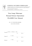
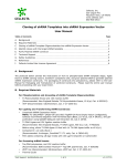
Product Manual ViraBind™ Lentivirus Purification Kit Catalog Number VPK-104 10 preps FOR RESEARCH USE ONLY Not for use in diagnostic procedures Introduction Lentivirus vectors based on the human immunodeficiency virus-1 (HIV-1) have become promising vectors for gene transfer studies. The advantageous feature of a lentivirus vector is the ability of gene transfer and integration into dividing and non-dividing cells. The pseudotyped envelope with vesicular stomatitis virus envelope G (VSV-G) protein broadens the target cell range. Lentiviral vectors have been shown to deliver genes to neurons, lymphocytes and macrophages, cell types that previous retrovirus vectors could not be used. Lentiviral vectors have also proven to be effective in transducing brain, liver, muscle, and retina in vivo without toxicity or immune responses. Recently, the lentivirus system is widely used to integrate siRNA efficiently in a wide variety of cell lines and primary cells both in vitro and in vivo. Lentivirus particles are produced from 293T cells through transient transfection of 3 or 4 plasmids that encodes for the components of the virion. Viral medium containing viral particles produced by packaging cells within 48-72 hr can be harvested and frozen. To obtain a higher titer, pseudovirus supernatant can be concentrated by ultracentrifuging. As a consequence, the ultracentrifugation step also concentrates cellular debris, membrane fragments, and denatured proteins derived from culture media of virus-producing cells. This unwarranted material in the crude vector preparation is toxic to target cells, especially primary cells, and may cause immunogenic reactions in experimental animal models by in vivo vector administration. Therefore, to reduce undesirable effects and increase gene transfer efficiency, the purification of virus vector becomes essential. ViraBind™ Lentivirus Purification Kit does not involve ultracentrifugation, instead it is based on the unique properties of Lentivirus envelop proteins. The entire procedure takes 30 minutes. Each preparation has a capacity up to 2.0 x 10⁸ IFUs. ViraBind™ Lentivirus Purification Kit provides an efficient system for quick lentiviral purification with high recovery (>90%). The system may be adapted to purification of other viral types, such as adenovirus and retrovirus. Related Products 1. LTV-100: 293LTV Lentiviral Cell Line 2. LTV-200: ViraDuctin™ Lentivirus Transduction Kit 3. VPK-100: ViraBind™ Adenovirus Purification Kit 4. VPK-107: QuickTiter™ Lentivirus Titer Kit (Lentivirus-Associated HIV p24) 5. VPK-108-H: QuickTiter™ Lentivirus Quantitation Kit (HIV p24 ELISA) 6. VPK-112: QuickTiter™ Lentivirus Quantitation Kit Kit Components 1. LTV Purification Filter (Part No. 90200): Five filters 2. 10X LTV Wash Buffer (Part No. 90201): One bottle – 100 mL. 3. 2X LTV Elution Buffer (Part No. 90203): One bottle – 50 mL of 50 mM Tris, pH 7.5, 5 mM Mg2Cl, 2 M NaCl. 2 4. 1X Regeneration Solution (Part No. 90204): One bottle – 50 mL. Materials Not Supplied 1. Lentivirus packaging plasmid mix and expression construct 2. Transfection Reagent 3. HEK 293T cells and cell culture growth medium 4. Cell culture centrifuge 5. Glycerol 6. 0.45 µm filter 7. 10 or 30-mL size syringe with a Luer-Lok® tip Storage Store all kit components at room temperature. Safety Considerations Remember that you will be working with samples containing infectious virus. Follow the recommended NIH guidelines for all materials containing BSL-2 organisms. Preparation of Reagents 1X LTV Wash Buffer: Prepare a 1X LTV Wash Buffer by diluting the provided 10X stock 1:10 in deionized water. Store the diluted solution at room temperature. 1X LTV Elution Buffer: Prepare a 1X LTV Elution Buffer by diluting the provided 2X stock 1:2 in deionized water. Store the diluted solution at room temperature. Pseudovirus Production The following procedure is suggested for a 10cm dish and may be optimized to suit individual needs. Please refer to the user manual when a lentivirus expression systems from Invitrogen or System Biosciences is used. 1. Use HEK 293T cells that have been passaged 2-3 times prior to transfection. Culture these cells until the monolayer is 70-80% confluent. 2. Replace the cell culture media with new growth media, 10 mL per 10 cm dish. 3. Transfect cells with packaging plasmid mix and your expression construct. Lipofectamine™, please refer to Invitrogen’s Lipofectamine™ reagent manual. When use 4. After 36-48 hrs, harvest all 10 mL medium in a 15 mL conical tube and centrifuge for 5 min at 3000 rpm to pellet the cell debris. Filter the supernatant through a 0.45 µm low protein binding filter. 5. The viral supernatant can be stored at -80ºC or immediately purified (see purification instructions below). Note: Freezing and thawing may result in 2-3 fold loss of viral titer after each cycle. 3 Purification Protocol 1. Prior to application of the viral supernatant to the purification filter, the supernatant is clarified by passing through a 0.45 µm sterile filter. 2. Attach a syringe to the purification filter, and add 5 mL of 1X LTV Wash Buffer to pre-rinse the filter. Slowly allow the viral supernatant to pass through the purification filter by gravity flow, and save the flow-through. To ensure maximal recovery, pass the flow-through through the same filter again. Note: When the flow through noticeably slows down during loading viral sample or reapplying the first flow-through for the second time, gently apply pressure with syringe plunger. To ensure maximal virus binding, try to keep the flow rate at less than 10 mL/min, we recommend flow through at dropwise (3-5ml/min). 3. Thoroughly wash the purification filter with 10 mL of 1X LTV Wash Buffer using gravity flow or gently applying moderate pressure with syringe plunger. Repeat the wash step twice, using 10 mL of 1X LTV Wash Buffer each wash. 4. Position a collection tube under the purification filter, add 2-3 mL of 1X LTV Elution Buffer (25 mM Tris, pH 7.5, 2.5 mM Mg2Cl, 1 M NaCl) and allow it to pass through (gravity flow or moderate pressure). Note: Air bubbles between syringe and filter will slow down the elution, to avoid them by pipetting a few times, be careful not stab the membrane filter. When apply moderate pressure, it’s critical to keep the flow rate slow at drop by drop to ensure the maximal yield. 5. Add glycerol to a final concentration of 10% to the purified virus or dialyze the viral solution into a desired buffer. Aliquot and store the final purified virus solution at -80ºC. 6. The purification filter, used in step 4, can be used twice. Upon completion of the purification, add 10 mL of 1X LTV Regeneration Solution to the filter, followed by 5 mL of 1X LTV Wash Buffer. Wrap the regenerated filter in parafilm and store at 4ºC until the next use. Notes: The purification filter is designed to be used ONLY twice to ensure high virus recovery (>90%), our results show the virus-binding capacity drops significantly when used more than two times. The Regeneration Solution should remove any virus or protein remaining on the filter, however to avoid cross-contamination, we suggest that when the filter is used for the second time, the same pseudovirus is applied. Example of Results The following figures demonstrate typical purification results. One should use the data below for reference only. This data should not be used to interpret actual results. 4 GFP Positive Cells (%) 120 100 80 60 40 20 El ut io n W as h Th ro ug h Fl ow Vi ra lS up er na t an t 0 Figure 1: Purification of pseudotyped GFP lentivirus. GFP lentiviral supernatant was purified according to the above Purification Instructions. Each fraction, obtained during purification, and its dilution were used to infect Hela cells for 48 hr in the presence of 5 µg of Polybrene (5 µg/ml). GFP positive cells were scored under high magnification fields. References 1. Naldini, L., U. Blomer, P. Gallay, D. Ory, R. Mulligan, F. H. Gage, I. M. Verma, and D. Trono (1996) Science 272, 263-267. 2. Verma, I. M., and N. Somia (1997) Nature 389, 239-242 3. Kafri, T., U. Blomer, D. A. Peterson, F. H. Gage, and I. M. Verma (1997) Nat. Genet. 17, 314-317. 4. Beyer, W. R., M. Westphal, W. Ostertag, and D. von Laer (2002) J. Virol. 76, 1488-1495. Recent Product Citations 1. Zhou, C. et al. (2015). Lhx8 mediated Wnt and TGFβ pathways in tooth development and regeneration. Biomaterials. doi:10.1016/j.biomaterials.2015.06.004. 2. Ying, S.-Y. et al. (2006). MicroRNA Protocols, Chapter 24, Humana Press. Warranty These products are warranted to perform as described in their labeling and in Cell Biolabs literature when used in accordance with their instructions. THERE ARE NO WARRANTIES THAT EXTEND BEYOND THIS EXPRESSED WARRANTY AND CELL BIOLABS DISCLAIMS ANY IMPLIED WARRANTY OF MERCHANTABILITY OR WARRANTY OF FITNESS FOR PARTICULAR PURPOSE. CELL BIOLABS’ sole obligation and purchaser’s exclusive remedy for breach of this warranty shall be, at the option of CELL BIOLABS, to repair or replace the products. 5 In no event shall CELL BIOLABS be liable for any proximate, incidental or consequential damages in connection with the products. Contact Information Cell Biolabs, Inc. 7758 Arjons Drive San Diego, CA 92126 Worldwide: +1 858-271-6500 USA Toll-Free: 1-888-CBL-0505 E-mail: [email protected] www.cellbiolabs.com 2004-2015: Cell Biolabs, Inc. - All rights reserved. No part of these works may be reproduced in any form without permissions in writing. 6