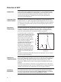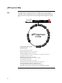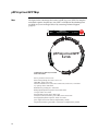
1
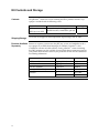
pShooter™ Vector (pEF/myc vector) For Intracellular Targeting of Recombinant Proteins and Antibodies Catalog no. V891-20 Rev. Date: 14 July 2010 Manual part no. 28-0179 MAN0000659 User Manual ii Table of Contents Kit Contents and Storage ............................................................................................................ iv Introduction ................................................................................................................... 1 Product Description.......................................................................................................................1 Methods ......................................................................................................................... 3 General Guidelines ........................................................................................................................3 Cloning into pEF/myc/nuc ..........................................................................................................5 Transfection of Mammalian Cells................................................................................................6 Detection of Fusion Proteins.........................................................................................................9 Troubleshooting ...........................................................................................................................11 Detection of GFP ..........................................................................................................................12 Appendix...................................................................................................................... 13 EF-1α Promoter.............................................................................................................................13 pEF/myc/nuc Map ......................................................................................................................14 Features of pEF/myc/nuc...........................................................................................................15 pEF/myc/nuc/GFP Map ............................................................................................................16 Technical Support ........................................................................................................................17 Purchaser Notification.................................................................................................................18 References .....................................................................................................................................19 iii Kit Contents and Storage Contents The pShooter™ manual for vectors utilizing the EF-1α promoter and the c-myc epitope is included with the following vector. Vector pEF/myc/nuc pEF/myc/nuc/GFP Shipping/Storage Amount Catalog no. 20 μg each (40 μl of 0.5 μg/μl vector in 10 mM Tris-HCl, 1 mM EDTA, pH 8.0) V891-20 All vectors are shipped at room temperature. Upon receipt, store at –20°C. Products Available Primers to sequence your insert in the pEF/myc vectors and antibodies to the cmyc epitope are available from Invitrogen. In addition, Geneticin® is also Separately available for selection of stable cell lines. Lastly, pShooter™ vectors containing the CMV promoter are also available. You may find that one promoter expresses your protein better than the other in your particular cell line. See the table below for ordering information. Vector Amount pEF Forward Primer 2 μg, lyophilized N623-02 BGH Reverse Primer 2 μg, lyophilized N575-02 Anti-myc Antibody 25 Westerns R950-25 Anti-myc-HRP Antibody 25 Westerns R951-25 1g 11811-023 5g 11811-031 ® Geneticin iv Catalog no. Introduction Product Description Background The final location of a protein within a cell depends upon ‘targeting sequences’ encoded within the sequence of a protein. The presence of a nuclear localization sequence within a protein or at the N- or C-terminus, directs the protein to the nucleus. Description The pShooter™ vector is designed to express and target your recombinant protein to the desired intracellular location in mammalian cells. They were originally designed to target single-chain antibodies (scFvs) to specific intracellular locations (Persic et al., 1997a; Persic et al., 1997b). The pShooter™ vector described in this manual is a 5.5 kb expression vector that expresses your recombinant protein as a fusion to a targeting sequence (if necessary). Proteins are targeted to the nucleus (Fisher-Fantuzzi & Vesco, 1988). Expression is driven by the strong, constitutive human EF-1α promoter (Mizushima & Nagata, 1990). The table below summarizes the above features. Vector pEF/myc/nuc Desired Location Nucleus Targeting Signal(s) 3X (DPKKKRKV) In addition, the vector uses the same backbone (pcDNA3) which includes the bovine growth hormone polyadenylation sequence, an f1 origin, the SV40 origin, the neomycin resistance gene, the SV40 late polyadenylation sequence, pUC origin, and the ampicillin resistance gene (Persic et al., 1997b). For more information on all of the above features, see page 15 in the Appendix. Uses of the pShooter™ Vector Targeting Recombinant Proteins The vector can be used to direct any recombinant protein to a particular intracellular location. However, success may be dependent on the specific protein used. To help analyze experiments, the vector is supplied with an optimized form of green fluorescent protein (cycle 3 GFP) cloned into the vector as a control. Guidelines for assaying cycle 3 GFP fluorescence are also provided (see page 12). Continued on next page 1 Product Description, continued Uses of the pShooter™ Vector, continued Targeting Antibodies The pShooter™ vector was originally designed for the targeting of scFvs to a specific intracellular location for intracellular immunization (Biocca & Cattaneo, 1995; Cattaneo & Biocca, 1997; Persic et al., 1997a). In this technique, an antibody which is inhibitory for a protein's function can be directed to the same compartment as the protein itself to inactivate the protein. The pShooter™ vector retains all of the features cited in Persic, et al., 1997a. Some of these features are summarized below. The restriction sites in the multiple cloning site were chosen because they are rare in both human and mouse antibody variable regions and have been removed from the rest of the vector. Vectors consist of a number of functional cassettes flanked by unique restriction sites, with junctional DNA reduced to a minimum. The nuclear localization signal is designed to be at the C-terminus of a scFv, positioned away from the antigen binding site, to reduce potential problems of steric hindrance. scFvs derived from phage antibody libraries can be easily cloned in from compatible vectors (e.g. pHEN; (Hoogenboom et al., 1991)) or amplified incorporating compatible ends. For more information on cloning antibodies and antibody domains, refer to Persic, et al., 1997a. For an example in which these vectors have been used in intracellular immunization to inhibit function within a cell, see Gargano and Cattaneo, 1997. 2 Methods General Guidelines Introduction This section contains general information on propagation and maintenance of the pShooter™ vector and guidelines for E. coli transformation. Additional information is provided on the following pages: To develop a cloning strategy, refer to the multiple cloning sites on page 5. A map of the targeting vector is on page 14. A map of the control vector is on page 15. Full sequences of any of the vectors described in this manual may be obtained by downloading them from our Web site (www.invitrogen.com) or by calling Technical Support (see page 17). General Molecular Biology Techniques For help with DNA ligations, E. coli transformations, restriction enzyme analysis, purification of single-stranded DNA, DNA sequencing, and DNA biochemistry, refer to Molecular Cloning: A Laboratory Manual (Sambrook et al., 1989) or Current Protocols in Molecular Biology (Ausubel et al., 1994) (See References, page 19). E. coli Strain Many E. coli strains are suitable for the growth of this vector including TOP10F´ (Catalog no. C615-00) and DH10B™. We recommend that you propagate vectors containing inserts in E. coli strains that are recombination deficient (recA) and endonuclease A deficient (endA). For your convenience, TOP10F´ and DH10B™ are available as chemically competent or electrocompetent cells (TOP10F´ only) from Invitrogen. Item ™ E. coli Transformation Quantity Catalog no. One Shot TOP10F´ (chemically competent cells) 21 x 50 μl C3030-03 Max Efficiency® DH10B™ (chemically competent cells) 5 x 0.2 ml 18297-010 Electrocomp™ TOP10F´ 5 x 80 μl C665-55 You may use any method you wish to prepare competent E. coli for transformation. Select transformants on LB plates containing 50-100 μg/ml ampicillin. Continued on next page 3 General Guidelines, continued Propagation and Maintenance of Plasmids Cloning into the pShooter™ Vectors In order to propagate and maintain the pShooter™ vector, we recommend that you transform the plasmids into E. coli and prepare glycerol stocks for long-term storage. Transform plasmids into E. coli as follows: Use a small amount of the supplied vector stock solution and the transformation method of choice to transform a recA, endA E. coli strain like TOP10F´, DH10B™, INVαF´, DH5αF´, or equivalent. Select transformants on LB plates containing 50-100 μg/ml ampicillin. Select a transformant and grow a log phase culture for a glycerol stock. Prepare glycerol stocks by mixing 0.85 ml of the log phase culture with 0.15 ml of sterile glycerol. Transfer the resulting solution to a cryovial and store at -80°C. Diagram for the multiple cloning sites is provided on page 5 to help you clone your gene of interest in frame with the desired targeting signal and/or the c-myc epitope for detection. For help with PCR, restriction digests, and ligations, refer to general molecular biology texts (Ausubel et al., 1994; Sambrook et al., 1989). 4 MEND ION AT RECOM Transform ligation mixtures into competent E. coli as using the method of choice, and plate the cells on LB plates containing 50-100 μg/ml ampicillin. Select 10 to 20 transformants and analyze your construct by restriction enzyme digestion or sequencing to ensure that your insert is cloned in the correct orientation. If you wish to sequence your insert, use the pEF Forward and BGH Reverse primers (Catalog nos. N623-02 and N575-02, respectively) to confirm that your gene is correctly fused to the targeting signal and/or the c-myc epitope. Cloning into pEF/myc/nuc Special Considerations The ATG in the Nco I site is part of a Kozak consensus sequence (ANNATGG) (Kozak, 1987; Kozak, 1990). If you can clone in frame or flush with this ATG, it will facilitate expression of your protein. To efficiently target your protein to the nucleus, the nuclear localization signal (NLS) from SV40 large T antigen has been triplicated and placed downstream of the multiple cloning site for C-terminal fusion to your protein (Fisher-Fantuzzi & Vesco, 1988). Note that this signal will not be removed from your protein upon entry to the nucleus. If you clone in-frame with the NLS you will also be in frame with the c-myc epitope. The NLS and the c-myc epitope will add ~5 kDa to your protein. Note that you may have to use PCR to facilitate in-frame cloning with the ATG (if desired) and the NLS. pEF/myc/nuc MCS The multiple cloning site below shows part of the 5´ untranslated region from the EF-1α promoter. For more information on the EF-1α promoter, see page 13. Restriction sites are labeled to indicate the cleavage site. The multiple cloning site has been confirmed by sequencing and functional testing. pEF Forward 1081 ATGTAATTCT CCTTGGAATT TGGCCTTTTT GAGTTTGGAT CTTGGTTCAT TCTCAAGCCT priming site 1141 Pst I Sal I 1205 Nco I 3´ end of EF-1a Intron 1 CAGACAGTGG TTCAAAGTTT TTTTCTTCCA TTTCAGGTGT CGTGAACACG TGGCCACC ATG GCC Met Ala 5´ End of EF-1a Exon 2 Xho I Not I CAG GTG CAG CTG CAG GTC GAC CTC GAG ATC AAA CGG GCG GCC GCA GAT CCA AAA Gln Val Gln Leu Gln Val Asp Leu Glu Ile Lys Arg Ala Ala Ala Asp Pro Lys NLS #2 1259 NLS #1 NLS #3 AAG AAG AGA AAG GTA GAT CCA AAA AAG AAG AGA AAG GTA GAT CCA AAA AAG AAG Lys Lys Arg Lys Val Asp Pro Lys Lys Lys Arg Lys Val Asp Pro Lys Lys Lys myc epitope 1289 AGA AAG GTA GAT ACG GCC GCA GAA CAA AAA CTC ATC TCA GAA GAG GAT CTG AAT Arg Lys Val Asp Thr Ala Ala Glu Gln Lys Leu Ile Ser Glu Glu Asp Leu Asn BGH Reverse priming site 1367 GGG GCC GCA TAG TCTAGAAGCT CGCTGATCAG CCTCGACTGT GCCTTCTAGT TGCCAGCCAT Gly Ala Ala *** 1427 CTGTTGTTTG CCCCTCCCCC GTGCCTTCCT TGACCCTGGA AGGTGCCACT CCCACTGTCC BGH polyadenylation signal 1487 TTTCCTAATA AAATGAGGAA ATTGCATCGC ATTGTCTGAG TAGGTGTCAT TCTATTCTGG 5 Transfection of Mammalian Cells Introduction General information is provided below for transfection of mammalian cells with the pShooter™ vector. Positive control vector is supplied with each vector to optimize transfection conditions for your cell line. pShooter™ vectors have been tested in Chinese Hamster Ovary (CHO) and COS cells. A sample transfection is provided on the next page for CHO cells. Plasmid Preparation Once you have confirmed that your gene is in the correct reading frame, prepare plasmid DNA for transfection. Plasmid DNA for transfection into eukaryotic cells must be very clean and free from phenol and sodium chloride. Contaminants will kill the cells and salt will interfere with lipid complexing decreasing transfection efficiency. We recommend isolating DNA using the S.N.A.P.™ MidiPrep Kit (10-200 μg DNA, Catalog no. K1910-01) or CsCl gradient centrifugation. Methods of Transfection For established cell lines (e.g. COS, CHO), consult original references or the supplier of your cell line for the optimal method of transfection. It is recommended that you follow exactly the protocol for your cell line. Pay particular attention to medium requirements, when to pass the cells, and at what dilution to split the cells. Further information is provided in Current Protocols in Molecular Biology (Ausubel et al., 1994). Methods for transfection include calcium phosphate (Chen & Okayama, 1987; Wigler et al., 1977), lipid-mediated (Felgner et al., 1989; Felgner & Ringold, 1989) and electroporation (Chu et al., 1987; Shigekawa & Dower, 1988). Invitrogen offers the Calcium Phosphate Transfection Kit for mammalian transfection and Lipofectamine™ 2000 Reagent for lipid-mediated transfection. Catalog No. 11668-019 Description ™ Lipofectamine 2000 Reagent Quantity 1.5 ml Expression of Your Fusion Protein No matter which method of transfection you elect to use, it is very important to perform a time course to optimize expression and targeting of your particular protein. Be sure to transfect enough cells to collect time points, particularly if you are using immunofluorescence or a functional assay. Methods of Detection There are a variety of methods for detection, depending on what protein you are expressing and targeting. Visual Method. If you want to be sure that your protein is targeting to the correct location, use immunofluorescence (see page 9). Functional Assay. If you are targeting a protein that inhibits or alters the function of another protein, you may have a visual assay (e.g. changes in cell morphology) or an enzymatic assay. Continued on next page 6 Transfection of Mammalian Cells, continued Sample Transfection Procedure Below is a sample transfection procedure using Lipofectamine™ 2000, available from Invitrogen (Catalog no. 11668-019) and a GFP control vector. Day 1 1. Plate 2 x 10⁵ CHO KI cells in 3 ml F12 Nutrient mixture, Ham, 10% FBS into one 60 mm plate. Day 2 2. Prepare plasmid DNA lipid complexes according to the instructions provided in the Lipofectamine™ 2000 Reagent manual, which is available for downloading from our Web site (www.invitrogen.com) or by contacting Technical Support (see page 17). 3. Add the DNA lipid complexes to the cells. 4. Incubate the cells at 37°C in a CO2 incubator. Days 3, 4, and 5 Monitor fluorescence of GFP with a fluorescent microscope as described on page 12, and take photographs at 24, 48, and 72 hours. Stable Transfection (Geneticin® Resistance) For stable transfection, the pShooter™ vector contains the resistance factor to Geneticin®. Geneticin® blocks protein synthesis in mammalian cells by interfering with ribosomal function. It is an aminoglycoside, similar in structure to neomycin, gentamycin, and kanamycin. Expression in mammalian cells of the bacterial aminoglycoside phosphotransferase gene (APH), derived from Tn5, results in detoxification of Geneticin® Selective Antibiotic (Southern & Berg, 1982). Geneticin® Selection Guidelines Geneticin® is available from Invitrogen (Catalog no. 11811-031). Use as follows: 1. Prepare Geneticin® in a buffered solution (e.g. 100 mM HEPES, pH 7.3). 2. Use 100 to 1000 μg/ml of Geneticin® in complete medium. 3. Calculate concentration based on the amount of active drug (check the lot label). 4. Test varying concentrations of Geneticin® on your cell line to determine the concentration that kills your cells (kill curve). Cells differ in their susceptibility to Geneticin®. Cells will divide once or twice in the presence of lethal doses of Geneticin® Selective Antibiotic, so the effects of the drug take several days to become apparent. Complete selection can take from 2 to 4 weeks of growth in selective medium. Continued on next page 7 Transfection of Mammalian Cells, continued Linearization of Vectors for Stable Transfection While linearizing a plasmid is not necessary to obtain stable transfectants, it ensures that the vector does not integrate in a way that disrupts the gene of interest. Possible restriction enzymes you could use to linearize your particular construct are listed below. Vector 8 Sites Location All vectors Pvu I, Sca I Ampicillin resistance gene pEF/myc vectors Kpn I, EcoR I, Hind III 5´ end of EF-1α promoter pEF/myc GFP control vectors Kpn I, EcoR I 5´ end of EF-1α promoter Detection of Fusion Proteins Introduction To ensure that your protein is targeted correctly, it is important to visualize its cellular location. Inclusion of the c-myc epitope allows detection by immunofluorescence although you can use antibody to your own protein. A basic protocol is included for your convenience. Other protocols may be appropriate. Detection of Fusion Proteins Antibodies to the c-myc epitope are available from Invitrogen and can be used to detect expression of your fusion protein by immunofluorescence (see below) or Western blot. Note that the c-myc epitope will add an additional 1.5 kDa to your protein. The table below describes the antibodies available and ordering information. The amount supplied is sufficient for 25 Westerns and 2-3 immunofluorescence experiments. Antibody Anti-myc Anti-myc-HRP Basic Immunofluorescent Labeling of Cells Purpose Detects 10 amino acid epitope derived from the c-myc protein (Evan et al., 1985) See above. Provided as an HRP conjugate for time-saving detection. Catalog no. R950-25 R951-25 Antibodies can be used for immunofluorescence using standard techniques (Ausubel et al., 1994). A basic protocol is supplied below for adherent cells. For more information, refer to Chapter 14.6 in Current Protocols in Molecular Biology. 1. Cool the cells on ice. (Culture cells in a 3- to 5-cm dish. Cells should be confluent or as close to confluent as possible.) 2. Aspirate off the culture medium and wash the cells with +4°C PBS. 3. Remove PBS and fix cells for either 30 minutes in 2% paraformaldehyde/0.1% Triton X-100 or 15 minutes in 100% methanol at -20°C. Note: Be sure to wash the cells thoroughly with methanol or they will freeze. 4. Remove fixative and wash the cells twice with cold PBS (~5 minutes/wash). 5. Dilute primary antibody in PBS to a final concentration of 5 to 10 μg/ml. Prepare enough antibody to cover cells. 6. Centrifuge antibody for 2 minutes at 13,500 x g (+4°C) to precipitate any particulate matter. 7. Carefully layer primary antibody onto the cells until they are just covered and incubate for 1 hour at +4°C. 8. Remove antibody and wash four times with cold PBS (~5 minutes/wash). 9. Dilute labeled secondary antibody in PBS to a final concentration of 5 to 10 μg/ml. Prepare enough antibody to cover cells. 10. Centrifuge antibody for 2 minutes at 13,500 x g (+4°C) to precipitate any particulate matter. 11. Layer secondary antibody over cells and incubate for 1 hour at +4°C. 12. Remove antibody and wash four times with cold PBS (~5 minutes/wash). Store cells in PBS. Analyze cells by fluorescence immediately; or cover dishes, wrap in aluminum foil, and refrigerate. Be sure to examine preparations within 24 hours or the fluorescence will fade. Continued on next page 9 Detection of Fusion Proteins, continued Patterns of Expression Transformation of the vector expressing your gene of interest or the control vectors should give the following expression patterns using immunofluorescence or fluorescence (cycle 3 GFP). Nuclear expression: Recombinant proteins should be primarily localized to the nucleus. If you have trouble expressing and targeting your protein, read the section on the positive control vectors below and the Troubleshooting section on page 11. Color graphics are available in our Catalog showing expression and targeting of cycle 3 GFP in CHO cells. In addition, the original paper by Persic, et al., shows targeting of recombinant antibodies by immunofluorescence using the monoclonal antibody 9E10 (Anti-myc Antibody) in COS cells (Persic et al., 1997a). Using the Positive Controls Each of the pShooter™ vectors described in this manual is also provided with a control vector expressing cycle 3 GFP. These vectors may be used to: Optimize transfection conditions for your cell line Confirm that the targeting signals function properly in your cell line For more information on the control vector, see page 15. For information on detection of cycle 3 GFP, see page 12. 10 Troubleshooting Troubleshooting Use the table below to troubleshoot expression and targeting. Problem No targeting observed Reason Solution Low expression levels Could be a variety of reasons. Check for expression by Western blot. You may have to optimize transfection conditions (use the cycle 3 GFP control vector to evaluate transfection). Many of the other solutions below may help. No expression of your protein Check for expression by Western blot. If your protein is not expressed, sequence your construct to confirm that it is in frame with the targeting sequence. Cell line may not recognize targeting signal Check for targeting using the appropriate GFP control vector. Non-specific labeling The c-myc tag is derived from an endogenous protein (c-myc) Transfect with the empty vector (negative control) and assay for immunofluorescence. You may need to use a different tag or use antibody to your protein Difficulty expressing protein in stable clones Protein is toxic when redirected to another compartment Selection of stable clones may lead to down regulation of the protein. Try a different promoter for expression. Continuous culture may lead to loss of protein expression Remember to prepare an early set of back-up stocks. Some proteins (e.g., antibodies expressed intracellularly) may give a very good immunofluorescent signal, but may not be detectable in a Western blot. This may be due to aggregation and/or precipitation of the antibody, so be sure your SDSPAGE samples are well solubilized. 11 Detection of GFP Introduction Cycle 3 GFP has been optimized for expression in E. coli and mammalian cells. Fluorescent yield is >40-fold over wild-type GFP, yet it has the same excitation maxima (395 nm and 478 nm for primary and secondary excitation) and emission maxima (507 nm). Guidelines for detection and optimization of expression are described below. Construct of the Control Vectors The control vectors were synthesized by amplifying a 716 bp fragment from pαGFP (Crameri et al., 1996) using oligomers that introduced a Pst I site at the 5´ end and a Not I site at the 3´ end of cycle 3 GFP. In addition each of the oligomers was specifically designed to clone in frame with the targeting sequence and/or the c-myc epitope. Detection of Fluorescence To detect fluorescent cells, it is important to pick the best filter set to optimize detection. The primary excitation peak of cycle 3 GFP is at 395 nm. There is a secondary excitation peak at 478 nm. Excitation at these wavelengths yields a fluorescent emission peak with a maximum at 507 nm (see below). 630 610 590 570 550 530 510 490 470 450 430 410 390 370 350 330 310 Relative Fluorescence Excitation and Emission Spectra for SuperGFP Use of the best filter set will ensure 700 that the optimal regions of the cycle 3 507 GFP spectra are excited and passed 600 (emitted). For example, the FITC 500 filter set that we use excites cycle 3 GFP with light from 460 to 490 nm, 400 which covers the secondary excitation peak. The filter set passes 300 light from 515 to 550, allowing detection of most of the GFP 200 395 478 fluorescence. Standard FITC filters 100 easily suit most purposes; however, it is important to keep in mind that 0 fluorescence will be affected by the sample assayed and the filter you Wavelength (nm) choose For general information about GFP fluorescence and detection, refer to Current Protocols in Molecular Biology. Detection of Transfected Cells After transfection, allow the cells to recover for 12 to 48 hours before assaying for fluorescence. Note: Most media fluoresce because of the presence of riboflavin (Zylka & Schnapp, 1996) and may interfere with detection of cycle 3 GFP fluorescence. Medium can be removed and replaced with PBS to alleviate this problem. Estimate the total number of cells before assaying for fluorescence. Then check your plate for fluorescent cells. You can use fluorescence to estimate transfection efficiency and normalize any subsequent assay for your gene of interest. Optimizing Expression It is recommended that a time course be performed to determine the optimal time to assay for transient expression of GFP. Optimal times may vary from 12 to 96 hours from the time of transfection depending on cell line. 12 Appendix EF-1α Promoter Description The diagram below shows all the features of the EF-1α promoter used in the pShooter™ vector (Mizushima & Nagata, 1990). Features are marked as per Uetsuki, et al., 1989. The original sequence has been mutagenized to remove the Pst I, Bgl II, Afl II, and Xho I restriction sites. In addition, Kpn I, EcoR I, Hind III, Pml I, and Nco I were introduced by PCR. The EF-1α promoter can be excised using Kpn I, EcoR I, or Hind III and Pml I or Nco I. Kpn I EcoR I Hind III 1 GTACCGAATT CAAGCTTCGT GAGGCTCCGG TGCCCGTCAG TGGGCAGAGC GCACATCGCC 61 CACAGTCCCC GAGAAGTTGG GGGGAGGGGT CGGCAATTGA ACCGGTGCCT AGAGAAGGTG 121 GCGCGGGGTA AACTGGGAAA GTGATGTCGT GTACTGGCTC CGCCTTTTTC CCGAGGGTGG Start of Transcription TATA box 181 GGGAGAACCG TATATAAGTG CAGTAGTCGC CGTGAACGTT CTTTTTCGCA ACGGGTTTGC Exon I 5´ end of Intron 1 241 CGCCAGAACA CAGGTAAGTG CCGTGTGTGG TTCCCGCGGG CCTGGCCTCT TTACGGGTTA 301 TGGCCCTTGC GTGCCTTGAA TTACTTCCAC CTGGCTCCAG TACGTGATTC TTGATCCCGA 361 GCTGGAGCCA GGGGCGGGCC TTGCGCTTTA GGAGCCCCTT CGCCTCGTGC TTGAGTTGAG 421 GCCTGGCCTG GGCGCTGGGG CCGCCGCGTG CGAATCTGGT GGCACCTTCG CGCCTGTCTC 481 GCTGCTTTCG ATAAGTCTCT AGCCATTTAA AATTTTTGAT GACCTGCTGC GACGCTTTTT 541 TTCTGGCAAG ATAGTCTTGT AAATGCGGGC CAGGATCTGC ACACTGGTAT TTCGGTTTTT 601 GGGCCCGCGG CCGGCGACGG GGCCCGTGCG TCCCAGCGCA CATGTTCGGC GAGGCGGGGC 661 CTGCGAGCGC GGCCACCGAG AATCGGACGG GGGTAGTCTC AAGCTGGCCG GCCTGCTCTG 721 GTGCCTGGCC TCGCGCCGCC GTGTATCGCC CCGCCCTGGG CGGCAAGGCT GGCCCGGTCG 781 GCACCAGTTG CGTGAGCGGA AAGATGGCCG CTTCCCGGCC CTGCTCCAGG GGGCTCAAAA 841 TGGAGGACGC GGCGCTCGGG AGAGCGGGCG GGTGAGTCAC CCACACAAAG GAAAAGGGCC 901 TTTCCGTCCT CAGCCGTCGC TTCATGTGAC TCCACGGAGT ACCGGGCGCC GTCCAGGCAC 961 CTCGATTAGT TCTGGAGCTT TTGGAGTACG TCGTCTTTAG GTTGGGGGGA GGGGTTTTAT 1021 GCGATGGAGT TTCCCCACAC TGAGTGGGTG GAGACTGAAG TTAGGCCAGC TTGGCACTTG 1081 ATGTAATTCT CCTTGGAATT TGGCCTTTTT GAGTTTGGAT CTTGGTTCAT TCTCAAGCCT 1141 CAGACAGTGG TTCAAAGTTT TTTTCTTCCA TTTCAGGTGT CGTGAACACG TGGCCACC ATG G.. Met ... 5´ end of Exon 2 Sp 1 Sp 1 Sp 1 Sp 1 Sp 1 Ap 1 3´ end of Intron 1 Pml I Nco I 13 pEF/myc/nuc Map The figure below summarizes the features of pEF/myc/nuc. The complete nucleotide sequence for pEF/myc/nuc is available for downloading from our Web site (www.invitrogen.com) or by contacting Technical Support (see page 17). Nco I Pst I Sal I Xho I Not I Map myc epitope 3X Nuclear targeting sequences P -1a EF BGH pA f1 or i ri 40 o SV Neomy cin Ampicilli pEF/myc/nuc 5.5 kb n Comments for pEF/myc/nuc 5536 nucleotides p U C or i S Term 0p V4 A EF-1a promoter: bases 6-1192 EF Forward priming site: bases 1133-1153 Multiple cloning site: bases 1197-1248 Nuclear localization site (3X): bases 1250-1273, 1274-1297, 1298-1321 myc epitope: bases 1334-1363 BGH Reverse priming site: 1399-1416 BGH polyadenylation sequence: bases 1398-1612 f1 origin: bases 1675-2088 SV40 promoter/origin: bases 2153-2460 Neomycin (G418) resistance gene (ORF): bases 2468-3262 SV40 polyadenylation sequence: bases 3279-3517 pUC origin: bases 3700-4373 (Complementary strand) Ampicillin resistance gene (ORF): 4518-5378 (Complementary strand) 14 Features of pEF/myc/nuc Table The table below summarizes the features of the pShooter™ vector, derived from pcDNA3. Changes to the vector backbone are noted. Feature Benefit Human EF-1α promoter Permits efficient, high-level expression of your recombinant protein (Kim et al., 1990; Mizushima & Nagata, 1990; Uetsuki et al., 1989). For more detailed information on this promoter, see the next page. Multiple cloning site Allows insertion of your gene. Nuclear targeting sequence pEF/myc/nuc only Permits efficient targeting of your protein to the nucleus. Sequence is triplicated to ensure proper localization. Isolated from SV40 large T antigen (Fisher-Fantuzzi & Vesco, 1988). c-myc epitope (Glu-Gln-Lys-Leu-Ile-Ser-Glu-GluAsp-Leu) Allows detection of your recombinant protein by immunofluorescence with the Anti-myc Antibody (Catalog no. R950-25) (Evan et al., 1985). ER retention signal pEF/myc/ER only Permits retention of your protein in the ER (Munro & Pelham, 1987). TAG termination codon For efficient termination of translation. Bovine growth hormone (BGH) polyadenylation signal Efficient transcription termination and polyadenylation of mRNA (Goodwin & Rottman, 1992). f1 origin Allows rescue of single-stranded DNA. SV40 early promoter and origin Allows efficient, high-level expression of the neomycin resistance gene and episomal replication in cells expressing SV40 large T antigen (i.e. COS). Nco I site removed by sitedirected mutagenesis. Neomycin resistance gene Selection of stable transfectants in mammalian cells (Southern & Berg, 1982). Tn5 sequence removed and the Kozak sequence improved by PCR at the 5´ end of the ORF. Nco I, Pst I, and BssH II sites removed by site-directed mutagenesis. SV40 polyadenylation signal Efficient transcription termination and polyadenylation of mRNA. pUC origin High-copy number replication and growth in E. coli. ApaL I site removed by site-directed mutagenesis. Ampicillin resistance gene (β-lactamase) Selection of vector in E. coli. ApaL I site removed by sitedirected mutagenesis. 15 pEF/myc/nuc/GFP Map Not I The figure below summarizes the features of pEF/myc/nuc/GFP. The complete nucleotide sequence for pEF/myc/nuc/GFP is available for downloading from our Web site (www.invitrogen.com) or by contacting Technical Support (page 17). Pst I Map GFP 3X Nuclear targeting sequence a F-1 PE Term myc epitope BGH pA f1 or i ri 40 o SV n Neo m y cin Ampicilli pEF/myc/nuc/GFP 6.2 kb Comments for pEF/myc/nuc/GFP 6232 nucleotides p U C or i S 0 V4 pA EF-1a promoter: bases 6-1192 EF Forward priming site: bases 1133-1153 GFP ORF: bases 1220-1936 Nuclear localization site (3X): bases 1946-1969, 1970-1993, 1994-2017 myc epitope: bases 2030-2059 BGH Reverse priming site: 2095-2112 BGH polyadenylation sequence: bases 2094-2308 f1 origin: bases 2371-2784 SV40 promoter/origin: bases 2849-3156 Neomycin (G418) resistance gene (ORF): bases 3164-3958 SV40 polyadenylation sequence: bases 3975-4213 pUC origin: bases 4396-5069 (Complementary strand) Ampicillin resistance gene (ORF): 5214-6074 (Complementary strand) 16 Technical Support Web Resources Contact Us Visit the Invitrogen website at www.invitrogen.com for: Technical resources, including manuals, vector maps and sequences, application notes, SDSs, FAQs, formulations, citations, handbooks, etc. Complete technical support contact information Access to the Invitrogen Online Catalog Additional product information and special offers For more information or technical assistance, call, write, fax, or email. Additional international offices are listed on our website (www.invitrogen.com). Corporate Headquarters: 5791 Van Allen Way Carlsbad, CA 92008 USA Tel: 1 760 603 7200 Tel (Toll Free): 1 800 955 6288 Fax: 1 760 602 6500 E-mail: [email protected] Japanese Headquarters: LOOP-X Bldg. 6F 3-9-15, Kaigan Minato-ku, Tokyo 108-0022 Tel: 81 3 5730 6509 Fax: 81 3 5730 6519 E-mail: [email protected] European Headquarters: Inchinnan Business Park 3 Fountain Drive Paisley PA4 9RF, UK Tel: +44 (0) 141 814 6100 Tech Fax: +44 (0) 141 814 6117 E-mail: [email protected] SDS Safety Data Sheets (SDSs) are available at www.invitrogen.com/sds. Certificate of Analysis The Certificate of Analysis provides detailed quality control and product qualification information for each product. Certificates of Analysis are available on our website. Go to www.invitrogen.com/support and search for the Certificate of Analysis by product lot number, which is printed on the box. Limited Warranty Invitrogen (a part of Life Technologies Corporation) is committed to providing our customers with high-quality goods and services. Our goal is to ensure that every customer is 100% satisfied with our products and our service. If you should have any questions or concerns about an Invitrogen product or service, contact our Technical Support Representatives. All Invitrogen products are warranted to perform according to specifications stated on the certificate of analysis. The Company will replace, free of charge, any product that does not meet those specifications. This warranty limits the Company’s liability to only the price of the product. No warranty is granted for products beyond their listed expiration date. No warranty is applicable unless all product components are stored in accordance with instructions. The Company reserves the right to select the method(s) used to analyze a product unless the Company agrees to a specified method in writing prior to acceptance of the order. Invitrogen makes every effort to ensure the accuracy of its publications, but realizes that the occasional typographical or other error is inevitable. Therefore the Company makes no warranty of any kind regarding the contents of any publications or documentation. If you discover an error in any of our publications, report it to our Technical Support Representatives. Life Technologies Corporation shall have no responsibility or liability for any special, incidental, indirect or consequential loss or damage whatsoever. The above limited warranty is sole and exclusive. No other warranty is made, whether expressed or implied, including any warranty of merchantability or fitness for a particular purpose. 17 Purchaser Notification Limited Use Label License No. 60: EF-1alpha Promoter 18 EF-1alpha promoter products are sold under license for research purposes only. The use of this product for any commercial purpose, including but not limited to, use in any study for the purpose of a filing of a new drug application, requires a license from: Mochida Pharmaceutical Co., Ltd., 7, Yotsuya 1-Chome, ShinjukuKu, Tokyo 160, Japan. Tel: 81-3-3225-5451; Fax: 81-3-3225-6091. References Ausubel, F. M., Brent, R., Kingston, R. E., Moore, D. D., Seidman, J. G., Smith, J. A., and Struhl, K. (1994) Current Protocols in Molecular Biology, Greene Publishing Associates and Wiley-Interscience, New York Biocca, S., and Cattaneo, A. (1995) Intracellular Immunization: Antibody Targeting to Subcellular Compartments. Trends Cell Biol. 5, 248-252 Cattaneo, A., and Biocca, S. (1997) Intracellular Antibodies: Development and Applications, Landes Bioscience, distributed by Academic Press, San Diego, CA Chen, C., and Okayama, H. (1987) High-Efficiency Transformation of Mammalian Cells by Plasmid DNA. Mol. Cell. Biol. 7, 2745-2752 Chu, G., Hayakawa, H., and Berg, P. (1987) Electroporation for the Efficient Transfection of Mammalian Cells with DNA. Nucleic Acids Res. 15, 1311-1326 Crameri, A., Whitehorn, E. A., Tate, E., and Stemmer, W. P. C. (1996) Improved Green Fluorescent Protein by Molecular Evolution Using DNA Shuffling. Nature Biotechnology 14, 315-319 Evan, G. I., Lewis, G. K., Ramsay, G., and Bishop, V. M. (1985) Isolation of Monoclonal Antibodies Specific for c-myc Proto-oncogene Product. Mol. Cell. Biol. 5, 3610-3616 Felgner, P. L., Holm, M., and Chan, H. (1989) Cationic Liposome Mediated Transfection. Proc. West. Pharmacol. Soc. 32, 115-121 Felgner, P. L. a., and Ringold, G. M. (1989) Cationic Liposome-Mediated Transfection. Nature 337, 387388 Fisher-Fantuzzi, L., and Vesco, C. (1988) Cell-Dependent Efficiency of Reiterated Nuclear Signals in a Mutant Simian Virus 40 Oncoprotein Targeted to the Nucleus. Mol. Cell. Biol. 8, 5495-5503 Gargano, N., and Cattaneo, A. (1997) Rescue of a Neutralising Antiviral Antibody Fragment from an Intracellular Polyclonal Repertoire Expressed in Mammalian Cells. FEBS Lett. 414, 537-540 Goodwin, E. C., and Rottman, F. M. (1992) The 3´-Flanking Sequence of the Bovine Growth Hormone Gene Contains Novel Elements Required for Efficient and Accurate Polyadenylation. J. Biol. Chem. 267, 16330-16334 Hoogenboom, H. R., Griffiths, A. D., Johnson, K. S., Chiswell, D. J., Hudson, P., and Winter, G. (1991) Multi-Subunit Proteins on the Surface of Filamentous Phage: Methodologies for Displaying Antibody (Fab) Heavy and Light Chains. Nucleic Acids Res. 19, 4133-4137 Kim, D. W., Uetsuki, T., Kaziro, Y., Yamaguchi, N., and Sugano, S. (1990) Use of the Human Elongation Factor 1a Promoter as a Versatile and Efficient Expression System. Gene 91, 217-223 Kozak, M. (1987) An Analysis of 5´-Noncoding Sequences from 699 Vertebrate Messenger RNAs. Nucleic Acids Res. 15, 8125-8148 Kozak, M. (1990) Downstream Secondary Structure Facilitates Recognition of Initiator Codons by Eukaryotic Ribosomes. Proc. Natl. Acad. Sci. USA 87, 8301-8305 Mizushima, S., and Nagata, S. (1990) pEF-BOS, a Powerful Mammalian Expression Vector. Nucleic Acids Res. 18, 5322 Munro, S., and Pelham, H. R. B. (1987) A C-Terminal Signal Prevents Secretion of Luminal ER Proteins. Cell 48, 899-907 Persic, L., Righi, M., Roberts, A., Hoogenboom, H. R., Cattaneo, A., and Bradbury, A. (1997a) Targeting Vectors for Intracellular Immunization. Gene 187, 1-8 Continued on next page 19 References, continued Persic, L., Roberts, A., Wilton, J., Cattaneo, A., Bradbury, A., and Hoogenboom, H. R. (1997b) An Integrated Vector System for the Eukaryotic Expression of Antibodies or Their Fragments After Selection from Phage Display Libraries. Gene 187, 9-18 Sambrook, J., Fritsch, E. F., and Maniatis, T. (1989) Molecular Cloning: A Laboratory Manual, Second Ed., Cold Spring Harbor Laboratory Press, Plainview, New York Shigekawa, K., and Dower, W. J. (1988) Electroporation of Eukaryotes and Prokaryotes: A General Approach to the Introduction of Macromolecules into Cells. BioTechniques 6, 742-751 Southern, P. J., and Berg, P. (1982) Transformation of Mammalian Cells to Antibiotic Resistance with a Bacterial Gene Under Control of the SV40 Early Region Promoter. J. Molec. Appl. Gen. 1, 327-339 Uetsuki, T., Naito, A., Nagata, S., and Kaziro, Y. (1989) Isolation and Characterization of the Human Chromosomal Gene for Polypeptide Chain Elongation Factor-1a. J. Biol. Chem. 264, 5791-5798 Wigler, M., Silverstein, S., Lee, L.-S., Pellicer, A., Cheng, Y.-C., and Axel, R. (1977) Transfer of Purified Herpes Virus Thymidine Kinase Gene to Cultured Mouse Cells. Cell 11, 223-232 Zylka, M. J., and Schnapp, B. J. (1996) Optimized Filter Set and Viewing Conditions for the S65T Mutant of GFP in Living Cells. BioTechniques 21, 220-226 ©2010 Life Technologies Corporation. All rights reserved. For research use only. Not intended for any animal or human therapeutic or diagnostic use. The trademarks mentioned herein are the property of Life Technologies Corporation or their respective owners. 20 Notes 21 Notes 22 Corporate Headquarters 5791 Van Allen Way Carlsbad, CA 92008 T: 1 760 603 7200 F: 1 760 602 6500 E: [email protected] For country-specific contact information, visit our web site at www.invitrogen.com