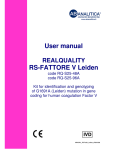
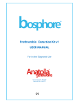
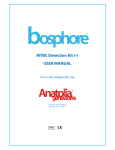
1
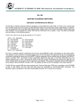
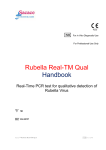
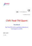
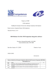
®
FVL Detection Kit v1
USER MANUAL
For in vitro Diagnostic Use
®
Document Code: MB16v2f
Approval Date: July 2011
Contents
Page
1. Product Description
1
2. Content
1
3. Storage
1
4. Required Materials and Devices
1
5. Important Notes and Safety Instructions
2
6. Product Use Limitations
2
7. Mutation
2
8. Method
3
9. Procedure
4
9.1. DNA Isolation
4
9.2. Kit Components
4
9.2.1. PCR Mix
4
9.2.2. Detection Mix 1
4
9.2.3. Positive Control
4
9.3. Preparing the PCR
5
9.4. Programming the Montania® 483 Real-Time PCR Instrument
5
10. Analysis
8
11. Troubleshooting
9
12. References
9
13. Symbols
9
14. Contact Information
10
ii
Code: MB16v1f
Date: April 2011
1. PRODUCT DESCRIPTION
Bosphore® FVL Detection Kit v1 detects Factor V Leiden mutation; namely G1691A (a change of
glutamic acid to arginine) in human biological samples. Wild type F5 allele is amplified and
fluorescence detection is accomplished using the HEX filter. Mutant F5 allele is amplified and
fluorescence detection is accomplished using the FAM filter.
2. CONTENT
Bosphore® FVL Detection Kit v1 is composed of Real-Time PCR reagents.
Component
1
2
3
4
REAGENT
dH2O
PCR Mix
Detection Mix1
Positive Control
100
Tests
(1000 µl)
(1375 µl)
(110 µl)
(44 µl)
50 Tests
(1000 µl)
(688 µl)
(55 µl)
(22 µl)
25 Tests
(1000 µl)
(344 µl)
(28 µl)
(15 µl)
3. STORAGE
Bosphore® FVL Detection Kit v1 PCR reagents should be stored at -20°C. Repeated thawing and
freezing (>3x) should be avoided since it may reduce sensitivity. If the components are to be used
in small amounts, they should be frozen in aliquots.
While preparing the PCR; the components should not be exposed to room temperature for
more than 10 min and the detection mix components should not be exposed to light more than 12 min. We recommend preparing the PCR on a cooling block and keeping the detection mixes
within a closed container.
The components maintain their stability until the expiry dates on the labels, if they are stored at
advised conditions.
4. REQUIRED MATERIALS AND DEVICES
•
Montania® 483 Real-Time PCR Instrument (Anatolia Geneworks), or another Real-Time
PCR system with FAM and HEX filters (iCycler, iQ5, CFX–BioRad, LightCycler 1.5, 2.0,
480-Roche, 7500 Real-Time PCR System-ABI, Stratagene Mx3005P, Mx3000P-Agilent,
LineGeneK, LineGene 9600-Bioer, Rotorgene 2000, 3000, 6000, Q-Qiagen)
•
0.2 ml Thin-Wall PCR tubes or strips
•
Magnesia® 16 Nucleic Acid Extraction System / Magnesia® Genomic DNA Whole Blood
Kit (Anatolia Geneworks),or other high quality genomic DNA extraction kits/systems
•
Deep freezer (-20°C)
•
Desktop centrifuge with rotor for 2 ml. microcentrifuge tubes
1
Code: MB16v1f
Date: April 2011
•
Calibrated adjustable micropipettes
•
DNAse, RNAse, pyrogen free micropipette tips with filters
•
DNAse, RNAse, pyrogen free 1.5 or 2 ml. microcentrifuge tubes
•
Disposable laboratory gloves
5. IMPORTANT NOTES AND SAFETY INSTRUCTIONS
Important!:
•
The product should be delivered on dry ice. Check for presence of dry ice upon arrival.
•
Check for the expiry dates on the box and tube labels, upon arrival. Do not use expired
products or components.
•
Calibrated or verified micropipettes, DNAse, RNAse, pyrogen free micropipette tips
with filters, and DNAse, RNAse, pyrogen free microcentrifuge tubes should be used.
•
Before starting a test procedure, all components should be thoroughly thawed. After
thawing, all components should be centrifuged briefly (spin-down for 3-5 seconds),
and mixed well to ensure homogeneity prior to use.
•
The kit components should be kept on ice or a cooling block until the reaction is
prepared, and they should be quickly returned to -20ºC.
•
PCR and nucleic acid isolation must be performed in different compartments. Samples
should be stored separately to avoid contact with the kit components.
•
All the biological wastes produced during the nucleic acid isolation step; including the
blood samples and material contacted with them, should be discarded into medical
waste and disposed safely.
6. PRODUCT USE LIMITATIONS
•
All the components may exclusively be used for in vitro diagnostics.
•
This product should be used in accordance with this user manual.
•
This product is to be used by personnel specially trained to perform in vitro diagnostic
procedures.
7. MUTATION
Factor V Leiden is the name of the single base mutation in the gene (referred to as F5) for
coagulation Factor V protein molecule. This mutation causes a missense substitution of glutamic
acid for an arginine at amino acid 506 or 534 depending on the accepted start-position. Since this
is the cleavage site for activated protein C (APC) to inactivate Factor V, this substitution prevents
2
Code: MB16v1f
Date: April 2011
efficient inactivation of factor V (resistance to activated protein C) resulting in an increased
tendency to form abnormal blood clots that causes hypercoagulability disorder (Thrombophilia).
[1], [2]
Factor V Leiden, which is the common inherited form of thrombophilia occurs in 3%-5% of
the general population and in 20%-40% of patients with venous thromboembolic disease. The risk
of blood clot mainly depends on if a person inherits one or two copies of the factor V Leiden
mutation. Individuals inheriting one copy of the mutation (Heterozygous for the factor V Leiden
defect ) have a 5-10 fold increased risk of thrombosis, while people inheriting two copies of the
mutation (homozygous for the factor V Leiden defect), one from each parent, may have up to 50100 fold increased risk of developing this type of blood clot than wild-type (WT) healthy
individuals. [3], [4]
8. METHOD
Bosphore® FVL Detection Kit v1 is based on the Real-Time PCR method. Polymerase chain
reaction is a technique that is used for amplification of a DNA region. The reaction occurs by the
repeating cycles of heating and cooling. The main components of PCR are primers, dNTPs, Taq
polymerase enzyme, buffer solution and template. Primers are small synthetic DNA those anneal to
the specific regions of the template in order to start the synthesis. dNTPs are the building blocks of
the amplified products. Taq polymerase amplifies the DNA template. Buffer solution provides the
pH adjustment required for the reaction and template, as referred, is the target region for
synthesis.
In Real Time PCR technique, in contrast to conventional PCR, PCR product can be monitored
during the reaction. Therefore, Real-Time PCR obviates the need for further analysis methods like
gel electrophoresis, whereby minimizing the risk of contamination. Dual-labeled probes employed
in the reaction in addition to the conventional PCR reagents, enable detection of the amplified
target with increased sensitivity.
The test utilizes the 5’ exonuclease activity of Taq Polymerase to cleave a dual-labeled
fluorescent hydrolysis probe during the extension phase. The probe is labeled at the 5’ end with a
fluorescent reporter, and at the 3’end with another fluorescent molecule that acts as a quencher for
the reporter. When the two fluorophores are in close proximity, even if the reporter is excited by
light, no reporter fluorescence can be detected. During the elongation step, Taq Polymerase
encounters and cleaves the probe bound to the template. As the reporter is relieved from the
suppressing effect of the quencher, fluorescence signal can be detected.
3
Code: MB16v1f
Date: April 2011
As the PCR product accumulates, the fluorescence generated by the reporter increases. The
point at which the signal rises above background level and becomes detectable, is called the
threshold cycle (CT).
Bosphore FVL Detection Kit v1 employs multiplex PCR. F5 DNA, whether wild-type or mutant, is
amplified in a single reaction, using sequence-specific primers against mutant and wild-type
alleles. The fluorescent signal generated by the mutant-type F5 gene amplification is detected by a
probe labeled at the 5’ end with FAM, through the FAM channel. In contrast, the fluorescent signal
generated by the wild-type allele amplification, is detected by a second probe (labeled at the 5’
end with a different reporter molecule, HEX) through the HEX channel.
9. PROCEDURE
9.1. DNA Isolation
We recommend that the Magnesia® 16 Nucleic Acid Extraction System / Magnesia® Genomic
DNA Whole Blood Kit (Anatolia Geneworks) isolation system is used with Bosphore® FVL Detection
Kit v1. The DNA isolation should be performed according to the manufacturers’ instructions. The
starting volume is 400 µl; the elution volume is 60 µl.
9.2. Kit Components
9.2.1. PCR Mix
HotStarTaq DNA Polymerase: HotStarTaq DNA Polymerase is a modified form of a
recombinant 94 kDa DNA polymerase, originally isolated from Thermus aquaticus, cloned into
E.Coli. The enzyme is provided in an inactive form. It is activated by a 15-minute 95 ºC incubation
step. This prevents the formation of misprimed products and primer-dimers during reaction setup
and the first denaturation step, leading to high PCR specificity and accurate quantification. PCR
Buffer: contains Tris-Cl, KCl, (NH4)2SO4, 8 mM MgCl2, pH 8.7 (20ºC ). dNTP Mix: Contains ultrapure
quality dATP, dGTP, dCTP ve dTTP/dUTP.
9.2.2. Detection Mix 1
Detection Mix 1 contains F5 gene -specific forward and reverse primers (10 µM each) and
two dual-labeled probes against wild-type and mutant F5 allele (1.5 µM each).
9.2.3. Positive Control
The positive control contains heterozygous F5 gene containing one copy of the wild-type and
mutant F5 allele each. It should be included in the PCR to efficiently test whether the samples
being analyzed are positive or negative. The threshold cycle for the positive control is given in the
acceptance criteria table (Section 10. Analysis). Threshold cycles higher than the acceptance criteria
may indicate an efficiency loss in the reaction.
4
Code: MB16v1f
Date: April 2011
9.3. Preparing the PCR
Positive control should be added into the PCR reaction together with the samples and the
negative control (PCR-grade water). Make sure that all the kit components are thawed before use.
Refer to the table below for preparing the PCR. It is for only one reaction, multiply these values with
the sample number to find the values required for the master mix. While preparing master mixes
for more than 3 samples, an extra 10% should be added to the total sample number.
PCR Mix
Detection Mix 1
dH2O
Sample DNA
Positive/Negative Control
12.5 µl
1.15 µl
6.35 µl
Toplam Hacim
25.0 µl
5 µl
Pipette 20 µl of the master mix into the PCR tubes or strips, and add 5 µl of DNA (sample/positive or
negative control). Close the tube cap. Make sure that the solution in each tube is at the bottom of
the tube. Centrifuge if necessary.
9.4. Programming the Montania® 483 Real-Time PCR Instrument
The thermal protocol for Bosphore® FVL Detection Kit v1 is composed of an initial denaturation
for activation the HotStarTaq DNA Polymerase, a two-step amplification cycle and a terminal hold.
The real-time data is collected at the second step of the amplification cycle.
Initial denaturation
Denaturation
Annealing and Synthesis
(Data Collection)
Hold
95°C
95°C
64°C
14:30 min.
00:30 min.
00:45 min.
22°C
05:00 min.
35 cycles
Montania® 483 Real-Time PCR Instrument is installed and calibrated as it is delivered to the end
user.
In order to establish an appropriate link between the system components, first the thermal
cycler and the optical module, and then the PC and the software should be started.
Before starting a Real-Time PCR reaction using the Bosphore® Kits, the following steps should
be completed:
•
Choose the filter pairs to be used (FAM and HEX),
•
Identify unknown samples, positive and negative controls,
•
Select the correct thermal protocol.
5
Code: MB16v1f
Date: April 2011
These steps are described below:
From the main menu of the Montania® 483 Real-Time PCR Instrument, “File” and then “New” is
selected. “Create a new Experiment” is selected. In the “Select Channel” window channels 1 (FAM)
and 2 (HEX) are selected (Fig. 1). Samples, positive and negative controls are identified in the
“Module Edit” menu (Fig. 2). To select the thermal protocol “Gene Amplification” menu is used. The
“Open” button in the “Experiment Program” is clicked and the appropriate thermal protocol is
selected. (Fig. 3a). The thermal cycles of the selected protocol is displayed. The experiment starts by
clicking the “Start” button (Fig. 3b).
Fig. 1: Filter Selection in Montania® 483
Fig. 2: Sample Location and Identification
6
Code: MB16v1f
Date: April 2011
Fig. 3a: Selecting the Thermal Protocol
Fig. 3b: Starting the Experiment
10. ANALYSIS
By the end of the thermal protocol, the Montania® 483 Real-Time PCR Instrument software
automatically calculates the baseline cycles and the threshold.
Example of an amplification curve is given in Fig. 4.
7
Code: MB16v1f
Date: April 2011
Fig. 4: Amplification Curve of a Bosphore® FVL Detection v1 test
Analysis of the results should be performed by trained personnel who have received the
required training for analyzing Real-Time PCR data. We recommend that the test results must be
evaluated by an expert clinician, taking the patient’s clinical findings and the results of other tests
into consideration. The non-exponential signals that cut the threshold in the last 5 cycles, and with
a Rn lower than 0.2 should not be considered as a positive amplification. Please consult the
manufacturer in case of problems with analyzing the results.
All analysis is done automatically in routine use. However, when the trained personnel, who
have received the required training from manufacturer, consider it as necessary, the system allows
pulling down the threshold as much as possible in order to detect slight amplifications. In this case,
attention should be paid to keep the threshold line above the background.
Amplification should be observed from both FAM and HEX filters of the heterozygous positive
control during the test. The amplification of both filters from the tested samples should be
compared with that of the positive control. Test results should not be reported unless the assay
results meet the criteria stated above. Please contact the manufacturer if an impairment in the
product’s performance is observed (See the last page for contact information).
The qualitative results of the test are displayed on the “Report Mode” screen. The samples that
cross the threshold in Channel 1 (FAM) and Channel 2 (HEX) are displayed as positive, for mutant
and wild-type alleles respectively.
The following table shows the possible results and their interpretation:
8
Code: MB16v1f
Date: April 2011
Signal in both FAM and HEX
No signal in FAM, signal in HEX
Signal detected only in the FAM filter
No signal in FAM and HEX
Heterozygous mutant: The sample has both
wild-type and mutant F5 alleles.
Homozygous wild-type: The sample has only
wild-type FV DNA
Homozygous mutant: The FV DNA in the
sample is composed of mutant alleles
The test should be repeated
11. TROUBLESHOOTING
Please contact the manufacturer in case of a problem during a run.
Signal from FAM/HEX Filter in the Negative Control
Contamination
Use filter-tips. Repeat PCR with new kit components.
The Threshold is Above Low Signals
The threshold should be manually
Using the mouse pull the threshold down until it cuts the low
adjusted
signals. Avoid the background and the signal from negative
control.
13. REFERENCES
1. Bertina, R.M., et al., Mutation in blood coagulation factor V associated with resistance to
activated protein C. Nature, 1994. 369(6475): p. 64-7.
2. Genetic home reference, NIH Resources, Factor V Leiden thrombophilia, August 2010
3. Herrmann, Koesling, et al., Prevalence of factor V Leiden mutation in various populations.
Genet. Epidemiology, 1997. 14(4): p. 403-411.
4. Epidemiology: Rees DC, Cox M, Clegg JB. World distribution of Factor V Leiden. Lancet
1995;346:1133–4
14. SYMBOLS
Use by
Lot/Batch
REF
Catalog number
Temperature limitation
Caution, consult accompanying documents
Manufacturer
IVD
In Vitro Diagnostic Medical Device
9
Code: MB16v1f
Date: April 2011
15. CONTACT INFORMATION
®
Anatolia Tanı ve Biyoteknoloji A.Ş.
Egitim Mh. Kasap Ismail Sk.
No:10/23 Kadikoy 34722
ISTANBUL-TURKEY
Phone: +90 216 330 04 55
Fax: +90 216 330 00 42
E-mail: [email protected]
www.anatoliageneworks.com
Registered Trademarks: Anatolia Geneworks® Montania®, Magnesia® and Bosphore® are registered
trademarks of Anatolia Tani ve Biyoteknoloji Inc.
10
Code: MB16v1f
Date: April 2011