1
Canterbury DHB
P&P Manuals
14.
Volume D - Nursing Standards, Policies & Procedures
Wound Care
Wound Care
14.1
Introduction
Purpose
This section is intended as a guide to assist staff in the best wound
management practices and is an addition to the formal
documentation of the clinical records.
For the purposes of the section, the term ‘wound’ is used to
describe a variety of conditions where skin integrity is
compromised.
Scope
This section is for the use of all nursing staff.
Associated documents
Older Persons Health Specialist Service Manuals
•
Volume D – Nursing Standards, Policies and Procedures
•
CDHB Volume 12 – Fluid and Medication Management
- Entonox Administration
•
Nursing Care Plan and Clinical Notes
•
Braden Pressure Risk Assessment (OPHSS 0514)
•
Mattress Selection Guide
•
Carville, K (2007) Wound Care Manual. Western Australia:
Silver Chain
Smith & Nephew Wound Care Product Catalogue in the wards and
departments.
Resource People
The Wound Care Resource Group is composed of Wound
Resource delegates from each area and is the first resource
available should assistance be required for a patient’s wound.
The Clinical Nurse Specialist – Wound Care is also available and
can be contacted via the Telephone Operator.
Smith & Nephew Wound Care Product representative.
Written referral faxed to Nurse Maude for compression bandaging.
Authorised By: Director of Nursing
Older Persons Health Specialist Service
Issue Date: July 2009
Issue No. 2
Page 303 of 383
Canterbury DHB
P&P Manuals
Volume D - Nursing Standards, Policies & Procedures
Wound Care
Technical advice from Intermed for VAC dressings and mattress
hiring – phone 0800 333 444.
Department of Vascular. Endovascular and Transplant Surgery:
It is agreed that this service will provide a weekly, on site service
for assessment of inpatients of Older Persons Health Specialist
Services at The Princess Margaret Hospital (TPMH).
VASCULAR REGISTRAR:
The Vascular Registrar will be available to travel to TPMH on
Tuesday afternoons, on an “as needed” basis.
The following process must be followed:
•
The requesting team phones the Vascular Registrar prior to
1200MD each Tuesday to discuss the request
•
The phone call is followed by a faxed , written Consult
form providing appropriate clinical details
The Vascular Registrar is advised of the requesting ward cost
centre code to enable transport arrangements (i.e. taxi chit)
Contact Numbers:
VASCULAR REGISTRAR Pager no. 8107 Fax – 80352
Authorised By: Director of Nursing
Older Persons Health Specialist Service
Issue Date: July 2009
Issue No. 2
Page 304 of 383
Canterbury DHB
P&P Manuals
14.2
Volume D - Nursing Standards, Policies & Procedures
Wound Care
Wound Management Standard
Individual wound management is provided in an environment that
optimises the healing process, using knowledge, skills and
available resources within an inter-disciplinary team concept.
14.2.1
Forms
Wound Assessment and Treatment Form (OPHSS 0330)
Braden Assessment Tool (Ref: OPHSS 0514)
Authorised By: Director of Nursing
Older Persons Health Specialist Service
Issue Date: July 2009
Issue No. 2
Page 305 of 383
Canterbury DHB
P&P Manuals
14.3
Volume D - Nursing Standards, Policies & Procedures
Wound Care
Patient Assessment
Factors affecting Wound Healing
Restoring skin integrity is a systemic process that occurs from the
inside out. Effective wound management depends on the provision
of systemic support.
Objective
To effectively manage wounds, addressing the 3 priorities:
1 Individual patient factors affecting wound repair.
2 Causative factors.
3 Topical therapy (In the context of Wound Management,
Topical Therapy is any product that is applied topically to the
wound) – remembering that topical therapy cannot compensate
for an underling pathological condition or host deficiency.
14.3.1
Indications of deficiencies in systemic support
Poor Tissue Perfusion
•
Anaemia.
•
Hypotension.
•
Hypoxia.
•
Hypovolaemia.
•
Poor capillary refill.
•
Altered blood glucose levels.
•
Immunosuppressed – Neutropenia alone will not significantly
impair wound healing but because of increased risk of infection
care must be taken in wound prevention and management.
•
Aged patients – often more susceptible to injury and less able
to heal.
•
Corticosteroids – steroids are known to inhibit epithelial
proliferation and to exert powerful anti-inflammatory effects.
Poor Nutrition
•
Body weight
•
Alcoholism
•
Dehydration
Authorised By: Director of Nursing
Older Persons Health Specialist Service
Issue Date: July 2009
Issue No. 2
Page 306 of 383
Canterbury DHB
P&P Manuals
Volume D - Nursing Standards, Policies & Procedures
Wound Care
•
Oedema
•
Anorexia
•
Dry skin
•
Sparse hair
•
Lethargy
•
Protein, vitamin and mineral deficiencies
Systemic Infections
•
Fever
•
Malaise
•
Pallor
•
Altered vital signs
•
Altered mental status
•
Diminished urine output
•
Leucocytosis
(Blood, wound, urinary and sputum cultures must be taken to
establish the cause of infection and systemic antibiotic therapy.)
14.3.2
Conditions affecting wound healing
•
Continence Status
•
Diabetes
•
Respiratory Disease
•
Peripheral Vascular Disease
•
Renal and Hepatic Disease
•
Malignancy
•
Haematopoietic abnormalities
•
Malnutrition
•
Medication
Any systemic condition that adversely affects health status can
negatively affect wound healing.
Intervention for compromised pulmonary or cardiovascular
function must be treated as indicated.
Authorised By: Director of Nursing
Older Persons Health Specialist Service
Issue Date: July 2009
Issue No. 2
Page 307 of 383
Canterbury DHB
P&P Manuals
14.4
Volume D - Nursing Standards, Policies & Procedures
Wound Care
The Healing Process
Wound healing consists of three stages:
•
Inflammatory Stage (0-6 days)
•
Proliferative Stage (3-24 days)
•
Maturation Stage (3weeks – 2years)
These time factors may vary according to the nature, severity and
type of wound.
14.4.1
Inflammatory Stage
The Inflammatory Stage begins with the defensive reaction when
the tissue is disrupted by a wound. Vasoconstriction, platelet plug
formation and blood coagulation occur to protect from excessive
blood loss and exposure to bacterial contamination. This is
followed by a destructive reaction where vasodilatation increases
oxygen and nutrients to the damaged tissue and mobilisation of
white blood cells to ingest bacteria and tissue debris. When dead
tissue and infection is cleared the inflammatory stage will subside.
If not, the prolonged inflammatory phase will delay healing.
14.4.2
Proliferation Stage
Fibrin in the clot breaks down and is replaced by granulating tissue
composed of fibroblasts and collagen. This tissue is very fragile.
Wound contraction occurs, reducing the wound area. Epithelial
cells at the basal layer begin to degenerate, migrating towards the
wound surface.
Re-epithelialisation will not occur over unhealthy tissue where dirt,
foreign bodies or contaminated areas exist. Epithelialisation occurs
only over living tissue, thus, in the presence of dehydrated eschar,
new cells will burrow deeper towards moist tissue, resulting in a
deeper scar. Epithelialisation occurs three times faster in a warm,
moist environment.
14.4.3
Maturation Stage
This stage may last for weeks, months or years, depending on the
wound. This is the time when the newly formed scar tissue
strengthens vascularity decreases and the scar begins to flatten out.
Authorised By: Director of Nursing
Older Persons Health Specialist Service
Issue Date: July 2009
Issue No. 2
Page 308 of 383
Canterbury DHB
P&P Manuals
14.5
Volume D - Nursing Standards, Policies & Procedures
Wound Care
Principles of Wound Management
The restoration of skin integrity is a systemic process that occurs
from the inside out.
Objective
The aim of wound management is to promote progressive healing
of tissue.
Effective wound management depends on:
•
Elimination or control of causative factors.
•
Provision of systemic support.
•
Implementation of appropriate topical therapy.
Assessment prior to implementing a Wound Management Plan should include:
•
Causative factors.
•
Systemic factors.
•
Local (wound environment) factors.
Authorised By: Director of Nursing
Older Persons Health Specialist Service
Issue Date: July 2009
Issue No. 2
Page 309 of 383
Canterbury DHB
P&P Manuals
14.6
Volume D - Nursing Standards, Policies & Procedures
Wound Care
Wound Assessment and Documentation
Objective
•
To make an accurate assessment of the wound by assessing the
patient as a whole, the immediate cause of the wound and any
underlying pathophysiology and to identify any factors that
may delay healing.
•
To recognise the healing process by identifying healthy
granulation tissue and epithelialisation.
Associated Documents
Wound Assessment and Treatment Sheet (OPHSS 0330)
Braden Pressure Risk Assessment (OPHSS 0514)
Mattress Selection Guide
Nursing Care Plan and Clinical Notes
Carville, K (2007) Wound Care Manual. Western Australia: Sliver
Chain
Complete Nursing Initial Assessment
Step
Action
Assess the wound and document the following significant details on the
Wound Assessment and Treatment Sheet
1.
Duration of the wound.
2.
Likely aetiology (differential diagnosis).
3.
Wound description (does it look unusual?).
4.
Site of wound (draw diagram).
5.
Size of wound (maximum length and width/tracing). Attach tracing to
Wound Assessment Sheet.using Vitatrak
6.
Depth (superficial or deep)
7.
Exudate (light, moderate, heavy)
8.
Infection (odour, purulence or cellulitis; signs of inflammation; pain).
Note: Some dressings generate their own exudate and odour.
Authorised By: Director of Nursing
Older Persons Health Specialist Service
Issue Date: July 2009
Issue No. 2
Page 310 of 383
Canterbury DHB
P&P Manuals
Volume D - Nursing Standards, Policies & Procedures
Wound Care
Step
9.
Action
Colour classification (eg. stages of healing)
Black
Yellow
Red
Pink
Necrotic
Sloughy
Granulating
Epithelialising
Treat the most severely affected area first (Black > yellow > red > pink).
Due to the complicated nature of wounds, minor limitations to this
classification can occur.
10.
Condition of surrounding skin (wet/dry/eczema).
Note: If eczema is present elsewhere on the body the patient should be
considered for referral to the Dermatology Clinic for patch testing as up to
25% of patients may be allergic to part of their current treatment.
11.
Pain management
12.
Haematoma formation – assessment for surgically closed wounds
•
Tense wound
•
Red or bluish discolouration of skin
•
Pain
•
Bleeding (abnormal amount)
Note:
1
Know the difference between:
Clinically Infected Wounds (eg.
• cellulitis is present –
• pain,
• redness,
• swelling,
• heat,
• systemic temperature,
• uncharacteristic odour and
• purulent exudate)
and
Colonised Wounds (eg. organisms present in wound – often
patient’s own flora) – small numbers with no “host” reaction.
Clinically infected wounds should be treated with systemic
antibiotics as prescribed by medical staff.
2
Surgical Wounds – Also look for the presence of haematoma
formation in assessing surgically closed wounds and, if present,
document this in the patient’s record.
Authorised By: Director of Nursing
Older Persons Health Specialist Service
Issue Date: July 2009
Issue No. 2
Page 311 of 383
Canterbury DHB
P&P Manuals
Volume D - Nursing Standards, Policies & Procedures
Wound Care
Signs/symptoms of haematoma include:
•
Bleeding through dressing.
•
Tense wound.
•
Dark red or bluish discolouration of skin.
•
Pain
Authorised By: Director of Nursing
Older Persons Health Specialist Service
Issue Date: July 2009
Issue No. 2
Page 312 of 383
Canterbury DHB
P&P Manuals
14.7
14.7.1
Volume D - Nursing Standards, Policies & Procedures
Wound Care
Creating an Ideal Environment for Wound Healing
Wound Cleansing
Sterile Normal Saline is considered the solution of choice. When
cleansing a wound, solutions should be warmed to body
temperature to prevent the wound cooling. If the wound is cooled,
the body takes several hours to raise it back to temperature causing
a slowing in the wound healing process.
Sterile Normal Saline can be delivered in a variety of ways, e.g.
•
bottled for irrigation
•
small ampoules
•
compressed canisters
Most other solutions are detrimental to the wound healing process,
causing slowing of healing, or toxic effects such as interrupting the
formation of fibroblasts.
Tap water in combination with a medisponge can be used for the
cleansing of some wounds such as leg ulcers and burns without
detrimental effects as long as the wound is dressed immediately
afterwards and the wound is wrapped in gladwrap for the journey
from bathroom to dressing station. This can have a positive
psychological effect for the patients who may have been unable to
wash themselves properly for some time.
Bathing is not recommended as water is absorbed into chronic
wounds and very heavy exudate will occur for the next 2-3 days.
14.7.2
Topical Therapy
Topical therapy creates a local wound environment that supports
and facilitates the repair process by:
•
Removing necrotic tissue and slough.
•
Identifying and eliminating infection.
•
Obliterating dead space.
•
Absorbing excess exudate.
•
Maintaining a moist wound surface.
•
Providing thermal insulation.
•
Protection of the healing wound.
Authorised By: Director of Nursing
Older Persons Health Specialist Service
Issue Date: July 2009
Issue No. 2
Page 313 of 383
Canterbury DHB
P&P Manuals
Volume D - Nursing Standards, Policies & Procedures
Wound Care
Debridement and the Removal of Necrotic Tissue and Slough
The presence of necrotic tissue and excess slough at a wound site
delays healing and increases the risk of clinical infection
developing. The risk of wound infection rises in proportion to the
amount of necrotic tissue present in the wound.
The goal for any necrotic wound is the timely debridement of the
eschar. This removes the source of infection by eliminating the
culture medium and also eliminates a physical obstacle to
epithelisation and contraction.
Surgical debridement under a general or local anaesthetic is the
most rapid method of obtaining a clean wound bed. It may not,
however, be appropriate for elderly or debilitated patients.
Other conservative forms of debridement can be used, these are:
1.
Autolytic debridement. This may occur naturally and can be
enhanced with the use of Hydrogels, hydrocolloids. The
surrounding skin must be protected from maceration with the
use of barrier wipes.
2.
Sharp conservative debridement should only be carried out
by a Registered Nurse with advanced wound care knowledge
and with the support of the Clinical Nurse Specialist for
Wound Management.
Hard black or brown necrotic tissue should be re-hydrated and
conditions created that favour the body’s natural debriding
processes. A Hydrogel or similar debriding agent should be used.
Slough is dead tissue caused by injury or inflammation spreading
from healthy tissue, which detaches leaving a granulating surface
and can be removed using the above methods of debridement.
Contraindications:
•
Lack of expertise in the procedure (Sibbald et al 2000)
•
Non-healing ulcer from insufficient vascular supply
•
Cellulitis
•
Patient – medically unfit
•
Anticoagulants
Advantages:
•
Most rapid and effective dorm of debridement
•
Improves local wound perfusion
Authorised By: Director of Nursing
Older Persons Health Specialist Service
Issue Date: July 2009
Issue No. 2
Page 314 of 383
Canterbury DHB
P&P Manuals
Volume D - Nursing Standards, Policies & Procedures
Wound Care
Identifying and Eliminating Infection
The presence of wound exudate is not always a clear indication that
a wound has become infected. The isolation of bacteria alone from
a patient’s wound is not always sufficient evidence on which to
diagnose a wound infection.
Most chronic open wounds are heavily colonised by
microorganisms that do not appear to delay the healing process. It
is therefore only necessary to take a wound swab to identify microorganisms and to determine their antibiotic sensitivity if a wound is
showing the clinical signs and symptoms of infection such as:
•
•
•
•
•
•
•
•
•
Cellulitis is present
Pain
redness
Swelling,
Heat
Systemic temperature
Uncharacteristic odour and
Purulent exudate
Systemic temperature
The signs of wound infection described above may not be seen in
the very young and the very elderly due to an immature or
impaired immune system.
Note: In the elderly, the first evidence of infection may be
generalised septicaemia accompanied perhaps by a subnormal
temperature.
Wound Screening
When a wound exhibits clinical signs of infection, a surface sample
can be taken to determine:
•
the pathogen causing the infection
•
appropriate antibiotic therapy
•
appropriate management of the wound and patient
Authorised By: Director of Nursing
Older Persons Health Specialist Service
Issue Date: July 2009
Issue No. 2
Page 315 of 383
Canterbury DHB
P&P Manuals
Volume D - Nursing Standards, Policies & Procedures
Wound Care
Surface Sample Technique
Step
Action
1.
Clean the wound. Collect the sample after the wound has been cleansed.
Cleaning will remove irrelevant debris and wound dressing products. A
sample from a cleansed wound will show the micro-organisms in the
wound bed.
2.
Zigzag swab across the wound whilst rotating the swab between your
fingers.
Aspiration
An aspirate of pus is more useful to determine the pathogen
causing the infection.
Obliterating Dead Space
The term “dead space” refers to areas of tissue destruction
underlying intact surfaces such as a sinus tract formation. Such
areas provide a fluid medium for bacterial growth and can
contribute to abscess formation. If the wound is granulating and
contracting, sinus tracts also pose the risk of superficial wound
closure over a fluid filled defect.
The “dead space” should be obliterated by light packing.
Authorised By: Director of Nursing
Older Persons Health Specialist Service
Issue Date: July 2009
Issue No. 2
Page 316 of 383
Canterbury DHB
P&P Manuals
Volume D - Nursing Standards, Policies & Procedures
Wound Care
Absorbing Excess Exudate
Normal Exudate
An increase in permeability of small vessels occurs following
injury, which allows leakage of a protein rich fluid. As a result, an
inflammatory exudate is formed on the wound surface. It will be
greatest where the tissues are oedematous or the inflammation
intense. Exudate forms an important part of the wound’s defence
system as it contains proteins, including antibodies.
A healing wound produces decreasing amounts of exudate until
epithelialisation is complete. Seen as a yellow stain on the
dressing, it is a normal part of the healing process.
Excess Exudate
There is a delicate balance between the need for a moist wound
environment and the need to remove excess exudate that can result
in sloughing tissue. Exotoxin and cell debris present in the exudate
retards healing by perpetuating the inflammatory response.
Large amounts of exudate can macerate surrounding skin and
dilute the wound healing factors and nutrients at the wound
surface. In addition, bacterial toxins in the exudate may inhibit the
wound repair process.
The correct wound care dressing depends on the amount of
exudate.
Maintaining a Moist Wound Surface
Moisture helps wounds heal. A moist wound/dressing environment
facilitates recruitment of vital host defence and the necessary cell
population, such as the macrophage that helps promote wound
healing. An added benefit of moist wounds is decreased pain at
rest, during ambulation and during dressing changes.
The use of moisture retentive dressings (eg. dressings capable of
maintaining a warm, moist environment) has been shown to
provide an optimal environment that accelerates healing and
promotes tissue growth.
Applying the Appropriate Topical Therapy
There is no one ideal dressing for all wounds. A decision about
wound management follows a thorough assessment of the patient
and wound using the guidelines and the experience available.
Note: To promote wound healing, dressing changes should be
minimised. Check rationale for frequency of dressing changes.
Authorised By: Director of Nursing
Older Persons Health Specialist Service
Issue Date: July 2009
Issue No. 2
Page 317 of 383
Canterbury DHB
P&P Manuals
14.7.3
Volume D - Nursing Standards, Policies & Procedures
Wound Care
Types of Dressings
Refer to manufacturer’s sheet for indication for use and
contraindications, located in the Assessment room on Ground Floor
of TPMH, opposite the ambulance bay.
Film Membrane e.g. Op Site, Flexigrid
Description
•
Sterile, thin, semi-permeable, hypoallergenic, adhesive coated
films.
• Variably transparent, depending on product.
May cling to itself during application – needs skill to apply.
Indications for Use
•
Suitable for relatively shallow wounds.
•
Used prophylactically to prevent pressure sores (minimises
friction), retention dressings e.g. for cannulas and in theatres
for operative surgery and to hold other dressings in place.
Contraindications
•
Excessive exudate may accumulate under dressing.
•
May be some adhesive trauma on removal, especially on
inflamed and fragile skin.
•
Extreme care should be exercised when using with elderly
patients.
Hydrogels e.g. Intra-site gel/Duoderm gel, Solosite
Description
•
Sterile gel, high water content.
•
Secondary dressing required e.g. film membrane.
•
Gel can be introduced into a small sinus via a quill.
•
Removal is facilitated by irrigation with warmed saline.
Indications for Use
•
•
•
All wound healing from debridement to protection of
granulation tissue.
Absorbs excess exudate and produces a moist environment.
Causes reduction in pain for the patient.
Authorised By: Director of Nursing
Older Persons Health Specialist Service
Issue Date: July 2009
Issue No. 2
Page 318 of 383
Canterbury DHB
P&P Manuals
Volume D - Nursing Standards, Policies & Procedures
Wound Care
•
•
•
Promotes autolysis and softening of dry eschar.
Re-hydrates the wound and debrides slough.
Alternative choice to chlorinated solutions.
Hydrocolloids e.g. Comfeel/Duoderm, etc.
Description
•
Requires no secondary dressing.
•
Waterproof – patient can shower.
•
Easy to use, comfortable and removable without causing
trauma.
Indications for Use
•
Dressings promote formation of granulation tissue and provide
pain relief by keeping nerve endings moist.
•
Suitable for de-sloughing and for light to medium exudate.
•
Initially dressings may need changing daily. Once exudate has
diminished, dressings may be left in place for up to 7 days.
Heavy exudate leads to frequent changes of dressing due to
leakage.
Contraindications
Dressings are “interactive” in contact with wound exudate.
May release degradation products from dressing into the wound.
Infected or heavily exudating wounds.
Alginates e.g. Algisite M, Seasorb, Kaltostat
Description
•
Manufactured from various seaweeds.
Indications for Use
•
Highly absorbent. Most appropriate for medium to heavily
exuding wounds, venous leg ulcers, pressure sores, fungating
wounds, infected wounds.
•
Choose a dressing slightly larger than the wound. Do not cut
down to the actual size of the wound – the periphery should be
dressed.
Contraindications
•
Not suitable for dry wounds.
Authorised By: Director of Nursing
Older Persons Health Specialist Service
Issue Date: July 2009
Issue No. 2
Page 319 of 383
Canterbury DHB
P&P Manuals
Volume D - Nursing Standards, Policies & Procedures
Wound Care
Foams – Hydrophilic, polyurethane dressings e.g. Allevyn pad, Allevyn Ag
Description
•
Highly absorbent.
•
Seal edges with film.
•
Alleyvn Ag provides bacterial efficacy against a broad
spectrum of gram positive, gram negative, yeasts, anaerobes
and resistant bacteria including MRSA
Contraindications
•
Adherence to the wound bed if used on too lightly exudating
wounds.
•
Moderate to heavily exudating wounds.
•
Primary or secondary dressings infected or non infected
wounds.
Foam Cavity Wound Dressing e.g. Allevyn Cavity
Description
•
Hydrophilic polyurethane foam ‘chips’ held together by a low
adherent perforated film.
•
Available in circular and tubular sizes.
•
Highly absorbent.
•
Easy to apply.
•
Requires a secondary dressing e.g. foam sheet, film membrane.
Indications for Use
•
Large, deep, cavity wounds, e.g. pressure sores, surgical
incisions, Pilonidal sinuses.
•
Comfortable causes no trauma on removal.
•
Can be left in situ approximately 3-4 days depending on
volume of exudate.
Hydrofibre (Aquacel) Aquacel Ag
Description
•
Highly absorbent.
•
Requires secondary dressing.
•
Aquacel Ag provides bacterial efficacy against a broad
spectrum of gram positive, gram negative, yeasts, anaerobes
and resistant bacteria including MRSA
Authorised By: Director of Nursing
Older Persons Health Specialist Service
Issue Date: July 2009
Issue No. 2
Page 320 of 383
Canterbury DHB
P&P Manuals
Volume D - Nursing Standards, Policies & Procedures
Wound Care
Indications for Use
•
Moderate-heavy exudating wounds.
Contraindications
•
Not suitable for dry wounds.
1 Carbonet
Description
•
Requires primary dressing.
Indications for Use
•
Malodorous wounds.
•
Once carbon is struck through it is inactivated.
2 Carboflex
Description
•
Multi-layered dressing alginate/hydrofibre/carbon.
Indications for Use
•
Moderate exuding wounds that are malodorous.
•
Once strike through occurs it is inactivated.
Medicated Gels
Metronidazole Gel
Description
•
Requires secondary dressing.
•
Apply daily for 5-7 days.
Indications for Use
•
Must be prescribed – only for use with known anaerobic
infections.
Large Wound Drainage Bags
Description
•
For a very heavily exuding wound unable to be controlled by
daily or BD absorptive dressing.
Authorised By: Director of Nursing
Older Persons Health Specialist Service
Issue Date: July 2009
Issue No. 2
Page 321 of 383
Canterbury DHB
P&P Manuals
Volume D - Nursing Standards, Policies & Procedures
Wound Care
Low Adherent – Melolin
Description
•
Low adhesive absorbent pad.
•
Requires secondary dressing e.g. film or adhesive tape.
Indications for Use
•
Light exudating wounds.
Exudry
Description
•
Anti-shear layer requires a 2 dressing.
Indications for Use
•
Very high exudating wounds.
•
Reduces the risk of maceration to the surrounding tissue.
Iodosorb
Description
•
Progressively releases iodine-killing micro-organisms.
•
Absorbs fluids and forms a protective gel.
•
Requires secondary dressing e.g. film.
Indications for Use
•
Infected exudating wounds.
•
Change dressing when saturated with fluid and colour lost.
Contraindications
•
Iodine is absorbed systematically. Should not be used on
patients with known or suspected sensitivity to iodine or
patients with a past history of thyroid disorder.
Authorised By: Director of Nursing
Older Persons Health Specialist Service
Issue Date: July 2009
Issue No. 2
Page 322 of 383
Canterbury DHB
P&P Manuals
Volume D - Nursing Standards, Policies & Procedures
Wound Care
Inadine
Description
• Povidone-iodine, non adherent dressing
Indications for Use:
•
Indicated for prophylaxis and treatment of infection in
minor burns, leg ulcers, superficial skin loss injuries
and as a dressing for adjunctive therapy in the treatment
of infected ulcerative wounds
Contraindications:
•
Inadine should not be used on patients who are sensitive
to iodine or povidone-iodine
Acticoat/Acticoat 7
Description
•
Silver dressings consist of layers of rayon inner core
sandwiched between layers of silver coated, low adherent
polythene net.
Indications for Use
•
Prevents and reduces infection.
•
Remains effective up to 3-7 days.
•
Use for pressure areas, venous ulcers, diabetic ulcers and
burns.
Contraindications
•
Do not use on patients with a known sensitivity to silver.
•
Do not use on patients undergoing MRI.
Adhesive Tape/Dressings
Description
•
Dressings to be removed with remove wipes.
•
Before applying an adhesive dressing wipe the skin clean with
skin- prep barrier wipes and allow to dry.
Indications for Use
•
Protection provides moist wound environment.
Contraindications
•
Frail skin/high to moderate exudating wounds.
Authorised By: Director of Nursing
Older Persons Health Specialist Service
Issue Date: July 2009
Issue No. 2
Page 323 of 383
Canterbury DHB
P&P Manuals
Volume D - Nursing Standards, Policies & Procedures
Wound Care
Skin care
Remove
•
Gentle, effective adhesive remover to assist the removal of
dressings and tapes.
•
Do not apply directly to wound surface.
Skin Prep
•
Preparation that cleans, disinfects, protects and improves
adhesion of dressings.
•
Apply to skin around the wound taking care not to touch the
wound surface.
•
Wait to dry prior to applying dressing.
Authorised By: Director of Nursing
Older Persons Health Specialist Service
Issue Date: July 2009
Issue No. 2
Page 324 of 383
Canterbury DHB
P&P Manuals
14.8
14.8.1
Volume D - Nursing Standards, Policies & Procedures
Wound Care
Negative Pressure Dressings
Vacuum Assisted Closure (VAC)
Objective
To ensure the VAC/ dressing is applied safely by approved
personnel
Rationale
Decision to apply negative pressure dressings requires consultation
with Medical Staff, Clinical Nurse Specialist – Wound Care
Resource People
Refer to External Contractor for VAC dressings, OPHSS Clinical
Nurse Specialist – Wound Care
Scope
Registered Nurses.
Enrolled Nurse/Nurse Assistant under the direction of a Registered
Nurse.
Associated documents
CDHB Policy & Procedure Manual, Volume 10 – Infection
Control
Hand washing
Wound Assessment and Treatment form
Patient Care Plan and Clinical Notes
Equipment
•
Dressing trolley with rubbish bag attached.
•
Sterile dressing pack
•
Sterile Normal Saline – amount will depend upon wound size
(30-100mL).
•
Non-sterile gloves
•
Sterile gloves
•
20mL syringe
•
pairs of sterile scissors
Authorised By: Director of Nursing
Older Persons Health Specialist Service
Issue Date: July 2009
Issue No. 2
Page 325 of 383
Canterbury DHB
P&P Manuals
Volume D - Nursing Standards, Policies & Procedures
Wound Care
•
•
Sterile forceps preferably toothed for debridement (optional).
Foam and film (small, medium or large) dependent on wound
size.
•
VAC canister
•
VAC/
•
“Convacare” or “Skin Prep” barrier wipes
•
Thin Hydrocolloid to “picture frame” wound
Procedure
Step
Action
Rationale
1.
Apply dressing on the documented
instruction of medical staff/Wound
Care Consultant
2.
Explain the procedure to the patient.
To ease patient anxiety and gain their
co-operation.
3.
Administer prescribed analgesia.
When suction is initially applied it
can be uncomfortable.
4.
Assemble the equipment on a trolley.
5.
Social handwash
To maintain standard precautions.
Refer to OPHSS manual Volume I –
Infection Control.
6.
Set up the equipment on the trolley,
using a “no touch technique”. Warm
the normal saline (warmed to skin
temperature by placing the saline
ampoules in a bowl of hot tap water
for 10 minutes prior to setting up
dressing pack).
To maintain a sterile field.
7.
Place an incontinent sheet and the
the paper dressing drape out of the
dressing pack under the wound area
to be dressed.
Provides a barrier between the
patient and the bedding
8.
Place on non-sterile gloves to
Refer: Procedure for Removal of
remove previous dressing if required. VAC, and Negative Pressure
Dispose of the old dressing and the
Dressings.
non-sterile gloves in the rubbish bag.
Warm saline reduces temperature
loss and inhibition of fibroblasts to
function.
Authorised By: Director of Nursing
Older Persons Health Specialist Service
Issue Date: July 2009
Issue No. 2
Page 326 of 383
Canterbury DHB
P&P Manuals
Step
9.
Volume D - Nursing Standards, Policies & Procedures
Wound Care
Action
Rationale
Inspect the wound, assessing the
following:
To monitor/record progress of
wound
•
Optimise peri wound
Wound bed – appearance, tissue
type
•
Peri wound skin condition
•
Odour – present
Monitor for infection
10.
Procedural hand wash.
To maintain standard precautions.
11.
Don sterile gloves.
12.
Using the syringe draw up warmed
normal saline and irrigate the wound.
13.
Remove excess saline from wound
with the syringe and dry surrounding
skin with sterile gauze.
14.
Remove any loose or non-adherent
slough with sterile blade and sterile
forceps.
15.
Wipe the “barrier wipes” around the
periwound tissue.
To prevent trauma to the skin
through repeated removal of the film
seal.
16.
Apply a thin hydrocolloid around
wound edges before application of
the foam.
To avoid foam on periwound skin
17.
Using the second pair of sterile
scissors, cut foam to the shape of the
wound (this piece should fit within
the wound and not overlap the
edges).
.18
Check whether the rubber disc of the
connector extends beyond the wound
edges. If so:
To prevent trauma to the periwound
tissue.
To protect the skin from the foam
and connector.
19
Cut another piece of foam big
enough to protect disc surface and
place on top of first piece of foam
To prevent pressure and blistering
from the disc on the skin.
(IT MUST NOT OVERLAP THE
HYDROCOLLOID)
20
Apply film with NO tension and
wide margins. Secure by removing
each section as labelled.
Tension will cause blistering to
surrounding tissue when the vacuum
is applied
Authorised By: Director of Nursing
Older Persons Health Specialist Service
Issue Date: July 2009
Issue No. 2
Page 327 of 383
Canterbury DHB
P&P Manuals
Step
Volume D - Nursing Standards, Policies & Procedures
Wound Care
Action
Rationale
21.
Cut a hole in film where connector
disc will be placed
22
Remove the outer support layer and
coloured edges
23
Remove gloves and dispose in
rubbish bag
24
Clamp tubing, connect the tube from
the foam on the patient and the tube
from the canister together
25.
Lock canister into machine
26.
Turn the machine on.
27.
Unclamp tubes.
If continuing previous settings
28.
Warn the patient for whom this is a
change of dressing that the vacuum
is commencing and may be
uncomfortable.
29.
Adjust intensity setting if pt finds too
painful
Press “therapy on”
If new patient at commencement of therapy
30.
Press arrow on the machine for
“New” and it will go through the
menu.
31.
VAC Target will be set to
appropriate level by the External
contractor (normally 75-125mmhg)
set at.
32.
Press “OPTIONS” pad it will display
“Continuous” or “Intermittent”
therapy –whichever is flashing is the
current setting. Push “Other” option
arrow on the machine to change.
Authorised By: Director of Nursing
Older Persons Health Specialist Service
Issue Date: July 2009
Issue No. 2
Page 328 of 383
Canterbury DHB
P&P Manuals
Step
33.
Volume D - Nursing Standards, Policies & Procedures
Wound Care
Action
Rationale
Press “OPTIONS” pad and it will
display: “Reset timer” to (0) by
pushing the arrow indicated on the
machine.
Continuous therapy always for first
48 hours of commencement of VAC
therapy, then intermittent –if
tolerated by the patient and no
undermining of tissue, otherwise
continue on “Continuous”.
34.
Press “OPTIONS” pad only at the
onset of treatment and display:
“Configuration done” will appear,
then “therapy off”
35.
Warn the patient, the vacuum is
about to be activated
Activation of the vacuum can cause
pain/discomfort.
When the Vacuum is working the
foam compresses
36.
Press “therapy on”
37.
Dispose of equipment, placing
rubbish into biohazard rubbish bag.
38.
Social hand wash
39.
Document the following on Wound
Assessment and Treatment Sheet and
Patient Care Plan
•
Wound bed – appearance, tissue
type
•
Peri wound skin condition
•
Odour – present
•
Date of the next change of
dressing
As a guideline, dressings are
changed:
•
12 – 24 hours – if wound is
infected
•
48 hours – 1st dressing after
application
then
•
Dressings changed Mondays,
Wednesdays and Fridays.
Authorised By: Director of Nursing
Older Persons Health Specialist Service
Issue Date: July 2009
Issue No. 2
Page 329 of 383
Canterbury DHB
P&P Manuals
Step
40.
Volume D - Nursing Standards, Policies & Procedures
Wound Care
Action
Rationale
Document the following in the
patient’s clinical notes and wound
assessment and treatment form:
•
Pressure setting (eg 125mmHg)
•
Type of therapy (eg Continuous
or Intermittent)
Authorised By: Director of Nursing
Older Persons Health Specialist Service
Issue Date: July 2009
Issue No. 2
Page 330 of 383
Canterbury DHB
P&P Manuals
14.8.2
Volume D - Nursing Standards, Policies & Procedures
Wound Care
Removal of all forms of Negative Dressings
Objective
To ensure the removal of a negative pressure dressing is
undertaken safely by approved personnel.
Rationale
External provider is available for consultation for appropriate
removal.
Scope
•
Registered Nurses.
•
Enrolled Nurse/Nurse Assistant under direction of a Registered
Nurse
Associated documents
CDHB Volume 10- Infection Control
Equipment
•
Dressing trolley with rubbish bag attached.
•
Non-sterile gloves
•
Plastic apron
•
Adhesive remover wipes
•
Sterile Normal Saline, volume dependent on wound size (30100mL).
Authorised By: Director of Nursing
Older Persons Health Specialist Service
Issue Date: July 2009
Issue No. 2
Page 331 of 383
Canterbury DHB
P&P Manuals
Volume D - Nursing Standards, Policies & Procedures
Wound Care
Procedure
Step
1.
Action
Rationale
Change of dressing will be
determined by:
• the type of foam used
• the wound bed tissue
• how quickly the granulation
tissue forms and whether it is
growing into the foam
Removal of the negative pressure
dressing will be anywhere from 1272 hours.
This decision is usually made by the
person applying the dressing, CNM
and/or Medical Staff and the
requirement will be documented in
the clinical notes.
As a guideline, dressings are
changed:
•
12 – 24 hours – if wound is
infected
•
48 hours – 1st dressing after
application
72 hours if granulation tissue is
not growing into foam or using
denser foam.
•
2.
Explain the procedure to the patient.
To ease the patient anxiety and gain
co-operation.
3.
Administer prescribed analgesia.
Removal of the foam may be painful.
Authorised By: Director of Nursing
Older Persons Health Specialist Service
Issue Date: July 2009
Issue No. 2
Page 332 of 383
Canterbury DHB
P&P Manuals
Step
Volume D - Nursing Standards, Policies & Procedures
Wound Care
Action
Rationale
4.
If the foam was adhered at the last
dressing change as ascertained from
This releases suction of foam and
the documentation in the patient’s
reduces adherence to the wound bed
clinical record.
• Turn off the vacuum 30 mins
prior to removal
• Clamp off the suction or turn off To aid removal of foam.
the machine
• Social handwash
• Inject down the tubing inserted
into the foam, or into the foam
itself 20 – 30mL of sterile saline,
warmed to skin temperature by
placing the ampoules in a bowl of
hot water while preparing the
trolley for approx 10 minutes
5.
Place an incontinent sheet and the
paper dressing drape out of the
dressing pack under the wound area
to be dressed.
6.
Don non sterile gloves.
7.
Ease the film off (use adhesive
removal wipes) and discard in
rubbish bag.
8.
Remove foam and discard in rubbish
bag.
9.
Assess peri wound skin for signs of
pressure from foam or tubing
Provides a barrier between the
patient and the bedding
To reduce trauma to peri wound skin.
Document in the clinical notes any
red areas, or signs of foam or tubing
impression on the periwound skin.
Cover areas with thin Hydrocolloid
and alter the position on the wound
where the tubing exits the wound
If there are signs of necrosis then
contact the medical staff.
10.
Inspect the wound, assessing the
following:
• Wound bed – appearance, tissue
type
• Peri wound skin condition
• Odour – present
Authorised By: Director of Nursing
Older Persons Health Specialist Service
Issue Date: July 2009
Issue No. 2
Page 333 of 383
Canterbury DHB
P&P Manuals
Step
Volume D - Nursing Standards, Policies & Procedures
Wound Care
Action
Rationale
11.
Remove gloves and discard in
rubbish bag.
12.
Social handwash.
13.
Apply new negative pressure
dressing or ordered alternative.
Refer to application procedure.
14.
Wipe the machine with a sodium
hypochlorite solution to remove any
body fluid splashes.
To minimise the risk of cross
infection.
Do not clean with phenol solution
(eg. Prephen)
Phenol damages plastic
15.
Document on the Wound
Assessment and Treatment Sheet:
• Wound bed – appearance, tissue
type
• Condition of periwound skin
• Dressing product applied
16.
This procedure is usually performed
in conjunction with the reapplication
of a VAC or negative Pressure
dressing.
However, if the therapy is being
discontinued then document in the
patient’s clinical record the reason
for discontinuing therapy and who
made the decision.
Authorised By: Director of Nursing
Older Persons Health Specialist Service
Issue Date: July 2009
Issue No. 2
Page 334 of 383
Canterbury DHB
P&P Manuals
14.8.3
Volume D - Nursing Standards, Policies & Procedures
Wound Care
Hiring of a VAC
Objective
To document the procedure to hire a Vac machine from contracted
provider.
Conditions of Hire
After Hours a VAC machine should not need to be hired. An
interim dressing can be applied (eg cavity foam or alginate rope to
control exudates) for a maximum of 24 hours until a machine can
be acquired.
Approval for hire must be obtained from Charge Nurse Manager
The contracted provider may be unable to provide a machine for
24-48 hours due to demand.
Scope
Charge Nurse Manager (CNM)
Associated documents
CDHB Supply Department Internal Requisition form. (QB00300)
Procedure
Step
Action
Rationale
1.
Following consultation with patient,
nursing and medical staff document
therapy decision in clinical records.
2.
Obtain authorisation from the Charge
Nurse Manager
3.
Complete an internal requisition
form clearly stating machine and
equipment required, patient’s name,
ward and cost centre number. CNM
to sign form.
4.
The foam (depending on size) and
canisters may be available from the
Wound Product Cupboard or the
equipment can be ordered from
Supply Department.
Essential for record of payment
Authorised By: Director of Nursing
Older Persons Health Specialist Service
Issue Date: July 2009
Issue No. 2
Page 335 of 383
Canterbury DHB
P&P Manuals
Step
5.
Volume D - Nursing Standards, Policies & Procedures
Wound Care
Action
Rationale
Fax the internal requisition form to
Supply Department.
Staple original form to the patient’s
wound care assessment and
treatment sheet.
Ring Supply Dept for a purchase
order number.
6.
Ring contracted provider and
organise delivery by phoning 0800
333 444 or 021 441122 and ask for
Christchurch Rep.
They will require the purchase order
number.
Authorised By: Director of Nursing
Older Persons Health Specialist Service
Issue Date: July 2009
Issue No. 2
Page 336 of 383
Canterbury DHB
P&P Manuals
14.8.4
Volume D - Nursing Standards, Policies & Procedures
Wound Care
To Return a Hired VAC
Objective
To document the procedure to return a VAC/Mini VAC machine
hired from contracted provider
Scope
•
Registered Nurse
•
Enrolled/Student Nurse under the supervision of a Registered
Nurse.
•
Ward Clerk under the supervision of a Registered Nurse.
Associated documents
Delivery Note (note date & time of return)
Original CDHB Supply Department Internal requisition form
(QB00300)
Equipment
Hired VAC/
Procedure
Step
Action
Rationale
1
Document the decision to
discontinue therapy in the patient’s
clinical record. An entry made by a
Ward Clerk is to be countersigned by
an RN.
2
Give an explanation to the patient.
3
Remove machine from patient.
Refer to procedure for removal.
4
Dispose of disposables (eg. tubing,
foam and canister) by closing clamps
and placing in yellow plastic rubbish
receptacles
Authorised By: Director of Nursing
Older Persons Health Specialist Service
Issue Date: July 2009
Issue No. 2
Page 337 of 383
Canterbury DHB
P&P Manuals
Step
Volume D - Nursing Standards, Policies & Procedures
Wound Care
Action
Rationale
5
Wipe the machine with a sodium
hypochlorite solution to remove any
body fluid splashes.
6
Note date and time of return on
delivery note.
7
Document on the Assessment and
Treatment form the date therapy
discontinued.
8
Ring external contractor to organise
for the machine to be picked up by
phoning 0800 333 444 or 021
441122 and ask for Christchurch
Rep.
9
Return intact VAC dressing products
to the wound care cupboard.
To minimise the risk of cross
infection.
For their records.
Authorised By: Director of Nursing
Older Persons Health Specialist Service
Issue Date: July 2009
Issue No. 2
Page 338 of 383
Canterbury DHB
P&P Manuals
Volume D - Nursing Standards, Policies & Procedures
Wound Care
14.9
Pain Management
Pain is a very individual experience. To try and define pain in
terms of another person’s perception is both inaccurate and
inappropriate. “Pain is whatever the experiencing person says it is,
existing wherever they say it does” (McCaffery, 1979). Therefore,
the implication is that the person is believed. This encompasses
verbal and non-verbal expressions of pain.
Nursing actions should be designed to enhance the patient’s
opportunity to make decisions and/or to actively participate in and
feel more in control of a situation.
For example:
An adult should be given the choice of having a support person
with them. Privacy and screening is applicable for all procedures.
In all situations the patient should be prepared emotionally by
ensuring adequate information is given prior to the procedure and
by offering timely pain relief that is appropriate.
14.9.1
Pain Management Considerations
CDHB Volume 12 – Fluid and Medication Management Manual
Nursing Initial Assessment Form
Entonox Gas
•
As prescribed by the medical staff
•
Useful for change of dressings and orthopaedic injuries.
•
May be used effectively in adults.
Morphine Sulphate
•
IV increments as prescribed.
•
Weight of patient and allergy record must be established.
Paracetamol Elixir / Codeine Elixir
•
As prescribed by medical staff.
•
Given one hour prior to the procedure.
•
IM or oral are the preferred methods of treatment.
•
Useful where there has been an allergic type response.
Phenergan
Authorised By: Director of Nursing
Older Persons Health Specialist Service
Issue Date: July 2009
Issue No. 2
Page 339 of 383
Canterbury DHB
P&P Manuals
14.10
14.10.1
Volume D - Nursing Standards, Policies & Procedures
Wound Care
Types and Nature of Wounds
Mechanical Damage
This is created by those forces that are applied externally to the
skin. Each may occur in isolation or in combination with other
mechanical injuries.
Pressure Wounds
Pressure is the most familiar form of mechanical damage. Pressure
ulcers most commonly occur over a bony prominence such as the
trochanter, sacrum or calcaneus. The tissue damage associated with
a pressure ulcer is greatest at the bone-tissue interface, therefore
these wounds extend typically into the subcutaneous tissue or
deeper (eg. muscle, tendon or bone). Necrotic tissue will be
presented initially.
Figure 5: Wound Care – On the back
Figure 6: Wound Care – On the side
Authorised By: Director of Nursing
Older Persons Health Specialist Service
Issue Date: July 2009
Issue No. 2
Page 340 of 383
Canterbury DHB
P&P Manuals
Volume D - Nursing Standards, Policies & Procedures
Wound Care
Figure 7: Wound Care – Sitting
Ischium
General Nursing Interventions to reduce Pressure
Identify the ‘at risk’ patient. Refer to Policy and Procedure
Braden scale to be completed on admission.
Implement preventative actions. Prevention involves maintenance
of healthy skin, frequent repositioning and the appropriate
utilisation of support surfaces.
Shear Wounds
Shear force is created by the interaction of both gravity and friction
(resistance) against the surface of the skin. Friction is always
present when shear force is present. The classic example of shear is
when a patient is in a semi-Fowler’s position. While the torso
slides downward to the foot of the bed, the bed surface generates
enough resistance that the skin over the sacrum tends to remain in
the same location.
Figure 8: Wound Care – Shear & Friction
Two forces contribute
to pressure ulcers.
Opposite, but parallel,
sliding motions (shear)
– like bone moving
down and skin up –
compress blood vessels.
Surfaces rubbing
(friction) can cause skin
to break down.
Authorised By: Director of Nursing
Older Persons Health Specialist Service
Issue Date: July 2009
Issue No. 2
Page 341 of 383
Canterbury DHB
P&P Manuals
Volume D - Nursing Standards, Policies & Procedures
Wound Care
Ultimately, the skin is held in place while the skeletal structures
pull the body (by gravity) towards the foot of the bed.
Consequently blood vessels in the area are stretched and angulated.
Such changes may create small vessel thrombosis and tissue death.
Causes of Shear Injury
Shear injury is predominantly localised at the sacrum or coccyx
and is most commonly a consequence of elevating the head of the
bed or of improper transfer technique.
General Nursing Interventions to Reduce Shear
•
The primary intervention nurses should use sliding sheets to
reduce shear when repositioning the patient. This method
would eliminate drag on the sacrum.
•
Elevation of the bed should be limited to no more than 30° and
be limited to short periods of time.
•
Position feet against a footboard.
Patients at Risk of Pressure and Shear Wounds
•
Mobility impaired.
•
Bed or chair confined.
•
Loss of bowel or bladder function.
•
Poor nutrition.
•
Lower mental awareness.
•
Impaired circulation.
•
Warfarin and/or long term steroids
Friction Wounds
Skin injured by friction results from two surfaces rubbing together
and has the appearance of an abrasion. This type of injury is
frequently seen on elbows heels and sacrum. Tissue necrosis does
not occur with friction.
Authorised By: Director of Nursing
Older Persons Health Specialist Service
Issue Date: July 2009
Issue No. 2
Page 342 of 383
Canterbury DHB
P&P Manuals
Volume D - Nursing Standards, Policies & Procedures
Wound Care
General Nursing Interventions to Reduce Friction
14.10.2
•
Interventions involve the use of protective sheepskin over the
elbows or heels and moisturisers sparingly applied to
vulnerable areas to maintain proper hydration of the epidermis.
•
Transparent adhesive dressings and skin sealants can also be
effective at reducing friction. These would be contraindicated if
the friction were sufficient to loosen the dressing.
•
Reduce shear as friction always occurs in combination with
shear.
Chemical Damage
Common causes are faecal incontinence, harsh solutions such as
Betadine, improper use of products and drainage around
percutaneous tubes or drains.
The presence of chemicals on the skin is a common source of skin
damage. The presence of these solutions on the skin will destroy or
erode the epidermis. Early manifestations start with erythema or an
erythematous macular rash and can quickly progress to
denudement if exposure continues.
General Nursing Interventions to Protect Skin from Chemical
Damage
Moisture-barrier ointments, gentle skin cleansing and creative uses
of skin barriers are the cornerstone to the prevention of chemical
irritation when patients are identified as at risk.
•
Identification of the ‘at risk’ patient.
•
Protection of the skin around catheters or drains.
•
Avoidance of the presence of harsh substances on the skin.
•
Appropriate use of skin-care products.
•
Contact the prescriber if the patient is on a laxative and
suffering from faecal incontinence.
Patients at Risk of Chemical Damage
Loss of bowel/bladder function.
Uncontrolled exudate/drainage from wound/fistula/stoma.
Excessive washing/rubbing.
Authorised By: Director of Nursing
Older Persons Health Specialist Service
Issue Date: July 2009
Issue No. 2
Page 343 of 383
Canterbury DHB
P&P Manuals
14.10.3
Volume D - Nursing Standards, Policies & Procedures
Wound Care
Vascular Ulcers
Ulcerations, particularly on the legs or feet, can occur as the result
of venous hypertension, arterial insufficiency or neuropathy, or a
combination of these factors. Although these types of lesions
commonly develop incidentally to benign trauma, (eg. by bumping
against the leg of a chair) each ulcer has very distinct
distinguishing features, pathologic processes and treatment
regimen.
The inter-disciplinary management regimen for all leg ulcers
should include:
•
Accurate assessment of the underlying cause of the ulcer.
•
Treating the cause of the ulcer.
•
Providing the optimum local environment for wound healing.
•
Preventing complications.
•
Preventing ulcer recurrence and providing patient education.
Causes of Lower Extremity Ulcers include:
•
Venous hypertension
•
Arterial disease
•
Diabetes Mellitus
•
Malignancy
•
Rheumatoid Arthritis
•
Trauma
•
Insect bites
Arterial Ulcers
Arterial ulcers are not as common as venous ulcers. However, they
are often more complex to manage because of co-existing diseases
and complications. The difficulty in healing these ulcers lies in the
lack of adequate arterial perfusion to the affected tissue.
Description
Arterial ulcers are typically deep ulcers with a pale wound bed and
distinct wound margins. May also have a ‘punched out’
appearance. They may have black/necrotic tissue.
Ischaemic changes in the leg may also be visible and include thin,
hairless leg, thickened toenails and dry epidermis. The feet may
develop redness/rubor when dependent and pedal pulses are likely
to be absent or diminished.
Authorised By: Director of Nursing
Older Persons Health Specialist Service
Issue Date: July 2009
Issue No. 2
Page 344 of 383
Canterbury DHB
P&P Manuals
Volume D - Nursing Standards, Policies & Procedures
Wound Care
Pain is a common complaint, often with exercise, at night or while
resting and is a guide to the severity of the condition. This pain is
exacerbated when the patient is in a recumbent position but can
quickly be resolved by dangling the leg over the edge of the bed –
arterial perfusion is thus enhanced with gravity.
Location
Arterial ulcers are frequently located on the feet, between toes or
on the tips of toes and the lower leg.
Venous Ulceration
Lower leg ulceration is caused by venous hypertension that may
develop as a result of incompetent perforator veins, and/or deep
veins. Adequate perfusion of local tissue becomes increasingly
difficult in the presence of venous hypertension.
Description and Location
Venous ulcers are characterised as being:
•
Located in the ‘gaiter area’ (midcalf to heel)
•
Shallow
•
Irregularly shaped
•
Painless to moderately painful
•
High to moderately exuding
The lower leg is commonly oedematous and the surrounding skin
may have a dry scale dermatitis, a woody texture
(lipodermatosclerosis) and a reddish-brown discolouration
(Haemosiderin staining).
Neuropathy
Neuropathy places the patient at risk of injury including trauma
related to repetitive stress e.g. from poorly fitting shoes, misshapen
nails and thermal injuries from hot water.
Description and Location
The foot is the common site for ulcers due to the presence of
peripheral neuropathy and peripheral vascular disease. Repeated
trauma, especially prolonged pressure, can cause ulceration on the
sole of the foot, especially under the head of the first metatarsal
bone, over enlarged bunions and on the bony prominences of the
toes or at the heel while on prolonged bed rest.
Authorised By: Director of Nursing
Older Persons Health Specialist Service
Issue Date: July 2009
Issue No. 2
Page 345 of 383
Canterbury DHB
P&P Manuals
Volume D - Nursing Standards, Policies & Procedures
Wound Care
Diabetic Ulcers
Diabetics with pre-existing leg or foot ulcers are extremely
complex in their cause.
There are two contributing factors; arterial insufficiency, trauma
and peripheral neuropathy (insensate foot). These factors delay
wound healing and increase their vulnerability to infection.
Gangrene may develop secondary to poor circulation and infection.
The patient requires amputation of the affected area and may
proceed to lower limb amputation.
Prevention
•
Regular foot inspection by podiatrist.
•
Good fitting shoes.
•
Assessment of weight bearing loads.
•
Toenail care by diabetic podiatrists.
•
Removal of hyperkerotitic areas by diabetic podiatrists.
•
Blood sugar control
Intervention
•
Early management of cuts/infections.
•
Referral to diabetic podiatrists.
•
Avoid maceration of surrounding skin.
Authorised By: Director of Nursing
Older Persons Health Specialist Service
Issue Date: July 2009
Issue No. 2
Page 346 of 383
Canterbury DHB
P&P Manuals
14.11
Volume D - Nursing Standards, Policies & Procedures
Wound Care
Wound Care Product Cupboard
Purpose
The purpose of the Wound Care Product Cupboard is to aid nursing
staff to access quality wound care products not normally stored on
the ward
The products available in the cupboard are either expensive one-off
purchases, products used in complex wound care or some of the
least used products.
For example, some stock is available only in large quantities
making it uneconomical for ward areas to purchase. Ward areas
may access the cupboard for the exact amount required and be
charged accordingly.
Location
Ground floor opposite Ambulance Bay.
Contact Person
Charge Nurse Manager (CNM) or Duty Nurse Manager
Accessing the Cupboard
•
Obtain the key (No:) from the Duty Nurse Manager or
Orderlies and sign for it.
•
Choose the appropriate product for your requirements. If
unsure, consult the chart on the inside of the door or consult
Clinical Nurse Specialist – Wound Care (OPHSS).
•
Record the following details in the book provided in the
cupboard:
- The Patient Identification Label.
- The product selected and the quantity.
- Any other relevant information.
- Signature, printed name, designation
- Ward or Department.
- Date
Authorised By: Director of Nursing
Older Persons Health Specialist Service
Issue Date: July 2009
Issue No. 2
Page 347 of 383
Canterbury DHB
P&P Manuals
Volume D - Nursing Standards, Policies & Procedures
Wound Care
Restocking the Cupboard
The Clinical Nurse Specialist – Wound Care (OPHSS)) restocks
the cupboard weekly and the Ward or Department is charged for
the product used.
Auditing the service
The Clinical Nurse Specialist – Wound Care (OPHSS) carries out a
monthly check of the cupboard and completes an audit.
14.11.1
List of Products
List displayed in wound care cupboard and in wards.
1. Medisponge/Surgisponge
2. Large Hydrocolloid
3. Carbon Dressing
4. Cuticerin/Adaptic
5. Alginate Sheets
6. Alginate rope for packing – woven and unwoven
7. Hydrofibre sheets
8. Large/Medium/Small VAC foam dressings
9. Vac Canister
10. Mini vac foam and drape
11. Iodosorb
12. Inadine
13. Acticoat
14. Aquacel Aq
15. Exudry
Recommended Ward Stock – 1A
List displayed in wound care cupboard and in wards clinic rooms.
White board to communicate products needed and/or borrowed
Recommended Stock for Wards 1B, 2B, 3A, K1 & K2
List displayed in wound care cupboard and in wards and
departments.
Authorised By: Director of Nursing
Older Persons Health Specialist Service
Issue Date: July 2009
Issue No. 2
Page 348 of 383
Canterbury DHB
P&P Manuals
Volume D - Nursing Standards, Policies & Procedures
Wound Care
Recommended Stock for Wound Care Cupboard
List displayed in wound care cupboard and in wards and
departments.
14.12
Latex Allergy Free Wound Products
•
Airstrip
•
IV 3000 (Smith & Nephew)
•
Allevyn – all varieties
•
Jelonet
•
Bactigras
•
OpSite
•
Carbonet
•
OpSite Post Op
•
Cica-Care Adhesive Gel
Sheet
•
OpSite Wound-Flexigrid
•
•
Remove Wipes
Comfeel
•
•
Skin-Prep Wipes/Spray
Elastogauze
•
•
SoloSite Gel
EXU-DRY
•
•
Hypafix
•
IntraSite Gel
Tapes (use only steristrips or
mefix to tape eyes, secure
ETT/LMAs and IVs)
•
Iodosorb
•
Triple Care Cream
•
Triple Care Cleanser
All products are available via Supply Department
Authorised By: Director of Nursing
Older Persons Health Specialist Service
Issue Date: July 2009
Issue No. 2
Page 349 of 383
Canterbury DHB
P&P Manuals
14.13
Volume D - Nursing Standards, Policies & Procedures
Wound Management
Treatment Interventions
Cause
Location
Characteristics
Prevention Interventions
Treatment
Pressure
Bony prominence
in immobile
patients.
Starts with non blanching
erythema or ecchymosis
(bruise colouring).
Deep lesions.
Risk assessment of skin integrity and
Braden Scale performed on admission
Establish turning schedule
Redistribute weight over larger surface
area utilising sliding sheet
.
Keep pressure off heels.
Use positioning aids, pillows and
mattresses.
Don’t use air rings.
Complete Incident Form
Complete Braden Scale on Admission
Note Post Med Hx e.g. PVD, diabetes,
paralysis etc.
Remove pressure, reduce shear/
friction.
Keep off affected area.
Complete Braden Assessment Referral
to Dietitian
Shear
Areas exposed to
bed or chair
surface.
Shallow or deep.
Tissue damage may
present as haematoma.
Limit elevating head of bed to no more
than 30 degrees & for limited times.
Prevent sliding down bed.
Raise foot of bed.
Correct use of sliding sheets to reposition
patients.
Friction
Areas exposed to
bed or chair
surface.
Superficial.
Apply transparent dressing.
Use sheepskin elbow or heel protectors.
Gently apply moisturisers to skin.
Reduce shear (friction occurs in
combination with shear).
Authorised By: Director of Nursing
Older Persons Health Specialist Service
Issue Date: July 2009
Issue No: 2
Page 350 of 383
Canterbury DHB
P&P Manuals
Volume D - Nursing Standards, Policies & Procedures
Wound Management
Cause
Location
Characteristics
Prevention Interventions
Treatment
Epidermal
Stripping
Areas exposed to
tape.
Irregular shape and
superficial.
Apply tape without tension.
Use micropore tape.
Remove tape – slowly peel tape away from
anchored skin.
Avoid tape on skin.
Secure dressings with tubular stockinette.
Fragile skin.
Prepare with barrier wipe.
Hydrocolloid/foam dependent on
exudate levels.
Secure with tubular stockinette or
carefully applied bandage.
Use remove to facilitate ease of
removal
Chemical
Areas exposed to
urine, stool or
drainage.
Superficial erythema
Erythematous macular
rash.
Gentle skin cleansing (no harsh soaps or
rough cloths).
Use moisture-barrier ointments in
moderation.
Prevent drainage from contacting the skin.
Incontinence management bowel training
prompted voiding programmes.
Remove source of chemical irritation.
Protect surrounding skin – barrier wipe
if skin not broken.
“No Sting” 3M if skin broken and need
to apply an appliance.
Wound drainage or stoma bag.
Venous
hypertension
Midcalf to heel
Hyperpigmentation.
Oedema of
surrounding tissue.
Shallow.
Irregular shape.
Compression therapy.
Periodic elevation.
Avoid trauma.
Control underlying medical disorder.
Referral to dietitian.
Elevate legs.
Avoid tight socks.
Dressings to absorb exudate.
Compression therapy – to be applied by
approved external contractor only
Authorised By: Director of Nursing
Older Persons Health Specialist Service
Issue Date: July 2009
Issue No: 2
Page 351 of 383
Canterbury DHB
P&P Manuals
Volume D - Nursing Standards, Policies & Procedures
Wound Management
Cause
Location
Characteristics
Prevention Interventions
Treatment
Ischaemia
Feet, toes and
lower leg.
Distal areas of
trauma.
Surrounding tissue cool
and pale.
Diminished or absent
pulses.
Delayed capillary refill.
Pain made worse by
walking.
Deep ulcers with pale
wound bed.
Distinct wound margins.
Often ‘punched out’
appearance
Thin hairless leg.
Thickened toenails
Dry epidermis
Referral to multi-disciplinary team e.g.
Diabetologist, Dietitian, Podiatrist,
Vascular Surgeon, Physiotherapist.
Avoid compression.
Avoid constricting garments.
Avoid trauma.
Remoisturise the epidermis.
Hydration.
Elimination of nicotine and caffeine,
avoidance of cold.
Do not elevate legs.
Relieve pressure on heels when in bed.
Keep wounds dry.
Trim lifting eschar/necrotic tissue.
Hydrate necrotic tissue under specific
instruction.
Avoid maceration of surrounding skin.
Avoid nicks to skin.
Radiation
Areas exposed to
radiotherapy.
Erythema.
Often painful.
Enlarges to the margins of
the irradiated skin.
Protect from unnecessary trauma, friction,
shear, pressure, adhesives or chemicals.
Refer to Radiology Department as
required.
Authorised By: Director of Nursing
Older Persons Health Specialist Service
Issue Date: July 2009
Issue No: 2
Page 352 of 383
Canterbury DHB
P&P Manuals
Volume D - Nursing Standards, Policies & Procedures
Wound Management
Cause
Location
Characteristics
Prevention Interventions
Neuropathy
Areas of sensory
loss exposed to
trauma or
pressure.
Common in patients with
diabetes.
Reduce pain/sensation.
Punched out ulcer
appearance.
Callous often surrounds
ulcer edge.
May be in well-perfused
extremity.
Prevent trauma from chemical, thermal and Dependent on wound bed assessment
mechanical sources.
treat appropriately.
Elevate heels off mattress.
Avoid maceration of surrounding skin.
Referral to orthotics for footwear.
Referral to Podiatrists, Diabetic Centre.
Surgical
incision
Areas exposed to
surgery.
Acquired as the result of
an operative procedure.
Apply non-adherent absorptive dressing.
Surgeon preference. Tape/Mefix
Allergies
Areas exposed to
allergen.
Weeping epidermis.
Crusting.
Oedema.
Erythema.
Vesicles.
Avoid allergen.
Anti-inflammatory agents may be
warranted.
Remove allergen.
Absorb exudate, protect skin.
Foam
Non adherent dressing
Accidental
Injury
Areas exposed to
trauma.
Acquired as the result of
accidental trauma.
Depends on wound.
Education about accident prevention.
ACC45 form must be completed for
any accidental injuries that will require
ongoing treatment.
Authorised By: Director of Nursing
Older Persons Health Specialist Service
Issue Date: July 2009
Issue No: 2
Page 353 of 383
Treatment
Canterbury DHB
P&P Manuals
14.14
Volume D - Nursing Standards, Policies & Procedures
Wound Management
Specific Wound Types
14.14.1
Skin Tears
Definition
A break in the skin integrity.
An acute partial thickness wound involving separation of the
epidermis and dermis, mainly on the extremities of older adults.
Management of Skin Tears
Step
Action
1
Achieve haemostasis.
2
Re-hydrate skin flap by placing onto jelonet or saline soaked gauze.
3
Irrigate wound bed/skin tear with warm saline to remove debris and
haematoma.
4
Replace skin flap – approximate skin edges, do not apply tension. Use steri
strips if necessary but not over the tip of the flap. Deficits may heal by
secondary intention.
5
Apply non-adherent dressing e.g. mepitel, cuticerin or adaptic, gauze and
bandage. Algisite M may be applied underneath to help stop bleeding.
Renew the dressing in 5 to 7 days or if breakthrough appears.
6
Elevate the limb, rest and give pain relief.
7
Initiate further prevention strategies and educate the patient.
8
Document in clinical notes and nursing care plan
Patients at risk of skin Tears
Fragile skin, especially with use of adhesive dressings.
Oedematous skin
Immunosuppressed.
Elderly.
Dementia.
Poor nutrition.
Warfarin and/or steroid use
References
Joanna Briggs institute (2008) Aged Care Manual –Registered
Nurses
Authorised By: Director of Nursing
Older Persons Health Specialist Service
Issue Date: July 2009
Issue No: 2
Page 354 of 383
Canterbury DHB
P&P Manuals
14.14.2
Volume D - Nursing Standards, Policies & Procedures
Wound Management
Split Skin Grafts
Skin grafts are recognised as a specialised procedure and, if
unfamiliar to staff, contact the Clinical Nurse Specialist – Wound
Care OPHSS
Definition
A thin shaving of skin (epidermis and part of the dermal layer)
removed surgically and applied to recipient site, sometimes sutured
or stapled into place, dressed and a fixation bandage applied.
The first dressing is usually done 5 days post operatively.
The graft is considered “taken” if it appears pink and is adherent to
the wound base. The sutures or staples are removed at this stage .A
new non adhesive dressing i.e.: mepitel, cuticerin is applied and
then PFA. This is all then bandaged into place. This is then
redressed at an appropriate date (eg. 4 – 5 day’s time) depending
on the state of the graft.
Other appearances of the graft:
•
White appearance indicates graft not taken.
•
Complete absence of graft indicates a haemolytic strep A
infection.
•
Yellow/green exudates, sloughy, moth-eaten appearance may
indicate a pseudomonas infection.
Inspect the graft earlier:
If the bandages or dressings have become loose or have fallen off.
If an infection is suspected.
If strike-through occurs.
If the bandages have become wet e.g. with urine.
14.14.3
Full Thickness Graft
Small grafts applied mostly to the facial area onto a clean surgical
base.
These are sutured in place and a pack of PFA wool is tied into
place over the graft to apply pressure to aid the graft to “take”.
The first dressing is usually applied at 5 days when the tie-over
pack (T.O.P.) and sutures are removed. (See instructions for Split
Skin Donors)
These grafts often appear whitish at first dressing.
Authorised By: Director of Nursing
Older Persons Health Specialist Service
Issue Date: July 2009
Issue No: 2
Page 355 of 383
Canterbury DHB
P&P Manuals
14.14.4
Volume D - Nursing Standards, Policies & Procedures
Wound Management
Split Skin Graft Donor Sites
Definition
This is the raw area left on the skin after a thin shaving of
epidermis and dermis has been removed surgically. It is similar to a
graze.
Associated Documents:
Caville, Keryln, (2007). Wound Care Manual. Silver Chain:
Western Australia
Guidelines for Alginates as Donor Site Dressings
Guidelines for Donor Site Dressings
Usual Donor Sites
•
Upper inner thigh.
•
Upper inner arm.
•
Buttock
•
Donor sites can be taken anywhere. The size of the site varies
depending on the amount of skin required. The skin can be
meshed to cover a greater area.
Once the skin is taken and haemostasis has been obtained a
dressing is applied. This area then usually heals within 10 – 14
days if the dressing is undisturbed.
Dressing Choices include
• Mefix with secondary dressing (can be reduced 2-3 days).
•
Alginates with a secondary dressing (can be reduced in 2-3
days).
A donor dressing is taken down earlier than the usual 10-14 days
If an unpleasant odour or increasing pain is detected or experienced
at the site. As this could indicate an infection, send a bacterial swab
for microbiological examination.
If strike-through occurs, remove the appropriate layers of the
dressing and repad or redress as necessary.
If bandages or primary dressing slips off.
If bandages/dressings become wet e.g. non-waterproof type
dressings.
Authorised By: Director of Nursing
Older Persons Health Specialist Service
Issue Date: July 2009
Issue No: 2
Page 356 of 383
Canterbury DHB
P&P Manuals
14.14.5
Volume D - Nursing Standards, Policies & Procedures
Wound Management
Full Thickness Skin Graft Donor Site
Definition
A small area of skin consisting of epidermis and full thickness
dermis is surgically removed from a defined donor region. The
donor site is then directly sutured.
Common donor sites are
•
Pre or Post Auricular
•
Supraclavicular region
Sutures are removed between 7-10 days.
Site usually heals with no complications as ordinary suture lines.
Authorised By: Director of Nursing
Older Persons Health Specialist Service
Issue Date: July 2009
Issue No: 2
Page 357 of 383
Canterbury DHB
P&P Manuals
14.15
Volume D - Nursing Standards, Policies & Procedures
Wound Management
Wound Management
14.15.1
Dressings – Sterile / Aseptic Procedure
Policy
Aseptic techniques will be used to promote wound healing with
ongoing monitoring of granulation and healing
Purpose
To promote wound healing by the use of an aseptic technique.
To ensure staff are able to monitor granulation and healing and act
accordingly.
Rationale
Wounds should be reviewed at least weekly by Medical Staff and
documented in clinical records.
Scope
Medical staff
Registered Nurses
Enrolled Nurse/Nurse Assistant under supervision of a Registered
Nurse
Nursing Students under direction of a Registered Nurse
Associated Documents
CDHB, Volume 10 – Infection Control
Clinical notes QMR0003
Wound Assessment and Treatment Form (Ref: OPHSS 0330)
Initial Nursing Assessment (Ref: OPHSS 0500, OPHSS 0478)
Patient Care Plan (Ref: OPHSS- AT&R 0614, OPHSS PSE 0468)
Definitions
•
Asepsis is the purposeful prevention of the transfer of infection.
Asepsis is an overarching technique or measure that controls
the number of micro-organisms present.
Can be medical or surgical aseptic technique. Refer to the
Infection Control Manual, Aseptic Technique.
Authorised By: Director of Nursing
Older Persons Health Specialist Service
Issue Date: July 2009
Issue No: 2
Page 358 of 383
Canterbury DHB
P&P Manuals
Volume D - Nursing Standards, Policies & Procedures
Wound Management
•
Sterile as a process refers to the elimination of all microorganisms including spores. (Infection Control Manual, Vol.
10).
•
Trolley with rubbish bag/hazard bag
•
Disposable dressing packs
•
0.9% Sodium Chloride solution – sterile
•
Sterile instruments if required, e.g. sterile forceps, scissors
•
Pre-packed disposable gauze swabs
•
Sterile choice of dressings
•
Adhesive tape of choice
•
Unsterile gloves
•
Sterile Gloves
•
Any other wound care product required.
Equipment
Note: For large or infected/or specific sterile dressings (eg. PICC
line/CVC dressings) sterile gloves should be added to equipment.
Procedure
Step
Action
Rationale
Pre Dressing
1
Wash and dry hands or use alcohol
hand gel.
To decrease skin contaminants.
2
Wipe dressing trolley with detergent
and warm water.
To decontaminate the trolley.
3
Attach rubbish bag to side of trolley
with adhesive tape and ensure that
this side is nearest to the patient.
For rubbish disposal and also ensures
that rubbish is not going over sterile
field thus contaminating sterile area.
4
Prepare patient and environment,
ensuring windows, doors and screens
are closed.
Ensures patient’s privacy and
minimises air flow.
5
Wash and dry hands or use alcohol
hand gel.
Decreases Nurse’s skin
contaminants.
6
Open sterile disposable dressing
pack after checking that pack has not
been damaged or opened.
Ensures that sterility is maintained.
Authorised By: Director of Nursing
Older Persons Health Specialist Service
Issue Date: July 2009
Issue No: 2
Page 359 of 383
Canterbury DHB
P&P Manuals
Step
Volume D - Nursing Standards, Policies & Procedures
Wound Management
Action
Rationale
7
Peel open the pack at the indicated
corner and open, sliding the pack out
on to the trolley with the folded ends
facing you.
Decreases the need of handling the
pack if folded ends are facing you.
8
Open the pack by pulling each of its
folded corners out – do not touch the
inside of the cover. Then, by either
taking out one sterile plastic forcep
or by using your hand underneath the
open pack, arrange your contents
ready for use.
Ensures sterility is maintained.
9
Pour 0.9% Sodium Chloride solution
and any other solutions required into
the divided container (remembering
to pour away from label).
So these are ready for immediate use
without having to interrupt sterile
procedure.
To avoid obscuring of the label.
Tap water may be used in
appropriate cases i.e.: leg ulcers.
This may be achieved by either
bathing the leg using a bowl of warm
H2O and medisponge or
alternatively showering the dressings
off. The wound/s should be covered
with gladwrap to return to room for
the dressings Infection control
policies must be followed.
Warm normal saline to blood
temperature.
10
11
Using non-touch technique, open
other sterile dressing equipment and
allow contents to drop on to the
sterile field e.g. scissors, sterile
forceps, gauze squares and
steristrips.
Maintains asepsis.
Remove old dressing by using either
unsterile gloves or by using plastic
forceps – all of which are discarded
after task complete.
Prevents cross-infection.
Note: If adhered, the dressing may
be saturated off with 0.9% Sodium
Chloride or alternatively showered
off and covered with glad wrap to
return to room for the dressing.
For easy removal and ensuring that
damage is avoided to new epithelial
cells and helps with patient’s
comfort.
Prepares all necessary equipment
without having to interrupt sterile
procedure.
Authorised By: Director of Nursing
Older Persons Health Specialist Service
Issue Date: July 2009
Issue No: 2
Page 360 of 383
Canterbury DHB
P&P Manuals
Volume D - Nursing Standards, Policies & Procedures
Wound Management
Dressing
12
Wash hands using aseptic technique
(sterile gloves may be put on after
this stage if indicated).
Maintains asepsis
13
Place sterile dressing sheet in
position holding corners only.
Maintains sterile field.
14
Using 0.9% Sodium Chloride soaked
gauze, clean the wound starting at
the centre moving outwards,
remembering to go from clean to
dirty.
15
Discard each swab after one wipe
into paper bag. Swab down the side
of the wound and discard swab
before gong to repeat this process for
other side.
16
Dress the wound as per wound care
sheet/care plan.
17
Secure the dressing with
bandage/tape.
To avoid cross-infection.
Ensures that the dressing will not be
accidentally removed.
Post Dressing
18
Ensure that the patient is
comfortable.
19
Discard all disposable material and
place in rubbish bag on trolley, then
dispose of by placing in rubbish bin
in sluice room. If metal forceps used,
place directly into the blue bin ready
for sterilising.
Prevents cross-infection.
20
Wipe the dressing trolley with
detergent and warm water or
alternatively with an impregnated
detergent wipe.
To decontaminate the trolley
21
If the dressing trolley is
contaminated with Blood or body
fluid, cleaning must be followed by
disinfection using diluted
Chlorwhite. See CDHB Infection
Control Manual Volume 10.
Authorised By: Director of Nursing
Older Persons Health Specialist Service
Issue Date: July 2009
Issue No: 2
Page 361 of 383
Canterbury DHB
P&P Manuals
Volume D - Nursing Standards, Policies & Procedures
Wound Management
22
Wash hand with liquid soap and
water or alcohol hand gel.
23
Document and report any
irregularities to Medical staff.
Ensures prompt and effective
treatment.
Also, if required, record in other
associated documents e.g. clinical
notes QMR0003, Patient Care Plan,
Wound Assessment & Treatment
Sheet/s.
Authorised By: Director of Nursing
Older Persons Health Specialist Service
Issue Date: July 2009
Issue No: 2
Page 362 of 383
Canterbury DHB
P&P Manuals
14.15.2
Volume D - Nursing Standards, Policies & Procedures
Wound Management
Pressure Area Risk Assessment and Prevention
Policy
The skin integrity of all patients will be assessed on admission and
then as appropriate using the Braden Assessment Tool. OPHSS
0514
Early intervention is indicated for the patient at risk of developing
pressure areas.
Purpose
To ensure early intervention is initiated for any patient at risk of
altered skin integrity.
Scope
Registered Nurse
Student Nurse under direct supervision of a Registered Nurse
Enrolled Nurse/Nurse Assistant under direction of a Registered
Nurse
Associated Documents
Patient Care Plan (Ref AT & R – TPMH 0614 ; PSE – TPMH 0468
Pressure Area Prevention Flow Chart
Braden Assessment Tool (OPHSS 0514)
Mattress Selection Guide
Falls Risk Assessment Strategies (OPHSS 0297)
Quality Improvement Event Reporting Form (Ref: 1077)
Procedure
Step
1
Action
Early assessment intervention for all patients at risk of altered skin
integrity: on admission, if the patient's condition deteriorates, if the patient
undergoes prolonged anaesthetic, or if any significant event occurs that
would alter the patient's ability to maintain their own skin integrity.
Authorised By: Director of Nursing
Older Persons Health Specialist Service
Issue Date: July 2009
Issue No: 2
Page 363 of 383
Canterbury DHB
P&P Manuals
Step
Volume D - Nursing Standards, Policies & Procedures
Wound Management
Action
2
If skin integrity is compromised/altered or if the patient already has a
pressure ulcer, the patient must then be assessed using the Braden Pressure
Assessment Tool.
Note: If the patient is admitted with an existing pressure ulcer a Quality
Improvement Event Reporting Form (Ref: 1077) should be completed.
3
If the patient is assessed as a high risk, reassess pressure risk daily.
Check all the potential pressure points twice each shift and on turns and
document skin integrity status in the patient's clinical records.
4
If the patient is assessed as a moderate risk, reassess pressure risk twice
weekly. Check skin integrity each shift and document skin integrity status
in the patient's clinical records.
5
If the patient is assessed as a low risk, reassess pressure risk weekly and
document skin integrity status in the patient's clinical records.
6
To protect skin integrity, all at risk patients are encouraged or assisted to
change position 2 hourly or less, if there is an assessed need. A 30 degree
movement from side is all that is required. The use of a sliding sheet is
recommended for turning
Figure 9: Areas to check Skin Integrity
Authorised By: Director of Nursing
Older Persons Health Specialist Service
Issue Date: July 2009
Issue No: 2
Page 364 of 383
Canterbury DHB
P&P Manuals
Volume D - Nursing Standards, Policies & Procedures
Wound Management
Step Action
7
Correctly reposition all 'at risk' patients to relieve pressure e.g. No more
than a 30 degree angle when in semi-recumbent position.
Reduce risk of friction and shear by preventing the patient from slipping
down the bed. Position patients with loss of sacral sensation in an upright
sitting position.
8
Use foam wedges/pillows to support limbs.
Note: Use of bags of saline or water filled gloves is discouraged as they
have the potential to rupture or leak.
9
Use skin preparations (eg. Barrier Wipe) prior to the application of
tape/plaster and assess skin for reaction to tape/plaster.
10
Report any redness, blistering, bruising or interruption in skin integrity over
pressure sites immediately.
Complete documentation indicating action and treatment in the Clinical
Notes, Care Plan, Wound Sheet Form and the Quality Improvement/Event
Reporting Form (Ref 1077).
11
Keep susceptible skin sites clean and dry and apply protective barrier
creams.
12
Allocate at risk patients the appropriate pressure relieving devices, as per
flowchart (page 364).
13
A Quality Improvement/Event Reporting Form (Ref 1077) should also be
completed for potential or actual development of pressure sores.
Authorised By: Director of Nursing
Older Persons Health Specialist Service
Issue Date: July 2009
Issue No: 2
Page 365 of 383
Canterbury DHB
P&P Manuals
Volume D - Nursing Standards, Policies & Procedures
Wound Management
14.15.3
Flowchart – Pressure Area Prevention
14.15.4
Braden Assessment Tool
This form is available on the intranet OPHSS 0514
Authorised By: Director of Nursing
Older Persons Health Specialist Service
Issue Date: July 2009
Issue No: 2
Page 366 of 383
Canterbury DHB
P&P Manuals
14.16
Volume D - Nursing Standards, Policies & Procedures
Wound Management
Hiring Pressure Relieving Mattresses
Policy
The appropriate assessment and interventions must be performed
before a pressure-relieving mattress is hired.
Purpose
To ensure all the steps are adhered to before a pressure-relieving
mattress is hired from a company representative
Scope
This policy applies to the wards and departments of OPHSS
Personnel permitted to authorise the hire of a Pressure Relieving Mattress
Charge Nurse Manager
Duty Nurse Manager
Procedure
Step
Action
1
A Pressure Area Assessment form is to be completed by Nursing staff and
the patient identified as a moderate or high risk using the Braden Scale.
A moderate or high-risk category indicates the use of a pressure-relieving
mattress.
2
Use the Mattress Selection Guide to determine the appropriate mattress.
Note: The Therakair mattress is no longer used at TPMH as it is deemed
unsuitable for our patients.
3
Check OPHSS stock for the appropriate item before hiring from an external
supplier..
4
Complete an internal requisition form and obtain the signature of a Charge
Nurse Manager, or Duty Nurse Manager. Ensure that the mattress type,
patient’s name and exact location are detailed on the requisition.
5
Telephone external provider and order the mattress (refer to Flowchart for
contact details).
6
Reassess the patient using Pressure Area Assessment Form. If the condition
of the patient changes, arrange return of the mattress in a timely manner by
phoning the external provider.
7
It is the nurse’s responsibility to assemble the mattress on arrival and
disassemble for collection.
Authorised By: Director of Nursing
Older Persons Health Specialist Service
Issue Date: July 2009
Issue No: 2
Page 367 of 383
Canterbury DHB
P&P Manuals
14.16.1
Volume D - Nursing Standards, Policies & Procedures
Wound Management
Returning Hired Mattresses
Ring external provider to collect mattress.
Internally supplied mattresses are to be wiped down, reboxed and
returned to Sterile Stock Store cupboard.
Note: Therakair mattresses are not currently used within the
OPHSS division as they are deemed unsuitable for our patients.
Authorised By: Director of Nursing
Older Persons Health Specialist Service
Issue Date: July 2009
Issue No: 2
Page 368 of 383
Canterbury DHB
P&P Manuals
14.17
Volume D - Nursing Standards, Policies & Procedures
Visitrak Wound Surveillance Tool
Visitrak Wound Surveillance Tool
Policy
To ensure the appropriate use of the Visitrak Wound Surveillance Tool.
Purpose
To monitor wound dimensions using the Visitrak Wound Surveillance
Tool and gauge effectiveness of wound management techniques.
Rationale
Chronic/slow to heal wounds will be monitored to gauge effectiveness of
product selection, use and management.
Scope
•
Registered Nurses who have completed recognised Wound
Assessment Training as authorised by the Director of Nursing.
•
Registered Nurse trained in use of Visitrak Wound Surveillance Tool.
Associated Documents
•
Volume 10 – Infection Control Manual
•
Volume I – Infection Control local policies
•
Visitrak Manufacturer’s Handbook.
•
Wound Assessment Form (OPHS 0330)
•
Pressure areas
•
Chronic ulcers
•
Surgical wounds
•
Amputee wounds
•
Skin tears
•
Chronic non-healing wounds
Types of Wounds
Authorised By: Director of Nursing
Older Persons Health Specialist Service
Issue Date: July 2009
Issue No: 2
Page 369 of 383
Canterbury DHB
P&P Manuals
Volume D - Nursing Standards, Policies & Procedures
Visitrak Wound Surveillance Tool
Equipment
•
Dressing trolley with rubbish bag attached.
•
Sterile dressing pack
•
Sterile normal saline 0.9% 30-100mL
•
Warmed H2O may be used in appropriate cases ie.: leg ulcers
•
Visitrak digital – with stylus
•
Permanent marker pen
•
Visitrak depth probe
•
Visitrak grid
Procedure
Step
Action
1.
Explain procedure to patient to ease anxiety, gain co-operation and
informed consent.
2.
Administer analgesia as required.
3.
Assemble equipment on trolley with rubbish bag.
4.
Social hand wash as Standard Precautions.
5.
Assemble equipment using ‘no touch’ technique and warm normal saline
to maintain sterile field.
6.
Make Visitrak digital ready for use with grid, probe and permanent
marker pen.
7.
Place non-sterile gloves on trolley to remove dressing as necessary.
8.
Inspect wound:
–
wound bed – appearance, tissue type
–
peri-wound skin condition
–
odour
9.
Procedural social hand wash.
10.
Remove white backing paper from Visitrak Grid.
11.
Place grid over wound, position top of tracing grid in direction of patient’s
head.
12.
Trace around edge of wound margin onto grid using a permanent marker
pen.
Note: Care must be taken to remain in contact with the grid area.
13.
If the wound is too large to fit within the grid area of one tracing grid,
overlap second grid and trace across both grids.
Authorised By: Director of Nursing
Older Persons Health Specialist Service
Issue Date: July 2009
Issue No: 2
Page 370 of 383
Canterbury DHB
P&P Manuals
Volume D - Nursing Standards, Policies & Procedures
Visitrak Wound Surveillance Tool
Step
Action
14.
Remove contaminated wound contact layer from the tracing grid quickly
and carefully dispose of as clinical waste.
15.
Place the tracing grid on the surface of the Visitrak digital unit, fitting
holes over the pegs on the unit surface. Place the cover sheet on top of the
tracing grid and secure over the pegs of Visitrak digital.
16.
Place the stylus at a point on the wound tracing. Press the switch on the
stylus and wait until the tracing symbol appears in the display.
17.
While holding the cover sheet/tracing grid securely on the surface of the
unit, trace the wound outline without lifting the stylus.
Note: Ensure that tracing is started and finished at the same point.
18.
When processing is complete, the unit will beep and display the
measurement in cm2 in the digital display.
19.
Place Visitrak probe into the deepest part of the wound and measure the
depth using the scale.
20.
Using your thumb, mark where you would expect the skin surface to be
and assess the depth using the scale on the depth indicator.
21.
Record the depth in the box under the wound depth symbol.
22.
Dispose of the depth probe as clinical waste.
23.
Document in patient’s clinical record and on Wound Assessment Care
Plan (tracing to be retained in clinical record) dated and signed.
Further functions are available. Follow step by step guidelines in user manual..
Authorised By: Director of Nursing
Older Persons Health Specialist Service
Issue Date: July 2009
Issue No: 2
Page 371 of 383
Canterbury DHB
P&P Manuals
14.18
Volume D - Nursing Standards, Policies & Procedures
Visitrak Wound Surveillance Tool
Glossary of Common terms in Wound care
30° tilt technique
The positioning of patients so that risk of pressure sore
development is decreased. The patient is placed in a laterally
inclined position, supported with pillows, with their back making a
30° angle with the support surface.
Abscess
A local collection of necrotic tissue, bacteria and white cells known
as pus. This collection of infection is retained within a wall that is
formed of phagocytes and strands of fibrin.
Anaerobes
Obligate anaerobes are bacteria that do not tolerate free oxygen and
grow where there is no air or where there is a low oxidationreduction potential. Examples include Clostridium Perfringens,
Bateroides Fragilis.
Angiogenesis
The generation of new blood vessels that are initially seen at the
base of a wound.
Ankle brachial pressure index (ABPI)
A Doppler ultrasound test used to determine the presence and
degree of peripheral arterial disease in patients with leg ulcers.
Ankle flare
Distension of the small vessels that appears around the ankle and
heel and is associated with varicose vein formation, venous
hypertension and venous ulceration.
Autolysis
The breakdown of devitalised tissue by leucocytes.
Bacteroides
Anaerobic gram-negative bacillus normally found in the
oropharynx and also in faeces. In wound infections it is usually
regarded as an opportunist organism.
Callous
A build up of keratinised skin. This is a reaction to persistent
pressure.
Cavitation
The generation of a temporary cavity as a result of a high-velocity
missile (bullet) entering soft tissue.
Authorised By: Director of Nursing
Older Persons Health Specialist Service
Issue Date: July 2009
Issue No: 2
Page 372 of 383
Canterbury DHB
P&P Manuals
Volume D - Nursing Standards, Policies & Procedures
Visitrak Wound Surveillance Tool
Cellulitis
A spreading non-suppurative infection of the soft tissue.
Collagen
A protein generated by fibroblasts that provides the supportive
network of connective tissue.
Colonisation
Multiplication of microorganisms without a corresponding host
reaction.
Commensals
Microorganisms that do not react with their host, that is, they are
non-pathogenic and become part of the host’s normal flora
Complement (C)
A complex group of proteins and other factors found in serum and
other body fluids that are normally inactive.
Contamination
Presence of microorganisms but without their multiplication.
Contraction
A function of the healing process in granulating wounds where the
edges of the wound are drawn towards each other.
Cytokine
A chemical messenger. See also Growth Factors.
Debridement
The removal of devitalised tissue and foreign matter from a wound.
Doppler ultrasound
Used to record the ankle brachial pressure index.
Ecchymosis
Escape of blood into the tissues, producing a large and blotchy area
of superficial discoloration (bruise).
Elastin
The main component of elastic tissue. Elastic-fibres are found in
the extracellular matrix of skin, blood vessels and lungs, giving
them the ability to recoil after transient stretch.
Endolethium
The cells that line the blood vessels.
Authorised By: Director of Nursing
Older Persons Health Specialist Service
Issue Date: July 2009
Issue No: 2
Page 373 of 383
Canterbury DHB
P&P Manuals
Volume D - Nursing Standards, Policies & Procedures
Visitrak Wound Surveillance Tool
Endotoxin(s)
Produced mainly by gram-negative organisms e.g. E. coli,
Pseudomonas aeruginosa and salmonella. These toxins do not
diffuse into the interstitial fluid but are liberated when the cell
eventually dies.
Epithelium or epithelial tissue
The tissue that covers the body surface, lines body cavities and
forms glands.
Epithelialisation
The final stage of the proliferative phase.
Erythema
A redness of the skin due to hyperaemia.
Eschar
A scab consisting of dried serum and devitalised dermal cells.
Exotoxin(s)
They are produced mainly by gram-positive organisms e.g.
Clostridium tetani, Streptococcus pyogenes, Staphylococcus
aureus. These toxins diffuse easily into the interstitial fluid and are
highly toxic.
Extracellular matrix
Consists of ground substance and fibres. The ground substance is
an amorphous gel-like material that fills the space between the
cells and contains interstitial fluid and proteoglycans. The fibres
consist of collagen, elastin and reticular fibres, the amounts and
proportions of which vary depending on the type of tissue being
scrutinised.
Fibroblast
The cells that form fibrous tissue.
Gram staining
A staining technique that allows for the differentiation of bacteria
into gram-positive and gram-negative.
Granulation
The formation of new tissue filling the defect that takes place
during the proliferative phase of healing. The name is derived from
the fact that the buds of new tissue take on the appearance of small
granules.
Growth factors
Peptides that are sub-set of cytokines vital for cell proliferation.
Authorised By: Director of Nursing
Older Persons Health Specialist Service
Issue Date: July 2009
Issue No: 2
Page 374 of 383
Canterbury DHB
P&P Manuals
Volume D - Nursing Standards, Policies & Procedures
Visitrak Wound Surveillance Tool
Healing by first intention
Also called primary intention – wounds in this category are closed,
thus leaving a minimal defect.
Healing by second intention
This occurs when a wound is left open and allowed to heal by
granulation.
Healing by third intention
Sometimes called tertiary intention or delayed closure. Here the
wound is left open often to assist drainage and is then closed
surgically at a later date.
Histamine
A protein that causes local dilation of blood vessels. The
subsequent increased permeability allows leakage of fluid into the
tissues and swelling results.
Hypergranulation
Granulation tissue that is raised above the periwound area.
Hypertrophic scar
Develops soon after injury as a result of wounding from any cause
e.g. vaccination, acne or surgery. More common in larger
scars/burns. Unlike keloids these scars do not invade the skin
beyond the wound margin.
Hypovolaemia
Reduced intravascular fluid volume.
Infection
Microorganisms are not only present but are multiplying and
producing an associated host reaction. This reaction may take
various forms and identification of infection can prove difficult for
the novice. See also contamination and colonisation.
Interleukins
Literally, between leucocytes, are growth factors that accumulate in
areas where T-cells have been activated.
Keloid
A thick protuberance of scar tissue. These outgrowths of excessive
collagen continue to grow for some considerable time (years) and
can invade the healthy periwound skin. Do not confuse keloid with
hypergranulation or hypertrophic scar.
Authorised By: Director of Nursing
Older Persons Health Specialist Service
Issue Date: July 2009
Issue No: 2
Page 375 of 383
Canterbury DHB
P&P Manuals
Volume D - Nursing Standards, Policies & Procedures
Visitrak Wound Surveillance Tool
Lipodermatosclerosis
A brown staining of the lower limb as a result of the breakdown of
haemoglobin. The condition is closely associated with venous
hypertension and leg ulceration.
Maceration
A softening or sagginess of the tissue caused by retention of
excessive moisture.
Necrosis
The local death of tissue. This tissue is often black/brown in colour
and leathery in texture.
Overgranulation
See Hypergranulation
Phagocytosis
The process of engulfing foreign matter by macrophages or
neutrophils.
Rubor
Redness
Slough
Devitalised tissue that has a yellow/white/grey hue.
Topical therapy
Any product that is applied topically to a wound.
Vaculitis
Inflammation of small arteries or veins with resulting fibrosis and
thrombi formation. It is usually associated with rheumatoid
disease.
Authorised By: Director of Nursing
Older Persons Health Specialist Service
Issue Date: July 2009
Issue No: 2
Page 376 of 383
Canterbury DHB
P&P Manuals
14.19
Volume D - Nursing Standards, Policies & Procedures
Visitrak Wound Surveillance Tool
Acknowledgements
Acknowledgments go to the Christchurch Hospital Wound Care
Group (1993 – 1996) and to the following people who contributed to
the development of this Resource Section.
Pam Mitchell, Clinical Nurse Consultant, Plastic Surgery.
Teresa Foote, Staff Nurse, Ward 20.
Trish Martin, Staff Nurse, Ward 24.
Sharon Cassidy, Staff Nurse, Ward 21.
Julie Cheeseman, Staff Nurse, Ward 30.
Figures 1 – 4 courtesy of KCI Intermed.
Acknowledgment also to The Princess Margaret Hospital Wound Care
Resource Group (2006)
Lois Askin, Staff Nurse Ward 2B
Robyn Brassell-Brian, Staff Nurse Ward 1A
Chris Keown, Staff Nurse Ward 2B
Keryn Schroeder, Charge Nurse Manager Ward 1A
Content reviewed in 2009 by
Kathy Peri, Director of Nursing OPHSS
Rachel White Clinical Nurse Specialist – Wound Care OPHSS
Authorised by:
Kathy Peri, Director of Nursing
Date of Issue
July 2009
Authorised By: Director of Nursing
Older Persons Health Specialist Service
Issue Date: July 2009
Issue No: 2
Page 377 of 383
Canterbury DHB
P&P Manuals
14.19.1
Volume D - Nursing Standards, Policies & Procedures
Visitrak Wound Surveillance Tool
Pressure Area Risk Assessment and Prevention
Policy
Pressure ulcer prevention requires collaboration involving the
patient, family/whanau and IDT. It is important to identify those at
risk of pressure ulcers on admission and throughout the duration of
hospitalisation. Therefore, skin integrity of all patients will be
assessed on admission and then as appropriate using the Braden
Scale assessment tool for assessing pressure risk.
Early intervention is vital for the patient at risk of developing
pressure areas.
Purpose
To ensure prevention, early detection intervention is initiated for
any patient at risk of pressure ulcer.
Preventive Protocol
•
Determine and eliminate the cause of the damage
•
Client/carer education regarding risk and preventative
factors
•
Increase activity and mobilisation
•
Discourage smoking
•
Promote good nutrition
•
Skin hygiene – maintain pH, cleanse and protect from
excreta/ body fluids, regular moisturising
•
Incontinence management
•
Regular repositioning regimes
•
Appropriate use of protective devices and clothing
•
Appropriate use of lifting equipment
•
Appropriate selection of support surfaces
Scope
Registered Nurse
Student Nurse under direct supervision of a Registered Nurse
Enrolled Nurse/Nurse Assistant under direction of a Registered
Nurse
IDT
Authorised By: Director of Nursing
Older Persons Health Specialist Service
Issue Date: July 2009
Issue No: 2
Page 378 of 383
Canterbury DHB
P&P Manuals
Volume D - Nursing Standards, Policies & Procedures
Visitrak Wound Surveillance Tool
Associated Documents
Nursing Care Plan and Clinical notes
Braden Pressure Risk Assessment Tool (OPHSS 0514)
Mattress Selection Guide
OPHSS Falls Risk Assessment and Strategies (OPHSS 0297)
CDHB Quality Initiative and Event Report Form (Ref 1077)
Carville, K. (2007). Wound Care manual. Western Australia: Silver
Chain.
Australian Wound Management Association Inc. (2001). Clinical
guidelines for the prediction and prevention of pressure
ulcers. Western Australia: Cambridge Publishing.
Joanna Briggs Institute. (2008). Pressure ulcers – prevention of
pressure related damage. Best Practice, 12 (2).
Reference
Australian Wound Management Association. (2001)
Clinical practice guidelines for the prediction and prevention of
pressure ulcers (abridged version). Western Australia: Cambridge
Publishing
Procedure
Step
Action
1
Early assessment intervention for all patients at risk of altered skin
integrity: on admission, if the patient's condition deteriorates, if the patient
undergoes prolonged anaesthetic, or if any significant event occurs that
would alter the patient's ability to maintain their own skin integrity.
2
If skin integrity is at risk or if the patient already has a pressure ulcer, the
patient must then be assessed using the Braden Pressure Assessment Tool.
Note: If the patient is admitted with an existing pressure ulcer a Quality
Improvement Event Reporting Form (Ref: 1077) should be completed.
3
If the patient is assessed as a high risk, reassess pressure risk daily.
Check all the potential pressure points twice each shift and on turns and
document skin integrity status in the patient's clinical records.
4
If the patient is assessed as a moderate risk, reassess pressure risk twice
weekly. Check skin integrity each shift and document skin integrity status
in the patient's clinical records.
5
If the patient is assessed as a low risk, reassess pressure risk weekly and
document skin integrity status in the patient's clinical records.
6
Allocate at risk patients the appropriate pressure relieving devices, as per
Braden Assessment Tool.
Authorised By: Director of Nursing
Older Persons Health Specialist Service
Issue Date: July 2009
Issue No: 2
Page 379 of 383
Canterbury DHB
P&P Manuals
Step
Volume D - Nursing Standards, Policies & Procedures
Visitrak Wound Surveillance Tool
Action
7
Report any redness, blistering, bruising or interruption in skin integrity over
pressure sites immediately.
Complete documentation indicating action and treatment in the Clinical
Notes, Care Plan , Wound Sheet Form and the Quality Improvement Event
Reporting Form (Ref: 1077)
8
To protect skin integrity, all at risk patients are encouraged or assisted to
change position 2 hourly or less, if there is an assessed need. A 30 degree
movement from side is all that is required. The use of a sliding sheet is
recommended for turning. Keep susceptible skin sites clean and dry and
apply protective barrier creams.
9
Correctly reposition all 'at risk' patients to relieve pressure e.g. no more
than a 30 degree angle when in semi-recumbent position.
Reduce risk of friction and shear by preventing the patient from slipping
down the bed. Position patients with loss of sacral sensation in an upright
sitting position.
10
Use foam wedges/pillows to support limbs.
11
A Quality Improvement Event Reporting Form (Ref: 1077) should also be
completed for potential or actual development of pressure sores
Figure 10: Areas to check Skin Integrity
Authorised By: Director of Nursing
Older Persons Health Specialist Service
Issue Date: July 2009
Issue No: 2
Page 380 of 383
Canterbury DHB
P&P Manuals
14.19.2
Volume D - Nursing Standards, Policies & Procedures
Visitrak Wound Surveillance Tool
Flowchart – Pressure Ulcer Prevention Strategies
Authorised By: Director of Nursing
Older Persons Health Specialist Service
Issue Date: July 2009
Issue No: 2
Page 381 of 383


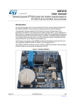

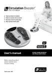
![Généralités sur les plaies [Mode de compatibilité]](http://vs1.manualzilla.com/store/data/006481798_1-f95064b5e0feec6dd6119ac587e78945-150x150.png)