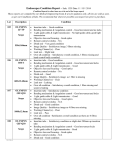
1
INSTRUCTION MANUAL
S200 Susie Simon
S201 Susie Simon with Ostomy
S203 Susie
S204 Simon
S205 Simple Simon
S206 Simple Susie
General Patient Care
Heart and Lung Sounds
Blood Pressure Auscultation
Injection Training
GYN Training
Gaumard® Scientific
14700 SW 136 St.
Miami, FL 33196-5691
e-mail: [email protected]
©2002-07 Gaumard Scientific
All Rights Reserved
PLEASE READ THE FOLLOWING INSTRUCTIONS PRIOR TO
COMMENCING TRAINING EXERCISES ON YOUR NEW MANIKIN.
HANDLE YOUR SIMULATOR IN THE SAME MANNER AS YOU WOULD
HANDLE YOUR PATIENT - WITH CARE AND CONSIDERATION.
SHOULD YOU HAVE ANY QUESTIONS AFTER READING THIS
INSTRUCTIONAL MANUAL, PLEASE CALL
Worldwide 305-971-3790
USA 800-882-6655
FAX 305-667-6085
e-mail: [email protected]
2
TABLE OF CONTENTS
SECTION 1 - INTRODUCTION
5
1. Susie Simon Simulator Family
5
2. Options
7
3. Contents
8
4. Assembly
9
SECTION 2 - GENERAL CARE CAPABILITIES
10
1. Bandaging
10
2. Eyes/Ophthalmological Exercises
10
3. Teeth and Tongue
10
4. Hygienic Care
10
5. Injection Sites
11
7. Male and Female Organs
14
8. Range of Simulated Movement
14
9. Ears, Nose and Throat
14
10. Tracheotomy
14
11. Stomach
15
12. Transverse Colostomy, Ileostomy, and Suprapubic Stoma
15
13. Intestinal Tract
16
14. Urinary System
16
15. Female Catheterization
16
16. Male Catheterization
17
17. Decubitus Ulcers (optional)
19
3
18. Injection Training Arm and Hand (optional)
20
SECTION 6 - HEART AND LUNG SOUNDS (optional)
22
SECTION 7 - BLOOD PRESSURE TRAINING ARM (optional)
28
SECTION 8 - GYNECOLOGIC EXAMINATION (optional)
31
SECTION 9 - GENERAL NOTES
31
1. Lubrication
31
2. Catheters
31
3. Emptying the reservoir system
31
4. Filling the bladder
32
5. Internal Cleaning
32
6. Removal of internal tanks
32
4
SECTION 1 - INTRODUCTION
This is an operating guide for six (6) simulators in the Susie Simon family which includes both male
and female platforms.
Susie Simon Simulator Family
Model
Name
Number
200
Susie
Simon
201
Susie
Simon
203
Susie
204
Simon
205
Simple
Simon
206
Simple
Susie
• Full-size adult manikin
f
f
f
m
m
f
• Soft, lifelike faceskin
√
√
√
√
√
√
√
√
√
√
√
√
√
√
√
√
√
√
√
√
√
√
√
√
√
√
√
√
√
√
√
√
√
√
√
√
√
√
√
• Interchangeable male organ
√
√
√
• Bends at waist as in human
√
√
√
√
√
√
√
√
√
√
√
√
√
√
√
√
√
√
√
√
√
√
Features
• Stylish wig for haircare
exercises and surgical
draping
• Movable jaw with tongue
• Removable upper and lower
dentures for oral hygiene
• Realistic eyes in eyesockets
For ophthalmic exercises
• Ear canal for otic drops
and irrigation
• Mouth, nose and tracheotomy
openings for naso-gastric
lavage and gavage
• 360° intramuscular injection
site in right and left upper
arm
• Intramuscular injection
site in buttock
• Enema administration
capability
5
√
Susie Simon Simulator Family
Model
Name
Number
• Realistic urethral passage
and bladder for
catheterization exercises
• Elevating pillow increases
bladder pressure for
increased flow during
catheterization
(male or female)
• Vaginal douching and
pap smear exercises with
realistic vagina and cervix
200
Susie
Simon
201
Susie
Simon
203
Susie
204
Simon
√
√
√
√
√
√
√
√
√
√
√
• No drip thumb latch
with audible “click”
provides secure seal
between ostomies and
internal tanks
• Soft, realistic hands,
feet, fingers, and toes
• Bathing and bandaging
activity
• Detachable and removable
internal tanks
• New modular tricuspid valve
permits male or female
catheterization
with soft silicone catheters
206
Simple
Susie
√
√
• Sculpted stomas
• Transverse colos-tomy,
ileostomy, and
suprapubic stoma
205
Simple
Simon
√
√
√
√
√
√
√
√
√
√
√
√
√
√
√
√
√
√
√
√
√
√
√
√
√
√
√
√
√
√
√
√
√
√
6
√
OPTIONS
• Intravenous training arm for injection,
infusion, and blood collection procedures
S261
$220
• Advanced intravenous training arm with subtle
venous network. Specify right or left arm
S262.100
$305
• Arterial and venous patient training
arm for arterial stick exercises,
arteriovenous anastomosis exercises,
hemodialysis set-up, skin/tissue suture
exercises, as well as intramuscular
and subcutaneous injection training,
intravenous infusion and injection,
and blood collection procedures.
Specify right or left arm
S263
$625
• Blood pressure arm with programmable BP
auscultation tutor. See p.31 for additional details
S270
$650
• Bilateral IM sites, arms and legs
S266.4
$75.00
• Site specific heart and lung sounds kit with
stethoscope. See details p. 36-37
S200.100
$595
• Set of 2 decubitus ulcers, with one ulcer depicting
the initial stage of ulceration and tissue infection;
the other graphically depicting suppuration and
deeply infected stage
S264
$60
• Ulcerated foot
S265
$60
• Deltoid, chest, and abdominal
incisions
S266.1, S266.2, S266.3
ea $30
• Amputation stump for bandaging
with a unique design that permits easy
placement on the manikin
S267
$60
• Additional arm and buttock injection sites
S268.1, S268.2
ea. $ 30
• Carrying bag
S269
$85
7
Contents
Upper body. An optional blood pressure arm may be attached to Susie’s left side and an optional IV arm to
her right.
Lower body with legs and feet. A waist rod joins the upper and lower torsos. The blue bulb shown near
the right knee is used to inflate an internal cushion lifting the uterus and bladder anteriorly providing
increased urinary flow.
8
Assembly
To assemble Susie, unscrew one knob at either end of the waist rod, pull rod out.
Make sure the white guide tube remains in place.
Within the upper torso locate the stomach reservoir. Connect the tube from the stomach
to the port shown above. The click valve on this tube is normally closed. You are now
ready to attach the lower torso of the manikin to the upper torso.
9
Ease the lower torso into the upper torso, being careful not to disengage the stomach
reservoir.
Line up the holes and slide the waist rod through the white guide tube. Replace the
waist knob and finger tighten.
10
SECTION 2 - GENERAL CARE CAPABILITIES
1. Bandaging
The fingers and toes of this simulator are separated to permit bandaging exercises.
The surface of the manikin is smooth and resistant to water, oil, and liniments.
2. Eyes/Ophthalmologic Exercises
The head has removable eyes that open and close permitting the following exercises:
• Administration of orbital medicines into the conjunctival sac
• Removal of foreign bodies
• Eye irrigation
3. Teeth and Tongue
The teeth and tongue are of normal size and may be removed.
4. Hygienic Care
The head of the female simulators are supplied with a wig, permitting instruction in
combing, shampooing, and head draping. The manikin surface is water resistant so
that bathing exercises may be practiced.
11
5. Injection Sites
Sites in the upper left and right arm, as well as optional sites in the left and right thigh,
allow administration of intramuscular injections. Sites are removable. Inside each site is
a sponge to absorb the injectate. There is also a site in the upper gluteal region to
permit intramuscular injections in the buttocks. All injection sites are easily removed
and replaced. Numerous injection sites are located in the optional injection training arm
and hand described later.
IM site on shoulder
Subcutaneous site on optional injection training arm
12
IM site on upper buttock
Optional IM sites on left and right thighs
13
7. Male and Female Organs
If your simulator has interchangeable male and female organs, note a red adaptor at the
opening of the urethra for female catheterization exercises. This red adaptor will be
removed when the male organ is used for catheterization.
8. Range of Simulated Movement
The joints are strong and their movements are lifelike and realistic. The manikin bends
at the waist. The head and jaw articulate.
9. Ears, Nose and Throat
Left ear - the interior of the ear contains a simulated ear canal with a capacity of
10 ml, facilitating syringing exercises.
Nasal/oral openings: both are connected to the stomach reservoir/tank, so that a
#10 Levine tube may be used to demonstrate tube feeding and gastric suction.
A gastric reservoir (capacity: 850 ml) is provided, with an opening for
gastrostomy.
REMEMBER TO ALWAYS USE A LUBRICANT PRIOR TO INTRODUCTION OF A
LEVINE TUBE OR ANY OTHER INVASIVE DEVICE.
10. Tracheostomy
An curved cavity is located at the sternal notch for placement of a lubricated trach tube.
You may inflate the cuff of a Shiley 8,
14
11. Stomach
The upper torso also includes a stomach tank into which a #10 Levine tube may be
used to demonstrate tube feeding and gastric suction. A gastrostomy port connects
directly to the stomach tank from the red flange located near the waist. ALWAYS USE
A LUBRICANT WHEN INTRODUCING THE LEVINE TUBE.
12. Transverse Colostomy, Ileostomy, and Suprapubic Cystostomy
The creation of an ostomy port, a temporary or permanent excretory opening, is an
important part of abdominal surgery. The simulator demonstrates the appearance of
ostomy openings. The Susie Simon S201 has anatomically sculptured stomas of a
transverse colostomy, ileostomy, and suprapubic cystostomy, which may be performed
as a result of abdominal surgery. Conventional ostomy drainage and irrigation
exercises can be performed.
The ostomy sites connect to reservoirs of appropriate size, and disposable or
permanent ostomy bags may be applied to all openings. Exercises in skin preparation
and stoma hygiene, as well as treatment of skin conditions around the sites may also be
practiced. The reservoirs may be cleansed by introducing a solution of soap and water
or detergent with a 60 cc. syringe. Alternatively, the reservoirs can be removed from
the lower torso and cleaned. Note that the Simple Simon 205 and the Simple Susie 206
do not have ostomies or internal tanks.
15
13. Intestinal Tract
Administration of an enema may be performed on all manikins except the S205 and
S206. The legs articulate sufficiently to permit enema exercises with the manikin on its
back. The enema should be introduced with an anal nozzle of small diameter.
Remember to use a lubricant.
PLEASE NOTE: A non-return valve is built into the anal canal to prevent fluid spilling
during instillation. The enema reservoir capacity is approximately 750 ml.
14. Urinary System
The urethral passage and the bladder (capacity: approximately 1800 ml) are connected
by a valve assembly to make catheterization exercises more lifelike. Fluid can be
withdrawn from the bladder after the insertion of a #18 French catheter. The suprapubic
opening may be used for filling the bladder or for drainage exercises. Please note that
repeated sterilization can cause a variance in catheter diameters. An older device
might permit fluid leakage. Therefore, different catheters should be inserted to
determine a proper fit.
NOTE: ALWAYS USE A LUBRICANT WHEN INTRODUCING A CATHETER.
15. Female Catheterization
Bladder catheterization may be required to remove urine. This procedure must be
conducted under aseptic conditions to prevent the subsequent infection or inflammation
of the urinary tract. A suprapubic cystostomy opening is present for practice in
cystostomy management and maintenance. When practicing catheterization, the labia
minora must be separated to examine the urethral opening, as in the female patient.
The realistic simulation of the vulva area also permits instruction in asepsis and
disinfection. When actually performing catheterization on the simulator, a "one eye"
#18 French catheter is recommended. Smaller catheters may cause leakage.
16
Always lubricate the distal end of the catheter. Once the catheter is in place, use the
blue squeeze bulb to increase bladder pressure and assure a good flow of urine.
16. Male Catheterization (All models except Simple Susie S206)
Male catheterization is performed in the upright or recumbent position by the attachment
of the male organ. The flexible vinyl male organ contains the urethra, which is
connected to an internal urinary bladder through a one-way valve. A suprapubic
cystostomy opening is also present for practice in cystostomy management and
maintenance. When performing catheterization, the penis must be manipulated to
permit passage of the catheter, as in the male patient. The realistic simulation of the
male genitalia also permits instruction in asepsis and disinfection. When actually
performing catheterization, a "one eye" #18 French catheter is recommended for the
most efficient use of the simulator. The simulator also demonstrates the appearance of
the ostomy opening in the patient who has had a suprapubic stoma as a result of
surgery on the bladder or prostate. All suprapubic cystostomy drainage and irrigation
exercises can be performed.
17
In order to perform male catheterization, this red flange must be removed and retained.
Attach the male organ by inserting the tube into the urethra and securing with Velcro
18
NOTE: ALWAYS USE A LUBRICANT WHEN INTRODUCING A CATHETER.
17. Decubitus Ulcers (optional)
A decubitus ulcer is caused by prolonged pressure in a patient confined to bed and in
one position for a long period of time. They are also known as pressure sores or bed
sores. The simulator is supplied with two of these ulcers. These ulcers are
anatomically accurate. The first decubitus ulcer illustrates the initial stage of ulceration.
The second decubitus ulcer illustrates the suppuration or pus/deeply infected stage.
19
18. Patient Training Arm and Hand Injection Simulator (optional)
This simulator is a training tool for infusion, blood collection, intravenous injection,
intramuscular injection, TB screening and subcutaneous injection exercises.
The simulator is attached to the manikin, and is to be used connected to a blood
dispensing bag. You may use the metal stand supplied or a conventional IV pole. The
arm is also supplied with an amount of synthetic blood concentrate, and a spare arm
skin. The arm and hand contain venous grooves, which are fitted with soft latex tubes
that simulate the consistency of the veins. A translucent, pliable vinyl skin, which is
removable and washable, is stretched over the venous structure, simulating the normal
adult arm.
The arm features the following: (1) subcutaneous injection areas on the volar side of
the forearm and the lateral side of the upper arm; (2) an intramuscular injection site in
the deltoid area; and (3) two veins in the dorsum of the hand for additional intravenous
training techniques.
In addition, the training arm contains simulated cephalic, basilic, antecubital, radial and
ulnar veins. Simulated blood may be placed in the dispensing bag, which is equipped
with a squeeze bulb. Applying pressure via the squeeze bulb permits the veins to stand
out, simulating a clenched fist or tourniquet situation. Release of pressure simulates
collapsed veins. Use of the squeeze bulb permits the palpability of the veins to be
varied, as seen in routine hospital or emergency situations.
Note that the use of cannulas/needles larger than 21 gauge will shorten the life of
the venistructure.
20
REPLACING THE SKIN AND VEINS
1. To remove the vinyl outer skin, start at the top of the arm and remove by rolling it
down and over the wrist. Use of water based silicone or talcum powder will ease
movement.
2. Select a new skin and heat it in warm soapy water to a temperature of about 125
degrees F or about 50 degrees C. Dry the skin, and insert it onto the arm at the
fist and pull the new skin up into place.
3. To replace the veins make sure you are not allergic to latex. Gaumard uses very
pure latex veins to produce the best possible self-sealing possible.
CLEANING & REPAIR OF THE PATIENT TRAINING ARM AND HAND
1. The skin of the training arm can be cleaned with a mild detergent, or soap and
water. After drying the arm, lightly dust it with talcum powder. This will keep the
training arm supple and easy to use.
2. If the venous system is blocked, first check that the tubes are not kinked. If
blockage
persists, remove the fist and flush veins with water.
3. Indelible marks made with ballpoint pens, ink or magic markers will remain.
.
21
SECTION 6 - HEART AND LUNG SOUNDS (optional)
INTRODUCTION
This teaching system is used for auscultation training. RFID sensors are hidden
beneath the skin in a total of 13 locations; nine on the front and four on the back.
Included is a Virtual Stethoscope™ which is powered by a 9V cell. Remove the battery
housing and place a fresh battery inside, being sure to make secure connections.
Menus of available heart and lung sounds are attached listing the location, the sound
and a brief description of the physiological condition associated with each sound.
Hear the appropriate heart or lung sound as the bell of the stethoscope is moved across
the front or the back of the torso. An external speaker, which must be powered through
a wall outlet, is supplied so that the Instructor can allow the classroom to hear what the
student is hearing through the stethoscope.
22
The vest overlay contains RFID tags that communicate with the Virtual Stethoscope so
that the heart or lung sound appropriate for that location is heard. The vest is supplied
with blue stick-on dots that may be easily removed by the Instructor.
The four lung fields may also be heard on the back of the patient.
23
LOCATION
HEART SOUND
COMMENT
Base Right
Base Sound
Patient has a normal heart with mild anemia.
The heart is hyperdynamic and has elevated
cardiac output. S2 is accentuated at the base.
Fixed Split S2
Patient has an atrial septal defect which
increases flow through the right heart,
prolongs RV systole and also produces a midsystolic murmur (MSM) because of increased
flow through the RV outflow tract.
Physiological
Split S2
The splitting of S2 is easily heard during
inspiration and the second sound is single
during expiration. The second component of
the split sound (P2) is accentuated.
Split S2
S2 is variably split during mid-inspiration, as
three beats are repeated.
Paradoxical Split
S2
The splitting of S2 is heard during expiration,
but the sound becomes single during
inspiration.
(The background noise is increased during
inspiration.)
Opening Snap
Patient has mitral stenosis, responsible for an
early crisp diastolic sound heard at the base
0.08 seconds after S2. S1 is usually loud at
the base, which reflects mitral stenosis.
Friction Rub
Patient has uremic pericarditis, which leads to
rubbing of roughened visceral and parietal
pericardial surfaces against one another. The
3 component rub exists during deep
inspiration.
Base Left
Left Side
Sternal
Border
24
LOCATION
HEART SOUND
COMMENT
Apex
Apex Sound
Patient has a normal heart with mild anemia.
The heart is hyperdynamic and has elevated
cardiac output.
Mid-Systolic Click
Patient has mitral prolapse, which produces a
mid-systolic click heard during inspiration.
S3 Sound
Patient has a readily heard third heart sound.
S3 occurs later in diastole than the opening
snap.
Intermittent S4
Patient has left ventricular hypertrophy, and
has a fourth sound (S4) which is not heard
on every cycle. The sound is presystolic,
about 0.1 second before S1.
Starr-Edwards
Valve
This ball-in-cage mitral prosthesis has a
mechanical closing sound (S1) and one or
more diastolic sounds caused by the ball
bouncing within the cage.
25
LOCATION
LUNG SOUND
COMMENT
Trachea
Tracheal Sounds
Expiration sounds are louder, have a higher
pitch, and are of longer duration than during
inspiration. The silent period or pause
following expiration is longer than the one
between expiration and inspiration.
Stridor Sounds
Patient has marked respiratory distress, and a
narrow aperture between the vocal cords that
produces a high pitched tone during both
inspiration and expiration. During the end of
expiration, there is an abrupt drop in pitch.
Bronchial Sounds
Breath sounds are similar to tracheal sounds
in that the expiratory phase is louder and lasts
longer than the inspiratory phase. The major
distinguishing characteristic is the high
pitched, hard quality of the expiratory phase.
Wheezing Sounds
These musical wheezing sounds are often
heard in asthma patients. During inspiration,
the wheeze is slightly higher in pitch than
during expiration. Wheezing in asthmatics is
often present in either one or both phases of
respiration.
Upper Anterior
(Two Sites)
Lower Anterior
(Two Sites)
Bronchial Sounds
Breath sounds are similar to tracheal sounds
in that the expiratory phase is louder and lasts
longer than the inspiratory phase. The major
distinguishing characteristic is the high
pitched, harsh quality of the expiratory phase.
26
LOCATION
LUNG SOUND
COMMENT
Posterior
(Four Sites)
Wheezing Sounds
These musical wheezing sounds are often
heard in asthma patients. During inspiration,
the wheeze is slightly higher in pitch than
during expiration. Wheezing in asthmatics is
often present in either one or both phases of
respiration.
Pleural Friction
This sound probably originates from the
friction of inflamed pleural surfaces moving
against one another. The sound is repetitive
as long as the breathing pattern and position
remain constant. Similar to but lower in pitch
than crackles.
Medium-Fine
Crackles
These noises begin about mid-inspiration and
progressively increase in intensity up to the
end of expiration. Coarse crackles are also
audible in the early expiratory phase of some
of the breaths.
Ronchi, Crackles
Coarse crackles are present during both
inspiration and expiration. There are also
some very low pitched repetitive sounds that
are ronchi. High pitched squeaks are also
audible against a background of bronchial
breath sounds.
Coarse Crackles
Coarse crackles begin at the onset of
inspiration and diminish in intensity and
prevalence toward the end of inspiration.
Expiration is not audible.
Pulmonary Edema
Coarse and medium crackles appear toward
the end of inspiration and continue into
expiration.
The respiratory rate is rapid and expiratory
phase is “bronchial” in character. These
features exist during respiratory distress and
congestion.
27
SECTION 7 - BLOOD PRESSURE TRAINING ARM (optional)
Assembly
Connect the electrical cable leading to the blood pressure arm to the BP Auscultation
Tutor, being careful to not damage the four pins found within the cable connector.
Connect the power supply to the Tutor. You may have to supply a mechanical adaptor
to access the wall outlet in your area. Now connect the long clear tube extending from
the sphygmomanometer assembly to the BP Tutor.
Wrap the BP cuff around the left arm midway between the deltoid and elbow.
28
Operation
Turn the power switch “ON”. Do not press “Calibrate”. Note you may palpate the wrist
and feel the radial pulse. The BP Tutor normally remembers the previous settings. We
use these settings to simulate a hypertensive patient having an auscultation gap. The
challenge for the student is to recognize that the systolic is 150 not 120. The 150 may
suggest hypertension, while 120 would be incorrectly interpreted as normal.
Systolic
Diastolic
Upper auscultation gap
Lower auscultation gap
Pulse
150
90
140
120
80
K1 starts here
K4 ceases then K5 silence
K sounds cease but pulse continues
K sounds resume
Calibration
Calibration may be required. In the event the Instructor notes that the recorded pressure
varies significantly from the pressure on the sphygmomanometer, you may recalibrate
as follows:
1. Press Calibrate, then Start, and the display will show “CAL 000”
2. Press Calibrate, then Start, again and the display will show “CAL 128” Inflate the
cuff to read 128 on the dial of the sphygmomanometer.
3. Press Calibrate, then Start, again and the display will show “CAL 256” Inflate the
cuff to read 256 on the dial of the sphygmomanomater.
4. Now inflate the blood pressure cuff to read 256 and press Calibrate Start again.
5. At this time, the BP Tutor will display the reading on the sphygmomanometer.
Lower the cuff pressure to zero and watch the display track the
sphygmomanometer.
29
Blood Pressure Training
Turn the Tutor ON and observe the previously selected values:
1. Press Systolic and observe the value. If you agree, press Systolic again and that
number is locked in. To increase or decrease the Systolic pressure, press the UP
or DOWN red arrows. When the desired value is reached, press Systolic once
more and the new value is locked in.
2. Proceed with Diastolic, Upper Auscultation Gap, Lower Auscultation Gap, and
Heart Rate in the same manner. Once selected, each value will disappear so that
the student cannot see them.
3. Instruct the student to take the blood pressure by attaching the BP cuff and
placing the bell of a conventional stethoscope over the speaker concealed in the
antecubital region of the arm near the elbow.
4. Instruct the student to pump up the cuff until the radial pulse is no longer felt.
Now slowly release the cuff pressure until the first Korotkoff sound is heard
indicating the Systolic pressure. Decrease cuff pressure further and hear K2, K3,
K4. The K5 sound is silence. If an auscultation gap was programmed, the
student will not hear Korotkoff sounds between the upper and lower limits.
5. Ask the student to record the observed systolic pressure, the diastolic pressure,
the limits of auscultation gap (if any) as well as the pulse rate. Compare the
values observed by the student with those originally selected in order to assess
student competency.
30
SECTION 8 – GYNECOLOGIC EXAMINATION
The Susie Simon features a full-size adult female lower torso, consisting of
the abdomen and pelvis suitable for practice in vaginal speculum
examination and diaphragm sizing and fitting.
A tenaculum forceps can be used to grasp the cervix and pull it toward the student for
examination.
SECTION 9 - GENERAL NOTES
1. Lubrication
ALWAYS USE A LUBRICANT WHEN INTRODUCING A CATHETER OR INVASIVE
DEVICE
2. Catheters - Troubleshooting
There may not be an immediate outflow of water on introduction of the catheter into the
bladder. Should blockage occur, use the blue squeeze bulb at the side of the lower
torso to increase pressure in the bladder.
3. Emptying the Reservoir System
To remove the remaining fluid from the bladder reservoir after catheterization
exercises are complete, lift the abdominal cover and remove the reservoir.
31
4. Filling of the Bladder
The bladder should be filled through the suprapubic opening. This may be done in one
of two ways. Instillation of water (approximately 500 ml into the1800 ml tank) through
introduction of an appropriate funnel at the suprapubic site; or, by using a catheter with
a large syringe.
5. Cleaning
The manikin may be cleaned with a mild detergent, or with soap and water. Do not
clean with harsh abrasives.
• Indelible marks made with ballpoint pens, ink, or markers will remain.
• Store the manikin in a cool area in the box provided. Do not stack heavy materials
on top of the box
• Do not wrap the manikin or any Gaumard product in newsprint.
6. Removal of Internal tanks (reservoirs)
The lower torso contains several reservoirs for patient care exercises. Each is
connected using “click” connectors permitting that reservoir to be removed and cleaned
or replaced as needed.
SHOULD YOU HAVE ANY QUESTIONS AFTER READING THIS INSTRUCTION
MANUAL, PLEASE CONTACT OUR CUSTOMER SERVICE DEPARTMENT FOR
FURTHER ASSISTANCE:
800-882-6655 Toll Free USA
305-971-3790 Worldwide
305-667-6085 Fax
OR
E-MAIL US AT:
[email protected]
Gaumard Scientific
14700 SW 136 Street
Miami, FL 33196
www.gaumard.com
32
33