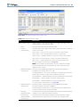
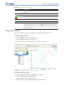
1
Living Image® Software
User’s Manual
Version 4.0
© 2002-2009 Caliper Corporation. All rights reserved.
PN 126545
Caliper Life Sciences
68 Elm Street
Hopkinton, MA 01748
USA
1.877.522.2447 (US)
1.508.435.9500
Fax: 1.508.435.3439
E-mail: [email protected]
www.caliperls.com
Discovery in the Living Organism, IVIS Imaging System, Living Image, DLIT, and FLIT are either registered
trademarks or trademarks of Caliper Life Sciences, Inc. The names of companies and products mentioned herein
may be the trademarks of their respective owners. Apple, Macintosh and QuickTime are registered trademarks of
Apple Computer, Inc. Microsoft, PowerPoint and Windows are either registered trademarks or trademarks of
Microsoft Corporation in the United States and/or other countries. Adobe and Illustrator are either registered
trademarks or trademarks of Adobe Systems Incorporated in the United States and/or other countries.
Contents
1 Welcome . . . . . . . . . . . . . . . . . . . . . . . . . . . . . . . . . . . . . . . . . 1
1.1 What’s New In Living Image 4.0 Software . . . . . . . . . . . . . . . . . . . . . . . . . . . . . . 1
1.2 About This Manual . . . . . . . . . . . . . . . . . . . . . . . . . . . . . . . . . . . . . . . . . . 2
1.3 Contacting Caliper Technical Support . . . . . . . . . . . . . . . . . . . . . . . . . . . . . . . . . 3
2 Overview of Imaging & Image Analysis . . . . . . . . . . . . . . . . . . . . . . . . . 5
2.1 Workflow Overview . . . . . . . . . . . . . . . . . . . . . . . . . . . . . . . . . . . . . . . . . . 5
2.2 Imaging Modes . . . . . . . . . . . . . . . . . . . . . . . . . . . . . . . . . . . . . . . . . . . . . 7
2.3 About Image Sequences . . . . . . . . . . . . . . . . . . . . . . . . . . . . . . . . . . . . . . . 10
2.4 Image Display & Analysis . . . . . . . . . . . . . . . . . . . . . . . . . . . . . . . . . . . . . . 11
3 Getting Started . . . . . . . . . . . . . . . . . . . . . . . . . . . . . . . . . . . . . 15
3.1 Starting the Living Image Software . . . . . . . . . . . . . . . . . . . . . . . . . . . . . . . . . 15
3.2 Initializing the IVIS Imaging System . . . . . . . . . . . . . . . . . . . . . . . . . . . . . . . . 17
3.3 Checking the System Temperature . . . . . . . . . . . . . . . . . . . . . . . . . . . . . . . . . 17
3.4 About the IVIS Acquisition Control Panel & Auto Exposure Feature . . . . . . . . . . . . . . . 18
3.5 Tracking System& User Activity . . . . . . . . . . . . . . . . . . . . . . . . . . . . . . . . . . 19
4 Luminescent or Fluorescent Imaging . . . . . . . . . . . . . . . . . . . . . . . . 21
4.1 Acquire a Luminescent Image . . . . . . . . . . . . . . . . . . . . . . . . . . . . . . . . . . . . 21
4.2 Acquire a Fluorescent Image With Epi-Illumination . . . . . . . . . . . . . . . . . . . . . . . . 24
4.3 Acquire a Fluorescent Image With Transillumination . . . . . . . . . . . . . . . . . . . . . . . . .27
4.4 Acquire a Sequence Using the Imaging Wizard . . . . . . . . . . . . . . . . . . . . . . . . . . . 30
4.5 Manually Set Up an Image Sequence . . . . . . . . . . . . . . . . . . . . . . . . . . . . . . . . 37
4.6 Manually Saving Image Data . . . . . . . . . . . . . . . . . . . . . . . . . . . . . . . . . . . . 41
4.7 Exporting Image Data . . . . . . . . . . . . . . . . . . . . . . . . . . . . . . . . . . . . . . . . 42
5 X-Ray Imaging . . . . . . . . . . . . . . . . . . . . . . . . . . . . . . . . . . . . . 43
5.1 Acquire an X-Ray Image . . . . . . . . . . . . . . . . . . . . . . . . . . . . . . . . . . . . . . 43
5.2 Acquire an Image Sequence That Includes X-Ray Images . . . . . . . . . . . . . . . . . . . . . 48
5.3 Measuring Relative Density . . . . . . . . . . . . . . . . . . . . . . . . . . . . . . . . . . . . . 55
6 Kinetic Imaging . . . . . . . . . . . . . . . . . . . . . . . . . . . . . . . . . . . . 59
6.1 Kinetic Acquisition . . . . . . . . . . . . . . . . . . . . . . . . . . . . . . . . . . . . . . . . . 59
6.2 Viewing & Editing Data in the Kinetic Acquisition Window . . . . . . . . . . . . . . . . . . . . 63
6.3 Saving Kinetic Data . . . . . . . . . . . . . . . . . . . . . . . . . . . . . . . . . . . . . . . . . 65
6.4 Playing Kinetic Data . . . . . . . . . . . . . . . . . . . . . . . . . . . . . . . . . . . . . . . . . 66
7 Working With Data . . . . . . . . . . . . . . . . . . . . . . . . . . . . . . . . . . . 71
7.1 Opening Data . . . . . . . . . . . . . . . . . . . . . . . . . . . . . . . . . . . . . . . . . . . . 71
7.2 About the Image Window & Tool Palette . . . . . . . . . . . . . . . . . . . . . . . . . . . . . . 75
7.3 Viewing Image Information . . . . . . . . . . . . . . . . . . . . . . . . . . . . . . . . . . . . . 77
7.4 Adjusting Image Appearance . . . . . . . . . . . . . . . . . . . . . . . . . . . . . . . . . . . . 79
7.5 Correcting Image Data . . . . . . . . . . . . . . . . . . . . . . . . . . . . . . . . . . . . . . . . 81
ii
Living Image Software User’s Manual
7.6 Viewing Intensity Data & Making Measurements . . . . . . . . . . . . . . . . . . . . . . . . . . .83
7.7 Creating a Transillumination Overview . . . . . . . . . . . . . . . . . . . . . . . . . . . . . . . 90
7.8 Overlaying Multiple Images . . . . . . . . . . . . . . . . . . . . . . . . . . . . . . . . . . . . . 91
7.9 Rendering Intensity Data in Color . . . . . . . . . . . . . . . . . . . . . . . . . . . . . . . . . . 93
7.10 Exporting or Printing Images . . . . . . . . . . . . . . . . . . . . . . . . . . . . . . . . . . . . 95
7.11 Editing an Image Sequence . . . . . . . . . . . . . . . . . . . . . . . . . . . . . . . . . . . . . 97
7.12 Creating an Image Sequence from Individual Images . . . . . . . . . . . . . . . . . . . . . . . 98
8 Working With ROI Tools . . . . . . . . . . . . . . . . . . . . . . . . . . . . . . . . 101
8.1 About ROIs . . . . . . . . . . . . . . . . . . . . . . . . . . . . . . . . . . . . . . . . . . . . . 101
8.2 Quick Guide: Drawing Measurement ROIs on an Image or Sequence . . . . . . . . . . . . . . . 102
8.3 ROI Tools . . . . . . . . . . . . . . . . . . . . . . . . . . . . . . . . . . . . . . . . . . . . . . 104
8.4 Measurement ROIs . . . . . . . . . . . . . . . . . . . . . . . . . . . . . . . . . . . . . . . . . . 106
8.5 Subject ROIs . . . . . . . . . . . . . . . . . . . . . . . . . . . . . . . . . . . . . . . . . . . . . 110
8.6 Measuring Background-Corrected Signal . . . . . . . . . . . . . . . . . . . . . . . . . . . . . . 110
8.7 Kinetic ROIs . . . . . . . . . . . . . . . . . . . . . . . . . . . . . . . . . . . . . . . . . . . . . 112
8.8 Managing ROIs . . . . . . . . . . . . . . . . . . . . . . . . . . . . . . . . . . . . . . . . . . . 115
8.9 Managing the ROI Measurements Table . . . . . . . . . . . . . . . . . . . . . . . . . . . . . . . 126
9 Image Math . . . . . . . . . . . . . . . . . . . . . . . . . . . . . . . . . . . . . . . 133
9.1 Creating a New Image Using Image Math . . . . . . . . . . . . . . . . . . . . . . . . . . . . . . 133
9.2 Subtracting Tissue Autofluorescence . . . . . . . . . . . . . . . . . . . . . . . . . . . . . . . . . 136
10 Planar Spectral Image Analysis . . . . . . . . . . . . . . . . . . . . . . . . . . . . 139
10.1 Image Sequence Requirements . . . . . . . . . . . . . . . . . . . . . . . . . . . . . . . . . . . 139
10.2 Planar Spectral Image Analysis . . . . . . . . . . . . . . . . . . . . . . . . . . . . . . . . . . 139
10.3 Viewing Graphical Results . . . . . . . . . . . . . . . . . . . . . . . . . . . . . . . . . . . . . 142
10.4 Managing Planar Spectral Imaging Results . . . . . . . . . . . . . . . . . . . . . . . . . . . . . 144
11 Spectral Unmixing . . . . . . . . . . . . . . . . . . . . . . . . . . . . . . . . . . . 147
11.1 Image Sequence Requirements . . . . . . . . . . . . . . . . . . . . . . . . . . . . . . . . . . . 147
11.2 Performing Spectral Unmixing . . . . . . . . . . . . . . . . . . . . . . . . . . . . . . . . . . . 147
11.3 Spectra Window . . . . . . . . . . . . . . . . . . . . . . . . . . . . . . . . . . . . . . . . . . 152
11.4 Spectral Unmixing Parameters . . . . . . . . . . . . . . . . . . . . . . . . . . . . . . . . . . . 154
11.5 Spectral Unmixing Options . . . . . . . . . . . . . . . . . . . . . . . . . . . . . . . . . . . . . 155
12 Reconstructing a 3D Surface . . . . . . . . . . . . . . . . . . . . . . . . . . . . . 159
12.1 Generating a Surface . . . . . . . . . . . . . . . . . . . . . . . . . . . . . . . . . . . . . . . . 160
12.2 Managing Surfaces . . . . . . . . . . . . . . . . . . . . . . . . . . . . . . . . . . . . . . . . . 164
12.3 Export or Import a Surface . . . . . . . . . . . . . . . . . . . . . . . . . . . . . . . . . . . . . 164
13 Point Source Fitting . . . . . . . . . . . . . . . . . . . . . . . . . . . . . . . . . . 167
13.1 Image Sequence Requirements . . . . . . . . . . . . . . . . . . . . . . . . . . . . . . . . . . . 167
13.2 Displaying the Point Source Fitting Tools . . . . . . . . . . . . . . . . . . . . . . . . . . . . . 167
13.3 Point Source Fitting . . . . . . . . . . . . . . . . . . . . . . . . . . . . . . . . . . . . . . . . . 168
Contents iii
13.4 Checking the Point Source Fitting Results . . . . . . . . . . . . . . . . . . . . . . . . . . . . . 170
13.5 Exporting Results . . . . . . . . . . . . . . . . . . . . . . . . . . . . . . . . . . . . . . . . . . 171
13.6 Managing Results . . . . . . . . . . . . . . . . . . . . . . . . . . . . . . . . . . . . . . . . . . 172
13.7 Point Source Fitting Tools . . . . . . . . . . . . . . . . . . . . . . . . . . . . . . . . . . . . . 173
14 3D Quantification Database . . . . . . . . . . . . . . . . . . . . . . . . . . . . . . 179
14.1 Preparing & Imaging the Samples . . . . . . . . . . . . . . . . . . . . . . . . . . . . . . . . . 179
14.2 Creating a Quantification Database . . . . . . . . . . . . . . . . . . . . . . . . . . . . . . . . 180
14.3 Managing Quantification Results . . . . . . . . . . . . . . . . . . . . . . . . . . . . . . . . . . 183
15 3D Reconstruction of Sources . . . . . . . . . . . . . . . . . . . . . . . . . . . . 185
15.1 Reconstructing Luminescent Sources . . . . . . . . . . . . . . . . . . . . . . . . . . . . . . . . 188
15.2 Reconstructing Fluorescent Sources . . . . . . . . . . . . . . . . . . . . . . . . . . . . . . . . 198
15.3 3D Reconstruction Results . . . . . . . . . . . . . . . . . . . . . . . . . . . . . . . . . . . . . 200
15.4 Checking the Reconstruction Quality . . . . . . . . . . . . . . . . . . . . . . . . . . . . . . . 204
15.5 Measuring Sources . . . . . . . . . . . . . . . . . . . . . . . . . . . . . . . . . . . . . . . . . 206
15.6 Displaying Luminescent & Fluorescent Sources on One Surface . . . . . . . . . . . . . . . . . 209
15.7 Exporting a 3D Scene . . . . . . . . . . . . . . . . . . . . . . . . . . . . . . . . . . . . . . . . 209
15.8 3D Tools Overview . . . . . . . . . . . . . . . . . . . . . . . . . . . . . . . . . . . . . . . . . 211
15.9 3D Tools - Surface . . . . . . . . . . . . . . . . . . . . . . . . . . . . . . . . . . . . . . . . . 211
15.10 3D Tools - Source . . . . . . . . . . . . . . . . . . . . . . . . . . . . . . . . . . . . . . . . . 213
15.11 3D Tools - Registration . . . . . . . . . . . . . . . . . . . . . . . . . . . . . . . . . . . . . . 214
15.12 3D Tools - Animate . . . . . . . . . . . . . . . . . . . . . . . . . . . . . . . . . . . . . . . . 221
15.13 DLIT/FLIT Troubleshooting . . . . . . . . . . . . . . . . . . . . . . . . . . . . . . . . . . . 227
Appendix A IVIS Acquisition Control Panel . . . . . . . . . . . . . . . . . . . . . . . 229
A.1 Control Panel . . . . . . . . . . . . . . . . . . . . . . . . . . . . . . . . . . . . . . . . . . . . 229
A.2 Manually Setting the Focus . . . . . . . . . . . . . . . . . . . . . . . . . . . . . . . . . . . . . 233
Appendix B Preferences . . . . . . . . . . . . . . . . . . . . . . . . . . . . . . . . . 235
B.1 General Preferences . . . . . . . . . . . . . . . . . . . . . . . . . . . . . . . . . . . . . . . . . 236
B.2 User Preferences . . . . . . . . . . . . . . . . . . . . . . . . . . . . . . . . . . . . . . . . . . . 238
B.3 Acquisition . . . . . . . . . . . . . . . . . . . . . . . . . . . . . . . . . . . . . . . . . . . . . . 239
B.4 Theme . . . . . . . . . . . . . . . . . . . . . . . . . . . . . . . . . . . . . . . . . . . . . . . . 240
B.5 Tissue Properties . . . . . . . . . . . . . . . . . . . . . . . . . . . . . . . . . . . . . . . . . . 243
Appendix C Detection Sensitivity . . . . . . . . . . . . . . . . . . . . . . . . . . . . 245
C.1 CCD Detection Efficiency . . . . . . . . . . . . . . . . . . . . . . . . . . . . . . . . . . . . . . 245
C.2 Binning . . . . . . . . . . . . . . . . . . . . . . . . . . . . . . . . . . . . . . . . . . . . . . . 246
C.3 Smoothing . . . . . . . . . . . . . . . . . . . . . . . . . . . . . . . . . . . . . . . . . . . . . . 248
Appendix D Image Data Display & Measurement . . . . . . . . . . . . . . . . . . . . 249
D.1 Image Data . . . . . . . . . . . . . . . . . . . . . . . . . . . . . . . . . . . . . . . . . . . . . 249
D.2 Quantifying Image Data . . . . . . . . . . . . . . . . . . . . . . . . . . . . . . . . . . . . . . . 251
D.3 Flat Fielding . . . . . . . . . . . . . . . . . . . . . . . . . . . . . . . . . . . . . . . . . . . . . 254
iv
Living Image Software User’s Manual
D.4 Cosmic Ray Corrections . . . . . . . . . . . . . . . . . . . . . . . . . . . . . . . . . . . . . . . 254
Appendix E Luminescent Background Sources & Corrections . . . . . . . . . . . . 255
E.1 Electronic Background . . . . . . . . . . . . . . . . . . . . . . . . . . . . . . . . . . . . . . . 255
E.2 Background Light On the Sample . . . . . . . . . . . . . . . . . . . . . . . . . . . . . . . . . . 256
E.3 Background Light From the Sample . . . . . . . . . . . . . . . . . . . . . . . . . . . . . . . . . 258
Appendix F Fluorescent Imaging . . . . . . . . . . . . . . . . . . . . . . . . . . . . 261
F.1 Description and Theory of Operation . . . . . . . . . . . . . . . . . . . . . . . . . . . . . . . . 261
F.2 Filter Spectra . . . . . . . . . . . . . . . . . . . . . . . . . . . . . . . . . . . . . . . . . . . . . 263
F.3 Working with Fluorescent Samples . . . . . . . . . . . . . . . . . . . . . . . . . . . . . . . . . 264
F.4 Image Data Display . . . . . . . . . . . . . . . . . . . . . . . . . . . . . . . . . . . . . . . . . 265
F.5 Fluorescent Background . . . . . . . . . . . . . . . . . . . . . . . . . . . . . . . . . . . . . . . 267
F.6 Subtracting Instrument Fluorescent Background . . . . . . . . . . . . . . . . . . . . . . . . . . 271
F.7 Adaptive Background Subtraction . . . . . . . . . . . . . . . . . . . . . . . . . . . . . . . . . . 272
F.8 Subtracting Tissue Autofluorescence Using Background Filters . . . . . . . . . . . . . . . . . . 273
Appendix G Planar Spectral Imaging . . . . . . . . . . . . . . . . . . . . . . . . . . 275
G.1 Planar Spectral Imaging Theory . . . . . . . . . . . . . . . . . . . . . . . . . . . . . . . . . . . 275
G.2 Optical Properties . . . . . . . . . . . . . . . . . . . . . . . . . . . . . . . . . . . . . . . . . . 277
G.3 Luciferase Spectrum . . . . . . . . . . . . . . . . . . . . . . . . . . . . . . . . . . . . . . . . . 277
G.4 An Example of Planar Spectral Imaging . . . . . . . . . . . . . . . . . . . . . . . . . . . . . . . 278
G.5 Optimizing the Precision of Planar Spectral Analysis . . . . . . . . . . . . . . . . . . . . . . . . 281
Appendix H DLIT & FLIT Reconstruction of Sources . . . . . . . . . . . . . . . . . . 283
H.1 Determining Surface Topography . . . . . . . . . . . . . . . . . . . . . . . . . . . . . . . . . . 283
H.2 Converting Light Emission to a Photon Density Map . . . . . . . . . . . . . . . . . . . . . . . 284
H.3 Defining the Linear Relationship Between a Source and Photon Density . . . . . . . . . . . . . 285
H.4 Determining the Best Approximate Solution to the Linear System . . . . . . . . . . . . . . . . . 286
H.5 Source & Tissue Properties . . . . . . . . . . . . . . . . . . . . . . . . . . . . . . . . . . . . . 286
Appendix I IVIS Syringe Injection System . . . . . . . . . . . . . . . . . . . . . . . . 289
I.1 Controlling the Infusion Pump . . . . . . . . . . . . . . . . . . . . . . . . . . . . . . . . . . . . 289
I.2 Tracking Infusion in the Maximum vs. Time Graph . . . . . . . . . . . . . . . . . . . . . . . . . 291
I.3 Closing the Infusion Pump Control Panel . . . . . . . . . . . . . . . . . . . . . . . . . . . . . . 291
Appendix J Menu Commands, Tool Bar, & Shortcuts
. . . . . . . . . . . . . . . . . 293
1 Welcome
What’s New In Living Image 4.0 Software . . . . . . . . . . . . . . . . . . . . . . 1
About This Manual . . . . . . . . . . . . . . . . . . . . . . . . . . . . . . . . . . 2
Contacting Caliper Technical Support . . . . . . . . . . . . . . . . . . . . . . . . 3
The Living Image software controls image acquisition on IVIS imaging systems, and
provides tools for optimizing image display and analyzing images.
1.1 What’s New In Living Image 4.0 Software
The major new features in the Living Image software include:
New Feature or Updated Tool
Page
X-Ray Imaging
Acquire X-ray images on the IVIS Lumina XR imaging system. Co-register an X-ray image
with a luminescence, fluorescence, or photographic image.
43
Determine relative density in an x-ray image using ROIs
55
3D Surfaces
Select a particular region of the subject to reconstruct
160
Export a 3D surface or a user-selected portion of the surface in DICOM format. Multi-frame
export option supports 3D CT reconstruction in third party software.
164
Expanded export and import capabilities for reconstructed 3D surfaces (XMH, DXF, STL, IV)
164
3D Sources
Living Image software automatically selects the data to include in the reconstruction based
on the Threshold% value
188
Select particular data for the reconstruction
191
Quantify the number of cells in a luminescent source or dye molecules in a fluorescent source
179
Display luminescent or fluorescent sources on one surface
188
Export source voxels or source surfaces
188
Kinetic Imaging
Acquire ventral kinetic data using the Ventral Imaging Chamber
59
Spectral Unmixing
A spectral unmixing wizard for image sequence setup
149
Distinguish the spectral signatures of up to four different fluorescent or luminescent reporters
The Spectra window displays the spectra plots of the unmixed results. Add other spectra
to the plot (from the Living Image source library or the spectrum associated with an ROI)
152
View user-selected probes in the composite view
151
Apply signal thresholds
149
ROIs
Automatically draw ROIs on all images in the Sequence View window.
102
General
Activity log tracks session duration for each user and IVIS statistics (for example, the
number and type of acquired images, fluorescent lamp usage, X-ray tube usage).
19
2
Living Image Software User’s Manual
1.2 About This Manual
This user manual explains how to acquire image data on an IVIS imaging system and
analyze images using the Living Image software. The manual provides detailed
instructions and screenshots that depict the system response.
NOTE
Sometimes the screenshots in the manual may not exactly match those displayed on
your screen.
For more details on your IVIS imaging system, please see the appropriate system manual.
Conventions Used In the Manual
Convention
Example
Menu commands are bolded.
To open image data, select File
main bar.
Toolbar button names are bolded.
To open image data, click the Open Dataset button
Numbered steps explain how to carry
out a procedure.
Document names are italicized.
Note information
→Open Dataset on the
1. To start the Living Image software, click the
the desktop.
icon on
Living Image Software User’s Guide
NOTE
A note presents pertinent details on a topic.
or
Note: Notes may also appear in this format.
Caution information
!
CAUTION
CAUTION! A caution note warns you that your
actions may have nonreversible consequences or
may cause loss of data.
Important information
!
.
IMPORTANT
ALERT! Important information advises you of
actions that are essential to the correct
performance of the instrument or software.
Chapter 1 | Welcome
3
Living Image Help
There are several ways to obtain help on the software features:
•
•
•
To view a tooltip about a button function, put the mouse cursor over the button.
To view a brief description about an item in the user interface, click the
button, then click the item.
toolbar
Press F1 or select Help →User Guide on the menu bar to display the Living Image
Software User’s Manual (.pdf).
1.3 Contacting Caliper Technical Support
If you need technical support, please contact Caliper at:
Telephone:
1.877.LabChip (877.522.2447) Toll Free in the United States
1.508.435.9761
E-mail:
[email protected]
Fax:
1.508.435.0950
Address:
Caliper Life Sciences
68 Elm Street
Hopkinton, MA 01748
USA
4
Living Image Software User’s Manual
[This page intentionally blank.]
2 Overview of Imaging & Image Analysis
Workflow Overview . . .
Imaging Modes . . . . . .
About Image Sequences .
Image Display & Analysis
.
.
.
.
.
.
.
.
.
.
.
.
.
.
.
.
.
.
.
.
.
.
.
.
.
.
.
.
.
.
.
.
.
.
.
.
.
.
.
.
.
.
.
.
.
.
.
.
.
.
.
.
.
.
.
.
.
.
.
.
.
.
.
.
.
.
.
.
.
.
.
.
.
.
.
.
.
.
.
.
.
.
.
.
.
.
.
.
.
.
.
.
.
.
.
.
.
.
.
.
.
.
.
.
.
.
.
.
.
.
.
.
.
.
.
.
. 5
. 7
10
11
This chapter provides a brief overview of imaging and image analysis.
2.1 Workflow Overview
The Living Image software provides image acquisition, viewing, and analysis functions
for IVIS Imaging Systems. Figure 2.1 shows the steps to acquire an image. Figure 2.2
shows an example sequence acquisition workflow.
➊ Start the Living Image
software
& initialize
the IVIS Imaging System
➋ In the control panel:
• Select the imaging
modes (for example,
luminescent &
photograph)
• Set the imaging
parameters
• When you are ready to
acquire the images,
click Acquire
➍ The image window & Tool
Palette appear when
acquisition is finished
Figure 2.1 Steps to acquire a luminescent or fluorescent image
➌ Enter image label
information (optional)
6
Living Image Software User’s Manual
➊ Start the Living Image software
the IVIS Imaging System
& initialize
➋ In the control panel:
• Click Sequence Setup
• Click the Imaging Wizard button
➌ In the Imaging Wizard, double-click
Bioluminescence
or Fluorescence
and
step through the wizard
➍ In the control panel, click Acquire Sequence
Imaging Wizard
➎ Enter image label
information (optional)
➏ The image window appears and displays the images
as they are acquired
Figure 2.2 Steps to acquire an image sequence
Chapter 2 | Overview of Imaging & Image Analysis
7
2.2 Imaging Modes
Table 2.1 shows the imaging modes that are available on IVIS Imaging Systems. Table
2.2 shows examples of the different types of images.
You can acquire:
•
Single images, for example, a luminescent image and a photograph. After
acquisition, the Living Image software automatically coregisters images to generate
an overlay image.
•
An image sequence - a collection of images that are grouped together in a single
folder (Figure 2.3).
Table 2.1 IVIS Imaging Systems & imaging modes
Imaging Mode
IVIS Imaging System
Lumina II
Lumina XR
100 Series
200 Series
Spectrum
Kinetic
Photograph
3
3
3
3
3
3
Luminescent
3
3
3
3
3
3
Fluorescent
3
3
3
3
3
3
3
3
Structure
3
X-ray
Kinetics
3
NOTE
For details on your IVIS Imaging System, please see the imaging system hardware
manual.
Double-click an image in the Sequence View
to open it in a separate window.
Figure 2.3 Example image sequence. Overlay images: luminescent image on photograph
8
Living Image Software User’s Manual
Table 2.2 Image types
Imaging Mode
Description
Photograph
A short exposure of the subject illuminated by
the lights located in the ceiling of the imaging
chamber. The photographic image is displayed
as a grayscale image.
Luminescent
A longer exposure of the subject taken in
darkness to capture low level luminescence
emission. The luminescent image is displayed in
pseudocolor that represents intensity. For more
details on luminescent image data, see
Appendix D, page 249.
Example
Luminescent image
Overlay: Luminescent
image on photograph
Fluorescent
An exposure of the subject illuminated by
filtered light. The target fluorophore emission is
captured and focused on the CCD camera.
Fluorescent image data can be displayed in units
of counts or photons (absolute, calibrated), or in
terms of efficiency (calibrated, normalized). For
more details on fluorescence image data, see
Appendix F, page 261
Fluorescent image
Overlay: Fluorescent
image on photograph
Chapter 2 | Overview of Imaging & Image Analysis
Table 2.2 Image types
Imaging Mode
Description
Structure
A structured light image of parallel laser
scanned across the subject. The surface
topography of the subject is determined from
the structured light image.
X-ray
An exposure of the subject using the X-ray
energy source on the Lumina XR. The X-ray
image is displayed as a grayscale image.
Example
X-ray image
Overlay: Fluorescent
image on X-ray image
Kinetic
A series of images captured on the IVIS Kinetic
Imaging System that enables visualization of
luminescent or fluorescent signals in real time.
Play back kinetic data in
real time or view a
particular frame(s)
9
10
Living Image Software User’s Manual
2.3 About Image Sequences
A sequence is a collection of images that are grouped together in a single folder. A
sequence may include images that are acquired during the same session and are intended
to be grouped together. For example, images taken at different time points or an image
sequence for DLIT or FLIT 3-D tomographic analysis.
Images that were acquired during different sessions can also be grouped together to form
a sequence (for more details, see page 98). For example, a time series could be
constructed from images acquired on different days following an experimental treatment.
Some types of analyses are performed on an image sequence (see Table 2.3). The
sequence requirements (number and type of images) depend on the type of analysis.
Table 2.4 shows the types of analyses that are possible on the different IVIS Imaging
Systems.
Table 2.3 Analyses that require an image sequence
Analysis
Description
Page
Planar spectral image
Computes the total flux and average depth of a luminescent source
below the surface.
10
Display multiple fluorescent or
luminescent reporters
Uses the Image Overlay function to display multiple luminescent or
fluorescent images on one photographic image.
91
Subtract tissue autofluorescence
using blue-shifted background filters
Uses the image math feature to subtract a background image from
the primary image.
133
Point Source Fitting
Estimates the optical properties of tissue, the location and power of
a point source, or the fluorescent yield of fluorophores.
167
Spectral unmixing
Removes tissue autofluorescence from a fluorescence image.
147
DLIT
Reconstructs the brightness and 3D location of luminescent
sources.
189
FLIT
Reconstructs the brightness and 3D location of fluorescent sources.
198
Table 2.4 IVIS Imaging System capabilities
Acquire a Sequence for...
IVIS Imaging System
Lumina
Lumina XR
100 Series
200 Series
Spectrum
Kinetic
optional*
optional*
optional*
yes
yes
optional*
Displaying multiple fluorescent or
luminescent reporters
yes
yes
yes
yes
yes
yes
Subtracting tissue autofluorescence
using blue-shifted background filters
yes
yes
yes
yes
yes
yes
Point Source Fitting
no
no
no
yes
yes
no
optional*
optional*
optional*
yes
yes
optional*
DLIT
no
no
no
yes
yes
no
FLIT Analysis
no
no
no
yes
yes
no
Planar spectral image analysis
Spectral unmixing
*Optional, requires premium filters
Chapter 2 | Overview of Imaging & Image Analysis 11
2.4 Image Display & Analysis
You’ll find most of the tools for adjusting image display and analyzing images in the Tool
Palette. The Tool Palette automatically appears when acquisition is finished or when you
open image data. Its contents depend on the type of image data that is active. Figure 2.4
shows the tools that are available for an image or a kinetic sequence. Figure 2.5 shows the
Tool Palette for an example image sequence.
Click a section of the Tool Palette
to show or hide the tools.
Kinetic sequence
Overlay image
Image Adjust Tools
(page 79)
Corrections/Filtering Tools
(page 81)
Image Information Tools
(page 83)
ROI Tools
(page 101)
• Tune the photograph
brightness, gamma
(similar to contrast),
or opacity
• Subtract dark background
from the image data
• Display x,y coordinates
and intensity data at a
user-selected location
on the image
• Set the image display
color scale minimum
and maximum
• Specify pixel binning
• Measure counts or photons
in a user-specified region of
interest (ROI) and compute
measurement statistics (for
example, average, min,
max, standard deviation)
• Select a color table
for image display
• Apply flat field correction
to the image data
• Smooth the pixel signal
• Display a histogram of
pixel intensities in an
image
• Plot the intensity (y-axis)
at each pixel (x-axis)
along a user-specified
line in the image
• Measure distance in an
image
• Measure efficiency, radiant
efficiency, or NTF
efficiency in the ROI and
compute measurement
statistics (for fluorescent
images only)
Figure 2.4 Tools that are available for a single image (luminescent, fluorescent, X-ray) or a kinetic sequence
12
Living Image Software User’s Manual
Image sequence (luminescent or fluorescent)
Tool Palette for an Image Sequence
Image Adjust Tools(page 99)
ROI Tools (page 101)
• Tune the photograph brightness,
gamma (similar to contrast), or
opacity
• Measure counts or photons in a
user-specified region of interest
(ROI) and compute measurement
statistics (for example, average,
min, max, standard deviation)
• Set the image display color scale
minimum and maximum
• Measure efficiency in the ROI and
compute measurement statistics
(for fluorescent images only)
• Select a color table for image
display
Analyses Requiring an Image Sequence
Planar Spectral
Imaging
(page 139)
Determines the
average depth and
total photon flux of a
luminescent point
source in a userspecified region of
interest. Analyzes a
sequence of
luminescent images
acquired using
different emission
filters.
Surface Topography
(page 159)
Analyzes structured
light images to
reconstruct the animal
surface. A surface is
required for point
source fitting and 3D
reconstruction of
luminescent or
fluorescent sources
inside a subject
Point Source Fitting
(Advanced Option)
(page 167)
DLIT 3D
Reconstruction
(page 189)
Estimates the optical
properties of tissue,
the location and power
of a point source, or
the fluorescent yield
of fluorophores.
A 3-dimensional
reconstruction of the
subject that
estimates the depth
and intensity of a
luminescent lightemitting source.
Figure 2.5 Tools palette for an example luminescent image sequence
FLIT 3D
Reconstruction
(page 198)
A 3-dimensional
reconstruction of the
subject that
estimates the
location and intensity
of a fluorescent lightemitting source.
Spectral Unmixing
(page 147)
Extracts the signal
of one or more
fluorophores from
the tissue
autofluorescence.
Distinguishes the
spectral signatures
of different
fluorescent or
luminescent
reporters when
more than one
reporter is used in
the same animal
model.
Chapter 2 | Overview of Imaging & Image Analysis 13
Additional tools are available in the menu bar (click Tools on the menu bar).
Tool
Description
Page
Well Plate
Quantification
Analyzes images of known serial dilutions of luminescent cells or
fluorescent dye molecules and generates a quantification database.
The software uses the quantification database to determine the
number of cells in a DLIT source or the number of cells or dye
molecules in a FLIT source.
179
Image Overlay
Displays multiple luminescent or fluorescent images on one
photograph.
91
Colorize
Renders luminescence or fluorescence data in color, enabling you to
see both intensity and spectral information in a single view. The tool
provides a useful way to visualize multiple probes or scale probe
signals that are not in the visible range.
93
Transillumination Generates an overview image for each filter pair that includes the data
Overview for
from all of the transillumination locations. The overview image can be
<name>_SEQ
analyzed using the tools in the Tool Palette.
90
Image Math
133
A method for mathematically combining two images (add, multiply, or
subtract). Use image math to remove autofluorescence from a
fluorescent image.
14
Living Image Software User’s Manual
[This page intentionally blank.]
3 Getting Started
Starting the Living Image Software . . . .
Initializing the IVIS Imaging System . . . .
Checking the System Temperature . . . .
About the IVIS Acquisition Control Panel &
Tracking System& User Activity . . . . . .
. . . . . . . .
. . . . . . . .
. . . . . . . .
Auto Exposure
. . . . . . . .
. . . . .
. . . . .
. . . . .
Feature
. . . . .
.
.
.
.
.
.
.
.
.
.
.
.
.
.
.
.
.
.
.
.
.
.
.
.
.
.
.
.
.
.
.
.
.
.
.
15
17
17
18
19
This chapter explains how to start the Living Image software and initialize the IVIS
Imaging System. After it is initialized, the imaging system is ready to acquire images.
3.1 Starting the Living Image Software
The Living Image software on the PC workstation that controls the IVIS Imaging System
includes both the acquisition and analysis features. The Living Image software on other
workstations includes only the analysis features. For information on installing the
software, see the Installation Guide included on the Living Image CD ROM. By default,
the software is installed at:
PC: C:Programs:Caliper Life Sciences: Living Image
Macintosh: Applications:Caliper Life Sciences: Living Image
NOTE
All components of the IVIS Imaging System should be left on at all times due to the long
cooling time required to reach operating (demand) temperature. It is also important to
leave the system on to enable automatic overnight electronic background
measurements. Periodically rebooting the computer is permissible and does not affect
the camera operation.
To start the software:
1. PC Users: Click the Windows Start menu button
and select All Programs →
Living Image. Alternatively, click the Living Image software icon
on the desktop.
Macintosh Users: Click the Living Image icon
from the application folder.
— The main window appears (Figure 3.1).
on the desktop or run the software
16
Living Image Software User’s Manual
Figure 3.1 Living Image main window
2. Select a user ID from the drop-down list or enter a new User ID (up to three letters),
and click OK.
— The control panel appears if the workstation controls the IVIS Imaging System
(Figure 3.2). For more details on the control panel, see Appendix A, page 229.
Menu bar (for more details, see Appendix J, page 293)
Toolbar
Activity window
Figure 3.2 Living Image main window and acquisition control panel
NOTE
The Living Image software on the PC workstation that controls the IVIS Imaging System
includes both the acquisition and analysis features. The Living Image software on other
workstations includes only the analysis features. Macintosh users have access to the
analysis features only.
Chapter 3 | Getting Started 17
3.2 Initializing the IVIS Imaging System
The imaging system must be initialized each time the Living Image software is started, or
if the power has been cycled to the imaging chamber or the camera controller (a
component of some IVIS Imaging Systems). The initialization procedure moves every
motor-driven component in the system (for example, stage and lens) to a home position,
resets all electronics and controllers, and restores all software variables to the default
settings. Initialization may be useful in error situations. For further details on instrument
operation, see your IVIS Imaging System hardware manual.
To initialize the IVIS Imaging System:
3. Start the Living Image software (double-click the
icon on the desktop).
4. In the control panel that appears, click Initialize.
— You will hear the motors move.
Figure 3.3 IVIS® Acquisition control panel
NOTE
The control panel is only available on the workstation that controls the imaging system.
The items available in the control panel depend on the particular IVIS Imaging System
and the imaging mode selected (luminescent or fluorescent, Image Setup or Sequence
Setup mode).
3.3 Checking the System Temperature
The temperature box in the IVIS acquisition control panel indicates the temperature status
of the charge coupled device (CCD) camera (Figure 3.4). After you initialize the system,
the temperature box turns green when the temperature is locked at the demand
temperature (-90 ° C or -105 ° C for IVIS Systems cooled by a Cryotiger® unit), indicating
the instrument is ready for operation and image acquisition.
The demand temperature for the CCD camera is fixed. Electronic feedback control
maintains the CCD camera temperature to within a few degrees of the demand
temperature. The default temperature of the stage in the imaging chamber is 37 ° C, but
may be set to a temperature from 25-40 ° C.
18
Living Image Software User’s Manual
Click the temperature box to
view the demand and
measured temperatures of
the CCD camera and stage
Temperature box color indicates:
System not initialized
System is initialized, but CCD camera temperature is
out of range and not ready for imaging
System is initialized and CCD camera is at or within
acceptable range of the demand temperature and
locked. The system is ready for imaging.
Figure 3.4 IVIS acquisition control panel
NOTE
The items in the control panel depend on the particular IVIS Imaging System and the
imaging mode selected (luminescent or fluorescent, Image Setup or Sequence Setup
mode). For more details on the control panel, see Appendix A, page 229.
The IVIS Imaging System is ready for imaging after the system is initialized and the
operating (demand) temperature of the CCD camera is reached (locked).
3.4 About the IVIS Acquisition Control Panel & Auto Exposure Feature
The control panel (Figure 3.5) provides the image acquisition functions. For details on the
imaging parameters in the control panel, see Appendix A, page 229.
The auto exposure setting is useful in situations where the signal strength is unknown or
varies widely, for example during a time course study. When you choose auto exposure
(Figure 3.5), the system acquires an image at maximum sensitivity, then calculates the
required settings to achieve, as closely as possible, an image with a user-specified target
max count. If the resulting image has too little signal or saturated pixels, the software
adjusts the parameters and takes another image.
In most cases, the default auto exposure settings provide a good luminescent or
fluorescent image. However, you can modify the auto exposure preferences to meet your
needs. For more details, see page 239.
Chapter 3 | Getting Started 19
To acquire an image using auto exposure,
click the
arrow and select Auto.
Luminescence
imaging settings
Fluorescence
imaging settings
Photographic
imaging settings
Structured light
imaging settings
X-ray imaging
settings
Figure 3.5 IVIS acquisition control panel
NOTE
The options available in the IVIS acquisition control panel depend on the selected
imaging mode, the imaging system, and the installed filter wheel or lens option.
3.5 Tracking System& User Activity
The Activity window shows the imaging system activities. The software creates and saves
a log of the system activities related to data acquisition, for example, the type and number
of acquired images, fluorescent lamp usage, X-ray tube accumulated usage, and kinetic
camera usage. This information may be useful for Caliper field service engineers to
understand the imaging system behavior over time or for troubleshooting. You’ll find the
activity log at C:Program Files\Living Image.
Activity window
Figure 3.6 Activity window
20
Living Image Software User’s Manual
The software tracks user time on the system (hr/min/sec per user ID) from logon until
switching users or system shut down. The software creates a separate record for each
month (for example, LI_USAGE_<MONTH>_2009.csv) located at C:Program
Files\Caliper Life Sciences\Living Image\Usage).
Software Help
There are several ways to obtain help on the software features:
•
•
To view a tooltip about a button function, put the mouse cursor over the button.
To view a brief description about an item in the user interface, click the
button, then click the item.
toolbar
Press F1 or select Help →User Guide on the menu bar to display the Living Image
Software User’s Manual (.pdf).
4
Luminescent or Fluorescent Imaging
Acquire a Luminescent Image . . . . . . . . . . . .
Acquire a Fluorescent Image With Epi-Illumination .
Acquire a Fluorescent Image With Transillumination
Acquire a Sequence Using the Imaging Wizard . . .
Manually Set Up an Image Sequence . . . . . . . .
Manually Saving Image Data . . . . . . . . . . . .
Exporting Image Data . . . . . . . . . . . . . . . .
.
.
.
.
.
.
.
.
.
.
.
.
.
.
.
.
.
.
.
.
.
.
.
.
.
.
.
.
.
.
.
.
.
.
.
.
.
.
.
.
.
.
.
.
.
.
.
.
.
.
.
.
.
.
.
.
.
.
.
.
.
.
.
.
.
.
.
.
.
.
.
.
.
.
.
.
.
.
.
.
.
.
.
.
.
.
.
.
.
.
.
.
.
.
.
.
.
.
.
.
.
.
.
.
.
. 5
. 8
11
14
21
25
26
The IVIS Imaging System is ready to acquire images after the system is initialized and the
CCD camera reaches operating (demand) temperature (locked). This chapter explains how
to acquire a luminescent or fluorescent image or image sequence.
This section explains how to acquire an image in the following modes:
•
•
•
Luminescent ( see below)
Fluorescent with epi-illumination (page 8)
Fluorescent with transillumination (page 11)
4.1 Acquire a Luminescent Image
1. Start the Living Image software (double-click the
icon on the desktop).
2. Initialize the IVIS Imaging System and confirm or wait for the CCD temperature to
lock. (For more details, see page 17.)
3. In the control panel, put a check mark next to Luminescent and select Auto exposure
(click the
arrow).
When you select Auto exposure, the software automatically determines the binning
and F/Stop settings. Alternately, you can manually set the exposure, binning, and F/
Stop. For more details on these control panel settings, see page 225.
Figure 4.1 Acquisition control panel
4. Put a check mark next to Photograph and select Auto exposure (click the
arrow).
5. Make a selection from the Field of View drop-down list. For more details on the field
of view, see page 227.
6. Set the Focus:
6
Living Image Software User’s Manual
•
Select use subject height from the Focus drop-down list and use the
the keyboard arrows to specify a subject height (cm).
arrows or
or
•
Select Manual focus from the Focus drop-down list. (For more details on manual
focusing see page 229.)
7. If necessary, click
in the control panel to operate in single image mode.
NOTE
In single image mode, the
button appears in the control panel. Click this
button to set up sequence acquisition.
8. To acquire an overlay image (coregistered images) for display after acquisition, put a
check mark next to Overlay.
NOTE
To view the subject(s) inside the chamber before image acquisition, take a photograph.
Uncheck the Luminescent option, choose the Photograph and Auto options, and click
Acquire.
9. When you are ready to capture the image, click Acquire.
The information entered here appears
in the image label (Figure 4.4).
Figure 4.2 Edit Image Labels box
10. In the Edit Image Labels box that appears (Figure 4.2), enter information about the
image and click OK.
NOTE
You can enter image label information at any time during or after acquisition. If you do
not want to enter image information, click Cancel.
11. If this is the first image of the session, you are prompted to enable the autosave
function.
Chapter 4 | Luminescent or Fluorescent Imaging
7
Figure 4.3 Autosave prompt
12. To enable autosave, click Yes in the prompt and choose a folder in the dialog box that
appears.
If autosave is enabled, all images acquired during the session are automatically saved
to a user-selected folder. You can choose a different folder at any time (select
Acquisition →Auto-Save on the menu bar).
Image acquisition proceeds. During acquisition, the control panel Acquire button
becomes a Stop button. To cancel the acquisition, click Stop in the control panel. The
image window appears when acquisition is completed (Figure 4.4).
Tool palette
Image label
Check the image min and
max in the color scale to
determine whether the
signal of interest is above
the noise level and below
CCD saturation
Figure 4.4 Overlay (luminescent image on photograph) in the image window
Table 4.1 Image window
Item
Description
Units
Select the measurement units for the image display from this drop-down list. The
available units depend on the type of image data. For more details on
measurement units, see page 247.
Display
A list of image types available for display, for example, overlay. For more details
on the different types of image displays, see Table 2.2, page 8.
Note: If the acquisition included more than two imaging modes (for example,
luminescent, x-ray, and photograph), additional drop-down lists appear so you can
conveniently choose any two images to overlay.
8
Living Image Software User’s Manual
Table 4.1 Image window (continued)
Item
Description
Info
Click to display or hide the image label. The image label includes information you
enter in the Edit Image Labels dialog box (Figure 4.2) and other information
automatically recorded by the software.
Opens a dialog box that enables you to export the active view as a graphic file.
Creates a preview picture (snapshot) of the image or thumbnails that the Living
Image browser displays when the data is selected. For more details on the
browser, see page 71.
Preview picture of the selected data
Color Scale
Provides a reference for the pixel intensities in a luminescent or fluorescent
image. Pixels less than the color scale minimum do not appear in the image. Pixels
greater than the color scale maximum are displayed in the maximum color
4.2 Acquire a Fluorescent Image With Epi-Illumination
Epi-illumination provides an excitation light source located above the stage. For more
information about fluorescent imaging, see page 257.
1. Start the Living Image software (double-click the
icon on the desktop).
2. Initialize the IVIS Imaging System and confirm or wait for the CCD temperature to
lock. (For more details, see page 17.)
3. In the control panel, put a check mark next to Fluorescent and select Auto exposure
(click the
arrow).
When you select Auto exposure, the software automatically determines the binning
and F/Stop settings. Alternately, you can manually set the exposure, binning, and F/
Stop. For more details on these control panel settings, see page 225.
Figure 4.5 Acquisition control panel
Chapter 4 | Luminescent or Fluorescent Imaging
9
4. Select an excitation and emission filter from the drop-down lists. For more
information about the standard filter sets, see Table F.1, page 260.
5. Put a check mark next to Photograph and select Auto exposure (click the
arrow).
6. Make a selection from the Field of View drop-down list. For more details on the field
of view, see page 227.
7. Set the Focus:
•
Select use subject height from the Focus drop-down list and use the
the keyboard arrows to specify a subject height (cm).
arrows or
or
•
Select Manual focus from the Focus drop-down list. (For more details on manual
focusing see page 229.)
8. If necessary, click
in the control panel to operate in single image mode.
NOTE
In single image mode, the
button appears in the control panel. Click this
button to set up sequence acquisition.
9. To acquire an overlay image (coregistered images) for display after acquisition, put a
check mark next to Overlay.
NOTE
To view the subject(s) inside the chamber before image acquisition, take a photograph.
Uncheck the Fluorescent option, choose the Photograph and Auto options, and click
Acquire.
10. When you are ready to capture the image, click Acquire.
The information entered here appears in
the image label (Figure 4.8, page 10).
Figure 4.6 Edit Image Labels box
11. In the Edit Image Labels box that appears, enter information about the image and click
OK.
10
Living Image Software User’s Manual
NOTE
You can enter image label information at any time during or after acquisition. If you do
not want to enter image information, click Cancel.
12. If this is the first image of the session, you are prompted to enable the autosave
function.
Figure 4.7 Autosave prompt
13. To enable autosave, click Yes in the prompt and choose a folder in the dialog box that
appears.
If autosave is enabled, all images acquired during the session are automatically saved
to a user-selected folder. You can choose a different folder at any time (select
Acquisition →Auto-Save on the menu bar).
Image acquisition proceeds. During acquisition, the control panel Acquire button
becomes a Stop button. To cancel the acquisition, click Stop in the control panel. The
image window appears when acquisition is completed (Figure 4.8).
Tool palette
Image label
Check the image min and
max in the color scale to
determine whether the
signal of interest is above
the noise level and below
CCD saturation
Figure 4.8 Overlay (fluorescent image on photograph) in the image window
For more details on the items in the image window, see Table 4.1, page 7.
Chapter 4 | Luminescent or Fluorescent Imaging 11
4.3 Acquire a Fluorescent Image With Transillumination
Transillumination provides an excitation light source located below the stage. FLIT
reconstruction of fluorescent sources analyzes a transilluminated image sequence. For
more information about fluorescent imaging, see page 257 .
NOTE
Transillumination is only available on the IVIS Spectrum Imaging System.
1. Start the Living Image software (double-click the
icon on the desktop).
2. Initialize the IVIS Imaging System and confirm or wait for the CCD temperature to
lock. (For more details, see page 17.)
3. In the control panel, put a check mark next to Fluorescent and Transillumination.
Figure 4.9 Acquisition control panel
4. Select an excitation and emission filter from the drop-down lists. For more
information about the standard filter sets, see Table F.1, page 260.
5. Click Setup.
If you are prompted to acquire a subject photograph, click Yes.
6. In the Transillumination Setup box that appears (Figure 4.10), choose the location for
transillumination and image acquisition (click a square).
12
Living Image Software User’s Manual
Single location mode
acquires one image at
the location marked
by a green square .
Figure 4.10 Opening the Transillumination Setup dialog box
Table 4.2 Transillumination Setup box
Item
Description
Move Motors to Selected
Spot
Transillumination motors will move to the grid location selected in
the Transillumination Setup dialog box.
Mask Grid points To
Subject
When setting up a transillumination sequence, choose this option to
automatically select only the grid locations within the subject
boundaries. Grid locations outside the subject are masked.
Raster Scan
If this option is not selected, the software generates one image per
transillumination location per filter pair. For example, a sequence
setup that includes 20 locations using two filters will generate 20
images. If the raster scan option is selected, the software takes all
of the images from the transillumination locations and adds them
together into one image.
The raster scan option may be helpful when trying to determine the
optimal excitation and emission filters for a particular fluorescent
probe.
Grid Type
Select a grid type from the drop-down list: 15x23, 11x23, 5x10, or
8x12 well plate, Xenogen Sparse Mask, 6x8x1cm.
Update Photograph
Click to acquire a new photographic image. If the chamber door is
opened during transillumination setup, you are prompted to acquire
a new photograph.
Clear Selections
Clears selected/ highlighted transillumination locations on the grid.
7. Confirm that the Lamp Level is set to High.
8. Make a selection from the Field of View drop-down list. For more details on the field
of view, see page 227.
Chapter 4 | Luminescent or Fluorescent Imaging 13
9. Set the Focus:
•
Select use subject height from the Focus drop-down list and use the
the keyboard arrows to specify a subject height (cm).
arrows or
or
•
Select Manual focus from the Focus drop-down list. (For more details on manual
focusing see page 229.)
10. If necessary, click
in the control panel to operate in single image mode.
NOTE
In single image mode, the
button appears in the control panel. Click this
button to set up sequence acquisition.
11. To acquire an overlay image (coregistered images) for display after acquisition, put a
check mark next to Overlay.
NOTE
To view the subject(s) inside the chamber before image acquisition, take a photograph.
Uncheck the Fluorescent option, choose the Photograph and Auto options, and click
Acquire.
12. When you are ready to capture the image, click Acquire.
The information entered here appears in
the image label (Figure 4.8, page 10).
Figure 4.11 Edit Image Labels box
13. In the Edit Image Labels box, enter information about the image and click OK.
NOTE
You can enter image label information at any time during or after acquisition. If you do
not want to enter image information, click Cancel.
14. If this is the first image of the session, you are prompted to enable the autosave
function.
14
Living Image Software User’s Manual
Figure 4.12 Autosave prompt
15. To enable autosave, click Yes in the prompt and choose a folder in the dialog box that
appears.
If autosave is enabled, all images acquired during the session are automatically saved
to a user-selected folder. You can choose a different folder at any time (select
Acquisition →Auto-Save on the menu bar).
Image acquisition proceeds. During acquisition, the control panel Acquire button
becomes a Stop button. To cancel the acquisition, click Stop in the control panel. The
image window and Tool Palette appear when acquisition is completed (Figure 4.8,
page 10).
4.4 Acquire a Sequence Using the Imaging Wizard
To acquire an image sequence, first specify the acquisition parameters for each image in
the sequence editor (Figure 4.14). The Imaging Wizard provides a convenient way to do
this for some imaging applications (Table 4.3). The wizard guides you through a series of
steps, prompting you for the information that the software needs to set up the sequence in
the sequence editor.
If you don’t use the wizard, you can set up a sequence manually (for more details, see
page 21).
Figure 4.13 Imaging Wizard
Table 4.3 Imaging Wizard options
Bioluminescence
Description
Open Filter
Acquires an image at maximum sensitivity.
Planar Spectral
Analyze the sequence to compute the average depth and total
photon flux of a luminescent point source in a region of interest
(ROI).
Page
139
Chapter 4 | Luminescent or Fluorescent Imaging 15
Table 4.3 Imaging Wizard options (continued)
Bioluminescence
Description
Page
Spectral Unmixing
Analyze the sequence to determine spectral signature of different
reporters in the same image and calculate the contribution of each
reporter on each pixel in the image.
147
DLIT
Apply the DLIT algorithm to the sequence to reconstruct the 3D
surface topography of the subject and the position, geometry, and
strength of the luminescent sources.
171
Fluorescence
Description
Page
Filter Pair
Use the data to make fluorescence measurements.
Spectral Unmixing/
Filter Scan
Analyze a sequence to extract the signal of one or more fluorophores
from the tissue autofluorescence. Helps you determine the optimum
excitation and emission filter for a probe.
147
FLIT
Apply the FLIT™ algorithm to the sequence to reconstruct the 3D
surface topography of the subject and the position, geometry, and
strength of the fluorescent sources.
180
NOTE
For details on acquiring a sequence on the Lumina XR that includes X-ray images, see
Chapter 5, page 43.
Sequence Setup Using the Imaging Wizard
1. Click Sequence Setup in the control panel (Figure 4.14).
The sequence editor appears.
2. If necessary, click the Remove button
editor.
3. Click the Imaging Wizard button
and select All to clear the sequence
.
NOTE
The Imaging Wizard is not available until the IVIS Imaging System is initialized and the
demand temperature is locked.
16
Living Image Software User’s Manual
Sequence Editor
Double-click here to set
up a bioluminescence
image sequence
Double-click here to set
up a fluorescence
image sequence
Imaging Wizard
If this screen does not appear
when the wizard starts, click
(on
Restart Wizard
the wizard screen) to restart the
wizard.
Figure 4.14 Opening the sequence editor and Imaging Wizard
4. In the wizard, choose bioluminescence or fluorescence imaging.
5. In the next wizard screen, choose the type of image sequence that you want to acquire.
Step through the rest of the wizard.
When you complete the wizard, the sequence information appears in the sequence
editor (Figure 4.16).
Chapter 4 | Luminescent or Fluorescent Imaging 17
Imaging Wizard - Bioluminescence options
Imaging Wizard - Fluorescence options
Figure 4.15 Choose the type of image sequence
NOTE
The imaging options available in the Imaging Wizard depend on the IVIS Imaging System
and the installed filter set.
Sequence Editor
Figure 4.16 Control panel and sequence editor during acquisition
Each row in the sequence editor specifies the acquisition parameters for one image in the
sequence. For more details on the sequence editor, see Table 4.5, page 23.
Acquire the Image Sequence
1. Confirm that the IVIS Imaging System is initialized and the CCD temperature is
locked. (For more details, see page 17.)
2. When you are ready to acquire the images, click Acquire Sequence in the control
panel.
The Edit Image Labels box appears.
18
Living Image Software User’s Manual
The information entered here appears
in the image label (Figure 4.19).
Figure 4.17 Edit Image Labels box
3. In the Edit Image Labels box, enter information about the image and click OK. If you
do not want to enter image information, click Cancel.
4. If this is the first image of the session, you are prompted to enable the autosave
function.
Figure 4.18 Autosave prompt
5. To enable autosave, click Yes in the prompt and choose a folder in the dialog box that
appears.
If autosave is enabled, all images acquired during the session are automatically saved
to a user-selected folder. You can choose a different folder at any time (select
Acquisition →Auto-Save on the menu bar).
Image acquisition proceeds. The Sequence View window appears and displays the
images as they are acquired. The Tool Palette appears when acquisition is completed
(Figure 4.19).
6. To stop acquisition, click the Stop in the control panel. To pause acquisition, click
Pause in the control panel.
Chapter 4 | Luminescent or Fluorescent Imaging 19
Image label
Tool palette
Sequence View window
Check the image min and max in the color scale
to determine whether the signal of interest is
above the noise level and below CCD saturation
Figure 4.19 Sequence View window & Tool Palette
Double-click an image in the sequence to open it in a separate image window.
NOTE
The Spectra window is available if the acquisition included multiple wavelengths. The
Spectra window provides a convenient way to view probe spectra from the factoryinstalled library and ROIs. For more details, see page 152.
Table 4.4 Sequence View window
Item
Description
Units
Select the measurement units for the image display from this drop-down list. The
available units depend on the type of image. For more details on measurement
units, see page 247.
Use Saved
Colors
If this option is chosen, data are displayed using a user-specified color palette. For
example, after you load data, specify a color table in the Image Adjust tools, and
save the data. The user-specified color table is automatically applied whenever the
data are loaded.
20
Living Image Software User’s Manual
Table 4.4 Sequence View window (continued)
Item
Description
Options
Layout - Choose a display option for the images in a sequence (Default, Dynamic, or
Film Strip). For example, here is Film Strip mode:
Sort by - Options for ordering images in the sequence window. This option only
applies to images that were opened using the “Load as Group” function in the LIving
Image browser.
Default - Order in which the images are stored in the folder
TimeStamp - Ascending order of the image acquisition time
UserID - Ascending alphanumeric order of the user ID
Display - Choose the types of information to display with each image.
In this example, exposure
time and binning factor are
displayed on each image
Info
Click to show or hide the image label information (Figure 4.19).
Opens all of the images in the sequence.
Closes all open images.
Opens the Edit Sequence dialog box that enables you to add or remove images from
the sequence.
Enables you to export the active image as a graphic file (for example, .png, .dcm).
Chapter 4 | Luminescent or Fluorescent Imaging 21
Table 4.4 Sequence View window (continued)
Item
Description
Creates a preview picture (snapshot) of the image or thumbnails that the Living
Image browser displays when the data is selected.For more details on the browser,
see page 71.
Preview picture of the selected data
4.5 Manually Set Up an Image Sequence
This section explains how to set up an image sequence if you do not use the Imaging
Wizard. You can save the sequence parameters in the sequence editor to a Living Image
Sequence Setup file (.xsq).
For details on image acquisition, see Acquire the Image Sequence, page 17.
NOTE
To create an image sequence, it may be convenient to edit a sequence setup generated
by the Imaging Wizard or an existing sequence setup (.xsq). Save the revised sequence
setup to a new name.
1. Click Sequence Setup in the control panel.
The sequence editor appears.
2. If necessary, click the Remove button
editor.
and select All to clear the sequence
22
Living Image Software User’s Manual
Sequence Editor
Figure 4.20 Opening the sequence editor
3. Choose a subject and probe.
Figure 4.21 Choose a subject and probe
4. In the control panel, specify the settings for the first luminescence or fluorescence
image in the sequence and the photograph. (For details on the imaging parameters in
the control panel, see page 225.)
NOTE
If you choose the photograph Reuse option in the control panel (Figure 4.22), the IVIS
System acquires only one photograph for the entire sequence. If this option is not
chosen, the system acquires a photograph for each image in the sequence.
5. Click the Add button
.
The acquisition parameters appear in the sequence editor (Figure 4.22).
6. Repeat step 4 to step 5 for each image in the sequence.
7. To set a time delay between each acquisition, enter a time (minutes) in the Delay box
in the sequence editor.
8. To save the sequence setup information (.xsq):
a. In the sequence editor, click the Save button
.
Chapter 4 | Luminescent or Fluorescent Imaging 23
b. In the dialog box that appears, select a destination directory, enter a file name, and
click Save.
Reuse option
Figure 4.22 Control panel and sequence editor with image settings
Each row in the sequence editor specifies the acquisition parameters for one image in the sequence.
Table 4.5 Sequence Editor
Item
Description
Starts the Imaging Wizard.
Displays a dialog box that enables you to select and open a
sequence setup (.xsq), sequenceinfo.txt, or clickinfo.txt file.
Displays a dialog box that enables you to save the information in
the sequence editor to a sequence setup file (.xsq).
Display Photographic Settings Choose this option to include the photograph exposure time,
binning, and F/Stop in the sequence editor.
If a subject and probe are specified (optional), the software uses
the information to automatically set parameters in the Surface
Topography, DLIT, FLIT, Spectral Unmixing, and Planar Spectral
Imaging tools. If a subject or probe is not selected here, the
default parameters appear in the tool palette.
Number of Segments
The sequence specified in the sequence editor is called a
segment. Choose this option to set the number of segments to
acquire and the time delay between segments. This is useful for
acquiring data for kinetic analysis.
Delay
Specifies a time delay between each segment acquisition.
Applies the selected cell value to all cells in the same column.
Deletes the selected row from the sequence editor.
Updates the selected row in the sequence editor with the
acquisition parameters in the control panel.
Inserts a row above the currently selected row using the
information from the control panel.
Adds a new row at the end of the sequence setup list.
24
Living Image Software User’s Manual
Editing Image Parameters
You can edit parameters in the sequence editor or in the control panel.
To edit a parameter in the sequence editor:
1. Double-click the cell that you want to edit.
Figure 4.23 Control panel and sequence editor
2. Enter a new value in the cell or make a selection from the drop-down list.
To apply the new value to all of the cells in the same column, click
.
3. Click outside the cell to lose focus.
To edit a parameter in the control panel:
1. In the sequence editor, select the row that you want to modify.
2. In the control panel, choose new parameter values and/or imaging mode.
3. Click
in the sequence editor.
Inserting Images in a Sequence
Method 1:
1. In the sequence editor, select the row next to where you want to insert the image.
2. Set the imaging mode and parameters in the control panel.
3. To insert the new image above the selected row, click
.
Method 2:
Select the row(s) of interest and right-click the sequence editor to view a shortcut menu
of edit commands.
Chapter 4 | Luminescent or Fluorescent Imaging 25
Figure 4.24 Sequence editor, edit commands in the shortcut menu
Table 4.6 Sequence editor, shortcut menu edit commands
Command
Description
Copy row(s)
Copies the selected row(s) to the system clipboard.
Select All
Selects all rows in the sequence editor.
Delete row(s)
Deletes the selected row(s) from the sequence editor.
Replace Row(s)
Replaces the row(s) selected in the sequence editor with the rows in
the system clipboard.
Note: The Replace function is only available when the number of rows
in the system clipboard is the same as the number of rows selected in
the sequence editor.
Paste Row(s)
Adds copied rows to end of the sequence.
Removing Images From a Sequence
Method 1:
1. Select the row(s) that you want to delete.
2. Click
and choose Selected from the drop-down list.
Method 2:
Select the row(s) of interest and right-click the sequence editor to view a shortcut menu
of edit commands (Figure 4.24).
4.6 Manually Saving Image Data
When you acquire the first image(s) of a session, you are prompted to enable the autosave
feature. If autosave is enabled, all images acquired during the session are automatically
saved to this folder. You can choose a different folder at any time (select Acquisition →
Auto-Save on the menu bar).
This section explains how to manually save data if you do not want to use the autosave
feature.
1. Turn off the autosave feature: select Acquisition on the menu bar and remove the
check mark next to Auto Save.
2. After you acquire an image or image sequence, click the Save button . Alternatively,
select File →Save on the menu bar.
3. In the dialog box that appears, select a directory and click OK.
26
Living Image Software User’s Manual
NOTE
The software automatically includes the user ID, and a date and time stamp with the
data.
4.7 Exporting Image Data
You can save the active image view in different file formats (for example, .bmp, .dcm).
1. Open an image or image sequence.
2. Click the Export Graphics button
.
Figure 4.25 Exporting an image to a graphic file
3. In the dialog box that appears, select a directory, choose a file type, and enter a file
name.
4. Click Save.
NOTE
To export a sequence to DICOM (.dcm) format, select Export →DICOM on the menu
bar. This creates a directory that contains the .dcm files and a SequenceInfo.txt.
5 X-Ray Imaging
Acquire an X-Ray Image . . . . . . . . . . . . . . . . . . . . . . . . . . . . . . 27
Acquire an Image Sequence That Includes X-Ray Images . . . . . . . . . . . . 32
Measuring Relative Density . . . . . . . . . . . . . . . . . . . . . . . . . . . . 39
X-ray images can be acquired on the Lumina XR Imaging System. An X-ray image,
luminescent or fluorescent image, and a photograph can be acquired at the same time. You
can choose two images to create an overlay (Figure 5.1).
Figure 5.1 Example overlay: fluorescence image on X-ray image
5.1 Acquire an X-Ray Image
This section explains how to acquire an X-ray image on the Lumina XR Imaging System.
For information on including a luminescent or fluorescent image in the acquisition, see
Chapter 4, page 5.
1. Start the Living Image software (double-click the
icon on the desktop).
2. Initialize the Lumina XR Imaging System and confirm or wait for the CCD
temperature to lock. (For more details, see page 17.)
3. In the control panel, put a check mark next to X-Ray (Figure 5.2).
NOTE
To enable X-ray acquisition, verify that the X-ray enabling key on the front of the Lumina
XR is set to ON, and the orange X-ray enable button has been depressed and is
illuminated.
28
Living Image Software User’s Manual
X-ray acquisition
settings
Figure 5.2 Lumina XR control panel
4. Select the Auto exposure time (click the
arrow). Alternately, manually set the
exposure, binning, and F/Stop. (For more details on the control panel settings, see
page 225.)
5. Make a selection from the Energy drop-down list.
Table 5.1 Lumina XR energy options
Energy Option
Suitable For
X-Ray Energy Level
Animal
Living subject
35 Kv 100 μA, filtered X-rays
Specimen
Non-living specimen
28 Kv 100 μA, unfiltered X-rays
NOTE
A Caliper field service engineer can customize the default X-ray energy levels (Table
5.1) to settings from 5.0 - 40 Kv and from 1-100 μA with or without the low energy X-ray
absorbing (Al) filter.
6. Set the Field of View: Make a selection from the Field of View drop-down list. For
more details on the field of view, see page 227.
NOTE
To view the subject(s) inside the chamber before image acquisition, take a photographic
image (uncheck the luminescent or fluorescent option, choose the Photographic and
Auto options, and click Acquire).
7. Set the Focus:
•
Select use subject height from the Focus drop-down list and use the arrows or the
keyboard arrows to specify a subject height (cm).
NOTE
The subject height for X-ray images is restricted to 2.8 cm or less. The subject height is
critical to ensure proper optical and X-ray overlay. The subject height is used to
determine the X-ray resizing coefficient. Select a subject height which suits the region
of interest.
or
Chapter 5 | X-Ray Imaging 29
•
Select Manual focus from the Focus drop-down list. (For more details on manual
focusing see page 229.)
8. If you want to acquire a photograph, set the Photograph image settings:
a. Put a check mark next to Photograph.
b. Enter an exposure time or choose the Auto option.
c. Confirm the binning and f/stop defaults or enter new values.
9. If necessary, click
in the control panel to operate in single image mode.
NOTE
In single image mode, the
button appears in the control panel. Click this
button to set up sequence acquisition. (For more details on setting up a sequence, see
page 32.)
10. When you are ready to acquire the image, click Acquire.
The Edit Image Labels box appears.
The information you enter here appears
in the image label (Figure 5.6).
Figure 5.3 Edit Image Labels box
11. In the Edit Image Labels box, enter information about the image and click OK (Figure
5.3). If you do not want to enter image information, click Cancel.
Image acquisition proceeds. During acquisition, the Acquire button becomes a Stop
button. To cancel the acquisition, click Stop. When acquisition is complete, the image
window appears (Figure 5.6).
12. If this is the first image of the session, you are prompted to enable the autosave
function.
30
Living Image Software User’s Manual
Figure 5.4 Autosave prompt
13. To enable autosave, click Yes in the prompt and choose a folder in the dialog box that
appears.
If autosave is enabled, all images acquired during the session are automatically saved
to a user-selected folder. You can choose a different folder at any time (select
Acquisition →Auto-Save on the menu bar).
The acquisition proceeds and the control panel warns you that X-ray radiation is being
produced (Figure 5.5). The image window and Tool Palette appear when acquisition is
completed (Figure 5.6)
14. If you need to stop the acquisition, click Stop in the control panel
X-ray energy
warning
Figure 5.5 Control panel during X-ray image acquisition
.
Click to stop
the acquisition
Chapter 5 | X-Ray Imaging 31
Image label
Tool palette
Check the image min and
max in the color scale to
determine whether the
signal of interest is above
the noise level and below
CCD saturation
Figure 5.6 Overlay (fluorescent image on X-ray image) in the image window
NOTE
It may be necessary to use the Image Adjust tools to optimize the overlay display. Use
the Opacity control to adjust the appearance of the overlay. For more details on adjusting
image appearance, see page 79.
Table 5.2 Image window
Item
Description
Units
Select the measurement units for the image display from this drop-down list. The
available units depend on the type of image data. For more details on
measurement units, see page 247.
Display
Select the image type (for example, X-ray) that you want to display from this dropdown list. For more details on the different types of image displays, see Table 2.2,
page 8.
Note: If the acquisition included more than two imaging modes (for example,
luminescent, X-ray, and photograph), additional drop-down lists appear so you can
conveniently choose any two images to overlay.
Info
Click to display or hide the image label information.
Opens a dialog box that enables you to export the active view as a graphic file.
32
Living Image Software User’s Manual
Table 5.2 Image window (continued)
Item
Description
Takes a “snapshot” that is displayed with the data in the Living Image Browser.
Living Image browser
Snapshot
Color scale
Provides a reference for the pixel intensities in a luminescent or fluorescent
image. Pixels less than the color scale minimum do not appear in the image. Pixels
greater than the color scale maximum are displayed in the maximum color
Image label
Information about the image that the software automatically records and userspecified information entered in the Edit Image Label dialog box.
5.2 Acquire an Image Sequence That Includes X-Ray Images
To acquire an image sequence, first specify the acquisition parameters for each image in
the sequence editor (Figure 5.7). The Imaging Wizard provides a convenient way to do
this for some imaging applications. The wizard guides you through a series of steps,
prompting you for the information that the software needs to set up the sequence in the
sequence editor. If you don’t use the wizard, you can set up a sequence manually (for more
details, see page 21).
Sequence Setup Using the Imaging Wizard
1. Click Sequence Setup in the control panel (Figure 5.7).
The sequence editor appears.
2. If necessary, click the Remove button
editor.
3. Click the Imaging Wizard button
and select All to clear the sequence
.
NOTE
The Imaging Wizard is not available until the IVIS Imaging System is initialized and the
demand temperature is locked.
Chapter 5 | X-Ray Imaging 33
Select the type of
subject and probe from
the drop-down lists.
Sequence Editor
Double-click here to set
up a bioluminescence
image sequence
Double-click here to set
up a fluorescence
image sequence
Imaging Wizard
If this screen does not appear
when the wizard starts, click
(on
Restart Wizard
the wizard screen) to restart the
wizard.
Figure 5.7 Opening the sequence editor and starting the Imaging Wizard
4. In the wizard, choose bioluminescence or fluorescence imaging (Figure 5.7).
5. In the next wizard screen, choose the type of image sequence that you want to acquire.
Step through the rest of the wizard
6. To include an X-ray image or photograph in the sequence, put a check mark next to
the X-ray or Photograph option when you set the imaging parameters in the wizard
(Figure 5.8).
34
Living Image Software User’s Manual
Figure 5.8 Imaging Wizard, imaging parameters
Table 5.3 Imaging Wizard
Item
Description
Imaging Subject
Choose the type of subject from this drop-down list.
Exposure Parameters
The Auto Settings exposure option is the default. To manually set the
exposure parameters, select the Manual Settings option. For more
details on the exposure parameters, see page 225.
To include a photograph or X-ray image in the
acquisition, put a check mark next to the
Photograph or X-ray option.
NOTE
In the control panel, the Photograph and X-ray Reuse option is selected
by default (Figure 5.9). This means the same X-ray image and
photograph will be used if camera conditions do not change (for
example, binning or F/Stop). If you do not want to reuse the X-ray image
or photograph, you can manually edit the image sequence in the
sequence editor (for more details, see page 24). Alternately, remove
the check mark next to Reuse in the control panel before you begin the
Imaging Wizard.
Field of View
Sets the size of the stage area to be imaged by adjusting the position of
the stage and lens.The FOV is the width of the square area (cm) to be
imaged. A smaller FOV gives a higher sensitivity measurement, so it is
best to set the FOV no larger than necessary to accommodate the
subject or area of interest. The FOV also affects the depth of field (range
in which the subject is in focus). A smaller FOV results in a narrower
depth of field, but gives a higher resolution image. Select the FOV by
choosing a setting (A, B, or C) from the drop-down list. For more details
on the calibrated FOV positions, see Table A.3, page 229.
Chapter 5 | X-Ray Imaging 35
Table 5.3 Imaging Wizard
Item
Description
Focus
Drop-down list of focusing methods available:
Use subject height - Choose this option to set the focal plane at the
specified subject height.
Manual - Choose this option to open the Focus Image window so that
you can manually adjust the stage position. For more details on manual
focusing, see page 36
Options
Time Series Study - Choose this option to specify the number of
segments to acquire and a time delay between segments. This option is
useful for acquiring data for kinetic analysis.
Note: The sequence specified in the sequence editor is called a
segment.
Restart Wizard
Returns the wizard to the starting screen.
Reuse photograph & reuse X-ray options
Figure 5.9 Control panel and sequence editor
7. Complete the rest of the Imaging Wizard.
When you complete the wizard, the sequence information appears in the sequence
editor (Figure 5.10).
X-ray reuse option is selected by default.
One X-ray image is acquired for the sequence.
Figure 5.10 Control panel and sequence editor
36
Living Image Software User’s Manual
Acquire the Image Sequence
1. Confirm that the IVIS Imaging System is initialized and the CCD temperature is
locked. (For more details, see page 17.)
2. When you are ready to acquire the images, click Acquire Sequence in the control
panel.
The Edit Image Labels box appears.
The information entered here appears
in the image label (Figure 5.13).
Figure 5.11 Edit Image Labels box
3. In the Edit Image Labels box, enter information about the image and click OK. If you
do not want to enter image information, click Cancel.
4. If this is the first image of the session, you are prompted for an autosave location. All
images acquired during the session are automatically saved to this folder. You can
choose a different folder at any time (select Acquisition →Auto-Save on the menu
bar).
To select a folder for autosaved data, click Yes in the prompt and choose a folder in
the dialog box that appears.
Figure 5.12 Autosave prompt appears if this is the first acquisition of the session
Image acquisition proceeds. The Sequence View window appears and displays the
images as they are acquired. The Tool Palette appears when acquisition is completed
(Figure 5.13).
5. To stop acquisition, click the Stop in the control panel. To pause acquisition, click
Pause in the control panel.
Chapter 5 | X-Ray Imaging 37
Tool palette
Image label
Check the image min and max in the color scale
to determine whether the signal of interest is
above the noise level and below CCD saturation
Figure 5.13 Image sequence and Tool Palette
Double-click an image in the sequence to open it in a separate image window.
Table 5.4 Image window: Sequence view
Item
Description
Units
Select the measurement units for the image display from this drop-down list. The
available units depend on the type of image data. For more details on measurement
units, see page 247.
Use Saved
Colors
If this option is chosen, data are displayed using a user-specified color palette. For
example, after you load data, specify a color table in the Image Adjust tools, and
save the data. The user-specified color table is automatically applied whenever the
data are loaded.
38
Living Image Software User’s Manual
Table 5.4 Image window: Sequence view (continued)
Item
Description
Options
Layout - Choose a display option for the images in a sequence. For example, here is
Film Strip mode:
Sort by - Options for ordering images in the sequence window:
Default - Order in which the images are stored in the folder
TimeStamp - Ascending order of the image acquisition time
UserID - Ascending alphanumeric order of the user ID
Display - Choose the types of information to display with each image.
In this example, exposure
time and binning factor are
displayed on each image
Info
Click to show or hide the image label information (Figure 5.13).
Opens all of the images in the sequence.
Closes all open images.
Opens the Edit Sequence dialog box that enables you to add or remove images from
the sequence.
Enables you to export the active image as a graphic file (for example, .png, .dcm).
Chapter 5 | X-Ray Imaging 39
Table 5.4 Image window: Sequence view (continued)
Item
Description
Creates a preview picture (snapshot) of the image or thumbnails that the Living
Image browser displays when data are selected. For more details on the browser,
see page 71.
Preview picture of the selected data
5.3 Measuring Relative Density
As density increases, tissue absorbs more X-ray energy and appears darker in a raw
Lumina XR X-ray image (Figure 5.14). To enable measurements of this X-ray absorption,
the raw X-ray image is mathematically transformed, resulting in an image where denser
material appears lighter and intensity measurements are proportional to the tissue density
(Figure 5.14).
Figure 5.14 X-ray images: raw (left) and in absorption units (right)
The Living Image software displays transformed X-ray images by default. To display raw
X-ray images, remove the check mark next to X-Ray Absorption in the Corrections /
Filtering tools (Figure 5.15).
40
Living Image Software User’s Manual
If this option is not selected, no
mathematical transformation is
applied to the data and raw X-ray
images are displayed (Figure 5.14).
Figure 5.15 Corrections/Filtering tools
When the X-ray data has been corrected for absorption, you can evaluate relative bone
density by comparing the signal intensities of measurement ROIs. The ROI intensity
increases with increasing tissue density.
NOTE
When acquiring X-ray images for bone density evaluation, it is best if the subject does
not fill the entire field of view since the X-ray absorption measurements are referenced
to the exposed scintillator plate.
To determine relative bone density:
1. Load an X-ray image. (For more details on opening image data, see page 71.)
2. Confirm that the X-ray absorption correction (default) is applied (Figure 5.15).
3. In the ROI tools, select Measurement ROI from the Type drop-down list (Figure 5.16).
Figure 5.16 X-ray image with absorption correction
Chapter 5 | X-Ray Imaging 41
4. To select the ROI shape:
a. Click the Circle
or Square
button.
b. On the drop-down list that appears, select the number of ROIs that you want to add
to the image.
— The ROI(s) and intensity label(s) appear on the image. If you are working with a
sequence, open an image to show the ROI intensity.
5. Adjust the ROI position:
a. Place the mouse pointer over the ROI. When the pointer becomes a
ROI.
, click the
b. Drag the ROI.
6. Adjust the ROI dimensions:
a. Place the mouse pointer over the ROI. When the pointer becomes a
ROI.
, click the
b. Place the mouse pointer over an ROI handle
handle to resize the ROI.
. Drag the
so that it becomes a
NOTE
You can also change the ROI position or size using the adjustment controls in the ROI
Properties box (see “Moving an ROI,” page 119 and “Editing ROI Dimensions,” page 120).
7. Click the Measure button
.
The ROI intensity measurements appear in the X-ray image and the ROI measurements
table appears. For more details on the table, see “Managing the ROI Measurements Table,”
page 126. For information on how to save ROIs, see page 123.
Figure 5.17 Measurement ROIs on X-ray image
42
Living Image Software User’s Manual
[This page intentionally blank.]
6 Kinetic Imaging
Kinetic Acquisition . . . . . . . . . . . . . . . .
Viewing & Editing Data in the Kinetic Acquisition
Saving Kinetic Data . . . . . . . . . . . . . . .
Playing Kinetic Data . . . . . . . . . . . . . . .
. . . . .
Window
. . . . .
. . . . .
.
.
.
.
.
.
.
.
.
.
.
.
.
.
.
.
.
.
.
.
.
.
.
.
.
.
.
.
.
.
.
.
.
.
.
.
.
.
.
.
.
.
.
.
.
.
.
.
43
47
49
50
The IVIS Kinetic Imaging System is ready to acquire kinetic data after the system is
initialized and the CCD camera reaches operating (demand) temperature (locked).
6.1 Kinetic Acquisition
1. Start the Living Image software (double-click the
icon on the desktop).
2. Initialize the IVIS System and confirm or wait for the CCD temperature to lock. For
more details, see page 17.
3. If you are acquiring ventral images (requires the subject be placed in the Ventral
Imaging Chamber), choose the VIC option in the control panel.
4. When you are ready to begin imaging, click Kinetics in the control panel.
The Kinetic Acquisition window appears.
Figure 6.1 Control panel (top) and Kinetic Acquisition window (bottom)
5. Select the type of data to acquire and set the acquisition parameters. (For more details
on the acquisition parameters, see Table 6.1, page 45.)
44
Living Image Software User’s Manual
Select the type data to acquire
(luminescent or fluorescent) &
choose the Overlay option to
acquire photographs
After acquisition, choose
the type of data to display
To adjust a setting using the thumb
wheel, put the mouse arrow on the
wheel, then click and hold the
mouse button while you move the
mouse arrow left or right.
Figure 6.2 Kinetic Acquisition window
6. Click the Record button
changes to a Stop button
to start acquisition. (After acquisition begins, the button
.) To stop acquisition, click the Stop button
.
The maximum vs. time graph appears when kinetic acquisition begins and plots the
maximum intensity signal in each frame. The graph provides a convenient way to look
for signal trends or select particular frames for viewing.
• Click a point in the graph to view
the corresponding image
(frame)
• Put the mouse pointer over the
graph to view a tooltip that
shows the frame number and
time
• Right-click the graph to view the
available shortcut menu of
graph display options
Figure 6.3 Maximum vs. time graph
Chapter 6 | Kinetic Imaging 45
Table 6.1 Kinetic acquisition settings
Item
Description
Select the type of data to acquire (luminescent or fluorescent) from this
drop-down list. Choose the Overlay option to acquire photographs.
Overlay
Choose this option to acquire photographs.
Dynamic Range
14 bit - If this option is chosen, the signal intensities range from 0 to
16383 counts per pixel.
16 bit - If this option is chosen, the signal intensities range from 0 to
65535 counts per pixel.
Note: The14 bit dynamic range enables faster imaging.
Exposure Time
(msecs)
The exposure time for the luminescent image. Shorter exposure times
enable faster frame rates; longer exposure times provide greater
sensitivity. The14 bit dynamic range enables faster imaging by attaining
a higher frame rate at the cost of a smaller dynamic range.
Binning
Controls the pixel size on the CCD camera. Increasing the binning
increases the pixel size, sensitivity, and frame rate, but reduces spatial
resolution. Using larger binning for a luminescent or fluorescent image
can significantly improve the signal-to-noise ratio. The loss of spatial
resolution at high binning is often acceptable for in vivo images where
light emission is diffuse. For more details on binning, see Appendix C,
page 242.
Recommended binning: 1-4 for imaging of cells or tissue sections, 4-8 for
in vivo imaging of subjects, and 8-16 for in vivo imaging of subjects with
very dim sources.
F/Stop
Sets the size of the camera lens aperture.The aperture size controls the
amount of light detected and the depth of field. A larger f/stop number
corresponds to a smaller aperture size and results in lower sensitivity
because less light is collected for the image. However, a smaller aperture
usually results in better image sharpness and depth of field.
In kinetic mode, the photographic and luminescent (or fluorescent) image
are acquired at the same F/Stop. For more details on f/stop, see Appendix
C, page 241.
46
Living Image Software User’s Manual
Table 6.1 Kinetic acquisition settings (continued)
Item
Description
EM Gain
Multiplies the signal in real time. This option is useful for boosting low
signals above the background noise. For kinetic imaging, the EM gain may
be set to 50, 100, or 250. For conventional 16-bit still image acquisition,
EM gain may be set to Off, 50, 100, or 250.
Excitation Filter
A drop-down list of fluorescence excitation filters. For fluorescent
imaging, choose the appropriate filter for your application (GFP, DsRed,
Cy5.5, or ICG). For luminescent imaging, Block is selected by default. If
you select Open, no filter is present.
Lamp Level
Sets the illumination intensity level of the excitation lamp used in
fluorescent imaging (Off, Low, High, and Inspect).
Low - This setting is approximately 18% of the High setting.
Inspect -Turns on the illumination lamp so that you can manually inspect
the excitation lamp.
Note: Make sure that the filters of interest are selected in the filter dropdown lists before you select Inspect. The Inspect operation automatically
positions the selected filters in the system before turning on the lamp.
Subsequent changes to the filter popup menus will have no effect until
another Inspect operation is performed.
Photograph Light
Level
Controls the brightness of the lights at the top of the imaging chamber
that are used to acquire photographic images.
Accumulate
Select this option to view the cumulative intensity signal in real time.
When this option is chosen, the software computes and visualizes the
cumulative signal in each frame.
Color Scale
Auto - If this option is chosen, the software chooses the color scale
minimum and maximum. Note: Do not choose this option if the
Accumulate option is selected.
Minimum - A user-specified threshold for the color scale minimum that is
applied to the data if the Auto option is not selected. Intensity signals less
than the minimum are not displayed.
Maximum - A user-specified threshold for the color scale maximum that
is applied to the data if the Auto option is not selected.
File Size
Displays the file size of the kinetic stream (.dcm) being acquired. The file
size display is only available in the Kinetic Acquisition panel.
Save
Click to select an option for saving the data:
Save Current Image - Saves the currently selected frame (single image,
photograph, and read bias).
Save Accumulated Image - Saves the accumulated signal for the selected
frames (.tiff).
Save Kinetic Data - Saves all selected photographic, luminescent or
fluorescent images (frames) and the read bias image (.dcm). The signal is
not accumulated.
Done
Closes the Kinetic Acquisition window
Chapter 6 | Kinetic Imaging 47
Accumulated Signal
The Accumulate option enables you to view increasing signals in real time. If you plan to
accumulate signals, it is recommended that you perform a test acquisition to optimize
settings so that the photographic image, luminescent, or fluorescent signal is not
saturated.
To perform a test acquisition:
1. Confirm that the Accumulate option is selected. Do not select the Auto color scale
option.
).
2. Start the acquisition (click the Record button
3. If the photographic image is saturated, stop the acquisition (click the
reduce the photograph light level.
!
button) and
CAUTION
CAUTION! Extended acquisition of saturated images can shorten the life of the EMCCD
and should be avoided.
4. Restart the acquisition. If necessary, repeat step 3 and step 4.
6.2 Viewing & Editing Data in the Kinetic Acquisition Window
After stopping acquisition, you can view the data in the Kinetic Acquisition window.
1. To start the playback, click the Play button
changes to a Stop button
.).
2. To stop the playback, click the Stop button
. (After playback starts, the button
.
3. To view a particular frame, do either of the following:
•
•
Move the top frame slider or enter a frame number in the box next to the frame slider
Click a location in the Maximum vs. Time graph
1. To select a particular range of kinetic data, move the start and end frames selection
handles. Alternately, enter a frame number in the box next to each slider. Only the
selected frames will be played back or saved.
NOTE
Kinetic data (.dcm) can also be edited in the Image window. For more details, see
page 91.
48
Living Image Software User’s Manual
Frame number 121
Current frame number. Enter a new number
or use the top slider to view another frame.
Start frame in a user-specified data range
= Position of the displayed (current) frame
= Position of the start frame
= Position of the end frame
Figure 6.4 Acquisition window
Viewing Options
After acquisition has been stopped, right-click the image to access a shortcut menu of
viewing options.
Table 6.2 Kinetic view options
Item
Description
Zoom Area
To magnify a particular area, draw a box around the area that you want to
zoom in on, right-click the area and select Zoom Area on the shortcut
menu.
Zoom In
Incrementally magnifies the view.
Zoom Out
Incrementally reduces the magnification.
Reset Zoom
Returns the image to the default display magnification.
Pan View
Enables you to view a different area of a magnified image. To view
another area of the image, choose this option, then click and hold the
pointer while you move the mouse over the image.
Crop Area
To crop the image, draw a rectangle over the area of interest in the image,
then right-click the area in the box and select Crop Area on the shortcut
menu.
Draw Grid
Displays a grid over the frame.
Draw Scale
Displays a scale along the x- and y-axis of the frame.
Insert Tag
Displays a tag with x,y pixel information at a user-selected location of the
image. To insert a tag, right-click a location in the image and choose Insert
Tag on the shortcut menu.
Remove Tag
Removes a user-selected tag from the image.
Chapter 6 | Kinetic Imaging 49
Table 6.2 Kinetic view options (continued)
Item
Description
Remove All Tags
Removes all tags from the frame.
Display Color Bar
Choose this option to display the color scale.
Display Color Min/Max Choose this option to display the color scale minimum and maximum.
Display Image Min/
Max
Choose this option to display the minimum and maximum signal.
6.3 Saving Kinetic Data
The IVIS Kinetic instrument enables you to acquire a real-time data stream which can
generate very large files. The file size limit for DICOM data is 2GB. Kinetic data
acquisition automatically stops when this file size limit is reached. Table 6.3 shows how
binning conditions affect the total number of frames that can be collected in overlay or
luminescent/fluorescent only mode.
Table 6.3 Frames collected per 1 GB DICOM file
Binning Level
Frame Size
Overlay Mode
Luminescent or
Fluorescent Only
DICOM File Size
Total Frames Collected
Bin1
2 MB
250
500
Bin2
512 KB
975
1950
Bin 4
128 KB
3900
7800
Bin 8
32 KB
15600
31250
Bin 16
8 KB
62500
125000
1 GB
To save data:
1. In the Kinetic Acquisition window, click the Save button and select a save option.
Save Option
Description
Save ➞ Current Image
Saves the currently displayed frame.
Save ➞ Accumulated
Image
Save
➞
Kinetic Data
Saves the accumulated signal for the selected frames (.tiff).
Note: It is not necessary to select the Accumulate option to save
an accumulated image.
Saves the data (photographic frames, all luminescent or
fluorescent frames, and read bias) in DICOM format (.dcm).
2. In the Edit Image Labels box that appears, enter information for the image label and
click OK. If you do not want to enter label information, click Cancel.
50
Living Image Software User’s Manual
Kinetic Acquisition window
Image window
Tool palette
Edit Image Labels box
Figure 6.5 The Edit Image Labels box, Image window, and Tool Palette appear when you save an
image or kinetic stream
NOTE
You can edit and analyze kinetic data in the Image window.
6.4 Playing Kinetic Data
In the image window, you can:
•
•
•
Play kinetic data
Select and view a particular image
Select a range of images and extract as a separate kinetic data set
Chapter 6 | Kinetic Imaging 51
Current image
Current image number (top
slider position). To select a
particular image, enter a new
number or move the top
slider.
Start frame (image) in
the selected data range
(left slider position)
Use the bottom sliders to
select a range of data for
viewing or export
End frame (image) in the selected
data range (right slider position)
Figure 6.6 Image window, kinetic data
Figure 6.7 Image window, selecting kinetic data for export
Put the mouse arrow over a bottom slider to view a tooltip that shows the number of selected
images (frames)
52
Living Image Software User’s Manual
Table 6.4 Image window, kinetic data
Item
Description
Play
Starts playing kinetic data.
Stop
Stops playing kinetic data.
Edit and Save
xxxxoptions
Shows or hides the bottom sliders that enable you to select a range of
data and the Extract button that provides save options for the userselected image or data.
Accumulate
If this option is chosen, the software computes and displays the
cumulative intensity signal. Choose this option and playback the kinetic
data to visualize accumulation as it happens.
Extract
Click to select a save option for the current image or selected data.
Extract Current Image Displays the current image in a new image window.The software
prompts you to save the image when you close the image window.
Extract Accumulated
Image
The software computes the cumulative signal for each image (sum of
the signal in all images up to and including the current image), then
displays the cumulative signal of the current image in a new image
window. The software prompts you to save the image when you close
the image window.
Extract Kinetic Data
Choose this option if you want to save a particular range of images.
Opens the Browse For Folder dialog box that enables you to select
where to save the selected data.
Viewing Kinetic Data
1. Open the kinetic data.
2. To start playing the kinetic data, click the Play button . If you want to start the
playback at a particular image, first move the top slider to the starting image, then
click the Play button .
3. To stop playing data, click the Stop button
.
4. To view the cumulative signal during playback, choose the Accumulate option. If the
accumulated image maximum exceeds the current color scale range, use the image
adjust tools to adjust the color scale.
Exporting Kinetic Data
You can select a range of images for export to DICOM format (includes photographs,
intensity signal, and read bias) or to a movie.
button (Figure 6.7).
1. In the image window, click the
2. If you want to select a particular range of data for export, use the frame range selection
to select the data. Use the left slider to select the start image and the right slider to
select the end image in the data range of interest.
The top slider automatically moves to denote the location of the current image with
respect to the selected data range.
3. To export the selected data to a movie:
a. Click Extract and choose Save as a Movie.
b.
In the dialog box that appears, select a folder, enter a name for the movie, and
choose the file format (for example, .mpg4).
Chapter 6 | Kinetic Imaging 53
Exporting an Image from a Kinetic Data Set
1. To select an image, move the frame slider or enter a frame number in the spin box.
Export Graphics button
Spin box
Figure 6.8 Image window, selecting an image for export
2. Click Extract and choose Extract Current Image.
A new image window appears and displays the selected image.
3. To save a snapshot of the current image, click the Export Graphics button
in the image
window. In the dialog box that appears, select a destination folder, enter a file name, select a
file type, and click Save.
54
Living Image Software User’s Manual
[This page intentionally blank.]
7 Working With Data
Opening Data . . . . . . . . . . . . . . . . . . . . .
About the Image Window & Tool Palette . . . . . .
Viewing Image Information . . . . . . . . . . . . .
Adjusting Image Appearance . . . . . . . . . . . .
Correcting Image Data . . . . . . . . . . . . . . . .
Viewing Intensity Data & Making Measurements . .
Creating a Transillumination Overview . . . . . . .
Overlaying Multiple Images . . . . . . . . . . . . .
Rendering Intensity Data in Color . . . . . . . . . .
Exporting or Printing Images . . . . . . . . . . . .
Editing an Image Sequence . . . . . . . . . . . . .
Creating an Image Sequence from Individual Images
.
.
.
.
.
.
.
.
.
.
.
.
.
.
.
.
.
.
.
.
.
.
.
.
.
.
.
.
.
.
.
.
.
.
.
.
.
.
.
.
.
.
.
.
.
.
.
.
.
.
.
.
.
.
.
.
.
.
.
.
.
.
.
.
.
.
.
.
.
.
.
.
.
.
.
.
.
.
.
.
.
.
.
.
.
.
.
.
.
.
.
.
.
.
.
.
.
.
.
.
.
.
.
.
.
.
.
.
.
.
.
.
.
.
.
.
.
.
.
.
.
.
.
.
.
.
.
.
.
.
.
.
.
.
.
.
.
.
.
.
.
.
.
.
.
.
.
.
.
.
.
.
.
.
.
.
.
.
.
.
.
.
.
.
.
.
.
.
.
.
.
.
.
.
.
.
.
.
.
.
71
75
77
79
81
83
90
91
93
95
97
98
7.1 Opening Data
You can open (load) data from the:
•
•
Living Image browser (see below)
Toolbar or menu bar (page 74)
Multiple data sets can be open at the same time.
NOTE
To open a recently viewed file, select File →Recent Files on the menu bar.
Loading Data From the Living Image Browser
The Living Image Browser provides a convenient way to browse and preview data, view
information about the data, and load the data.
To start the browser:
1. Click the Browse button . Alternately, select File →Browse on the menu bar.
2. In the dialog box that appears, select the folder of interest and click OK.
The Living Image Browser appears (Figure 7.1).
72
Living Image Software User’s Manual
Living Image browser
Figure 7.1 Opening the Living Image Browser
NOTE
The next time you start the Living Image software and open the Browse For Folder box,
the software automatically returns to the last folder visited.
The Living Image browser displays the selected data along with the user ID, label
information, and camera configuration information.
- image
- image sequence
- kinetic data or image exported as DICOM file
3. To expand a sequence, click the + sign next to
.
4. To view data properties, right-click an item and select Properties on the shortcut
menu.
5. To open data, do one of the following:
•
•
•
•
Double-click the data row
Right-click the data name and select Load on the shortcut menu
Select the data row and click Load.
Double-click the thumbnail
The image(s) and Tool Palette are displayed. Open data is highlighted in green in the
browser (Figure 7.2).
Chapter 7 | Working With Data
73
Figure 7.2 Image sequence opened
Multiple data sets can be open at the same time.
Table 7.1 Living Image browser
Item
Description
Hide Browse View
Closes the browser table.
Close Preview
Closes the image preview box.
Label Set
A drop-down list of the available label sets which specify image information
(column headers) that is displayed in the Living Image browser.
Add to List
If you choose this option, the data that you select in the Browse for Folder
box is added to the Living Image browser. If this option is not chosen, the
data that you select in the Browse for Folder box replaces the contents of
the Living Image browser, except for loaded data.
Browse
Opens the Browse For Folder box.
View
The name of the Living Image browser configuration (the column headers
and their order in the browser).
Configure
Opens a dialog box that enables you create and save custom Living Image
browser configurations.
Note: To reorder a column in the browser, click the column header, then
press the mouse key while you drag the header left or right. Release the
mouse key to set the new position.
74
Living Image Software User’s Manual
Table 7.1 Living Image browser (continued)
Item
Description
Load as Group
Enables you to select particular images that you want to view as a
sequence. The images may be acquired during different sessions.
To select adjacent images in the browser, press and hold the Shift key
while you click the first and last file in the selection.
To select non-adjacent images in the browser:
PC users: Press and hold the Ctrl key while you click the images in the
browser
Macintosh users: Press and hold the Cmd key (apple key) while you click
the images in the browser.
Note: The Load as Group option is only available when two or more
images (non-kinetic) are selected in the browser.
Load
Opens the selected image or image sequence.
Remove
Removes a user-selected image sequence(s) from the browser.
Close
Closes the Living Image browser.
Opening Data from the Menu or Toolbar
NOTE
To open a recently viewed file, select File →Recent Files on the menu bar.
1. Click the Open button
on the toolbar. Alternately, select File →Open on the menu
bar.
2. In the Open box that appears, double-click the file of interest. Alternately, select the
data and click Open.
File types:
Click*.txt - an image (Living Image file format)
Sequence*.txt - an image sequence (Living Image file format)
*.dcm - kinetic data or an image that was exported to a DICOM file
Figure 7.3 Opening data from the toolbar or menu bar
Chapter 7 | Working With Data
75
7.2 About the Image Window & Tool Palette
An image, image sequence, or kinetic data set is displayed in an image window. Multiple
image windows can be open at the same time.
Tool palette
Double-click an image in
the sequence to view it in
a separate image window
Figure 7.4 Image windows, sequence view and single image
The options available in the image window depend on the type of active image data.
Table 7.2 Image window
Item
Description
Units
Select the measurement units for the image display from this drop-down list. The
available units depend on the type of image data. For more details on
measurement units, see page 251.
Info
Click to display or hide the image label. The image label includes information you
enter in the Edit Image Labels dialog box (see page 22) and other information
automatically recorded by the software.
Opens a dialog box that enables you to export the active view as a graphic file.
Takes a “snapshot” that is displayed with the data in the Living Image Browser.
For more details on the browser, see page 71.
Living Image browser
Snapshot
76
Living Image Software User’s Manual
Table 7.2 Image window (continued)
Item
Description
Display
A list of image types available for display, for example, overlay. For more details
on
the different types of image displays, see Appendix A, page 205.
(single image)
Note: If the acquisition included more than two imaging modes (for example,
luminescent, x-ray, and photograph), additional drop-down lists appear so you can
choose any two images to overlay.
To change the foreground or background, click the button or select from the dropdown list.
Color Scale
Provides a reference for the pixel intensities in a luminescent or fluorescent
(single image) image. Pixels less than the color scale minimum do not appear in the image. Pixels
greater than the color scale maximum are displayed in the maximum color
Options
(image
sequence)
Layout - Choose a display option for the images in a sequence (Default, Dynamic,
or Film Strip). For example, here is Film Strip mode:
Sort by - Options for ordering images in the sequence window:
Default - Order in which the images are stored in the folder
TimeStamp - Ascending order of the image acquisition time
UserID - Ascending alphanumeric order of the user ID
Display - Choose the types of information to display with each image.
In this example, exposure
time and binning factor are
displayed on each image
Opens all of the images in a sequence.
Closes all open images of a sequence.
Chapter 7 | Working With Data
77
Table 7.2 Image window (continued)
Item
Description
Opens the Edit Sequence dialog box that enables you to add or remove images
from a sequence.
The Tool Palette appears when you open an image or sequence.
Click to expand a tool.
Figure 7.5 Tool palette
The options available in the Tool Palette depend on the type of active image data. For an
overview of the tools, see Figure 2.4 on page 11and Figure 2.5 on page 12.
7.3 Viewing Image Information
At acquisition, the software captures image information that includes all of the text
information that is associated with an image, for example, camera parameters and any
image label information entered at acquisition (Figure 7.6).
Click Info to display the image label and acquisition information
Image label
Figure 7.6 Image window displaying image information
78
Living Image Software User’s Manual
Another way to view information about images is available in the View menu.
1. Open an image or sequence.
2. Select View →Image Information on the menu bar.
The Image Information window appears.
3. Choose an image by making a selection from the Sequences drop-down list and the
Images drop-down list (Figure 7.7).
Drop-down list of open sequences.
Choose Individual Images from the
list to show the open single images in
the Images drop-down list.
Drop-down list of images in the selected sequence.
Or a list of single images if “Individual Images” is
selected in the Sequences drop-down list.
Choose the Show All
Sections option to
display all categories of
image information.
Figure 7.7 Viewing image information
4. To view information of interest, select a category in the upper box to show the
associated information in the lower box. For example, select luminescent image in the
upper box to show the luminescent image acquisition parameters.
Editing the Image Label
You can edit the image label information after acquisition.
1. Open an image.
2. Select Edit →Image Labels on the menu bar.
3. In the Edit Image Labels box that appears (Figure 7.8), edit the information. You can
also select a new label set to apply to the image or sequence.
Chapter 7 | Working With Data
79
Figure 7.8 Edit Label dialog box
4. When you are finished, click OK.
The image information is updated.
5. Save the image to save the updated image information (select File →Save As on the
menu bar).
7.4 Adjusting Image Appearance
Use the image adjust tools to adjust the appearance of an image (Figure 7.9).
NOTE
Not all tools are available for all image display modes.
Image Adjust tools
Color scale Min & Max
Figure 7.9 Tool palette, Image Adjust tools
80
Living Image Software User’s Manual
Table 7.3 Image Adjust tools
Item
Description
Click this button to incrementally zoom out on the image (reduces the image
dimensions in the image window). Note: The zoom tools are also available in the
shortcut menu when you right-click the image (Ctrl-click for Macintosh users).
Click this button to incrementally zoom in on the image (incrementally magnifies
the image in the image window).
Click this button to magnify the area inside a rectangle that you draw using a
click-and-drag operation. (Sets the dimensions of the magnified area equal to
image window dimensions.)
Click this button to return the image to the default display magnification.
Click this button to move a magnified image (pan) in the image window. For
more details, see page 81.
Click this button to hide or display the image min/max information in the image
window
Click this button to hide or display the color scale in the image window
Click this button to hide or display the color scale min/max information in the
image window
Photo
Adjustment
Brightness - Click and move the slider left or right to adjust the brightness of an
image displayed in overlay or photograph mode. Alternatively, enter a brightness
value.
Gamma - Click and move the slider left or right to adjust the gamma of an image
displayed in overlay mode. Alternatively, enter a gamma value. (Gamma is
related to image contrast.)
Opacity - Click and move the slider left or right to adjust the opacity of the
pseudocolor luminescent data of an image displayed in overlay mode.
Alternatively, enter an opacity value.
Color Scale
Min - The minimum pixel intensity associated with the color scale for an image.
Pixels less than the minimum value are not displayed.
Max - The maximum pixel intensity associated with the color scale for an image.
Pixels greater than the maximum value are displayed in the maximum color.
Limits
Auto - If this option is chosen, the software sets the Min and Max values to
optimize image display and suppress background noise. The Min and Max
settings can be manually adjusted to further optimize the image display for your
needs.
Full - Choose this option to set the Max and Min values to the maximum and
minimum data values in the image.
Manual - Choose this option to enter Max and Min values for the image display.
Individual - Applies a separate color table to each image in a sequence. Note:
This option is only available when an image sequence is active.
Color Table
Click the drop-down arrow to select a color table for the image
data. (For more details on color tables, see Pseudocolor Images, page 249.)
Reverse - Choose this option to reverse the selected color table.
Logarithmic Scale - Choose this option to apply a log scale to the relationship
between numerical data and the color range in the color table. A log scale
increases the range of meaningful numerical data that can be displayed.
Chapter 7 | Working With Data
81
Magnifying or Panning in the Image Window
To incrementally zoom in or out on an image:
Click the
or
button. Alternately, right-click the image and select Zoom In or Zoom
Out on the shortcut menu.
To magnify a selected area in an image:
1. Click the
button. Alternatively, right-click the image and select Area Zoom on the
shortcut menu.
2. When the pointer becomes a +, draw a rectangle around the area that you want to
magnify.
The selected area is magnified when you release the mouse button.
To reset the magnification (remove magnification):
Click the
button. Alternately, right-click the image and select Reset Zoom on the
shortcut menu.
To pan the image window:
NOTE
Panning helps you view different areas of a magnified image. If the image has not been
magnified, you cannot pan the image.
1. Click the
button.
2. When the pointer becomes a
., click and hold the pointer while you move the mouse.
7.5 Correcting Image Data
Use the Corrections/Filtering tools to subtract background or apply corrections to the
image data. (For more details on sources of background, see Appendix E, page 255.) You
can also apply smoothing and soft binning to the image data. (For more information on
binning and smoothing, see Appendix C, page 245.)
Read Bias Subtraction and Flat Field
Correction are default mandatory corrections
in photons mode. In counts mode, these
corrections can be cleared.
Figure 7.10 Tool palette, Corrections/Filtering tools
82
Living Image Software User’s Manual
Table 7.4 Tool palette, Corrections/Filtering tools
Tool
Description
Read Bias
Subtraction/Dark
Charge Subtraction
Select this check box to subtract dark background from the image data. If
a dark charge image is available for the imaging conditions, the dark
background image, including read bias noise, will be subtracted.
Otherwise, only read bias noise will be subtracted. For more details on
background, see Appendix E, page 255.
Note: In Radiance (Photons) mode, dark background or read bias
subtraction is a mandatory default. In counts mode, the check box can be
cleared.
Flat Field Correction
Select this check box to apply a lens correction factor to the image data.
For more details on flat field correction, see Appendix D, page 254. Note:
In photons mode, flat field correction is a mandatory default. In counts
mode, the check box can be cleared.
Cosmic Correction
Select this check box to correct image data for cosmic rays or other
ionizing radiation that interact with the CCD. For more details on cosmic
correction, see Appendix D, page 254.
Adaptive FL
Background
Subtraction
Opens the Photo Mask Setup box that enables you to set the photo mask
for adaptive fluorescent background subtraction. For more details on
adaptive fluorescent background subtraction, see Appendix F, page 272.
Binning
Specifies the number of pixels in the image data that are grouped together
to form a larger pixel (called soft binning). Binning changes the pixel size
in the image (Figure 7.11). For more details on binning, see Appendix C,
page 246.
Smoothing
Computes the average signal of the specified number of pixels and
replaces the original signal with the average signal (Figure 7.11).
Smoothing removes signal noise without changing pixel size.
Click this button to return the binning or smoothing to the previous setting
and update the image.
Binning at acquisition = 8, no smoothing
Binning = 2, smoothing = 5x5
Figure 7.11 Example of binning and smoothing image data
Chapter 7 | Working With Data
83
7.6 Viewing Intensity Data & Making Measurements
The Image Information tools enable you to view intensity data and measure distance on
an image. You can view pixel data in different formats:
Image Information
Description
See Page
x,y coordinates and
associated intensity
The x,y pixel coordinates of the mouse pointer location in the
image and the intensity (counts or photons) at that location.
84
Histogram
Histogram of pixel intensities in an image.
85
Line profile
Plots a line graph of intensity data at each pixel along a userspecified horizontal or vertical line in the image
86
Figure 7.12 Tool palette, Image Information tools
Table 7.5 Tool palette, Image Information tools
Tool
Description
Click this button to display a histogram of pixel intensity. For more details, see
page 85.
Click this button to display a line profile. For more details, see page 86.
Click this button to display the distance measurement cursor in the image
window. For more details, see page 88.
Click this button to draw and measure a rectangle on an image. For more details,
see page 89.
Click this button to display/hide a scale on the x and y-axis of the image window.
Click this button to display/hide a grid the image window.
Units
Choose the units (cm or pixels) for distance measurements in the image window.
84
Living Image Software User’s Manual
Table 7.5 Tool palette, Image Information tools (continued) (continued)
Tool
Description
Image
Binning - The binning applied to the image. Note: If soft binning is applied to the
image data, and the binning level is changed from 8 to 16, the new binning is
indicated as 8x2.
Width/Height - The FOV dimensions. If the selected units. Note: If “Pixels” is
selected from the Units drop-down list, the dimensions are provided in terms of
binned pixels.
Image X,Y - The x,y pixel coordinates of the mouse pointer location in the image.
Image Data - The intensity (counts or photons) at the pixel location of the mouse
pointer.
Crop/Distance
The x,y pixel coordinates at the upper left corner of the crop tool.
OR
The x,y pixel coordinates at the “A” end of the distance.
The x,y pixel coordinates at the lower right corner of the crop tool.
OR
The x,y pixel coordinates at the “B” end of the distance.
The width and height of the image crop tool.
OR
Δx, Δy from the A to B end of the distance measurement cursor.
For more details, see page 88 and 88.
Viewing X,Y Coordinates & Intensity Data
1. Open an image, and the Image Information tools, choose Cm or Pixels from the Units
drop-down list.
2. Put the mouse pointer over a location of in the image.
The x,y coordinates and intensity data are displayed in the Tool Palette.
x,y coordinates &
intensity data at the
pointer location
Figure 7.13 x,y coordinates and intensity data at the mouse pointer location
Chapter 7 | Working With Data
85
Image Histogram
The image histogram plots a frequency distribution of the pixel intensities in an image.
The software sorts the intensities into groups or bins (x-axis) and plots the number of
pixels per bin (y-axis).
To view the image histogram:
1. Open an image, and in the Image Information tools, click the Image Histogram button
.
Figure 7.14 Viewing a histogram of pixel intensities
NOTE
By default the Auto min/max range of the image data determines the histogram range
and bins (the software sets the min and max values to optimize image display and
suppress background noise). To display the histogram using the full intensity range of
the image, click Full in the Histogram window.
2. To edit the minimum or maximum bin intensity, enter a new value in the Min Bin or
Max Bin box, or click the
arrows.
3. To edit the number of bins, enter a new value in the # Bins box or click the
arrows.
NOTE
In the Overlay display mode, the histogram plots the luminescent data. To obtain a
histogram of the photograph, select Photograph from the Display drop-down list.
Table 7.6 Histogram window
Item
Description
Full
Displays the histogram using the full intensity range of the image.
Min Bin
The lowest intensity bin.
Max Bin
The highest intensity bin.
# Bins
The total number of bins.
Opens a dialog box that enables you to export the histogram (.csv).
Copies the histogram to the system clipboard.
86
Living Image Software User’s Manual
Table 7.6 Histogram window (continued)
Item
Description
Opens the print dialog box.
Line Profile
The line profile plots intensity (y-axis) at each pixel (x-axis) along a user-specified line
in the image. The line profile is automatically updated when you change the line position.
NOTE
In the Overlay display mode, the line profile plots the luminescent data. To obtain a
histogram of the photograph, select Photograph from the Display drop-down list.
To display the line profile:
1. Open an image, and in the Image Information tools, click the Line Profile button
A line appears on the image and the Line Profile window appears.
Figure 7.15 Viewing a line profile of pixel intensities
2. To view the line profile at another location in the image, put the mouse pointer over
the line. When the pointer becomes a , drag the line over the image. The blue line
determines the pixel intensities that are plotted in the line profile graph.
The line profile is updated as you move the line move over the image.
.
Chapter 7 | Working With Data
87
Table 7.7 Line Profile window
Item
Description
Line Orientation
Choose Vertical, Horizontal, or Free Hand from the drop-down list to set the
orientation of the line in the image window. The Free Hand orientation
enables you to drag each line segment endpoint to a user- selected position.
Width
Sets the line width.
Position
Line position (pixels).
X Min
arrows
Displays the minimum and maximum value of the x-axis. Use the
to change the x-axis min or max. If photons is selected in the image window,
the x-axis units = pixels. If counts is selected in the image window, the x-axis
units = cm. To display the range available for the Min or Max, place the
mouse pointer over the Min or Max edit box.
X Max
Y Min
Y Max
arrows
Displays the minimum and maximum value of the y-axis. Use the
to change the y-axis min or max. To display the range available for the Y Min
or Y Max, place the mouse pointer over the Min or Max edit box.
Click to reset the X and Y Min and Max values to the defaults.
Full Scale
Select this option to display the full X and Y-axis scales.
Logarithmic Scale
Select this option to apply a log scale to the y-axis.
Enables you to choose the grid line pattern to display in the line profile
window.
Exports the line profile data to a .csv or .txt file.
Copies the line profile graph to the system clipboard.
Opens the Print dialog box.
88
Living Image Software User’s Manual
Making Measurements
To measure distance with the measurement cursor:
1. Open an image, and in the Image Information tools, click the Distance Measurement
Cursor button .
A measurement cursor (
) appears on the image (Figure 7.16). The Tool
Palette shows the position and length of the cursor.
Measurement
cursor position
& length
Figure 7.16 Measurement cursor
The Tool Palette displays the measurement cursor position and length.
2. To change the cursor position or size, drag the A or B end of the cursor to a new
location on the image.
The measurement information in the Tool Palette is updated.
3. To hide the cursor, click the
button.
Table 7.8 Measurement cursor position & length
Item
Description
Pixel x,y coordinates of position A on the cursor.
Pixel x,y coordinates of position B on the cursor.
Length of the cursor from A to B (number of pixels), vertical distance from
A to B (number of pixels).
Distance
Length of the cursor from A to B (number of pixels).
Chapter 7 | Working With Data
89
1. Open an image, and in the Image Information tools, click the Image Crop button
.
To measure distance using the crop box:
Crop box position
& dimensions
Figure 7.17 Using a crop box to make measurements
2. When the mouse pointer changes to a +, draw a rectangle on the area of interest.
3. To change the size or position of the crop box, drag a handle
the box.
4. To delete the crop box from the image, click the
at a corner or side of
button.
Table 7.9 Crop box position & dimensions
Item
Description
x,y coordinates at the upper left corner of the box.
x,y coordinates of lower right corner of the box.
Box width and height.
Distance
Length of the diagonal from the upper left to lower right corner of the box.
90
Living Image Software User’s Manual
Tagging an Image
An image tag displays the x,y pixel coordinates of the location, and the pixel intensity (z,
counts or photons). You can apply a tag at a user-specified location in an image.
To apply a tag:
1. Right-click a location in the image.
2. Select Insert Tag on the short cut menu.
Figure 7.18 Insert a tag on an image (left), move the tag label (right)
To move a tag:
1. Position the mouse pointer over the tag.
2. When the hand tool appears , use a click-and-drag operation to move the tag, then
click the mouse to set the tag location.
A line between the pixel and the tag identifies the location associated with the tag.
7.7 Creating a Transillumination Overview
The transillumination overview tool combines the images of a FLIT sequence (a
fluorescence sequence acquired in transillumination mode) into a single image. All of the
individual fluorescent signals are stacked over one photograph and the intensity is
summed. One overview is created per filter pair. If two filter pairs were used during
acquisition, then two overview images will be created.
All transillumination locations are displayed simultaneously; a tool tip displays the
transillumination position when you mouse over a transillumination point. An overview
image is displayed in photon units and can be analyzed using the tools in the Tool Palette.
NOTE
If you choose the Raster Scan option in the Transillumination Setup box, the overview
image is automatically generated. For more details, see page 28.
1. Load a sequence that was acquired in fluorescence transillumination mode.
2. Select Tools →Transillumination Overview for <name>_SEQ on the menu bar.
The overview appears.
Chapter 7 | Working With Data
91
Figure 7.19 Transillumination overview
7.8 Overlaying Multiple Images
The image overlay tool provides a convenient way to view multiple reporters in one
image. You can use the image overlay tool to display multiple luminescence or
fluorescence images on one photographic image.
To coregister multiple images:
1. Acquire an image sequence using the appropriate filters for each reporter. Alternately,
create a sequence from images acquired during different sessions. (For more details,
see page 98.)
2. Open the image sequence.
Figure 7.20 Image sequence
92
Living Image Software User’s Manual
3. Open one of the images and optimize the image display using the color scale Min and
Max sliders in the Image Adjust tools.
To view all images in the sequence, click the Display All button
(overlay mode) in a separate image window.
to open each image
4. Select Tools→Image Overlay for <sequence name>_SEQ on the menu bar.
The image overlay window appears and shows the first photograph in the sequence.To
view a different photograph, make a selection from the photograph drop-down list.
Figure 7.21 Image Overlay window
5. To overlay all images, click the
button.
The overlay appears. The photograph is at the bottom of the stack and the last
fluorescent or luminescent image in the list is at the top of the stack.
Figure 7.22 Generated overlay
Chapter 7 | Working With Data
93
Table 7.10 Image Overlay window
Item
Description
Units
Choose the type of units for displaying the fluorescent or luminescent
image. For more details on measurement units, see page 251.
Photograph
A drop-down list of the photographs in the image sequence.
Fluorescent or
Luminescent Images
The sequence images.
Copies the overlay to the system clipboard.
Click to export the overlay to a graphic file.
Click to include all fluorescent or luminescent images in the overlay.
Click to remove all fluorescent or luminescent images from the
photograph.
Image Adjust
Tools for adjusting the appearance of the highlighted fluorescent or
luminescent image. Adjustments can only be made on one image at a
time.
Min - The minimum pixel intensity associated with the color scale for
an image. Pixels less than the minimum value are not displayed.
Max - The maximum pixel intensity associated with the color scale for
an image. Pixels greater than the maximum value are displayed in the
maximum color.
Opacity - Controls the opacity of the fluorescent or luminescent image.
Color Table
Tools selecting and modifying the color scale associated with an image.
Color Scale Type - Choose BlackLevel to show black at the low end of
the color scale. Choose WhiteLevel to show white at the low end of the
color scale.
Click the drop-down arrow to select a color
table for the image data. (For more details on color tables, see
Pseudocolor Images, page 249.)
Reverse - Choose this option to reverse the selected color table.
Logarithmic Scale - Choose this option to apply a log scale to the
relationship between numerical data and the color range in the color
table. A log scale increases the range of meaningful numerical data that
can be displayed.
Palette label
To include a brief line of text next to the color scale, enter text in the
palette label box, then press the Enter key. To remove the text from
the image window, delete the text in the palette label box and press
Enter.
Scales per Column
Sets the number of color scales to display in a column.
7.9 Rendering Intensity Data in Color
The colorize tool renders luminescence or fluorescence data in color, enabling you to see
both intensity and spectral information in a single view. The tool provides a useful way
to visualize multiple probes or scale probe signals that are not in the visible range.
NOTE
The colorize tool is only available if Show Advanced Options is selected in the general
preferences (see page 236).
94
Living Image Software User’s Manual
To view colorized intensity data:
1. Open an image sequence.
Figure 7.23 Microplate images
Images were acquired using different combinations of excitation and emission filters. The
samples are quantum dot nanocrystals (700 or 800 nm).
2. Select Tools →Colorize on the menu bar.
The software renders each luminescent or fluorescent image in color and combines
them into a single image (Figure 7.24).
Figure 7.24 Colorize view
Chapter 7 | Working With Data
95
Table 7.11 Colorize tools
Item
Description
Colorize
Color Range
The color map indicates the color range of the selected camera setup from
short to long wavelength. The two sliders determine the lower and upper limits
of the color range that is used to render color. The parts of the color map
outside the selected range are not used in the color rendering process. By
default, the entire color range is selected.
Filter Range
The wavelength range of the luminescent images in the sequence. The two
sliders determine the lower and upper end of the filter range. Only the parts of
the image that are within the selected wavelength range are colorized. By
default, the entire filter range is selected.
Color Camera
VIS
Regular camera setup that mainly renders color in the visible range. It is similar
to the color response of a commercial digital camera. NIR fluorophores appear
dark red to invisible using the VIS camera setup.
NIR
A special camera setup that extends the color response into the near infrared
range. Near infrared fluorophores appear red to purple using the NIR camera
setup.
Log Scale
If this option is chosen, the dynamic range of the brightness in the image is
compressed using a log scale. This improves the visibility of dark areas in the
image.
Real Color
If this option is chosen, the colors are rendered using the wavelengths that
directly correspond to the camera setup. For example, GFP appears green
using real color rendering.
If this option is not chosen, the original wavelength range of the image is
modified to include the entire visible wavelength range of the camera setup.
This helps improve the color contrast.
7.10 Exporting or Printing Images
The Image Layout window (Figure 7.25) provides a convenient way to:
•
•
•
Annotate and export an image (for example, .bmp)
Print an image
Copy an image to the system clipboard
1. To open the Image Layout window, select View →Image Layout Window on the
menu bar.
2. To paste the active image into the Image Layout window, click the
button.
3. To resize the image, drag a handle
at a corner of the image.
4. To reposition the image in the window, drag the image.
96
Living Image Software User’s Manual
Figure 7.25 Image Layout window
Table 7.12 Image Layout window
Item
Description
Clears the Image Layout window.
Note: If you do not clear the layout (click the button) before you close the
Image Layout window, the same window contents are displayed the next time
the window is opened
Opens a dialog box that enables you to save the Image Layout window
contents to a graphic file.
Pastes the active image in the Image Layout window.
Copies the contents of the Image Layout window to the system clipboard.
Pastes the contents of the system clipboard to the Image Layout window.
Rectangle drawing tool
Ellipse drawing tool
Pointer tool
Arrow and line drawing tool
Select an the item in the Image Layout window. To move the item to the front
or back in the window, choose an option from the
drop-down list.
Deletes the selected image.
Chapter 7 | Working With Data
97
Table 7.12 Image Layout window (continued)
Item
Description
A drop-down list of formatting options for the Image Layout window. For
example, the 2x2 layout style provides 4 separate layout areas in the window.
A different image can be pasted into each layout area.
To apply notes to an image, enter text in the annotation box and press Enter.
Drag the text to the location of interest in the image.
Opens a dialog box that enables you to select a font or edit the font style and
size.
Opens a color palette that enables you to select a font color or specify a
custom font color.
Opens a text editor that enables you to edit the selected text.
7.11 Editing an Image Sequence
You can add or remove individual images from a sequence. Only individual images, not
an image sequence, can be added to a sequence.
1. Open the image sequence that you want to edit.
2. If you plan to add images to the sequence, browse for the images that you want to add
in the Living Image ® browser. (For more details on browsing, see page 71.)
NOTE
If you plan to add images to the sequence, browse for the images that you want
to add in the Living Image browser. (For more details on the browser, see
page 71.)
3. In the image window, click the Edit button
.
Single images in the
Living Image Browser
that can be added to
the sequence
Images in the
active sequence
Images that have
been removed from
the active sequence
Figure 7.26 Opening the Edit Sequence dialog box
4. In the Edit Sequence box that appears, choose the image(s) to add or remove (retire)
from the sequence (Figure 7.26).
98
Living Image Software User’s Manual
To add an image to the sequence, select an image from the “Browser Images” and click
Copy. To remove an image from the sequence, choose an image from “Sequence
Clicks” and click Retire.
5. To restore a retired image to the sequence, select the retired image and click
Reactivate.
6. To reorder the sequence, select an image and click Move Up or Move Down.
NOTE
The Move Up and Move Down buttons are only available when the sequence view
window displays images in the default sort order. If the TimeStamp or UserID sort order
is selected, the images cannot be reordered.
7. When you are finished editing the sequence, click Close.
The updated image sequence is displayed.
7.12 Creating an Image Sequence from Individual Images
You can create a sequence from images acquired during different sessions.
1. In the Living Image Browser, browse for the images of interest. (For more details on
browsing, see page 71.)
NOTE
Browse for individual images (which may or may not be part of a sequence), not image
sequences.
Individual images (highlighted blue in this example) that may or may not be part
of a sequence can be selected for grouping into a new sequence.
Images loaded in the browser as part of a sequence (highlighted pink in this example).
These images can also be selected for grouping into another sequence.
Figure 7.27 Living Image browser
2. In the browser, select the images that you want to group together.
To select adjacent images in the browser, press and hold the Shift key while you click
the first and last file in the selection.
Chapter 7 | Working With Data
99
To select non-adjacent images in the browser:
•
PC users - Press and hold the Ctrl key while you click the images of interest in the
browser.
•
Macintosh users - Press and hold the Cmd key (apple key) while you click the
images of interest in the browser.
3. Click Load as Group.
The image thumbnails are displayed together in an image window. For details on how
to save or export the image data, see Chapter 3, page 36.
Organizing Images
When multiple image windows are open, you can organize them in a cascade or tile
arrangement.
Choose Window →Cascade or Window →Tile on the menu bar.
Figure 7.28 Image windows: cascade (top) or tiled (bottom)
100 Living Image Software User’s Manual
[This page intentionally blank.]
8 Working With ROI Tools
About ROIs . . . . . . . . . . . . . . . .
Quick Guide: Drawing Measurement ROIs
ROI Tools . . . . . . . . . . . . . . . . .
Measurement ROIs . . . . . . . . . . . .
Subject ROIs . . . . . . . . . . . . . . .
Measuring Background-Corrected Signal
Kinetic ROIs . . . . . . . . . . . . . . .
Managing ROIs . . . . . . . . . . . . . .
Managing the ROI Measurements Table .
. .
on
. .
. .
. .
. .
. .
. .
. .
. . . . . . . . . . . . .
an Image or Sequence
. . . . . . . . . . . . .
. . . . . . . . . . . . .
. . . . . . . . . . . . .
. . . . . . . . . . . . .
. . . . . . . . . . . . .
. . . . . . . . . . . . .
. . . . . . . . . . . . .
.
.
.
.
.
.
.
.
.
.
.
.
.
.
.
.
.
.
.
.
.
.
.
.
.
.
.
.
.
.
.
.
.
.
.
.
.
.
.
.
.
.
.
.
.
.
.
.
.
.
.
.
.
.
55
56
58
60
64
64
66
69
80
8.1 About ROIs
A region of interest (ROI) is a user-specified area in an image (Figure 8.1). The ROI tools
enable you to create three types of ROIs: measurement, average background, or subject
ROI (Table 8.1). During a session, the Living Image software records information about
the ROIs you create and computes statistical data for the ROI measurements. The ROI
Measurements table displays the data and provides a convenient way to review or export
ROI information (Figure 8.1). (For more details, see Managing the ROI Measurements
Table, page 80.)
ROI Measurements table
Figure 8.1 Example measurement ROIs and ROI measurements table
56
Living Image Software User’s Manual
Table 8.1 Types of ROIs
Type of ROI
Measurement ROI
Average Background ROI
Measures the signal
intensity in an area of
an image.
Measures the average signal
intensity in a user-specified area of
the image that is considered
background.
Available Shapes
Identifies a subject animal in an
image.
Note: Using this type of ROI is optional.
If the animal has significant
autoluminescence or autofluorescence,
you can determine a backgroundcorrected signal in a measurement ROI
by subtracting an average background
ROI from a measurement ROI.
Note: Using this type of ROI is
optional. It provides a convenient
way to automatically associate
(link) a measurement and average
background ROI for backgroundcorrected ROI measurements
when there is significant
autoluminescence or
autofluorescence.
• Manual
• Manual
• Manual
• Automatic
• Free draw
• Automatic
Description
Available ROI
Drawing
Methods
Subject ROI
• Free draw
Circle, square, grid, or
contour
• Free draw
Circle or square
Square
8.2 Quick Guide: Drawing Measurement ROIs on an Image or Sequence
These steps provide a quick guide on how to apply a measurement ROI to an image or
image sequence. For more details about measurement ROIs, see page 60.
1. Open an image or sequence and click ROI Tools in the Tool Palette.
2. In the ROI tools, select Measurement ROI from the Type drop-down list.
3. Click the Contour button
. For an image or sequence, select Auto All from the
drop-down list. For kinetic data, select Kinetic ROI.
The software automatically draws measurement ROIs on all images. The ROI label
shows the total intensity in the ROI. If you are working with a sequence, open an image
to view the intensity label.
Figure 8.2 Open an image from a sequence to see the ROI intensity measurements
Chapter 8 | Working With ROI Tools
57
4. If it is necessary to adjust the ROI boundaries, change any of the auto ROI parameters
(use the slider or
arrows) (Figure 8.3).
Threshold % - Specifies the minimum per cent of peak pixel
intensity that a pixel must have to be included in an ROI
identified by the software.
Figure 8.3 Auto ROI parameters
NOTE
After the ROIs have been created, right-click an ROI to view a shortcut menu of ROI
commands (Ctrl-click for Macintosh users). The shortcut menu provides easy access to
many functions for managing ROIs and viewing ROI properties.
5. Click the Measure button
in the ROI tools to show the ROI Measurements table.
Figure 8.4 ROI Measurements table
The ROI Measurements table displays data for all ROIs created in images or sequences
during a session (one ROI per row). The table provides a convenient way to review and
export ROI data. For more details on the table, see “Managing the ROI Measurements
Table,” page 80.
58
Living Image Software User’s Manual
8.3 ROI Tools
Table 8.2 provides brief explanations for the ROI tools.
The ROI tools that appear in the Tool Palette
depend on the type of ROI selected from
the Type drop-down list, and whether an
image or sequence is active.
Figure 8.5 ROI tools
Table 8.2 ROI tools
Item
Description
Click to select the number of circle ROIs to add to the active image.
Click to select the number of square ROIs to add to the active image.
Click to specify the grid pattern for a measurement ROI that you want to add to
the active image. This tool is useful for an image of a multi-well culture plate or
microplate.
Click and select Auto All to automatically draw ROIs in the image using the auto
ROI parameters. Click and select Auto 1 to automatically draw one ROI at a userselected location using the auto ROI parameters. For more details on using the
auto ROI features, see page 61.
Click to display the ROI Measurements table or compute intensity signal in an
ROI.
Click to display a drop-down list of options to delete an ROI(s) in the active image.
For more details, see page 78.
Note: These commands do not delete the ROIs that are saved to the system
(listed in the Menu Name drop-down list).
Apply to
Sequence
Choose this option to apply the selected ROI to all images in a sequence.
Chapter 8 | Working With ROI Tools
59
Table 8.2 ROI tools (continued)
Item
Description
Type
Choose the ROI type from the drop-down list:
Measurement - Measures the signal intensity in an area of an image.
Average Bkg - Measures the average signal intensity in a user-specified area of
the image that is considered background.
Subject ROI - Identifies a subject animal in an image. The software automatically
associates a measurement and an average bkg ROI that are included in the same
subject ROI. Using this type of ROI is optional.
Save ROIs
Name
The name of the selected ROI set or the default name for a new ROI set.
Delete
Deletes the selected ROI set from the system. Note: This permanently removes
the ROI from the system.
Load
Applies the ROI set selected from the Name drop-down list to the active image.
Save
Saves the ROI set in the active image.
Note: This is a global save (the ROI is saved to the system) and the ROI set can
be loaded onto any image. If you use the File ➞ Save commands to save an
image that includes an ROI, the ROI is saved with the image only (not a global
save) and is not available for loading onto other images. For more details, see
Saving ROIs, page 77.
Auto ROI
Parameters
Parameters that specify how the auto ROI tool draws an ROI. Note: These are
advanced options that are only available if “Show Advanced Options” is selected
in the general preferences.
Threshold % If the Auto All or Auto 1 method is selected, the Threshold % specifies the
minimum per cent of peak pixel intensity that a pixel must have to be included in
an ROI identified by the software.
Note: After ROIs are drawn on an image, if you modify the Threshold% (move the
slider or enter a new value) the software automatically updates the ROIs.
Lower
Limit
Specifies a multiple (1 to 10) of the color scale minimum that sets the lower
threshold for identifying an ROI. For example, if the lower limit = 2 and the color
scale minimum = 1000 counts, then the auto ROI tool will only draw an ROI on
areas of 2000 counts or greater. This helps create ROIs only in the visible range.
Minimum
Size
Sets the minimum size of an ROI (measured in pixels). For example if the
minimum size is set at 50, then ROIs created on the image must be greater than
50 pixels in size.
Preview
If this option is chosen, the software draws the ROI each time a parameter is
changed. ROI parameters can be saved without drawing the ROI.
Use Bkg
Offset
Choose this option to measure background-corrected signal. This is typically used
to remove natural animal background luminescence, and should not be confused
with the dark-charge and read-bias 'background' corrections that are applied (by
default) to the raw CCD data to remove electronic noise before any
measurements. For more details, see page 64.
Replace
ROIs
If this option is chosen, all auto ROIs are replaced when new ROI(s) are created.
Restore
Defaults
Restores the factory-set defaults for the auto ROI parameters.
Save/Load
Click to display or hide the tools that enable you to save, load, or delete auto ROIs
in the active data. Note: The save function saves parameters, the not actual ROIs.
This means that when you load saved auto ROI parameters, the software draws
a new ROI using the saved values (Threshold%, Lower Limit, Minimum Size).
60
Living Image Software User’s Manual
8.4 Measurement ROIs
To obtain the intensity signal in a user-specified area of an image, draw a measurement
ROI on the image. There are three ways to draw measurement ROIs:
See
Page
Drawing Method
Description
Manual
Places one or more ROIs (circular, square, or grid shape) on the image.
56
Automatic
The software automatically locates and draws an ROI(s) on the image.
To do this, the software locates the peak pixel intensities in the image
and searches the neighborhood around a peak pixel. A pixel is included
in the ROI if the pixel intensity is greater than the threshold%, a userspecified percentage of the peak pixel intensity.
61
Free draw
Draw line segments that define the ROI.
63
Manually Drawing a Measurement ROI
1. Open an image or image sequence, and in the ROI tools, select Measurement ROI from
the Type drop-down list.
2. Select the ROI shape:
a. Click the Circle
, Square
, or Grid button
.
The grid shape is useful for drawing a grid of ROIs on an image of a well plate.
b. On the drop-down list that appears, select the number of ROIs that you want to add
to the image or the grid ROI dimensions.
The ROI(s) and intensity label(s) appear on the image. If you are working with a
sequence, open an image to show the ROI intensity.
Figure 8.6 Placing two circular ROIs on the image
3. Adjust the ROI position:
Chapter 8 | Working With ROI Tools
a. Place the mouse pointer over the ROI. When the pointer becomes a
ROI.
61
, click the
b. Drag the ROI.
4. Adjust the ROI dimensions:
a. Place the mouse pointer over the ROI. When the pointer becomes a
ROI.
, click the
b. Place the mouse pointer over an ROI handle
handle to resize the ROI.
. Drag the
so that it becomes a
NOTE
You can also change the ROI position or size using the adjustment controls in the ROI
Properties box (see “Moving an ROI,” page 73 and “Editing ROI Dimensions,” page 74).
5. Click the Measure button
.
The ROI measurements and table appear. For more details on the table, see “Managing
the ROI Measurements Table,” page 80 .
For information on how to save ROIs, see page 116.
Automatically Drawing Measurement ROIs
The Living Image® software can automatically identify all of the ROIs in an image or
image sequence that meet the auto ROI parameter thresholds or draw one ROI at a userspecified location.
1. Open an image or image sequence, and in the ROI tools, select Measurement ROI from
the Type drop-down list.
2. Click an ROI shape button (Circle , Square , or Contour ) and select Auto All
from the drop-down list.
The ROIs appear on the image or sequence thumbnails.
Figure 8.7 Automatically drawing measurement ROIs detected by the software
3. Click the Measure button
in the ROI tools to show the ROI Measurements table.
62
Living Image Software User’s Manual
Figure 8.8 ROI Measurements table
The ROI Measurements table displays data for all ROIs created in images or sequences
during a session (one ROI per row). The table provides a convenient way to review and
export ROI data. For more details on the table, see “Managing the ROI Measurements
Table,” page 80
4. Double-click a thumbnail to view the ROI measurements on the image (Figure 8.9).
Figure 8.9 ROI measurements on image
To automatically draw an ROI at a user-specified location:
1. Open an image.
2. Click an ROI shape button (Circle
from the drop-down list.
The create tool appears on the image.
, Square
, or Contour
) and select Auto 1
Chapter 8 | Working With ROI Tools
63
Figure 8.10 ROI create tool
3. Use the ring
to move the create tool to the area where you want to draw the ROI,
then click Create.
The ROI appears on the image and the ROI label displays the intensity signal.
4. To draw another ROI on the image, repeat step 2. to step 3.
For information on how to save ROIs, see page 77.
Drawing an ROI Using the Free Draw Method
1. Open an image, and in the ROI tools, select the type of ROI that you want to draw from
the Type drop-down list.
2. Click an ROI shape button (Circle , Square , or Contour ) and select Free
Draw from the drop-down list. In this example, the Contour shape
was selected for
the free draw method.
The ROI shapes that are available depend on the type of ROI selected.
3. If you selected:
or
- Use the pointer (+) to draw the ROI.
- Use the pointer (+) to click around the area of interest and draw line segments that
define the ROI. Right-click when the last point is near the first point in the ROI.
64
Living Image Software User’s Manual
Figure 8.11 Drawing an ROI using the free draw method
8.5 Subject ROIs
A subject ROI identifies a subject animal in an image. It provides a convenient way to
automatically associate (link) a measurement and average background ROI for
background-corrected ROI measurements when there is significant autoluminescence or
autofluorescence. (For more details on background-corrected ROI measurements, see
page 64.) Using a subject ROI is optional.
To draw a subject ROI using the auto ROI feature:
1. Select Subject ROI from the Type drop-down list.
2. Click the
button.
3. Select Auto All.
To manually draw a subject ROI:
4. Select Subject ROI from the Type drop-down list.
5. Click the
button, and select 1.
6. Position the subject ROI so that it includes the measurement ROI(s) and the associated
average background ROI.
8.6 Measuring Background-Corrected Signal
If a subject has significant autoluminescence or autofluorescence, you can obtain a
background-corrected ROI measurement by subtracting an average background ROI from
a measurement ROI. The software computes:
Background-corrected intensity signal = Average signal in the measurement ROI Average signal in the average background ROI
Chapter 8 | Working With ROI Tools
65
NOTE
This is an optional "background" correction that is applied in addition to the electronic
dark-charge and read-bias corrections that are applied to the raw CCD data.
The Image Adjust tools and zoom feature are helpful for selecting an appropriate area for
an ROI. By setting the image minimum close to zero and zooming in on a background area
in the image, you can determine where naturally occurring background luminescence or
autofluorescence is present. For more details on the Image Adjust tools and the zoom
feature, see , page 99 and Magnifying or Panning in the Image Window, page 81.
To measure background-corrected signal:
1. Draw one or more measurement ROIs on the subject. (For more details, see page 64.)
2. Draw an average background ROI on the subject:
a. Select Average Bkg ROI from the Type drop-down list.
b. Click the Square
or Circle
button and select 1.
The ROI is added to the image.For more details on adjusting the ROI position or
dimensions, see page 73 and page 74.
NOTE
The average background ROI and measurement ROI do not need to be the same shape
or size because the software computes the average intensity signal in each ROI.
3. Use one of the following methods to associate the average background ROI with one
or more measurement ROIs:
Method 1: Draw a subject ROI that includes
the measurement ROI and the average
background ROI. For details on how to draw a
subject ROI, see page 64
66
Living Image Software User’s Manual
Method 2: Right-click the measurement ROI
and select Set BkG ROI to Bkg X on the
shortcut menu that appears.
Method 3:
a. Right-click a background ROI and select Properties on the shortcut menu.
b. In the ROI Properties box that appears, click the Bkg ROI tab and put a check
mark next to Use as BKG for future ROIs in.
8.7 Kinetic ROIs
Kinetic ROIs help you track signal sources on an unanesthetized, mobile subject. The
software automatically creates a separate ROI in each frame based on the user-specified
auto ROI settings. As a result, kinetic ROIs are continuously displayed during kinetic data
playback. You can draw a kinetic ROI using any of the methods or shapes in Table 8.1,
page 56.
Chapter 8 | Working With ROI Tools
67
NOTE
Large kinetic data sets may require more time to create, plot, and measure the ROIs
because the software first applies corrections to a frame (specified in the Corrections/
Filtering Tool Palette), then draws the ROIs in the frame. The process can be aborted at
any time.
These steps provide a quick guide on how to apply a measurement ROI to kinetic data.
For more details about measurement ROIs, see page 60.
1. Open the kinetic data and click ROI Tools in the Tool Palette.
2. In the ROI tools, select Measurement ROI from the Type drop-down list.
3. Click the Contour button
and select Kinetic ROI.
The create tool appears on the image.
Figure 8.12 ROI create tool
4. Use the ring
to move the create tool to the area where you want to draw the ROI,
then click Create.
The ROI and label appear on the image.
NOTE
When drawing kinetic ROIs on kinetic data with multiple sources, it is recommended that
you start with the brightest source, then the next brightest, and so on in order to create
ROIs that can be distinguished based on the signal strength.
5. If it is necessary to adjust the ROI boundaries or change any of the auto ROI
parameters (use the slider or
arrows):
Threshold % - Specifies the minimum per cent of peak pixel intensity that a pixel must
have to be included in an ROI identified by the software
6. Click Done to hide the create tool.
The kinetic data playback starts and shows the ROI in each image.
68
Living Image Software User’s Manual
NOTE
After the ROIs have been created, right-click an ROI to view a shortcut menu of ROI
commands (Ctrl-click for Macintosh users). The shortcut menu provides easy access to
many functions for managing ROIs and viewing ROI properties.
7. To measure intensity in the ROI, click the Measure button
in the Tool Palette.
The Kinetic ROI Measurements table appears (Figure 8.13).
8. To view ROI measurements for all images, click the
arrow next to Current Frame
and select All Frames, then click the Refresh button.
Kinetic ROI measurements are
displayed in a separate tab
Figure 8.13 Kinetic image window and ROI measurements table
Plotting Kinetic ROI Measurements
The kinetic ROI plot provides a convenient way to view and compare kinetic ROI
measurements across user-selected image frames from the same or different kinetic data
sets.
1. Open one or more kinetic data sets.
2. Draw kinetic ROIs on the data sets in which you want to measure and compare ROIs.
3. In the ROI tools, click the ROI Measurements button
The ROI measurements table appears.
.
Chapter 8 | Working With ROI Tools
69
Figure 8.14 Plot Kinetic ROI Measurements tab
4. Click the Plot Kinetic ROI Measurements tab.
5. Make a selection from the Measurement Unit and ROI Measurement drop-down lists.
6. Select a data set and an ROI.
7. Click Plot ROI Measurements.
8. To add other ROI data to the graph, repeat step 6 to step 7.
8.8 Managing ROIs
In the ROI Properties box, you can view information about an ROI, change the position
of the ROI on the image, and edit the ROI label or line characteristics
Viewing ROI Properties
1. To view ROI properties, do one of the following:
• Double-click an ROI in the image
•
•
Right-click the ROI and select Properties from shortcut menu that appears.
Select the ROI, then select View →Properties on the menu bar.
The ROI Properties box appears (for more details see Figure 8.17).
2. To view properties for another ROI, click the ROI in the image. Alternately, select an
ROI from the ROI drop-down list in the ROI Properties dialog box (Figure 8.15).
70
Living Image Software User’s Manual
ROI selected in
the image. To
view properties
for another ROI,
select another
ROI from the
drop-down list or
click an ROI in
the image.
Figure 8.15 Opening the ROI Properties dialog box
Chapter 8 | Working With ROI Tools
71
ROI selected
in the image
Background ROI selected in the image
Label of the ROI
selected in the
image. Doubleclick to edit.
Selected image
Measurement ROI selected in the image
Figure 8.16 ROI Properties, Bkg ROI tab
The items in the ROI Properties box depend on the type of ROI selected in the image. For more
details see Table 8.3, page 72.
72
Living Image Software User’s Manual
Info tab
Subj ROI tab
Drop-down list
of subject ROIs
in the image
Information about
the ROI selected
from the drop-down
list above
Enter label
information
here for the
subject ROI
selected above
Figure 8.17 ROI Properties, Subj tab and Info tab
The items in the ROI Properties box depend on the type of ROI selected in the image. For more
details see Table 8.3, page 72.
Table 8.3 ROI Properties dialog box
Item
Description
ROI
A drop-down list of ROIs in the active image or image sequence. To select an
ROI, double-click the ROI in the image or make a selection from the drop-down
list.
ROI Label
Click to edit the selected ROI label name.
Image Number
A drop-down list of open images.
BKG ROI tab
The Bkg ROI tab shows a drop-down list shows all average background ROIs in
the click number selected above that can be linked to a user-specified
measurement ROI or subject ROI (selected from the drop-down list at the top of
the dialog box).
Subj ROI
The Subject ROI tab shows a drop-down list of all subject ROIs in the image
number selected above that can be linked to a user-specified measurement ROI
or average background ROI (selected from the drop-down list at the top of the
dialog box).
The Bkg ROI tab shows a drop-down list shows all average background ROIs in
the click number selected above that can be linked to a user-specified
measurement ROI or subject ROI (selected from the drop-down list at the top of
the dialog box).
Info tab
Information about the ROI selected in the image.
ID
User-entered information about a subject ROI.
Chapter 8 | Working With ROI Tools
73
Table 8.3 ROI Properties dialog box
Item
Description
Label
Label name of the selected subject ROI.
Lock Position
Choose this option to lock the position of the ROI selected in the image.
Xc
X-coordinate of the ROI selected in the image.
Yc
Y-coordinate of the ROI selected in the image.
Lock Size
Choose this option to lock the dimensions of the ROI selected in the image.
Width
Width (pixels or cm) of the ROI selected in the image (for more details on setting
the units, see ROI Dimensions, page 82).
Height
Height (pixels or cm) of the ROI selected in the image.
Line Size
Specifies the ROI line thickness. To change the line thickness, enter a new value
or click the up/down arrows .
Line Color
Specifies the color of the ROI line. To select a line color, click the Browse button
.
Done
Click to close the ROI Properties box and apply any new settings, including:
• Linkage between a measurement ROI and subject ROI (for more details, see
Drawing an ROI Using the Free Draw Method, page 63).
• ROI size dimensions or position
• Subject ROI ID information
Moving an ROI
There are two ways to move an ROI on an image:
•
•
Drag the ROI to a new location
Edit the settings in the ROI Properties box
NOTE
An ROI cannot be moved if it was created using the auto ROI tool or if the ROI position
is locked.
To drag an ROI:
1. Put the mouse pointer over the ROI so that it becomes a
2. Drag the ROI.
arrow.
3. Release the mouse button when the ROI is properly positioned.
To move an ROI using the ROI Properties dialog box:
1. Double-click the ROI in the image.
The ROI Properties box appears and displays the position and dimensions of the
selected ROI.
74
Living Image Software User’s Manual
Position of the ROI
selected in the image
Figure 8.18 ROI Properties dialog box
2. To set ROI position, enter new Xc (pix) and Yc (pix) values in the ROI Properties box.
3. To rotate the ROI clockwise, enter the degrees in the Angle (deg) box and click outside
the box.
4. To lock the current ROI position, choose the Lock Position option.
NOTE
The ROI position cannot be changed until the Lock Position option is cleared.
Editing ROI Dimensions
There are two ways to resize a circle or square ROI:
•
•
Drag a handle on the ROI
Edit the settings in the ROI Properties box
NOTE
You cannot change the size of an ROI that was created using the auto ROI or free draw
tool.
To resize an ROI using a handle:
1. Select the ROI and put the mouse pointer over a handle ( ) on the ROI.
2. When the pointer becomes a
arrow, drag the handle.
Chapter 8 | Working With ROI Tools
To resize an ROI using the ROI Properties box:
1. Double-click the ROI in the image.
The ROI Properties box appears and displays the positions and dimensions of the
selected ROI.
Dimensions of the ROI
selected in the image
Figure 8.19 ROI Properties dialog box
2. Enter a new width or height value in the ROI Properties box.
3. To lock the current ROI size, choose the Lock Size option.
NOTE
The ROI size cannot be changed until the Lock Size option is cleared.
75
76
Living Image Software User’s Manual
Editing the ROI Line
1. Double-click the ROI that you want to edit.
The ROI Properties box appears (Figure 8.20).
Brightness slider
Cross hairs in the
custom color field
Figure 8.20 ROI Properties dialog box and Select Color box
2. To edit the ROI line thickness, enter a new value in the Line Size box. Alternately,
click the arrows.
3. To change the ROI line color:
a. Click the Browse button
.
b. The Select Color box appears.
c. To select a basic color for the ROI line, click a basic color swatch, and click OK.
d. To define a custom color, drag the crosshairs in the custom color field, adjust the
brightness slider, and click Add to Custom Colors.
e. To select a custom color for the ROI line, click a custom color swatch, and click
OK.
Chapter 8 | Working With ROI Tools
77
Move or Edit the ROI Label
To move the ROI label:
1. Put the mouse pointer over the ROI label.
2. When the pointer becomes a , drag the label, and then click to release the label at
the new location (Figure 8.21).
Edit the ROI
label here
Figure 8.21 Move or edit the ROI label
To edit the ROI label:
1. Double-click the ROI of interest. Alternately, right-click the ROI (Ctrl-click for
Macintosh users) and select Properties on the shortcut menu.
2. In the ROI Properties box that appears, edit the name in the ROI Label box and click
Done (Figure 8.21).
Saving ROIs
The software automatically saves ROIs with an image. The ROI measurements are saved
to the AnalyzedClickInfo.txt file associated with the image. ROIs are saved per user and
can be applied to other sequences.
To save ROIs to the system:
1. In the Name drop-down list, confirm the default name or enter a new name for the
ROI(s).
78
Living Image Software User’s Manual
Figure 8.22 Name and save the ROIs to the system
2. Click Save.
The ROI(s) from the image are saved to the system and can be selected from the Name
drop-down list.
To load ROIs on an image:
1. Open an image.
2. In the ROI tools, make a selection from the Name drop-down list and click Load.
NOTE
If you load ROI(s) onto an image, then draw additional ROIs, the Save button changes
to Overwrite. If you want to save this collection of ROIs using the existing name, click
Overwrite.
Deleting ROIs
You can delete ROIs from an image or permanently remove ROIs from the system.
To delete ROIs from an image:
1. In the ROI tools, click the
button.
Chapter 8 | Working With ROI Tools
Figure 8.23 Removing ROIs from an image
2. In the drop-down list that appears, select a delete command.
The specified ROIs are deleted from the image.
NOTE
This does not delete ROIs saved to the system (global save).
To permanently remove ROIs from the system:
1. Select the ROI(s) that you want to delete from the drop-down list of saved ROIs.
2. Click Delete (Figure 8.24).
Figure 8.24 Removing ROIs from the system
79
80
Living Image Software User’s Manual
8.9 Managing the ROI Measurements Table
The ROI Measurements table shows information and data for the ROIs created during a
session. The ROI measurements can be displayed in units of counts or photons, or in terms
of efficiency. For more details, see Quantifying Image Data, page 247.
Viewing the ROI Measurements Table
1. Click the
button. Alternately, select View →ROI Measurements on the menu bar.
Select the type of data to include
in the table. For more details, see
Table 8.4, page 81.
Grid ROIs
Figure 8.25 Opening the ROI Measurements table
Column headers in the table include ROI information, ROI measurements and dimensions, and
information about the image recorded at acquisition.
Chapter 8 | Working With ROI Tools
81
Figure 8.26 ROI Measurements table, kinetic ROIs
Table 8.4 ROI Measurements table
Item
Description
Measurement Types
Make a selection from the drop-down list to specify the type of ROI
measurements to include in the table.
None
Excludes ROI measurements from the table.
Counts
(luminescence)
Includes Total Counts, Avg Counts, Stdev Counts, Min Counts, and Max
Counts in the table.
Total Counts = the sum of all counts for all pixels inside the ROI.
Avg Counts = Total Counts/Number of pixels or super pixels.
Stdev Counts = standard deviation of the pixel counts inside the ROI
Min Counts = lowest number of counts in a pixel inside the ROI.
Max counts = highest number of counts in a pixel inside the ROI.
For more details on count units, see page 247.
Note: These numbers are displayed if the units selected in the ROI
Measurements table and the image are the same. Otherwise, N/A
appears in each column.
Radiance (Photons)
(fluorescence)
Total Flux = the radiance (photons/sec) in each pixel summed or
integrated over the ROI area (cm2) x 4π.
Average Radiance = the sum of the radiance from each pixel inside the
ROI/number of pixels or super pixels (photons/sec/cm2/sr).
Stdev Radiance = standard deviation of the pixel radiance inside the ROI
Min Radiance = lowest radiance for a pixel inside the ROI.
Max Radiance = highest radiance for a pixel inside the ROI.
For more details on photon units, see page 248.
Radiant Efficiency
(fluorescence)
Epi-fluorescence - Fluorescence emission radiance per incident
excitation irradiance: p/sec/cm 2/sr/μW/cm 2
Transillumination fluorescence - Fluorescence emission radiance per
incident excitation power: p/sec/cm2/sr/mW
Efficiency
(epi-fluorescence)
Fluorescent emission yield normalized to the incident excitation intensity
(radiance of the subject/illumination intensity)
NTF Efficiency
(transillumination
fluorescence)
Fluorescent emission image normalized to the transmission image
measured with the same emission filter and open excitation filter.
82
Living Image Software User’s Manual
Table 8.4 ROI Measurements table (continued)
Item
Description
Image Attributes
Make a selection from the drop-down list to specify the click number
(image file) information to include in the table. Click attributes include
label name settings and camera settings.
None
Excludes image attributes from the table.
All Possible Values
Includes all of the image attributes (for example, label name settings and
camera settings) in the table.
All Populated Values Includes only the image attributes with values in the table.
Living Image
Universal
ROI Dimensions
Includes all Living Image Universal label name settings in the table.
Make a selection from the drop-down list to specify the ROI dimensions
to include in the table.
None
Excludes the ROI area, x,y-coordinates, and dimensions from the table.
Pixels
Includes ROI area, x,y-coordinates, and dimensions (in pixels) in the
table.
cm
Includes ROI area, x,y-coordinates, and dimensions (in cm) in the table.
Copy
Selected
Copies the selected row(s) in the table to the system clipboard.
All
Copies all rows in the table to the system clipboard.
Refresh
Updates the ROI Measurements table (for example, after you draw new
ROIs, move an ROI, and close or open image data).
Configure
Displays the Configure Measurements box that enables you to specify
and organize the data categories (column headers) for the table.
Export
Displays the Save Measurements box so that the data can be saved to a
.txt or .csv file.
Note: Grid ROI measurements exported to a .csv file can be opened in a
spreadsheet application like Microsoft® Excel ®.
Close
Closes the ROI Measurements table.
Chapter 8 | Working With ROI Tools
83
Configuring the ROI Measurements Table
You can customize the data and information (column headers) in the ROI Measurements
table. Several predefined categories are available in the Measurement Types, Click
Attributes, and ROI Dimensions drop-down lists.
1. To reorder the columns, drag a column header (left or right) in the table.
2. To change the measurement units, make a selection from the Measurement Types
drop-down list.
Figure 8.27 ROI Measurements table
3. To include image information in the ROI table, make a selection from the Image
Attributes drop-down list.
4. To include ROI dimensions in the table, select units (Pixels or cm) from the ROI
Dimensions drop-down list.
To create & save a custom table configuration:
1. In the ROI Measurements table, click Configure.
The Configure Measurements box appears.
Click to reorder the
available items in
ascending or descending
alphabetical order
Available items (column headers)
that can be added to the table
Figure 8.28 Configure Measurements dialog box
Items (column headers)
currently in the table
84
Living Image Software User’s Manual
2. Do either of the following:
Select a configuration that you want to modify from the User Lists drop-down
OR
Select Customized (Unsaved) from the User Lists drop-down to create a new
configuration
3. To add an item to the table, click an item in the Available Item list and then click Add.
4. To remove an item from the table, select the item that you want to remove in the
Selected Items list, and click Remove.
Items in the ROI
Measurements table
Figure 8.29 Configure Measurements dialog box
5. To reorder an item in the Selected Items list, select the item and click Move Up or
Move Down.
The columns in the ROI Measurements table are updated.
6. To save the table configuration, enter a name in the Name box and click Save.
NOTE
You cannot overwrite a factory loaded configuration. You can modify a factory loaded
configuration and save it to a new name.
To delete a custom table configuration:
1. Select the configuration from the User Lists drop-down and click Delete.
NOTE
Factory loaded table configurations cannot be deleted.
Chapter 8 | Working With ROI Tools
85
Copying or Exporting the ROI Measurements Table
To export the table:
1. In the ROI Measurements table, click Export.
2. In the dialog box that appears, select a folder and enter a name for the file (.txt), then
click Save.
To copy the table to the system clipboard:
Copy selected rows - Select the rows of interest and click Copy. Alternatively, select the
rows, then right-click the table and choose Copy on the shortcut menu.
All rows - Click Select All and then click Copy. Alternately, press Ctrl+A, then right
click the table and choose Copy on the shortcut menu.
Figure 8.30 Copy all rows in the ROI Measurements table to the system clipboard
86
Living Image Software User’s Manual
[This page intentionally blank.]
9 Image Math
Creating a New Image Using Image Math . . . . . . . . . . . . . . . . . . . . . 87
Subtracting Tissue Autofluorescence . . . . . . . . . . . . . . . . . . . . . . . 90
The Living Image software provides tools that enable you to mathematically combine two
images to create a new image. The primary use of image math is to subtract tissue
autofluorescence background from signal.
LIving Image Tool
Use This Tool To...
Image Math
Mathematically combine (add, multiply, subtract, or divide) two
user-specified images.
See Page
87
Image Math
Remove autofluorescence from a fluorescent image.
90
To perform image math, open an image sequence or a group of images. For more details
on creating a sequence from individual images, see page 98.
9.1 Creating a New Image Using Image Math
1. Load an image sequence.
2. Select Tools →Image Math for <name>_SEQ on the menu bar.
Figure 9.1 Opening the Image Math window
3. In the Image Math window that appears, select an image of interest from box A and
box B.
The Image Math window shows a thumbnail of image A, image B, and the new image.
88
Living Image Software User’s Manual
Click to export the
image to a graphic file
Figure 9.2 Image Math window and new image
NOTE
For more details on items in the Image Math window, see Table 9.1, page 89.
4. Select a mathematical function from the Result drop-down list.
5. To include a scaling factor (k) in the function, enter a value for k.
6. To view the new image, click Display Result for Measuring.
To save the new image:
1. Click the Save button . Alternatively, select File →Save on the menu bar.
2. In the dialog box that appears, select a directory, and click Save.
Chapter 9 | Image Math
89
A folder of data is saved to the selected location (AnalyzedClickInfo.txt, ClickInfo.txt,
luminescent and photographic TIF images).
To export the image to a graphic file:
1. Click the Export button (Figure 9.2).
2. In the dialog box that appears, select a directory, enter a file name, and select the file
type from the Save as type drop-down list.
3. Click Save.
Table 9.1 Image Math window
Item
Description
Color Ranges for A and B
Full - Choose this option to set the Max and Min values to the
maximum and minimum data values in the image.
Auto - When this option is chosen, the software sets the Min and
Max values to optimize image display and suppress background
noise. The Min and Max settings can be manually adjusted to
further optimize the image display for your needs.
Note: The color scale does not affect the image math result.
Color Ranges for Result
Image
Full - See above.
Auto - See above.
Min = 0 - Choose this option to set the minimum data value to zero.
Results
Drop-down list of mathematical functions that can be used to
generate the new image, including:
A - B*k
A + B*k
A * B*k
A/B*k if Counts(B)>k (Useful for fluorescence tomography.)
k, Image Math window
A user-specified scaling factor applied in the results function.
k, Fluorescent Background
Subtraction window
The software computes k = the ratio of the autofluorescent signal
measured using the background filter to the autofluorescent signal
measured using the excitation filter in a region on the animal where
no fluorophore is present.
Compute ‘k’ from ROI
This option is useful for subtracting fluorescence background. Draw
the same ROI in both images on an area considered background. In
the “Compute ‘k’ from ROI” drop-down list, select the same ROI in
each image.
with Photo from
Choose this option to display the new image in overlay mode using
the selected photographic image. (This option is only available if one
of the selected images is an overlay.
Display Result for
Measuring
Opens the image generated by image math in an image window.
90
Living Image Software User’s Manual
9.2 Subtracting Tissue Autofluorescence
To remove tissue autofluorescence from image data, the IVIS Imaging System
implements a subtraction method using blue-shifted background filters that emit light at
a shorter wavelength (Table 9.2).
Table 9.2 Example emission, excitation, and background filters for acquiring data that will be
corrected for tissue autofluorescence
Emission
Filter
Fluorophore
GFP
Excitation Filter
(Primary Image)
Background Filter
(Background Image)
Passband (nm)
515-575
445-490
410-440
DsRed
575-650
500-550
460-490
Cy5.5
695-770
615-665
580-610
ICG
810-875
710-760
665-695
The objective of using a background filter is to excite the tissue autofluorescence without
exciting the fluorophore. To reduce autofluorescence signal in the primary image data,
use the image math tool to subtract the background filter image from the primary
excitation filter image. For more details on tissue autofluorescence, see Appendix F,
page 269
The software computes: Background-corrected signal = (A - B) × k
where:
A = primary image (acquired using the excitation filter)
B = background image (acquired using the background filter)
k = (primary signal/background signal)
The background signal is obtained from a measurement ROI that is located in an area
where no fluorophore signal is present. The scale factor k accounts for different levels of
tissue autofluorescence due to different excitation wavelengths and filter transmission
characteristics.
After you acquire an image sequence that includes a primary and background image, use
the image math tool to subtract tissue autofluorescence. (For more details on acquiring an
image sequence, see Chapter 3, page 23.)
Chapter 9 | Image Math
91
To subtract tissue autofluorescence:
1. Load the image sequence that includes the primary and background fluorescent
images.
Figure 9.3 Image sequence
2. Open either the primary or background image and:
a. Optimize the image display using the color scale Min and Max sliders in the Image
Adjust tools.
b. Draw a measurement ROI on an area of the animal that represents background
signal (area where no fluorophore signal is present).
NOTE
You only need to draw the ROI on one of the images. The software copies the ROI to the
other image.
Figure 9.4 Draw measurement ROI on an area that represents background signal
92
Living Image Software User’s Manual
3. Select Tools →Image Math for <name>_SEQ on the menu bar.
4. In the Image Math window that appears, select the primary image in box A. Select the
background image in box B.
For more details on items in the Image Math window, see Table 9.1, page 89.
5. Select the math function 'A-B*k' in the Result drop-down list.
Figure 9.5 Select a math function and view the mathematical result
6. Click
and select the ROI (created in step 2) from the drop-down list.
The background-corrected signal is displayed.
7. To view the mathematical result (overlay mode) in a separate image window, click
Display Result For Measuring.
If necessary, use the Color Scale Min and Max sliders in the Image Adjust tools to
adjust the image display.
To save the new image:
1. Click the Save button . Alternately, select File →Save on the menu bar.
2. In the dialog box that appears, select a directory, and click Save.
A folder of data is saved to the selected location (AnalyzedClickInfo.txt, ClickInfo.txt,
luminescent and photographic TIF images).
To export the new image to a graphic file:
1. Click the Export button .
2. In the dialog box that appears, select a directory, enter a file name, and select the file
type from the Save as type drop-down list.
3. Click Save.
10 Planar Spectral Image Analysis
Image Sequence Requirements . . . . . .
Planar Spectral Image Analysis . . . . . .
Planar Spectral Image Analysis . . . . . .
Viewing Graphical Results . . . . . . . . .
Managing Planar Spectral Imaging Results
.
.
.
.
.
.
.
.
.
.
.
.
.
.
.
.
.
.
.
.
.
.
.
.
.
.
.
.
.
.
.
.
.
.
.
.
.
.
.
.
.
.
.
.
.
.
.
.
.
.
.
.
.
.
.
.
.
.
.
.
.
.
.
.
.
.
.
.
.
.
.
.
.
.
.
.
.
.
.
.
.
.
.
.
.
.
.
.
.
.
.
.
.
.
.
.
.
.
.
.
93
93
93
96
98
The Living Image software applies planar spectral image analysis to a sequence to
determine the average depth and total photon flux of a luminescent point source in a userspecified region of interest. For more information on planar spectral image analysis, see
Appendix G, page 271.
10.1 Image Sequence Requirements
Use the Imaging Wizard to setup the image sequence required for planar spectral image
analysis. (For more details on the Imaging Wizard, see page 25.) At a minimum, the
sequence must include a photographic and luminescent image at the first wavelength and
a luminescent image at a second wavelength (560-660).
10.2 Planar Spectral Image Analysis
1. Load the image sequence that you want to analyze.
Figure 10.1 Sequence for planar spectral image analysis
2. In the Tool Palette, click Planar Spectral Imaging.
3. In the Analyze tab, select the emission filter wavelengths for the analysis (Figure
10.2).
It is recommended that you do not include a wavelength in the analysis if the signal is
less than or equal to the autoluminescent background. If autoluminescent background
94
Living Image Software User’s Manual
is a concern, you can create a background ROI and link it to the measurement ROI
prior to planar spectral analysis. (For more details, see Measuring BackgroundCorrected Signal, page 64.)
Emission filter wavelengths
selected for the analysis
Figure 10.2 Planar spectral imaging tools, Analyze tab
For more details on the Analyze tab, see page 95.
4. In the ROI List drop-down, select All or a particular ROI for the analysis. If there is
no measurement ROI, draw an ROI that includes the area for analysis. (For more
details on drawing ROIs, see page 56.)
You only need to draw the ROI(s) on one image in the sequence. The software copies
the ROI(s) to all other images of the sequence during the analysis. The ROI should
include as much of the light emission from a single source as possible.
Figure 10.3 Selecting ROIs for planar spectral image analysis
Chapter 10 | Planar Spectral Image Analysis
95
5. Choose the tissue properties:
a. In the Properties tab, make a selection from the Tissue Properties drop-down list.
Choose the tissue type most representative of the area of interest. Muscle is a good
choice for a generic tissue type.
The software automatically sets the internal medium index of refraction based on
the selection in the Tissue Properties list
Analyze tab
Properties tab
Results tab
Figure 10.4 Planar spectral imaging tools
6. Make a selection from the Source Spectrum drop-down list (Firefly in this example).
7. Click Analyze in the Analyze tab.
The Results tab displays the computed average depth (mm) and total flux (photon/sec)
of the luminescent point source in the specified ROI(s). For more details on the results,
see page 96.
Table 10.1 Planar spectral imaging tools
Item
Description
Analyze tab
Sequence
Name of sequence used for the analysis.
Tissue, Source
The tissue properties and source spectrum selected in the Properties
tab.
Select Filters
In the Filter box, select the acquisition wavelengths for the images in
the selected sequence. To select non-adjacent wavelengths, press and
hold the Ctrl key while you click the wavelengths. (Macintosh users,
press and hold the Cmd key while you click the wavelengths.)
ROI List
A drop-down list of the ROIs in the active image.
96
Living Image Software User’s Manual
Table 10.1 Planar spectral imaging tools (continued)
Item
Description
Analyze
Click to perform the spectral analysis.
Properties tab
Tissue Properties
Drop-down list of the absorption and scattering properties of various
tissues.
Internal medium index
of refraction
Tissue index of refraction that is automatically specified when you
select a tissue property.
Source Spectrum
Drop-down list of luminescent sources.
Plot
Tissue Properties - Click to display graphs (cm-1 vs. nm) of the
absorption coefficient (μa), effective attenuation coefficient (μeff), and
reduced scattering coefficient (μ’s).
Source Spectrum - Click to display the spectrum of the selected
luminescent source (intensity versus wavelength, normalized to one).
Results tab
Spectral Results
ROI - Name of the analyzed ROI.
Depth (mm) - Estimated depth of the point source.
Total Flux (phot/s) - Estimated total photon flux from the point source.
Plot Linear Fit
Displays a graph of normalized intensity versus the effective
attenuation coefficient (μeff, the optical property of the tissue selected
in the Tissue Properties drop-down list) along with the linear fit to these
data determined by the spectral analysis code.
Plot Intensity
Displays a graph of normalized intensity versus wavelength. Intensity is
normalized by the selected source spectrum and filter transmission
properties.
Export
Opens a dialog box that enables you to save the results to a text file
(.txt).
Save Results
Name - A drop-down list of saved results. Includes the default name for
new unsaved analysis results (SpIm_<name>).
Delete - Deletes the selected results.
Load - Opens the selected results.
Save - Saves the analysis results (results name appears in the Name
drop-down list).
10.3 Viewing Graphical Results
1. In the Results tab, select an ROI.
2. Click Plot Intensity or Plot Linear Fit.
The linear fit graph plots the logarithm of the intensity, normalized to the selected
source spectrum and the filter transmission properties, against the optical property of
the tissue (μeff). The slope of the line is the source depth. If any of the measured points
(in red) deviate significantly from the straight line fit, then the analysis results may be
suspect. The horizontal error bars represent the uncertainty in the optical properties
(usually estimated at ±10%). The vertical error bars represent noise in the image.
Chapter 10 | Planar Spectral Image Analysis
97
Select an ROI
Figure 10.5 Viewing the linear fit graph
Figure 10.6 Intensity graph
The intensity graph displays a graph of the measured intensity in the selected ROI at each
wavelength in the analysis. The intensity is normalized to the selected source spectrum
and the filter transmission properties.
To export graph data:
1. Click the Export Data button .
2. In the dialog box that appears, select a directory for the data and enter a file name
(.csv).
The data can be opened in a spreadsheet application such as Microsoft® Excel ®.
98
Living Image Software User’s Manual
10.4 Managing Planar Spectral Imaging Results
Go to the Results tab to select results that you want to view or manage.
Figure 10.7 Planar spectral imaging tools, Results tab
To save results:
1. Select the results of interest (Splm_<name>) from the Name drop-down list.
2. Click Save.
The planar spectral imaging results are saved with the image.
To view results:
1. Select the results of interest from the Name drop-down list.
2. Click Load.
To delete results:
1. Select the results that you want to delete from the Name drop-down list.
2. Click Delete.
To copy selected results:
1. Right-click the results (row) of interest select Copy Selected from the shortcut menu
that appears.
The selected results are copied to the system clipboard.
To copy all results:
1. In the Results tab, right-click the results table and select Copy All from the shortcut
menu that appears.
All of the results table is copied to the system clipboard.
Chapter 10 | Planar Spectral Image Analysis
99
To export results:
1. Right-click the results table and select Export Results from the shortcut menu that
appears.
In the dialog box that appears, choose a folder for the results, enter a file name (.txt),
and click Save.
100 Living Image Software User’s Manual
[This page intentionally blank.]
11 Spectral Unmixing
Image Sequence Requirements
Performing Spectral Unmixing
Spectra Window . . . . . . . .
Spectral Unmixing Parameters
Spectral Unmixing Options . .
.
.
.
.
.
.
.
.
.
.
.
.
.
.
.
.
.
.
.
.
.
.
.
.
.
.
.
.
.
.
.
.
.
.
.
.
.
.
.
.
.
.
.
.
.
.
.
.
.
.
.
.
.
.
.
.
.
.
.
.
.
.
.
.
.
.
.
.
.
.
.
.
.
.
.
.
.
.
.
.
.
.
.
.
.
.
.
.
.
.
.
.
.
.
.
.
.
.
.
.
.
.
.
.
.
.
.
.
.
.
.
.
.
.
.
.
.
.
.
.
.
.
.
.
.
101
101
106
108
109
The Living Image software applies spectral unmixing to distinguish the spectral
signatures of different fluorescent or luminescent reporters and calculate the respective
contribution of each on every pixel of an image. Use spectral unmixing to:
•
Extract the signal of one or more fluorophores from the tissue autofluorescence.
Images can be acquired using epi-illumination (excitation light above the stage) or
transillumination (excitation light below the stage)
•
Analyze luminescent or fluorescent images when more than one reporter is used in
the same animal model
11.1 Image Sequence Requirements
Use the Imaging Wizard to set up the image sequence that is required for spectral
unmixing. For more details on the wizard, see page 25.
If you do not use the Imaging Wizard to set up the image sequence, it is recommended
that the image sequence include images acquired using several filters that sample the
emission or excitation spectra at multiple points across the entire range. Make sure that
the band gap between the excitation and emission filters is sufficiently large (for example,
>35 nm) so that the excitation light does not leak through the emission filter where it can
be detected by the CCD.
11.2 Performing Spectral Unmixing
1. Load the image sequence.
In the example in Figure 11.1, the fluorophore is Quantum Dots 800. Images were
acquired using a 675 nm excitation filter and emission filters from 720 to 820 nm in
20 nm increments.
102 Living Image Software User’s Manual
Excitation
wavelength
Emission
wavelengths of
the sequence
Figure 11.1 Sequence for spectral unmixing
2. In spectral unmixing tools, click the Analyze tab, and put a check mark next to the
emission wavelengths that you want to include in the analysis.
3. Click Start Unmixing.
The Spectral Unmixing Wizard appears and shows the purple data mask that specifies
the analysis area. By default, the data mask includes the entire subject.
Default data mask applied by the software
Data mask manually applied to a userselected area
Figure 11.2 Selecting the data mask
For more details on the data mask options, see Table 11.1.
Chapter 11 | Spectral Unmixing 103
4. If you do not want to analyze the entire subject, draw a data mask on a user-selected
area using the data mask options.
Table 11.1 Data mask options
Option
Description
Photograph
If this option is chosen, the software automatically draws the data mask
so that it includes the entire the photograph.
Threshold
arrows to adjust the
If necessary use the threshold slider or
mask so that it matches the underlying subject photograph as
closely as possible without including any area outside the
subject image
Draw Mask
Choose this option to manually draw a data mask on an area of the
photograph.
Rectangle
Specifies a rectangular shape for the manual data mask.
Ellipse
Specifies an elliptical shape for the manual data mask.
5. Click Next in the wizard.
In this screen, you will select the subject type and signals (components) to unmix.
➊ Choose a subject type from the drop-down list.
➋ Choose one or both types of background
signals.
➌ If the probe does not automatically appear in
this list, select the probe(s). If you are not
sure about the probe that was used, choose
“Unknown”.
Figure 11.3 Choose the component to unmix
NOTE
Select at least two, but no more than four components to unmix.
Table 11.2 Spectral unmixing wizard, choose the components to unmix
Item
Description
Imaging Subject
A drop-down list of subject types.
104 Living Image Software User’s Manual
Table 11.2 Spectral unmixing wizard, choose the components to unmix (continued)
Item
Description
Background Signals
Any undesired fluorescence that the camera detects; for example,
autofluorescence from the animal, food, or instrument background.
Probe Information
Specify the probes.
- Click to add a probe.
- Click to remove the selected probe.
Match Probe Labels
If the probe names are specified, the software attempts to
automatically match the unmixed spectra with the specified probe
names. Note: A correct match is not guaranteed due to the complexity
of the in vivo spectra and filter sections, especially when only part of
the emission/excitation spectrum is sampled.
Use Constraints
Choose this option to apply the recommended constraints when
performing spectral unmixing. For more details on the constraints, see
page 109. To disable the constraints, uncheck this option. Alternately,
in the Options tab of the Spectral Unmixing tools, click Reset Values
and then click Update.
Number of components
to unmix
The total number of components (background and probe signals)
selected for unmixing.
6. Click Finish when you are done choosing the components to unmix.
The unmixed images and results are displayed (Figure 11.4). The results include a .
signal distribution map of each unmixed result and a composite image that includes all
of the fluorescent signals, each displayed in a different color.
Figure 11.4 Spectral unmixing results
7. To analyze an unmixed image, double-click the image.
Chapter 11 | Spectral Unmixing 105
The image appears in a separate image window and the Tool Palette is available. This
enables you to make ROI measurements and image adjustments that are saved with the
image.
8. To adjust the composite image, double-click the composite image.
The composite image is displayed in a separate window.
Figure 11.5 Composite image window
Table 11.3 Composite image window
Item
Description
Units
The type of data displayed in the composite image.
Image list
A list of the images that comprise the composite (background
component(s), probe(s), and a photograph).
Min/Max
Sets the minimum and maximum count to display in the image.
Brightness
Adjusts the brightness of the component signals.
Logarithmic Scale
Choose this option to display signals using a logarithmic scale. This may
be useful when probe signal strengths differ significantly, for example,
a bright source and a dim source.
Color
Shows the color of the figure legend for the image selected in the
image list. Double-click the color swatch to open a color palette that
enables you to select a new color for the figure legend.
Label
The name of the image selected in the image list. To edit the name,
double-click the name in this box. Right-click the label name to show a
short-cut menu of edit commands (for example, Cut, Copy, Paste).
Copies the composite image to the system clipboard.
Click to export the composite image to a graphic file (for example, .jpg).
106 Living Image Software User’s Manual
Table 11.3 Composite image window (continued)
Item
Description
Opens the Print dialog box.
11.3 Spectra Window
The Spectra window plots the normalized spectra of the unmixed results.
List of the spectra in the
results. Add/remove a
check mark to show/hide
Spectrum Plot
Figure 11.6 Spectra window
Table 11.4 Spectra window
Item
Description
Enables you to select a spectrum to add to the graph.
From Library - Choose this option to select a probe from the Living Image
database. The library includes spectra of different sources obtained using
excitation and emission filters.
From ROI - Choose this option to display a spectrum calculated for a userselected ROI.
Deletes the spectrum selected in the spectrum list from the plot.
Type
The type of spectrum.
UMX - A spectrum generated by the spectral unmixing algorithm.
LIB - A user-selected library spectrum. The library includes spectra obtained
of different sources obtained using excitation and emission filters.
ROI - A spectrum calculated for a user-selected ROI.
Chapter 11 | Spectral Unmixing 107
Table 11.4 Spectra window (continued)
Item
Description
Name
The spectrum identifier used by the unmixing algorithm. The name cannot be
modified.
Label
The spectrum name in the spectrum plot key. Double-click the label to edit it.
The plot line color for a particular spectrum. For the UMX type spectrum, it is
also the color in the composite image. Double-click a color swatch to open the
color palette that enables you to change the plot line color for the spectrum.
Normalized
Normalizes ROI spectra to library spectra.
Legend
Click to show or hide the spectra plot legend.
Red Shift
Adjusts library and ROI spectra to compensate for tissue absorption
(simulates the red spectral shift of a spectrum produced by a signal that is
located at a depth of 5 mm in tissue). Note: This option is only available if
“Show Advanced Options” is selected in the general preferences. For more
details, see page 232.
Adding Spectra to the Plot
You can add library spectra or a spectrum calculated for an ROI to the plot.
To display library spectra:
1. Click the
button and select From Library.
A new row appears in the spectrum list.
2. In the new row, select a probe name from the drop-down list.
Figure 11.7 Selecting a library spectrum to display
To add spectra from an ROI:
1. Create an ROI on an image and apply it to the sequence.
1. Click the
button and select From ROI.
A new row appears in the spectrum list.
2. In the new row, select an ROI name from the drop-down list.
108 Living Image Software User’s Manual
To remove a spectrum:
1. In the spectrum list, select the spectrum (row) that you want to remove.
2. Click the
button.
11.4 Spectral Unmixing Parameters
The Results tab in the Spectral Unmixing Tool Palette shows the optimized fit parameters
used by the software to derive the spectral unmixing results (Figure 11.8).
Figure 11.8 Tool palette, Spectral unmixing tools, Results tab
Table 11.5 Spectral unmixing tools, Results tab
Item
Description
Spectral Unmixing Results
Number of
Iterations
The number of iterations that the algorithm used.
Number of
Components
The number of components unmixed.
Number of
Wavelengths
The number of wavelength pairs used in the analysis.
Number of Samples The number of pixel samples used in the analysis.
Lack of Fit (%PCA)
The fitting residue compared to the data filtered by principal component
analysis.
Lack of Fit (% EXP)
The fitting residue compared to the experimental data.
Divergence
Counter
The number of divergences that occurred.
Maximum
Iterations
The maximum number of iterations allowed.
Denoise (PCA)
Indicates how much of the data was filtered by principal component
analysis.
Normalization
The normalization method used in the analysis.
Non-negativity
Method
The non-negativity method used in the analysis.
Chapter 11 | Spectral Unmixing 109
Table 11.5 Spectral unmixing tools, Results tab (continued)
Item
Description
Weighting Mode
The weighting method applied to the data.
Column Weighting
Mode
Indicates if column-wise weighting was used.
Row Weighting
Mode
Indicates if row-wise weighting was used.
Save Results
Name
The name of the selected spectral unmixing results.
Delete
Removes the selected spectral unmixing results from the system.
Save
Saves the spectral unmixing results.
11.5 Spectral Unmixing Options
In the spectral unmixing tools, the Options tab shows the user-modifiable parameters in
the spectral unmixing algorithm (Figure 11.9). It is recommended that you first perform
spectral unmixing using the default settings. Then, if necessary, change the option
settings and reanalyze the data.
Figure 11.9 Spectral unmixing options
Table 11.6 Spectral unmixing options
Option
Constraints
Description
The constraints for unmixing the components.
Reset Values
Returns all constraint settings to the default values.
Init
The method for generating the initial guess of the spectrum for the
selected component. “Auto” means this is automatically determined by
the software. Alternatively, you can used a loaded spectrum as the initial
guess.
110 Living Image Software User’s Manual
Table 11.6 Spectral unmixing options (continued)
Option
Description
Fix
This option determines whether the spectrum is allowed to change. If
this option is chosen, the spectrum of that component is not updated
during unmixing.
HP
Sets a high pass filter for the spectrum. Signal below the HP cut-off
frequency is forced to zero. Choose N/A to turn off the high pass filter.
Otherwise, the value represents the high pass cut-off frequency. This
constraint can help isolate components that are physically mixed and
difficult to distinguish.
LP
Sets a low pass filter for the spectrum Signal above the LP cut-off
frequency is forced to zero. Choose N/A to turn off the low pass filter.
Otherwise, the value represents the cut-off frequency of the low pass
cut-off frequency. This constraint can help isolate components that are
physically mixed and difficult to distinguish.
Unimod
Choose this option to apply the unimodality constraint. Unimodality
forces the spectrum to have only one peak (one extremum). however,
small magnitude extrema are allowed if they are less than the Unimod
Tolerance value. This tolerance value limits the rising slope of the second
spectral peak. For example, 5% tolerance means that the increase in
magnitude of the neighboring nodes cannot exceed 5%.
Sort
Choose this option to automatically sort the unmixed spectra in
ascending order of their center wavelength.
Force
Choose this option to force the first component to non-zero throughout
the image.
Denoise by PCA
Determines how much of the data will be filtered by principal component
analysis. Stronger denoising means less principal components will be
used in the data and more details will be lost. Stronger denoising also
may slow down the unmixing.
Unimod Tolerance (%)
The threshold for the unimodality constraint. It is the percentage of
overshoot allowed for the second spectral peak.
PCA
Mode
Standard - Principle component analysis performed on the original data.
Correlation - Principle component analysis performed on the correlation
matrix of the original data.
Covariance - Principle component analysis performed on the covariance
matrix of the original data. n
Explained Variance
Click to display the PCA variance plot (Figure 11.10).
Biplot
Click to display the biplot graph (Figure 11.11).
Update
Click to redo the spectral unmixing results with updated constraints.
Chapter 11 | Spectral Unmixing 111
PCA Explained Variance
The PCA Explained Variance histogram shows the part of variance (y-axis) that can be
explained by a number of principal components (x-axis).
Figure 11.10 PCA explained variance histogram
PCA Biplot
The PCA biplot is a visualization tool for principal component analysis. It shows a
simultaneous display of n observations (pixels) and p variables (wavelengths) on a twodimensional diagram.
Figure 11.11 PCA biplot
112 Living Image Software User’s Manual
[This page intentionally blank.]
12 Reconstructing a 3D Surface
Generating a Surface . . . . . . . . . . . . . . . . . . . . . . . . . . . . . . .
Managing Surfaces . . . . . . . . . . . . . . . . . . . . . . . . . . . . . . . .
Export or Import a Surface . . . . . . . . . . . . . . . . . . . . . . . . . . . .
114
118
118
A surface is a 3D reconstruction of the animal surface (topography) derived from
structured light images. The Living Image software requires a surface to perform some
types of analyses (Figure 12.1).
You can:
•
•
•
Save a surface and use it for any of the analyses shown below
Export a surface for viewing in other 3D viewer applications
Import a surface
For more details on how the software generates a surface, see page 283.
Generate a surface for:
Point Source Fitting
Estimates the optical
properties of tissue, the
location and power of a
point source, or the
fluorescent yield of
fluorophores (page 121)
DLIT Analysis
3D reconstruction
of luminescent
sources (page 143)
Figure 12.1 Analyses that require a surface
GADLIT Analysis
3D reconstruction
of luminescent
sources (page 149)
FLIT Analysis
3D reconstruction
of fluorescent
sources displayed
as voxels
(page 180)
114 Living Image Software User’s Manual
12.1 Generating a Surface
1. Load the image sequence for the reconstruction.
For example, a sequence that was acquired for DLIT analysis.
2. In the surface topography tools, make a selection from the Object drop-down list (nude
mouse, fur mouse, or phantom).
3. Select an orientation (dorsal or ventral).
4. Select a smoothing level.
5. Click Reconstruct.
The Tomography Analysis box appears. By default, the entire subject is selected for
the reconstruction.
The green rectangle
selects the region to
reconstruct.
Figure 12.2 Surface topography tools and tomography analysis box
6. If you want to reconstruct only a particular region of the subject, resize the rectangle
(drag a green handle ) so that it includes only the area of interest.
7. Click Next.
The purple data mask appears. The mask is an overlay on the subject image that defines
the area of interest for the surface topography reconstruction. The mask should match
the underlying photograph of the subject as closely as possible without including any
area outside the subject image.
Chapter 12 | Reconstructing a 3D Surface 115
Figure 12.3 Data mask (purple)
8. If it is necessary, adjust the threshold value so that the mask fits the subject image as
closely as possible. To change the threshold, do one of the following:
•
•
•
Press the left or right arrow keys on the keyboard.
Move the Threshold slider left or right.
Click the
arrows or enter a new value in the box.
1. Click Finish.
The surface and 3D tools appear in the Tool Palette. For more details on the Tool
Palette, see page 207.
3D view toolbar
Surface
Figure 12.4 3D view and 3D tools in the toolbar and Tool Palette
116 Living Image Software User’s Manual
f
Table 12.1 3D view toolbar
Tool
Description
Image Tools
A drop-down list of tools for viewing and working with the surface.
Select
to:
• Click and display measurement dimensions in the coronal, sagittal, or
transaxial view (in the 3D view window).
• Drag a measurement cursor in the coronal, sagittal, or transaxial view and
display measurement dimensions. (For details on measurement cursors, see
page 88.)
Select
to zoom in or out on the image (use a click-and-drag operation).
Select
to move the subject in the window (use a click-and-drag operation).
Select
to rotate the subject around the x, y, or z axis (use a click-and-drag
operation).
Click to hide or show the x,y,z-axis display in the 3D view window.
Click to hide or show coronal, sagittal, and transaxial planes through the surface
in the 3D view window.
Click to show or hide a bounding box around the surface.
Click to show or hide a grid under the surface.
Select this tool from the drop-down list to change the view perspective (top,
bottom, left, right, front, back, or perspective view). For examples of the views,
see Figure 12.6.
Select this tool from the drop-down list to display the perspective view.
Click to show or hide measurement cursors in the coronal, sagittal, or transaxial
views.
After you perform DLIT or FLIT analysis, click a voxel in the 3D reconstruction,
then click this button to display measurements for the voxel in the 3D tools
(source voxel measurements).
Enables you to save the 3D view to a graphic file (for example, .jpg).
Changing the View Perspective
Figure 12.6 shows examples of the available views. You can view the surface from
different perspectives by doing one of the following:
•
•
•
Select
to change the view (Figure 12.4)
Alternately, click the surface in the 3D View window, then press the V key to cycle
through the different views of the surface
Select
to display the perspective view (Figure 12.4)
Chapter 12 | Reconstructing a 3D Surface 117
View name
Figure 12.5 Surface, perspective view
Top
Bottom
Front
Back
Left
Right
Figure 12.6 Alternate views of the surface
118 Living Image Software User’s Manual
12.2 Managing Surfaces
After the surface is saved, it can be shared by the Point Source Fitting, DLIT, or FLIT
tools.
Surface name
Figure 12.7 Tool palette, Surface topography tools
Item
Description
Name
Name of the selected surface.
Delete
Removes the selected surface from the system.
Load
Opens the selected surface.
Save
Saves a surface to the selected name.
Overwrite
Saves the surface and overwrites the previous surface results.
12.3 Export or Import a Surface
A surface can be shared with other users or viewed in other 3D viewer applications.
1. Load a surface.
2. Select File →Export (or Import) →3D Surface on the menu bar.
3. In the dialog box that appears, select a folder, enter a file name, and select a file type
(see Table 12.2).
NOTE
Importing a surface by this method is for viewing purposes only, not for coregistration
with 3D reconstructions in LIving Image software. To import a surface or other organs
for coregistration purposes, import an organ atlas. For more details, see page 215.
Chapter 12 | Reconstructing a 3D Surface 119
Table 12.2 Surface file types
Export Option
Description
Export
Import
Surface mesh
(.xmh)
A native file format of the Living Image software that is
used to exchange 3D surface information between Living
Image software and other third party analysis tools. It is
based on a basic indexed face set format which stores all
of the vertex information first, then stores the triangle
information in terms of indexes into the vertex list.
yes
yes
AutoCAD DXF
(.dxf)
Drawing exchange format that is compatible with most
DXF file viewers.
yes
yes
VRML 1.0 (.wrl)
VRML 1.0 (.wrl) - VIrtual reality modeling language format
that is compatible with most VRML viewers.
yes
no
Open Inventor (.iv)
The ASCII version of the IV file format which is supported
by all IV viewers.
yes
yes
STL (.stl or ASCII
format)
Stereo lithography binary format compatible with most
STL viewers.
yes
yes
(binary)
120 Living Image Software User’s Manual
[This page intentionally blank.]
13 Point Source Fitting
Image Sequence Requirements . . . . .
Displaying the Point Source Fitting Tools
Point Source Fitting . . . . . . . . . . .
Checking the Point Source Fitting Results
Exporting Results . . . . . . . . . . . .
Managing Results . . . . . . . . . . . .
Point Source Fitting Tools . . . . . . . .
.
.
.
.
.
.
.
.
.
.
.
.
.
.
.
.
.
.
.
.
.
.
.
.
.
.
.
.
.
.
.
.
.
.
.
.
.
.
.
.
.
.
.
.
.
.
.
.
.
.
.
.
.
.
.
.
.
.
.
.
.
.
.
.
.
.
.
.
.
.
.
.
.
.
.
.
.
.
.
.
.
.
.
.
.
.
.
.
.
.
.
.
.
.
.
.
.
.
.
.
.
.
.
.
.
.
.
.
.
.
.
.
.
.
.
.
.
.
.
.
.
.
.
.
.
.
.
.
.
.
.
.
.
.
.
.
.
.
.
.
121
121
122
124
125
126
127
The point source fitting algorithm is a tool for advanced users that can be used to estimate
the optical properties of tissue, the location and power of a point source, or the fluorescent
yield of fluorophores.
13.1 Image Sequence Requirements
The software analyzes the images in a sequence acquired in one of the following imaging
modes:
•
•
•
•
Luminescence
Transillumination fluorescence (bottom-illuminated fluorescence)
Epi-illumination fluorescence (top-illuminated fluorescence)
Transmission
The point source fitting algorithm requires an image sequence that includes one or more
images and a structured light image.
13.2 Displaying the Point Source Fitting Tools
The default Tool Palette does not include the point source fitting tools. These tools are
available if the advanced options are selected in the user preferences.
1. Select Edit →Preferences on the menu bar.
2. In the dialog box that appears, put a check mark next to Show Advanced Options, and
click OK (Figure 13.1).
The point source fitting tools appear in the Tool Palette (Figure 13.1). For more details
on the tools, see page page 127.
122 Living Image Software User’s Manual
Figure 13.1 General Preferences
13.3 Point Source Fitting
Point source fitting is performed separately on each image in a sequence.
1. Open the image sequence that you want to analyze.
Select an image
for analysis
Figure 13.2 Select an image for point source fitting
2. In the Analyze tab of the Point Source Fitting tools, select an image in the sequence.
3. In the Surface Topography tools, generate or load a surface. For more details on
generating the surface, see page 113.
Chapter 13 | Point Source Fitting 123
NOTE
Confirm that the surface is good quality. If necessary, regenerate the surface using a
higher level of smoothing.
4. Click the Params tab.
The default starting values for the source location, power, and tissue optical properties
are displayed. The software automatically selects the correct model type for the image
data.
Figure 13.3 Point Source Fitting tools, Params tab
For more details on the tools, see page 127.
5. To fix a parameter starting value, click the unlocked icon
closed lock .
so that it becomes a
6. If you want to construct the source only in a region of interest, make a selection from
the Mask drop-down list.
7. Confirm the angle limit and spatial filter defaults or enter new values.
8. To specify different starting values for the optical properties:
a. In the Properties tab, make a selection from the Tissue Properties drop-down list.
b. Confirm the internal medium index of refraction or enter a new value.
9. In the Params tab, click LM Fitting.
The source appears on the surface and the Results tab displays the point source fitting
results. For more details on the Results tab, see page 130.
124 Living Image Software User’s Manual
Figure 13.4 Point source fitting results
13.4 Checking the Point Source Fitting Results
1. In the Results tab, click Photon Density Maps.
The Photon Density Maps window appears.
Select an image from the drop-down list
Figure 13.5 Point source photon density map
2. Select the image from the Image sources drop-down list.
3. Compare the simulated and measured photon densities.
4. To rotate the surface and view it from a different angle, move the thumb wheel to the
left or right.
Chapter 13 | Point Source Fitting 125
Thumb wheel
Figure 13.6 Rotating the surface
5. To view the photon density profile at another location, drag the cross hairs or click a
location.
13.5 Exporting Results
To export all results:
6. In the Results tab, click Export results
.
7. In the dialog box that appears, select the destination folder for the results and click
OK.
The results include a .txt, .csv, .xsc (source information), and a .xmh (surface) file.
To export user-selected results:
1. Right-click the item of interest in the results list, and select Export Results on the
shortcut menu.
2. In the dialog box that appears, choose a folder for the results, enter a file name (.txt),
and click Save.
126 Living Image Software User’s Manual
13.6 Managing Results
Figure 13.7 Point Source Fitting tools, Results tab
To save results:
1. Select the results of interest (LMFIT_<name>) from the Name drop-down list.
2. Click Save.
The point source fitting results are saved with the image.
To view results:
1. Select the results of interest from the Name drop-down list.
2. Click Load.
To delete results:
1. Select the results that you want to delete from the Name drop-down list.
2. Click Delete.
To copy selected results:
1. Right-click the results (row) of interest and select Copy Selected from the shortcut
menu that appears.
The selected results are copied to the system clipboard.
To copy all results:
1. In the Results tab, right-click the results table and select Copy All from the shortcut
menu that appears.
All of the results table is copied to the system clipboard.
Chapter 13 | Point Source Fitting 127
13.7 Point Source Fitting Tools
If the image sequence does not include a structure light image, the point source firing
tools do not appear in the Tool Palette.
The Analyze tab shows the active image sequence.
Select an image for analysis.
Figure 13.8 Point Source Fitting tools, Analyze tab
Table 13.1 Analyze tab
Item
Description
Image #
Image number in the active sequence.
ExWL
Excitation wavelength
EmWL
Emission wavelength
MinRadiance
Minimum surface radiance used for model fitting.
x,y
x- and y-coordinates of the bottom illumination source.
128 Living Image Software User’s Manual
Starting parameter values for the analysis
Figure 13.9 Point Source Fitting tools, Params tab
Table 13.2 Params tab
Item
Description
Model Type
Transmission - This model assumes that the light source comes from
under the animal and is transmitted through the entire subject.
Bioluminescence - Model for luminescent data.
Transillumination Fluorescence - Model for fluorescent data acquired
with bottom illumination.
Epi-illumination Fluorescence - Model for fluorescent data acquired
with top illumination.
Angle Limit (deg)
The angle limit refers to the angle between the object surface normal
and the optical axis. The optical axis can be considered to be a line
perpendicular to the stage. The surface normal is a line perpendicular
to a plane tangent to the surface point. For example, in a dorsal view of
a mouse, the highest point on its back would have a normal line
perpendicular to the stage. In this case the angle is zero. The side of a
mouse abdomen would have a normal line parallel to the stage, so the
angle here would be close to 90°.
The software uses luminescent image data for surface elements that
are less than the angle limit. The default angle limit setting is 70° for
the IVIS Imaging System 200 Series or the IVIS Spectrum. For IVIS
Spectrum or 200 Series data, if there is significant signal on the side of
the subject, a larger angle of 70-85° can be used.
Spatial Filter
Filters out the noisy data at the mouse edges. A setting of 0.1 means
that the analysis includes 90% of the data from the center of mass to
the edges.
Chapter 13 | Point Source Fitting 129
Table 13.2 Params tab
Item
Description
Parameter starting
values
Note: Selecting a tissue Properties tab automatically updates MuaEm,
MusEm/ MuaEx, and MusEx in the Params tab.
x, y, or z
Source coordinates.d
F-yield/Power
Fluorescence yield/strength of illumination or luminescence source.
MuaEm
Absorption coefficient at the emission wavelength.
MusEm
Reduced scattering coefficient at the emission wavelength.
MuaEx
Absorption coefficient at the excitation wavelength.
MusEx
Reduced scattering coefficient at the excitation wavelength.
Restore Defaults
Resets the model type, algorithm starting parameters and algorithm
options to the default values.
Mask
A drop-down list of ROIs in the selected image. Select an ROI to
compute only the source in the ROI.
Statistics Weighting
Choose this option to apply a statistical weighting technique to help
reduce the error associated with high radiance measurements.
LM Fitting
Click to begin the point source fitting.
Select other starting values for
the optical properties here
Figure 13.10 Point Source Fitting tools, Properties tab
130 Living Image Software User’s Manual
Table 13.3 Properties tab
Item
Description
Tissue Properties
Make a selection from this drop-down list to specify starting values for
the parameters other than the defaults. Note: Selecting a tissue
property automatically updates MuaEm, MusEm/ MuaEx, and MusEx in
the Params tab.
Internal medium index
of refraction
The internal medium index of the tissue selected from the Tissue
Properties drop-down list. You can also enter a user-specified value.
Figure 13.11 Point Source Fitting tools, Results tab
Table 13.4 Results tab
Item
Description
Error Estimation
Starting ChiSqure
Error between the measured and simulated photon density at the start
of the analysis.
Ending ChiSqure
Error between the measured and simulated photon density at the end
of the analysis.
Parameters
MuaEm (/cm)
Absorption coefficient at the emission wavelength.
MusEx (/cm)
Reduced scattering coefficient at the excitation wavelength.
Mueff (/cm)
Effective attenuation coefficient
Mueff =
Diff (cm)
3Mua ( Mua + Mus )
Diffusion coefficient, Diff = (Mua +Mus)/3
Chapter 13 | Point Source Fitting 131
Table 13.4 Results tab (continued)
Item
Description
X location of the source
X-coordinate of the source location.
Y location of the source
Y-coordinate of the source location.
Z location of the source
Z-coordinate of the source location.
Misfit improvement
percentage 0.0%
The error between the measurement and the calculated fit.
132 Living Image Software User’s Manual
[This page intentionally blank.]
14 3D Quantification Database
Preparing & Imaging the Samples . . . . . . . . . . . . . . . . . . . . . . . .
Creating a Quantification Database . . . . . . . . . . . . . . . . . . . . . . .
Managing Quantification Results . . . . . . . . . . . . . . . . . . . . . . . .
133
134
137
It is possible to determine the number of cells in a DLIT source or the number of dye
molecules or cells in a FLIT source if a quantification database is available. The database
is derived from an analysis of images of known serial dilutions of luminescent cells or
fluorescent cells or dye molecules.
14.1 Preparing & Imaging the Samples
1. Prepare a well plate (4 x 6, 6 x 4, 8 x 12, or 12 x 8 well format) that contains a dilution
series of luminescent cells or fluorescent dye at four or more concentrations.
2. Include at least four background wells that contain diluent only.
3. Place the well plate on the IVIS stage, positioning it so that it is centered and squared
in the field of view.
4. Acquire the images:
Bioluminescent samples - Acquire one 'Open' filter image of the well plate
Fluorescent samples - Acquire reflectance-illumination Filter Scan images using the
appropriate excitation and emission bandpass filters.
The well plate in Figure 14.1 contains a dilution series of a sample at four
concentrations. The image sequence is a filter scan set of images with the excitation
filter centered at 465 nm for all the images, and emission filter images centered at 520
nm, 540 nm, 560 nm, and 580 nm.
Figure 14.1 Well plate data
134 Living Image Software User’s Manual
14.2 Creating a Quantification Database
1. Load the well plate image sequence.
2. Select Tools →Well Plate Quantification for “<name>_SEQ” on the menu bar.
The Well Plate Quantification window appears.
3. Choose the Dye molecules or Cells option.
Figure 14.2 Well Plate Quantification window
4. Select the well plate dimensions from the Well Plate Type drop-down list.
The first image in the sequence opens and a grid ROI appears on the image.
Figure 14.3 Well plate image with grid ROI
5. Adjust the grid ROI to closely fit the plate wells.
Chapter 14 | 3D Quantification Database 135
6. In the well plate table, select the sample wells, and click Set (Figure 14.4). Clicking a
row or column header selects the entire row or column.
Figure 14.4 Select the sample wells and enter the number of cells or molecules
7. Enter the dilution values in the table cells.
8. Choose the Apply to Sequence option.
9. Choose the Background Wells option.
10. In the well plate table, select the background wells and click Set.
Figure 14.5 Set the background wells
136 Living Image Software User’s Manual
11. Click Quantify.
The results are displayed
Figure 14.6 Quantification plot and results
12. Check the linear fit of the data for each image in the quantification plot.
A good straight line fit gives confidence to the results values. Large deviations from a
straight line could indicate possible issues with the dilution series or errors when
entering sample dilutions.
Table 14.1 Quantification results
Item
Description
Excitation (nm)
The excitation and emission filter wavelengths for the image.
'Excitation' and 'Emission' filters will be specified for fluorescent
images, and the 'Open' filter for 'Emission' will be specified for
bioluminescent images.
Emission (nm)
Extinction Coeff
A measure of excitation photon absorption interaction with the well
plate samples based on a base-10 logarithmic derivation. The quantum
efficiency factor of the conversion of the absorbed photon to the
emission wavelength is also included.
Cross Section
A measure of excitation photon absorption interaction with the well
plate samples based on a natural logarithmic derivation. The quantum
efficiency factor of the conversion of the absorbed photon to the
emission wavelength is also included.
Chapter 14 | 3D Quantification Database 137
14.3 Managing Quantification Results
The quantification results can be saved with the image sequence and as a calibration
database that is made available in the DLIT or FLIT 3D reconstruction tools (in the
Properties tab). When you define the properties for performing a 3D reconstruction and a
calibration database is specified, the 3D reconstruction results will be displayed in
calibrated units for cell numbers or molecule quantities in picomole units.
Save the results with
the image sequence
Save the results to a database that is
available for DLIT or FLIT analyses
Figure 14.7 Save the quantification results
Table 14.2 Managing quantification results
Item
Description
Delete - Removes the active quantification results from the image
sequence.
Load - Opens quantification results from the sequence path.
Save - Saves the quantification results with the selected image
sequence.
Overwrite - Saves the results with the selected image sequence and
overwrites previous results.
Delete - Deletes the database from the system.
Load - Opens quantification results from the system path.
Save - Saves the quantification results to a system database that is
available for DLIT or FLIT reconstruction.
Overwrite - Saves the results to the selected database name and
overwrites previous results.
138 Living Image Software User’s Manual
[This page intentionally blank.]
15 3D Reconstruction of Sources
Reconstructing Luminescent Sources . . . . . . .
Reconstructing Fluorescent Sources . . . . . . .
3D Reconstruction Results . . . . . . . . . . . . .
Checking the Reconstruction Quality . . . . . . .
Measuring Sources . . . . . . . . . . . . . . . . .
Displaying Luminescent & Fluorescent Sources on
Exporting a 3D Scene . . . . . . . . . . . . . . .
3D Tools Overview . . . . . . . . . . . . . . . . .
3D Tools - Surface . . . . . . . . . . . . . . . . .
3D Tools - Source . . . . . . . . . . . . . . . . .
3D Tools - Registration . . . . . . . . . . . . . . .
3D Tools - Animate . . . . . . . . . . . . . . . . .
DLIT/FLIT Troubleshooting . . . . . . . . . . . . .
. . . . . . .
. . . . . . .
. . . . . . .
. . . . . . .
. . . . . . .
One Surface
. . . . . . .
. . . . . . .
. . . . . . .
. . . . . . .
. . . . . . .
. . . . . . .
. . . . . . .
.
.
.
.
.
.
.
.
.
.
.
.
.
.
.
.
.
.
.
.
.
.
.
.
.
.
.
.
.
.
.
.
.
.
.
.
.
.
.
.
.
.
.
.
.
.
.
.
.
.
.
.
.
.
.
.
.
.
.
.
.
.
.
.
.
.
.
.
.
.
.
.
.
.
.
.
.
.
.
.
.
.
.
.
.
.
.
.
.
.
.
.
.
.
.
.
.
.
.
.
.
.
.
.
142
152
154
158
160
163
163
165
165
167
168
175
181
The Living Image software provides algorithms for 3D reconstruction of luminescent or
fluorescent sources (tomographic analysis). For more details on the DLIT or FLIT
algorithm, see Appendix H, page 279
3D Reconstruction
Algorithm
Description
Page
Diffuse Tomography (DLIT)
DLIT provides a complete 3D reconstruction of the
luminescent source distribution within the subject. DLIT
places no constraints on the geometry or spatial variation of
the source strength throughout the volume. DLIT is a more
versatile algorithm than GADLIT and is well-suited for
analyzing complex and spatially extended luminescent
sources. The 3D recontracting is presented as voxels. If a
luminescent calibration database is available, the number of
cells per source can be determined in addition to source
intensity (photons/sec).
143
Note: DLIT is the recommended algorithm for 3D
reconstruction of luminescent sources.
Genetic Algorithm Diffuse
Tomography (GADLIT)
GADLIT is based on a simpler model than DLIT. The source
distribution is assumed to consist of one or more isolated
regions of quasi-uniform source strength. The 3D
reconstruction is presented as a surface surrounding each
source region. GADLIT is well-suited for analyzing simple
and compact luminescent sources.
143
Note: GADLIT is only available if the “Show Advanced
Options” is selected in the user preferences (see
page 236).
Fluorescent Tomography
(FLIT)
FLIT provides a complete 3D reconstruction of the
fluorescent source distribution within the subject.The 3D
reconstruction is presented as voxels. If a fluorescent
calibration database is available, the number of fluorophore
molecules or cells per source can be determined in addition
to the total fluorescence yield.
152
The input data to the FLIT algorithm for 3D reconstruction of fluorescent light sources
includes:
•
A surface that defines the surface of the subject.
140 Living Image Software User’s Manual
•
A sequence of images acquired at different transillumination source positions using
the same excitation and emission filter at each position. Use the Imaging Wizard to
acquire the images.
The input data to the DLIT or GADLIT algorithm for a 3D reconstruction of luminescent
light sources includes:
•
•
A surface that defines the surface of the subject.
A sequence of two or more images of the light emission from the surface of the
subject acquired at different filter bandwidths (Table 15.1). Use the Imaging Wizard
to acquire the images.
Table 15.1 IVIS System filters for luminescence & fluorescence tomography
IVIS Imaging System
Filters
Bandwidth (nm)
200 Series
6 emission filters, 550-670 nm
20
Spectrum
10 excitation filters, 415-760 nm
20
18 emission filters, 490-850 nm
Figure 15.1 shows an example 3D reconstruction workflow.
Chapter 15 | 3D Reconstruction of Sources 141
➊ Load an image sequence (see page 142
for image sequence requirements)
➌ Select tissue and source
properties
➋ Generate or load a surface
➍ Reconstruct and view source measurements
Figure 15.1 Basic workflow for 3D reconstruction of sources
142 Living Image Software User’s Manual
15.1 Reconstructing Luminescent Sources
General Considerations
Animal Requirements
The best surface topography reconstruction is obtained from nude mice. It is possible to
perform 3D imaging on white or light-colored furred mice if the fur is reasonably smooth
over the mouse surface. Therefore it is recommended that you comb the fur before
imaging to eliminate any "fluffy" areas that may trigger artifacts during the surface
topography reconstruction. In this case, it is recommended that you shave the animals or
apply a depilatory. 3D reconstructions are currently not possible on black or dark-colored
furred mice.
Luminescent Exposure vs. Luciferin Kinetic Profile
It is important to consider the luciferin kinetic profile when you plan the image sequence
acquisition. The DLIT algorithm currently assumes a flat luciferin kinetic profile.
Therefore, to optimize the signal for DLIT 3D reconstruction, carefully plan the start and
finish of image acquisition and ration the exposure time at each emission filter so that the
sequence is acquired during the flattest region of the luciferin kinetic profile.
Image Sequence Requirements
Use the Imaging Wizard to setup the image sequence required for DLIT or GADLIT
analysis. For more details on the Imaging Wizard, see page 26.
NOTE
DLIT is the recommended algorithm for 3D reconstruction of luminescent sources.
If you plan to manually set up the sequence, Table 15.2 shows the recommended image
sequence. Analyzing more images usually produces more accurate results. At a minimum,
the sequence must include data from at least two different emission filters (560-660 nm):
•
•
•
Emission filter #1: Photographic, luminescent
Emission filter #2: Luminescent image
One structured light image
Table 15.2 Recommended image sequence for DLIT or GADLIT analysis
Image Type
Emission Filter Options
560
Photograph
✓
Structured light
✓
Luminescent
✓
580
600
620
640
660
Select the Reuse option in the control panel.
✓
✓
✓
✓
✓
NOTE
It is recommended that the binning level be the same for all of the luminescent images.
For more information on the DLIT algorithm and user-modifiable parameters, see
Appendix H, page 283.
Chapter 15 | 3D Reconstruction of Sources 143
Steps to Reconstruct Luminescent Sources Using DLIT
1. Load an image sequence.
2. Generate or load a surface using the Surface Topography tools. For details on
generating the surface, see page 113.
3. In the Tool Palette, choose DLIT 3D Reconstruction.
The Analyze tab shows the data that the algorithm automatically selects for the
reconstruction (Figure 15.2). For more details about the Threshold %, see page 146.
Figure 15.2 Analyze tab
4. In the Properties tab, make a selection from the “Tissue Properties” and “Source
Spectrum” drop-down lists (Figure 15.3).
“Muscle” is usually the best choice of tissue properties for general in vivo
reconstructions.
144 Living Image Software User’s Manual
The selected plot type is displayed below.
Figure 15.3 Properties tab
5. To view the tissue properties (μa, μeff, μ’s) for the tissue and source you selected, make
a selection from the Plot drop-down.
6. To compute the number of cells per source, select a luminescent calibration database.
For details on generating a luminescent calibration database, see page 133.
7. In the Analyze tab, click Start.
The Data Preview window appears and displays the image data that will be included
in the reconstruction. Usually, no data adjustment is required. However, it is possible
to exclude or include user-selected pixel data from the analysis. For more details, see
page 146.
Figure 15.4 Data Preview window (right)
8. Click Reconstruct.
Chapter 15 | 3D Reconstruction of Sources 145
The reconstruction requires about 1-5 minutes, depending on the parameter settings
and the processor speed. When the analysis is finished:
•
•
•
The 3D View window displays the animal surface and the reconstructed sources
In the Tool Palette, the Results tab displays the results data and the algorithm
parameter values
The 3D Tools appear after a reconstruction is generated or loaded. For more details
on the 3D Tools, see page 165-175.
For details on managing results (for example, save, load, or delete), see page 157.
3D View toolbar
Figure 15.5 DLIT reconstruction results
f
Table 15.3 3D view toolbar
Tool
Description
Image Tools
A drop-down list of tools for viewing and working with the surface or DLIT
results.
or
- Rotates or spins the surface in the x, y, or z-axis direction.
- Moves the surface in the x or y-axis direction.
- Zooms in or out on the image. To zoom in, right-click (Cmd key (apple key)
toward the bottom of the
+click for Macintosh users) and drag the
window. To zoom out, right-click and drag the
window.
toward the top of the
Displays the x,y,z-axis display in the 3D view window.
Displays coronal, sagittal, and transaxial cross-sections through the subject in
the 3D view window.
Displays a bounding box around the subject.
146 Living Image Software User’s Manual
Table 15.3 3D view toolbar (continued)
Tool
Description
Displays a grid under the subject.
Select this tool from the drop-down list to change the view perspective (top,
bottom, left, right, front, back, or perspective view). For examples of the views,
see Figure 15.34, page 173.
Select this tool from the drop-down list to display the perspective view.
Rotates the 3D reconstruction results in the 3D view window (3D scene). Click
the + or - key to increase or decrease the rotation speed.To stop the rotation,
click the 3D scene or the
button.
Displays measurement cursors in the coronal, sagittal, or transaxial views.
Click this button, then select a source or a point in a source to obtain source
measurements (total flux, volume, center of mass, host organ) in the 3D tools
(Source tab). For more details, see page 160.
Copies or pastes voxels or a source surface so that DLIT or GADLIT and FLIT
reconstructions can be displayed on one surface. For more details, see
page 163.
Enables you to save the 3D view to a graphic file (for example, .jpg).
Data Preview Window
The Data Preview window shows the image data that the algorithm automatically selects
for reconstruction. In special cases, you may want to include or exclude particular data
from this default selection. There are two ways to do this:
•
Change the Threshold % value - Applying a Threshold % value excludes or includes
some pixels from the reconstruction. The software computes the minimum and
maximum pixel values of an image based on an histogram of pixel intensities. If
Threshold % = 0.5%, then pixels with intensity less than 0.5% of the maximum
intensity value are excluded from the reconstruction. The Threshold % can be edited
for individual images. The Data Preview window is updated when you change the
Threshold % value.
•
Region selection - Use the pencil tool to mark particular regions to include in the
reconstruction. This may be useful for noisy images with high intensity pixels where
changing the Threshold % value is not helpful. You can also use this method to focus
on particular sources to reconstruct and ignore others.
To change the Threshold % for a selected image:
1. Open the Data Preview window and click Start in the Analyze tab (Figure 15.6).
2. Click an image in the Data Preview window.
NOTE
Changes to Threshold % are applied to the selected image only. To apply the change to
all images, choose the Select All option.
3. Click Data Adjustment.
Chapter 15 | 3D Reconstruction of Sources 147
4. In the window that appears, enter a new Threshold % value.
The new Threshold % appears in the Analyze tab.
5. To reset the Threshold % to the default value (for the selected images), click Restore
Threshold.
Data Preview window
The red outline
indicates the
image selected for
data adjustment
Set the Threshold % here.
Figure 15.6 Adjusting the Threshold %
148 Living Image Software User’s Manual
To select particular regions for reconstruction:
1. Open the Data Preview window as shown in Figure 15.6.
2. Click Data Adjustment.
3. In the window that appears, choose the Draw option and put the mouse pointer over
appears.
the image so that the pencil tool
4. To automatically select all pixels in a source, right-click with the region with the
pencil tool.
Alternately, put the pencil over the image and click the mouse key or press and hold
the mouse key while moving the pencil over an area of the image.
NOTE
If the pencil tool markings are applied to the image, only the marked pixels are included
in the analysis.
Use these tools to select particular image data
to include in the analysis
Figure 15.7 Selecting regions to include in the reconstruction
Table 15.4 Region Selection Tools
Item
Description
Draw
Choose this option to display the pencil tool
. Use this tool to apply
markings that select regions to include in the reconstruction.
Erase
Choose this option to display the eraser tool. Use the eraser to remove
pencil tool markings (exclude pixels from the image).
Painting size
Adjusts the width of the pencil tool mark or the eraser tool.
Segment
Colors available for the pencil tool.
Opacity
Adjusts the opacity of the pencil tool markings.
Reset
Removes all pencil tool markings.
Chapter 15 | 3D Reconstruction of Sources 149
Steps to Reconstruct Luminescent Sources Using GADLIT
5. Load an image sequence.
6. Generate or load a surface using the Surface Topography tools. For details on
generating the surface, see page 113.
7. In the Tool Palette, choose GADLIT 3D Reconstruction.
The Analyze tab shows the data that the algorithm automatically selects for the
reconstruction (Figure 15.2). For more details about the Threshold %, see page 146.
Figure 15.8 Analyze tab
8. In the Properties tab, make a selection from the “Tissue Properties” and “Source
Spectrum” drop-down lists (Figure 15.3).
“Muscle” is usually the best choice of tissue properties for general in vivo
reconstructions.
150 Living Image Software User’s Manual
The selected plot type is displayed below.
Figure 15.9 Properties tab
9. To view the tissue properties (μa, μeff, μ’s) for the tissue and source you selected, make
a selection from the Plot drop-down.
10. To compute the number of cells per source, select a luminescent calibration database.
For details on generating a luminescent calibration database, see page 133.
11. In the Analyze tab, click Start.
The Data Preview window appears and displays the image data that will be included
in the reconstruction. Usually, no data adjustment is required. However, it is possible
to exclude or include user-selected pixel data from the analysis. For more details, see
page 146.
Figure 15.10 Data Preview window (right)
12. Click Reconstruct.
Chapter 15 | 3D Reconstruction of Sources 151
The reconstruction requires about 1-5 minutes, depending on the parameter settings
and the processor speed. When the analysis is finished:
•
•
•
The 3D View window displays the animal surface and the reconstructed sources
In the Tool Palette, the Results tab displays the results data and the algorithm
parameter values
The 3D Tools appear after a reconstruction is generated or loaded. For more details
on the 3D Tools, see page 165-175.
For details on managing results (for example, save, load, or delete), see page 157.
3D View toolbar (for more details see page 145)
Figure 15.11 GADLIT reconstruction results
152 Living Image Software User’s Manual
15.2 Reconstructing Fluorescent Sources
Image Sequence Requirements
Use the Imaging Wizard to setup the image sequence required for FLIT analysis. (For
more details on the Imaging Wizard, see page 26.) If you plan to manually set up the
sequence, Figure 15.12 shows the an example image sequence. Acquire the images using
transillumination on the IVIS Spectrum Imaging System using the same excitation and
emission filters from at least four source locations that form a rectangle.
Acquire a fluorescent image,
structured light image, and
photograph at the first
transillumination location
Acquire a fluorescent image at
the remaining transillumination
locations.
Figure 15.12 Example sequence setup for FLIT
Steps to Reconstruct Fluorescent Sources
1. Load an image sequence.
2. Generate or load a surface. For details on generating the surface, see page 113.
3. In the Tool Palette, choose FLIT 3D Reconstruction.
The Analyze tab shows the images that the algorithm automatically selects for the
reconstruction based on an appropriate signal level (Figure 15.2). For more details
about the Threshold %, see page 146.
Figure 15.13 FLIT 3D Reconstruction tools, Analyze tab
Chapter 15 | 3D Reconstruction of Sources 153
4. Select the type of image displayed in the Sequence View window: Radiance or NTF
Efficiency.
5. In the Properties tab, make a selection from the “Tissue Properties” and “Source
Spectrum” drop-down lists (Figure 15.3).
“Muscle” is usually the best choice of tissue properties for general in vivo
reconstructions.
The selected plot type is displayed below.
Figure 15.14 FLIT 3D Reconstruction tools, Properties tab
6. To view the tissue properties (μa, μeff, μ’s) for the tissue you selected, make a selection
from the Plot drop-down.
7. To include the number of fluorescent molecules/source in the results, select a
luminescent calibration database.
For details on generating a luminescent calibration database, see page 133.
8. In the Analyze tab, click Start.
9. The Data Preview window appears and displays the image data that will be included
in the reconstruction. Usually, no data adjustment is required. However, it is possible
to exclude or include user-selected pixel data from the analysis. For more details, see
page 146.
You can also include or exclude image data by adding or removing the check mark
next to the images listed in the Analyze tab (Figure 15.13).
10. Click Reconstruct.
The reconstruction requires about 1-5 minutes, depending on the parameter settings
and the processor speed. When the analysis is finished:
•
•
The 3D View window displays the surface and the reconstructed sources
In the Tool Palette, the Results tab displays the results data and the algorithm
parameter values (Figure 15.16).
154 Living Image Software User’s Manual
•
The 3D Tools appear in the Tool Palette. For more details on the 3D Tools, see
page 165-175.
For details on managing results (for example, save, load, or delete), see page 157.
3D View toolbar
Figure 15.15 FLIT results: 3D View window and Results tab
For details on the 3D View toolbar, see Table 15.3, page 145.
15.3 3D Reconstruction Results
The Results tab displays information about the photon density, voxels, and algorithm
parameters.
DLIT or FLIT Results
NOTE
For more details on DLIT, see Appendix H, page 279.
Sometimes adjusting the DLIT algorithm parameters
improves the fit of the simulated photon density to the
measured photon density data.
Figure 15.16 Example DLIT 3D reconstruction results
Chapter 15 | 3D Reconstruction of Sources 155
Table 15.5 DLIT or FLIT 3D reconstruction results
Item
Description
Final voxel size (mm)
The voxel size (length of a side, mm) that produces the optimum
solution to the DLIT or FLIT analysis.
Number of voxels
The number of voxels that describe the light source(s).
Reduced Chi2
A measure of the difference between the computed and measured
photon density maps at the optimum solution. A smaller χ2 value
indicates a better quality of fit.
Starting voxel size
The voxel size at the start of the analysis. The length of the side of
the voxel cube in mm units for the coarsest initial grid size in the
adaptive gridding scheme.
Nsurf (best)
The number of surface element data analyzed per wavelengths/
images.
Total surf samples
The total number of surface element data analyzed for all
wavelengths/images.
Threshold angle
The angle that the object surface normal makes with the optical
axis. The optical axis can be considered to be a line perpendicular to
the stage. The default setting for this limit is 70° for IVIS Spectrum
or IVIS 200 data.
Uniform Surface Sampling
TRUE = the option is chosen and the surface data for each
wavelength is sampled spatially uniformly on the signal area. FALSE
= the option is not chosen and the N brighter surface elements are
used as data in the reconstruction.
NNLS Weighted Fit
TRUE = the option is chosen and the DLIT or FLIT algorithm weights
the wavelength data inversely proportional to data uncertainty in the
NNLS reconstruction. FALSE = the option is not chosen
Bkgd Fluorescence (FLIT)
The background fluorescence is derived from the data. Background
fluorescence can be due to non-specific dye or tissue
autofluorescence.
Image Threshold
The percentage of the minimum radiance at each wavelength (DLIT)
or source location (FLIT) is of the maximum radiance. This defines
the minimum intensity included in the data.
Tissue Properties
The tissue properties for modeling the photon propagation.
Source Spectrum
The emission spectrum of the type of luminescent source.
Photon Density Maps
Click to view the photon density maps. For more details, see
page 158.
Export Results
Opens a dialog box that enables you to save the results (.csv).
Save Results
Name
The default name for the active DLIT or FLIT results. Select results
from this drop-down list.
Delete
Click to delete the selected DLIT or FLIT results.
Load
Click to load the selected DLIT or FLIT results.
Save
Click to save the active DLIT or FLIT results.
Overwrite
If you reanalyze saved results, click to save the new results and
overwrite the previous results.
156 Living Image Software User’s Manual
GADLIT Results
Choose all sources or a particular source to
display on the animal surface. View the
parameters for the selected source(s) below.
Figure 15.17 GADLIT results
Table 15.6 GADLIT 3D reconstruction results
Item
Description
Sources
A list of the reconstructed sources. Choose all, none, or a particular
source from this list for display.
The total number of
sources
The number of sources in the reconstruction.
Source strength
(photon/s)
The sum of the luminescent source intensities.
Source location
The location of the source center of mass.
Source volume
The total volume (mm3) of a source.
Min Radiance
The minimum radiance at each wavelength used in the reconstruction.
Index of Refraction
The internal medium index of refraction that is associated with the userselected tissue.
Tissue Properties
The tissue properties for modeling the photon propagation.
Source Spectrum
The emission spectrum of the type of luminescent or fluorescent
source.
Photon Density Maps
Click to view the photon density maps. For more details, see page 158.
Export Results
Opens a dialog box that enables you to save the results (.csv).
Save Results
Name
The default name for the active DLIT or FLIT results. Select results from
this drop-down list.
Delete
Click to delete the selected results.
Load
Click to load the selected results.
Save
Click to save the active results.
Chapter 15 | 3D Reconstruction of Sources 157
Table 15.6 GADLIT 3D reconstruction results (continued)
Item
Overwrite
Description
If you reanalyze saved results, click to save the new results and
overwrite the previous results.
Managing Reconstruction Results
Results name
Figure 15.18 3D analysis results
To save 3D results:
1. In the Results tab of the DLIT/FLIT 3D reconstruction tools, confirm the default name
or enter a new name.
2. Click Save.
The results are saved to the sequence click number folder and are available in the
Name drop-down list.
To open 3D results:
1. In the Results tab, make a selection from the Name drop-down list.
2. Click Load.
The 3D results appear in the 3D View window
To copy user-specified results to the system clipboard:
1. In the Results tab, select the text of interest.
2. Right-click the results table and select Copy Selected from the shortcut menu that
appears.
To copy all results:
1. In the Results tab, right-click the results table and select Copy All from the shortcut
menu that appears.
All of the results table is copied to the system clipboard.
158 Living Image Software User’s Manual
To export results:
1. In the results tab, right-click the results table and select Export Results from the
shortcut menu that appears.
2. In the dialog box that appears, choose a folder for the results, enter a file name, and
click Save.
The exported results are saved in .csv file format.
15.4 Checking the Reconstruction Quality
To check the quality of a 3D reconstruction, it is useful to compare the measured and
simulated photon density plots. The photon density is closely related to the measured
radiance. Photon density is the steady state measure of the number of photons in a cubic
millimeter. Light sources inside the tissue contribute to photon density in other portions
of the tissue.
The reconstruction algorithms first convert the luminescent image of surface radiance to
photon density just below the animal surface because this is what can be observed. Then
the algorithm solves for point source locations inside the tissue which would produce the
observed photon density near the surface.
To view photon density maps:
1. After the reconstruction is finished or results are loaded, click Photon Density Maps
in the Results tab.
The photon density maps for all wavelengths are displayed (Figure 15.19).
2. To rotate the surface and view it from a different angle, move the thumb wheel to the
left or right
Use the thumb wheel to rotate the surfaces
Figure 15.19 Photon density maps
3. Select a wavelength from the drop-down list
Chapter 15 | 3D Reconstruction of Sources 159
The photon density profiles at the crosshairs location are displayed. In a good
reconstruction, the simulated (red) photon density curves closely resemble the
measured (blue) photon density curves.
620 nm wavelength selected
Figure 15.20 Simulated and measured photon density plots
4. To view the photon density profile at another location on the animal surface, drag the
cross hairs or click a point on the photon density map.
Table 15.7 Photon Density Maps window
Item
Description
Image sources
A list of images used in the reconstruction. Select all images or a
particular image number to display.
Angle of View
The thumb wheel position. Turn the thumb wheel to rotate the surface
on the vertical axis.
Log Scale
Choose this option to display the photon density using a log scale.
Simulated
The photon density computed from DLIT, GADLIT, or FLIT source
solutions which best fit the measured photon density.
Measured
The photon density determined from the image measurements of
surface radiance.
Horizontal Profile
The photon density line profile at the horizontal plane through the
subject at the crosshairs location.
Vertical Profile
The photon density line profile at the vertical plane through the subject
at the crosshairs location.
Position (mm)
Horizontal Profile: The y-axis position of the crosshairs horizontal line.
Vertical Profile: The x-axis position of the crosshairs vertical line.
160 Living Image Software User’s Manual
15.5 Measuring Sources
Here is a convenient way to measure the source (total flux or total florescence yield, or if
calibrated, the abundance in cells or picomoles. The volume and center of mass are also
reported in the 3D Tools-Source tab.
Determine Source Center of Mass
1. Click the
toolbar button and then drawing a box around the source.
2. Click Center of mass in the 3D tools-Source tab (Figure 15.21).
The integrated source intensity, volume, and center of mass are displayed in the Source
tab. The coronal, sagittal, and transaxial planes intersect at the center of mass of the
selected source(Figure 15.22)
To select a source, click the Measure Source
button, then draw a box around the source
The source intensity integrated within the
selection box is reported in the Source tab
Figure 15.21 Selecting source voxels in the 3D View window
Measure Source Depth
1. Select the source (see Figure 15.21).
button.
2. Click the
The distance from the center of mass to the surface is measured in the three planes
(Figure 15.22).
•
Coronal and transaxial planes display the shortest distance from the center of mass
to the surface
•
The sagittal plane displays the distance from the center of mass to the bottom of the
subject.
3. To display slice planes through the center of mass, click the
details on planes, see page 161.
button. For more
Chapter 15 | 3D Reconstruction of Sources 161
Figure 15.22 Slice planes through the source center of mass & distance measurements from the
source center of mass to the surface
Viewing Coordinates
1. In the Coronal, Sagittal, or Transaxial windowpane, click a location in the
reconstruction slice.
The coordinates (mm) of the position are displayed. The coordinates are updated when
you press and hold the mouse button while you drag the cursor.
•
•
•
Coronal plane - Displays the x-y coordinates.
Sagittal plane - Displays the y-z coordinates.
Transaxial plane - Displays the x-z coordinates.
Figure 15.23 Viewing y,z coordinates in the sagittal plane
Displaying Slices Through a Reconstruction
1. Click a location on a source. Alternately, click the
toolbar button, draw a box
around a source, then click Center of mass in the 3D Source tools.
2. Click the
toolbar button.
The Coronal, Sagittal, and Transaxial windowpanes show a slice through the surface
taken by the associated plane.
162 Living Image Software User’s Manual
Sagittal plane
Transaxial plane
Coronal plane
Figure 15.24 Planes cutting a reconstruction
3. To move a plane, put the mouse cursor over a line in the coronal, sagittal, or transaxial
arrow, drag the line.
windowpane. When the cursor becomes a or
The view is updated in the windowpanes as you move the line.
Figure 15.25 Moving the coronal plane
Chapter 15 | 3D Reconstruction of Sources 163
15.6 Displaying Luminescent & Fluorescent Sources on One Surface
If an experiment includes luminescent and fluorescent reporters, DLIT or GADLIT and
FLIT reconstructions can be displayed on one surface.
NOTE
If the DLIT/GADLIT and FLIT image sequences are acquired during the same session,
the generated surfaces are nearly identical.
1. Load a DLIT or GADLIT reconstruction and a FLIT reconstruction.
2. Choose one of the reconstructions, click the
button and select Copy source voxels.
If copying a GADLIT source, select Copy source surface.
3. In the other reconstruction, click the
Paste source surface.
button and choose Paste source voxels or
15.7 Exporting a 3D Scene
The items in the 3D View comprise a 3D scene. For example, the 3D scene in Figure 15.26
includes a surface and voxels. The 3D scene can be exported to DICOM format and
viewed in third party software.
3D scene
Figure 15.26 3D scene
To export the 3D scene:
1. Load the results that you want to export.
2. Select File→Export →3D Scene as DICOM on the menu bar.
3. In the dialog box that appears, set the export options, and click Export.
For more details on the 3D Scene Exporter, see Table 15.8.
164 Living Image Software User’s Manual
Figure 15.27 3D Scene Exporter dialog box
4. In the Browse For Folder dialog box that appears, choose a folder for the DICOM files
and click OK.
During the export operation, the 3D View window displays the each slice in the export.
For example, if Transaxial Slice is selected for export, then the transaxial windowpane
cycles through a display of each exported slice.
Table 15.8 3D Scene Exporter dialog box
Item
Description
Save DICOM as:
Single-Frame DICOMs - Exports multiple files that contain a single
frame each.
Multi-Frame DICOM - Exports a single file that contains multiple
frames.
Note: Choose the Single-Frame or Multi-Frame DICOM option,
depending on the third party software you will use to import and view
the 3D scene. Some applications cannot reconstruct multi-frame
DICOM files.
Slice Orientation
Choose transaxial, coronal, or sagittal slices for the export.
Export voxels using
original resolution
Choose this option to export source voxels without any smoothing or
binning. The original resolution of the source voxels is the resolution
obtained after DLIT or FLIT reconstruction (approximately 1mm
resolution).
Total Slices
Parameters that determine the number and resolution of the slices to
export.
Slice spacing
Pixel spacing
Slice dimensions
Solid mesh
The intensity of the pixels inside the surface is set to a small value so
that the exported surface appears as a solid structure.
Axial range
The axial range min/max can be use to select a particular section of the
mouse surface or 3D scene for export. The default values select the
entire surface along the axis selected in the Slice Orientation dropdown list.
Hollow mesh
The intensity of pixels inside the surface is set to zero so that the
exported surface appears as a hollow empty structure.
Chapter 15 | 3D Reconstruction of Sources 165
15.8 3D Tools Overview
The Tool Palette includes the 3D Tools after you reconstruct or load a surface or 3D
sources. The tools are organized by tabs:
3D Tools
Functions
Page
Surface Tools
Adjust the appearance of the reconstructed animal surface
and photon density maps
See below
Source Tools
Adjust the appearance of reconstructed sources, make
source measurements, export voxel measurements
167
Registration Tools
Display organs on the reconstructed surface, adjust the
location or scale of organs on the surface, import an organ
atlas
168
Animate Tools
Display preset animations of the 3D View scene. Enables you
to create custom animations and record an animation to a
movie file.
175
15.9 3D Tools - Surface
Use these tools to adjust the appearance of the reconstructed animal surface and photon
density maps.
Figure 15.28 Surface tools and example DLIT reconstruction with photon density maps
Table 15.9 Surface tools
Item
Description
Display Subject
Surface
Choose this option to display the surface in the 3D View window.
166 Living Image Software User’s Manual
Table 15.9 Surface tools (continued)
Item
Description
Drawing styles for the surface.
Point cloud
Wire frame
Surface face
Wire frame &
surface face
Shading styles for the surface.
Surface face
Smooth surface
xxxxface
Reflect surface
xxxxface
Reflect smooth
surface face
Click to open the color palette from which you can select a display color for the
surface and the cross section views.
Opacity
Adjusts the surface opacity.
Display Photon
Density Map
Choose this option to display the photon density on the surface.
Apply
Choose measured or simulated photon density maps for display.
Wavelengths
(DLIT)
Choose the data to display in the photon density map.
Images (FLIT)
Color Table
Color scheme for the photon density map.
Reverse
Choose this option to apply the colors of the selected color table in reverse
order. For example, the BlackRed color table represents the source intensity
(photons/sec) from low to high using a color scale from black to red. If Reverse
is chosen, the source intensity (photons/sec) from low to high is represented
using the color scale from red to black.
Log Scale
Choose this option to apply a logarithmic scale to the photon density scale.
Chapter 15 | 3D Reconstruction of Sources 167
15.10 3D Tools - Source
Use the source tools to:
•
•
•
Adjust the appearance of sources in DLIT, GADLIT, or FLIT reconstructions
Make source measurements (page 160)
Export voxel measurements (.csv)
Figure 15.29 Source tools and example DLIT reconstruction
Table 15.10 Source tools
Item
Description
Display Source
Surface
Choose this option to display the source surfaces reconstructed using DLIT,
GADLIT, or FLIT. A surface will be wrapped around the currently displayed
voxels. Adjust the voxel display by moving the Threshold slider.
Drawing styles for the source surface (see “Display Source Surface”).
Shading styles for the source surface (see “Display Source Surface”).
Click to open the color palette from which you can select a display color for
the source surface.
Opacity
Adjusts the source surface opacity.
168 Living Image Software User’s Manual
Table 15.10 Source tools (continued)
Item
Description
Display Voxels
Choose this option to display the sources reconstructed using DLIT.
Threshold
Choose this option to apply a minimum threshold intensity to the voxel
display.
(DLIT/FLIT only)
Gradation
(DLIT/FLIT only)
Use this slider to set a threshold for the percentage voxel intensity above
which voxels are opaque and below which voxels will gradually face to
transparent. The percentage voxel intensity is the percentage relative to the
maximum intensity.
Voxel size
The 3D grid-spacing size for interpolation of the reconstructed source.
Smoothing
The smoothing box filter size.
Display voxels as
The voxel display mode (cubes, spheres, points, or texture).
Color Table
Color scheme for voxel display.
Reverse
Choose this option to apply the colors of the selected color table in reverse
order to the photon density scale. For example, the BlackRed color table
represents the source intensity (photons/sec) from low to high using a color
scale from black to red. If Reverse is chosen, the source intensity (photons/
sec) from low to high is represented using the color scale from red to black.
Log Scale
Choose this option to apply a logarithmic scale to the color table.
Quantification
(DLIT)
For uncalibrated sources, the total flux measured for the sources selected
using the Measure Source tool
. For calibrated sources, this unit will be in
[cell] units. For details on using this tool, see page 160.
Quantification
(FLIT)
For uncalibrated sources, the fluorescence yield measured for the voxels
selected using the Measure Source tool
. Fluorescence yield is expressed
in units of [pmol M-1cm -1] here for uncalibrated sources. For calibrated
sources, this unit will be in either [cells] or [pmol]. For details using this tool,
see page 160.
Volume
Volume of the selected source (mm3).
Center of Mass
(DLIT, GADLIT, or
FLIT)
The weighted average x, y, and z-coordinates of the selected voxels, where
the weights are the flux of each highlighted voxel.
Host Organ
The location of the selected source can be referenced to an organ atlas, and
the organ from the atlas that is closest to the source will be reported here.
This information is available if you select and register an organ atlas with the
reconstruction. For more details, see page 173.
Click to compute the center of mass for the source selected with the
Measure Source tool
. For details using this tool, see page 160.
Export Voxels
Enables you to export the voxel measurements in their x-, y-, and zcoordinates and source intensities (.csv file).
15.11 3D Tools - Registration
Use the registration tools to:
•
•
•
•
Display organs in the surface (page 170)
Manually adjust the location or scale of organs in the surface (page 170)
Check the organ fit (page 172)
Import an organ atlas (page 173)
You can check the organ fit in the 3D View window (page 172)
Chapter 15 | 3D Reconstruction of Sources 169
Figure 15.30 3D registration tools and surface with fitted organs (“skin” not displayed)
Table 15.11 Registration tools
Item
Description
Use this tool to manually adjust the scale of location of organs. For more
details, see page 170.
Fits the organs to the surface using a linear transformation that keeps the
shape of the atlas surface.
Fits the organs to the surface using linear transformation and volume
deformation.
If you manually change the location, orientation, or scale of an organ(s), you
can click Reset to restore the default size and position of the selected organs.
Display Organs
Choose this option to display the organs on the surface. Organs that are
check marked will be displayed. For more details, see page 170.
Drawing styles for the organs (see “Display Organs”).
Shading styles for the organs (see “Display Organs”).
Opacity
Adjusts the opacity of the organ display.
Organ Atlas
Choose a type of organ atlas.
170 Living Image Software User’s Manual
Table 15.11 Registration tools (continued)
Item
Description
Click to select all organs in the database and display them on the surface.
Click to clear the selected organs and remove all organ diagrams from the
surface.
Displaying Organs With the Reconstruction
1. Load reconstruction results and confirm that the surface is in the perspective view
(click the
toolbar button in the 3D View window or press the R key).
2. In the 3D registration tools, choose the Display Organs option and select an organ
atlas.
The organs in the selected atlas appear on the surface.
3. To fit the organs to the surface, click a registration tool:
Rigid registration - Performs linear transformation, but keeps the shape of
the atlas surface.
Full registration - Performs linear transformation and volume deformation.
NOTE
For an optimum fit when there is a large difference between the orientation or size of the
atlas organs and surface, first use the transformation tool to manually register the
surface and atlas organs, then click a registration tool to automatically fit the organs.
(For more details on manual registration, see below.)
4. If necessary, adjust the opacity of the organs using the slider or enter a number in the
box.
The organs are easier to view if you uncheck Skin in the Organs list.
5. To clear all organs from the surface, click the Deselect All button
particular organ, remove the check mark next to the organ name.
. To hide a
6. To display a specific organ(s), choose the organ name. To display all organs on the
surface, click the Select All button
.
NOTE
If you manually change the location, orientation, or scale of an organ(s), you can click
Reset button
to restore the default size and position of the selected organs.
To manually adjust the scale or location of organs:
1. Load reconstruction results and confirm that the surface is in the perspective view
(click the
toolbar button in the 3D View window or press the R key).
2. In the 3D registration tools, choose the Display Organs option and select an organ
atlas.
The organs in the selected atlas appear on the surface. In Figure 15.31, only Skin is
selected.
3. Click the Transform tool button
.
Chapter 15 | 3D Reconstruction of Sources 171
The transform tool appears.
Transform tool
Figure 15.31 Displaying the transform tool
4. To adjust the x,y, or z-position of the organ, drag the transform tool.
5. To return the transform tool to the default location, click the Reset button
.
6. Press the Tab key to put the transform tool in scale mode.
A red cube
appears at each corner of the transform tool.
7. To increase or decrease (scale) the size of the organ, drag a red cube at a corner of the
transform tool. To restrict scaling to a particular axis, press the X, Y, or Z key, then
drag a red cube.
8. Press the Tab key again to put the transform tool in rotate mode.
A red, green, and blue circle appear around the surface.
172 Living Image Software User’s Manual
The selected circle line is thicker
Figure 15.32 Rotating the organ on the surface
9. To rotate the organ on the x,y, or z-axis, click the blue, green, or red circle and drag
the mouse arrow in the direction of interest.
To return the digital organ to the default position and size, click the Reset button
then
button.
10. To turn off the transform tool, click the Transform tool button
.
To check the organ fit:
1. Check the fit in the coronal, sagittal, and transaxial windowpanes.
2. Click the Change view toolbar button .
The Top view is displayed.
Figure 15.33 Skin (pink) fitted to surface (gray)
3. Press the V key or the
button to display alternative views of the surface.
,
Chapter 15 | 3D Reconstruction of Sources 173
Top
Bottom
Back
Front
Left
Right
Figure 15.34 Alternate views of the surface
Importing an Organ Atlas
An organ atlas (.iv, .dxf, or .stl, one organ per file) consisting of segmented organ surfaces
derived from an MRI or CT scan can be imported into the Living Image software for
registration with the animal surfaces derived from IVIS data. Organ files must be
segmented from MRI or CT 3D volumetric data in third party medical imaging analysis
software.
NOTE
The imported atlas must include a surface (skin) file which delineates the animal surface.
The file name must include the word “skin”, for example rat skin.iv.
1. Load a DLIT or FLIT image sequence that is associated with the mouse comprising the
organ files in *.iv, *.dxf or *.stl format.
2. Select File →Import →Organ Atlas on the menu bar.
3. In the dialog box that appears, click Add Organ Files (Figure 15.35).
174 Living Image Software User’s Manual
Figure 15.35 Import Organ Atlas dialog box
4. In the next dialog box that appears, select all of the files (.iv, .dxf, .stl) that you want
to include in the atlas (one file per organ) and click Open.
5. In the Select Skin Mesh drop-down list, select the skin organ file, which must include
‘skin’ in the file name.
6. Click Generate Mesh Coefficients.
7. Enter a name for the atlas and click Save Organ Atlas.
The organ atlas (.atlas) is created and is added to the Organ Atlas drop-down list (in
the 3D tools, Registration tab).
Chapter 15 | 3D Reconstruction of Sources 175
15.12 3D Tools - Animate
The Living Image software can create an animation from a sequence of 3D views or key
frames. For example, an animation can depict a rotating 3D scene (Figure 15.36). The
animation (series of key frames) can be recorded to a movie file (.mov, .mp4, or .avi).
Keyframe 1
Keyframe 2
Keyframe 3
Keyframe 4
Keyframe 5
The
box shows the key frames in the current animation setup. Click a key frame in
this box to display the associated 3D view and time stamp (position in the time scale (0100) at which the frame occurs in the animation).
Click
to view the animation composed of the key frames.
Figure 15.36 Individual 3D views (key frames) in the preset animation “Spin CW on Y-Axis”
Use the animation tools to:
•
•
•
•
•
View a preset animation (derived from a factory loaded animation setup) (page 176)
Create a custom animation (generated from your custom animation setup) (page 178 )
Save an animation setup (page 180)
Record an animation to a movie file (page 180)
Edit an animation setup (page 180)
Table 15.12 3D animation tools
Item
Description
Time Scale%
The time stamp of a key frame in the animation on a time scale of 0-100. For
example, if the animation is 10 sec long and includes five key frames:
Key frame 1: Time stamp= 0; first frame of the animation.
Key frame 2: Time stamp = 25%; frame occurs 2.5 seconds after the start
of animation.
Key frame 3: Time stamp = 50%; frame occurs 5.0 seconds after the start
of animation.
Key frame 4: Time stamp = 75%; frame occurs 7.5 seconds after the start
of animation.
Key frame 5: Time stamp = 100%; last frame of the animation.
Presets
A drop-down list of predefined animation setups.
Key frame
A 3D view. The software interpolates the key frames to create intermediate
frames in real time, then generates an animated sequence from all of the
frames. Each successive key frame in a sequence should differ slightly from
the preceding one, so that motion is smoothly depicted when the frames are
shown at a proper frame rate (frames/second). The Living Image software
provides preset key frames or you can specify the 3D views for the key
frames.
176 Living Image Software User’s Manual
Table 15.12 3D animation tools (continued)
Item
Description
Preset Key Frame
Factor
Determines how many key frames are used to generate one revolution in a
spinning animation (No. of frames = (4 x Key Frame Factor) + 1). Increasing
the key frame factor reduces the time period between key frames and
creates the appearance of finer movement. Decreasing the key frame factor
increases the time period between key frames and creates the appearance
of coarser movement.
FPS
Frames displayed per second in the animation sequence.
Creates a new key frame from the current 3D view.
Updates the selected key frame to the current 3D view.
Deletes a selected or all key frames from the key frame box.
Moves a selected key frame up in the key frame box.
Moves the selected key frame down in the key frame box.
Total Duration
The total time of the animation sequence.
Play
Click to view the animation sequence defined by the current key frames and
animation parameters.
Record
Displays a dialog box that enables you to save the current animation to a
movie (.mov, .mp4, or .avi).
Animation Setup
Load
Displays a dialog box that enables you to open an animation setup (.xml).
Save
Displays a dialog box that enables you to save the current key frames and
animation parameters to an animation setup (.xkf).
Viewing a Preset Animation
Preset animations are factory loaded animation setups. They include predefined key
frames which are used to generate the animation.
Table 15.13 Preset animations
Name
Choose This Animation Setup to...
Spin CW
Rotate the 3D reconstruction clockwise.
Spin CCW
Rotate the 3D reconstruction counterclockwise.
Zoom In
Magnify the 3D reconstruction.
Fade In
Increase opacity from 0-100%.
Fade Out
Decrease opacity from 100-0%.
1. Open the reconstruction results.
2. Select properties to display in the 3D View window (for example, organs, sources,
surface, or photon density maps).
3. In the 3D tools, click the Animate tab.
Chapter 15 | 3D Reconstruction of Sources 177
Select a preset animation
Figure 15.37 3D animation tools
For more details on the animation tools, see Table 15.12, page 175
4. If necessary, clear the key frame box (click the
button and select Delete All).
5. To view a preset animation, make a selection from the Presets drop-down list. See
Table 15.13 for a description of the preset animations.
After a preset animation is selected, a list of the key frames appears.
Figure 15.38 Key frames for the selected animation
6. To view the animation, click Play.
NOTE
You can view multiple animations sequentially. For example, if you select Spin CW on
X-Axis and Spin CW on Y-axis from the Presets drop-down list, the animation shows the
3D reconstruction spinning clockwise on the x-axis, then spinning clockwise on the yaxis.
178 Living Image Software User’s Manual
Creating a Custom Animation
To create an animation, specify a custom animation setup or edit an existing setup.
1. Open 3D reconstruction results.
2. Select properties to display in the 3D View window (for example, organs, sources,
surface, or photon density maps).
Figure 15.39 3D animation tools
3. In the 3D tools, click the Animation tab.
4. If necessary, clear the key frame box (click the
5. To capture the first key frame, click the
button and select Delete All.)
button.
The first key frame is added to the key frame box.
6. Adjust the position of the reconstruction in the 3D View using an image tool (for
, or
). For more details on the image tools, see page 145.
example,
7. Click the
button.
The second key frame is added to the key frame box.
Chapter 15 | 3D Reconstruction of Sources 179
Key Frame 1
Key Frame 2
Key Frame 3
Key Frame 4
Figure 15.40 Example key frames for a custom animation
8. Repeat step 6 to step 7 until all of the key frames are captured. For details on how to
edit the key frame sequence, see page 180.
Click a key frame to display the associated 3D view and the time stamp (position in
the time scale (0-100) at which the frame occurs in the animated sequence).
9. Confirm the defaults for FPS (frames per second) and Total Duration (length of
animation) or enter new values.
FPS x Total Duration = No. of frames generated to create the animation. The number
of generated frames should be ≥ to the number of key frames. Otherwise, the frames
may not be properly animated.
10. To view the animation, click Play. To stop the animation, click Stop.
An animation setup (series of key frames) can be saved (.xkf) or recorded to a movie
(.mov, mp4, .avi).
180 Living Image Software User’s Manual
To save an animation setup:
1. Click Save.
2. In the dialog box that appears, select a directory and enter a file name (.xkf)
To record the animation to a movie:
1. Click Record.
2. In the dialog box that appears, choose a directory, enter a file name (.mov, mp4, .avi),
and click Save.
To edit an animation setup:
1. Open a reconstruction.
2. Open an animation setup. To select a predefined setup, make a selection from the
Preset drop-down list.
To select a saved user-defined setup:
a. Click Load.
b. In the dialog box that appears, select an animation setup (.xkf).
Figure 15.41 List of key frames in the selected animation
3. To add a key frame:
a. Adjust the position of the reconstruction in the 3D view using an image tool (for
, or
). For more details on the image tools, see page 145.
example,
b. Click the
button.
4. To reorder a key frame in the sequence, select the key frame and click the
arrow.
5. To update a key frame:
a. Select the key frame and adjust the 3D view.
b. Click the
button.
6. To delete a key frame:
a. Select the key frame that you want to remove.
b. Click the
button and select Delete Current.
or
Chapter 15 | 3D Reconstruction of Sources 181
15.13 DLIT/FLIT Troubleshooting
Issue
Solution
No sources in solution
• In DLIT or FLIT, this can occur if the surface is not correct. That is, if
a surface is imported into the 3D View from another source other than
from the Structured Light Analysis.
• In FLIT, if there is very little signal detected at the surface and the
'Background' box is checked in the Params tab, then it is possible that
only the background fluorescence is simulated.
Surfaces are spiky
• The most common source of spiky surfaces are folds in the animal
skin or animal fur, which corrupt the desired smooth lines projected
on the animal from the laser galvanometer.
– Choose the 'Fur Mouse' option for 'Subject'
– Smoothing the surface by using the 'Smooth' feature in the Surface
Topography tools can help improve the surface
Bad Photon Density fit
• The optical properties or source spectrum may have been chosen
erroneously. For example, for mice, 'Muscle' optical property is
appropriate while 'XPM-2' is only appropriate for the Caliper phantom.
182 Living Image Software User’s Manual
Appendix A | IVIS Acquisition Control Panel 229
Appendix A IVIS Acquisition Control Panel
Control Panel . . . . . . . . . . . . . . . . . . . . . . . . . . . . . . . . . . .
Manually Setting the Focus . . . . . . . . . . . . . . . . . . . . . . . . . . .
229
233
A.1 Control Panel
The control panel provides the image acquisition functions (Figure A.1).
To acquire an image using auto
exposure, click the
arrow and select
Luminescence
imaging settings
Fluorescence
imaging settings
Photographic
imaging settings
Structured light
imaging settings
X-ray imaging
settings
Figure A.1 IVIS acquisition control panel, auto exposure selected
NOTE
The options available in the IVIS acquisition control panel depend on the selected
imaging mode, the imaging system, and the filter wheel or lens option that are installed.
Table A.1 IVIS acquisition control panel
Item
Description
Luminescent
Choose this option to acquire a luminescent image.
Fluorescent
Choose this option to acquire a fluorescent image.
Exposure time
The length of time that the shutter is open during acquisition of a photographic
or luminescent image. The luminescent or fluorescent signal level is directly
proportional to the exposure time. The goal is to adjust the exposure time to
produce a signal that is well above the noise (>600 counts recommended), but
less than the CCD camera saturation of ~60,000 counts.
Luminescent exposure time is measured in seconds or minutes. The minimum
calibrated exposure time is 0.5 seconds. The exposure time for fluorescent
images is limited to 60 seconds to prevent saturation of the CCD. There is no
limit on the maximum exposure time for luminescent images; however, there
is little benefit to exposure times greater than five minutes.The signal is linear
with respect to exposure time over the range from 0.5 to 10 minutes.
Integration times less than 0.5 seconds are not recommended due to the finite
time required to open and close the lens shutter.
230 Living Image Software User’s Manual
Table A.1 IVIS acquisition control panel (continued)
Item
Description
Binning
Controls the pixel size on the CCD camera. Increasing the binning increases the
pixel size and the sensitivity, but reduces spatial resolution. Binning a
luminescent image can significantly improve the signal-to-noise ratio. The loss
of spatial resolution at high binning is often acceptable for in vivo images where
light emission is diffuse. For more details on binning, see Appendix C,
page 246.
Recommended binning: 1-4 for imaging of cells or tissue sections, 4-8 for in vivo
imaging of subjects, and 8-16 for in vivo imaging of subjects with very dim
sources.
F/stop
Sets the size of the camera lens aperture.The aperture size controls the amount
of light detected and the depth of field. A larger f/stop number corresponds to
a smaller aperture size and results in lower sensitivity because less light is
collected for the image. However, a smaller aperture usually results in better
image sharpness and depth of field.
A photographic image is taken with a small aperture (f/8 or f/16) to produce the
sharpest image and a luminescent image is taken with a large aperture (f/1) to
maximize sensitivity. For more details on f/stop, see Appendix C, page 245.
Excitation Filter
A drop-down list of fluorescence excitation filters. For fluorescent imaging,
choose the appropriate filter for your application. For luminescent imaging,
Block is selected by default. If you select Open, no filter is present. For
systems equipped with spectral imaging capability, choose the appropriate
emission filter for your application.
Note: The excitation filter selection automatically sets the emission filter
position.
Emission Filter
A drop-down list of fluorescence emission filters located in front of the CCD
lens. The emission filter wheel is equipped with filters for fluorescence or
spectral imaging applications. The number of filter positions (6 to 24) depends
on the system. For luminescent imaging, the Open position (no filter) is
automatically selected by default.
Photograph
Choose this option to automatically acquire a photograph. The illumination lights
at the top of the imaging chamber are on during a photographic image so that
the system can acquire a black and white photograph of the sample(s).
Note: You can adjust the appearance of the photographic image using the
Bright and Gamma controls (see , page 99).
X-ray
Choose this option to acquire an X-ray image.
Structure
Choose this option to take a structured light image (an image of parallel laser
lines scanned across the subject) when you click Acquire. The structured light
image is used to reconstruct the surface topography of the subject which is an
input to the Diffuse Luminescence Imaging Tomography (DLIT™) algorithm that
computes the 3D location and brightness of luminescent sources.
When this option is chosen, the f/stop and exposure time are automatically set
to defaults for the structured light image (f/8 and 0.2 sec, respectively). The
spatial resolution of the computed surface depends on the line spacing of the
structured light lines. The line spacing and binning are automatically set to the
optimal values determined by the FOV (stage position) and are not usermodifiable.
Overlay
If this option is chosen, the system automatically displays the overlay after
acquisition is completed (for example, luminescent image on photograph).
Lights
Turns on the lights located at the top of the imaging chamber.
Appendix A | IVIS Acquisition Control Panel 231
Table A.1 IVIS acquisition control panel (continued)
Item
Description
Fluor Lamp
Level
Sets the illumination intensity level of the excitation lamp used in fluorescent
imaging (Off, Low, High, and Inspect). The Low setting is approximately 18%
of the High setting. Inspect turns on the illumination lamp so that you can
manually inspect the excitation lamp.
Note: Make sure that the filters of interest are selected in the filter drop-down
lists before you select Inspect. The Inspect operation automatically positions
the selected filters in the system before turning on the lamp. Subsequent
changes to the filter popup menus will have no effect until another Inspect
operation is performed.
Field of View
Sets the size of the stage area to be imaged by adjusting the position of the
stage and lens. The FOV is the width of the square area (cm) to be imaged. A
smaller FOV gives a higher sensitivity measurement, so it is best to set the FOV
no larger than necessary to accommodate the subject or area of interest. The
FOV also affects the depth of field (range in which the subject is in focus). A
smaller FOV results in a narrower depth of field. Select the FOV by choosing a
setting from the drop-down list. For more details on the calibrated FOV
positions A-E, see Table A.3, page 233.
Service
Click to move the stage to a position for cleaning the imaging chamber below
the stage. Only available on the IVIS Imaging System 200 Series and IVIS
Spectrum.
Load
Click to move the stage from the cleaning position back to the home position.
XFOV-24
Note: This check box is only available on an IVIS® System that includes the XFO24 lens option. When the XFO-24 lens is installed, choose the XFOV-24 option.
For more details on how to install the XFO-24 lens, see the XFOV-24 Lens
Instructions.
!
IMPORTANT
ALERT! If you remove the XFO-24 lens from the system, be sure to remove
the check mark from the XFOV-24 check box.
Subject height
(cm)
Sets the position of the focal plane of the lens/CCD system by adjusting the
stage position. The subject height is the distance above the stage that you are
interested in imaging. For example, to image a mouse leg joint, set the subject
height to a few mm. To image the uppermost dorsal side of a mouse, set the
subject height to the 1.5 - 2.0 cm. The default subject height is 1.5 cm.
!
IMPORTANT
ALERT! The IVIS® System has a protection system to prevent instrument
damage, however always pay close attention to subject height, particularly on
the IVIS Imaging System 200 Series. For example, it is possible for a large
subject (10 cm ventral-dorsal height) to contact the top of the imaging chamber
if you set the subject height = 0 and choose a small FOV.
Focus
Drop-down list of focusing methods available:
Use subject height - Choose this option to set the focal plane at the specified
subject height.
Manual - Choose this option to open the Focus Image window so that you can
manually adjust the stage position. For more details on manual focusing, see
page 36.
232 Living Image Software User’s Manual
Table A.1 IVIS acquisition control panel (continued)
Item
Description
Temperature
The temperature box color indicates the temperature and status of the system:
System not initialized.
System initialized, but the CCD temperature is out of range.
System is initialized and the CCD temperature is at or within acceptable range of
the demand temperature and locked. The system is ready for imaging.
Click the temperature box to display the actual and demand temperature of the
CCD and stage. For more details, see page 17.
Acquire
Click to acquire an image using the settings and options selected in the control
panel or to acquire an image sequence specified in the Sequential Setup table.
Sequence
Setup
Click to display the Sequence Editor so that you can access the Imaging Wizard,
specify and manage sequence acquisition parameters, or open sequence
acquisition parameters (xsq). For more details on setting up an image sequence,
see page 23.
Image Setup
Click to close the Sequence Editor.
Initialize
Click to initialize the IVIS Imaging System. For more details on initializing the
system, see page 17.
Table A.2 Additional controls for the IVIS Imaging System 200 Series or IVIS Spectrum
Item
Description
Alignment grid
Choose this option to activate a laser-generated alignment grid on the
stage when the imaging chamber door is opened. The alignment grid is
set to the size of the selected FOV. The grid automatically turns off after
two minutes. If subject alignment is not completed in two minutes, place
a check mark next to Enable Alignment Grid to turn on the grid.
Note: The horizontal cross hair of the alignment grid is offset
appropriately to take into account the height entered in the Subject
height box.
Focus
Scan Mid Image - Choose this option in the Focus drop-down list to set
the focal plane at the maximum dorso-ventral height of the subject at the
middle of the animal. This focusing method uses the laser to scan
horizontally across the middle of the subject to determine the maximum
subject height along this line. This option is well suited for animal
imaging because the peak height is clearly identified as the maximum
height on the dorsal side along the mid-plane of the animal.
Note: This focusing method is not recommended for microplates or
when using a high magnification field of view (FOV A = 4.0 cm). In these
situations, Manual or Subject Size focus methods are recommended.
Transillumination
Setup
(IVIS Spectrum only)
Choose this option to display the transillumination setup window that
enables you to select the locations for image acquisition using bottom
illumination that originates beneath the stage.
Appendix A | IVIS Acquisition Control Panel 233
w
Table A.3 Typical field of view (FOV) settings
FOV
Setting
IVIS Imaging System
Lumina
Lumina XR
100 Series
200 Series
Spectrum
Kinetic
FOV (cm)
a Position
5
5
10
4
4
4
B
7.5
7.5
15
6.5
6.5
7
C
10
10
20
13
13
10
D
12.5
12.5 a
25
22.5 (19.5)c
22.5 (19.5)c
12
E
24 b
N/A
N?A
22.5 (26)c
22.5 (26)c
24b
D is not available for X-ray imaging on the Lumina XR.
b Available
c Some
A
with removable lens option XFOV-24. Not available on the IVIS Lumina XR.
imaging systems may have the FOV in parentheses. FOV 19.5 and 26 were replaced by FOV 22.5.
A.2 Manually Setting the Focus
The IVIS Imaging System automatically focuses the image based on subject height. If you
do not want to use the automatic focus feature, you can manually set the focus.
1. In the control panel, choose Manual Focus in the Focus drop-down list.
The Manual Focus window appears.
Figure A.2 Opening the Manual Focus window
2. To mark the center of the camera in the window, put a check mark next to Display
CCD Center.
3. Select the size of the step increment that the stage moves: Coarse, Normal, or Fine.
4. Click Up or Down to move the stage and change the focus.
5. If necessary, select another F/stop setting from the drop-down list and adjust the light
level using the arrows.
6. Click Update to apply the settings.
234 Living Image Software User’s Manual
The resulting focal plane (cm above the stage) is automatically entered in the Subject
height box.
7. Click OK when the image is focused.
Appendix B Preferences
General Preferences
User Preferences . .
Acquisition . . . . .
Theme . . . . . . .
Tissue Properties . .
.
.
.
.
.
.
.
.
.
.
.
.
.
.
.
.
.
.
.
.
.
.
.
.
.
.
.
.
.
.
.
.
.
.
.
.
.
.
.
.
.
.
.
.
.
.
.
.
.
.
.
.
.
.
.
.
.
.
.
.
.
.
.
.
.
.
.
.
.
.
.
.
.
.
.
.
.
.
.
.
.
.
.
.
.
.
.
.
.
.
.
.
.
.
.
.
.
.
.
.
.
.
.
.
.
.
.
.
.
.
.
.
.
.
.
.
.
.
.
.
.
.
.
.
.
.
.
.
.
.
.
.
.
.
.
.
.
.
.
.
.
.
.
.
.
.
.
.
.
.
.
.
.
.
.
236
238
239
240
243
You can manage user IDs and specify defaults for some parameters that are associated
with the user ID selected at the start of a new session.
To view the user-modifiable preferences after you log on, select Edit →Preferences on
the menu bar.
Figure B.1 Preferences dialog box
NOTE
Any changes made to the Preferences are implemented at the start of the next session.
The Acquisition tab is only available in the Living Image software that controls the IVIS
Imaging System.
236 Living Image Software User’s Manual
B.1 General Preferences
Figure B.2 General preferences
Table B.1 General preferences
Item
Description
Start Up Defaults
Dock Tool Palette - Choose this option to set the position of the Tool
Palette in the application window. Choose left or right.
Note: To undock the Tool Palette, click on the palette title bar and drag
it a distance greater than its width.
Window Size
Specifies the dimensions of the main application window.
Width, Height - Sets the dimensions of the image window.
Restore Defaults - Click to apply the default settings.
Apply Individual Color
Scale for Sequences
Choose this option to apply a separate color scale to each thumbnail of
a sequence. If this option is not chosen, all of the thumbnails are
displayed using the same color scale.
Show Transillumination
Locations
Choose this option to display a cross hair at each transillumination
location when you load transillumination data. When you mouse over a
cross hair, a tool tip displays the transillumination coordinates. If this
option is not chosen, you can choose the Transillumination Location
option in the sequence view window to display the transillumination
locations.
Show Advanced
Options
If this option is chosen, the:
• Tool Palette includes the Point Source Fitting and the GADLIT tools
• Image window includes the Colorized View tab
Show Activity Window
on:
A drop-down list of options for when to display the activity log (Figure
B.3).
Save Settings
Save float-corrected image - Saves an image after all corrections are
applied (read bias subtraction, flat field correction, cosmic correction).
Color Selections - Applies the color settings of the active image data to
subsequently opened image data.
Folder Locations - Sets the default folder path to the current folder path
setting. Click the Export button
in the image window to view the
current folder path setting (Figure B.3).
Window Size & Position - Applies the active image window size and
position settings to subsequently opened image data.
Most Recently Used Dataset History - Applies the active image window
size and position settings to subsequently opened image data.
Appendix B | Preferences 237
Table B.1 General preferences (continued)
Item
Description
Display ROI Label As
Measurement
Photons - Select the type of measurement (in photons) to show in the
ROI label.
Counts - Select the type of measurement (in counts) to show in the ROI
label.
Some of the general preferences specify how the main application window is organized.
To undock the Tool Palette, click on the palette title bar and drag it a distance greater than
its width. To dock the Tool Palette in the main window, drag the palette to the right or left
side of the window and release.
Tool palette
Activity window
Figure B.3 Main application window
238 Living Image Software User’s Manual
B.2 User Preferences
Figure B.4 User preferences
Table B.2 User preferences
Item
Description
User’s Settings
Existing User ID - The user ID displayed in the log on dialog box at startup
New User ID - Opens the Add New User box. A new user is added to the
Existing User ID drop-down list.
Delete User ID - Deletes the user selected from the Existing User ID dropdown list.
Preferences/
Defaults
Edit User label Choices - Opens a dialog box that enables you to edit the
Living Image Universal label set
Default Units - Choose counts or radiance (photons) for image display.
Appendix B | Preferences 239
B.3 Acquisition
Figure B.5 Acquisition preferences, Auto Exposure
Table B.3 Auto exposure settings
Item
Description
Luminescent/Fluorescent
Auto Exposure Preferences
First Preference
Second Preference
Third Preference
During auto exposure, the software acquires a luminescent or
fluorescent image so that the brightest pixel is approximately
equal to the user-specified target max count.
If the target max count cannot be closely approximated by
adjusting the first preference (for example, exposure time), the
software uses the first and second or first, second and third
preferences to attempt to reach the target max count during
image acquisition.
Target Max Count
(Minimum)
Range Values
Exp Time (sec)
Binning
F/Stop
Restore Defaults
A user-specified intensity.
The minimum and maximum values define the range of values for
exposure time, F/Stop, or binning that the software can use to
attempt to reach the target max count during image acquisition.
Click to apply default settings.
240 Living Image Software User’s Manual
Figure B.6 Acquisition preferences, Camera Settings
Table B.4 Camera settings
Item
Description
Default Image Exposure Sets the default exposure settings that appear in the IVIS acquisition
control panel.
Default Image Binning
Standard - Binning choices include Small, Medium and Large. These are
predetermined, factory-loaded binning values that depend on the
imaging system camera.
Manual - Allows the user to choose a binning value (1,2,4,or 16)
Auto Save
Specifies the folder where images are automatically saved. Click the
button to select a folder.
Restore Defaults
Click to apply the default settings.
B.4 Theme
Figure B.7 Image view preferences
Table B.5 Image view preferences
Item
Description
Color Palette
Use these controls to select a color table for luminescent and
fluorescent image data. Choose the Reverse option to reverse the
min/max colors of the selected color table.
Appendix B | Preferences 241
Table B.5 Image view preferences (continued)
Item
Description
Use saved color palette
while loading datasets
If this option is chosen, data are displayed using a user-specified
color palette. For example, after you load data, specify a color table
in the Image Adjust tools, and save the data. The user-specified color
table is automatically applied whenever the data are loaded.
Background & Text Color
Sets the colors for the background and text the image window. To
change a color, click the
button that opens the color palette.
ROI Color
Sets the colors for the ROI outline. To change a color, click the
button that opens the color palette.
Luminescent - Color of the ROI outline on a luminescent image.
Fluorescent - Color of the ROI outline on a fluorescent image.
Restore Defaults
Click to apply the default settings.
242 Living Image Software User’s Manual
Figure B.8 3D view preferences
Table B.6 3D view preferences
Item
Description
Color Theme
Predefined color schemes available for the 3D View window. Click the
button to restore the defaults for the selected color theme.
Background Color
Settings that modify the appearance of the background in the 3D View
window.
Solid Color - Choose this option to apply a non-gradient background
color to the 3D view in the image window.
Gradient Color - Choose this option to apply a gradient background color
to the 3D view in the image window. Top = the color at the top of the
window; Bottom = the color at the bottom of the window.
Surface & Text Color
Settings that modify the display of the surface and text in the 3D View
window.
Color Palette
Source voxels - Choose a color table for voxel display.
Reverse - Choose this option to reverse the min/max colors of the
selected color table.
Restore Defaults
Click to apply the default settings.
Appendix B | Preferences 243
B.5 Tissue Properties
Figure B.9 Set the default tissue properties preferences (left) for the Properties tab in the
Planar Spectral Imaging DLIT, or FLIT tools.
Table B.7 Tissue properties preferences
Item
Description
Tissue Properties
Choose a default tissue type that is most representative of the
area of interest. This tissue type will be used if a Subject Type
is not selected in the Imaging Wizard and saved during
acquisition.
Source Spectrum
Choose the default luminescent source. This Source Spectrum
will be used if a Subject Type is not selected in the Imaging
Wizard and saved during acquisition for DLIT sequences.
Plot
Tissue Properties - Choose this option to display a graph of the
absorption coefficient (μa), effective attenuation coefficient
(μeff), and reduced scattering coefficient (μ’s or μsp).
Source Spectrum - Choose this option to display the source
spectrum for DLIT reconstructions.
244 Living Image Software User’s Manual
[This page intentionally blank.]
Appendix C | Detection Sensitivity 245
Appendix C Detection Sensitivity
CCD Detection Efficiency . . . . . . . . . . . . . . . . . . . . . . . . . . . . .
Binning . . . . . . . . . . . . . . . . . . . . . . . . . . . . . . . . . . . . . .
Smoothing . . . . . . . . . . . . . . . . . . . . . . . . . . . . . . . . . . . .
245
246
248
The parameters that control the number of photons collected (signal) and the image
background (noise) determine the sensitivity of low light imaging. To maximize
sensitivity, the goal is to increase signal and decrease background
Several factors affect the number of photons collected, including the lens f/stop, image
magnification, size and detection efficiency (quantum efficiency) of the CCD, transport
efficiency of the imaging optics, and the image exposure time.
C.1 CCD Detection Efficiency
IVIS Imaging Systems use a back-thinned, back-illuminated CCD cooled to -90° to -105°
C (depending on the system). This type of CCD provides high quantum efficiency of over
80% across the visible and near infrared part of the spectrum.
Figure C.1 shows detection efficiencies for several commonly used photon detectors. The
back-illuminated CCD has the highest efficiency, particularly in the 600-800 nm region
of the spectrum, the area of greatest interest for in vivo imaging.
Figure C.1 Quantum efficiencies
Bialkali photocathode (▲), back-illuminated CCD (◆), and front-illuminated CCD (■). IVIS systems
use back-illuminated CCDs.
Lens Aperture
IVIS Imaging Systems are equipped with a high-light-collection f/1 lens. The sensitivity
of the IVIS Imaging System can be adjusted by changing the f/stop setting that controls
the lens aperture. The detected signal scales approximately as 1/(f/stop) 2. For maximum
sensitivity, select f/1, the largest aperture setting on the IVIS Imaging System (Figure
C.2). This provides the greatest light collection efficiency, but results in the minimum
depth of field for the image. The depth of field refers to the depth over which the image
appears to be in focus and is determined by the f/stop and the field of view (FOV).
At f/1, the depth of field ranges from ~0.2 cm at FOV= 3.9 cm (IVIS Imaging System 200
Series only) to ~2 cm at FOV= 25 cm. You can use the manual focus option on the Control
panel to easily assess the depth of field at any f/stop and FOV setting. For more details on
246 Living Image Software User’s Manual
manual focusing, see page 36. Generally f/1 is recommended for low light luminescent
images and f/2 or f/4 is recommended for brighter luminescent or fluorescent images.
Figure C.2 Lens f/stop positions.
Left: lens wide open at f/1; right: lens closed down at f/8.
Image Exposure Time
The image exposure time also affects sensitivity. The number of photons collected is
directly proportional to the image exposure time. For example, an image acquired over a
two minute exposure contains twice as many detected photons as an image acquired over
a one minute exposure. Longer exposure times are usually beneficial when imaging very
dim samples. However, this may not always be true because some types of background,
dark charge in particular, increase with exposure time. (For more details on backgrounds,
see Appendix E, page 255.) An IVIS Imaging System has extremely low background that
enables exposures of up to 30 minutes. However, animal anesthesia issues and luciferin
kinetics limit practical exposure times for in vivo imaging to 5-10 minutes.
Field of View (FOV)
The FOV indirectly affects sensitivity. Changing the FOV without changing the binning
or the f/stop does not significantly affect sensitivity. However, CCD pixels are effectively
smaller at a smaller FOV (higher magnification) so that higher levels of binning can be
applied without loss of spatial resolution.
For example, an image acquired at binning=4 and FOV=20 cm has the same spatial
resolution as an image acquired at binning=8 and FOV=10 cm. Due to the increase in
binning, the latter image has a four-fold increase in sensitivity compared to the former.
C.2 Binning
A charge coupled device (CCD) is a photosensitive detector constructed in a twodimensional array of pixels. After an image is acquired, each pixel contains an electrical
charge that is proportional to the amount of light that the pixel was exposed to. The
software measures the electrical charge of each CCD pixel and assigns a numerical value
(counts). (For more details on counts and other measurement units, see Appendix D,
page 249.) The resulting image data comprise a two-dimensional array of numbers; each
pixel contains the counts associated with the amount of light detected.
IVIS Imaging Systems are equipped with a CCD that ranges from 1024× 1024 to
2048× 2048 pixels in size, and thus have a high degree of spatial resolution. At binning=1,
each pixel is read and the image size (number of pixels) is equal to the physical number
of CCD pixels (Figure C.3).
Appendix C | Detection Sensitivity 247
Binning =
CCD pixel
Binning =
Binning =
Signal 4 times larger. Signal 16 times larger.
Spatial size doubled. Spatial size quadrupled.
Figure C.3 A small segment of the CCD.
At binning = 2, 4 pixels are summed together; at binning = 4, 16 pixels are summed.
At binning=2, four pixels that comprise a 2× 2 group on the CCD are summed prior to read
out and the total number of counts for the group is recorded (Figure C.3). This produces
a smaller image that contains one fourth the pixels compared to an image at binning=1.
However, due to summing, the average signal in each pixel is four times higher than at
binning=1. At binning=4, 16 pixels are summed prior to read out.
Binning significantly affects the sensitivity of the IVIS Imaging System. Binning at
higher levels (for example, ≥ 4) improves the signal-to-noise ratio for read noise, an
electronic noise introduced into the pixel measurement at readout. If four pixels are
summed before readout, the average signal in the summed pixel (super pixel) is four times
higher than at binning=1.
The read noise for the super pixel is about the same as it was for the individual pixels.
Therefore, the signal-to-noise ratio for the read noise component of the image noise is
improved by a factor of four. Read noise is often the dominant source of noise in in vivo
images, so a high binning level is a very effective way to improve the signal-to-noise
ratio.
Unfortunately, binning reduces the spatial resolution in an image. For example, at
binning=2, a super pixel is twice as wide as a pixel at binning=1. This results in a factor
of two loss in image spatial resolution. However, for in vivo imaging, the added sensitivity
is usually more important than the spatial resolution. Further, since in vivo signals are
often diffuse due to scattering in tissue, little is gained by increasing spatial resolution.
(For more background on the propagation of light through tissue, see Diffusion Model of
Light Propagation Through Tissue, page 276.) In such cases, high levels of binning may
be appropriate (up to 10 or 16, depending on the CCD of the IVIS Imaging System). If
signal levels are high enough that sensitivity is not an issue, then it is better to image at a
lower binning level (two or four) in order to maintain a higher degree of spatial resolution.
NOTE
For application-specific questions regarding the appropriate binning level, please
contact Caliper Corporation.
The IVIS System Control panel provides several binning options. The actual binning
numbers associated with these settings depends on the CCD chip and type of image (Table
C.1). These choices should satisfy most user needs. However, if you want to manually
control binning, you can specify Manual Binning in the Living Image Tools-PreferenceCamera Settings box.
248 Living Image Software User’s Manual
Table C.1 Binning settings
Binning
Camera
EEV
ROPER
SITe
Andor
Medium Lumin
Bin 8
Bin 5
Bin 4
Bin 4
Small (high-resolution) Lumin
Bin 4
Bin 2
Bin 2
Bin 2
Large (high-sensitivity) Lumin
Bin 16
Bin 10
Bin 8
Bin 8
Medium Photo
Bin 4
Bin 2
Bin 2
Bin 2
Small (high-resolution) Photo
Bin 2
Bin 1
Bin 1
Bin 1
You can also apply soft binning after an image is acquired. Conceptually, soft binning is
the same as hardware binning⎯groups of pixels are summed and a smaller, lower
resolution image is produced. However, in soft binning the summing is performed
digitally on the stored image data, not on the electronic charge before readout as in
hardware binning.
Although soft binning does not improve the signal-to-noise ratio for read noise, it may
enhance the signal visibility because it reduces the statistical scatter of nearby pixel
contents. Usually, hardware binning is preferred, but if it is not possible to take another
image, applying soft binning to the data may provide a worthwhile solution.
C.3 Smoothing
Smoothing is a filtering method that reduces noise in the image data. To apply smoothing,
the software replaces the intensity of each pixel with the average intensity of a nearby
pixel neighborhood that includes the pixel. Figure C.4 shows a 3x3 pixel neighborhood.
Smoothing does not change the pixel size and helps:
•
•
Eliminate outlier pixel values that are extremely high or low.
Reduce noise (fluctuations) in the image to help reveal small signals.
Center pixel value = the mean value of
the nine pixels in the 3x3 neighborhood
Figure C.4 3x3 pixel neighborhood
Appendix D Image Data Display & Measurement
Image Data . . . . . . .
Quantifying Image Data
Flat Fielding . . . . . .
Cosmic Ray Corrections
.
.
.
.
.
.
.
.
.
.
.
.
.
.
.
.
.
.
.
.
.
.
.
.
.
.
.
.
.
.
.
.
.
.
.
.
.
.
.
.
.
.
.
.
.
.
.
.
.
.
.
.
.
.
.
.
.
.
.
.
.
.
.
.
.
.
.
.
.
.
.
.
.
.
.
.
.
.
.
.
.
.
.
.
.
.
.
.
.
.
.
.
.
.
.
.
.
.
.
.
.
.
.
.
.
.
.
.
.
.
.
.
.
.
.
.
249
251
254
254
D.1 Image Data
Scientific Image Data
Scientific image data is a two-dimensional array of numbers. Each element of the array
(pixel) is associated with a number that is proportional to the light intensity on the
element. A charge coupled device (CCD) camera used for scientific imaging is essentially
an array of photo-sensitive pixels and each pixel collects photons during an image
exposure.
The subsequent electronic readout provides a photon intensity number associated with
each pixel. In a bright area of the image, more photons are detected and the photon
intensity number is greater than the number in a dim area of the image.
The image data can be visualized in different ways, including pseudocolor images
(generated by the Living Image software), contour plots, or isometric displays.
Graphic Image Data
A graphic image is a two-dimensional array of pixels with a color assigned to each pixel.
There are several schemes for digitally storing the images. For example, a common
scheme assigns a red-green-blue (RGB) color code to each pixel. The RGB code defines
how much of each color to apply in order to create the final pixel color. Color photographs
or color screenshots are examples of RGB images.
An RBG image is also a two-dimensional array of numbers, but unlike a scientific image,
the numbers are only color codes and are not related to light intensity. A graphic image
can be exported to a graphic display application.
Pseudocolor Images
An image can be generated from scientific image data by assigning a color to each
numerical value and plotting the array so that each pixel is filled with the color that
corresponds to its numerical value. A color table defines the relationship between the
numerical data and the displayed color. For example, a grayscale color table assigns black
to the smallest number in the array, white to the largest number, and shades of gray to the
values in between (Figure D.1). The resulting image is equivalent to a black and white
photograph. An illuminated photographic image acquired on an IVIS Imaging System is
an example of a grayscale pseudoimage.
The reverse rainbow color table is also commonly used and assigns violet to the smallest
number on the array, red to the largest number, and all of the spectral colors of the
rainbow to the values in between (Figure D.1).
250 Living Image Software User’s Manual
Color table
associated
with the data
A photograph is a gray
scale pseudoimage
Overlay mode: pseudocolor image of the
luminescent data is overlaid on a photograph
Figure D.1 Example pseudoimages
A pseudocolor scheme is typically used to display the numerical contents of scientific
image data like the luminescent or fluorescent images acquired on an IVIS Imaging
System. The pseudocolor scheme makes it easy to see areas of bright light emission. The
amount of light emission can be quantified using measurement ROIs. (For more details,
page 102.)
Measurement data is independent of the colors displayed in the pseudocolor image. You
can change the appearance of the image data without affecting the underlying numeric
pixel values. For example, you apply a different color table to the data or adjust the range
of numeric values associated with the color table. Measurements that quantify pixel data
produce the same results independent of the appearance of the pseudocolor display.
A pseudocolor image can be converted to an RGB color code and saved as an RGB image.
The RGB image looks like a pseudocolor image, but does not include the numerical
information derived from the light detected in each pixel. Therefore, the amount of light
in an RGB image cannot be quantified.
Overlays
In the overlay display mode, the pseudocolor luminescent or fluorescent image is
displayed on the associated grayscale photographic image (Figure D.1). Pixels in the
luminescent or fluorescent image that are less than the minimum color table setting are
not displayed. As a result, the lowest intensity color in the table is transparent and this
enables you to view the underlying photographic image in regions where the luminescent
light emission is low.
Appendix D | Image Data Display & Measurement 251
D.2 Quantifying Image Data
The Living Image software can quantify and display scientific image data several types
of measurements.
Table D.1 Data display units
Data Display
Description
Recommended For:
Counts
An uncalibrated measurement of the
photons incident on the CCD camera.
Image acquisition to ensure that
the camera settings are property
adjusted.
Radiance (photons)
A calibrated measurement of the photon
emission from the subject.
Luminescent measurements
Radiant Efficiency
Epi-fluorescence - A fluorescence
emission radiance per incident excitation
power.
Fluorescent measurements
(fluorescence)
Transillumination fluorescence Fluorescence emission radiance per
incident excitation power
Efficiency (epifluorescence)
Fluorescent emission normalized to the
incident excitation intensity (radiance of
the subject/illumination intensity)
Epi-fluorescent measurements
NTF Efficiency
Fluorescent emission image normalized
to the transmission image measured
with the same emission filter and open
excitation filter.
Transillumination fluorescent
measurements
Counts
When image data is displayed in counts, the image pixel contents are displayed as the
numerical output of the charge digitizer on the charge coupled device (CCD) (Figure D.2).
The counts measurement (also known as analog digitizer units (ADU) or relative
luminescence units (RLU)) is proportional to the number of photons detected in a pixel.
Counts are uncalibrated units that represent the raw amplitude of the signal detected by
the CCD camera. A signal measured in counts is related to the photons incident on the
CCD camera. The signal varies, depending on the camera settings (for example,
integration time, binning, f/stop, or field of view setting).
All IVIS Imaging Systems include a CCD digitizer that is a 16-bit device, which means
that the signal count range is from zero to 65,535. Sometimes the displayed signal count
may appear outside of this range due to corrections applied to the image data (for
example, background corrections).
252 Living Image Software User’s Manual
Select Counts, Photons, or
Efficiency for the image data
In counts mode, the ROI measurements table includes:
Total Counts - Sum of all counts for all pixels inside the ROI
Average Counts - Total Counts/Number of pixels or superpixels
Quantity ROI Pixels - Number of binned pixels inside the ROI
Area (CCD pixels) - Number of unbinned CCD pixels inside the ROI
Figure D.2 ROI measurements (counts mode)
Photons
When image data is displayed in photons, the photon emission from the subject or
radiance is displayed in photons/sec/cm2/sr. Counts are a relative measure of the photons
incident on the CCD camera and photons are absolute physical units that measure the
photon emission from the subject.
The radiance unit of photons/sec/cm2/sr is the number of photons per second that leave a
square centimeter of tissue and radiate into a solid angle of one steradian (sr) (Figure D.3).
Figure D.3 Isotropic radiation
Isotropic radiation from a cell is called photon flux (photons/sec). When cells occur in tissue,
photon emission from the tissue surface is called surface radiance (photons/sec/cm2/sr).
Appendix D | Image Data Display & Measurement 253
A steradian can be thought of as a three-dimensional cone of light emitted from the
surface that has a unit solid angle. Much like a radian is a unit of arc length for a circle,
a steradian is a unit of solid angle for a sphere. An entire sphere has 4π steradians. Lens
systems typically collect light from only a small fraction of the total 4π steradians.
When image data is displayed in photons mode, the units change to photons/sec/cm2/sr.
These are units of photon radiance on the surface of the animal. A very important
distinction between these absolute physical units and the relative units of counts is that
the radiance units refer to photon emission from the subject animal itself, as opposed to
counts that refers to photons incident on the detector.
Measurements in units of radiance automatically take into account camera settings (for
example, integration time, binning, f/stop, and field of view). As a result, images of the
same subject acquired during the same session have the same signal amplitude regardless
of the camera settings because the radiance on the animal surface does not change. The
advantage of working with image data in photons mode is that camera settings can be
changed during an experiment without having to adjust the images or the measured ROI
data. Images or ROI data can be quantitatively compared across different IVIS Imaging
Systems.
Caliper Corporation calibrates the camera settings of each IVIS Imaging System at 600
nm. The response of the CCD is relatively flat (~10%) over the range from 500-700 nm
which includes the spectral variation found in bacterial or firefly luciferase. Therefore,
calibration is accurate over this range.
Efficiency
The fluorescent signal detected from a sample depends on the amount of fluorophore
present in the sample and the intensity of the incident excitation light. The excitation light
incident on the sample stage is not uniform over the field of view (FOV). At FOV=10,
there is a slightly dished illumination profile due to the close proximity of the stage to the
illumination reflectors, while the profiles for the other stage locations are peaked near
their center. The illumination intensity profile varies by up to ±30% across the entire FOV
(Figure D.4).
Figure D.4 Illumination profiles at different FOVs
Measurements were taken at the center of the FOV on the IVIS Imaging System 100 Series.
Displaying fluorescent image data in terms of efficiency eliminates the variable excitation
light from the measurement and enables a more quantitative comparison of fluorescent
signals. When you select efficiency for the image data (Figure D.2), the software
normalizes the fluorescent emission image to a reference image and computes:
Efficiency = Radiance of the subject/Illumination intensity
254 Living Image Software User’s Manual
Prior to instrument delivery, Caliper Life Sciences generates a reference image of the
excitation light intensity (no emission filter) incident on a highly reflective white plate
for each excitation filter at every FOV and lamp power. The data are stored in the Living
Image folder.
Image efficiency data does not have units. The efficiency number for each pixel
represents the fraction of fluorescent photons relative to each incident excitation photon
and is typically in the range of 10 -2 to 10 -9. When ROI measurements are made, the total
efficiency within the ROI is the efficiency per pixel integrated over the ROI area, so the
resulting units of total efficiency is area or cm2.
D.3 Flat Fielding
Flat fielding refers to the uniformity of light collected across the field of view (FOV). A
lens usually collects more light from the center of the FOV than at the edges. The Living
Image software provides a correction algorithm to compensate for the variation in the
collection efficiency of the lens. This enables uniform quantitation of ROI measurements
across the entire FOV.
To apply the correction algorithm, choose the Flat Field Correction option in the
Corrections/Filtering tools. The algorithm multiplies each pixel by a predetermined scale
factor. The scale factor for each pixel depends on its distance from the center of the image.
The scale factor near the center of the field of view is one, but can be up to two or three
near the corners on the IVIS Imaging System 100 Series. (The IVIS Imaging System 200
Series has a larger lens with a smaller flat field correction.)
You may notice an increase in noise near the edges and corners of the FOV when flat field
correction is applied– this is normal.
D.4 Cosmic Ray Corrections
Cosmic rays are extraterrestrial high-energy particles that register a false signal on a CCD
detector. Cosmic rays as well as other sources of ionizing radiation cause infrequent
interactions (a few per minute) on the CCD. These interactions result in large signals that
are usually isolated to a single pixel, making them easy to correct.
The Living Image software searches for isolated, high amplitude hot pixels and replaces
them with a collective average of surrounding pixels. The Cosmic Correction option
should always be selected for in vivo image data because hot pixels can significantly
affect an ROI measurement.
Cosmic ray correction is not recommended when imaging very small objects such as
individual cells. An individual cell may only light up one or two pixels and can sometimes
be misinterpreted as a cosmic ray. In this case, clear the Cosmic Correction option in the
Corrections/Filtering tools to avoid filtering out single-cell images.
Appendix E Luminescent Background Sources & Corrections
Electronic Background . . . . . . . . . . . . . . . . . . . . . . . . . . . . . .
Background Light On the Sample . . . . . . . . . . . . . . . . . . . . . . . .
Background Light From the Sample . . . . . . . . . . . . . . . . . . . . . . .
255
256
258
The background sources of light from luminescent images are inherently very low. This
appendix discusses sources of background and how to manage them. Due to the extreme
sensitivity of the IVIS Imaging System, residual electronic background (dark current) and
luminescent emission from live animals (autoluminescence) are measurable and must be
taken into account.
For information on fluorescent background, see Appendix F, page 267.
E.1 Electronic Background
The cooled CCD camera used in an IVIS Imaging System has electronic background that
must be accurately measured and subtracted from the image data before the light intensity
is quantified. Raw data that is not corrected for electronic background results in erroneous
ROI measurements. Incorrect background subtraction may also result in serious errors.
However, it is not necessary to subtract the electronic background when making a simple
visual inspection of an image.
The types of electronic background include:
•
Read bias - An electronic offset that exists on every pixel. This means that the zero
photon level in the readout is not actually zero, but is typically a few hundred counts
per pixel. The read bias offset is reproducible within errors defined by the read
noise, another quantity that must be determined for quantitative image analysis.
•
Dark current - Electronic background generated by the thermal production of charge
in the CCD. To minimize dark current, the CCD is cooled during use.
Read Bias & Drift
Prior to a luminescent image exposure, the Living Image software initiates a series of
zero-time exposures (image readout) to determine a read bias measurement.
If a dark charge background is available for the luminescent image, the average bias offset
for the read bias image stored with the dark charge measurement is compared to the
average bias offset determined with the read bias measurement made prior to the image.
The difference, or drift correction, is stored with the luminescent image data, and is later
used to correct minor drift (typically less than two counts/pixel) that may occur in the bias
offset since measuring the dark charge background.
If a dark charge background is not available at the time of the luminescent image
exposure, the software checks to see if the selected image parameters warrant a dark
charge measurement (large binning and long exposure time). If a dark charge image is not
required, the read bias will be used. If a dark charge is recommended, the software
provides the option of using the read bias measurement instead. Since the read bias is by
far the largest component of background, using a read bias measurement instead of a dark
charge measurement is often acceptable. If read bias is used instead of a dark charge
background, the read bias image is stored with the image data rather than the usual
background information.
256 Living Image Software User’s Manual
If the amount of dark charge associated with an image is negligible, read bias subtraction
is an adequate substitute for dark charge background subtraction. Dark charge increases
with exposure time and is more significant at higher levels of binning. A good rule of
thumb is that dark charge is negligible if:
τ B2 < 1000
where τ is the exposure time (seconds) and B is the binning factor.
Under these conditions, dark charge contributes less than 0.1 counts/pixel and may be
ignored.
Dark Charge
Dark charge refers to all types of electronic background, including dark current and read
bias. Dark charge is a function of the exposure time, binning level, and camera
temperature. A dark charge measurement should be taken within 48 hours of image
acquisition and the system should remain stable between dark charge measurement and
image acquisition. If the power to the system or camera controller (a component of some
IVIS Imaging Systems) has been cycled or if the camera temperature has changed, a new
dark charge measurement should be taken.
The dark charge is measured with the camera shutter closed and is usually performed
automatically overnight by the Living Image software. The software acquires a series of
zero-time exposures to determine the bias offset and read noise, followed by three dark
exposures. The dark charge measurement usually takes more than three times as long to
complete as the equivalent luminescent exposure.
E.2 Background Light On the Sample
An underlying assumption for in vivo imaging is that all of the light detected during a
luminescent image exposure is emitted by the sample. This is not accurate if there is an
external light source illuminating the sample. Any reflected light will be detected and is
indistinguishable from emission from the sample.
The best way to deal with external light is to physically eliminate it. There are two
potential sources of external light: a light leak through a crack or other mechanical
imperfection in the imaging chamber or a source of external illumination.
IVIS Imaging Systems are designed to be extremely light tight and are thoroughly
checked for light leaks before and after installation. Light leaks are unlikely unless
mechanical damage has occurred. To ensure that there are no light leaks in the imaging
chamber, conduct an imaging test using the High Reflectance Hemisphere (Figure E.1).
A more subtle source of external illumination is the possible presence of light emitting
materials inside the imaging chamber. In addition to obvious sources such as the light
emitting diodes (LEDs) of electronic equipment, some materials contain phosphorescent
compounds.
NOTE
Do not place equipment that contains LEDs in the imaging chamber.
Phosphorescence is a physical process similar to fluorescence, but the light emission
persists for a longer period. Phosphorescent materials absorb light from an external source
(for example, room lights) and then re-emit it. Some phosphorescent materials may re-
Appendix E | Luminescent Background Sources & Corrections 257
emit light for many hours. If this type of material is introduced into the imaging chamber,
it produces background light even after the chamber door is closed. If the light emitted
from the phosphorescent material illuminates the sample from outside of the field of view
during imaging, it may be extremely difficult to distinguish from the light emitted by the
sample.
IVIS Imaging Systems are designed to eliminate background interference from these
types of materials. Each system is put through a rigorous quality control process to ensure
that background levels are acceptably low. However, if you introduce such materials
inadvertently, problems may arise.
Problematic materials include plastics, paints, organic compounds, plastic tape, and
plastic containers. Contaminants such as animal urine can be phosphorescent. To help
maintain a clean imaging chamber, place animal subjects on black paper (for example,
Artagain black paper, Strathmore cat. no. 445-109) and change the paper frequently.
Cleaning the imaging chamber frequently is also helpful.
!
IMPORTANT
ALERT! Use only cleaning agents approved by Caliper. Many cleaning compounds
phosphoresce! Contact Caliper technical support for a list of tested and approved
cleaning compounds.
If it is necessary to introduce suspect materials into the imaging chamber, screen the
materials by imaging them. Acquire an image of the material alone using the same settings
(for example, FOV and exposure time) that will be used to image the sample to determine
if the material is visible in the luminescent image.
Microplates (white, black, or clear plastic) can be screened this way. Screen all three
types with a test image. White plates appear extremely bright by IVIS Imaging System
standards and interfere with measurements. Black or clear plastic microplates do not
phosphoresce, making them better choices.
The High Reflectance Hemisphere provides a more definitive way to determine the
presence of an undesirable light source (Figure E.1). It is a small white hemisphere that
is coated with a non-phosphorescent material. A long exposure image of the hemisphere
should produce a luminescent image in which the hemisphere is not visible.
Figure E.1 High Reflectance Hemisphere and a plastic marker pen
Left: Photographic image. Right: Photograph with luminescent overlay. The hemisphere is
illuminated by phosphorescence emitted from the pen.
If any part of the hemisphere exhibits what appears to be luminescent emission, it is
actually the light reflected from a source illuminating the hemisphere. Observe the side
of the hemisphere that is illuminated to help determine the source location.
In Figure E.1, the pen appears very bright due to phosphorescent emission that is also
illuminating the portion of the hemisphere next to the pen. If the pen had been outside the
258 Living Image Software User’s Manual
field of view, it would not have been imaged, and the source of the phosphorescence
would be less obvious. However, the illumination of the hemisphere would still be very
apparent and indicative of a light pollution problem.
!
IMPORTANT
ALERT! Handle the High Reflectance Hemisphere by its black base plate while wearing
cotton gloves provided by Caliper. Skin oils can phosphoresce and will contaminate the
hemisphere. Latex gloves and the powder on them may also phosphoresce. If the
hemisphere becomes contaminated, contact Caliper technical support for a
replacement. There are no known agents that can clean the hemisphere. To check the
hemisphere for contamination, take several images of the hemisphere, rotating it slightly
between images. A glowing fingerprint, for example, will rotate with the hemisphere,
while a glowing spot due to external illumination most likely will not.
E.3 Background Light From the Sample
Another source of background is the natural light emitted from a sample that is not due to
emission from the source of interest in the sample. This type of background may be due
to a material associated with the experimental setup. For example, the cell culture medium
may phosphoresce. Materials should be screened so you can identify and eliminate
problematic materials. If a background source is phosphorescent and the phosphorescent
lifetime is relatively short, you can try keeping the sample in the dark for a long period
before imaging to reduce background light emission.
Occasionally there is no way to eliminate the natural light emission of the sample. The
natural light emission associated with living animals (autoluminescence) is a major area
of interest in in vivo luminescent imaging. Most animals exhibit a low level of
autoluminescence. Usually this is only a problem when looking for very low signals at the
highest levels of sensitivity.
Caliper has conducted tests to try to minimize the source of the background light emission
in mice.
Test Description
Observation
Test 1: Subject animals were housed in Background emission levels were not reduced. A
the dark 12 hours prior to imaging.
phosphorescent component in mouse fur or skin is not
the source of light emission.
Test 2: White-furred animals were
shaved prior to imaging
No increase or decrease in background emission levels.
Test 3: Alfalfa (known to be
phosphorescent) was eliminated from
the animal diet.
An alfalfa-free diet reduced background emission
slightly, but not significantly.
The sources of autoluminescence are not yet fully understood. No external sources have
been proven to cause natural light emissions, so it is possible that a chemiluminescent
process associated with metabolic activity in living animals is the source of animal
background. This is supported by the observation that the level of background light drops
significantly in euthanized animals.
In Figure E.2, the background light emission is clearly visible in the images of a control
white-furred mouse and a nude mouse. The images are five minute, high-sensitivity (high
binning) exposures. The average emission from a white-furred mouse and a nude mouse
is approximately 1600 photons/s/cm2/sr and 1000 photons/s/cm 2/sr, respectively. Since
these values are well above the lower limit of detection of the IVIS Imaging System (~100
Appendix E | Luminescent Background Sources & Corrections 259
photons/s/cm2/sr), the background light emission from the mouse determines the limit of
detection.
An approximation of this background (determined by making similar measurements on
either control animals or regions of the subject animal that do not contain the primary
signal) can be subtracted from ROI measurements. (For more information on ROI
measurements, see Chapter 8, page 110.)
Note that the background light emission is not uniform over the entire animal. In Figure
E.2, images of control animals (mice) show a somewhat higher background component
originating from the abdominal and thoracic regions. Therefore, care must be taken when
selecting a representative background area.
Photograph
Luminesce
nt overlay
Photograph
Luminescent
overlay
Figure E.2 Background light emission
Background light emission from a female white furred (Swiss Webster) (left) and a female nude
(Nu/nu) mouse (right).
Usually only very low signals at the highest level of sensitivity require this type of
background subtraction. For more information on how best to handle these types of
measurements, please contact Caliper technical support.
260 Living Image Software User’s Manual
[This page intentionally blank.]
Appendix F Fluorescent Imaging
Description and Theory of Operation . . . . . . . . . .
Filter Spectra . . . . . . . . . . . . . . . . . . . . . . .
Working with Fluorescent Samples . . . . . . . . . . .
Image Data Display . . . . . . . . . . . . . . . . . . .
Fluorescent Background . . . . . . . . . . . . . . . . .
Subtracting Instrument Fluorescent Background . . . .
Adaptive Background Subtraction . . . . . . . . . . . .
Subtracting Tissue Autofluorescence Using Background
. . . .
. . . .
. . . .
. . . .
. . . .
. . . .
. . . .
Filters
.
.
.
.
.
.
.
.
.
.
.
.
.
.
.
.
.
.
.
.
.
.
.
.
.
.
.
.
.
.
.
.
.
.
.
.
.
.
.
.
.
.
.
.
.
.
.
.
.
.
.
.
.
.
.
.
.
.
.
.
.
.
.
.
261
263
264
265
267
271
272
273
F.1 Description and Theory of Operation
System Components
The IVIS Imaging System 200 Series and IVIS Lumina offer built-in fluorescence
imaging capability as standard equipment. The IVIS Imaging System 100 and 50 Series
use the XFO-6 or XFO-12 Fluorescence Option to perform fluorescence imaging. The
fluorescence equipment enables you to conveniently change between luminescent and
fluorescent imaging applications (Figure F.1). For more details, see the IVIS Imaging
System 200 Series System Manual, the IVIS Lumina System Manual, or the XFO-6 or XFO12 Fluorescence Option Manual.
IVIS Imaging System 200 Series
IVIS Lumina,
IVIS Imaging System 50 or 100
Figure F.1 Fluorescent imaging hardware
A 150-watt quartz tungsten halogen (QTH) lamp with a dichroic reflector provides light
for fluorescence excitation. The relative spectral radiance output of the lamp/reflector
combination provides high emission throughout the 400-950 nm wavelength range
(Figure F.2). The dichroic reflector reduces infrared coupling (>700 nm) to prevent
overheating of the fiber-optic bundles, but allows sufficient infrared light throughput to
enable imaging at these wavelengths. The Living Image software controls the illumination
262 Living Image Software User’s Manual
intensity level (off, low, or high). The illumination intensity at the low setting is
approximately 18% that of the high setting.
Figure F.2 Relative spectral radiance output for the quartz halogen lamp with dichroic reflector.
The lamp output is delivered to the excitation filter wheel assembly located at the back of
the IVIS Imaging System (Figure F.3). Light from the input fiber-optic bundle passes
through a collimating lens followed by a 25 mm diameter excitation filter. The IVIS
Imaging System provides a 12-position excitation filter wheel, allowing you to select
from up to 11 fluorescent filters (five filters on older systems). A light block is provided
in one filter slot for use during luminescent imaging to prevent external light from
entering the imaging chamber. The Living Image software manages the motor control of
the excitation filter wheel.
Figure F.3 Excitation filter wheel cross section.
Following the excitation filter, a second lens focuses light into a 0.25 inch fused silica
fiber-optic bundle inside the imaging chamber. Fused silica fibers (core and clad), unlike
ordinary glass fibers, prevent the generation of autofluorescence.
The fused silica fiber bundle splits into four separate bundles that deliver filtered light to
four reflectors in the ceiling of the imaging chamber (Figure F.1). The reflectors provide
a diffuse and relatively uniform illumination of the sample stage. Analyzing image data
in terms of efficiency corrects for nonuniformity in the illumination profile. When the
efficiency mode is selected, the measured fluorescent image is normalized to a reference
illumination image. (For more details on efficiency, see page 253.)
The emission filter wheel at the top of the imaging chamber collects the fluorescent
emission from the target fluorophore and focuses it into the CCD camera. All IVIS
Imaging Systems require that one filter position on each wheel always be open for
luminescent imaging.
Appendix F | Fluorescent Imaging 263
IVIS Imaging System
Number of Emission Filter Wheel
Positions
Number of Available
Fluorescence Filters
200 Series
24 (two levels, each with 12 positions)
22 (60 mm diameter)
Lumina
8
7
100 or 50
6
5 (75 mm diameter)
F.2 Filter Spectra
High quality filters are essential for obtaining good signal-to-background levels
(contrast) in fluorescence measurements, particularly in highly sensitive instruments such
as the IVIS Imaging Systems. Figure F.4 shows typical excitation and emission fluorophore
spectra, along with idealized excitation and emission filter transmission curves. The
excitation and emission filters are called bandpass filters. Ideally, bandpass filters
transmit all of the wavelengths within the bandpass region and block (absorb or reflect)
all wavelengths outside the bandpass region. This spectral band is like a window,
characterized by its central wavelength and its width at 50% peak transmission, or full
width half maximum. Figure F.5 shows filter transmission curves of a more realistic nature.
Because the filters are not ideal, some leakage (undesirable light not blocked by the filter
but detected by the camera) may occur outside the bandpass region. The materials used in
filter construction may also cause the filters to autofluoresce.
100
10
1.0
0.1
0.01
0.001
Figure F.4 Typical excitation and emission spectra for a fluorescent compound.
The graph shows two idealized bandpass filters that are appropriate for this fluorescent
compound.
Figure F.5 Typical attenuation curves for excitation and emission filters.
264 Living Image Software User’s Manual
In Figure F.5, the vertical axis is optical density, defined as OD = -log(T), where T is the
transmission. An OD=0 indicates 100% transmission and OD=7 indicates a reduction of
the transmission to 10-7.
For the high quality interference filters in the IVIS Imaging Systems, transmission in the
bandpass region is about 0.7 (OD=0.15) and blocking outside of the bandpass region is
typically in the OD=7 to OD=9 range. The band gap is defined as the gap between the 50%
transmission points of the excitation and emission filters and is usually 25-50 nm.
There is a slope in the transition region from bandpass to blocking (Figure F.5). A steep
slope is required to avoid overlap between the two filters. Typically, the slope is steeper
at shorter wavelengths (400-500 nm), allowing the use of narrow band gaps of 25 nm. The
slope is less steep at infrared wavelengths (800 nm), so a wider gap of up to 50 nm is
necessary to avoid cross talk.
Fluorescent Filters and Imaging Wavelengths
Eight excitation and four emission filters come standard with a fluorescence-equipped
IVIS Imaging System (Table F.1). Custom filter sets are also available. Fluorescent
imaging on the IVIS Imaging System uses a wavelength range from 400-950 nm, enabling
a wide range of fluorescent dyes and proteins for fluorescent applications.
For in vivo applications, it is important to note that wavelengths greater than 600 nm are preferred. At
wavelengths less than 600 nm, animal tissue absorbs significant amounts of light. This limits the
depth to which light can penetrate. For example, fluorophores located deeper than a few millimeters
are not excited. The autofluorescent signal of tissue also increases at wavelengths less than 600 nm.
Table F.1 Standard filter sets and fluorescent dyes and proteins used with IVIS Imaging Systems
Name
Excitation Passband
(nm)
Emission Passband (nm) Dyes & Passband
GFP
445-490
515-575
GFP, EGFP, FITC
DsRed
500-550
575-650
DsRed2-1, PKH26, CellTracker™ Orange
Cy5.5
615-665
695-770
Cy5.5, Alexa Fluor® 660, Alexa Fluor®
680
ICG
710-760
810-875
Indocyanine green (ICG)
GFP Background
410-440
Uses same as GFP
GFP, EGFP, FITC
DsRed Background 460-490
Uses same as DsRed
DsRed2-1, PKH26, CellTracker™ Orange
Cy5.5 Background
580-610
Uses same as Cy5.5
Cy5.5, Alexa Fluor ® 660, Alexa Fluor®
680
ICG Background
665-695
Uses same as ICG
Indocyanine green (ICG)
F.3 Working with Fluorescent Samples
There are a number of issues to consider when working with fluorescent samples,
including the position of the subject on the stage, leakage and autofluorescence,
background signals, and appropriate signal levels and f/stop settings.
Tissue Optics Effects
In in vivo fluorescence imaging, the excitation light must be delivered to the fluorophore
inside the animal for the fluorescent process to begin. Once the excitation light is
absorbed by the fluorophore, the fluorescence is emitted. However, due to the optical
Appendix F | Fluorescent Imaging 265
characteristics of tissue, the excitation light is scattered and absorbed before it reaches the
fluorophore as well as after it leaves the fluorophore and is detected at the animal surface
(Figure F.6).
The excitation light also causes the tissue to autofluoresce. The amount of
autofluorescence depends on the intensity and wavelength of the excitation source and the
type of tissue. Autofluorescence can occur throughout the animal, but is strongest at the
surface where the excitation light is strongest.
Figure F.6 Illustration of the in vivo fluorescence process.
At 600-900 nm, light transmission through tissue is highest and the generation of
autofluorescence is lower. Therefore it is important to select fluorophores that are active
in the 600-900 nm range. Fluorophores such as GFP that are active in the 450-600 nm
range will still work, but the depth of detection may be limited to within several
millimeters of the surface.
Specifying Signal Levels and f/stop Settings
Fluorescent signals are usually brighter than luminescent signals, so imaging times are shorter,
typically from one to 30 seconds. The bright signal enables a lower binning level that produces
better spatial resolution. Further, the f/stop can often be set to higher values; f/2 or f/4 is
recommended for fluorescence imaging. A higher f/stop improves the depth of field, yielding a
sharper image. For more details on the f/stop, see Lens Aperture, page 245.
F.4 Image Data Display
Fluorescent image data can be displayed in units of counts or photons (absolute,
calibrated), or in terms of efficiency (calibrated, normalized). For more details, see
Quantifying Image Data, page 251.
If the image is displayed in photons, you can compare images with different exposure
times, f/stop setting, or binning level. When an image is displayed in terms of efficiency,
the fluorescent image is normalized against a stored reference image of the excitation
light intensity. Efficiency image data is without units and represents the ratio of emitted
light to incident light. For more details on efficiency, see page 253.
Fluorescent Efficiency
The detected fluorescent signal depends on the amount of fluorophore present in the
sample and the intensity of the incident excitation light. At the sample stage, the incident
excitation light is not uniform over the FOV. It peaks at the center of the FOV and drops
266 Living Image Software User’s Manual
of slowly toward the edges (Figure F.7). To eliminate the excitation light as a variable
from the measurement, the data can be displayed in terms of efficiency (Figure F.8).
Figure F.7 Illumination profiles for different FOVs on an IVIS Imaging System 100 Series measured
from the center of the FOV.
Choose Radiant Efficiency to
enable a more quantitative
comparison of fluorescent signals
Figure F.8 Fluorescent image data displayed in terms of radiant efficiency
When efficiency is selected, the fluorescent image data is normalized (divided) by a stored,
calibrated reference image of the excitation light intensity incident on a highly reflective white
plate. The resulting image data is without units, typically in the range of 10-2 to 10-9.
NOTE
On each IVIS Imaging System, a reference image of the excitation light intensity is
measured for each excitation filter at every FOV and lamp power. The reference images
are measured and stored in the Living Image folder prior to instrument delivery.
Appendix F | Fluorescent Imaging 267
F.5 Fluorescent Background
Autofluorescence
Autofluorescence is a fluorescent signal that originates from substances other than the
fluorophore of interest and is a source of background. Almost every substance emits some
level of autofluorescence. Autofluorescence may be generated by the system optics,
plastic materials such as microplates, and by animal tissue. Filter leakage, which may also
occur, is another source of background light.
The optical components of the IVIS Imaging Systems are carefully chosen to minimize
autofluorescence. Pure fused silica is used for all transmissive optics and fiber optics to
reduce autofluorescence. However, trace background emissions exist and set a lower limit
for fluorescence detection.
To distinguish real signals from background emission, it is important to recognize the
different types of autofluorescence. The following examples illustrate sources of
autofluorescence, including microplates, other materials, and animal tissue.
Microplate Autofluorescence
When imaging cultured cells marked with a fluorophore, be aware that there is
autofluorescence from the microplate as well as native autofluorescence of the cell.
Figure F.9 shows autofluorescence originating from four different plastic microplates.
The images were taken using a GFP filter set (excitation 445-490nm, emission 515575nm).
White polystyrene
Clear polypropylene
Clear polystyrene
Black polystyrene
Figure F.9 Examples of microplate autofluorescence emission
The black polystyrene plate emits the smallest signal while the white polystyrene plate emits
the largest signal. (Imaging parameters: GFP filter set, Fluorescence level Low, Binning=8,
FOV=15, f/1, Exp=4sec.)
Two types of autofluorescent effects may occur:
Overall glow of the material - Usually indicates the presence of autofluorescence.
268 Living Image Software User’s Manual
Hot spots - Indicates a specular reflection of the illumination source (Figure F.10). The
specular reflection is an optical illumination autofluorescence signal reflecting from
the microplate surface and is not dependent on the microplate material.
Figure F.10 Specular reflection.
The four symmetric hot spots on this black polystyrene well plate illustrate the specular
reflection of the illumination source. (Imaging parameters: GFP filter set, Fluorescence level
Low, Binning=8, FOV=15, f/1, Exp=4sec.)
Black polystyrene microplates are recommended for in vitro fluorescent measurements.
Figure F.9 and Figure F.10 show that the black polystyrene microplate emits the smallest
inherent fluorescent signal, while the white polystyrene microplate emits the largest
signal. The clear polystyrene microplate has an autofluorescent signal that is slightly
higher than that of the black microplate, but it is still low enough that this type of
microplate may be used.
Control cells are always recommended in any experiment to assess the autofluorescence
of the native cell.
Miscellaneous Material Autofluorescence
It is recommended that you place a black Lexan® sheet (Caliper part no. 60104) on the
imaging stage to prevent illumination reflections and to help keep the stage clean.
NOTE
The black paper recommended for luminescent imaging (Swathmore, Artagain, Black,
9"x12", Caliper part no. 445-109) has a measurable autofluorescent signal, particularly
with the Cy5.5 filter set.
Figure F.11 shows a fluorescent image of a sheet of black Lexan on the sample stage, as
seen through a GFP filter set. The image includes optical autofluorescence, light leakage,
and low level autofluorescence from inside the IVIS® System imaging chamber. The ringlike structure is a typical background autofluorescence/leakage pattern. The image
represents the minimum background level that a fluorophore signal of interest must
exceed in order to be detected.
Appendix F | Fluorescent Imaging 269
Figure F.11 Light from black Lexan
This image shows the typical ring-like structure of light from a sheet of black Lexan, a low
autofluorescent material that may be placed on the imaging stage to prevent illumination
reflections. (Imaging parameters: GFP filter set, Fluorescence level High, Binning=16,
FOV=18.6, f/2, Exp=5sec.)
Other laboratory accessories may exhibit non-negligible autofluorescence. The chart in
Figure F.12 compares the autofluorescence of miscellaneous laboratory materials to that
of black Lexan. For example, the autofluorescence of the agar plate with ampicillin is
more than 180 times that of black Lexan. Such a significant difference in autofluorescence
levels further supports the recommended use of black polystyrene well plates.
NOTE
It is recommended that you take control measurements to characterize all materials used
in the IVIS Imaging System.
Figure F.12 Comparison of autofluorescence of various laboratory materials to that of black Lexan
Despite the presence of various background sources, the signal from most fluorophores
exceeds background emissions. Figure F.13 shows the fluorescent signal from a 96-well
microplate fluorescent reference standard (TR 613 Red) obtained from Precision
Dynamics Co. Because the fluorescent signal is significantly bright, the background
autofluorescent sources are not apparent.
270 Living Image Software User’s Manual
Figure F.13 96 well plate fluorescent reference standard (TR 613 Red)
The fluorescent signal is strong enough to exceed background emissions. (Imaging parameters:
DsRed filter set, Fluorescence level Low, Binning=8, FOV=15, f/1, Exp=4sec.) Reference
standard TR 613 Red is available through Precision Dynamics Co, http://www.pdcorp.com/
healthcare/frs.html.
Animal Tissue Autofluorescence
Animal tissue autofluorescence is generally much higher than any other background
source discussed so far and is likely to be the most limiting factor in in vivo fluorescent
imaging. Figure F.14 shows ventral images of animal tissue autofluorescence for the GFP,
DsRed, Cy5.5, and ICG filter set in animals fed regular rodent food and alfalfa-free rodent
food (Harlan Teklad, TD97184). Animals fed the regular rodent diet and imaged using the
GFP and DsRed filter sets, show uniform autofluorescence, while images taken with the
Cy5.5 and ICG filter sets show the autofluorescence is concentrated in the intestinal area.
The chlorophyll in the regular rodent food causes the autofluorescence in the intestinal
area. When the animal diet is changed to the alfalfa-free rodent food, the autofluorescence
in the intestinal area is reduced to the levels comparable to the rest of the body. In this
situation, the best way to minimize autofluorescence is to change the animal diet to
alfalfa-free rodent food when working with the Cy5.5 and ICG filter sets. Control animals
should always be used to assess background autofluorescence.
Figure F.14 Images of animal tissue autofluorescence in control mice (Nu/nu females)
Animals were fed regular rodent food (top) or alfalfa-free rodent food (bottom). Images were
taken using the GFP, DsRed, Cy5.5, or ICG filter set. The data is plotted in efficiency on the same
log scale.
Figure F.15 shows a comparison of fluorescence and luminescence emission in vivo. In
this example, 3× 10⁶ PC3M-luc/DsRed prostate tumor cells were injected subcutaneously
Appendix F | Fluorescent Imaging 271
into the lower back region of the animal. The cell line is stably transfected with the firefly
luciferase gene and the DsRed2-1 protein, enabling luminescent and fluorescent
expression. The fluorescence signal level is 110 times brighter than the luminescence
signal. However, the autofluorescent tissue emission is five orders of magnitude higher.
In this example, fluorescent imaging requires at least 3.8× 10 5 cells to obtain a signal
above tissue autofluorescence while luminescent imaging requires only 400 cells.
Figure F.15 Fluorescent (left) and luminescent (right) images of stably transfected, dual-tagged
PC3M-luc DsRed cells.
The images show the signal from a subcutaneous injection of 3x10⁶ cells in an 11-week old male
Nu/nu mouse.
NOTE
When you make ROI measurements on fluorescent images, it is important to subtract the
autofluorescence background. For more details, see Subtracting Tissue
Autofluorescence, page 136.
F.6 Subtracting Instrument Fluorescent Background
The fluorescence instrumentation on an IVIS Imaging System is carefully designed to
minimize autofluorescence and background caused by instrumentation. However a
residual background may be detected by the highly sensitive CCD camera.
Autofluorescence of the system optics or the experimental setup, or residual light leakage
through the filters can contribute to autofluorescence background. The Living Image
software can measure and subtract the background from a fluorescence image.
Fluorescent background subtraction is similar to the dark charge bias subtraction that is
implemented in luminescent mode. However, fluorescent background changes day-today, depending on the experimental setup. Therefore, fluorescent background is not
measured during the night, like dark charge background is.
After you acquire a fluorescent image, inspect the signal to determine if a fluorescent
background should be subtracted (Figure F.16). If background subtraction is needed,
remove the fluorescent subject from the imaging chamber and measure the fluorescent
background (select Acquisition →Fluorescent Background →Measure Fluorescent
Background on the menu bar). In the Living Image software, the Sub Fluor Bkg check
box appears on the Control panel after a background has been acquired. You can toggle
the background subtraction on and off using this check box.
272 Living Image Software User’s Manual
NOTE
The fluorescence background also contains the read bias and dark charge. Dark charge
subtraction is disabled if the Sub Fluor Bkg option is checked.
Figure F.16 Comparison of dark charge bias subtraction (left) and fluorescent background
subtraction (right).
The autofluorescence from the nose cone and filter leakage have been minimized in the image
on the right by using Sub Fluor Bkg option.
F.7 Adaptive Background Subtraction
Adaptive background subtraction is a simple way to reduce the "instrument fluorescent
background" by fitting and removing the background using the existing image (for
example, the left image in Figure F.16).
Unlike the method described in section F.6, Subtracting Instrument Fluorescent
Background, where you acquire an actual instrument fluorescent background image by
removing the fluorescent subject from the imaging chamber to correct the background, the
new method uses software correction. To perform adaptive background subtraction:
•
•
•
Identify the fluorescent subject in the original image using the photo mask
The software automatically fits the instrument background to the whole image using the
pixels outside of the subject
The software subtracts the fitted instrument background from the original image
In most situations, such adaptive software correction works as effectively as the
traditional method except the following cases:
•
The subject is dark, making it is difficult to mask the subject using the photo (for
example, experiments that use black well plates)
•
The subject occupies most of the FOV (for example, high magnification or multiple
mice in the FOV). As a result, there is not enough information outside the subject
that can be used to help fit the background.
Appendix F | Fluorescent Imaging 273
F.8 Subtracting Tissue Autofluorescence Using Background Filters
High levels of tissue autofluorescence can limit the sensitivity of detection of exogenous
fluorophores, particularly in the visible wavelength range from 400 to 700 nm. Even in
the near infrared range, there is still a low level of autofluorescence. Therefore, it is
desirable to be able to subtract the tissue autofluorescence from a fluorescent
measurement.
The IVIS Imaging Systems implement a subtraction method based on the use of blueshifted background filters that emit light at a shorter wavelength (see Table 9.2, page 136).
The objective of the background filters is to excite the tissue autofluorescence without
exciting the fluorophore. The background filter image is subtracted from the primary
excitation filter image using the Image Math tool and the appropriate scale factor, thus
reducing the autofluorescence signal in the primary image data. (For more details, see
Chapter 9, page 136.) The assumption here is that the tissue excitation spectrum is much
broader than the excitation spectrum of the fluorophore of interest and that the spatial
distribution of autofluorescence does not vary much with small shifts in the excitation
wavelength.
Figure F.17 shows an example of this technique using a fluorescent marker. In this
example, 1× 10⁶ HeLa-luc/PKH26 cells were subcutaneously implanted into the left flank
of a 6-8 week old female Nu/nu mouse. Figure F.18 shows the spectrum for HeLa-luc/
PKH26 cells and the autofluorescent excitation spectrum of mouse tissue. It also shows
the passbands for the background filter (DsRed Bkg), the primary excitation filter
(DsRed), and the emission filter (DsRed). Figure F.17 shows the IVIS® images using the
primary excitation filter, the background excitation filer, as well as the autofluorescentcorrected image.
The corrected image was obtained using a background scale factor of 1.4, determined by
taking the ratio of the autofluorescent signals on the scruff of the animal. The numbers
shown in the figures are the peak radiance of the animal background within the region of
interest. In the corrected image, the RMS error is used to quantify the background. The
signal-to-background ratio of the original fluorescent image (DsRed filter) is 6.5. The
ratio increases to 150 in the corrected image, an improvement factor of 23. This
improvement reduces the minimum number of cells necessary for detection from 1.5× 10 5
to 6.7× 10 3.
Figure F.17 Example of the autofluorescent subtraction technique using a background excitation
filter.
a) primary excitation filter (DsRed), b) blue-shifted background excitation filter (DsRed Bkg), and
c) corrected data. The corrected image was obtained by subtracting the scaled background filter
image (multiplied by 0.47) from the primary filter image. The 6-week old female Nu/nu mouse
was injected subcutaneously with 1× 10 6 HeLa-luc/PKH26 cells in the left flank.
274 Living Image Software User’s Manual
Figure F.18 Spectral data describing the autofluorescent subtraction technique using a background
filter.
The graph shows the excitation and emission spectrum of PKH26 and the autofluorescent
excitation spectrum of mouse tissue. Also included are the spectral passbands for the blueshifted background filter (DsRed Bkg), the primary excitation filter (DsRed), and the emission
filter used with this dye.
Appendix G Planar Spectral Imaging
Planar Spectral Imaging Theory . . . . . .
Optical Properties . . . . . . . . . . . . .
Luciferase Spectrum . . . . . . . . . . . .
An Example of Planar Spectral Imaging . .
Optimizing the Precision of Planar Spectral
. . . . .
. . . . .
. . . . .
. . . . .
Analysis
.
.
.
.
.
.
.
.
.
.
.
.
.
.
.
.
.
.
.
.
.
.
.
.
.
.
.
.
.
.
.
.
.
.
.
.
.
.
.
.
.
.
.
.
.
.
.
.
.
.
.
.
.
.
.
.
.
.
.
.
.
.
.
.
.
.
.
.
.
.
275
277
277
278
281
The unique spectral signatures of the luciferase emission spectrum and the optical
properties of tissue enable the Living Image software to determine the depth and intensity
of light sources inside a living animal. The planar spectral imaging algorithm relies on a
diffusion model of light propagation in tissue and assumes a point source of light
embedded in a flat surface approximation of the mouse. The algorithm is designed to
provide a fast and robust method to approximate source location and brightness. The
analysis requires two or more single-view images at wavelengths between 560 and 660
nm.
The Diffuse Tomography (DLIT) algorithm is a more complete and accurate model. It
analyzes images of surface light emission to produce a three-dimensional (3D)
reconstruction of the luminescent light sources in a subject. For more details on DLIT
analysis, see Chapter 14, page 171 and Appendix H, page 283.
G.1 Planar Spectral Imaging Theory
An image acquired on an IVIS Imaging System is a diffuse projection on the surface of
the animal from the luminescent sources located deeper inside. Information about the
depth of the luminescent cells can help quantify the source brightness and provide
information on the location of the cells.
The Living Image software uses spectroscopic information from a single-view image to
estimate the depth of the luminescent cells. The method takes advantage of the fact that
firefly luciferase bioluminescence is emitted from 500 to 700 nm, a region of the spectrum
where there are major contrasts in tissue optical properties (Figure G.1).
In this portion of the spectrum, tissue absorption drops off dramatically between 500-580
nm (green/yellow wavelengths) and 600-750 nm (red wavelengths), due mainly to the
presence hemoglobin. As a result, the luminescent signal observed on the surface of the
animal is dependent on both the wavelength and the thickness of the tissue through which
it travels.
The depth and absolute photon flux of a single point source can be determined from two
or more images acquired at different wavelengths using relatively simple analytical
expressions derived from the diffusion model of the propagation of light through tissue.
276 Living Image Software User’s Manual
Figure G.1 Optical Properties of Mouse Tissue and Firefly Luciferase Spectra
The luminescent signal from firefly luciferase (right) is emitted from wavelengths of 500-700 nm,
which spans a region of the spectrum where there are major contrasts in the optical properties
of mouse tissue (left). The firefly spectrum was measured at 37°C using PC3M cells.
Diffusion Model of Light Propagation Through Tissue
Light propagating through tissue undergoes scattering and absorption. The diffusion
model assumes that scattering is the predominant phenomenon and the reduced scattering
coefficient µ' s >> absorption coefficient µ a. This is valid mostly for wavelengths in the
red and near infrared part of the spectrum. The model also assumes that the light is
produced by a single point source and that the tissues are optically homogeneous.
Under these conditions, if we model the animal surface as flat and infinite in extent and
integrate the light that is collected over the animal surface, the total integrated intensity
I(λ) is reduced to a relatively simple expression:
I(λ) = SK(λ) exp(-μeff d)
(1)
where S is the absolute total photon flux emitted by the luminescent source and d is the
source depth.
The term µ eff is the effective attenuation coefficient. It is determined by the tissue
coefficient of absorption (µ a) and reduced scattering (µ' s) that quantify the two main
phenomena light undergoes in tissue.
The function K(λ) is a more complex expression that is derived from the model and
includes terms that describe the effect of the tissue-air boundary on the light propagation.
Both µ eff and the function K are dependent on the wavelength, λ.
Equation 1 shows that if the total integrated intensity (ROI measurement) is measured at
several wavelengths, it is proportional to an exponential function of the product of the
depth and the optical property, µeff. Therefore, the steps to planar spectral image analysis
include:
•
•
•
Acquire two or more images at different wavelengths.
Measure the total integrated intensity on each image.
Fit the measured values to the exponential function of Equation 1.
The results of the fit are the total flux of the luminescence source S and the source depth d.
Appendix G | Planar Spectral Imaging 277
G.2 Optical Properties
Planar spectral image analysis requires prior knowledge of the tissue optical properties at
the wavelength used at image acquisition. The two main optical parameters are the:
•
Absorption coefficient (µ a) that defines the inverse of the mean path before photons
are absorbed by the tissue.
•
Reduced scattering coefficient (µ's) that defines the inverse of the mean path before
photons are scattered isotropically in the tissue.
The effective attenuation coefficient (µeff) is a function of the absorption and reduced
scattering coefficients:
µeff = (3µa (µ's + µa))1/2
(2)
Calculation of the function K in Equation 1 requires all three coefficients (µa, µ' s, and µ eff)
as input. The function K includes a term called the effective reflection coefficient to
account for the reflection of light at the air-tissue boundary due to a mismatch in the index
of refraction. The tissue index of refraction is generally assumed to be close to 1.4.
The model assumes that the tissues are optically homogeneous and the Living Image
software provides several factory set tissue optical property values to choose from.
G.3 Luciferase Spectrum
Analyzing spectrally filtered images requires knowledge of the spectral dependence of
luminescent light emission. The luciferase luminescence spectrum was measured in vitro
at 37° C and pH≈ 7 in various cell lines. This spectrum is used to normalize the photon
flux values that the software measures at each wavelength.
Source spectra for several reporters are included in the database, including firefly, click
beetle, renilla, and bacteria (Figure G.1).
NOTE
The firefly luciferase spectrum is temperature and pH dependent. The luciferase spectra
included in the software are only valid for measurements performed at 37° C and pH 7.07.5. If you use other temperature or pH conditions for an experiment, the associated
luciferase spectral curve is required for planar spectral image analysis. For more
information on the pH and temperature dependence of the luciferase spectrum, please
contact Caliper Corporation.
278 Living Image Software User’s Manual
G.4 An Example of Planar Spectral Imaging
Melanoma cells were injected intravenously into the tail vein of nude mice. After 13 days,
metastases developed in the lungs, kidney, and hind limb bone. An image sequence was
acquired on the IVIS Imaging System 200 Series using filters at six wavelengths from 560
to 660 nm, in 20 nm intervals.
NOTE
When using the 560 nm and 580 nm band pass filters, tissue optics result in a larger
attenuation of light (due mainly to hemoglobin absorption). A longer exposure time is
recommended at these wavelengths.
Figure G.2 shows the metastasis sites. The signals from the lungs and right kidney are well
defined in both animals. However, in the lower back area of the left mouse, the signals
are in close proximity, causing an artifact in the planar spectral analysis.
Figure G.2 Metastatic sites in nude mice
Mice were imaged 13 days after a tail vein injection of 5x10⁵ B16F10 melanoma cells. Imaging
parameters: high sensitivity binning, f/stop=1, FOV = C (13 cm), exposure time = 120 seconds
at 560 and 580 nm, exposure time = 60 seconds at all other wavelengths. This resulted in signals
of ~2000 counts on each image.
To perform the planar spectral analysis, draw a measurement ROI that captures the entire
signal of each site of interest without including a neighboring metastasis (Figure G.3).
After the ROI is defined, start the planar spectral analysis (for more details, see page 139).
The software:
•
•
•
Measures the total flux inside the ROI on each filtered image.
Normalizes the data to the luciferase spectrum (Plot of Intensity vs. Lambda, Figure
G.4).
Fits the normalized data to the analytical expression in Equation 1, page 276 where
S = absolute total photon flux emitted by the luminescence source and d = source
depth (Plot of Linear Fit Results, Figure G.4)
Appendix G | Planar Spectral Imaging 279
Figure G.3 Metastatic site
ROI includes the signal of the right kidney and separates it from other metastatic sites. The
signal coming from the lower back area is spread out due to the presence of two bright spots.
The dimmer signal in the lower bottom right of the image likely originates from the femoral bone
of the animal.
After the analysis is completed,
click a button to display
graphical results.
Figure G.4 Planar spectral analysis results
To estimate the cell count, divide the absolute photon flux by the flux per cell. This is best
determined by making independent in vitro measurements of the cell line used in the
experiment.
280 Living Image Software User’s Manual
The Plot of Linear Fit Results is weighted by the uncertainty of the measured images and
takes into account the uncertainty in the determination of the optical properties. The
precision of the method is largely determined by the known precision of the optical
properties. In most cases, the relative uncertainty in the depth determination is equal to
the relative uncertainty in the optical properties.
An analysis of the dorsal and ventral views of the mouse left lung in Figure G.5 results
in total flux values that are very similar. The measured depth values are also close,
indicating that the cells are distributed about the same distance from the front and back of
the animal.
Figure G.5 Planar spectral analysis results
Top: Dorsal view of the left lung, bottom: ventral view of the left lung
Appendix G | Planar Spectral Imaging 281
G.5 Optimizing the Precision of Planar Spectral Analysis
The accuracy of the planar spectral analysis is highly dependent on the quality of the:
•
•
Measured data for the firefly luciferase spectrum and the tissue optical properties.
Fit of the experimentally measured total flux at each wavelength to μeff (effective
attentuation coefficient).
In general, more experimental values produce a better fit of the data. It is particularly
important to be able to extract signals at all wavelengths to optimize the quality of the fit.
If the software detects no signal above the animal background level at 560 nm and 580
nm (the wavelengths that absorb the most light), the dynamic range of the optical
properties is reduced and with it, the precision of the fit.
If a luminescent signal is dim or buried deep in the tissue, it may barely exceed the tissue
autoluminescence at the shorter, more absorbing wavelengths (560 and 580 nm). In this
case, it is recommended that you subtract the tissue autoluminescence from the image
data. (For more details on subtracting tissue autoluminescence, see Appendix E,
page 258). It is also recommended that you inspect all images in the sequence to confirm
that the luminescent signal is greater than the tissue autoluminescence. If the luminescent
signal does not exceed the tissue autoluminescence at a particular wavelength, do not
include that wavelength in the analysis.
282 Living Image Software User’s Manual
[This page intentionally blank.]
Appendix H DLIT & FLIT Reconstruction of Sources
Determining Surface Topography . . . . . . . . . . . . . . . . . . . .
Converting Light Emission to a Photon Density Map . . . . . . . . . .
Defining the Linear Relationship Between a Source and Photon Density
Determining the Best Approximate Solution to the Linear System . . .
Source & Tissue Properties . . . . . . . . . . . . . . . . . . . . . . .
. . .
. . .
. .
. . .
. . .
.
.
.
.
.
283
284
285
286
286
Diffuse Tomography (DLIT) is a technique that analyzes images of the surface light
emission from a living subject to generate a three-dimensional (3D) reconstruction of
luminescent light source distribution inside the subject.
Fluorescent Tomography (FLIT) analyzes images of surface light emission to generate a
3D reconstruction of fluorescent light source distribution inside the subject.
NOTE
To reconstruct luminescent sources, the Living Image software requires a photograph,
a structured light image, and luminescent images obtained at two or more wavelength
filters from 560-660 nm. To reconstruct fluorescent sources, the software requires a
structured light and fluorescent images obtained using the same excitation and emission
filters at different transillumination source positions on the IVIS Spectrum.
To localize and quantify the light sources in a subject, the software:
•
Reconstructs the subject surface topography (surface) from structured light images.
The surface is defined by a set of connected polygons or surface elements.
•
Maps the surface radiance (photons/s/cm 2/steradian) to the photon density (photons/
mm 3) just beneath the surface of each element of the surface.
•
Divides the interior of the subject into a solid surface of volume elements or voxels.
Each voxel is considered to contain a point light source at its center that contributes
to the photon density at each surface element.
•
Defines equations that relate the source strength of each voxel to the photon density
at each surface element.
•
Determines the optimum approximate solution to the system of linear equations to
reconstruct the source strength in each voxel.
H.1 Determining Surface Topography
The software determines the surface topography from a structured light image. Parallel
laser lines are projected onto the subject to produce a structured light image (Figure H.1).
NOTE
If the Structure option is chosen in the Control panel, a structured light image is
automatically acquired.
The surface topography of the subject is determined by analyzing the displacement (Δx)
or bending of the laser lines as they pass over the subject. The displacement is defined as
the difference between where the line should fall on the stage in the absence of the subject
and where it appears in the image due to occlusion by the subject.
284 Living Image Software User’s Manual
Figure H.1 Parallel laser lines projected onto a subject.
Given knowledge of the angle θ, the height of the subject (h) can be determined by analyzing the
displacement, Δx, of the laser lines as they pass over the object.
The parallel lines are projected onto the surface of the subject at an angle (θ) . The angle
is known by instrument calibrations of the distance between the structured light projector
and the optical axis (D) and the distance between the stage and the structured light
projector (l) (Figure H.2).
Figure H.2 Structured light projector and subject.
D and l form two perpendicular sides of a triangle giving:
tan θ = D/l
Together Δx and h comprise a smaller version of this triangle. The height (h) can be
determined from:
h = Δx/tan θ
by measuring the displacement Δx.
The software utilizes fast numerical methods to rapidly evaluate Δx over the entire image
to determine the surface topography. The surface topography determination is limited to
the topside of the object facing the lens.
H.2 Converting Light Emission to a Photon Density Map
The input data to the FLIT algorithm for 3D reconstruction of fluorescent light sources
includes:
•
•
A surface that defines the surface of the subject.
A sequence of images acquired at different transillumination source positions using
the same excitation and emission filter at each position.Use the Imaging Wizard to
acquire the images.
Appendix H | DLIT & FLIT Reconstruction of Sources 285
The input data to the DLIT algorithm for a 3D reconstruction of luminescent light sources
includes:
•
•
A surface that defines the surface of the subject.
A sequence of two or more images of the light emission from the surface of the
subject acquired at different filter bandwidths (Table A.1). Use the Imaging Wizard
to acquire the images.
Table A.1 IVIS System filters for luminescence & fluorescence tomography
IVIS Imaging System
Filters
Bandwidth (nm)
200 Series
6 emission filters, 550-670 nm
20
Spectrum
10 excitation filters, 415-760 nm
20
18 emission filters, 490-850 nm
The IVIS Imaging System 200 Series and the IVIS Spectrum are absolutely calibrated so
that the electron counts on each CCD pixel can be mapped back to the surface of the object
to produce an absolute value of the surface radiance (photon/s/cm2/steradian) from each
imaged surface element (Figure H.3).
Figure H.3 Light emission from a surface element passes through the lens entrance pupil and is
recorded in the image.
The imaging system collects the light emitted from the surface element at an angle (θ e)
(measured with respect to the normal to the surface element) into the solid angle dΩ
subtended by the entrance pupil. The value of the surface radiance L(θ e) is directly related
to the photon density ρ (photons/mm 3) just inside the surface of the element.
H.3 Defining the Linear Relationship Between a Source and Photon Density
The software divides the interior of the subject into a solid mesh of volume elements
(voxels). Each voxel is considered to contain a point light source at its center. The index
i enumerates the set of voxels. S i is the value of the strength (or flux in photons/sec) of
the point source inside the ith voxel. The solid mesh defines a collection of point sources
that approximate the actual source distribution. The accuracy of the approximation is
improved by increasing the density of the solid mesh.
The reconstruction method is based on the principle that there is an approximately linear
relationship between the source strength in each voxel (Si) and the photon density (ρ j) at
each surface element described by a Green’s function Gij. The photon density at the jth
surface element is the sum of the contributions from all the voxels:
ρj ≅
∑Gij Si
i
(1)
286 Living Image Software User’s Manual
The Green's function contains information about the transport of photons through the
tissue and the effects of the tissue-air boundary. By using a planar boundary
approximation, the Green's function can be calculated analytically as a solution to the
diffusion equation. Having an analytic expression for G allows Equation 1 to be computed
very rapidly.
H.4 Determining the Best Approximate Solution to the Linear System
Once the Green's functions, G ij, are known, the goal is to solve Equation 1 for the source
strength S i in each voxel. The DLIT algorithm attempts to minimize χ2 (Equation 2) while
requiring that the source strength in each voxel is positive (Equation 3).
2
1
χ = ∑------2 ρ j – ∑G ij S i
σj
2
(2)
i
j
Si ≥ 0
(3)
A Non-Negative Least Squares algorithm is used to find the approximate solution which
minimizes χ2. In order to reduce the number of variables in the problem, the code only
uses surface elements with signal above a certain threshold (minimum radiance) and only
keeps the voxels that contribute significantly to these surface elements.
H.5 Source & Tissue Properties
DLIT analysis of spectrally filtered images requires knowledge of the spectral
dependence of luminescent light emission. Table H.1 shows the factory set source spectra
provided by the software.
NOTE
The source spectra is not an input to the 3D reconstruction of fluorescent sources.
Select a tissue or organ from th
drop-down list.
Choose “Source Spectrum”
from the Plot drop-down list to
display the selected spectrum.
This is required for DLIT.
Select a luminescent
source spectrum
Figure H.4 DLIT 3D reconstruction tools, Properties tab
Appendix H | DLIT & FLIT Reconstruction of Sources 287
Table H.1 Source spectra
Source Spectrum
Description
Bacteria
Bacterial luciferase
CB Green
Click beetle green luciferase
CB Red
Click beetle red luciferase
Firefly
Firefly luciferase
hRenilla
Sea pansy (Renilla reniformis) luciferase
XPM-2-LED
LED in the XPM-2 mouse phantom.
NOTE
The firefly luciferase spectrum is dependent on temperature and pH. The data provided
are valid only for measurements performed at 37° C and at pH 7.0-7.5. Selecting other
temperature and pH conditions for a specific experiment requires the use of the
associated spectral curve for the spectral analysis. For more information about pH and
temperature dependence of the luciferase spectrum, please contact Caliper Life
Sciences technical support.
You can view tissue optical property values (μeff, μ’s, μa) in the Tissue Properties dropdown list. The tissue properties are plotted as a function of wavelength. Select the tissue
or organ most representative of the source location. Muscle is a good choice for general
reconstructions in vivo.
NOTE
Default tissue optical properties and source spectra are specified in the Preferences box.
For more details, see Appendix B, page 243.
288 Living Image Software User’s Manual
[This page intentionally blank.]
Appendix I IVIS Syringe Injection System
Controlling the Infusion Pump . . . . . . . . . . . . . . . . . . . . . . . . . .
Tracking Infusion in the Maximum vs. Time Graph . . . . . . . . . . . . . . .
Closing the Infusion Pump Control Panel . . . . . . . . . . . . . . . . . . . .
289
291
291
The IVIS Syringe Injection system is designed for use with the IVIS Kinetic Imaging
System. You can control the infusion pump in the Living Image software or manually.
For more details on the setup and manual control of the infusion pump, see the IVIS
Syringe Injection System instructions from Caliper or the PHD 22/2000 Syringe Pump
Series User’s manual from Harvard Apparatus. Both are included on the Living Image
installation CDROM.
The IVIS Syringe Injection system can be used during kinetic or still image acquisition;
however, subjects must remain immobile.
I.1 Controlling the Infusion Pump
After the IVIS Kinetic Imaging System is initialized and locked, you can access the
infusion pump controls.
1. Select Acquisition →Infusion Pump Setup on the menu bar.
— The Infusion Pump control panel appears above the IVIS acquisition control panel.
Figure I.1 Display the Infusion Pump control panel
NOTE
If you are going to acquire kinetic data, open the infusion pump control panel before you
open the kinetic acquisition control panel. When the kinetic control panel is open, the
Acquisition menu is unavailable.
2. Set the volume and flow rate.
290 Living Image Software User’s Manual
Figure I.2 Volume & flow rate settings
3. Make a selection from the Syringe Type drop-down list (the associated syringe
diameter is automatically entered).
To enter a custom syringe:
a. Select Custom from the drop-down list.
b. Click OK in the dialog box that appears.
Figure I.3 Confirmation message for a custom syringe
c. Enter the syringe diameter in the infusion pump control panel.
NOTE
Custom syringe information that is entered in the infusion pump control panel is
not saved to the system.
4. To automatically start the infusion pump after data acquisition begins, choose
Auto Start After and enter the amount of seconds. For example, enter 10 to
start infusion 10 seconds after acquisition begins.
To manually start infusion, click Start Now.
5. To automatically stop infusion, choose Auto Stop After Acquisition. To
manually stop infusion at any time, click Stop Now.
If the auto stop option is not chosen and you do not manually stop the pump,
the pump continues to run after acquisition ends until the specified infusion
volume is reached.
NOTE
The information in the infusion pump control panel is saved in the click info file.
During acquisition, if you start infusion, then manually stop and restart the
infusion, only the last actual start and stop is saved to the click info file, not the
start/stop settings in the panel.
Appendix I | IVIS Syringe Injection System 291
I.2 Tracking Infusion in the Maximum vs. Time Graph
During kinetic acquisition, the blue shaded region in the Max vs. Time graph indicates the
infusion period. During acquisition, if you start infusion, then manually stop and restart
infusion, only the last actual start and stop is recorded in the Maximum vs. Time graph.
The graph stops recording infusion when acquisition stops (even though the pump may
not be stopped).
Figure I.4 Tracking infusion
I.3 Closing the Infusion Pump Control Panel
1. Close the kinetic control panel.
2. Click Acquisition →Infusion Pump Setup on the menu bar.
— The check mark is removed and the panel closes.
292 Living Image Software User’s Manual
[This page intentionally blank.]
Appendix J Menu Commands, Tool Bar, & Shortcuts
Figure J.1 Living Image toolbar
Table J.1 Menu bar commands and toolbar buttons
Menu Bar Command
Toolbar Description
Button
File
➞
Open
Displays the Open box so that you can select and open an image data file.
File
➞
Browse
Displays the Browse For Folder box so that you can select and an image
data folder. The selected folder is displayed in the Living Image browser.
File
➞
Save
Saves (overwrites) the AnalyzedClickInfo text file to update the analysis
parameters, but the original image data files are not altered.
File
➞
Save As
Displays the Browse For Folder box so that you can specify a folder in
which to save the image data. The original data is not overwritten.
➞ Import ➞ 3D Surface
File ➞ Import ➞ 3D Voxels
File ➞ Import ➞ Organ Atlas
File ➞ Export ➞ Image/
File
Sequence as DICOM
Opens a dialog box that enables you to import a surface.
Opens a dialog box that enables you to import a source volume.
Opens a dialog box that enables you to import an organ atlas (.atlas).
Opens the Browse for Folder dialog box that enables you to export the
active image data to DICOM format (.dcm).
File
➞
Export
➞
3D Surface
Opens a dialog box that enables you to save the 3D surface of the active
data to a file such as Open Inventor format (.iv).
File
➞
Export
➞
3D Voxels
Opens a dialog box that enables you to save the voxels from the active data
in Open Inventor format (.iv).
File ➞ Export
as DICOM
➞
3D Scene
Opens a dialog box that enables you to save a 3D reconstruction and/or
surface in DICOM format. The Multi-Frame DICOM option supports 3D CT
reconstruction in third party software.
File
➞
Print
Displays the Print box.
File
➞
Print Preview
Displays the Print Preview box that shows what will be printed.
File
➞
Recent Files
Shows recently opened data sets. Note: The number of files displayed can
be set in the Preferences box (select Edit ➞ Preferences and click the
Customize tab).
File➞ Exit
Closes the Living Image software.
Edit
➞
Copy
Copies the active image window to the system clipboard.
Edit
➞
Image Labels
Opens the Edit Image Labels dialog box that enables you to edit the label
set information for the active data.
➞ Preferences
View ➞ Tool Bar
View ➞ Status Bar
Edit
View
➞
Tool Palette
Opens the Preferences box.
Choose this option to display the toolbar.
Choose this option to display the status bar at the bottom of the main
window.
Choose this option to display the Tool Palette.
294 Living Image Software User’s Manual
Table J.1 Menu bar commands and toolbar buttons (continued)
Menu Bar Command
Toolbar Description
Button
View
➞
Activity Window
Displays the Activity window at the bottom of the main application
window. The Activity window shows a log of the system activity.
View
➞
Image Information
Displays the Image Information box that shows the label set and image
acquisition information for the active data.
ROI Properties
Displays the ROI Properties dialog box.
ROI Measurements
Displays the ROI Measurements table.
Image Layout
Opens the Image Layout window that enables you to paste an image of the
active data in the window.
➞
View ➞
View ➞
View
Window
View ➞ Acquisition Control
Panel
Displays the control panel.
Tools ➞ Well Plate
Quantification for ...
Opens the Well Plate Quantification window.
➞
Tools ➞
Tools
Image Overlay for...
Opens the Image Overlay window for the active data.
Colorize
Opens the Colorized View tab for the active sequence. Note: This menu
item is only available if “Show Advanced Options” is selected in the
Preferences.
Tools ➞ Transillumination
Overview
Tools
➞
Image Math for...
Acquisition ➞ Background
➞ Measure Dark Charge
Acquisition ➞ Background
➞ Add or Replace Dark
Charge
Acquisition ➞ Background
➞ Measure and Replace
Dark Charge
Combines the images of a FLIT sequence into a single image (intensities
are summed) that can be analyzed using tools in the Tool Palette.
Opens the Image Math window for the active data.
Opens a dialog box that enables you to acquire a dark charge
measurement.
Opens a dialog box that enables you to select an instrument luminescent
background. This background measurement is subtracted from
luminescent images.
Measures the dark charge under the same conditions as the currently
selected image. When the measurement is complete, the newly acquired
dark charge image will be included in the dataset of the current image,
replacing any existing dark charge image that may be present in the
dataset.
➞
Acquisition ➞ Background
View Available Dark
Charge
Opens a dialog box that enables you to view the dark charge
measurements for the system.
Acquisition ➞ Background
➞ Clear Available Dark
Charge
Opens a dialog box that enables you to remove the dark charge
measurements from the system.
Acquisition ➞ Background
➞ Auto Background Setup
Opens a dialog box that enables you to acquire background images, or
schedule or disable automatic background acquisition.
Acquisition ➞ Fluorescent
Background ➞ Measure
Fluorescent Background
Acquisition ➞ Fluorescent
Background ➞ Add or
Replace Fluorescent
Background
Acquisition ➞ Fluorescent
Background ➞ Measure and
Replace Fluorescent
Background
Starts a measurement of the instrument fluorescent background.
Opens a dialog box that enables you to select an instrument fluorescent
background measurement for the active image data. If the Sub Fluor Bkg
option is chosen in the control panel, the background measurement is
subtracted from the image data.
Opens a dialog box that enables you to select a fluorescent background
measurement.
Appendix J | Menu Commands, Tool Bar, & Shortcuts 295
Table J.1 Menu bar commands and toolbar buttons (continued)
Menu Bar Command
Toolbar Description
Button
Acquisition ➞ Fluorescent
Background ➞ View
Available Fluorescent
Background
Acquisition ➞ Fluorescent
Background ➞ Clear
Available Fluorescent
Background
➞
Window ➞
Window ➞
Window
➞
Window ➞
Window
Opens a dialog box that displays the fluorescent background
measurements for the system. If a fluorescent background is selected, the
Sub Fluor Bkg option appears in the control panel. Choose the Sub Fluor
Bkg option to subtract the user-specified background measurement from
the image data.
Opens a dialog box that enables you to remove the fluorescent background
measurements from the system.
Close
Closes the active image window.
Close All
Closes all image windows.
Cascade
Organizes the open image windows in a cascade arrangement (see
page 99).
Tile
Organizes the open image windows in a tiled arrangement (see page 99).
1. <Image or
Sequence name>
A list of the open image windows. Click a window in the list to make it the
active window (indicated by a check mark).
Window ➞ 2. <Image or
Sequence name>
Window
➞
etc.
Window ➞ Other Windows
➞ Browser Window
➞
Help ➞
Help ➞
Help ➞
Help
If the Living Image browser is open, makes it the active window and
displays it on top of all other open windows.
User Guide
Displays the online help.
About Living Image
Displays the online help index.
License information
Displays the license information.
Plug-in Information
Displays a list of tool plug-ins and tool palette plug-ins.
Click this button, then click an item in the user interface to display
information about the item.
Table J.2 Keyboard shortcuts
Keys
Shortcut Description
Ctrl + B
Opens the Living Image browser.
Ctrl + C
Copies the active image to the system clipboard.
Ctrl + D
Arranges open windows in a cascade.
Ctrl + O
Displays a dialog box that enables you to open data.
Ctrl + P
Open the Print dialog box.
Ctrl + S
Saves the active file or window.
Ctrl + T
Tiles the open windows.
Ctrl + W
Closes the active window.
Shift + F1
Changes the mouse pointer to the “What’s This” tool
.
Click this button, then click an item in the user-interface to display information about the item.
NOTE
Macintosh users use the Cmd key (apple key) instead of the Ctrl key.
296 Living Image Software User’s Manual
[This page intentionally blank.]
Index | 297
Index
Numerics
3D quantification database 179–183
create 180–182
manage results 183
samples 179
3D reconstruction
fluorescent sources 198–200
luminescent sources 188–194
reconstruct particular regions 194
3D reconstruction results
DLIT or FLIT 200–201
GADLIT 202–203
3D surface 159–165
3D tools 211–226
Animate tab 221–226
Registration 214–220
Source tab 213–214
Surface tab 211–212
3Dsurface
export or import 164–165
generate 160–161
manage 164
view perspective 162–163
A
accumulate signal 62, 63
acquire
image sequence using Imaging Wizard 30–34
image sequence with X-ray images 48–55
kinetic data 59
single fluorescent image (epi-illumination)
24–26
single fluorescent image (transillumination)
27–30
single luminescent image 21–24
single X-ray image 43–48
activity window 19
adaptive fluorescent background subtraction 272
adjusting image appearance 79
animation 221–226
custom 224–225
edit an animation setup 226
preset 222
animation tools 221–226
auto exposure feature 18
autofluorescence 110, 267
miscellaneous material 268–270
See tissue autofluorescence.
subtract using background filters 273–274
well plate 267–268
autoluminescence 110
automatically draw ROIs 107–109
average background ROI 102, 110
B
background
adaptive fluorescent background subtraction
272
fluorescent 267–272
light on sample 256–258
tissue autofluorescence 273–274
background light
from sample 258–259
on sample 256–258
background-corrected signal 110–112
band gap 264
bandpass filter 263
binning 82, 246–248
browse image data 71–74
browser 73
C
Caliper Corporation
technical support 3
cascade images 99
color table 249
colorize data 93–95
composite image 133–135
control panel 18, 229–232
conventions 2
copy
ROI measurements 128
correction/filtering tools
binning 82
cosmic correction 82
dark background subtraction 82
flat field correction 82
smoothing 82
cosmic correction 82
cosmic ray corrections 254
counts 251
crop box 89
D
dark background subtraction 82
dark charge 256
dark current 255
data
graphic image 249
scientific image 249
Data Preview window 192–194
298 | Living Image Software User’s Manual
detection efficiency 245
detection sensitivity
adjusting the lens aperture 245
exposure time 246
field of view 246
DICOM file
size limit 65
diffusion model 276
display units
counts 251
efficiency 253
photons 252
DLIT
sequence requirements 188
DLIT results 200–201
manage 203
DLIT/FLIT troubleshooting 227
drift correction 255
E
edit image label 78
edit image sequence 97–98
efficiency 253, 262, 265–266
electronic background
dark charge 256
dark current 255
drift 255
read bias 255
EM gain 62
export
image data 42
point source fitting results 171
surface 164–165
export image data 42
export images 95–97
exposure time 246
F
f/stop 245, 246
fluorescent imaging 265
field of view 246
filter
bandpass 263
fluorescent 264
filter spectra 263
flat field correction 82
flat fielding 254
FLIT
sequence requirements 198
FLIT results 200–201
manage 203
fluorescence
adaptive background subtraction 272
reconstruct 3D sources 198–200
fluorescence efficiency 265–266
fluorescence imaging components 261–263
fluorescent filters 264
fluorescent imaging
efficiency 262
f/stop 265
normalization 262
single image (epi-illumination) 24–26
focus manually 233–234, ??–234
G
GADLIT
sequence requirements 188
GADLIT results 202–203
graphic image data 249
H
help 20
High Reflectance Hemisphere 257
histogram 85
I
image
adjusting appearance 79
cascade 99
correct or filter 81–82
correcting/filtering tools 81–82
export 95–97
histogram 85
information 77
line profile 86
magnify or pan 81
measurements 88
pixel data 84
print 95–97
tag 90
tile 99
image data
browse 71–74
colorize 93–95
export 42
open 74
save manually 41
image layout window 96–97
image math 133–135
image overlay tool 91–93
image sequence
application-specific 10
create from individual images 98–99
edit 97–98
include X-ray images 48–55
manual setup 37–41
image window 75
Index | 299
imaging modes 7–9
imaging system capabilities 10
Imaging Wizard 30–34
import
organ atlas 219
surface 164–165
information about an image 77
infusion pump 289
infusion syringe injection system
control panel 290
infusion pump 289
initialization
See system initialization.
instrument fluorescent background
background
instrument fluorescent background 271
IVIS Imaging System
fluorescence imaging components 261–263
K
kinetic
acquisition settings 61
acquisition window 60
kinetic data
acquire 59
save 65, 66
view and edit 63–64
kinetic ROI
plot 114
draw 114, 113
L
lens aperture 245
line profile 86
Living Image browser 73
Living Image software
starting 15
luciferase spectrum 277
luminescence
reconstruct 3D sources 188–194
luminescent imaging (single image) 21–24
M
magnify image 81
manual
focusing 233–234, ??–234
save data 41
manual conventions 2
manual sequence setup 37–41
maximum vs time graph
tracking infusion 291
maximum vs. time graph 60
measurement ROI
automatically draw 107–109
measurement ROIs 102, 106–109
free draw 109
measurements 88
measuring relative density 55–57
menu commands 293
miscellaneous material autofluorescence 268–270
multiple reporters per photograph 91–93
N
NNLS
weighted fit 201
normalization 262
O
open image data 74
optical density 264
optical properties for planar spectral imaging 277
organ atlas - import 219
organ display 216–219
organ registration tools 214–220
overlay 250
overlaying images 91–93
P
pan image 81
PCA biplot 157
PCA explained variance 157
photon density 204
photon density map
measured 205
simulated 205
photon density maps 204
photon radiance 253
photons 252
pixel 249
pixel data 84
planar spectral analysis
optimizing precision 281
planar spectral image analysis 139–141
planar spectral imaging 275–281
diffusion model 276
intensity graph 142
linear fit graph 142
luciferase spectrum 277
optical properties 277
sequence requirements 139
tools 141–142
point source fitting 167–169
export results 171
manage results 172
results 170–172
sequence requirements 167
tools 167–??, 173–176, ??–177
preferences 235–??
300 | Living Image Software User’s Manual
print images 95–97
pseudocolor image 249
Q
quantification database 179–183
create 180–182
manage results 183
samples 179
R
radiance
photon 253
radiance units 252
read bias 255
reconstruct 3D fluorescent sources 198–200
reconstruct 3D luminescent sources 188–194
reconstruct particular pixels 194
reduced Chi2 201
ROI 101
automatically draw 107–109
background-corrected signal 110–112
delete 124
edit dimensions 120
free draw 109
managing 115
manually draw 106–??
measurement ROI
free draw 109
measurement ROIs 106–109
Measurements table 103
move 119
move or edit label 123
quick guide 102
ROI line 122
save 123
subject 110
tools 104–105
ROI (kinetic)
quick guide 113, 114
ROI Measurements table 126–128
configure 129–130
copy or export 131
ROI properties 115–119
ROI types
average background 102, 110
measurement 102
subject 102
S
save
data manually 41
ROI 123
kinetic data 66, 65
scientific image data 249
segment 39
sequence requirements
DLIT or GADLIT 188
FLIT 198
planar spectral imaging 139
point source fitting 167
spectral unmixing 147
smoothing 82, 248
software help 20
source depth 206
source spectrum 201
Spectra window 152–154
spectral imaging
See planar spectral imaging.
spectral unmixing 147–157
options 155–156
parameters 154–155
PCA biplot 157
PCA explained variance 157
results 150
sequence requirements 147
starting the software 15
starting the system
See system initialization.
steradian 253
subject ROI 102, 110
surface 159–165
export or import 164–165
generate 160–161
manage 164
view perspective 162–163
syringe injection system 289–291
system
initialization 17
optics autofluorescence 271
temperature 17
system activity 19
T
tag an image 90
technical support 3
temperature 17
threshold angle 201
tile images 99
tissue autofluorescence 273–274
eliminate by spectral unmixing 147–157
subtracting with background filters 136–138
tissue properties 201
tool palette 77
3D tools 211–226
correcting/filtering 81–82
point source fitting 167–??, 173–176, ??–177
ROI tools 104
Index | 301
toolbar 293
transillumination overview 90
troubleshooting DLIT/FLIT 227
U
uniform surface sampling 201
units
See display units.
user activity 19
user preferences 235–??
V
ventral imaging chamber 59
voxel 285
vsize
starting 201
W
well plate autofluorescence 267–268
X
X-ray
acquire image 43–48
acquire sequence 48–55
302 | Living Image Software User’s Manual