1
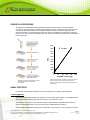
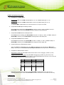
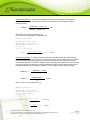
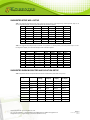
EPIGENTEK Complete Solutions for Epigenetics EpiQuik™ 8-OHdG DNA Damage Quantification Direct Kit (Fluorometric) Base Catalog # P-6004 PLEASE READ THIS ENTIRE USER GUIDE BEFORE USE Uses: The EpiQuik™ 8-OHdG DNA Damage Quantification Direct Kit (Fluorometric) is suitable for detecting oxidative DNA damage (8-OHdG) status using DNA isolated from any species such as mammals, plants, fungi, bacteria, and viruses in a variety of forms including, but not limited to, cultured cells, fresh and frozen tissues, paraffin-embedded tissues, and body fluid samples. Input DNA: The amount of DNA for each assay can be 100 ng to 300 ng. For optimal quantification, the input DNA amount should be 300 ng, as basal 8-OHdG is generally less than 0.01% of total DNA. Starting Material: Starting materials can include various tissue or cell samples such as cells from flask or microplate cultured cells, fresh and frozen tissues, paraffin-embedded tissues, blood, body fluid samples, etc. Internal Control: Both negative and positive DNA controls are provided in this kit. A standard curve can be performed (range: 5 to 200 pg of 8-OHdG) or a single quantity of 8-OHdG can be used as a positive control. Because 8-OHdG content can vary from tissue to tissue, and from normal and diseased states, or vary under treated and untreated conditions, it is advised to run replicate samples to ensure that the signal generated is validated. This kit will allow the user to quantify an absolute amount of 8-OHdG and determine the relative 8-OHdG states of two different DNA samples. Precautions: To avoid cross-contamination, carefully pipette the sample or solution into the strip wells. Use aerosol-barrier pipette tips and always change pipette tips between liquid transfers. Wear gloves throughout the entire procedure. In case of contact between gloves and sample, change gloves immediately. 110 Bi County Blvd. Ste. 122, Farmingdale, NY 11735 Tel: 1-877-374-4368 ■ Fax: 1-718-484-3956 ■ E-mail: [email protected] ■ Web: www.epigentek.com © Epigentek Group Inc. All rights reserved. Products are for research use only. Page 1 Printed 2014-09-23 P-6004 EPIGENTEK Complete Solutions for Epigenetics KIT CONTENTS Component 48 Assays Cat. #P-6004-48 96 Assays Cat. #P-6004-96 Storage Upon Receipt WB (10X Wash Buffer) 14 ml 28 ml 4°C BS (Binding Solution) 5 ml 10 ml RT NC (Negative Control , 10 µg/ml)* 10 µl 20 µl –20°C PC (Positive Control, 8-OHdG 1 µg/ml)* 10 µl 20 µl –20°C CA (Capture Antibody, 100 X) * 28 µl 55 µl 4°C DA (Detection Antibody, 1000 X)* 6 µl 12 µl –20°C ES (Enhancer Solution)* 5 µl 10 µl –20°C FD (Fluoro-Developer)* 10 µl 20 µl –20°C FE (Fluoro-Enhancer)* 10 µl 20 µl 4°C DB (Dilution Buffer) 4 ml 8 ml RT 8-Well Assay Strips (With Frame) 6 12 4°C User Guide 1 1 RT * Spin the solution down to the bottom prior to use. Note: The NC Negative Control I is an oligos containing no 8-OHdG. The PC Positive Control is an 8-OHdG oligos and normalized to have 100% of 8-OHdG . SHIPPING & STORAGE The kit is shipped in two parts: the first part at ambient room temperature and the second part on frozen ice packs at 4°C. Upon receipt: (1) Store NC, PC, DA, ES and FD at –20°C away from light; (2) Store WB, CA, DS, FE and 8-Well Assay Strips at 4°C away from light; (3) Store remaining components (BS and DB) at room temperature away from light. Note: Check if wash buffer, WB, contains salt precipitates before using. If so, warm (at room temperature or 37°C) and shake the buffer until the salts are re-dissolved. All components of the kit are stable for 6 months from the date of shipment, when stored properly. MATERIALS REQUIRED BUT NOT SUPPLIED Adjustable pipette Aerosol resistant pipette tips Microplate reader capable of reading fluorescence 530ex/590em 1.5 ml microcentrifuge tubes Incubator for 37°C incubation Plate seal or Parafilm M 110 Bi County Blvd. Ste. 122, Farmingdale, NY 11735 Tel: 1-877-374-4368 ■ Fax: 1-718-484-3956 ■ E-mail: [email protected] ■ Web: www.epigentek.com © Epigentek Group Inc. All rights reserved. Products are for research use only. Page 2 Printed 2014-09-23 P-6004 EPIGENTEK Complete Solutions for Epigenetics Distilled water 1X TE buffer pH 7.5 to 8.0 Isolated DNA of interest GENERAL PRODUCT INFORMATION Quality Control: Each lot of the EpiQuik™ 8-OHdG DNA Damage Quantification Direct Kit (Fluorometric) is tested against predetermined specifications to ensure consistent product quality. Epigentek guarantees the performance of all products in the manner described in our product instructions. Product Warranty: If this product does not meet your expectations, simply contact our technical support unit or your regional distributor. We also encourage you to contact us if you have any suggestions about product performance or new applications and techniques. Safety: Suitable lab coat, disposable gloves, and proper eye protection are required when working with this product. Product Updates: Epigentek reserves the right to change or modify any product to enhance its performance and design. The information in this User Guide is subject to change at any time without notice. Thus, only use the User Guide that was supplied with the kit when using that kit. Usage Limitation: The EpiQuik™ 8-OHdG DNA Damage Quantification Direct Kit (Fluorometric) is for research use only and is not intended for diagnostic or therapeutic application. Intellectual Property: The EpiQuik™ 8-OHdG DNA Damage Quantification Direct Kit (Fluorometric) and methods of use contain proprietary technologies by Epigentek. A BRIEF OVERVIEW 8-hydroxy-2'-deoxyguanosineis (8-OHdG or 8-oxo-dG) is an oxidized derivative of deoxyguanosine and is generated by hydroxyl radicals, singlet oxygen, and one-electron oxidants in cellular DNA. As a modified nucleoside base, 8-OHdG is considered important not only because of its abundance but also because of its mutagenic potential through G-to-T transversion mutations upon replication of DNA. 8OHdG also participates in epigenetic regulation of gene activation/repression by inhibiting the binding affinity of MBD proteins to the CpG sites of DNA. It is generally accepted that oxidatively damaged DNA can be repaired by base excision repair (BER) enzymes. Currently, 8-OHdG is widely accepted as a sensitive marker of oxidative DNA damage and oxidative stress. Evidence shows that increased levels of 8-OHdG are closely correlated with exposure to harmful environmental factors such as ionizing radiation, industrial chemicals, air pollution, cigarette smoking, and cancer chemotherapy. It has also been demonstrated that increased concentrations of 8-OHdG are pathogenically linked to a variety of age-associated diseases including cancer, coronary heart disease, diabetes and neurodegenerative diseases such as Alzheimer's disease. Compared with the urine 8-OHdG assay that mainly reflects the balance between oxidative damage and the repair rate of the whole body, directly quantifying the 8-OHdG content in different cells/tissues in normal and disease states would allow tissue-specific oxidative damage of DNA to be identified. Therefore, more 110 Bi County Blvd. Ste. 122, Farmingdale, NY 11735 Tel: 1-877-374-4368 ■ Fax: 1-718-484-3956 ■ E-mail: [email protected] ■ Web: www.epigentek.com © Epigentek Group Inc. All rights reserved. Products are for research use only. Page 3 Printed 2014-09-23 P-6004 EPIGENTEK Complete Solutions for Epigenetics useful information for the better understanding of oxidative damage-disease relationships, which benefits diagnostics and therapeutics of the disease, can be obtained. Several chromatography-based techniques such as HPLC-ECD and LC-MS are used for detecting 8OHdG in tissues and cells. However these methods are time consuming and have low throughput with high costs. The currently used competitive ELISA methods are also not conveniently applicable for cell/tissue 8-OHdG detection because they are less accurate and have an inability to use intact DNA isolated from cells or tissues directly. To address these problems, Epigentek offers the EpiQuik™ 8OHdG DNA Damage Quantification Direct Kit (Fluorometric) which uses a unique procedure to directly quantify 8-OHdG in cells/tissues. The kit has the following advantages and features: Fluorormetric assay with easy-to-follow steps for convenience and speed. The entire procedure can be completed within 3 hours and 45 minutes. High sensitivity, of which the detection limit can be as low as 1 pg of 8-OHdG. High specificity by detecting only 8-OHdG without cross-reactivity to 8-OHdG analogues such as dG, guanine, 8-OHGua and 8-OHG within the indicated concentration range of the sample DNA. Direct detection of 8-OHdG using intact DNA, which eliminates interference from high molecular weight compounds such as carbohydrates and proteins that are often seen in competitive 8-OHdG assays. Detection accuracy is highly correlated with and close to HPLC or LC-MS analysis. Highly convenient assay with direct use of DNA isolated from cells or tissues, no need for DNA digestion or hydrolysis. Universal positive and negative controls are included, which are suitable for quantifying 8-OHdG from any species. Strip-well microplate format makes the assay flexible for manual or high throughput analysis. Simple, reliable, and consistent assay conditions 110 Bi County Blvd. Ste. 122, Farmingdale, NY 11735 Tel: 1-877-374-4368 ■ Fax: 1-718-484-3956 ■ E-mail: [email protected] ■ Web: www.epigentek.com © Epigentek Group Inc. All rights reserved. Products are for research use only. Page 4 Printed 2014-09-23 P-6004 EPIGENTEK Complete Solutions for Epigenetics PRINCIPLE & PROCEDURE The EpiQuik™ 8-OHdG DNA Damage Quantification Direct Kit (Fluorometric) contains all reagents necessary for the quantification of Oxidative DNA damage (8-OHdG). In this assay, DNA is bound to strip wells that are specifically treated to have a high DNA affinity. 8-OHdG is detected using capture and detection antibodies. The detected signal is enhanced and then quantified fluorometrically by reading the fluorescence in a fluorescence microplate reader. The amount of 8-OHdG is proportional to the fluorescence intensity measured. 10000 R2 = 0.9968 9000 8000 7000 RFU 6000 5000 4000 3000 2000 1000 0 0 50 100 150 200 250 8-OHdG control (pg) Schematic procedure of the EpiQuik™ 8-OHdG DNA Damage Quantification Direct Kit (Fluorometric) 8-OHdG standard control was added into the assay wells at different concentrations and then measured with the EpiQuik™ 8-OHdG DNA Damage Quantification Direct Kit (Fluorometric). ASSAY PROTOCOL For the best results, please read the protocol in its entirety prior to starting your experiment. 1. Starting Materials Input DNA Amount: DNA amount can range from 100 ng to 300 ng per reaction. An optimal amount is 300 ng per reaction. Starting DNA may be in water or in a buffer such as TE. DNA Isolation: You can use your method of choice for DNA isolation. Epigentek offers a series of genomic DNA isolation kits for your convenience (see “Related Products” section). DNA Storage: Isolated genomic DNA can be stored at 4°C (short term) or –20°C (long term) until use. 110 Bi County Blvd. Ste. 122, Farmingdale, NY 11735 Tel: 1-877-374-4368 ■ Fax: 1-718-484-3956 ■ E-mail: [email protected] ■ Web: www.epigentek.com © Epigentek Group Inc. All rights reserved. Products are for research use only. Page 5 Printed 2014-09-23 P-6004 EPIGENTEK Complete Solutions for Epigenetics 2. Buffer and Solution Preparation a. Preparation of 1X Wash Buffer: 48-Assay Kit: Add 13 ml of WB 10X Wash Buffer to 117 ml of distilled water (pH 7.2-7.5). 96-Assay Kit: Add 26 ml of WB10X Wash Buffer to 234 ml of distilled water (pH 7.2-7.5). This Diluted WB 1X Wash Buffer can now be stored at 4°C for up to six months. b. Prepare Diluted CA Capture Antibody Solution: Dilute CA (Capture Antibody) with Diluted WB at a ratio of 1:100 (i.e., add 1 µl of CA to 100 µl of Diluted WB ). About 50 µl of this Diluted CA will be required for each assay well. c. Prepare Diluted DA Detection Antibody Solution: Dilute DA (Detection Antibody) with Diluted WB1 at a ratio of 1:1000 (i.e., add 1 µl of DA to 1000 µl of Diluted WB). About 50 µl of this Diluted DA will be required for each assay well. d. Prepare Diluted ES Enhancer Solution: Dilute ES Enhancer Solution with Diluted WB at a ratio of 1:5000 (i.e., add 1 µl of ES to 5000 µl of Diluted WB). About 50 µl of this Diluted ES will be required for each assay well. e. Prepare Fluorescence Development Solution: Add 1 µl of FD (Fluoro Developer) and 1 µl of FE (Fluoro Enhancer) to every 500 µl of DB (Dilution Buffer). About 50 µl of this Fluorescence Development Solution will be required for each assay well f. Preparation of Diluted Positive Control: Single Point Control Prep: Dilute PC Positive Control with 1X TE to 100 pg/µl (1 µl PC + 9 µl TE). Suggested Standard Curve Prep: First, dilute PC to 200 pg/µl (2 µl of PC + 8 µl of 1X TE). Then, further prepare 6 different concentrations with the 1 ng/µl diluted PC and 1X TE into 5, 10, 20, 50, 100 and 200 pg/µl according to the following dilution chart: Resulting PC Concentration Tube PC (200 pg/µl) 1X TE 1 1.0 µl 39.0 µl 5 pg/µl 2 1.0 µl 19.0 µl 10 pg/µl 3 1.0 µl 9.0 µl 20 pg/µl 4 1.0 µl 3.0 µl 50 pg/µl 5 2.0 µl 2.0 µl 100 pg/µl 6 3.0 µl 0.0 µl 200 pg/µl Note: Keep each of the diluted solutions (except Diluted WB 1X Wash Buffer) on ice until use. Any remaining diluted solutions, other than Diluted WB, should be discarded if not used within the same day. 3. DNA Binding 110 Bi County Blvd. Ste. 122, Farmingdale, NY 11735 Tel: 1-877-374-4368 ■ Fax: 1-718-484-3956 ■ E-mail: [email protected] ■ Web: www.epigentek.com © Epigentek Group Inc. All rights reserved. Products are for research use only. Page 6 Printed 2014-09-23 P-6004 EPIGENTEK Complete Solutions for Epigenetics a. Predetermine the number of strip wells required for your experiment. Carefully remove un-needed strip wells from the plate frame and place them back in the bag (seal the bag tightly and store at 4°C). b. Add 80 µl of BS Binding Solution to each well. c. Add 1 µl of NC, 1 µl of Diluted PC (see note below), and 300 ng of your Sample DNA (1-8 µl) into the designated wells depicted in Table 1 or Table 2. Mix solution by gently tilting from side to side or shaking the plate several times. Ensure the solution coats the bottom of the well evenly. Note: (1) For a single point control, add 1 µl of PC at a concentration of 100 pg/µl as prepared in Step 2f; For the standard curve, add 1 µl of Diluted PC at concentrations of 5 to 200 pg/µl (see the chart in Step 2). The final amounts should be 5, 10, 20, 50, 100 and 200 pg per well. (2) For optimal binding, sample DNA volume added should not exceed 8 µl. d. Cover strip plate with plate seal or Parafilm M and incubate at 37°C for 90 min. e. Remove the BS Binding Solution from each well. Wash each well with 150 µl of the Diluted WB 1X Wash Buffer each time for three times. 4. 8-OHdG DNA Capture a. Add 50 µl of the Diluted CA to each well, then cover and incubate at room temperature for 60 min. b. Remove the Diluted CA solution from each well. c. Wash each well with 150 µl of the Diluted WB each time for three times. d. Add 50 µl of the Diluted DA to each well, then cover and incubate at room temperature for 30 min. e. Remove the Diluted DA solution from each well. f. Wash each well with 150 µl of the Diluted WB each time for four times. g. Add 50 µl of the Diluted ES to each well, then cover and incubate at room temperature for 30 min. h. Remove the Diluted ES solution from each well. i. Wash each well with 150 µl of the Diluted WB each time for five times. 5. Signal Detection a. Add 50 µl of Fluorescence Development Solution to each well and incubate at room temperature for 2 to 4 min away from direct light. The Fluorescence Development Solution will turn pink in the presence of sufficient 8-OHdG products. b. Read the fluorescence on a fluorescence microplate reader within 2 to 10 min at 530ex/590em nm. Note: If the strip-well microplate frame does not fit in the fluorescence microplate reader, transfer the solution to a standard 96-well microplate. 6. 8-OHdG Calculation 110 Bi County Blvd. Ste. 122, Farmingdale, NY 11735 Tel: 1-877-374-4368 ■ Fax: 1-718-484-3956 ■ E-mail: [email protected] ■ Web: www.epigentek.com © Epigentek Group Inc. All rights reserved. Products are for research use only. Page 7 Printed 2014-09-23 P-6004 EPIGENTEK Complete Solutions for Epigenetics Relative Quantification: To determine the relative 8-OhdG status of two different DNA samples, a simple calculation for the percentage of 8-OHdG in your total DNA can be carried out using the following formula: (Sample RFU – NC RFU) ÷ S x 100% 8-OHdG % = (PC RFU – NC RFU) ÷ P S is the amount of input sample DNA in ng. P is the amount of input positive control (PC) in ng. Example calculation: Average RFU Average RFU Average RFU S is 300 ng P is 0.1 ng of NC is 1500 of PC is 5500 of Sample is 2100 (100 pg) (2100 – 1500) ÷ 300 8-OHdG % = (5500 – 1500) ÷ 0.1 x 100% = 0.005% Absolute Quantification: To quantify the absolute amount of 8-OHdG using an accurate calculation, first generate a standard curve and plot the OD values versus the amount of PC at each concentration point. Next, determine the slope (OD/ng) of the standard curve using linear regression (Microsoft Excel’s linear regression functions are suitable for such calculation) and also determine the most linear part (include at least 4 concentration points) of the standard curve for optimal slope calculation. Now calculate the amount and percentage of 8-OHdG in your total DNA using the following formulas: Sample RFU – NC RFU 8-OHdG (ng) = Slope 8-OHdG Amount (ng) S 8-OHdG % = x 100% S is the amount of input sample DNA in ng. Example calculation: Average RFU of NC is 1500 Average RFU of sample is 2100 Slope is 40000 RFU/ng S is 300 ng 8-OHdG (ng) = 2100 – 1500 40000 = 0.015 ng 0.015 8-OHdG % = 300 x 100% = 0.005% 110 Bi County Blvd. Ste. 122, Farmingdale, NY 11735 Tel: 1-877-374-4368 ■ Fax: 1-718-484-3956 ■ E-mail: [email protected] ■ Web: www.epigentek.com © Epigentek Group Inc. All rights reserved. Products are for research use only. Page 8 Printed 2014-09-23 P-6004 EPIGENTEK Complete Solutions for Epigenetics SUGGESTED STRIP WELL SETUP Table 1. The suggested strip-well plate setup using a single point positive control in a 48-assay format (in a 96-assay format, Strips 7 to 12 can be configured as Sample). The controls and samples can be measured in duplicate. Well # A B C D E F G H Strip 1 NC PC Sample Sample Sample Sample Sample Sample Strip 2 NC PC Sample Sample Sample Sample Sample Sample Strip 3 Sample Sample Sample Sample Sample Sample Sample Sample Strip 4 Sample Sample Sample Sample Sample Sample Sample Sample Strip 5 Sample Sample Sample Sample Sample Sample Sample Sample Strip 6 Sample Sample Sample Sample Sample Sample Sample Sample Table 2. The suggested strip-well plate setup for standard curve preparation in a 48-assay format (in a 96-assay format, Strips 7 to 12 can be configured as Sample). The controls and samples can be measured in duplicate. Well # A B C D E F G H Strip 1 NC PC 5 pg/µl PC10 pg/µl PC 20 pg/µl PC 50 pg/µl PC 100 pg/µl PC 200 pg/µl Sample Strip 2 NC PC 5 pg/µl PC10 pg/µl PC 20 pg/µl PC 50 pg/µl PC 100 pg/µl PC 200 pg/µl Sample Strip 3 Sample Sample Sample Sample Sample Sample Sample Sample Strip 4 Sample Sample Sample Sample Sample Sample Sample Sample Strip 5 Sample Sample Sample Sample Sample Sample Sample Sample Strip 6 Sample Sample Sample Sample Sample Sample Sample Sample SUGGESTED WORKING BUFFER AND SOLUTION SETUP Table 3. Approximate amount of required buffers and solutions for defined assay wells based on the protocol. Reagents 1 well 8 wells (1 strip) 16 wells (2 strips) 48 wells (6 strips) 96 wells (12 strips) Diluted WB 2.5 ml 20 ml 40 ml 120 ml 240 ml BS 80 µl 640 µl 1300 µl 3900 µl 8000 µl Diluted CA 50 µl 400 µl 800 µl 2400 µl 4800 µl Diluted DA 50 µl 400 µl 800 µl 2400 µl 4800 µl Diluted ES 50 µl 400 µl 800 µl 2400 µl 4800 µl DS 0.1 ml 0.8 ml 1.6 ml 4.8 ml 9.6 ml SS 0.1 ml 0.8 ml 1.6 ml 4.8 ml 9.6 ml NC N/A 0.5 µl – 1 µl 0.5 µl – 2 µl 1 µl – 4 µl 2 µl – 8 µl PC N/A 0.5 µl – 1 µl 0.5 µl – 2 µl 1 µl – 4 µl 2 µl – 8 µl 110 Bi County Blvd. Ste. 122, Farmingdale, NY 11735 Tel: 1-877-374-4368 ■ Fax: 1-718-484-3956 ■ E-mail: [email protected] ■ Web: www.epigentek.com © Epigentek Group Inc. All rights reserved. Products are for research use only. Page 9 Printed 2014-09-23 P-6004 EPIGENTEK Complete Solutions for Epigenetics TROUBLESHOOTING Problem Possible Cause Suggestion No signal in both the positive control and sample wells Reagents are added incorrectly. Check if reagents are added in the proper order and if any steps in the protocol may have been omitted by mistake. The well is incorrectly washed before DNA binding. Ensure the well is not washed prior to adding the positive control and sample. The bottom of the well is not completely covered by the BS Binding Solution. Ensure the solution coats the bottom of the well by gently tilting from side to side or shaking the plate several times. Incubation time and temperature are incorrect. Ensure the incubation time and temperature described in the protocol is followed correctly. Insufficient input materials. Ensure that a sufficient amount of positive control (> 50 pg) and sample (300 ng) is added into the wells. Incorrect fluorescence reading. Check if the appropriate fluorescence filters (530ex/590em) are used. Kit was not stored or handled properly. Ensure all components of the kit were stored at the appropriate temperature and the cap is tightly secured after each opening or use. The positive control is insufficiently added to the well in Step 3c. Ensure a sufficient amount of positive control DNA is added. The PC Positive Control is degraded due to improper storage conditions. Follow the Shipping & Storage guidelines of this User Guide for storage of PC Positive Control. Insufficient washing of wells. Check if washing recommendations at each step are performed according to the protocol. Contaminated by sample or positive control. Ensure the well is not contaminated by the sample or positive control DNA or from the use of contaminated tips. Incubation time is too long. The incubation time should not exceed 2 h at Step 3d, and not exceed 45 min at step 4d. Over development of fluorescence. Decrease the development time in Step 4a. Also RFU can be re-measured after diluting the solution already fluorescent in the wells with PBS at 1:5 or 1:10 ratio. The RFU ratio remains unchanged in the designated assay wells. No signal or weak signal in only the positive control wells High background present in the negative control wells or RFU in some of standard and sample wells is out of the fluorescent reading range. 110 Bi County Blvd. Ste. 122, Farmingdale, NY 11735 Tel: 1-877-374-4368 ■ Fax: 1-718-484-3956 ■ E-mail: [email protected] ■ Web: www.epigentek.com © Epigentek Group Inc. All rights reserved. Products are for research use only. Page 10 Printed 2014-09-23 P-6004 EPIGENTEK Complete Solutions for Epigenetics DNA sample is not properly extracted or purified. Ensure the DNA sample is in good quality. 260/280 ratio should be >1.7 with no or minimal RNA contamination No signal or weak signal only in sample wells Sample amount added into the wells is insufficient. Ensure a sufficient amount of DNA is used as indicated in Step 3. Little or no 8-OHdG contained in the sample. N/A Uneven fluorescent development Delayed fluorescence development in the wells. Ensure fluorescence development solution is added sequentially and is consistent with the order you added the other reagents (e.g., from well A to well G or from well 1 to well 12). If using 12 or more wells, a multi-channel pipette should be used so that the addition of fluorescence development solution can be completed as quickly as possible (<20 seconds). RELATED PRODUCTS DNA Sample Preparation P-1003 FitAmp™ General Tissue Section DNA Isolation Kit P-1004 FitAmp™ Plasma/Serum DNA Isolation Kit P-1006 DNA Concentrator Kit P-1007 FitAmp™ Gel DNA Isolation Kit P-1009 FitAmp™ Paraffin Tissue Section DNA Isolation Kit P-1017 FitAmp™ Urine DNA Isolation Kit P-1018 FitAmp™ Blood and Cultured Cell DNA Extraction Kit Methylated and Hydroxymethylated DNA Quantification P-1034 MethylFlash™ Methylated DNA Quantification Kit (Colorimetric) P-1035 MethylFlash™ Methylated DNA Quantification Kit (Fluorometric) P-1036 MethylFlash™ Hydroxymethylated DNA Quantification Kit (Colorimetric) P-1037 MethylFlash™ Hydroxymethylated DNA Quantification Kit (Fluorometric) Hydroxymethylated DNA Immunoprecipitation P-1038 MethylFlash™ Hydroxymethylated DNA Immunoprecipitation (hmeDIP) Kit DNA Damage and Repair OP-0001 EpiQuik™ Superoxide Dismutase Activity/Inhibition Assay Kit (Colorimetric) OP-0004 CytoX-Red Cell Proliferation/Cytotoxicity Assay Kit OP-0005 CytoX-Violet Cell Proliferation/Cytotoxicity Assay Kit P-6003 EpiQuik™ 8-OHdG DNA Damage Quantification Direct Kit (Colorimetric) P-6001 EpiQuik™ In Situ DNA Damage Assay Kit DNA Damage and Repair Antibodies (See http://www.epigentek.com/catalog/dna-damage-repair-c-35_70.html) 110 Bi County Blvd. Ste. 122, Farmingdale, NY 11735 Tel: 1-877-374-4368 ■ Fax: 1-718-484-3956 ■ E-mail: [email protected] ■ Web: www.epigentek.com © Epigentek Group Inc. All rights reserved. Products are for research use only. Page 11 Printed 2014-09-23 P-6004