1
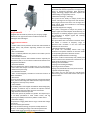
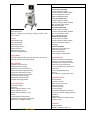
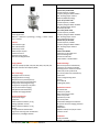
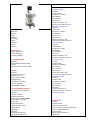
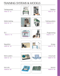
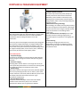
DOPPLER ULTRASOUND EQUIPMENT
MODEL 3300
Cost-Effective Ultrasound Imaging Solution for OB/GYN
With experienced insight into ultrasound industry, Apogee 3300,
the best choice of ultrasound system for medical ultrasound
practitioners who are seeking for balance of cost and
performance
As the entry level of Color Doppler ultrasound system integrated
with the latest imaging technologies, the Apogee 3300 provides
the configuration of a 17 inch medical LCD monitor, an 8 inch
touch screen and 4 probe connectors(3 active&1 docking), which
can support multiple probes, such as convex, linear, endocavity,
micro convex, 4D volumetric and bi-plane probes.
Value-added options:
1, Compound Imaging
The system has the ability to scan the target by multi-direction
beam forming thus easing echo artifacts and improving spatial
resolution
2, Panoramic Imaging
Extending wider view for doctors to scan large area tissue, the
system particularly allows doctors to monitor the scanning quality
via simultaneous display of B mode/ panoramic mode
3, 3D/4D Imaging
With built-in software, 3D images can be easily achieved together
with various processing functions such as rotation, zoom in/out,
and trim. Moreover, 4D volume probes enable the system to realtime display volumetric information of fetus or organ
conveniently and efficiently.
4, Auto IMT Measurement
The system automatically helps measure the Intima-Media
Thickness of the carotid artery wall, so as to evaluate
cardiovascular diseases such as hypertension diabetes
Distributed by DELTASYSTECH Romania
Quality Imaging Technology
1, Adaptive Speckle Reduction
The system may automatically track, identify and
intensify useful tissue-characteristic information.
Meanwhile, noise is filtered to increase S/N ratio,
enabling clearer tissue boundary and more obvious
image gradation, which is easy for distinguishing earlystage lesion tissues.
2, Multi-beam Forming Technology
This technology can multiply receiving and processing
scan lines of images from each element, which largely
increases the frame rate of images in B mode and 4D
mode and contributes to outstanding cardiac
performance.
3, The extended field of view displays more image
information without sacrificing image quality, a
convenient approach especially for scanning big-size
organs.
4, Linear Steering
By steering the imaging area of linear array,
sonographers can easily scan neighboring organs
without moving the probe, thus scanning becomes
more efficient easier.
MODEL 3500
Driving technology always brings changes for customers, so does
Apogee 3500 Omni
Equipped with extraordinary efforts by our company, Apogee
3500 Omni is the right choice with cost-effective benefits for
radiologists and cardiologists
Intelligent Clinical Solutions
1, TDI
It enables visible velocity indication of heart wall motion based on
Doppler effect, and provides supporting evidence for cardiac
diagnosis.
2, Built-in ECG Module
It helps to indentify different time phases in the cardiac cycle for
accurate diagnosis.
3, Auto IMT Measurement
It automatically measures Intima-Media Thickness especially for
carotid artery wall, so as to evaluate cardiovascular diseases such
as hypertension diabetes.
4, Anatomical M Mode
With free 360-degrees rotation sample lines, up to 3 line options,
it assists more exact analysis of cardiac structure movement even
in difficult heart positioning.
5, Tie Index Measurement
It offers echocardiography evaluation of ventricular function in
adults and children
6, Stress Echo
The stress echo package, including physical and pharmacological
stress, assists in observing how cardiac muscles respond to stress,
for diagnosis of coronary artery disease.
7, Color M Mode
By combining Color Flow Doppler with Motion Echocardiography,
it provides an effective way to evaluate the 2D/time relations
between cardiac flow and cardiac structure movements.
8, High-definition Phased Array Probe
It offers both options of phased array probes: low frequency(1.7
to 4.0 MHZ) specialized in adult cardiac exams, and hig
frequency(3.5-7.0MHz) exclusive for pediatric cardiac exams.
9, Panoramic Imaging
The panoramic imaging allows doctors to get a wide view of large
area tissues
10, Smart One Key Optimization
With only one button clicking, the system will help doctors to
achieve a reasonable good image in B mode and pulsed wave
Doppler mode, largely increasing the diagnosis efficiency.
Distributed by DELTASYSTECH Romania
Extraordinary Imaging Technology
We understand what the importance of image quality
means to ultrasound specialists, With pioneering
imaging technology, Apogee 3500 Omni explains the
understanding of image quality from us.
1, Multi-bean Forming T echnology
The system has the ability to multiply receive and
process scanning lines of images from each element,
which largely increases the frame rate of images in B
mode and 4D mode and contributes to outstanding
cardiac performance
2, Accurate Doppler Flow Imaging
The system is designed to analyze the position of
Doppler signals and make adaption simultaneously, for
the purpose of enhancing Doppler signals, increasing
the penetration of Doppler signals and reducing
Doppler artifacts.
3, Spectrum Compound Imaging
The system both emits and receives ultrasonic waves
and echoes in varieties of frequency range, to
guarantee both resolution in the near field and
penetration in the far field.
4, Broadband Harmonic Imaging
The system can successfully achieve both high
penetration and spatial resolution in Harmonic mode
by compounding varieties of Harmonic echoes.
5, Adaptive Speckle Reduction Technology
The system is able to automatically track, identify and
enhance useful tissue-characteristic information via 2
modes of SRT technology, as a result
Reducing noise and artifacts
Purifying tissue shading and edging
Improving contrast resolution
Helping early identification of tissue/structure lesion
6, Spatial Compound Imaging
The system can scan the target by multi-direction
beam forming thus to ease echo artifacts and improve
spatial resolutio
MODEL 3800
With the birth of the first volumetric probe in China, Our
company found itself determined to devote to women’s
healthcare
Apogee 3800 Touch is an extraordinary ultrasound imaging
system for women’s health, including obstetrics, gynecology,
maternal fetal medicine, assisted reproductive medicine and
breast medicine.
Exceptional 4D experience in OB/GYN
The system supports abdominal volumetric probes (resolution
priority/penetration priority) as well as a transvaginal volumetric
probe, to obtain 3D/4D images both in obstetrics and gynecology
application, providing more detailed and complete volumetric
information with multi-rendering modes, to assist the observation
and diagnosis of fetus wellness and uterus health.
Smart elastography for breast exams
The system supports high-definition linear probes with
elastography images to deliver stiffness information of tissues
with color code in real time and intelligent pressing guidance.
With the smart Elastography developed by our company, The
doctors can feel more convinced in early detection of breast
cancer.
Extraordinary Ultrasound Experience
1, High Clinical Productivity
Apogee 3800 Touch is facilitated with intelligent clinical functions
to provide user-friendly diagnostic experience.
2, Live Panoramic Imaging
Extending wider view for doctors to scan large area tissue, the
system particularly allows doctors to monitor the scanning quality
via simultaneous display of B mode/Panoramic mode. In addition,
when operating this function, doctors can erase the previous
image area and continue to generate better panoramic images.
3, Smart on key optimization
With one button pressed, the system smartly adjusts TGC and B
gain in B mode as well as base line, PRF and PW gain to obtain the
best B/W image and PW image for doctors.
4, Wireless image transmission to iPad/iPhone
This system offers doctors mobile working in the hospital and the
clinic via transmitting on-line ultrasound images to iPad/iPhone.
Doctors can access the on-scan images via iPad/iPhone even if
he/she is away from the ultrasound system.
Distributed by DELTASYSTECH Romania
A-one Imaging Technology
Apogee 3800 Touch inherits the proven imaging
technology of our color Doppler model, providing
outstanding image quality to support wellness of
mothers and babies.
1, Multi-beam Forming Technology
The technology largely increases the frame rate of
images in B mode, Color mode and 4D mode.
2, Wideband-beam Emission Technology
The technology remarkably eliminates artifacts and
guarantees high resolution in both near and far fields
of B mode.
3, Adaptive speckle Reduction Technology
The technology aims at reducing noise and artifacts,
purifying tissue shading and edging, improving
contrast resolution and helping early identification of
tissue/structure lesion
4, Spatial compound imaging
The technology helps to ease echo artifacts and
improve spatial resolution.
MODEL Q3
System Overview
Clinical Application
Abdomen - Obstetrics - Gynecology - Urology - Cardiac - Small
Parts - Vascular
Features
Task Indication Light
Built-in Workstation
Custom Keys Preset
Offline Analysis System
Probe Auto-Freeze
Optimization Key Preset
Real-time PIP Magnification
Imaging Modes
B, Dual B, Quad B, M, B/M, CFM, PDI, CDE, CDFI, PW, CW, THI
BW/Color Dual Real-Time Display Mode
Main Technology
DSC (Digital Scan Converter)
DBF (Digital Beam Forming)
RDA (Real-time Dynamic Aperture)
DRA (Dynamic Receiving Apodization)
DRF (Dynamic Receiving Focusing)
DFS (Dynamic Frequency Scan) 32Ch
THI (Tissue Harmonic Imaging)
SNR (Speckle Noise Reduction)
Freehand 3D Imaging Module
Physical Specification
Dimension & weight
Equipment:
530mm×650mm×1370mm; 47 kg
Package: (Wooden Carton)
680mm×710mm×1185mm; 72 kg
Monitor
15”High Definition & ResolutionProgressive-Scanning Color LCD Monitor
Up/Down Tilt, Left/Right Rotation
Probe Parameters
Convex Array Probe TC50
Central Frequency: 3.5MHz
Frequency Range:2.0MHz~6.0MHz
Max. Scanning Depth: 220mm
Abdomen/OB/GYN/Urology
Linear Array Probe TL40
Central Frequency: 7.5MHz
Frequency Range:5.0MHz~12.0MHz
Max. Scanning Depth: 90mm
Small Parts/Vascular
Intra-cavity Probe TC10
Central Frequency: 6.5MHz
Frequency Range:5.0MHz~10.0MHz
Max. Scanning Depth: 70mm
Trans-vaginal/Trans-rectal
Phased Array Probe TP16
Central Frequency: 3.0MHz
Frequency Range:2.0MHz~5.0MHz
Max. Scanning Depth: 200mm
Cardiology
Probe Port & Holder
Probe Ports: 3 Activated Ports
Automatic Recognition
Electronic Probe-shift
Probe Holders: 6 Probe Holders
Standard Package
Main Frame (with 15”LCD Monitor)
Customers can choose two probes
according to their need from Convex
Probe TC50, Linear probe TL40,
Intra-cavity Probe TC10, Phased Array
Probe TP16
Power cord/grounding cable
Fuse (2)
User’s Manual - Coupling Gel (0.25L)
Optional Accessories
17”LCD Monitor
Convex Array Probe
Micro Convex Array Probe
Linear Array Probe
Phased Array Probe
Intra-cavity Probe
Patient Data Management Software
THI (Tissue Harmonic Imaging) Software
DICOM 3.0
Pedal Switch
BW/Color Video/Laser/Ink-jet Printer
Puncture Trestle
Certificates
Quality Standards
ISO9001 - ISO13485 - CMD - CE
Distributed by DELTASYSTECH Romania
MODEL Q5
System Overview
Clinical Application
Abdomen - Obstetrics - Gynecology - Urology - Cardiac - Small
Parts - Vascular
Features
Task Indication Light
Built-in Workstation
Custom Keys Preset
Offline Analysis System
Probe Auto-Freeze
Optimization Key Preset
Real-time PIP Magnification
Imaging Modes
B, Dual B, Quad B, M, B/M, CFM, PDI, CDE, CDFI, PW, CW, THI
BW/Color Dual Real-Time Display Mode,
Main Technology
DSC (Digital Scan Converter)
DBF (Digital Beam Forming)
RDA (Real-time Dynamic Aperture)
DRA (Dynamic Receiving Apodization)
DRF (Dynamic Receiving Focusing)
DFS (Dynamic Frequency Scan) 32Ch
THI (Tissue Harmonic Imaging)
SNR (Speckle Noise Reduction)
Freehand 3D Imaging Module
Physical Specification
Dimension & weight
Equipment:
530mm×650mm×1370mm; 47 kg
Package: (Wooden Carton)
680mm×710mm×1185mm; 72 kg
Monitor
15”High Definition & ResolutionProgressive-Scanning Color LCD Monitor
Up/Down Tilt, Left/Right Rotation
Probe Parameters
Convex Array Probe TC50
Central Frequency: 3.5MHz
Frequency Range:2.0MHz~6.0MHz
Max. Scanning Depth: 220mm
Abdomen/OB/GYN/Urology
Linear Array Probe TL40
Central Frequency: 7.5MHz
Frequency Range:5.0MHz~12.0MHz
Max. Scanning Depth: 90mm
Small Parts/Vascular
Intra-cavity Probe TC10
Central Frequency: 6.5MHz
Frequency Range:5.0MHz~10.0MHz
Max. Scanning Depth: 70mm
Trans-vaginal/Trans-rectal
Phased Array Probe TP16
Central Frequency: 3.0MHz
Frequency Range:2.0MHz~5.0MHz
Max. Scanning Depth: 200mm
Cardiology
Probe Port & Holder
Probe Ports: 3 Activated Ports
Automatic Recognition
Electronic Probe-shift
Probe Holders: 6 Probe Holders
Standard Package
Main Frame (with 15”LCD Monitor)
Customers can choose two probes
according to their need from Convex
Probe TC50, Linear probe TL40,
Intra-cavity Probe TC10, PhasedArray
Probe TP16
Power cord/grounding cable
Fuse (2)
User’s Manual
Coupling Gel (0.25L)
Optional Accessories
17”LCD Monitor
Convex Array Probe
Micro Convex Array Probe
Linear Array Probe
Phased Array Probe
Intra-cavity Probe
Patient Data Management Software
THI (Tissue Harmonic Imaging) Software
DICOM 3.0
Pedal Switch
BW/Color Video/Laser/Ink-jet Printer
Puncture Trestle
Certificates
Quality Standards
ISO9001 - ISO13485 - CMD CE
Distributed by DELTASYSTECH Romania
MODEL Q6
System Overview
Application
Abdomen
Cardiac
Obstetrics
Gynecology
Urology
Small Parts
Vascular
Pediatrics
Electrical Power
Voltage: 100V~240V
Current: 7.0A/4.0A
Frequency: 50/60Hz
Physical Specifications
Equipment:
910mm×660mm×1470mm; 68kg
Package:
1035mm×725mm×1535mm; 128kg
Conditions
Operating
Temperature: 10°C~40°C
Humidity: 30%~75%
Pressure: 50kPa~106kPa
Shipping/Storage
Temperature: -15°C~50°C
Humidity: 10%~90%
Pressure: 50kPa~106kPa
Connectivity/Media/Peripherals
Transducer Ports: 3 (Activated)
USB Ports: 5
Hard Disc: ≥ 160GB
Footswitch: BNC/USB
Ethernet Port: 1 (10Mb/1000Mb)
External Display: DVI, VGA
External VCR
Digital Video Monitor
Compound Video Output
Barcode Reader
USB Modem
USB Printer
Peripherals/Storage Tray
Digital B/WThermal Printer
Transducers
Convex Array Probe
Frequency:
Central 3.3 MHz
Average Min. 3.15 MHz
Max. 3.85 MHz
Pitch: 0.468 mm
Radius: 60 mm
Number of Elements: 128
Geometric Focus Approx: 90 mm
Micro-Convex Array Probe:
Frequency:
Central 4 MHz
Average Min. 3.6 MHz
Max. 4.4 MHz
Pitch: 0.28 mm
Radius: 20 mm
Number of Elements: 128
Geometric Focus Approx: 90 mm
Linear Array Probe:
Frequency:
Central 10 MHz
Average Min. 7.2 MHz
Max. 8.8 MHz
Pitch: 0.390 mm
Radius: N/A
Number of Elements: 128
Geometric Focus Approx: 30 mm
Intra-Cavity Probe:
Frequency:
Central 6.5 MHz
Average Min. 5.85 MHz
Max. 7.15 MHz
Pitch: 0.205 mm
Radius: 10 mm
Number of Elements: 128
Geometric Focus Approx: 35 mm
Phased Array Probe:
Frequency:
Central 2.5 MHz
Average Min. 2.25 MHz
Max. 2.75 MHz
Pitch: 0.300 mm
Radius: N/A
Number of Elements: 64
Geometric Focus Approx: 30 mm
3D/4D Volume Probe
Imaging Modes
Overview
B, Dual B, Quad B
M, Color M, Anatomical M
Color, Dual Color, Simultaneous 2D/Color
Compound, Dual Compound
PW, CW, Triplex
CFM, PD, Directional PD, CD
Freehand 3D
Remote Diagnosis Module
DVD/CD Recorder
Image Storage
Storage Format:
PNG, AVI, MPEG, RAW(cine)
Export Video Format:
AVI, MPEG, DICOM
Export Image Format:
PNG, JPEG, BMP, GIF, DICOM
Cine Frame Capacity:
> 8000 frames
Stored Image Capacity:
> 50000 images
CD-R Storage
DVD Storage
Technology
TP-view Tech
Panoramic Imaging Tech
Speckle Reduction Tech
Dual & Triplex Synchronous Display
Elastography Tech
Directional Power Doppler
Real-time 3D Volume Imaging Tech
Expand-Pulse-Inversion Imaging Tech
Color Deflection Tech
Free-hand 3D Imaging Tech
Spatial-Compound Imaging Tech
Pulse-Inversion Harmonic Imaging Tech
Imaging Parameters Preset
Custom Key/Optimization Key
ECG (Optional)
ECG Module
ECG Lead
ECG Invert
ECG Baseline
ECG Gain
Embedded Computer
CPU:- Intel E4300 (Minimum)
Hard Drive (Patient) Data: - ≥ 500 GB
Hardware Specification
LCD Monitor
Size (Diagonal): 15”
Contrast Ratio: 1500:1
Resolution: 1024×768 pixels
Brightness: 250 cd/m2
Color Depth: 24bit
Rotate Angle: ±90°
Grey Levels: 256
Touch Screen
Size (Diagonal): 8.4”
Weight: 330g
Contrast Ratio: 600:1
Resolution: 1024×768 pixels
Brightness: 450 cd/m2
Standard 3D/4D, Advanced 3D/4D
Panoramic, Elastography
Elastography Comparative
Tissue Doppler Imaging (TDI)
Expand Pulse Imaging (EPI)
Combined Modes
Dual B, Quad B, B/M, B B/C, Dual B/C
B/PW, B/CW, B B/Power, Dual B/Power
Dual Compound, B/Elasto, B B/Elasto
B B/TDI, Dual B/TDI
B/C/M, B/C/PW, B/C/CW
B/Power/M, B/Power/PW, B/Power/CW
B/TDI/M, B/TDI/PW, B/TDI/CW
Standard 3D/4D Mode
Maps: 17
Line Density: 64~192
Rendering Quality: Low, Med, High
Frames per Volume: 3~125
Field of View (Max): 5°~75°
Viewing Options: A/B/C/VR, A/VR,
C/VR, V
Rendering Methods: Surface, SurfaceSmooth, Maximum, X-Ray
One Sweep 3D, 3D ROI, 3D Niche Mode,
Measurements, Sculpt, Annotations,
Contrast, Threshold
Advanced 3D/4D Mode
Maps: 17
Line Density: 64~192
Rendering Quality: Low, Med, High
Frames per Volume: 3~125
Field of View (Max): 5°~75°
Max Volume Rate: 3
Viewing Options: A/B/C/VR, A/VR,
C/VR, V
Rendering Methods: Surface, SurfaceSmooth, Maximum, X-Ray
Live 3D/4D imaging, One Sweep 3D,
Rendering 3D ROI, Multi-Slice,
3D Niche Mode, Measurements,
Sculpt, Annotations, Contrast, Threshold,
Save Cine-loops, Save Volume
Analysis Packages
OB
Cardiac
Abdominal
GYN
Urology
Vascular
Small Parts
Standard Package
Main Frame (with a 15”LCD Monitor)
Customers can select any 2 probes
according to their needs from Convex,
Intra-cavity, Linear and Phased Array
Probes.
Power cord/grounding cable
Fuse (2)
User’s Manual
Grey Levels: 64
Input Signal: LVDS
Compatible with bare hands/thin medical
gloves (e.g., latex, vinyl, nitrile)
Software Languages
English
German
Spanish
Italian
Czech
French
Russian
Simplified Chinese
Brazilian Portuguese
Imaging Performance
Startup Time (Max):
Avg. < 90 seconds
Preset Switching Time:
Avg. < 1 second
Storage Time (Image to Disk):
Avg. < 0.5 second
Distributed by DELTASYSTECH Romania
Coupling Gel (0.25L)
Optional Accessories
17”LCD Monitor
4D Imaging Module
Elastography Module
Convex Array Probe
Micro Convex Array Probe
Linear Array Probe
Phased Array Probe
4D Volume Probe
Intra-cavity Probe
DICOM 3.0
Pedal Switch
Laser, Ink-jet Printer
Puncture Trestle
Barcode Reader
ECG
UPS
Certificates
Quality Standards
ISO9001
ISO13485
CMD
CE Certificate
MODEL V3
Image Mode: B, 2B, 4B, M, B/M, CDFI, CDE, CCD, PW, CW.
Advanced Technology: 3D image supported.
One key image optimization: various of inner checking types/checking types edition and creation/making
superior image by pressing
of doctor according to different checking types.
English guide system: real time notice of operation steps, helping doctor get familiar with the operation of
device quickly.
Auto freezing protection of the probe: auto freeze the probe in the presetting time, lengthen the life of the
probe.
English report system: record the calculating parameters automatically, output English report directly; make the
inspection steps much
more efficient.
Expansion port: Video port, S-Video port, RS-232 port, USB2.0 port, VGA port, DICOM3.0 port
Probe supported
Convex probe, vaginal probe, linear probe, phrased array probe; micro convex probe
Distributed by DELTASYSTECH Romania