1
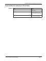
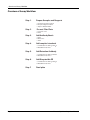
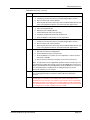
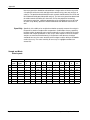
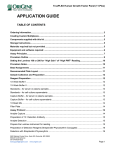
Procarta® Cytokine
Assay Kit
User Manual specifically for Cell
Culture Supernatant Samples
P/N 10482 Rev. A 061109
DRAFT June 11, 2009 10:31 pm
ProcartaCytokineTtile-Low_RES.fm
Affymetrix, Inc.
Procarta Cytokine Assay Kit User Manual
Copyright
© Copyright 2009, Affymetrix, Inc. All rights reserved.
Trademarks
Procarta is a registered trademark of Affymetrix, Inc. xMAP and Luminex are registered trademarks of the Luminex Corporation. Bio-Plex is a
registered trademark of Bio-Rad Laboratories.
Citing Procarta in Publications
When describing a procedure for publication using this product, we would appreciate it if you would refer to it as the Procarta® Cytokine Assay Kit.
If a paper cites a Procarta product and is published in a research journal, the lead author(s) may receive a travel stipend for use at a technology
conference or tradeshow by sending a copy of the paper to our technical support group at [email protected] or via fax at (510) 818-2610.
Disclaimer
Affymetrix, Inc. reserves the right to change its products and services at any time to incorporate technological developments. This manual is subject
to change without notice.
Although this manual has been prepared with every precaution to ensure accuracy, Affymetrix, Inc. assumes no liability for any errors or omissions,
nor for any damages resulting from the application or use of this information.
DRAFT June 11, 2009 10:31 pm
ProcartaCytokineTtile-Low_RES.fm
Contents
About the User Manual . . . . . . . . . . . . . . . . . . . . . . . . . . . . . . . . . . . . . . . . . . . . . . . . . 5
Who Should Read this Manual. . . . . . . . . . . . . . . . . . . . . . . . . . . . . . . . . . . . . . . 5
What this Manual Covers . . . . . . . . . . . . . . . . . . . . . . . . . . . . . . . . . . . . . . . . . . . 5
Safety Warnings and Precautions . . . . . . . . . . . . . . . . . . . . . . . . . . . . . . . . . . . . 5
Contacting Affymetrix . . . . . . . . . . . . . . . . . . . . . . . . . . . . . . . . . . . . . . . . . . . . . . 5
About the Procarta Cytokine Assay Kit . . . . . . . . . . . . . . . . . . . . . . . . . . . . . . . . . . . . . 6
Procarta Cytokine Assay Defined. . . . . . . . . . . . . . . . . . . . . . . . . . . . . . . . . . . . . 6
Available Kit Formats . . . . . . . . . . . . . . . . . . . . . . . . . . . . . . . . . . . . . . . . . . . . . . 6
Procarta Cytokine Assay Kit Contents and Handling Conditions . . . . . . . . . . . . . . . . . . 6
Kit Contents and Storage . . . . . . . . . . . . . . . . . . . . . . . . . . . . . . . . . . . . . . . . . . . 6
Kit Handling . . . . . . . . . . . . . . . . . . . . . . . . . . . . . . . . . . . . . . . . . . . . . . . . . . . . . 6
Required Materials and Equipment Not Provided . . . . . . . . . . . . . . . . . . . . . . . . . . . . . 7
Equipment . . . . . . . . . . . . . . . . . . . . . . . . . . . . . . . . . . . . . . . . . . . . . . . . . . . . . . 7
Overview of Assay Workflow . . . . . . . . . . . . . . . . . . . . . . . . . . . . . . . . . . . . . . . . . . . . . 8
Getting Started . . . . . . . . . . . . . . . . . . . . . . . . . . . . . . . . . . . . . . . . . . . . . . . . . . . . . . . . 9
Setting Up and Calibrating the Manifold. . . . . . . . . . . . . . . . . . . . . . . . . . . . . . . . 9
Operating the Manifold . . . . . . . . . . . . . . . . . . . . . . . . . . . . . . . . . . . . . . . . . . . . 10
Sample Preparation . . . . . . . . . . . . . . . . . . . . . . . . . . . . . . . . . . . . . . . . . . . . . . . . . . . .11
Preparing Samples. . . . . . . . . . . . . . . . . . . . . . . . . . . . . . . . . . . . . . . . . . . . . . . .11
Dilution for High Expression Cytokines . . . . . . . . . . . . . . . . . . . . . . . . . . . . . . . .11
Special Sample Preparation for TGF beta . . . . . . . . . . . . . . . . . . . . . . . . . . . . . .11
Standard Preparation . . . . . . . . . . . . . . . . . . . . . . . . . . . . . . . . . . . . . . . . . . . . . . . . . . 12
Options for Standard Curve . . . . . . . . . . . . . . . . . . . . . . . . . . . . . . . . . . . . . . . . 12
Option 1 . . . . . . . . . . . . . . . . . . . . . . . . . . . . . . . . . . . . . . . . . . . . . . . . . . . . . . . 12
Option 2 . . . . . . . . . . . . . . . . . . . . . . . . . . . . . . . . . . . . . . . . . . . . . . . . . . . . . . . 13
Performing the Assay. . . . . . . . . . . . . . . . . . . . . . . . . . . . . . . . . . . . . . . . . . . . . . . . . . 14
Troubleshooting . . . . . . . . . . . . . . . . . . . . . . . . . . . . . . . . . . . . . . . . . . . . . . . . . . . . . . 17
Possible Problems and Recommended Solutions . . . . . . . . . . . . . . . . . . . . . . . 17
Appendix . . . . . . . . . . . . . . . . . . . . . . . . . . . . . . . . . . . . . . . . . . . . . . . . . . . . . . . . . . . 19
Limit of Detection . . . . . . . . . . . . . . . . . . . . . . . . . . . . . . . . . . . . . . . . . . . . . . . . 19
Limit of Quantitation . . . . . . . . . . . . . . . . . . . . . . . . . . . . . . . . . . . . . . . . . . . . . . 19
Accuracy/ Recovery . . . . . . . . . . . . . . . . . . . . . . . . . . . . . . . . . . . . . . . . . . . . . . 19
Precision . . . . . . . . . . . . . . . . . . . . . . . . . . . . . . . . . . . . . . . . . . . . . . . . . . . . . . 19
Specificity . . . . . . . . . . . . . . . . . . . . . . . . . . . . . . . . . . . . . . . . . . . . . . . . . . . . . . 20
Sample and Blank Plate Layouts . . . . . . . . . . . . . . . . . . . . . . . . . . . . . . . . . . . . 20
Luminex Maintenance . . . . . . . . . . . . . . . . . . . . . . . . . . . . . . . . . . . . . . . . . . . . 21
Procarta Cytokine Assay User Manual
iii
About the User Manual
About the User Manual
Who Should Read Anyone that has purchased a Procarta® Cytokine Assay Kit from Affymetrix to
this Manual perform quantitative, multiplexed cytokine measurements from Cell Culture
Supernatant samples using the Luminex® Technology.
IMPORTANT Please note that we provide additional application specific manuals
for the Procarta assays for the following sample types:
♦
Serum/Plasma
♦
Tissue or Cell Lysates
♦
Bodily Fluids
Our most updated manuals can be obtained from our website at www.panomics.com
What this Manual This manual provides recommendations and step-by-step procedures for the
Covers following:
♦
Sample Procedure
♦
Standard Preparation
♦
Assay Procedure
♦
Troubleshooting
Safety Warnings ! WARNING ! Though the general procedure is very similar to other
and Precautions Luminex cytokine assays, there are subtle differences due to the
approach we have taken in developing the assay to have an improved
workflow. Please do not refer to other vendors’ manuals for performing
the assay. Please read the supplied User Manual and Product Insert
prior to starting the assay.
CAUTION All chemicals should be considered potentially hazardous. We recommend that
this product and its components be handled by those trained in laboratory techniques and be
used according to the principles of good laboratory practice.
CAUTION This kit contains small quantities of sodium azide. Sodium azide reacts with lead
and copper plumbing to form explosive metal azides. When disposing, flush drains with a large
volume of water to prevent azide accumulation. Observe all state and local regulations for
disposal.
Contacting For technical questions, please contact our technical support group by telephone at
Affymetrix 1-877-726-6642 option 3, or email at [email protected] (US and Canada).
In Europe, contact [email protected]. In Asia/Pacific, contact
[email protected]. For an updated list of FAQs and product support
literature, visit our website at www.panomics.com.
Procarta Cytokine Assay User Manual
Page 5
About the Procarta Cytokine Assay Kit
About the Procarta Cytokine Assay Kit
Procarta Cytokine The Procarta Cytokine assays are multiplex immunoassays based on xMAP®
Assay Defined detection technology developed by Luminex. This bead based multiplex assay kit
can quantitatively measure multiple cytokines from as little as 25 µL of Cell Culture
Supernatant samples in less than 4 hours. Procarta Cytokine kits can be used for up
to 6 months from the date of receipt.
Available Kit Procarta Cytokine Assay Kits are available as:
Formats ♦ Standard pre-mixed panels
♦
“By Request” user configured panels
Procarta Cytokine Assay Kits are available as single or ten plate 96-well formats and
contain all the reagents required to detect cytokines.
Procarta Cytokine Assay Kit Contents and Handling Conditions
Kit Contents and The Procarta Cytokine Assay Kit contains the following components. The kits are
Storage available as single plate or ten plate formats. Refer to the product insert for quantities
and details of components supplied.
Procarta Cytokine Kit components:
Component
Kit Handling ♦
Page 6
Storage
Reading Buffer
4°C
10X Wash Buffer
4°C
Detection Antibody, premixed
4°C
Antigen Standards, premixed, lyophilized
4°C
Streptavidin-PE (SAPE)
4°C
Antibody Beads
4°C
Filter Plate
4°C
Plate Seals
4°C
PCR 8 Strip Tube
4°C
Store the entire kit at 4°C
♦
Do not reuse or store resuspended antigen standards
♦
10X Wash Buffer and its 1X dilution can be stored at 4°C or room temperature for
up to 6 months
Procarta Cytokine Assay User Manual
Required Materials and Equipment Not Provided
Required Materials and Equipment Not Provided
Equipment
Item
Source
Vacuum filtration system
Millipore (P/N MAVM0960R and
WP6111560)
Microplate shaker
Labline model 4625 or
equivalent (must have 3 mm
orbit with ability to maintain 500
rpm)
Luminex or Luminex-based instrument
MiraiBio, Bio-Rad or other
Luminex instrument provider
Procarta Cytokine Assay User Manual
Page 7
Overview of Assay Workflow
Overview of Assay Workflow
Step 1.
Prepare Samples and Reagents
a. Collect and prepare samples
b. Prepare antigen standards
c. Prepare 1X Wash Buffer
Step 2.
Pre-wet Filter Plate
a. Let sit for 5 min
b. Filter
Step 3.
Add Antibody Beads
a. Filter
b. Wash once
c. Filter
Step 4.
Add samples/standards
a. Incubate 60 min shaking, 20-25 C
b. Wash/filter three times
Step 5.
Add Detection Antibody
a. Incubate 30 min shaking, 20-25 C
b. Wash/filter three times
Step 6.
Add Streptavidin-PE
a. Incubate 30 min shaking, 20-25 C
b. Wash/filter three times
Step 7.
Page 8
Read plate
Procarta Cytokine Assay User Manual
Getting Started
Getting Started
♦
Thoroughly read the Product Insert that is included with your kit. The
product insert includes the bead regions and may contain special
instructions for proper use of your “By Request” or custom panel.
♦
Turn on the Luminex machine and initiate the startup protocol. It takes 30
minutes for the laser to warm-up.
♦
Vortex the 10X Wash Buffer to dissolve all the salts. Mix 20 mL of the 10X Wash
Buffer with 180 mL deionized, sterile water. 1X Wash Buffer can be stored at 4 °C
or room temperature for up to 6 months.
♦
Ensure that the Millipore vacuum manifold has been properly setup and
calibrated.
♦
Change the pipet tips after every transfer and avoid creating bubbles when
pipetting.
♦
During the incubation steps, cover the Filter Plate with aluminum foil to prevent
photobleaching of the fluorescent beads.
♦
Be careful not to invert the plate or allow contents from one well to mix to another
well.
♦
We recommend using a multi-channel pipette whenever possible to reduce CVs.
♦
Bring all buffers to room temperature before setting up the assay. Store the
detection antibody, antibody beads, Streptavidin-PE, Samples, and reconstituted
standards on ice during the assay.
♦
When sealing the Filter plate with a Plate Seal, gently press your finger over the
Plate Seal. Too much pressure can cause force the fluid through the filter plate.
To avoid Filter Plate leakages, do not seal Filter Plates using a rubber roller (or
equivalent) as they apply significant pressure resulting in leakage.
♦
Seal all unused wells with an enclosed Plate Seal to ensure proper vacuum
pressure.
Setting Up and To set up and calibrate the manifold:
Calibrating the
Step
Action
Manifold
1
Set up the Filter Plate vacuum manifold as shown below. Follow the
manufacturer’s manual for details.
Procarta Cytokine Assay User Manual
Page 9
Getting Started
To set up and calibrate the manifold: (continued)
Step
2
Action
Calibrate the vacuum pressure using a used Filter Plate:
a. Place the Filter Plate on top of the manifold.
b. Turn on the vacuum.
c. Press down on all 4 corners of the Filter Plate to form a tight seal.
d. Adjust the pressure so that it takes 8-10 seconds to evacuate 200 μL of
wash buffer from the wells. If the vacuum is too high, beads can get
trapped or pulled through the filter.
e. Turn off vacuum as soon as the solution filters through the wells and
remove the plate from the manifold.
Operating the To operate the manifold:
Manifold
Step
Action
1
Check vacuum calibration periodically. As a general guideline, 200 μL of solution
should take approximately 8-10 seconds to clear the well of a Filter Plate.
2
For all filtration steps, turn on the vacuum pump, place the Filter Plate onto the
vacuum manifold and then filter the solution. Avoid splashing and
cross-contamination of wells during all wash steps.
IMPORTANT Do not allow the Filter Plates to air-dry following washes.
Immediately add the next component after each filtration step.
IMPORTANT We recommend performing all the wash steps next to the manifold
to minimize the amount of time that the beads are exposed to air.
3
Break the vacuum immediately after each solution has been completely filtered
from all wells (approximately 8-10 seconds).
Note
4
Page 10
Wells may filter at different rates.
Following the last wash in each series, blot the bottom of the Filter Plate
thoroughly with a paper towel to remove traces of 1X Wash Buffer. Avoid touching
the bottom of the Filter Plate with your fingers or to the bench during
manipulations. Immediately move to the next step to ensure that the beads are in
the appropriate buffered solution.
Procarta Cytokine Assay User Manual
Sample Preparation
Sample Preparation
Preparing Samples ♦
For optimal recovery and sensitivity, it is important to properly and consistently
prepare samples.
♦
Store all samples on ice after preparation and use immediately or aliquot and
store at –80°C. Avoid more than 2 freeze/thaw cycles.
♦
If you are using frozen samples, thaw completely on ice and mix well prior to use.
♦
Use fresh cell culture media (that is used to culture the experimental cells) to
dilute the standards and samples.
♦
A total volume of 50 µL/well of sample is needed and a minimum of 2 replicates
are recommended.
Dilution for High Sample dilution may be needed if the expression of those targets are outside the
Expression measurable range. If dilution is needed, use the cell culture media that was used to
Cytokines culture that particular cell line. However, most samples can be used directly in the
assay for all other available targets.
Special Sample TGF beta has a special protocol for sample preparation. The TGF beta assay can only
Preparation for detect the active form. The complexed form present must be acid treated and then
TGF beta neutralized converting the complexed form to the active form. The assay should be
processed as a single plex assay since the sample must be acid treated.
To access our most recent protocols, please visit our website at www.panomics.com.
Procarta Cytokine Assay User Manual
Page 11
Standard Preparation
Standard Preparation
Options for This section provides two different options for the creation of the standard curve.
Standard Curve Depending upon your application, we provide two instruction sets for creating an 8
point standard curve that either ranges from 40,000 pg/ml to 2.44 pg/ml or from
20,000 pg/ml to 1.22 pg/ml.
IMPORTANT Prior to preparing your standard curve, please thoroughly read your product
insert. Special instructions for preparation of the standard antigens for some “By Request”
panels may be included on your product insert.
Option 1 Option 1: 40,000 pg/ml to 2.44 pg/ml
Step
Action
1
Prior to preparing the standards, please ensure that the Luminex machine has
been turned on and calibrated. The laser will take 30 minutes to warm up.
2
Add 250 μL of cell culture media to the lyophilized antigen vial.
3
Vortex gently for 10 seconds and incubate on ice for 25 minutes. Transfer the entire
contents of the reconstituted antigen into the first tube of the PCR 8-Strip Tube
provided. Use of any other tube other than the Strip Tube provided may result in
poor standard recovery.
4
Prepare serial dilutions of the premixed standard as indicated below using the 8
Strip Tube provided:
a. Add 150 μL of the cell culture media to tubes 2-8. Follow the table below to
create the dilutions. Use a P200 pipette to transfer and mix the 50 μL of
standard from tube to tube.
b. Store on ice until ready to use. When transferring the standards to the filter
plate, we recommend using a multi-channel pipette.
Strip Tube
#
5
Page 12
Cell Culture
Media
(μL)
Antigen
Standard (μL)
Final Analyte
Concentration
in the Assay
Well (pg/mL)
1
—
250
40,000
2
150
50 from Tube 1
10,000
3
150
50 from Tube 2
2500
4
150
50 from Tube 3
625
5
150
50 from Tube 4
156
6
150
50 from Tube 5
39
7
150
50 from Tube 6
9.8
8
150
50 from Tube 7
2.44
Blank Well
50 μL per well
-
0
Proceed to Performing the Assay
Procarta Cytokine Assay User Manual
Standard Preparation
Option 2 Option 2: 20,000 pg/ml - 1.22 pg/ml
Step
Action
1
Prior to preparing the standards, please ensure that the Luminex machine has
been turned on and calibrated. The laser will take 30 minutes to warm up.
2
Add 500 μL of cell culture media to the lyophilized antigen vial.
3
Vortex gently for 10 seconds and incubate on ice for 25 minutes. Transfer 250 μL
to the first tube of the PCR 8-Strip Tube provided and store Strip Tubes on ice. Use
of any other tube other than the Strip Tube provided may result in poor standard
recovery.
4
Prepare serial dilutions of the premixed standard as indicated below using the 8
Strip Tube provided:
a. Add 150 μL of the cell culture media to tubes 2-8. Follow the table below to
create the dilutions. Use a P200 pipette to transfer and mix the 50 μL of
standard from tube to tube.
b. Store on ice until ready to use. When transferring the standards to the filter
plate, we recommend using a multi-channel pipette.
Strip Tube
#
5
Cell Culture
Media
(μL)
Antigen
Standard (μL)
Final Analyte
Concentration
in the Assay
Well (pg/mL)
1
—
250 from 500
20,000
2
150
50 from Tube 1
5000
3
150
50 from Tube 2
1250
4
150
50 from Tube 3
312.5
5
150
50 from Tube 4
78.12
6
150
50 from Tube 5
19.53
7
150
50 from Tube 6
4.88
8
150
50 from Tube 7
1.22
Blank Well
50 μL per well
-
0
Proceed to Performing the Assay
Procarta Cytokine Assay User Manual
Page 13
Performing the Assay
Performing the Assay
To perform the assay:
Step
Action
1
A blank layout is provided in the Appendix for recording your plate layout. Columns
1 and 2 are typically used for duplicate standards. Mark the sample and blank
wells you want to use.
2
Seal the un-used wells of the plate with a Plate Seal prior to starting the assay.
3
Pre-wet the Filter Plate:
a. Add 150 μL of Reading Buffer to each unsealed well.
b. Incubate 5 minutes at room temperature.
c. Remove the Reading Buffer with vacuum filtration. Reading Buffer should
clear wells within 8-10 seconds. If outside this range, see “Setting Up and
Calibrating the Manifold” on page 9.
IMPORTANT Do not invert the plate during the rest of this procedure.
4
Add the Antibody Beads:
a. Vortex the premixed Antibody Beads for 30 seconds at room temperature.
b. Add 50 μL of Antibody Beads to each well.
c. Remove buffer with vacuum filtration.
5
Wash the Antibody Beads:
a. Add 150 μL/well of 1X Wash Buffer and remove with vacuum filtration.
b. Blot the bottom of the Filter Plate thoroughly with paper towels to remove
residual buffer.
6
Add 50 μL/well of standards and samples as marked on the plate layout sheet. For
the blank samples add 50 μL/well of tissue culture media. Seal the plate gently
using a Plate Seal.
7
Incubate and wash the Filter Plate:
a. Completely wrap the Filter Plate with aluminum foil and shake at 500 rpm
for 60 minutes at room temperature. Optionally, if you decide to incubate
overnight, you can shake the plate at room temperature for 30 minutes,
transfer the plate to 4°C and store on a level surface. After overnight
incubation, remove the plate from 4°C and shake for 30 min at room
temperature prior to proceeding to the next step.
b. Carefully remove the Plate Seal to avoid splashing the plate contents.
c. Remove solution with vacuum filtration.
d. When washing the plate, we recommend using multi-channel pipette and a
large plastic reservoir for the wash buffer. We also recommend stationing
the wash buffer reservoir next to the manifold to minimize the amount of
time that the beads are exposed to air.
e. Wash the plate three times with 150 μL/well of 1X Wash Buffer. After the 3rd
wash, thoroughly blot the bottom of the Filter Plate with paper towels.
8
Add premixed Detection Antibodies:
a. Add 25 μL/well of the Detection Antibody.
b. Seal the Filter Plate with a new Plate Seal.
c. Wrap the Filter plate with aluminum foil and shake at 500 rpm for 30
minutes at room temperature.
Page 14
Procarta Cytokine Assay User Manual
Performing the Assay
To perform the assay: (continued)
Step
9
Action
Remove the solution with vacuum filtration and wash the plate:
a. Carefully remove the Plate Seal to avoid splashing the plate contents.
b. Remove solution with vacuum filtration.
c. Wash the plate three times with 150 μL/well of 1X Wash Buffer. After the 3rd
wash, thoroughly blot the bottom of the Filter Plate with paper towels.
10
Add Streptavidin–PE:
a. Vortex the Streptavidin–PE.
b. Add 50 μL/well of Streptavidin-PE.
c. Seal the Filter Plate with a new Plate Seal.
d. Completely wrap the Filter Plate with aluminum foil.
e. Shake at 500 rpm for 30 minutes at room temperature.
11
Remove the solution with vacuum filtration and wash the plate:
a. Carefully remove the Plate Seal to avoid splashing the plate contents.
b. Remove solution with vacuum filtration.
c. Wash the plate three times with 150 μL/well of 1X Wash Buffer. After the 3rd
wash, thoroughly blot the bottom of the Filter Plate with paper towels.
12
Prepare plate for analysis on a Luminex instrument:
a. Add 120 μL/well of the Reading Buffer.
b. Seal the Filter Plate with a new Plate Seal.
c. Shake at 500 rpm for 5 minutes at room temperature or until the Luminex
instrument is available.
d. Remove the Plate Seal before analyzing on the Luminex instrument.
Note The Filter Plate can be wrapped with aluminum foil and stored for up to 48
hours before proceeding. After storage, remove the Reading Buffer using vacuum
filtration and add 120 μL/well of Reading Buffer. Shake at 500 rpm for 5 minutes at
room temperature prior to reading. Delay in reading the plate may result in
decreased sensitivity for some analytes.
13
Please refer to your product insert when entering the bead analyte associations
into your designated Luminex acquisition software (Bio-Plex, MasterPlex,
xPonent).
14
Please refer to the previous section, “Standard Preparation” step 4, when
calculating the concentration of the samples and confirm which option for the
standard curve was used. The concentration of the samples can be calculated by
plotting the expected concentration of the standards against the MFI generated by
each standard. A 4PL or 5PL algorithm is recommended for the best curve fit. If 50
μL of sample was used, then no dilution factor is needed.
Procarta Cytokine Assay User Manual
Page 15
Performing the Assay
To perform the assay: (continued)
Step
15
Action
Analyze the plate following the respective operation manual for the Luminex or
Luminex-based instrument.
Software
Sample
Size
DD Gate
Timeout
Bead
Events/Be
ad Region
Luminex 100/
50 μL
7,500-15,000
25 sec
50 or 100
50 μL
4335-10,000
45 sec
50 or 100
50 μL
2,000-15,000
25 sec
50 or 100
50 μL
4,335-10,000
25 sec
50 or 100
IS100 v 2.318
Bioplex/
Bio-Rad
Miraibio/
Hitachi
Starstation/
ACS
16
When the Luminex beads are injected into the flow cell, a small percentage of the
beads will have a tendency to clump and go through the flow cell as doublets. The
DD Gate or Doublet Discriminator Gate will allow for discrimination of the doublets
from the singlet beads. When setting the DD Gate, you can follow the appropriate
settings from the table above. However, for the best results, you should adjust the
DD Gate and center the gates around the largest peak which is the singlet beads.
You can adjust the gates when processing the first sample.
The Bio-Plex® Suspension Array System allows calibration using Low or High
sensitivity settings. Perform the sensitivity selection during calibration, using
predetermined values of CAL2 RP1 target, as provided by Bio-Rad. Using the RP1
Low target value will provide results comparable to those obtained from the
Luminex 100. Using the RP1 High target value may increase detection sensitivity
for low cytokine protein concentrations but sacrifices part of the linear dynamic
range for high concentrations. We recommend RP1 Low target value for BioPlex.
The number of events per region can be 50 or 100. Please note that reading 100
events will take longer to read the entire plate.
IMPORTANT Check to ensure the probe height in the Luminex instrument is
adjusted appropriately for the Filter Plate.
IMPORTANT We recommend that you calibrate the Luminex or Luminex-based
instrument each day the assay is run.
IMPORTANT If there is a malfunction of the instrument or software during the
run, the plate can be re-processed on the Luminex machine. Remove the plate
from the instrument and remove the Reading Buffer by vacuum filtration.
Resuspend the beads in 120 μL of Reading Buffer and shake at 500 rpm for 5
minutes at room temperature. The plate may take longer to read since there will be
less beads in the plate.
Page 16
Procarta Cytokine Assay User Manual
Troubleshooting
Troubleshooting
Possible Problems
and
Recommended
Solutions
Observation
Possible Cause
Recommended Action
Filter plate Leakage or
Vacuum pressure too high
Adjust the vacuum pressure
as recommended in “Setting
Up and Calibrating the
Manifold” on page 9.
Filter Plate misaligned (at an
angle) during incubation or
processing steps
Set the Filter Plate on a flat,
level surface during
incubation/processing.
Leakage from capillary
action
After each vacuum step,
thoroughly blot the bottom
of the Filter Plate using
paper towels or absorbent
paper.
Too much force applied to
the Plate Seal
Apply a Plate Seal onto the
plate and gently press using
your index finger.
Filter plate is clogged
Samples clogging the filter
plate
Further dilute the samples
prior to adding to the plate.
High CVs
Samples and antigen
standards not stored on ice
Prepare the samples and
standards on ice before
setting up the assay.
Bottom of the Filter Plate is
not dry
After each vacuum step,
thoroughly blot the bottom
of the Filter Plate using
paper towels or absorbent
paper.
Contamination from
re-using the Plate Seal
Use a new Plate Seal for
each incubation step.
Incomplete washing
After adding the standards
and samples, it is very
important that any excess
standards are removed
during the wash step.
Contamination from Wash
Buffer
Be careful not to splash
Wash Buffer during wash
steps into adjacent wells.
Pipetting techniques
Using single channel
pipettes and poor pipette
techniques can cause high
CVs especially towards the
lower end of the dilution
curve.
Instrument calibrated at
high PMT settings
Calibrate the instrument
using the CAL2 Low RP1
target value.
Limited dynamic range
when using the BioPlex
software
Procarta Cytokine Assay User Manual
Page 17
Troubleshooting
Observation
Possible Cause
Recommended Action
Low bead count
Volume of bead solution is
too low
Add 120 μL/well Reading
Buffer and shake at 500 rpm
for 5 minutes at room
temperature to resuspend
beads before reading in the
Luminex instrument
High bead aggregation
Vortex the bead solution
well before using in the
assay and ensure that the
beads are properly mixed
during the incubation steps
Vacuum pressure too high
Confirm that it takes 8-10
seconds to evacuate 200 μL
of wash buffer
Filter ruptured due to
excess vacuum time
Do not use the vacuum over
10 seconds in any of the
steps
Dyes contained in the beads
are photo-bleached from
overexposure to light
Store bead solution and the
assay plate in the dark
Samples causing the
instrument to clog
May need to dilute the
sample or stop and shake
the plate in the middle of the
run
Probe height is incorrect
Refer to the Luminex
manual for proper
adjustment of the needle
height
Air bubble in the sample
loop
Refer to the Luminex
manual for proper removal
of the air bubble
Standards not reconstituted
and diluted correctly
Prepare fresh antigen
standards following the
instructions in “Standard
Preparation” on page 12
Beads stuck to the bottom
of the plate
Confirm that the plate
shaker is set to 500 rpm and
shaking for at least 5
minutes before reading
Did not use appropriate cell
culture media to prepare the
standards.
Use the same cell culture
media that was used to
culture the cells
Samples and antigen
standards were not stored
on ice
Prepare the samples and
standards on ice before
setting up the assay and let
set for 25 minutes
Low signal or sensitivity
Poor recovery
Page 18
Procarta Cytokine Assay User Manual
Appendix
Appendix
Limit of Detection The limit of detection (LOD) is the lowest analyte concentration that can be detected
in a sample but not necessarily quantified. LOD is based on the Mean Fluorescence
Intensity (MFI) generated by the Luminex instrument when measuring the assay
reporter molecule (SAPE) on the multi-fluorescent coded microspheres. The MFI and
the Standard Deviation (SD) of the assay blank is used to calculate the LOD. To
measure the LOD of an assay, the SD is multiplied by 2 and added to the MFI value of
the assay blank. If that total calculated value is lower than the lowest standard, then
the LOD would be considered lower than the lowest standard. The calculated LOD
can be measured by creating a linear curve fit of the lowest 3 standards and then
back-calculating the concentration of the assay blank using that linear curve fit
equation. LOD can be measured when the standards are prepared in at least
duplicate. Most cytokine assays have a LOD less than 1 pg/ml when measured in cell
culture supernatants samples and less than 10 pg/ml when measured in serum or
plasma.
Limit of The lower limit of quantitation (LLOQ) is the lowest analyte concentration that can be
Quantitation quantified with acceptable precision and accuracy. The upper limit of quantitation
(ULOQ) is the highest analyte concentration that can be quantified with acceptable
precision and accuracy. The LLOQ and LLOQ are expressed in concentration units
such as pg/ml. The algorithms used to create the standard curves will impact the
LOQ range. Depending upon the shape of the curve, a 5 parameter logarithmic curve
fit (5PL) may yield better accuracy compared to a 4 parameter logarithmic curve fit
(4PL). The standard curve is generated using 6 to 9 standards with increasing levels
of known standard antigens diluted in an appropriate standard matrix. The actual MFI
versus the known concentration of standards is plotted and a 4PL or 5PL algorithm is
used to generate a curve. This 4PL or 5PL curve fitting equation can calculate the
concentration of unknown samples using MFI as the input value. When the MFI of the
standard value is entered into the curve fit equation as an unknown, the
concentration value that is generated from the curve fit equation can be used to
evaluate the standard recovery. The standard recovery is calculated by taking the
ratio of this calculated concentration value divided by the expected amount of
standard and expressing that as a percentage. An acceptable range of recovery
should be between 70-130%. In addition, the LLOQ must be higher than the limit of
detection. The LLOQ for most cytokine assays in cell culture supernatant, serum and
plasma samples is less than 20 pg/ml.
Accuracy/ Accuracy is the closeness of test results obtained by the analytical method to the
Recovery true value. Accuracy is reported as the percent recovery of known added amounts of
analyte in the sample matrix. Percent recovery can be expressed relative to the
targeted spike concentration of analyte added to biological matrix. Accuracy should
be assessed using a minimum of 6 determinations over a minimum of 3
concentration levels (e.g. 3 concentrations/ 2 replicates) in a representative pool of
sample matrix (preferably the same pool of matrix used to prepare matrix controls).
The analyte concentrations tested should be targeted to the same region of the
standard curve as the matrix controls. In general, accuracy should be within the
range of 70 - 130% recovery of exogenous analyte.
Precision Precision is the degree of agreement among individual test results when the
analytical method is repeated for multiple measurements of a sample. Intra- and
Procarta Cytokine Assay User Manual
Page 19
Appendix
inter-assay precision should be evaluated from a single series of 10-20 assays with
2-4 replicates of the low, mid, and high matrix controls using a one-way analysis of
variance. The precision determined for matrix controls should confirm the LLOQ and
ULOQ determined by precision profile analysis. The inter-assay precision determined
for matrix controls should be less than 20% CV. For the purpose of evaluating
immunoassay precision, a different preparation of the standard curve must be used
for each assay. Ideally, precision should include data from different technicians as
well.
Specificity Specificity is the ability of an analytical method to accurately measure the analyte in
the presence of other sample matrix components. Qualification of assay specificity
includes studies of potential cross-reactive molecules as well as molecules that may
interfere with analyte quantitation. In our assays, dose-response curves of multiple
analytes are measured simultaneously in the presence and absence of antigen
standards to assess the cross-reactivity of the antigens and the affinity of antibodies
used in the assay. The cross reactivity of our assays is negligible and does not
exceed 5%.
Sample and Blank
Plate Layouts
Standards
Samples
1
pg/mL
2
pg/mL
3
4
5
6
7
8
9
10
11
12
A
40,000
40,000
1
1
8
8
16
16
24
24
32
32
B
10,000
10,000
2
2
9
9
17
17
25
25
33
33
C
2500
2500
3
3
10
10
18
18
26
26
34
34
D
625
625
4
4
11
11
19
19
27
27
35
35
E
156
156
5
5
12
12
20
20
28
28
36
36
F
39
39
6
6
13
13
21
21
29
29
37
37
G
9.8
9.8
7
7
14
14
22
22
30
30
38
38
H
2.44
2.44
Blank
Blank
15
15
23
23
31
31
39
39
Page 20
Procarta Cytokine Assay User Manual
Appendix
!
"
#
$
%
&
'
(
Luminex For optimal results, we strongly recommend that you clean the sample probe/needle
Maintenance on a regular basis according to the manufacturer’s recommendations. Probe/needles
that have rusty or salt deposits should be replaced (Luminex P/N CN-0007-01).
Clean the instrument after each run as indicated in the following table:
Step
Number of Times
Solution
Sanitize
2X
20% Bleach
Backflush
3X
None
Alcohol Wash
3X
70% Ethanol
Wash
4X
Distilled Water
Clean the instrument when shutting down as indicated in the following table
Step
Number of Times
Solution
Sanitize
2X
20% Bleach
Backflush
3X
None
Alcohol Wash
3X
70% Ethanol
Wash
4X
Distilled Water
Soak
1X
Water
Procarta Cytokine Assay User Manual
Page 21