1
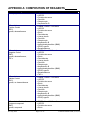
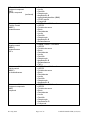
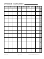
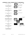
Human Adipogenesis Assay KitGlucocorticoid Analogues, 100 point assay kit Cat#: DIF-GLUC, DIF-GLUC-NC INSTRUCTION MANUAL ZBM0004.05 STORAGE CONDITIONS Frozen subcutaneous human preadipocytes Store in liquid nitrogen IMMEDIATELY upon receipt. No expiration date is applicable; however, the cells must be plated within 1 week of receiving the kit to account for the expiration of the kit components. Media, Reagents A & B, Buffers: Store at 2 - 8C. See kit label for expiration date Use reconstituted Glycerol Reagent A within 7 days. Glycerol Standard -20°C All Zen-Bio Inc products are for research use only. Not approved for human or veterinary use or for use in diagnostic or clinical procedures LIMITED PRODUCT WARRANTY This warranty limits our liability to replacement of this product. No other warranties of any kind, expressed or implied, including without limitation, implied warranties of merchantability or fitness for a particular purpose, are provided by Zen-Bio, Inc. Zen-Bio, Inc. shall have no liability for any direct, indirect, consequential, or incidental damages arising out of the use, the results of use, or the inability to use this product. ORDERING INFORMATION AND TECHNICAL SERVICES Zen-Bio, Inc. 3200 Chapel Hill-Nelson Blvd., Suite 104 PO Box 13888 Research Triangle Park, NC 27709 Telephone (919) 547-0692 Facsimile (FAX) (919) 547-0693 Toll Free 1-866-ADIPOSE (866)-234-7673 Electronic mail (e-mail) [email protected] World Wide Web http://www.zenbio.com Rev July 2010 Page 1 of 15 PATENT PROTECTED (6,153,432) INTRODUCTION The differentiation assay kits provide the tools to study the compounds that stimulate human adipocyte differentiation or lipogenesis. Such compounds may be PPAR agonists or a combination of thiazolidinediones and glucocorticoids that are potentially useful in the treatment of diabetes. This kit is designed to test compounds as potential glucocorticoid analogues, with dexamethasone at 1.0 M serving as the positive control. This kit contains sufficient reagents to assay 100 assay points in a 96 well format. ITEMS INCLUDED IN THE KIT Item Human Preadipocytes Frozen vial PM-1 DPC DNC DVC DMT MMPC MMNC MMVC MMT Wash buffer Lysis buffer Reagent A Description Human subcutaneous preadipocytes Preadipocyte medium (See Appendix A) Differentiation, Positive Control Differentiation, Negative Control Differentiation, Vehicle Control Differentiation Medium for Treatments Maintenance Medium for Positive Control Maintenance Medium for Negative Control Maintenance Medium for Vehicle Control Maintenance Medium for Treatments Reconstitute w/ 11.0 ml deionized water prior to use. Use reconstituted reagent Unit Quantity Item Storage VIAL 1 1 Liquid nitrogen BOTTLE VIAL VIAL VIAL BOTTLE TUBE TUBE TUBE BOTTLE BOTTLE BOTTLE 50ml 1 ml 1 ml 1 ml 300 ml 3 ml 3 ml 3 ml 300 ml 50ml 15ml 2 2 2 2 3 2 2 2 4 2 2 4°C 4°C 4°C 4°C 4°C 4°C 4°C 4°C 4°C 4°C 4°C BOTTLE 11ml 2 4°C BOTTLE 2.5ml 2 4°C VIAL 100 l 2 -20°C BOTTLE 2 ml EACH 3 --- ----- EACH 1 3 ----- --------- within 7 days. Reagent B Glycerol standard Reconstitute w/ 2.5 ml deionized water prior to use Glycerol @ 1mM [Reconstitute with 400 l Standards Diluent to make the 200 M glycerol standard; see page 5 for recommended dilution scheme] Standards Diluent Tray Data sheet Assay Plate, blank Clear polyvinyl tray for multi-channel pipetters Certificate of Analysis and protocol 96-well assay plates, blank PLATE 4°C Other equipment/reagents required but not provided with the kit: Single-channel pipetter Plate reader with a filter of 540 nm Rev July 2010 Page 2 of 15 Multi-channel pipetter Tubes to dilute standards PATENT PROTECTED (6,153,432) ASSAY PROCEDURE We strongly recommend testing all compounds in triplicate A. DIFFERENTIATION PROCEDURE On each day of the procedure, the appropriate medium must be warmed to 37 C prior to use. Note: This protocol is designed to accommodate a weekday work schedule. Any deviation from the recommended start day of Monday-Thursday may require weekend work. Day 1: This is the day the cells are plated. 1. Remove cells from liquid nitrogen and place immediately into a 37 C water bath and agitate while in bath. Be careful not to submerge the cap of the vial into water. Do not leave the vials in water bath after most of the content has thawed. Rinse the vials with 70% ethanol before taking them to the culture hood. 2. Upon thawing, transfer the cells to a sterile conical bottom centrifuge tube containing 10 ml of Preadipocyte Medium (cat # PM-1). 3. Centrifuge: 1,200 rpm (282Xg) / 20C / 5 minutes. Aspirate the supernatant. TAKE CARE TO NOT ASPIRATE ANY OF THE CELL PELLET. 4. The cell vial contains a minimum of 2.0 x 10⁶ viable cells; however, we recommend performing a cell count to determine a more exact number of cells. Resuspend the cell pellet in 2 ml Preadipocyte Medium, dilute an aliquot in trypan blue and count live (unstained) cells on a hemacytometer. The cell concentration required for approximately 40,000 cells / cm2 in the 96 well format with 150l /well is 1.3 x 10⁶ cells in 15 ml Preadipocyte Medium. 5. Plate cells in one of the 96 well plates provided in the kit. Do not agitate the plate, as cells will not plate evenly. 6. Place plate in 37C incubator, 5% CO2, 97% humidity. The cells will be maintained in the incubator after each manipulation until Day 14. Rev July 2010 Page 3 of 15 PATENT PROTECTED (6,153,432) GUIDE TO REAGENTS Well type # of wells Differentiation reagent Maintenance medium Positive control 3 DPC MMPC Negative control 3 DNC MMNC Vehicle control 3 DVC MMVC Treatment compounds 87* DMT MMT *Include any necessary solvent controls as treatments (see Note below). See Appendix A for description of reagents NOTE: Included in this kit are sufficient volumes of Differentiation Medium for Treatments (DMT) and Maintenance Medium for Treatments (MMT), based on using 10 ml of each medium per compound dilution for a maximum of 29 compounds tested in triplicate (87 wells remaining on a 96-well plate after accounting for 9 control wells). If a compound stock is too concentrated to accomplish the desired dilution, use an appropriate solution (not supplied) to prepare an intermediate concentration that would allow for a final volume of 10 ml. Also the positive control in this kit, dexamethasone, has a final solvent concentration of 0.005% ethanol. This low concentration does not affect the differentiation of adipocytes so the ethanol is not included in the vehicle control. If the concentration of any solvent for the compounds used is high enough to potentially alter differentiation, please include the solvent alone as a treatment. We do not recommend treating the cells with solutions exceeding 1% of any solvent, as higher concentrations may be toxic to the cells. Day 2: Begin the differentiation procedure using the 4 types of Differentiation reagents (see summary chart above and Appendix A). Plan to do all treatments and controls in triplicate. A blank plate map is included in these instructions to record the well treatments. Using the Differentiation Medium for Treatment (DMT), prepare treatments. Refer to the note above when preparing compound solutions. When all treatments are prepared, remove Preadipocyte medium from control wells. We recommend doing the treatments in small groups so the cells do not dry out. Pipet 150 l each Differentiation Positive Control, (DPC), Differentiation Negative Control (DNC), and Differentiation Vehicle Control (DVC) into appropriate wells. Remove media from experimental wells and pipet 150 l each Differentiation Medium for Treatment (DMT) containing compounds into appropriate wells. Rev July 2010 Page 4 of 15 PATENT PROTECTED (6,153,432) Day 8: Prepare treatments using the Maintenance Medium Treatment Compounds (MMT). Using a multi-channel pipetter remove media from all wells. Gently feed all wells with 150l of the appropriate Maintenance Medium (see chart above) that is provided with this kit. Day 15: Cells are now mature. Proceed to part B. The positive control wells should exhibit significantly greater lipid accumulation than the negative control wells or the vehicle control wells. Refer to page 11 for a picture of a typical positive control when the adipocytes are mature. Rev July 2010 Page 5 of 15 PATENT PROTECTED (6,153,432) B. TRIGLYCERIDE ASSAY 1. Warm the Wash Buffer and Lysis buffer in a 37oC water bath. 2. Prepare the Reagent B by adding 2.5ml deionized water per bottle and gently invert. DO NOT VORTEX! Use a pipette to ensure that the powder is completely dissolved. Keep at room temperature. Store in a light protected bottle. Reconstituted Reagent B is stable for 60 days refrigerated (2-8C); store any remaining solution refrigerated (2-8C). Bring Reagent B to room temperature. 3. Remove all media. Using about 15 ml of the wash buffer, wash the cells one time with 150 µl wash buffer. Label the disposable tray “wash buffer” and retain for later use. 4. Remove all Wash Buffer. Using a new tray, add 15 l Lysis buffer. Incubate at 37oC – 50oC for 20 minutes. 5. After the incubation is complete, visually confirm cell lysis by checking the wells under a microscope. If cells are not fully lysed, incubate for another 10 minutes. 6. Add 135 l warm Wash Buffer and mix the lysates by pipetting up and down three times. 7. Add 20 l Reagent B to each well. It is not necessary to mix at this time, however, gently tap the plate to help mix the reagents. Incubate the plate at 37oC for 2 hours. 8. Bring Reagent A and the glycerol standards to room temperature during this time. The Wash Buffer can also be kept at room temperature at this point. Warm the Standards Diluent to 37C. Prepare the standard curve as follows: Pipette 400 l of the Standards Diluent into the 1 mM glycerol standard tube provided and mix well by vortexing. This produces a diluted stock glycerol standard of 200 M. Pipette 250 l of Diluent into 6 tubes (not provided). Using the newly diluted stock glycerol solution, prepare a dilution series as depicted below. Mix each new dilution thoroughly before proceeding to the next. The 200 M stock dilution serves as the highest standard, and the Diluent serves as the zero standard. Rev July 2010 Page 6 of 15 PATENT PROTECTED (6,153,432) 400 l Stds Diluent 250 l 250 l 250 l 250 l 250 l 250 l Std 200 M 100 M 50 M 25 M 12.5 M 6.25 M 3.125 M Note: The above dilution series generates enough volume to perform the standard curve in duplicate. If you wish to perform the standard curve in duplicate, please note that eight fewer data points can be assayed with this kit. 9. Also at this time prepare the Reagent A by adding 11.0 ml deionized water per bottle and gently inverting. DO NOT VORTEX! Use a pipet to ensure that the powder is completely dissolved. Keep at room temperature. If using a Reagent A solution previously prepared and stored at 2-8C, also bring to room temperature. Make sure there is enough Reagent A from one solution to treat all the points in the assay. It may be necessary to combine solutions. Store in a light protected bottle. Reconstituted Glycerol Reagent A is stable for 7 days refrigerated (2-8C); store any remaining solution refrigerated (2-8C). 10. To a blank 96 well plate, add 80 l wash buffer to each well needed for the assay (NOTE: do not add Wash Buffer to the wells used for the standard curve). 11. Working with one row or column at a time, mix the lysates very well using a multi-channel pipet. Immediately transfer 20 l per well of the lysates to the corresponding well of the plate containing the wash buffer. This results in a Dilution Factor of 5. 12. Prepare the standard curve. Pipet 100 l of each standard into a well. (NOTE: Eight wells are necessary for the curve. If there are remaining wells on the assay plate, you can utilize the remaining wells. If not, a second plate is included in this kit). 13. Using the third tray, add 100 l Reagent A to samples and standards. Mix by pipetting up and down one time. Incubate at room temperature for 15 minutes. 14. Read at 540 nm using a microtiter plate reader. Rev July 2010 Page 7 of 15 PATENT PROTECTED (6,153,432) GLYCEROL STANDARD CURVE This kit is designed to show relative lipid accumulation of experimental treatments compared to controls. The assay is based on the equation 1 M Triglyceride yields 1M glycerol + Free Fatty Acids The reagent measures the concentration of glycerol released after lysing the cells and hydrolyzing the triglyceride molecules. The triglyceride concentration can then be determined from the glycerol values. Generate standard curve: see example below [DO NOT use this standard curve to generate your data. This is an example.] Subtract the OD value of the 0 M standard from all OD values including the standard curve. Glycerol (uM) 0 3.125 6.25 12.5 25 50 100 200 OD 0.048 0.059 0.07 0.098 0.127 0.2 0.353 0.649 OD 0.048 0.058 0.07 0.098 0.13 0.205 0.362 0.667 ODblank ODblank 0.011 0.022 0.05 0.079 0.152 0.305 0.601 0.01 0.022 0.05 0.082 0.157 0.314 0.619 Avg ODblank 0.048 0.059 0.070 0.098 0.129 0.203 0.358 0.658 Standard Curve 0.700 y = 0.003x + 0.053 R2 = 0.9997 0.600 0.500 0.400 Series1 Linear (Series1) 0.300 0.200 slope intercept R2 0.100 0.003 0.053 0.9997 0.000 0 50 100 150 200 250 G lycero l ( uM ) y = observed O.D. minus the blank x = concentration of glycerol in M To calculate x for each y, (i.e. to change the observed O.D. into glycerol concentration) use the following equation: y=(slope) times (x) plus intercept y=mx+b so x=(y-b)/m x=(y – 0.0006)/0.0014 where 0.0014= slope of the line and 0.0006= y intercept. Be careful to enter the proper sign for the y intercept value as it may be a negative number. Any OD values greater than the highest standard (200 M) should be suspect. The compound should be re-assayed using a lower dose of the compound at treatment OR a dilute solution of the condition medium at the time of the assay. Rev July 2010 Page 8 of 15 PATENT PROTECTED (6,153,432) The R2 value should be equal or greater then 0.98 for the standard curve to be valid. Any R2 values below 0.98, must have the standard curve run again. Solve for the Total Glycerol concentration (i.e. total triglyceride concentration) for each OD. Remember to include the Dilution Factor in the equation. Data is expressed as M Glycerol. FREQUENTLY ASKED QUESTIONS 1. Can I buy the reagents separately? The only reagents sold separately are Glycerol Reagent A (cat# RGTA-10) and the glycerol standard for the Triglyceride Assay kit (cat# TG-GLYSTAN). 2. Can I use another plate format besides 96 well? This kit is designed for the assay of A 96 well plate (100 assay points). We do not have a protocol for other formats. 3. Can I use this kit to measure total triglyceride in other cell lines and other human and non-human cells? Yes. The assay is not species specific. As long as the sample concentration is in the linear range, this kit should be able to detect it. 4. My cells did not lyse. What can I do? If cells are not fully lysed, incubate for another 10 minutes at 37oC - 50oC. Sometimes mixing by pipetting up and down several times is necessary for full lysis. 5. I do not have time to complete the assay. Can I freeze the samples? Yes. The cell lysates can be stored at -80C for a maximum of 7 days. Mix the thawed lysates in the plate by pipetting up and down several times. Allow all reagents and samples to reach room temperature BEFORE adding the Wash Buffer and Glycerol Reagent A to complete the assay. TROUBLESHOOTING Problem High background or the triglyceride reagent turns a darker color before the assay begins. Edge effects Inconsistent OD reading Rev July 2010 Suggestions Use clean tray and tips Change pipet tips frequently Ensure a saturated humidity in the incubator to prevent evaporation from the outside wells Be careful when pipetting to avoid bubbles. If bubbles persist, burst the bubbles using a large gauge needle prior to reading and read the plate again. Mix the lysates well before transferring the 10l to the Wash buffer plate. Page 9 of 15 PATENT PROTECTED (6,153,432) REFERENCES 1. Green, H. and Kehinde, O. (1974) Sublines of mouse 3T3 cells that accumulate lipid. Cell 1, 113-116. 2. Hauner, H., et al., (1989) J. Clin. Invest.(84), 1663-1670. 3. Kuri-Harcuch W, Wise LS, Green H. (1978) Interruption of the adipose conversion of 3T3 cells by biotin deficiency: differentiation without triglyceride accumulation. Cell 14:53-58. Rev July 2010 Page 10 of 15 PATENT PROTECTED (6,153,432) APPENDIX A: COMPOSITION OF REAGENTS Reagent Preadipocyte Medium Differentiation, Positive Control (DPC) agonist, dexamethasone Differentiation, Negative Control (DNC) agonist, dexamethasone, TNF- Differentiation, Vehicle Control (DVC) agonist, no dexamethasone Differentiation Medium for Treatment compound (DMT) agonist, compound Rev July 2010 Components DMEM / Ham's F-12 medium HEPES Fetal bovine serum Penicillin Streptomycin Amphotericin B DMEM / Ham's F-12 medium HEPES Fetal bovine serum Biotin Pantothenate Human insulin Penicillin Streptomycin Amphotericin B Isobutylmethylxanthine (IBMX) PPAR agonist Dexamethasone DMEM / Ham's F-12 medium HEPES Fetal bovine serum Biotin Pantothenate Human insulin Penicillin Streptomycin Amphotericin B Isobutylmethylxanthine (IBMX) PPAR agonist Dexamethasone TNF- DMEM / Ham's F-12 medium HEPES Fetal bovine serum Biotin Pantothenate Human insulin Penicillin Streptomycin Amphotericin B Isobutylmethylxanthine (IBMX) PPAR agonist DMEM / Ham's F-12 medium HEPES Fetal bovine serum Biotin Page 11 of 15 PATENT PROTECTED (6,153,432) Differentiation Medium for Treatment compound (DMT) agonist, compound (continued) Maintenance Medium Positive Control (MMPC) dexamethasone Maintenance Medium Negative control (MMNC) dexamethasone TNF- Maintenance Medium Vehicle control (MMVC) no dexamethasone Maintenance Medium Treatment compounds (MMT) compound Rev July 2010 Pantothenate Insulin Penicillin Streptomycin Amphotericin B Isobutylmethylxanthine (IBMX) PPAR agonist Compound DMEM / Ham's F-12 medium HEPES Fetal bovine serum Biotin Pantothenate Insulin Penicillin Streptomycin Amphotericin B Dexamethasone DMEM / Ham's F-12 medium HEPES Fetal bovine serum Biotin Pantothenate Insulin Penicillin Streptomycin Amphotericin B Dexamethasone TNF- DMEM / Ham's F-12 medium HEPES Fetal bovine serum Biotin Pantothenate Insulin Penicillin Streptomycin Amphotericin B DMEM / Ham's F-12 medium HEPES Fetal bovine serum Biotin Pantothenate Insulin Penicillin Streptomycin Amphotericin B Compound Page 12 of 15 PATENT PROTECTED (6,153,432) APPENDIX B: PLATE LAYOUT A B C D E F G H 1 2 3 4 5 6 7 8 9 10 11 12 Rev July 2010 Page 13 of 15 PATENT PROTECTED (6,153,432) APPENDIX C: DIFFERENTIATION PICTURES PREADIPOCYTE MATURE ADIPOCYTE nucleus Lipid droplets nucleus APPENDIX D: DIFFERENTIATION FLOWCHART DAY 1 PLATE CELLS. INCUBATE 24 HOURS 37C DAY 2 APPLY TREATMENTS AND CONTROLS INCUBATE 7 DAYS 37C DAY 8 CHANGE CELLS TO APPROPRIATE MAINTENANCE MEDIUM. INCUBATE 7 DAYS 37C DAY 15 FEED CELLS APPROPRIATE MAINTENANCE MEDIUM MOVE ON TO TRIGLYCERIDE ASSAY PROTOCOL Rev July 2010 Page 14 of 15 PATENT PROTECTED (6,153,432) APPENDIX E: TRIGLYCERIDE ASSAY FLOWCHART All media OOOOOOOOOOOO OOOOOOOOOOOO OOOOOOOOOOOO OOOOOOOOOOOO OOOOOOOOOOOO Remove all media from wells. 150 l Wash Buffer Wash with 150 l Wash Buffer Remove all Wash Buffer from wells and add 15 l Lysis Buffer. 150 l Wash Buffer OOOOOOOOOOOO OOOOOOOOOOOO OOOOOOOOOOOO OOOOOOOOOOOO OOOOOOOOOOOO Add 15 l Lysis Buffer o Incubate 20 minutes at 37 – 50 C Verify lysis and add 135 l warm Wash Buffer. Mix by pipetting up and down 3 times. Add 20 l Reagent B and incubate 2 o hours at 37 C. Add 135 l warm Wash Buffer OOOOOOOOOOOO OOOOOOOOOOOO OOOOOOOOOOOO OOOOOOOOOOOO OOOOOOOOOOOO Add 20 l Reagent B OOOOOOOOOOOO OOOOOOOOOOOO OOOOOOOOOOOO OOOOOOOOOOOO OOOOOOOOOOOO One hour prior to assay, reconstitute Glycerol Reagent A and prepare standards. Keep all at room temperature. Add 80 l Wash Buffer to a new plate. Mix lysates and transfer 20 l lysates to the wells containing Wash Buffer. Transfer 100 l of each standard to a new plate. Add 100 l Reagent A to samples and standards. o Incubate 15 minutes at 25 C (room temperature). Pop the bubbles in each well5 Add 80 l Wash Buffer OOOOOOOOOOOO OOOOOOOOOOOO OOOOOOOOOOOO OOOOOOOOOOOO OOOOOOOOOOOO Standards OOOOOOOOOOOO OOOOOOOOOOOO OOOOOOOOOOOO OOOOOOOOOOOO OOOOOOOOOOOO 20 l OOOOOOOOOOOO OOOOOOOOOOOO OOOOOOOOOOOO OOOOOOOOOOOO OOOOOOOOOOOO O O O O O GLYCEROL REAGENT A O O O O O An additional blank assay plate may be necessary for the assay of glycerol standards. Measure the optical density of each well at 540 nm using a spectrophotometer plate reader. Rev July 2010 Page 15 of 15 PATENT PROTECTED (6,153,432)