1
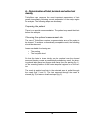
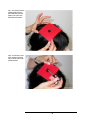
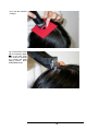
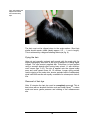
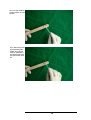
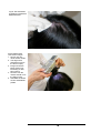
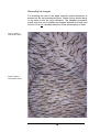
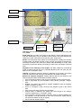
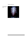
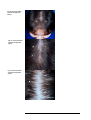
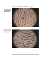
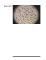
USER MANUAL
TrichoScan Professional Version 3.0
TRICHOLOG GmbH
79117 Freiburg, Germany
Datinf GmbH
72074 Tübingen, Germany
Manual TS 3.0 08/2007
Contents
Introduction
3
Background
3
Measuring method of TrichoScan
TrichoScan - Definitions
3
TrichoScan - Procedure
4
4
A – Determination of total, terminal, and vellus hair density and telogen hair counts
Preparing the patient
Choosing the optimal measurement site
Dying the hair
Removal of hair dye
Recording the images
6
6
6
9
9
14
B – Determination of anagen hair count
16
Software description
18
Image transmission
Manual control
Printing the results
Support
Certification
Initialization data
Parameters
18
18
18
19
19
19
20
Appendix
20
References
20
Examples of TrichoScan target sites
21
Examples of unsuitable TrichoScan images
24
Manual TS 3.0 08/2007
Introduction
Background
Hair loss or thinning hair is a common complaint in clinical dermatology, but patients seeking advice for hair loss are not necessarily going
bald. In established cases of androgenetic alopecia (AGA) characteristic patterns are easily discernible. However, the clinician is often
challenged by patients, especially females, with initial stages of AGA
where hair loss is reported but alopecia is not visibly discernable, or
where the effect of treatment attempts are hard to measure.
Numerous methods have been described to assess the rate of hair
growth. The techniques can be classified as either invasive (e.g. biopsies), semi-invasive (trichogram, unit area trichogram) or non-invasive
methods. However, while reviewing the capabilities of the different
methods, the common theme emerges that most techniques are of
little use to the clinician because they are time consuming, often
costly, or difficult to perform. Therefore, an operator- and patientfriendly, inexpensive, validated and reliable method of quantifying hair
growth is a rational need. TrichoScan is such a method, combining
standard epiluminescence microscopy (ELM) with automatic digital
image analysis, for the measurement of human hair.
Measuring method of TrichoScan
With the TrichoScan 3.0 version several different needs in hair science are met. Firstly, the most important parameters, such as total
terminal and vellus hair counts, can be analysed within the same day.
Secondly, the same target site can be used to calculate the number
of telogen hairs by mathematical approximation; and thirdly, with a
different hair shaving procedure, the anagen hair count is also measurable. This handbook will guide you through the different possibilities.
3
TrichoScan - Definitions
A:
TrichoScan-Professional V 3.0 is suitable for the analysis of
human scalp hair in androgenetic (pattern) alopecia.
B:
TrichoScan is a tool to monitor the most important hair parameters during treatment.
C:
TrichoScan-Professional V 3.0 is able to monitor total, vellus,
and terminal hair density and, by mathematical approximation,
the telogen and the anagen hair count.
D:
TrichoScan is not suitable for evaluating body hair or to
monitor other hair diseases such as alopecia areata.
E:
TrichoScan is not a diagnostic procedure.
F:
TrichoScan-Professional V 3.0 is not designed for clinical trials. It is made for the individual clinician in practice. We do not
accept any responsibility when the tool is used for clinical trials.
G:
The TrichoScan-analysis needs a clean and lightly pigmented
skin to enable good contrast with dark colored hair. Any remnants of the hair dye, dark melanocytic moles or dark scalp
skin will diminish the contrast to the hairs and the analysis will
not be possible.
F.
TrichoScan is a software program based on statistics and
definitions of hair patterns. The software cannot diagnose telogen or anagen hair loss like a histopathologist. However,
based on the biological behavior of those hairs, they can be
differentiated by mathematical approximation.
TrichoScan-Procedures
There are two possible basic procedures that provide different information with Trichoscan Professional V 3.0:
A - Determination of total, terminal, and vellus hair density (at the
time of hair clipping) and optional telogen hair counts (3 days after incomplete hair clipping).
B – Determination of anagen hair count (dyeing and measuring
hair 3 days after complete shaving).
4
TrichoScan – Hair parameters
Hair density (n/cm2): With the TrichoScan-Professional V 3.0 edition
it is possible to calculate the number of hairs detected (hair count)
and the hair density (hairs / cm2). Please note, that due to the image
resolution of digital cameras the TrichoScan software cannot detect
very fine hairs (approx. less than 10µm diameter). In addition,
TrichoScan cannot identify hairs which are too short for analysis (approx. less than 0.3mm in length). As digital camera image resolution
improves, these limitations may change in the future.
Terminal hair density (n/cm2): By definition a terminal hair is thicker
than 40µm. Trichoscan uses this value to identify terminal hairs in images. The number of terminal hairs relative to vellus hairs is also calculated and provided in the analysis results.
2
Vellus hair density (n/cm ): By definition a vellus hair is thinner than
40µm. Trichoscan uses this value to identify vellus hairs in images.
The number of vellus hairs relative to terminal hairs is also calculated
and provided in the analysis results.
Telogen hair density (n/cm2): By definition a terminal hair will not
grow whereas anagen hairs will. When images are taken three days
after hair clipping, growing hairs can be differentiated from nongrowing hairs based on different hair length. Trichoscan identifies
non-growing hairs as telogen hairs and growing hairs as anagen
hairs.
2
Anagen hair count (n/cm ): In the definition of the TrichoScan procedure, an anagen hair is a hair which is detectable three days after
complete hair shaving. Within this time only anagen hairs should
grow significantly.
5
A – Determination of total, terminal and vellus hair
density
TrichoScan can measure the most important parameters of hair
growth immediately following correct preparation of the scalp region
for analysis. The following steps are necessary.
Preparing the patient
There is no special recommendation. The patient may wash their hair
before the analysis.
Choosing the optimal measurement site
The use of TrichoScan requires a representative area of the scalp to
be shaved. To achieve a cosmetically acceptable result, the following
should be observed.
Areas unsuitable for shaving are:
•
•
The parting
The occipital whorl
So that the hairs in close vicinity can be combed over the shaved
area and thereby create an aesthetically satisfactory result, the shaving should take place two fingers width away from the parting (fig. 1),
on the receding hairline of the fronto-temporal regions or on the vertex.
The mask is applied and hair in the selected area is pulled through
the mask (figs. 2 and 3). The hair exposed through the mask is
shaved (fig. 4) to leave a small neat spot (fig. 5).
6
Fig 1: The shaving mask is
positioned about two fingers width away from the
parting or any other suitable measurement site.
Fig 2: The hair in the area
to be shaved is exposed
with a curved hook or with
pointed scissors.
7
Fig 3: Hair after exposure
is clipped.
Fig 4: Exposed hair, should
not be completely clipped
down to the bare scalp.
Best results are achieved
by a speedy and gentle
clipping process. Some
stubble must remain.
8
Fig 5: After clipping, the
clipped hairs can be
removed with sticky
tape.
The hair must not be clipped down to the scalp surface. Short hair
shafts should remain visible (ideally approx. 0,5 – 1 mm in length).
This is achieved by a diagonal shaving technique (fig. 4).
Dying the hair
Hairs do not normally contrast well enough with the scalp skin for
digital photography and need to be dyed in advance of taking digital
images. The dye product supplied with TrichoScan is best applied
using a wooden spatula after having been mixed 1:1 with development cream (figs. 6-10). The dye is applied onto the clipped scalp
area and must remain there for 15 minutes. Longer dying periods
lead to dyeing of the scalp skin, shorter periods lead to inadequately
dyed hair! Both results are equally unsuitable for subsequent evaluation.
Removal of hair dye
After 15 minutes the hair dye must be completely removed. This is
TM
best done with an alcoholic solution such as Kodan Spray , a clean
swab and some gentle pressure and rubbing of the measurement
site.
9
Fig 6: A small amount of
the dye supplied with the
Trichoscan kit is applied
onto a wooden spatula
or similar utensil.
Fig 7: The same amount
of dye development
cream is applied to the
spatula.
10
Fig 8: The dye and development solution are mixed
together.
Fig 9: After mixing of the
dye and development
solution the mixture is
ready for application to
the clipped scalp region
using the wooden spatula.
11
Fig 10: The dye must
remain on the scalp for
15 minutes.
After dying the hair, the area should be thoroughly cleansed using an
alcohol-based solution (Image 11 and 12).
Fig 11: Dye remnants
should be removed by
means of a swab and an
alcohol solution.
12
Fig 12: The area should
thoroughly be cleansed of
all dye remnants.
Fig13: Rules for best
TrichoScan images:
1. No hair dye remnants should remain.
2. The target area
should be wet from
the alcohol spray.
3. Images should be
taken without contaminating air bubbles present.
4. No hairs from the
outside should cross
the field of view.
5. Hair stubble of at least
0.5 mm should still be
present.
13
Recording the images
For recording, the lens of the digital camera's optical attachment is
pressed on the wet measurement area. Please use an alcohol spray
or tap water to wet the area sufficiently. This obligatory procedure
makes sure that no air bubbles are trapped between the scalp and
the lens. Oil is not a suitable alternative to the alcohol spray or water.
Fig14: A perfect
Trichoscan image.
Fig15: A perfect
TrichoScan image.
14
Suitable Images for TrichoScan
As an automated image analysis tool, the TrichoScan results strongly
depend on the image quality.
Suitable images include the following attributes:
-
Clean and sharp images with no hair dye remnants.
-
Wet hairs without larger air bubbles present.
-
In focus with no long hairs passing into the measurement
site from outside.
-
Hairs are evenly clipped and dark due to the hair dye.
-
Hairs have a minimum length of approximately 0.5 mm to
allow detection by the TrichoScan software. Stubbles that
are too short will escape the analysis as the software algorithms define hair as being longer than 0.5 mm.
-
Hairs that are straight. Avoid twisting the optics whilst
placed on the measurement site and be sure that all hairs
are evenly clipped.
-
TrichoScan analysis is not possible in darker skin types or
an area with dark melanocytic moles due to poor contrast
between skin and hair.
Then the image is ready for the analysis of total, terminal, and vellus
hair counts.
Telogen hair count
In the software sense a telogen hair is a non-growing hair. For this
analysis the hair should be clipped as above (figs. 1-5) during the first
office visit. The patient must return to your office 3 days after hair
clipping and then the target area receives the hair dye as described
(figs. 6-12) and an image is taken for TrichoScan analysis (figs. 1315). The software will measure the length of all hairs and by statistical
analysis will discriminate between growing versus non-growing hairs.
Please note that catagen and exogen hairs also do not grow significantly within this time period and will also be judged as non-growing
hairs. Therefore, the calculated telogen hair values will be a bit higher
than may normally be expected. For this analysis to be accurate a
very uniform hair clipping is mandatory to ensure the remaining hair
stubble is of equal length. For less expert hair clippers, we recommend the use the anagen hair count tool instead. An anagen hair
count requires less hair clipping consistency and so it is much easier
to obtain accurate results with this approach.
15
B – Determination of anagen hair count
The TrichoScan software defines anagen hairs based on the knowledge that anagen hair grows at approximately 0.3 mm/day whereas
telogen and catagen hairs do not grow. Therefore, anagen hair can
be calculated by shaving the measurement site; NOT with our
TrichoScan clipping machine which will leave some stubbles behind,
but with an ordinary single use razor, which will shave all hairs down
to the skin completely. Please control with a magnifying loupe and
confirm the shaving is indeed complete and no hairs can be detected
with the software. After three days the area is then dyed as described
above and the image is made. Please note that the calculated hair
density will be lower than normally expected as only anagen hairs are
measured. Please also note that there are no normal values for the
individual patient. During successful treatment, the anagen hair count
should increase and therefore this approach can be used to monitor a
treatment response. For this procedure you must change the
TrichoScan software’s analysis mode to “Anagen hair count”.
TrichoScan analysis
The recorded photographs are loaded into the TrichoScan software
which automatically proceeds with the first analysis. According to the
capacity of the computer used, the analysis may take up to one minute (800 MHz – Pentium III) - Faster computers increase the speed of
TM
the analysis. Macintosh computers cannot be used.
16
Left image (1)
Right image (2)
Progress indicator
Results
Diagram
Regulator for the
outlining of hair
lengths
Buttons for program navigation
Left image (1): Here the area to be analysed has a blue border once the image has
been loaded.
Right image (2): According to the stage of the analysis, various distinguishing features are marked which give an indication of the progress of the image analysis (red:
telogen hair, yellow: hairs that touch the border, green: anagen hair).
Result: The results area shows values for the analysed area, including the corrected
number of hairs, hair density (number of hairs per area), percentage of anagen hairs,
percentage of telogen hairs or density of anagen hairs (depending on mode), vellus
hair density, terminal hair density, percentage of vellus hairs, percentage of terminal
hairs.
Regulator for the outlining of hair lengths: The slider enables the maximal hair
length for defining telogen hairs to be manually adjusted by the user when the software is in the “Trichogram” mode.
Diagram: The diagram shows the frequency distribution of hair length. The red line
marks the maximum length of telogen hairs defined in the software analysis.
Buttons for program navigation: Clicking different elements and buttons in the
software program window enable different software features:
• Click on left image: Fading in and out of the borderline around recognized hairs.
• Click on right image: On- and off-switch for the indicator of anagen- / telogenhairs.
• Register: This button only applies to program installations used as temporary
software evaluation versions.
• Score: In “Trichogram” mode a scoring of the analysis is given in the colored
field.
• Print: Prints the analysis results for documentation.
• Change mode: To determine the anagen hair count change to the “Anagen”
mode (or change back to “Trichogram” mode).
• Cancel: Stops the analysis process. This button does not react immediately during evaluation, it may be necessary to wait a few seconds after clicking this button before the software is seen to respond.
• Close: Ends the analysis program.
17
Software description
Image transmission
The transmission of the digital image to the TrichoScan Software
takes place automatically. More details can be found in the documentation for the recording system.
Manual control
In case the pre-set demarcation line that distinguishes between anagen and telogen hairs does not produce satisfactory results, there is
the option of manually setting the maximum length of the telogen
hairs. This is achieved by moving the regulator situated above the
diagram. Color-coding of the hairs indicates the group they belong to.
•
•
Red: Telogen hair.
Yellow: Hair is touching the edge of the picture, grouping follows via a special statistical procedure (Product-LimitEstimation).
• Green: Anagen hair.
Printing the results
The image displayed on screen is the image that is printed (with adaptations taken into consideration, accordingly). Additionally, all of the
internal parameters are displayed beneath the picture. Thus, the reproducibility of the results is ascertained according to the print.
Additionally, information about the name and location of the software
license owner appears at the bottom edge of the picture.
18
Support
Up to date and additional information about TrichoScan can be found
on the Internet at www.trichoscan.com .
In case of problems concerning the recording images and technical
issues of the recording system, please contact the manufacturer.
Medical questions, such as enquiries about measurement results, can
be addressed to Prof. Rolf Hoffmann, MD in Freiburg, Germany
(Email: [email protected], Fax: ++49 761-6800113).
If you have any technical problems or TrichoScan software program
faults, please contact the TrichoScan software engineers (Email:
[email protected], Fax: ++49 7071-2536962).
The program was tested using many images with numerous parameters. However, there is the possibility that not all of the hairs are
correctly recognized in an image. In case recognizable hairs are not
identified by the TrichoScan software, please send the picture in
question via email to [email protected] for evaluation. These pictures will also be used to further improve and develop the TrichoScan
software system.
A research edition of TrichoScan is available for research purposes,
which is able to define additional hair growth parameters. This program version also has the capacity to analyse a greater number of
image recordings. For further information please contact Prof. Rolf
Hoffmann, MD (Email: [email protected]), or visit the TrichoScan
website at www.trichoscan.com.
Certification
A certification-list can be found on the TrichoScan website at
www.trichoscan.com. However, certification is only possible for medical doctors who have a licensed TrichoScan system. For more information please contact your distributor.
Initialization data
Initialization data is an integral part of the TrichoScan software. This
data should never be altered. Unauthorized changes of this file may
result in incorrect data output.
19
Appendix
References
[1] Rushton H, James KC, Mortimer CH
The unit area trichogram in the assessment of androgen-dependent
alopecia.
Br J Dermatol Oct 1983; 109(4):429-37
[2] de Lacharriere O, Deloche C, Misciali C, Piraccini BM, Vincenzi C,
Bastien P, Tardy I, Bernard BA, Tosti A
Hair diameter diversity: a clinical sign reflecting the follicle miniaturization.
Arch Dermatol. May 2001; 137(5):641-646
[3] Hoffmann R
TrichoScan: Ein neues Werkzeug für die digitale Haarzählung.
Hautarzt. Dec 2002; 53:798-804
[4] Hoffmann R
TrichoScan: combining epiluminescence microscopy with digital image analysis for the measurement of hair groth in vivo.
Eur J Dermatol. Jul-Aug 2001; 4(11):362-368
[5] Hoffmann R
TrichoScan: a novel tool for the analysis of hair growth in vivo.
J Investig Dermatol Symp Proc. 2003 Jun; 8(1):109-15
[6] Chamberlain AJ, Dawber RP
Methods of evaluating hair growth.
Australas J Dermatol. Feb 2003; 44(1):10-8. Review
[7] Hoffmann R
Trichoscan: what is new?
Dermatology. 2005;211(1):54-62. Review.
[8] Hoffmann R, Van Neste D.
Recent findings with computerized methods for scalp hair growth
measurements.
J Investig Dermatol Symp Proc. 2005 Dec;10(3):285-8.
20
TrichoScan target sites
Fig16: Recommended
TrichoScan target site
(stars).
21
Fig. 17: Recommended
TrichoScan target site
(stars).
Fig. 18 Recommended
TrichoScan target site
(stars).
Fig 19: Recommended
TrichoScan target site
(stars).
22
Fig 20: Recommended
TrichoScan target site
(stars).
Fig. 21: Recommended
TrichoScan target site
(stars).
Fig. 5: Recommended
TrichoScan target site
(stars).
23
Images not suitable for TrichoScan analysis
Fig. 23: No hair dye and the
black dot in the middle of
the image make this photograph unsuitable for
TrichoScan analysis.
Fig 24: Hairs from the outside cross the TrichoScan
target area making it unsuitable for analysis.
24
Fig. 25: Too many air
bubbles make this image
unsuitable for analysis.
25