1
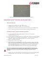
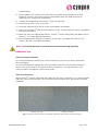
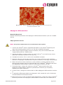
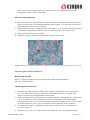
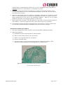
User Manual OriCellTM Fischer 344(F344) Rat Mesenchymal Stem Cells (MSCs) Cat. No. RAFMX-01001 Table of Contents Contents and Storage …………………………………………………………………………… 3 Product Introduction ………………………………………………………………………………… 3 Cell Characteristics and Identity …………………………………………………………………… 3 Product Application …………………………………………………………………………………… 3 TM Cyagen OriCell Fischer 344(F344) Rat MSCs Source ……………………………………… 4 General Handling Principles ……………………………………………………………………… 4 Culturing OriCellTM Fischer 344(F344) Rat MSCs Thawing and Establishing OriCell Passaging Cyagen OriCell TM TM Differentiation of OriCell TM Fischer 344(F344) Rat MSCs ……………………… 4 Fischer 344(F344) Rat MSCs ………………………………… 6 Fischer 344(F344) Rat MSCs …………………………………… 8 TM Cryopreservation of OriCell Fischer 344(F344) Rat MSCs ……………………………… 12 Appendix ………………………………………………………………………..…… 13 Troubleshooting ………………………………………………………………………………………… 13 Related Products ……………………………………………………………………………………… 14 References ……………………………………………………………………………………………… 14 Technical Support ………………………………………………………………… 15 CONTENTS AND STORAGE Product Name Fischer 344(F344) Rat Mesenchymal Stem Cells Catalog No. RAFMX-01001 Amount per Vial 1×10⁶ Cells Cryopreserved At Second Passage Storage Condition Liquid Nitrogen CAUTION: Please handle this product as a potentially biohazardous material. This product contains dimethyl sulfoxide (DMSO), a hazardous material, in the freezing medium. PRODUCT INTRODUCTION Mesenchymal stem cells (MSCs) are multipotent stem cells that can differentiate into a variety of cell types including osteocytes, adipocytes, and chondrocytes. MSCs proliferate quickly and are capable of generating a local immunosuppressive microenvironment, thus contributing to their wide application potentials in tissue engineering, cell therapy, and gene therapy. OriCellTM Fischer 344(F344)Rat Mesenchymal Stem Cells are derived from the bone marrow of Fischer 344(F344) Rats. They have a strong capacity for self-renewal while maintaining their multipotency. In addition, these cells have been tested for: Exogenous Factors: bacterial/fungal contamination, mycoplasma contamination, and endotoxin contamination. Characteristics: post-thaw viability, cell cycle, verification of undifferentiated state, and differentiation potential. This product is intended for laboratory research use only. It is not intended for diagnostic, therapeutic, clinical, household, or any other applications. CELL CHARACTERISTICS AND IDENTITY Strong capacity to expand. Can be passaged at least 5 times. Multipotent differentiation ability along the osteogenic, chondrogenic, and IMPI0032A2 RAFMX-01001 Page 3 of 14 adipogenic lineages. Positive for CD29, CD44, and CD90 (> 70%), and negative for CD34 and CD45 and CD11b (< 5%) in flow cytometry assays. PRODUCT APPLICATIONS Fischer 344(F344) Rat MSCs have become a popular research target due to their potential use in regenerative medicine and tissue engineering (in areas such as cardiovascular, neural, and orthopedic disease). OriCellTM Fischer 344(F344) Rat MSCs can be used as cell models to evaluate the immunoreactions, proliferation, immigration, and differentiation of MSCs both in vivo and in vitro. GENERAL HANDLING PRINCIPLES 1. Aseptic handling of the product is necessary throughout. 2. Once the cells have been established, always freeze several vials of OriCellTM Fischer 344(F344) Rat MSCs as a backup. Note: The OriCellTM Fischer 344(F344) Rat MSCs can be frozen/thawed at least two times. 3. For all studies, it is strongly recommended to use cells that are at, or under, an original passage number of 10. 4. For general maintenance of cells, we recommend the seeding density to be 2.03.0×104cells/cm2. 5. For general maintenance of cells, we recommend that the medium is changed if it becomes acidic (the pH indicator in the medium appears yellow). In general, change the growth medium every three days. 6. Do not let OriCellTM Fischer 344(F344) Rat MSCs overgrow as it will result in contact inhibition. When the cells are 80-90% confluent, subculturing the cells is strongly recommended. Note: We strongly recommend the use of OriCellTM culture media and other related reagents for optimal results. THAWING AND ESTABLISHING OriCellTM FISCHER 344(F344) RAT MSCs Materials Required OriCellTM Mesenchymal Stem Cell Growth Medium (Cat. No. GUXMX-90011) IMPI0032A2 RAFMX-01001 Page 4 of 14 Thawing and Establishing Fischer 344(F344) Rat MSCs 1. Pre-warm the fully supplemented (complete) OriCellTM MSC Growth Medium to 37°C. 2. Add 9 mL of OriCellTM MSC Growth Medium to a 15 mL conical tube. 3. Remove the cryovial of OriCellTM Fischer 344(F344)Rat MSCs from liquid nitrogen. 4. Quickly thaw the vial in a 37°C water bath until the last ice crystal disappears. For optimal results, be sure to finish the thawing procedure within 3 minutes. Be careful not to submerge the entire vial. Maximum cell viability is dependent on the rapid and complete thawing of frozen cells. Note: Results will be less than optimal if the cells are thawed for more than 3 minutes. 5. As soon as the cells are completely thawed, disinfect the outside of the cryovial with 70% v/v ethanol. 6. Use a pipette to transfer the cells to the 15 mL conical tube containing OriCellTM MSC Growth Medium inside a biosafety cabinet. Be careful not to introduce any bubbles during the transfer process. 7. Rinse the vial with 1 mL of the medium to reduce cell loss. Subsequently transfer this 1 mL of cell suspension into the conical tube. 8. Gently mix the cell suspension by slowly pipetting up and down. Be careful not to introduce any bubbles. 9. Centrifuge the cell suspension at 250 x g for 5 minutes. 10. Carefully aspirate off as much of the supernatant as possible and add 2-3 mL of fresh OriCellTM MSC Growth Medium (pre-warmed to 37°C). 11. Gently resuspend the cells in OriCellTM MSC Growth Medium. 12. Seed the cells into a T25 flask and add a sufficient amount of OriCellTM MSC Growth Medium. Gently rock the culture flask to evenly distribute the cells. 13. Incubate the flask at 37°C inside a 5% CO2 humidified incubator. 14. The next day, change the medium with fresh growth medium (pre-warmed to 37°C). 15. Change the growth medium every three days thereafter. 16. When the cells are approximately 80-90% confluent, they can be dissociated with Trypsin-EDTA and passaged. Note: Changing Medium 1. Warm an appropriate amount of medium to 37°C in a sterile container. Replace the spent medium with the pre-warmed, fresh medium. Once completed, return the flask to the incubator. 2. Avoid repeated warming and cooling of the medium. If the entire content is not needed for a single procedure, transfer only the required volume to a sterile secondary container. IMPI0032A2 RAFMX-01001 Page 5 of 14 Fig. 1. OriCellTM Fischer 344(F344) Rat Mesenchymal Stem Cells are established. PASSAGING OriCellTM FISCHER 344(F344)RAT MSCs Materials Required 0.25%Trypsin-0.04%EDTA (Cat. No. TEDTA-10001) Phosphate-Buffered Saline (1×PBS) (Cat. No. PBS-10001) OriCellTM Fischer 344(F344) Rat Mesenchymal Stem Cells (Cat. No. RAFMX-01001) OriCellTM Mesenchymal Stem Cell Growth Medium (Cat. No. GUXMX-90011) Passaging OriCellTM Fischer 344(F344) Rat MSCs 1. Pre-warm the OriCellTM MSC Growth Medium, 1×PBS, and 0.25%Trypsin0.04%EDTA solution to 37°C. 2. Carefully aspirate the spent medium from the 80-90% confluent monolayer of MSCs. 3. Add 1×PBS (6 mL for T75 flask, 3 mL for T25 flask). Be careful not to disturb the monolayer. Gently rock the flask back and forth to rinse the monolayer. 4. Aspirate 1×PBS off and discard. 5. Repeat steps 3-4 two or three times. 6. Add 0.25%Trypsin-0.04%EDTA solution (2-3 mL for T75 flask, 1 mL for T25 flask). Gently rock the flask back and forth to ensure that the entire monolayer is covered with the Trypsin-EDTA solution. Allow trypsinization to continue until the majority of the cells (approximately 80%) are rounded up. At this point, gently tap the side of the flask to release the majority of cells from the culture flask surface. Important: Avoid leaving cells exposed to the trypsin longer than necessary (no more than two minutes if using Cyagen’s trypsin-EDTA solution). Care should also be taken that the cells are not forced to detach prematurely as this may result in clumping. 7. After the cells are visibly detached, immediately add the pre-warmed OriCellTM MSC Growth Medium (6 mL for T75 flask, 3 mL for T25 flask) to neutralize the IMPI0032A2 RAFMX-01001 Page 6 of 14 trypsinization. 8. Gently pipette the medium over the cells to dislodge and resuspend the cells. Repeat 5-6 times until all the cells are dissociated from the flask and evenly dispersed into a single cell suspension. 9. Transfer the dissociated cells into a 15 mL conical tube. 10. Centrifuge at 250 x g for 5 minutes. 11. Carefully aspirate off as much of the supernatant as possible. 12. Add 2 mL of OriCellTM MSC Growth Medium to the conical tube and gently resuspend the cells thoroughly. 13. Plate the cells into appropriate flasks. OriCellTM Fischer 344(F344) Rat MSCs can be split at 1:2 or other appropriate ratios. 14. Add an appropriate amount of medium to the cells. Incubate the cells at 37°C inside a 5% CO2 humidified incubator. Note: Care should be taken to avoid introducing bubbles during pipetting. Additional Tips Time to Change Medium It is recommended to change the culture medium if there are too many dead cells after passaging. It is recommended to change the culture medium whenever the medium becomes acidic, even if the cells do not reach 80-90% confluency. The pH indicator in the culture medium will appear yellow when acidic. Time to Subculture When OriCellTM Fischer 344(F344) Rat MSCs are 80-90% confluent, it is recommended that the cells be subcultured. Do not let the cells overgrow as it will result in contact inhibition. Passage 3 at 40x TM Fig.2 Images of OriCell IMPI0032A2 RAFMX-01001 Passage 3 at 100x Fischer 344(F344) Rat Mesenchymal Stem Cells at passage 3. Page 7 of 14 TM OriCell FISCHER 344(F344) RAT MSC DIFFERENTIATION USING OriCellTM DIFFERENTIATION MEDIA OriCellTM Fischer 344(F344) Rat MSCs can differentiate into a variety of cell types including osteocytes, adipocytes, and chondrocytes. Osteogenic Differentiation Materials Required OriCellTM Mesenchymal Stem Cell Osteogenic Differentiation Medium (Cat. No. GUXMX-90021) Osteogenesis Protocol Note: The protocol listed below is for 6-well tissue culture plates. 1. Culture the OriCellTM Fischer 344(F344) Rat MSCs in OriCellTM Mesenchymal Stem Cell Growth Medium at 37°C in a 5% CO2 humidified incubator. 2. When cells are approximately 80-90% confluent, they can be dissociated with 0.25%Trypsin-0.04%EDTA (Cat. No. TEDTA-10001). 3. Reseed the MSCs in the growth medium at 3×10⁴ cells/cm2 in a 6-well tissue culture plate pre-coated with 0.1% gelatin solution. 4. Incubate the cells at 37°C inside a 5% CO2 humidified incubator. 5. When cells are approximately 60-70% confluent, carefully aspirate off the growth medium from each well and add 2 mL of OriCellTM Mesenchymal Stem Cell Osteogenic Differentiation Medium. 6. Feed cells every three days for 2-4 weeks by completely replacing the medium with fresh OriCellTM Mesenchymal Stem Cell Osteogenic Differentiation Medium (prewarmed to 37°C). 7. After 2-4 weeks of differentiation, cells can be fixed and stained with alizarin red S. Note: To prevent osteoblasts from detaching, it is recommended to change half of the medium every two days before analysis. Alizarin Red S Staining Analysis 1. After the cells have differentiated, remove the osteogenic differentiation medium from the wells and rinse with 1x phosphate-buffered saline (PBS). Fix cells with 2 mL of 4% formaldehyde solution for 30 minutes. 2. Rinse wells twice with 1x PBS. Stain the cells with 1 mL alizarin red S working solution for 3-5 minutes. 3. Rinse wells 2-3 times with 1x PBS. 4. Cells can now be visualized and analyzed under a microscope. IMPI0032A2 RAFMX-01001 Page 8 of 14 Fig. 3 OriCellTM Fischer 344(F344) Rat MSCs are differentiated into osteocytes and are stained with alizarin red S. Adipogenic Differentiation Materials Required OriCellTM Mesenchymal Stem Cell Adipogenic Differentiation Medium (Cat. No. GUXMX90031) Adipogenesis Protocol Note: The protocol listed below is for 6-well tissue culture plates. 1. Culture the OriCellTM Fischer 344(F344) Rat MSCs in the OriCellTM Mesenchymal Stem Cell Growth Medium at 37°C in a 5% CO2 humidified incubator. 2. When cells are approximately 80-90% confluent, they can be dissociated with Trypsin-EDTA (Cat. No. TEDTA-1000). 3. Reseed the MSCs in growth medium at 2x10⁴ cells/cm2 in a 6-well tissue culture plate with a medium volume of 2 mL per well. 4. Incubate the cells at 37°C in a 5% CO2 humidified incubator. 5. Feed the cells every three days until they are 100% confluent or post-confluent. Induction of adipogenic differentiation at post-confluency is strongly recommended. 6. When the cells are 100% confluent or post-confluent, carefully aspirate off the spent growth medium from the wells and add 2 mL of OriCellTM Mesenchymal Stem Cell Adipogenic Differentiation medium A (induction medium) per well. 7. Three days later, change the medium to OriCellTM Mesenchymal Stem Cell Adipogenic Differentiation medium B (maintenance medium) by completely replacing the spent medium A. 8. 24 hours later, change the medium back to MSC Adipogenic Differentiation medium A. 9. To optimally differentiate MSCs into adipogenic cells, repeat the cycle of induction and maintenance at least three times. 10. After three to five cycles of induction and maintenance, culture the cells in OriCellTM Mesenchymal Stem Cell Adipogenic Differentiation medium B for an additional 4-7 IMPI0032A2 RAFMX-01001 Page 9 of 14 days until the lipid droplets are big, round enough. During these days period, change the medium every three days. Oil Red O Stain Analysis 1. After the cells have differentiated, remove the MSC maintenance medium from the wells and rinse with 1x phosphate-buffered saline (PBS). Fix cells with 2 mL of 4% formaldehyde solution for 30 minutes. 2. Rinse wells twice with 1x PBS and stain cells with 1 mL of oil red O working solution (3:2 dilution with distilled water and filter with filter paper) for 30 minutes. 3. Rinse wells 2-3 times with 1x PBS. 4. Cells can now be visualized and analyzed under a microscope. Fig.4 OriCellTM Fischer 344(F344) Rat MSCs are differentiated into adipocytes and are stained with oil red O. Chondrogenic Differentiation Materials Required OriCellTM Mesenchymal Stem Cell Chondrogenic Differentiation Medium (Cat. No. GUXMX-90041) Chondrogenesis Protocol 1. Calculate the total number of MSC pellet cultures required for your experiment (2.5×10⁵ MSCs are needed to form each chondrogenic pellet). Transfer this amount of cells into an appropriate culture tube. 2. Wash the MSCs with Incomplete Chondrogenic Medium. Centrifuge the cells at 150 x g for 5 minutes at room temperature and then aspirate off the supernatant. Resuspend the cells in 1 mL of Incomplete Chondrogenic Medium per 7.5×10⁵ cells. Centrifuge again at 150 x g for 5 minutes and then aspirate off the medium. 3. Resuspend the MSCs in Complete Chondrogenic medium to a concentration of 5.0×10⁵ cells/mL. 4. Aliquot 0.5 mL (2.5×10⁵ cells) of the cell suspension into 15 mL polypropylene IMPI0032A2 RAFMX-01001 Page 10 of 14 culture tubes. Centrifuge the cells at 150 x g for 5 minutes at room temperature. DO NOT aspirate the supernatant or resuspend the pellet. 5. Loosen the caps of the tubes one half turn in order to allow gas exchange, and incubate the tubes at 37°C in a humidified atmosphere of 5% CO2. Do not disturb the pellets for 24 hours. 6. Feed the cell pellets every 2-3 days by completely replacing the medium in each tube (to avoid aspirating the pellets when aspirating the medium, attach a sterile 1200μL pipette tip to the end of the aspirating pipette). Add 0.5 mL of freshly prepared Complete Chondrogenic Medium to each tube. 7. After replacing the medium, flick the bottom of the tube to ensure that the pellet is free floating. Loosen the caps and return the tubes to the 37°C incubator. 8. Chondrogenic pellets should be harvested after 14-28 days in culture. Pellets may be formalin-fixed and paraffin-embedded for alcian blue stain analysis. Alcian Blue Staining Procedure 1. The tissue sample should be formalin-fixed and paraffin-embedded already. 2. Staining procedure: a) Deparaffinize slides and hydrate to distilled water. b) Stain in alcian blue solution for 30 minutes. c) Wash in running tap water for 2 minutes. d) Rinse in distilled water. e) Visualize under a light microscope and capture images for analysis. Blue staining indicates synthesis of proteoglycans by chondrocytes. Fig.5 OriCellTM Fischer 344(F344) Rat MSCs are differentiated into cartilages and are stained with alcian blue. IMPI0032A2 RAFMX-01001 Page 11 of 14 CRYOPRESERVATION OF CELLS USING OriCellTM CRYOPRESERVATION MEDIA OriCellTM NCR Protein-Free Cryopreservation Medium (Cat. No. NCPF-10001) is a protein-free, ready-to-use freezing medium. Its chemically-defined and protein-free formulation has been optimized to stem cells and primary cells, thus greatly enhancing the viability and integrity of these cells by protecting them from damage during the one-step freeze-thaw procedure. Unlike other conventional freezing media, which require a slow programmed freeze, this product allows the cells to be directly frozen at -80°C. Cryopreservation Note: Change the culture medium with fresh growth medium 24 hours before freezing. 1. Collect cells that are in the logarithmic growth phase. Perform a cell count to determine the viable cell density. 2. Centrifuge the cells for 3-5 minutes at 250 x g and 20°C. Remove and discard the supernatant using a pipette. 3. Resuspend the cell pellet in the OriCellTM NCR Protein-Free Cryopreservation Medium at a cell density of 105-106 cells/mL. 4. Dispense aliquots of the cell suspension into cryogenic storage vials that are properly labeled. 5. Place the vials directly in a -80°C freezer. After 24 hours, transfer the frozen vials to liquid nitrogen for long-term preservation. IMPI0032A2 RAFMX-01001 Page 12 of 14 APPENDIX Troubleshooting The table below lists some potential problems and solutions for culturing MSCs. Problem Low cell recovery rate Cause Solution The storage condition does not meet the requirements Purchase a replacement and store in liquid nitrogen for long-term preservation. Thawing of the cells takes too long Thaw cells for no more than 3 minutes. Cells are incompletely recovered after thawing After aspirating off medium, wash the tube with culture medium twice and transfer all of the cells to the dish. Cells are handled roughly Care should be taken to avoid introducing bubbles during pipetting. Also avoid vortexing and high-speed centrifugation Medium is not pre-warmed Warm medium to 37°C before recovery. Mycoplasma contamination Discard the cells in question and disinfect the laboratory environment before recovering the next batch of cells. Slow cell growth Over digestion Wash the cells with PBS 2-3 times to remove serum prior to trypsinization (serum will inhibit the function of trypsin). Control the digestion time. Cell aging IMPI0032A2 RAFMX-01001 Plating density is too low Increase the plating density. Inappropriate serum and medium Use Cyagen tailor-made culture media. If other serum and media products are used, please perform validation to ensure compatibility. Dead cells are not removed promptly Change the medium next day after recovery to ensure removal of all dead cells. Cell Contamination Discard the cells in question and disinfect the laboratory environment before recovering the next batch of cells. Plating density is too low Some stem cells can secrete factors to support cell growth. Therefore, a certain degree of plating density must be maintained; otherwise, it will lead to cell proliferation slow down and cell aging. Page 13 of 14 Over digestion Wash the cells with PBS 2-3 times to remove serum prior to trypsinization (serum will inhibit the function of trypsin). Cell aging Cells show spontaneous differentiation Ineffective induction of cell differentiation Control the digestion time. The passaging time is not appropriate The cells should be subcultured when reaching 80-90% confluency in order to avoid contact inhibition. DMSO is not completely removed during cell recovery Wash the cells with pre-warmed medium 2-3 times during recovery. Differentiation reagents need to be optimized Cell passage is too high Use Cyagen tailor-made differentiation media. Use cells at a low original passage number. Related Products Product Catalog Number OriCellTM Mesenchymal Stem Cell Growth Medium GUXMX-90011 TM OriCell Mesenchymal Stem Cell Osteogenic Differentiation Medium GUXMX-90021 OriCellTM Mesenchymal Stem Cell Adipogenic Differentiation Medium GUXMX-90031 OriCellTM Mesenchymal Stem Cell Chondrogenic Differentiation Medium GUXMX-90041 0.25%Trypsin-0.04%EDTA TEDTA-10001 Phosphate-Buffered Saline (1xPBS) PBS-10001 TM OriCell NCR Protein-Free Cryopreservation Medium NCPF-10001 References Jiang, Yuehua, Jahagirdar, Balkrishna N, and Reinhardt, R Lee.(2002)Pluripotency of mesenchymal stem cells derived from adult marrow. Nature 418:41-49. Hideya Yoshimura, Takeshi Muneta, and Akimoto Nimura. (2006)Comparison of rat mesenchymal stem cells derived from bone marrow, synovium, periosteum, adipose tissue, and muscle. Cell and Tissue Research 327:449-462. Cyagen Biosciences reserves all rights on the technical documents of its OriCellTM cell culture products. No part of this document may be reproduced or adapted for other purposes without written permission from Cyagen Biosciences. IMPI0032A2 RAFMX-01001 Page 14 of 14