1
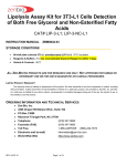
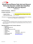
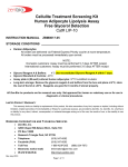
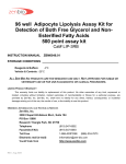
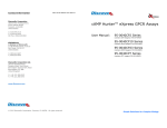
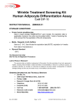
Lipolysis Assay Kit for 3T3-L1 Cells
Non-Esterified Fatty Acids Detection
100 point assay kit
Cat# LIP-2-L1; LIP-2-NC-L1
INSTRUCTION MANUAL
ZBM0041.00
STORAGE CONDITIONS
96-well plate cultured 3T3-L1 preadipocytes (LIP-2-L1) 37°C incubator
Reagents & Buffers: 4°C
Vehicle & Controls: -20°C
For in vitro Use Only
LIMITED PRODUCT WARRANTY
This warranty limits our liability to replacement of this product. No other warranties of any kind, expressed or implied, including
without limitation, implied warranties of merchantability or fitness for a particular purpose, are provided by Zen-Bio, Inc. Zen-Bio, Inc.
shall have no liability for any direct, indirect, consequential, or incidental damages arising out of the use, the results of use, or the
inability to use this product.
ORDERING INFORMATION AND TECHNICAL SERVICES
Zen-Bio, Inc.
3200 Chapel Hill-Nelson Blvd., Suite 104
PO Box 13888
Research Triangle Park, NC 27709
Telephone
(919) 547-0692
Facsimile (FAX)
(919) 547-0693
Toll Free
1-866-ADIPOSE
Electronic mail (e-mail)
[email protected]
World Wide Web
http://www.zenbio.com
Rev 8/15/2008
Page 1 of 9
(866)-234-7673
INTRODUCTION
Lipolysis plays a central role in the regulation of energy balance. Lipolysis is the process in which
triglycerides are hydrolyzed into glycerol and free fatty acids. This process releases free fatty acids (FFA) into the
bloodstream where they may be either re-esterified by the adipocyte or travel to other tissues and exert other
effects throughout the body. Elevated adipocyte lipolysis has been observed in obese and diabetic individuals
(Arner 1996). Alterations in lipolytic capacity have also been implicated in the susceptibility to obesity of AfricanAmerican individuals versus their Caucasian cohorts (Danadian et al. 2001).
The sympathetic nervous system plays a key role in the regulation of lipid mobilization. The main lipolytic
pathway involves beta-agonists ( -agonists), which activate -adrenergic receptors via the intracellular Gs proteins
in adipocytes. This leads to the activation of adenylate cyclase (AC), which then increases cyclic AMP (cAMP)
levels. Elevated cAMP acts as a second messenger to activate hormone sensitive lipase (HSL). HSL, the ratelimiting enzyme regulating adipocyte lipolysis, then catalyzes the hydrolysis of triglycerides and results in the
release of glycerol and FFA (increased lipolysis). Phosphodiesterases (PDE) are enzymes that hydrolyze cAMP
to 5’-AMP (5 prime adenosine monophosphate). This action results in a decrease in lipolysis. PDE inhibitors
increase intracellular cAMP levels. 3-isobutyl-1-methylxanthine (IBMX), a non-specific inhibitor of cAMP
phosphodiesterases (PDE), is used as the positive control if your test compounds are suspected PDE inhibitors.
Isoproterenol, a non-specific -adrenergic agonist is used as the positive control if your test compounds affect
lipolysis via -adrenergic receptors.
This lipolysis assay kit provides the tool to study chemical compounds that may influence lipolysis in
cultured human adipocytes.
Figure 1. Overview of adipocyte lipolysis
EPINEPHRINE
1,
NOREPINEPHRINE
3
2,
AR
AC
Gs
IR
PDE
ATP
cAMP
P
5’-AMP
PKA
TG
Per
HSL
FFA + glycerol
FFA + glycerol
bloodstream
Rev 8/15/2008
Page 2 of 9
ABBREVIATIONS:
AC
adenylate cyclase
AR
adrenergic receptors
Gs
G protein coupled receptor
FFA
free fatty acids
PKA
protein kinase
AMP adenosine monophosphate
ATP
adenosine triphosphate
IR
insulin receptor
PDE
phosphodiesterase
TG
triglyceride
PRINCIPLE OF THE ASSAY
Assessment of lipolytic activity is through a coupled reaction to measure non-Esterified fatty acids (NEFA)
released by adipocytes. The initial step, carried out by acyl-CoA synthetase (ACS), produces fatty acyl-CoA thiol
esters from the NEFA, ATP, Mg, and CoA in the reaction. The acyl-CoA derivatives react with oxygen in the
ACS
presence of acyl-CoA oxidase (ACOD) to
HCOOH + ATP + CoA
Acyl-CoA + AMP + PPi
produce hydrogen peroxide.
Hydrogen
(NEFA)
peroxide in the presence of peroxidase (POD)
ACOD
allows the oxidative condensation of 3Acyl-CoA + O2
2,3-trans-Enoyl-CoA + H2O2
methyl-N-ethyl-N-( -hydroxyethyl)-aniline with
4-aminoantipyrine which forms a purple
CH
NH
product that absorbs light at 550nm. This
N
N
C H OH
allows the concentration of NEFA to be
N
O
CH
POD
N
N
2H O
+
+
O
N
N
determined from the optical density measured
+ 4H O
C H OH
at 540 - 550nm.
2
2
2
5
2
4
2
5
2
4
2
2
ITEMS INCLUDED IN THE KIT
ITEM
Plate A
Assay Plate
Preadipocyte
Medium
Differentiation
Medium
Adipocyte Medium
LIP-2/3 Assay Buffer
Wash Buffer
Vehicle
Positive control
FFA Standard
FFA Diluent A
FFA Diluent B
FFA Reagent A
FFA Reagent B
Tray
DESCRIPTION
Cap
Color
UNIT
QTY
STORAGE
96 well plate 3T3-L1 preadipocytes
(LIP-2-L1 ONLY)
96-well assay plate, blank (for samples & standards
3T3-L1 Preadipocyte Medium (cat# PM-1-L1); 50ml
(LIP-2-L1 ONLY)
3T3-L1 Adipocyte Differentiation Medium (cat# DM-2L1); 15ml (LIP-2-L1 ONLY)
3T3-L1 Adipocyte Maintenance Medium (cat# AM-1-L1);
100ml (LIP-2-L1 ONLY)
100 ml
50 ml
0.1% DMSO in LIP-2/3 Assay Buffer
---
PLATE
1
37°C
---
PLATE
BOTTLE
2
1
----4°C
BOTTLE
1
4°C
BOTTLE
1
4°C
-----
BOTTLE
PURPLE
1 ml / VIAL
1
1
1
4°C
4°C
-20°C
BLUE
10 l /
VIAL
1
-20°C
AMBER
1
4°C
4°C
4°C
4°C
Isoproterenol, 10 mM in DMSO. Dilute to 1 M in Assay
Buffer before use! (i.e.1 l in 10 ml Assay Buffer)
1mM Stock. See page 5 for standard curve preparation
PINK
100 l /
VIAL
10.5ML
5.5ML
YELLOW
BOTTLE
1
1
1
PINK
BOTTLE
1
4°C
CLEAR
EACH
2
-----
YELLOW
Reconstitute using 10.5 ml FFA Diluent A. Discard
remainder after 10 days
Reconstitute using 5.5 ml FFA Diluent B. Discard
remainder after 10 days
For multi-channel pipetters, clear polyvinyl
Other equipment/reagents required but not provided with the kit:
Multi-channel Pipet , single channel pipet and pipet tips
Sterile trays for multi-channel pipetters during differentiation of cells
Plate reader with a filter of 540 nm
Incubator at 37oC
Large gauge needle
Tubes for dilution of standards
Rev 8/15/2008
Page 3 of 9
BOTTLE
ASSAY PROCEDURE
A. DIFFERENTIATION PROCEDURE
1. Preadipocytes are plated sub-confluent in 3T3-L1 Preadipocyte Medium (cat# PM-1-L1) and shipped the
next day via overnight delivery.
2. Incubate cells until they are 100% confluent (in about 4-5 days). Cells will need to be fed every other day
with PM-1-L1 during this time. See Table 1 for feeding volumes.
3. Once the cells are confluent, incubate an additional 48 hours before initiating differentiation.
4. Two days after the cells have been confluent, remove the Preadipocyte Medium (cat# PM-1-L1) and
replace with an appropriate volume 3T3-L1 Differentiation Medium (cat# DM-2-L1; see table 1 below for
recommended volumes). Incubate for 3 days.
5. Remove the 3T3-L1 Differentiation Medium and replace with 3T3-L1 Adipocyte Maintenance Medium.
Incubate for 2-3 days.
6. Feed cells every 2-3 days using 3T3-L1 Adipocyte Maintenance Medium until ready for assay. 3T3-L1
adipocytes are suitable for most assays 7-14 days post differentiation (see Table 1 and 3T3-L1 Growth
and Differentiation Feeding Schedule)
Table 1. Feeding Volumes
Format
Change PM-1-L1 to
PM-1-L1
OUT
IN
96 well plate
48 well plate
24 well plate
12 well plate
6 well plate
T-75 flask
T-25 flask
90 l/well
300 l /well
0.6 ml/well
1.2 ml/well
1.8 ml/well
12 ml/flask
4.2 ml/flask
Change PM-1-L1 to
DM-2-L1
OUT
IN
90 l/well
300 l /well
0.6 ml/well
1.2 ml/well
1.8 ml/well
12 ml/flask
4.2 ml/flask
150 l/well
500 l /well
1.0 ml/well
2.0 ml/well
3.0 ml/well
20 ml/flask
7 ml/flask
150 l / well
500 l /well
1.0 ml/well
2.0 ml/well
3.0 ml/well
20 ml/flask
7 ml/flask
Change DM-2-L1 to
AM-1-L1
OUT
IN
90 l /well
300 l /well
0.6 ml/well
1.2 ml/well
1.8 ml/well
12 ml/flask
4.2 ml/flask
120 l /well
400 l /well
0.8 ml/well
1.6 ml/well
2.4 ml/well
16 ml/flask
5.6 ml/flask
Change AM-1-L1 to
AM-1-L1
OUT
IN
90 l /well
300 l /well
0.6 ml/well
1.2 ml/well
1.8 ml/well
12 ml/flask
4.2 ml/flask
120 l /well
400 l /well
0.8 ml/well
1.6 ml/well
2.4 ml/well
16 ml/flask
5.6 ml/flask
3T3-L1 Growth and Differentiation Feeding Schedule
DAY
DAY
DAY
DAY
-2
0
3
5
proliferation
Feed
PM-1-L1
Feed
PM-1-L1
Feed
PM-1-L1
48 hrs
100%
confluent
*
Feed
DM-2-L1
Feed
AM-1-L1
DAY
DAY
DAY
DAY
DAY
9
11
13
15
7**
Feed
AM-1-L1
Feed
AM-1-L1
PREADIPOCYTE
Feed
AM-1-L1
Feed
AM-1-L1
Feed
AM-1-L1
MATURE ADIPOCYTE
nucleus
Lipid droplets
nucleus
* Once the cells are 100% confluent, incubate an additional 48 hours before initiating differentiation.
** 3T3-L1 adipocytes are suitable for most assays 7-14 days post differentiation
Rev 8/15/2008
Page 4 of 9
B. LIPOLYSIS PROCEDURE
1. Make your stock solution using whatever vehicle is appropriate for your test compounds. Dilute
your stock solutions to their final concentration in LIP-2/3 Assay Buffer (100 ml is available).
NOTE: if desired, maintain a constant concentration of solvent by preparing all compound
dilutions in the highest concentration of that solvent. Dilute your controls in assay buffer. Prepare
all vehicles as appropriate for your compounds, 0.1% DMSO has been included as the vehicle for
the positive controls. Include the Assay Buffer alone as a vehicle control. PLEASE NOTE: ZENBIO DOES NOT RECOMMEND THE USE OF SOLVENTS AT CONCENTRATIONS ABOVE 1%.
2. Remove 120 l medium from each well. Gently add 200 l Wash Buffer to all wells. Remove 200
l of the media and Wash Buffer from each well and replace with another 200 l Wash Buffer.
3. Remove all the media and Wash Buffer from the cells from triplicate wells. Treat the cells with
100 l of the test compounds resuspended in Assay Buffer three (3) wells at a time. Treat with
the diluted Isoproterenol as positive control. Use the Assay Buffer alone as one of the vehicle
controls. Please be sure to include both the vehicle provided in the kit and your vehicle (if your
test compounds are not dissolved in DMSO). The assay should be performed in triplicate.
o
4. Incubate the plates at 37 C-humidified incubator for 3 hours (for time course experiments the
longest time point recommended is 5 hours).
Note: Treatment times longer than 3 hours will
result in significant fatty acid reutilization by the adipocytes and may decrease signal relative to
total lipolysis activity.
5. Prepare the standard curve using the STANDARD SOLUTION as follows:
Briefly spin down the contents of the free fatty acid standard tube before reconstitution.
Standards are: 0, 1.4, 4.1, 12.3, 37, 111, and 333 M fatty acid. Prepare as follows:
The kit standard solution is the 1.0 mM standard. Pipette 60 l of Dilution Buffer into 6
tubes (not provided). Pipette 30 l of the FFA Standard Stock into a tube labeled 333 µM.
Prepare a dilution series as depicted below. Mix each new dilution thoroughly before
proceeding to the next. The Dilution Buffer alone serves as the zero standard.
Rev 8/15/2008
Page 5 of 9
30 l
30 l 30 l 30 l 30 l
30 l
Std
FFA
Std
333
M
111
M
37
M
12.3
M
4.1
M
1.4
M
6. Add 10.5ml FFA Diluent A to the FFA Reagent A bottle and gently invert. DO NOT VORTEX!
Store any remaining solution at 2-8 C; it is stable for 10 days after reconstitution refrigerated (28 C).
7. At the end of the incubation, 50
l of the conditioned media is removed and transferred to the
corresponding well of a blank plate for assessment of non-esterified fatty acids. [This is most
easily accomplished using a multi-channel pipet.] Add 50 l of each standard to empty wells.
8. Add the reconstituted FFA Reagent A to one of the disposable trays provided in the kit. Add 100
l of FFA Reagent A to each well. Gently shake the plate to ensure mixing. Place in a 37 oC
incubator for 10 minutes.
9. Add 5.5 ml FFA Diluent B to the FFA Reagent bottle and gently invert. Store any remaining
solution at 2-8 C; it is stable for 10 days after reconstitution refrigerated (2-8 C).
10. Add the reconstituted FFA Reagent B to the other disposable tray provided in the kit. Add 50 l of
FFA Reagent B to each well. Gently shake the plate to ensure mixing. Place in a 37 oC incubator
for 10 minutes.
11. Allow the plate to equilibrate to room temperature for 5 minutes. During this time, ensure that
there are no bubbles in the solution mixture. Use a large gauge needle or clean pipet tip to pop
any bubbles as this will result in inaccurate absorbance readings.
12. The optical density of each well is then measured at 540 nm.
Rev 8/15/2008
Page 6 of 9
FATTY ACID STANDARD CURVE
Generate standard curve: see example below
[DO NOT use this standard curve to generate your data. This is an example.]
Subtract the OD value of the 0 M standard from all OD values including the standard curve. Note: 1mM
standard is commonly omitted from analysis due to lack of linearity between 333 M and 1mM. Optionally, a 4parameter fit may be used to calculate standard curve.
OD
0.68
0.244
0.104
0.063
0.05
0.046
0.044
y = 0.0019x - 0.0045
R2 = 0.9995
OD - zero
0.636
0.2
0.06
0.019
0.006
0.002
0
Standard Curve
0.75
0.50
O.D.
M std
333
111
37
12.3
4.1
1.4
0
0.25
0.00
0
100
200
300
400
M standard
Data are expressed as M free fatty acids released.
OPTION: express data as Fold induction over appropriate vehicle
Fold induction = M free fatty acids SAMPLE
M free fatty acids VEHICLE
The R2 value should be equal or greater then 0.98 for the standard curve to be valid. Any R2 values below
0.98, must have the standard curve run again.
FREQUENTLY ASKED QUESTIONS
1. I do not have time to run the assay. Can I freeze the conditioned media in PLATE B? How long can I store
the samples before I complete the assay? Yes. The conditioned media in PLATE B can be immediately
stored at -80 C for a maximum of 7 days. Bring the conditioned media in PLATE B to room temperature
BEFORE adding the FFA Reagents A and B and completing the assay.
Rev 8/15/2008
Page 7 of 9
APPENDIX A: PLATE LAYOUT
A
B
C
D
E
F
G
H
1
2
3
4
5
6
7
8
9
10
11
12
Rev 8/15/2008
Page 8 of 9
APPENDIX B: LIP-2-L1 PROCEDURE FLOWCHART
Plate A = plate of mature 3T3-L1 adipocytes
ON DAY OF ASSAY
Make all test compound dilutions in
Assay Buffer.
Remove 120 l media from all wells.
Add 200 l Wash Buffer to all wells.
Plate A
120 l media
OOOOOOOOOOOO
OOOOOOOOOOOO
OOOOOOOOOOOO
OOOOOOOOOOOO
OOOOOOOOOOOO
200 l Wash Buffer
Plate A
Remove 120 l media & Wash
Buffer. Add another 200 l Wash
Buffer to all wells.
200 l Wash Buffer
OOOOOOOOOOOO
OOOOOOOOOOOO
OOOOOOOOOOOO
OOOOOOOOOOOO
OOOOOOOOOOOO
Add another 200 l
Wash Buffer
Plate A
Remove all media & Wash Buffer. Add 100 l
treatments/controls to 3 wells at a time.
OOOOOOOOOOOO
OOOOOOOOOOOO
OOOOOOOOOOOO
OOOOOOOOOOOO
OOOOOOOOOOOO
Remove 3 wells at a time
Add treatments 3 wells at a time
Incubate 3-5 hours at 37oC.
Plate A
Remove 50 l/well conditioned media from Plate A
to one of the blank assay plates provided. Add 50 l
FFA standards to empty wells.
Reconstitute FFA Reagent A using Diluent A.
Add 100 l/well. Incubate 10 minutes @ 37 C.
Reconstitute FFA Reagent B using Diluent B.
Add 50 l/well. Incubate 10 minutes @ 37 C.
Place at room temp. for 5 minutes. Pop any
bubbles in each well using a clean pipet tip or large
gauge needle.
Measure the optical density of each well
at 540 nm using a spectrophotometer
plate reader.
Rev 8/15/2008
Page 9 of 9
OOOOOOOOOOOO
OOOOOOOOOOOO
OOOOOOOOOOOO
OOOOOOOOOOOO
OOOOOOOOOOOO
OOOOOOOOOOOO
OOOOOOOOOOOO
OOOOOOOOOOOO
OOOOOOOOOOOO
OOOOOOOOOOOO
OOOOOOOOOOOO
OOOOOOOOOOOO
OOOOOOOOOOOO
OOOOOOOOOOOO
OOOOOOOOOOOO
blank plate
50 l
OOOOOOOOOOOO
OOOOOOOOOOOO
OOOOOOOOOOOO
OOOOOOOOOOOO
OOOOOOOOOOOO
100 l/well
FFA Reagent A
50 l/well
FFA Reagent B
OOO
OOO
OOO
OOO
OOO
OOO
OOO
OOO
OOO
OOO
An additional
plate may be
necessary for
the assay of
standards.