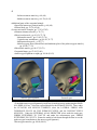
1
UNDERSTANDING
FEMALE PELVIC ANATOMY
IN 3D
USER’S MANUAL
BY
ANNE FRYE
LABRYS PRESS
AV DEPARTMENT
A DIVISION OF
MATRIX MIDWIFERY ENTERPRISES, LTD.
PORTLAND, OR
© 2013,
ALL RIGHTS RESERVED
1
TABLE OF CONTENTS
IS THIS THE MOST CURRENT VERSION OF THIS MANUAL?. . . . . . . . . . . . . . . . . . . . . . . . . . 3
INTRODUCTION. . . . . . . . . . . . . . . . . . . . . . . . . . . . . . . . . . . . . . . . . . . . . . . . . . . . . . . . . . . 4
GENERAL INFORMATION ABOUT THE 3D ANATOMY DVD. . . . . . . . . . . . . . . . . . . . . . . . . 5
UNDERSTANDING FEMALE PELVIC ANATOMY IN 3D DVD END-USER LICENSE
AGREEMENT. . . . . . . . . . . . . . . . . . . . . . . . . . . . . . . . . . . . . . . . . . . . . . . . . . . . . . . 5
Personal and Private Use by an Individual Licensee in the Developed World. . . . 6
Printing of PDF files. . . . . . . . . . . . . . . . . . . . . . . . . . . . . . . . . . . . . . . . . . . . . . 6
Group student use in a no-fee setting. . . . . . . . . . . . . . . . . . . . . . . . . . . . . . . . . 7
Individual educator use. . . . . . . . . . . . . . . . . . . . . . . . . . . . . . . . . . . . . . . . . . . . 7
Use as a Full-Time Practitioner.. . . . . . . . . . . . . . . . . . . . . . . . . . . . . . . . . . . . . 8
Use by an Independent Professional Educator. . . . . . . . . . . . . . . . . . . . . . . . . . 8
Use by an Educational Institution or Library in the Developed World. . . . . . . . . . 8
Library use of DVD by patrons.. . . . . . . . . . . . . . . . . . . . . . . . . . . . . . . . . . . . . 9
Uses by Individual Licensees Residing in Developing (i.e. third world)
Countries. . . . . . . . . . . . . . . . . . . . . . . . . . . . . . . . . . . . . . . . . . . . . . . . . . . . 9
WHAT IS ON THE FEMALE ANATOMY IN 3D DVD. . . . . . . . . . . . . . . . . . . . . . . . . . . . 14
MINIMUM SYSTEM REQUIREMENTS. . . . . . . . . . . . . . . . . . . . . . . . . . . . . . . . . . . . . . . 14
TO PLAY THE VIGNETTES ON YOUR COMPUTER. . . . . . . . . . . . . . . . . . . . . . . . . . . . . . 17
DETAILED INSTALLATION NOTES FOR THE INTERACTIVE PELVIC ANATOMY PROGRAM
. . . . . . . . . . . . . . . . . . . . . . . . . . . . . . . . . . . . . . . . . . . . . . . . . . . . . . . . . . . . . . . . 19
VIGNETTES INTRODUCING SOFT TISSUE REGIONS OF THE FEMALE PELVIS. . . . . . . . . . . .
DETAILS ABOUT PLAYING AND VIEWING THE VIGNETTES. . . . . . . . . . . . . . . . . . . . . .
The regions discussed. . . . . . . . . . . . . . . . . . . . . . . . . . . . . . . . . . . . . . . . . . . . . . .
Depiction of anatomical parts. . . . . . . . . . . . . . . . . . . . . . . . . . . . . . . . . . . . . . . . .
Vocabulary. . . . . . . . . . . . . . . . . . . . . . . . . . . . . . . . . . . . . . . . . . . . . . . . . . . . . . .
Closed captioning. . . . . . . . . . . . . . . . . . . . . . . . . . . . . . . . . . . . . . . . . . . . . . . . . .
Playing the vignettes on a computer. . . . . . . . . . . . . . . . . . . . . . . . . . . . . . . . . . . .
Playing the vignettes on a television.. . . . . . . . . . . . . . . . . . . . . . . . . . . . . . . . . . .
Repeat play (looping). . . . . . . . . . . . . . . . . . . . . . . . . . . . . . . . . . . . . . . . . . . . . . .
COMMENTS ON EACH VIGNETTE. . . . . . . . . . . . . . . . . . . . . . . . . . . . . . . . . . . . . . . . . .
Vignette 1: Urethrovaginal Sphincter Muscle Complex.. . . . . . . . . . . . . . . . . . . .
Vignette 2: Perineal Membrane Connective Tissue Complex. . . . . . . . . . . . . . . .
Vignette 3: Clitoral Complex. . . . . . . . . . . . . . . . . . . . . . . . . . . . . . . . . . . . . . . . .
Vignette 4: Levator Ani Muscle Complex. . . . . . . . . . . . . . . . . . . . . . . . . . . . . . .
Vignette 5: External Anal Sphincter.. . . . . . . . . . . . . . . . . . . . . . . . . . . . . . . . . . .
VIGNETTE SCRIPTS. . . . . . . . . . . . . . . . . . . . . . . . . . . . . . . . . . . . . . . . . . . . . . . . . . . .
22
22
22
22
22
23
23
23
23
23
23
23
24
24
24
25
2
INTERACTIVE 3D PELVIC ANATOMY PROGRAM E-LEARNING TOOL. . . . . . . . . . . . . . . . .
USING THE PROGRAM. . . . . . . . . . . . . . . . . . . . . . . . . . . . . . . . . . . . . . . . . . . . . . . . . .
Familiarizing Yourself with the Screen. . . . . . . . . . . . . . . . . . . . . . . . . . . . . . . . .
List to the Left of the Screen. . . . . . . . . . . . . . . . . . . . . . . . . . . . . . . . . . . . . . . . .
Scroll bar. . . . . . . . . . . . . . . . . . . . . . . . . . . . . . . . . . . . . . . . . . . . . . . . . . . . . .
Speaker. . . . . . . . . . . . . . . . . . . . . . . . . . . . . . . . . . . . . . . . . . . . . . . . . . . . . . .
Anatomical part. . . . . . . . . . . . . . . . . . . . . . . . . . . . . . . . . . . . . . . . . . . . . . . . .
Viewport. . . . . . . . . . . . . . . . . . . . . . . . . . . . . . . . . . . . . . . . . . . . . . . . . . . . . .
Moving the model. . . . . . . . . . . . . . . . . . . . . . . . . . . . . . . . . . . . . . . . . . . .
Orientation cube.. . . . . . . . . . . . . . . . . . . . . . . . . . . . . . . . . . . . . . . . . . . . .
Rotation icon. . . . . . . . . . . . . . . . . . . . . . . . . . . . . . . . . . . . . . . . . . . . . . . .
Names of anatomical parts.. . . . . . . . . . . . . . . . . . . . . . . . . . . . . . . . . . . . .
Selecting/deselecting parts. . . . . . . . . . . . . . . . . . . . . . . . . . . . . . . . . . . . . . . . . . .
Default setting–view all. . . . . . . . . . . . . . . . . . . . . . . . . . . . . . . . . . . . . . . . . .
Making parts appear/disappear. . . . . . . . . . . . . . . . . . . . . . . . . . . . . . . . . . . . .
Working with regions. . . . . . . . . . . . . . . . . . . . . . . . . . . . . . . . . . . . . . . . .
Isolating one part or region. . . . . . . . . . . . . . . . . . . . . . . . . . . . . . . . . . . . .
Working with individual parts. . . . . . . . . . . . . . . . . . . . . . . . . . . . . . . . . . .
Displaying the coxal (innominate or hip) bones and central pubic bone
region. . . . . . . . . . . . . . . . . . . . . . . . . . . . . . . . . . . . . . . . . . . . . . . .
Transparent parts.. . . . . . . . . . . . . . . . . . . . . . . . . . . . . . . . . . . . . . . . . . . . . . . . . .
Orientation of parts. . . . . . . . . . . . . . . . . . . . . . . . . . . . . . . . . . . . . . . . . . . . . . . . .
Focus.. . . . . . . . . . . . . . . . . . . . . . . . . . . . . . . . . . . . . . . . . . . . . . . . . . . . . . . . . . .
Zooming in and out.. . . . . . . . . . . . . . . . . . . . . . . . . . . . . . . . . . . . . . . . . . . . . . . .
List of anatomical parts.. . . . . . . . . . . . . . . . . . . . . . . . . . . . . . . . . . . . . . . . . . . . .
Notes on selected model parts.. . . . . . . . . . . . . . . . . . . . . . . . . . . . . . . . . . . . .
Female pelvis. . . . . . . . . . . . . . . . . . . . . . . . . . . . . . . . . . . . . . . . . . . . . . . .
Superficial transverse perineal muscle. . . . . . . . . . . . . . . . . . . . . . . . . . . .
Greater vestibular (Bartholin) glands. . . . . . . . . . . . . . . . . . . . . . . . . . . . .
Length of perineal body.. . . . . . . . . . . . . . . . . . . . . . . . . . . . . . . . . . . . . . .
Lateral fibers and tendinous plate of the pubococcygeal muscle. . . . . . . .
External anal sphincter models. . . . . . . . . . . . . . . . . . . . . . . . . . . . . . . . . .
Space between the external anal sphincter and coccyx. . . . . . . . . . . . . . . .
32
32
32
32
32
32
32
33
33
33
33
33
34
34
34
34
34
35
35
36
36
36
37
37
39
39
39
40
40
40
40
40
STUDYING WITH THE 3D ANATOMY PROGRAM. . . . . . . . . . . . . . . . . . . . . . . . . . . . . . . . . 41
BIBLIOGRAPHY. . . . . . . . . . . . . . . . . . . . . . . . . . . . . . . . . . . . . . . . . . . . . . . . . . . . . . . . . . 43
ABOUT THE GRAPHICS DESIGNER AND GAMING PROGRAMERS. . . . . . . . . . . . . . . . . . . . . 44
3
IS THIS THE MOST CURRENT VERSION OF THIS MANUAL?
If you are looking at the version of this manual included with the DVD, it may not be the
most current. To double-check on this, go to my website:
<www.midwiferybooks.com/anatomy_users_manual.pdf>. There you will find a link that
takes you right to the manual posted on my website, and the file date will be shown. The
help screen in the interactive 3D pelvic anatomy tool also points you to the website for this
manual, just in case there is a more recent version posted there.
<www.midwiferybooks.com/anatomy_users_manual.pdf>
Version date: August 2, 2013
4
INTRODUCTION
The programs on this DVD represent four years of weekly work sessions beginning in
March 2007 and ending in April of 2011. They were inspired by the dearth of illustrations
depicting the most current understandings of female pelvic soft tissue arrangements. In
researching the 6th edition of HEALING PASSAGE I found out many new things about female
pelvic anatomy that were either not explained or illustrated in textbooks or poorly explained
in the few that did try to include more recent information. No textbook that I could find to
date even attempted to show the pelvic floor muscles as they are in life–uplifted–rather than
sagging in a bowl shape, as they do after death due to lack of muscle tone. As a result, I was
inspired to create these tools as a way to help students, teachers and practitioners better
understand this anatomy. Please remember that, while I did exhaustive research and have
done my best to depict each part accurately, the work that is being done to understand these
new concepts is continuing. Therefore there may be mistakes in these presentations that will
have to be corrected in future versions. Nevertheless, I believe I have created a useful and
unique pair of tools that will be of benefit to the midwifery and obstetric communities. I
hope you find them helpful.
Anne Frye
May 20, 2013
5
GENERAL INFORMATION ABOUT THE 3D ANATOMY DVD
The material on the 3D Anatomy DVD has been designed to accompany the 6th edition
of the text HEALING PASSAGE: A MIDWIFE’S GUIDE TO THE CARE AND REPAIR OF THE
TISSUES INVOLVED IN BIRTH, by Anne Frye. The tools on the DVD augment the
information found in the Anatomy section of the text, but do not stand alone. For complete
information, reference to the anatomy portion of the text is essential. If you need a copy of
the text, it may be obtained by visiting the website: www.midwiferybooks.com. This DVD
is sold separately from the text and the anatomy portion of the text is included so that it will
be useful of all types of students of female pelvic anatomy.
UNDERSTANDING FEMALE PELVIC ANATOMY IN 3D DVD END-USER LICENSE
AGREEMENT
Please read through this section, if you do not agree with these stipulations, do not
remove the shrinkwrap on the DVD and arrange for a return. This agreement is quite
detailed with lots of legalize. The part most relevant to daily use, the types of permissions
granted according to licensing fees paid, is included at the beginning. I have tried to
anticipate the variety of ways licensees may wish to use this software. If you discover that
you want to use the material in a new way, for example you paid the individual price and
now want to start using it as an independent professional educator, you can simply call me
to pay the difference to bring yourself into compliance. If there is some question left
unanswered here, please call or email me to ask about legitimate uses that may not be
addressed in this document.
ACCEPTANCE OF THIS AGREEMENT
YOU ACCEPT AND AGREE TO BE BOUND BY THE TERMS OF THIS AGREEMENT BY
PROCEEDING WITH BREAKING AND REMOVING THE SHRINKWRAP ON THE
SOFTWARE PRODUCT OR BY INSTALLING, USING, OR COPYING THE SOFTWARE
PRODUCT. YOU MUST AGREE TO ALL OF THE TERMS OF THIS AGREEMENT BEFORE
YOU PROCEED TO REMOVE THE SHRINKWRAP FROM THE SOFTWARE PRODUCT. IF
YOU DO NOT AGREE TO ALL OF THE TERMS OF THIS AGREEMENT, YOU MUST NOT
OPEN, INSTALL, USE, OR COPY THE SOFTWARE PRODUCT.
LICENSING OF THE SOFTWARE PRODUCT
The two tools included on the DVD “Understanding Female Pelvic Anatomy in 3D”
(“collectively referred to as the “Software Product” or “DVD”) and accompanying documentation
are licensed and not sold. This Software Product is protected by copyright laws and treaties, as well
as laws and treaties related to other forms of intellectual property. Labrys Press or its subsidiaries,
affiliates, and suppliers (collectively “Labrys Press”) own intellectual property rights in the Software
Product. The Licensee’s (“you” or “your”) license to download, use, copy, or make changes to the
Software Product is subject to these rights and to all the terms and conditions of this End User
Licensing Agreement and Terms of Use (“Agreement”).
6
Categories of use licensing permissions
There are several categories of licenses (there are some special provisions for users in developing
countries described under a separate header in this Agreement). Licensing is priced according to the
type of use being sought. There categories are:
•Individual use (personal, private use, or use in a no-fee private study group comprised only of
students). This category includes permissions as follows:
-Individual educator use (Includes use for certain types of teaching up to twice yearly.)
-Limited practitioner use (Includes one-on-one teaching with up to 5 clients yearly or for
teaching up to 5 private clients and their partners yearly in groups [such as when you
offer your own childbirth education classes] in the course of your clinical practice.)
•Full-time practitioner use (Includes all of the above permissions plus use with an unlimited
number of clients for one-on-one teaching and for teaching private clients and their partners
in groups [such as when you offer your own childbirth education classes] in the course of
clinical practice.)
•Independent professional educator (For someone not affiliated with a formal school and who
is not teaching for any type of educational program that grants credentials to professionals
[CEU-granting presentations are ok] that will regularly show content to paying audiences
more than twice yearly.)
•Institutional use (For all types of formal educational programs.) While the permissions are the
same, there are two levels of pricing for institutional licensing:
-Direct-entry midwifery schools and their libraries, as well as all other types of certification
programs that are not university-based
-Institutional rate, all other categories (Universities and their libraries, Public libraries)
•Special licensing level for permanent residents and natives of developing countries
If you have paid for the DVD at an individual price, but now find you wish to use it when
teaching workshops, for example, you can bring your copy into compliance by simply calling the
office and paying the difference in the licensing fee.
Personal and Private Use by an Individual Licensee in the Developed World
Individual licensees are defined as students or professionals, including all types of educators,
who wish to use the DVD only for their own personal and private study. They may copy the contents
of this DVD to up to two of their personal computing devices that will not to be used simultaneously
for the viewing of the software. They may view the vignettes on any television or other device that
allows such viewing. The individual user may copy the files to a flash drive or other type of media
or storage device if necessary to transfer them to their computer or other compatible device. An
archival copy can be made to avoid loss should something happen to the original DVD.
When a computing device is retired, the user should first delete all of the software content from
the hard drive or removable drive before disposal or passing on the device to another for their use.
Once installed on their own computing devices, individual licensees may always show this
material to other interested individuals for the purpose of review so that they can decide if they wish
to purchase a license for themselves. Individual users should not charge for viewing this DVD, other
than as allowed for under in the terms described below.
Printing of PDF files: This DVD contains a PDF file of the user’s manual as well as a copy of the
anatomy portion of my text HEALING PASSAGE. You may print a copy of the user’s manual. You
7
may want to print it in color, as it contains a few color screen images.
The anatomy portion of the text is formatted to prevent inadvertent alteration. It is also indexed
and typos that appear in the print version have been corrected. It can be searched, moved to a
computer or tablet for reading on screen and the licensee is allowed to print one copy for personal
use only. It should not be duplicated or otherwise shared with others in any manner for any purpose.
If your one copy becomes lost or destroyed, you may print out another copy for your personal use.
Group student use in a no-fee setting: Small groups of students studying independently at a
private home or community room (but not at a school, school library or other educational institution)
may view the contents of an individually licensed software as a group, as long as the contents are not
copied to devices other than those owned by the individual licensee. Each student should have their
own text and their own DVD software for their private use at home. Students participating in a study
group that takes place at a school or school library should use the school’s copy. A student group
is defined as a gathering of students purely for the purpose of study; there should be no fees involved
for participating in the group and no expert teaching the class (that use falls in the next category).
Individual educator use: Individual licensees who offer independent educational presentations for
the sole purpose of education (ie., no professional credential is granted) or the granting of CEUs
(continuing education units), that is, a presentation not associated with a school or other educational
institution or certification program (except during childbirth education or similar classes offered to
the public), may use their personal software product license when teaching in venues such as at
conferences, workshops and similar offerings, whether free or presented for a fee up to two times
a year (use exceeding twice yearly that occurs on a regular basis [defined as more than two
consecutive years in a row] warrants licensing the software product at the independent educator’s
rate, you can call Labrys Press to report the new use and pay the difference to bring yourself into
compliance with this permissions agreement).
When offering CEUs licensee should include information about how the text and DVD can be
obtained verbally in their presentation and in writing in any handouts they may give out. If a
Powerpoint presentation is also used, this information could be included for students to copy down.
The following information should be included on a slide, in writing on a handout, or both (if both
media are used):
“UNDERSTANDING FEMALE PELVIC ANATOMY IN 3-D is designed to accompany the 6th
edition of the text HEALING PASSAGE: A MIDWIFE’S GUIDE TO THE CARE AND REPAIR OF
THE TISSUES INVOLVED IN BIRTH, by Anne Frye. The text and the DVD may be obtained
by visiting the website: www.midwiferybooks.com. For those who are not in the field of
maternity care, a copy of the anatomy portion of the HEALING PASSAGE is included on the
disk. Pricing varies according to the intended use, check the website.”
Limited practitioner use: The personal and private use license permits using the material for the
purpose of teaching up to 5 clients yearly on a one-on-one basis in the course of clinical care as well
as during childbirth education or similar classes taught by yourself for the same 5 or fewer private
clients up to twice yearly.
8
Use as a Full-Time Practitioner
This licensing level includes all permissions for personal and private use plus the use of the material
with an unlimited number of clients registered with your practice for one-on-one teaching or during
classes specific to your type of practice (such as childbirth or pelvic floor therapy classes) that are
only offered to clients registered with your practice. If you will be using the material in childbirth
or other types of classes that are offered to the general public (i.e., others who are not registered with
your clinical practice), please select the Independent Professional Educator level of licensing.
Use by an Independent Professional Educator
An independent professional educator is defined as a person who is not affiliated with a school
or university or professional practitioner certificate-granting program of any kind who will use the
DVD regularly to teach audiences of students or the public (such as during childbirth education
classes for pregnant parents) for a fee more often than twice yearly on a regular basis, whether or not
CEUs are granted. (What this means is you can be a certified instructor yourself but you are not
certifying others to become instructors in your classes, rather you are teaching students, other
professionals for CEUs only or the general public.) Regular use is defined as showing the DVD
more than twice yearly for two years in row. Apart from the number of and type of teaching
engagements, all other individual use permissions and restrictions apply.
Use by an Educational Institution or Library in the Developed World
DVDs for institutional use are licensed at a price that grants permission for repeated viewing and use
of the material by groups of tuition-paying students or the public, as would be the case for a pubic
library. Institutions include but are not limited to schools, colleges, medical libraries and universities
or any other formal educational programs, apart from short independent classes or workshops taught
by an individual or independent professional educator/licensee as described above.
Educational institutional use includes use by faculty or students at established in-house program,
during intermittent intensive classes with interposed independent/at-a-distance study or for any
at-a-distance training programs, in short, any type of program that grants some kind of certificate of
completion (other than CEUs [continuing education units]). This includes but is not limited to such
programs intended to credential professionals, including but not limited to such specialities as doula
training, childbirth educator certification, pelvic floor therapy training, nursing or midwifery
certificate training or credentialing venues of any other type.
At-a-distance programs may use the software product in webinars as they might in a regular
classroom but are not permitted to install the software on a server or in any other manner make it
available for access by students outside the “classroom” setting: students should be directed to obtain
individual licenses for their personal, private study.
School libraries should purchase a separate license for general use by library patrons of all types.
A department within an institution (including libraries) can license a single copy at the institutional
price for use by up to 5 (five) faculty members. Students that are registered in the classes of these
instructors may be granted access to the software in the library should they miss a class for the
purposes of catch-up. However, for general use by all library patrons, a second copy should be
obtained. Even if a faculty member has an individual license to use the software product privately,
the school should use a copy licensed at the appropriate institutional level for use in the classroom,
seminar, webinar or library setting.
Visiting faculty: The only exception to these rules is when a visiting instructor wishes to use the
9
software program in a presentation to be given at an institution where the faculty has never seen the
material before. In such cases, the visiting faculty may use the DVD in their presentation, which will
allow the students and resident faculty members to view the material. If the visiting instructor is also
connected to a college, university or other educational institution, ideally, the copy used should be
one that was licensed by the home institution of the visiting faculty member. If such a copy is not
available, the instructor may use their own personal copy in this special circumstance; the rules for
use by an independent educator apply. After that, however, the institution where the presentation
took place should obtain a copy for their continued use and for use by future visiting faculty if they
wish to incorporate this material into their program.
Library use of DVD by patrons: I recommend that a printed copy of the DVD User’s Manual and
a printed copy of the PDF Anatomy file (excerpt from the text) be given to the patron with the DVD
whenever it is to be used. If the library has a copy of the complete textbook, HEALING PASSAGE, it
can be included or at least recommended when the DVD is used, at the library’s discretion. Perhaps
a copy of the text can be purchased that will always accompany the audio/visual tools. Because the
disk includes a copyrighted program that must be installed on a computer before it can be used, I
highly recommend that the library designate this material as “reference use only” and install the
interactive 3D anatomy program on a library computer, for in-house patron use only.
Uses by Individual Licensees Residing in Developing (i.e. third world) Countries
DVDs licensed at the individual price rate may be used for any educational purpose if the
presentation is taking place in a developing country and the owner of the DVD is a native or
permanent resident of that country. All other restrictions about copying of the software product still
apply. Up to ten copies of the PDF file of the anatomy portion of the text may be made and
distributed to students. Permission to make additional copies must be requested by e-mail
([email protected]). If the licensee has access to funding that would cover the
institutional rate for the DVD, full payment of the direct-entry midwife institutional rate would be
appreciated but is not required, if the money could go to cover other necessary expenses.
RESPECT FOR COPYRIGHT
Using this software product in any manner other than as set forth in this Agreement without the
express permission of Anne Frye and Labrys Press constitutes software piracy. It’s the same as
coming by my table at a conference and taking a DVD, something you probably would not do. In
order to make my products available in the most useable format and at a reasonable price, I am
depending on you, my customers, to protect my copyrights and thus keep me in business. I appreciate
your understanding and support.
If you have somehow acquired a copy of this software that you did not pay for directly or that
was not a gift of the new, unopened DVD, it is an illegal copy. Call Labrys Press and pay for your
copy to bring yourself into compliance with this license or delete it from your device immediately.
RETURNS POLICY
Once the shrink-wrap is broken, the product is non-returnable except for detects in material or
workmanship. If the DVD is defective, contact Labrys Press immediately at 503-255-3378. You
will then be instructed about how to return it for a replacement.
10
TECHNICAL SUPPORT
Technical support from Labrys Press for Software Products is primarily limited to questions
regarding the use of the program itself once it is successfully installed. Labrys Press is not equipped
to provide computer hardware or operating system (Windows) support; for that you must consult an
independent computer expert at your own expense.
RESTRICTIONS ON COPYING
You may not copy any part of the Software Product except to the extent that licensed use
inherently demands the creation of up to two temporary copies stored in computer memory on two
devices and not permanently affixed on storage medium. Copying of this DVD or its contents for
the purpose of loaning, giving or selling it to another person or institution is prohibited. The only
exception to the selling rule is in the event that you discover that you cannot play the DVD on your
computer or that the program will not install. In this case only, you may transfer (sell) your license
to another person once you have contacted Labrys Press and explained the situation.
ARCHIVAL COPY PERMISSION
General terms: Your physical DVD will serve as your archival copy of the Software Content.
In the event of the release of a downloadable version, in addition to permission to install and use the
Software Product on two personal computing devices, you may make one archival copy of the
Software Product. The archival copy must be on a storage medium other than a hard drive, and may
only be used for the reinstallation of the Software Product. This Agreement does not permit the
installation or use of additional copies of the Software Product, or the installation of the Software
Product on more than two personal devices at any given time, on a system that allows shared used
of applications, on a multi-user network, or on any configuration or system of computers that would
be used to allow simultaneous viewing of the software.
MULTIPLE COPY AND USE PERMISSION MUST BE GRANTED INDIVIDUALLY
Other than as set forth in this Agreement, multiple copy use or installation is only allowed if you
obtain an appropriate licensing agreement for each user and each copy of the Software Product. For
further information regarding multiple copy licensing of the Software Product, please contact:
Representative: Labrys Press, 7528 NE Oregon St., Portland, OR 97213,
www.midwiferybooks.com
RESTRICTIONS ON TRANSFER
Without first obtaining the express written consent of Labrys Press, you may not assign your
rights and obligations under this Agreement, or redistribute, encumber, sell, rent, lease, sublicense,
or otherwise transfer your rights to the Software Product. The only exception to this is when a copy
has been purchased and you find it cannot be used on your computer. In this event, you may resell
your copy for the price you paid to another who can use it once you have obtained permission to do
so from Labrys Press.
RESTRICTIONS ON USE
Except as set forth in the section of this Agreement entitled “Categories of use licensing
permissions” you may not use, copy, or install the Software Product on any nonpersonal computing
11
system with more than one computer (i.e. a networked system, even one owned by the licensee that
is also used by persons other than the licensee), or permit the use, copying, or installation of the
Software Product by more than one user or on a computer that is not personally owned by the
licensee. You may not permit any third party to benefit from the use or functionality of the Software
via a rental, lease, timesharing, service bureau, hosting service, or other arrangement; or upload, host,
use or access the Software via a timesharing, service bureau, virtualization, application hosting or
other remote access arrangement.
If you hold multiple, validly licensed copies, you may not use, copy, or install the Software
Product on any system with more than the number of computers permitted by license, or permit the
use, copying, or installation by more users, or on more computers than the number permitted by
license. If a computer is retired and replaced by another, the software may be transferred to the new
device as long as it is deleted from the “old” device, in keeping with the license you purchased.
You may not decompile, “reverse-engineer,” disassemble, or otherwise attempt to derive the
source code for the Software Product.
RESTRICTIONS ON ALTERATION
You may not modify the Software Product or create any derivative work of the Software Product
or its accompanying documentation. Derivative works include but are not limited to translations.
You may not alter any files or libraries in any portion of the Software Product. You may not
reproduce the database portion or create any tables or reports relating to the database portion.
DISCLAIMER OF SOFTWARE PRODUCT WARRANTY
This DVD is distributed “as is” in the hope that it will be useful, but without warranty of any
kind, either expressed or implied, including, but not limited to, warranties of merchantability and
fitness for a particular purpose. While significant effort has been made to determine minimum
system requirements and to ensure that the software product will operate as expected when installed
on a system that meets these requirements, and to ensure that the DVD content is free of defects in
programing, no guarantees are made as to whether it will perform as expected on any given device
or computer. Labrys Press makes no warranty that operation of the Software Product will be secure,
error free, or free from interruption.
You must determine whether the software product sufficiently meets your requirements for
security and uninterruptability. You bear sole responsibility and all liability for any loss incurred due
to failure of the software product to meet your requirements. The entire risk as to the quality and
performance of the program is with you. Should the program prove defective, you assume the cost
of all necessary servicing, repair or correction as a result of installation.
In no event, unless required by applicable law, will the author be responsible for or liable to you
for damages, including any general, special, incidental or consequential damages arising out of the
installation or use or inability to use the program (including, but not limited to, loss of or damage
to data on any computer or information storage device, data being rendered inaccurate or losses
sustained by you or third parties or a failure of the program to operate with any other programs), even
if the author has been advised of the possibility of such damages.
12
LIMITED STORAGE MEDIUM WARRANTY
For a period of 60 days from the date of shipment or from the date that you download the
Software Product, as applicable, Labrys Press warrants that when properly handled and used under
normal conditions, the storage medium (i.e. DVD disk) on which the Software Product is shipped
will be free of defects in material and workmanship.
DISCLAIMER OF OTHER WARRANTIES AND LIMITATION OF LIABILITY
UNLESS OTHERWISE EXPLICITLY AGREED TO IN WRITING BY LABRYS PRESS,
LABRYS PRESS MAKES NO OTHER WARRANTIES, EXPRESS OR IMPLIED, IN FACT OR
IN LAW, INCLUDING, BUT NOT LIMITED TO, ANY IMPLIED WARRANTIES OF
MERCHANTABILITY OR FITNESS FOR A PARTICULAR PURPOSE OTHER THAN AS SET
FORTH IN THIS AGREEMENT OR IN THE DOCUMENTS PROVIDED WITH THE
SOFTWARE PRODUCT.
UNDER NO CIRCUMSTANCES SHALL LABRYS PRESS, ITS DIRECTORS, OFFICERS,
EMPLOYEES OR AGENTS BE LIABLE TO YOU OR ANY OTHER PARTY FOR INDIRECT,
CONSEQUENTIAL, SPECIAL, INCIDENTAL, PUNITIVE, OR EXEMPLARY DAMAGES OF
ANY KIND (INCLUDING LOST REVENUES OR PROFITS OR LOSS OF BUSINESS)
RESULTING FROM THIS AGREEMENT, OR FROM THE FURNISHING, PERFORMANCE,
INSTALLATION, OR USE OF THE SOFTWARE PRODUCT, WHETHER DUE TO A BREACH
OF CONTRACT, BREACH OF WARRANTY, OR THE NEGLIGENCE OF LABRYS PRESS OR
ANY OTHER PARTY, EVEN IF LABRYS PRESS IS ADVISED BEFOREHAND OF THE
POSSIBILITY OF SUCH DAMAGES. TO THE EXTENT THAT THE APPLICABLE
JURISDICTION LIMITS LABRYS PRESS'S ABILITY TO DISCLAIM ANY IMPLIED
WARRANTIES, THIS DISCLAIMER SHALL BE EFFECTIVE TO THE MAXIMUM EXTENT
PERMITTED.
LIMITATION OF REMEDIES AND DAMAGES
Your remedy for a breach of this Agreement or of any warranty included in this Agreement is
the replacement of the Software Product. Selection of whether to replace the DVD shall be solely
at the discretion of Labrys Press. Labrys Press reserves the right to substitute a functionally
equivalent copy of the Software Product as a replacement. If Labrys Press is unable to provide a
replacement or substitute Software Product or corrections to the Software Product, your sole
alternate remedy shall be a refund of the purchase price for the Software Product exclusive of any
costs for shipping and handling.
Any claim must be made within the applicable warranty period. All warranties cover only defects
arising under normal use and do not include malfunctions or failure resulting from misuse, abuse,
neglect, alteration, problems with electrical power, acts of nature, unusual temperatures or humidity,
improper installation, or damage determined by Labrys Press to have been caused by you. All limited
warranties on the Software Product are granted only to you and are non-transferable. You agree to
indemnify and hold Labrys Press harmless from all claims, judgments, liabilities, expenses, or costs
arising from your breach of this Agreement and/or acts or omissions.
13
GOVERNING LAW, JURISDICTION AND COSTS
This Agreement is governed by the laws of Oregon, without regard to Oregon's conflict or choice
of law provisions.
SEVERABILITY
If any provision of this Agreement shall be held to be invalid or unenforceable, the remainder
of this Agreement shall remain in full force and effect. To the extent any express or implied
restrictions are not permitted by applicable laws, these express or implied restrictions shall remain
in force and effect to the maximum extent permitted by such applicable laws.
CONTRACTURAL CHANGES
Labrys Press may change the terms of this agreement at any time. These changes will be made
known to users by being posted on the website www.midwiferybooks.com.
14
WHAT IS ON THE FEMALE ANATOMY IN 3D DVD
This DVD presents two e-learning tools consisting of a series of five brief films or vignettes,
introducing selected regions of female pelvic soft tissue anatomy and an interactive program to study
selected individual parts of the female pelvic anatomy in virtual 3D. There is also a copy of this user
manual as a PDF file. Any revisions will be noted in the file that can be found on my website by
clicking on the link to the file shown in the About window.
Finally, there is a PDF file of the anatomy portion of the HEALING PASSAGE textbook on the disk.
This is included so that anatomy students who are not maternity care specialists can have access to
this part of the text as an adjunct to learning. It cannot be printed out but can be copied to a
computer or tablet for display.
MINIMUM SYSTEM REQUIREMENTS
•PC Operating system (incl. 64-bit): Windows 8, Windows 7 (SP 1), Vista (SP2), Windows XP
(SP2)
•MAC computer built from 2006 on (with Intel processor) using Boot Camp or other Windows
compatibility software that allows booting with any of the above Windows operating systems
•PC with a Pentium 4, 1.5 processor
•512 MB RAM
•50 MB available disk space
•800×600 screen resolution, 24-bit color
•Real-time 3D graphics card with Direct X9 (3D9) or greater & Shader model 1.1
•DVD drive
•Manual input capability (i.e., mouse, trackball or tablet)
•Internet access (for installation and to download current version of User’s Manual only)
There are no returns on an opened Anatomy DVD package because there is no way to verify if the
program has been installed or not. To avoid disappointment, I have included details below about
how to tell if your computer system is likely to be compatible with the program. The following notes
are far beyond what other companies offer to try and help ensure compatibility and are provided for
your convenience.
Minimum requirements mean that the program installed and worked as expected on a computer that
was configured and had hardware matching these specifications. PLEASE NOTE THAT NOTHING
CAN GUARANTEE that your system will be fully compatible until the program has been installed,
as numerous unpredictable variables come into play with each computer. However, chances are
excellent that, if your system meets these criteria, the program will work as expected.
This detailed checklist explains how to determine if your system meets the above listed
minimum criteria to run the Interactive Pelvic Anatomy software:
•You will need one of these operating systems:
15
•Windows 8
•Windows 7 with Service Pack 1 or above
•Windows Vista with Service Pack 2
•Windows XP with Service Pack 2
NOTE: The program was tested and worked as expected on each of these operating systems prior
to release. However, I have no control over any changes that Microsoft may make to any of these
operating systems that may render them incompatible.
•Macintosh computer built in 2006 or later (i.e., that have an Intel processor) using Boot Camp or
other Windows compatibility software with any of the above Windows operating systems
installed in a dual boot configuration; otherwise this program will not run on an Apple computer.
(While there are programs that can be directly installed on a MAC that will allow you to run
certain Windows programs without having to have Windows installed, such configurations have
NOT been tested with this DVD.)
•PC with a Pentium 4, 1.5 GHz core processing unit (CPU) or equivalent (or a MAC with an Intel
processor; which should be true for any MAC built after 2005 [2006 or later])
•512 MB RAM (Random Access Memory)
NOTE: This just happens to be the amount of RAM on the oldest and least sophisticated computer
on which we tested the program; it may work just fine with fewer MB but this hasn’t been tested.
If you don’t have enough RAM, you will need to confirm how much RAM your computer can
take. Windows XP will not work with more than 2 GB RAM. Fortunately, if you need more
RAM, it is not that expensive and is easy to install, just make sure it matches the type you
already have. You can check your computer motherboard manual for details about the type of
RAM you need. If you don’t have the manual, you can look at the RAM chips already installed
to see what kind they are. You can also decide its time to buy a new system, because if you have
less than a GB of RAM that means you have a pretty old computer.
NOTE: To check on your type of CPU and the amount of RAM, click on START, then CONTROL
PANEL, then SYSTEM. These details appear in the center of the window. The Pentium 4 core
processor was introduced in November of 2000 and sold until August of 2008. Any CPU made
after 2000 will probably work just fine to run the program but most have not been tested.
•At least 50 MB of available hard drive space
NOTE: To check how much free space you have on your PC hard drive, click on START, then
COMPUTER (or MY COMPUTER), then right click on the C drive icon. A drop-down menu
should appear. At the bottom of the list choose PROPERTIES. The free space is listed above
the drive graphic.
•Screen resolution of at least 800 × 600 (however, the higher the resolution, the more smoothly the
program will work and the brighter and crisper the graphics will appear), with at least 24-bit
color (This measurement refers to color depth, which is the number of color values that can be
16
assigned to a single pixel in an image. Color depth can range from 1-bit [black-and-white] to 32bits [over 16.7 million colors]).
MONITOR TYPE & SIZE
RECOMMENDED
SCREEN RESOLUTION
CRT 15 inch
1024 × 1024
CRT 17 to 19 inch
1280 × 1024
CRT 20 inch or larger
1600 ×1200
LCD 19 inch standard ratio
1280 ×1024
LCD 20-inch standard ratio
1600 ×1200
LCD 10-22 inch widescreen
2680 × 1050
LCD 24-inch widescreen
1920 ×1200
Laptop 13-15 inch standard ratio
1400 ×1050
Laptop 13-15 inch widescreen
1280 × 800
Laptop 17 inch widescreen
1680 × 1050
NOTE: To check screen resolution and the ##-bits of color you are running on your PC:
•Go to START, CONTROL PANEL, DISPLAY (Or go to the START menu, click on HELP &
SUPPORT and enter “adjust display settings” in the search bar, click ENTER)
•In Windows XP, select the SETTINGS tab on the far right, look around for the resolution and ##-bit
color. If the color is set at 16-bit, try to change it to 24-bit, you may have to raise the screen
resolution to do so (see table above).
•In Windows 7, you go through the same steps but note that the “edit display” window looks quite
different than in older versions of Windows. The screen resolution drop down menu will appear
in the center of the first window. To find out the ##-bit color type, click on ADVANCED
SETTINGS, and then click the Monitor tab. Under Colors, select True Color (32-bit), and then
click OK. (If it won’t let you select at least 24-bit color [32-bit is fine too], check that your
resolution is set to the optimal level and then try again.)
•Real-time 3D graphics video card that is compatible with the following Microsoft OS software
utilities:
•Direct X9 (3D 9) or greater
•Shader model 1.1 or greater
(An ATI Radion 9800 Pro video card works, which was released in 2003)
NOTE: The video card is probably the most critical piece of hardware. While the vignettes should
play on any computer that has a DVD player with media software installed, the 3D graphics in
the program are a bit more demanding. The program is formatted like a very simple 3D game.
17
Newer PC computers running Windows 7 or later should all have video cards and other hardware
that supports the necessary graphics capabilities. Most PCs built after 2002 should support the
necessary graphics. MAC computers that were manufactured in 2006 or later will have graphics
cards that have this capability.
To find out if your PC computer supports Direct X9 (3D 9) or greater:
1. Open your START menu by clicking on your start button.
2. In Windows 7 type “dxdiag” in the search bar and click ENTER. (In older versions of Windows
or Vista, you may have to type “dxdiag” in the “Search programs and files” bar and then click
ENTER; this will open the DirectX Diagnostic Tool.)
3. In Windows 7, in the search findings, click on DxDiax under “programs” to open the tool. (In
older versions, the DxDiax window should open up on its own. Click the Display tab and check
your graphics card details under the Device heading and look through the list for “DirectX
version.” It must be 9 or greater.)
4. In Windows 7, the DxDiax window opens with the system tab displayed. Look at the bottom of
the list for the DirectX version. It must be 9 or greater.
5. The Shader model feature is linked to Direct X. Most video graphics cards equipped with
DirectX9 have Shader model 2.0 on board.
•DVD drive to install directly from the DVD. The files names can also be viewed on a computer
that does have a DVD player and then copied to and installed from a flash (stick or thumb) drive.
•Manual input device such as a mouse or trackball.
•Internet connection (for installation and to check for updates to the User’s Manual only).
NOTE: An internet connection allows the installer to down-load missing files that you may need
for the program to run on your computer. It also allows you to access the online version of the user
manual and the latest version of Adobe Acrobat Reader.
If your computer drive cannot play a DVD: If none of your computer drives can read a DVD you
will not be able to use the DVD to install the 3D Anatomy Tool or to view the vignettes on your
computer. You may copy the Interactive 3D Pelvic Anatomy program zip file to a CD or a flash (aka
stick or thumb) drive using someone else’s computer so that you can download it to your machine.
TO PLAY THE VIGNETTES ON YOUR COMPUTER
This disk contains both the interactive anatomy program files and the vignettes as DVD content,
so it is a little more complexly formatted than most DVDs. To play the DVD, go to the DVD
software on your computer and use it as you normally would. If you are not sure how to do that, or
if that doesn’t work as you expect, follow these instructions.
WINDOWS 8: The program will install in Windows 8 and the vignettes will play, however I have
not had an opportunity to sit down the Windows 8 to write down detailed instructions. It should be
pretty automatic.
18
WINDOWS 7: Place the DVD in the drive. The vignettes will probably launch on their own. If not,
go to the START Menu. Click on COMPUTER. This should open a window that shows a list of
drives attached to your computer. If something else is displayed, click on the word COMPUTER
in the left-hand column to bring up this list. Locate the DVD drive and wait till it displays “female
pelvic anatomy,” indicating that your computer has recognized the DVD.
Right click on this drive. A drop-down menu will appear. Find the “open autoplay” option and
click on it. An window specific to the DVD will pop up (it may appear behind other windows. If
so, you may need to minimize and/or move other windows aside to find it). Click on “play DVD
movie.” This should automatically start your DVD software.
WINDOWS VISTA: Place the DVD in the drive. In VISTA, the DVD may default to the DVD
portion of the disk. If it does, it may open your media software and display the DVD menu. If this
happens, your all set. Proceed to play the DVD as you normally would. It that does not happen, go
to the start menu, choose COMPUTER and find the DVD drive. It should say “female pelvic
anatomy.” Right-click on the DVD icon, then click on “play” to open the DVD portion of the disk.
WINDOWS XP: Place the DVD in the drive. In XP, the DVD defaults to program installation.
Therefore, once your computer has recognized the DVD, it is likely that another window will open.
It may ask about installing XNA Framework, or state that it cannot start the application due to it
being installed in a different location. Regardless, just click the X in the upper right hand corner to
exit this window. A little window may appear asking if you want to exit setup. If it does, click “ok.”
Once all the pop-up windows are gone, go to your start menu, click on MY COMPUTER, find your
DVD drive (which should now read “female pelvic anatomy”). Right click on this drive and choose
“play". (Note: do not choose “autoplay” as that will reinitiate the install process for the interactive
anatomy program.). Choosing “play” should take you right into your DVD player software and to
the menu for the vignettes.
SIMPLIFIED INSTALLATION NOTES FOR THE INTERACTIVE PELVIC ANATOMY PROGRAM
These notes should be sufficient if you are running Windows 7.
1. Turn on your computer and close any open applications.
2. If the anatomy program is already on your computer, delete it before proceeding.
3. If this is a first-time install, create a restore point (see Windows Help and Support for
instructions).
4. Ensure that your internet connection is operable.
5. Turn off your antivirus software.
6. Place the DVD in the disc drive.
7. The vignettes may start playing automatically. If so, close out of any multimedia windows that
may open.
8. The installation process should launch automatically. If it does not, open the DVD menu, open
the “Installer” folder and click on “setup.” The program should now proceed with the install.
9. If you are working in Windows 7 and a window pops up that says “the program may not have
installed correctly,” ignore it and close the window (this is a bug in Windows 7).
10. The install process will conclude with the program opening automatically, indicating that it has
19
installed correctly.
11. Turn on your antivirus software.
15. Double check the operation of other applications to ensure that all is working as it should.
If this process did not go as described or you want more hand-holding, please read the much more
detailed installation notes that follow.
DETAILED INSTALLATION NOTES FOR THE INTERACTIVE PELVIC ANATOMY PROGRAM
To install the Interactive 3D Pelvic Anatomy program for the first time:
1. Turn on your computer.
2. Close any programs that may be running. Press CTRL-ALT-DELETE together to access the task
manager, where you will see a list of programs that are running. Carefully look down the list and
close all other programs.
3. If this is a reinstall, delete the anatomy program from your computer before reinstalling it.
4. If this is a first-time install create a system restore point for your computer before installing any
new software (Windows 8, 7, Vista and XP all have this feature). Check the Windows Help and
Support files of your operating system to learn more about this feature. This is always a wise
precaution. By creating a restore point first, you will be able to return your system to the state
it was in before a program was installed, should any problems result from the installation. I
STRONGLY ENCOURAGE YOU TO SET A RESTORE POINT BEFORE PROCEEDING.
5. Ensure that your internet connection is up and running.
6. Turn off your virus protection software.
7. Insert the DVD in your DVD drive.
8. WINDOWS 7: A window that says something like “DVD RW drive (#:) Female pelvic anatomy”
may appear that offers you a choice of options. Be aware that you may have to close other
windows to find this box. If this window appears, click on “run setup.exe.”
9. WINDOWS XP and VISTA: A window may appear notifying you that FRAMEWORK needs to
be installed, this occurs because the DVD has defaulted to the “setup.exe” for installing the
program. Follow the instructions.
10. If no window appears, go to your START menu, click on COMPUTER (in XP it is MY
COMPUTER) and find your DVD drive. Right click on the icon and then click on “open.” Find
the INSTALLER folder and double click on it. Then double click the “setup” file to begin the
installation process.
11. At this point you’ll likely get a window saying the publisher cannot be verified (this occurs
because Tinderbox Entertainment is not registered with Microsoft). Click on “run.”
12. One way or another, once setup begins, it should proceed pretty much automatically. The
program will check your system to determine if your utility software meets the minimum criteria
for the program to run properly. If not, it will let you know what is needed to do so.
Issues that might arise:
You need a new version of Framework: The most likely issue will be that you are missing
the required version of XNA Framework Redistributable 3.1, a Microsoft utility file. If so,
you must first accept the Microsoft licensing agreement. Then you will confirm that you
20
want it to be installed and the program will automatically go to the web, download the file
and install it for you. Please note, Framework may take as long as 20 minutes to install,
especially on older versions of Windows; during this time the progress bar may not indicate
that anything is happening until a window pops up that says Framework has been installed.
NOTE: It is possible that installing the new Framework file could interfere with how an
older version of a program already installed on your computer works, but in most
instances, programs do not require framework files and it should not cause any problems.
Once the install process has completed, check to make sure your other programs are
working correctly, while doing so do not alter any important files. If a problem is
detected and you have created a restore point, it should be easy to take care of it by
returning your system to its previous state. However, the 3D anatomy program cannot run
without this version of Framework. If Framework causes problems, you will need to
consult a computer and Microsoft expert. There are no Microsoft experts on staff at
Labrys Press.
If Framework will not install: On rare occasions, all is going well but Framework refuses
to install. You can try rebooting your system and starting over from number 1 above. If that
doesn’t work, you will need to contact a computer-savvy expert to troubleshoot why your
particular system is not cooperating. I cannot help you with this, as I am not a Microsoft
expert. The program cannot run without this file. You should still be able to play the
vignettes.
Your graphics card is incompatible: During the install process or after it is done, when
the program tries to open, you may receive an error message along the lines of “The graphics
card must have a direct 3D 9 level driver and support pixel shader 1.1 and vertex shader 1.1.”
This means your graphics card does not meet the minimum requirements of the program.
If you must go shopping for a graphics card, ask the salesperson or call the manufacturer
of any card you are considering to make sure it is compatible. Any card designed to handle
basic 3D gaming should have the features listed above and new cards available today
probably have this capability. You should be able to find a suitable card for a very
reasonable price. If you must swap out your graphics card, make sure your motherboard is
compatible with the new card. A computer expert should be able to verify that it will work.
Once the new card is in place, you should uninstall and reinstall the anatomy program so that
it properly recognizes the new card.
13. Finishing installation: After the download and installation of Framework is complete,
installation of the program should proceed semi-automatically and will be over in a jiffy (there may
be another window or two in which you must click ok, so don’t leave). If nothing happens, you may
need to click on the setup file again to install the program (see step 10 above). A window may
appear that says the publisher cannot be verified (this occurs because Tinderbox Entertainment is
not registered with Microsoft). If this happens, click on “install.” In Windows 7, at some point
during installation, you may see a window that says that the program may not have installed
correctly, this seems to be a glitch in Windows 7. This appears in error, since the program has not
yet been installed. Simply click the X in the upper right hand corner of this box to exit this window.
The program should then install and open automatically, indicating success. A shortcut with a pelvis
21
icon will be added to your desktop and a link entitled “Labrys Press–Pelvic Anatomy”will appear
in your start menu. The drop down menu associated with the start menu link will have a link to the
program entitled “Interactive 3D Pelvic Anatomy” as well as a live web link to the most current
online version of this User’s Manual entitled “Interactive 3D Pelvic Anatomy online support.”
14. Turn your virus protection software back on.
15. Remember to double check the operation of other applications to ensure that all is working as
it should in conjunction with the new Framework file.
REINSTALLING THE PROGRAM:
If you already have any version of this program on your computer, it must be removed before a
new version can be installed. Do this by going to the start menu, clicking on Control Panel and
choosing “Programs and Features” in Windows & or 8 or “Add or Remove Programs” in Windows
XP. Find the interactive pelvic anatomy program, click on it and choose uninstall. Check the
Windows “Help and Support” files if you have more questions about uninstalling a program.
CUSTOMER SUPPORT
Any customer support from Labrys Press will be provided by myself, as this is a one-woman
business. The manual has a thorough explanation of how to use the tools on the DVD and it is
expected that you will have read the manual in its entirety before you call. Customer support for the
interactive anatomy program is limited to issues directly related to using the program once it is
successfully installed. The program is pretty simple and once installed, will either open or not,
depending upon whether your computer hardware will support 3D graphics.
If Framework causes problems, and you have set a restore point, it should be an easy matter to
recover your system to the condition it was in before you did anything, just look in Help and Support
in Windows and follow those instructions. If that doesn’t work or you did not bother to set a system
restore point, you will need to consult a computer expert who is knowledgeable about Microsoft
products to resolve the issue. In such cases, it is likely that the new version of Framework will have
to be completely removed from your system. Do not attempt to do this yourself and I cannot help
you as I am not a Microsoft expert. You will need to consult a computer expert.
22
VIGNETTES INTRODUCING SOFT TISSUE REGIONS OF THE FEMALE PELVIS
Five scripted vignettes presented in virtual 3D color graphics introduce selected soft
tissue regions of the female pelvis. These vignettes are organized in the same order that each
group of parts is discussed in the text. The descriptions on the DVD provide an overview
of each of these regions, which are fully explained in the text. The vignettes introduce the
shapes of the parts, how they are situated in the pelvis and how they relate to certain adjacent
parts. They are intended to enhance your understanding of each region in 3-dimensions. As
you study, reference to the drawings in the book along with reference to the images on the
DVD should help you grasp the shape of each part more clearly.
DETAILS ABOUT PLAYING AND VIEWING THE VIGNETTES
The regions discussed
These vignettes will be most useful if you sit at your computer before you read about each part
or region and play the appropriate vignette. The five soft tissue regions described in the vignettes
and the relevant page numbers where they are discussed in the text are as follows:
1. Urethrovaginal sphincter muscle complex, pp. 53-57
2. Perineal membrane connective tissue complex, pp. 57-60
3. Clitoral complex, pp. 60-70
4. Levator ani muscle complex, pp. 75-93
5. External anal sphincter, pp. 94-110
In private study, the vignettes should be used in conjunction with the interactive program.
Depiction of anatomical parts
This material does not depict the complete anatomy of the pelvis. Rather, it emphasizes anatomy
that is not well explained or illustrated in the mainstream literature, for which there is new
information and for which 3D modeling greatly enhances understanding. All anatomical parts are
shown as graphic representation; no attempt has been made to depict them as real tissue. This is to
emphasize their shape, location and relationship to each other and to make them easier to identify
separately. Each part is given a specific color. Texturing has been left out to simplify the images.
For more details on the creation of the model parts, see “Comments on each vignette” on page 23
and “Notes on selected model parts,” on page 39.
Vocabulary
I expect that this DVD will be viewed by diverse individual students as well as audiences in
academic settings where no time will be taken to explain vocabulary revisions such as those that
appear in the text (for example, the use of the Sanscrit word “yoni,” which means sacred space, the
source of all life, to replace the Latin derived term “vagina,” which means sheath for a sword). I
also expect that there will be times when the first language of the viewers will not be English. For
these reasons, standard English medical terminology has been used on the DVD.
23
Closed captioning
The vignettes have been closed captioned to accommodate those that are hearing impaired. The
captions cannot be turned off.
Playing the vignettes on a computer
You must have a disk player that can play a DVD. All new computers come with a combination
CD/DVD reader, however many older computers do not have the capability to read a DVD. If your
computer has a CD/DVD reader, you should be able to view the vignettes right away, no additional
software is required. To view the entire series of vignettes click PLAY ALL. To view an individual
vignette or the introductory information and credits, click on the appropriate icon.
Playing the vignettes on a television
If you cannot play a DVD on your computer, you can probably play it on your DVD player that
you use with your TV. Newer DVD players should be able to play the vignette portion of this disc
just fine. Insert the disk and choose what you want to view just as you would with any movie on any
DVD.
Repeat play (looping)
Each individual vignette (or the entire series of vignettes if you choose “play all”) are set to
“loop,” that is, each will play to the end and then restart until you interrupt them.
COMMENTS ON EACH VIGNETTE
These two learning tools represents one of the most complete and detailed presentations on
female pelvic soft tissue anatomy that is available anywhere. Exhaustive research went into this
work and every effort has been made to accurately represent each part. However, since the study of
this anatomy in living women is ongoing, since there are wide variations in normal anatomy among
individual women and since drawings of the newer understandings in the literature are very limited,
there may be mistakes that need to be corrected in future versions as more is understood.
The following comments refer only to the parts as depicted on the DVD, some of which, you will
notice, have been reproduced as drawings in the text. Please read about each anatomic region in the
text to gain a more complete understanding.
Vignette 1: Urethrovaginal Sphincter Muscle Complex
These muscles are quite interwoven with connective tissues and are nowhere near this distinct
in life. There was no good description of how close the urethrovaginal sphincter was to the skin
surface, so this is my best guess based on all available descriptions. You will note that three primary
muscles are mentioned and that four groups of muscle fibers are displayed: the transverse muscle
fibers are often left out of the few descriptions that are in the literature at present, but were described
by Oelrich (1983), the anatomist who put these parts on the map so to speak, so they are shown here.
Vignette 2: Perineal Membrane Connective Tissue Complex
This connective tissue is spread throughout the region of the anterior triangle and is not the
distinct shape you see in the DVD. The model shape does, however, more or less demarcate the
24
region filled by the connective tissue characterized as the perineal membrane and roughly its
dimensions. This connective tissue cannot be dissected as a discrete anatomical part, which is what
makes it so hard to draw and to visualize.
Vignette 3: Clitoral Complex
The clitoral complex includes the parts shown in this vignette but may include others, such as
the greater vestibular glands, that are not traditionally considered a part of the clitoral complex.
These models are based on MRI studies of living women, and therefore the shape and size of the
clitoral bulbs is much larger than is usually illustrated. The primary purpose of this vignette is to
show the relationship between the primary clitoral parts and the rest of the pelvic anatomy,
particularly the bony pelvis.
Vignette 4: Levator Ani Muscle Complex
Creating the levator ani muscle complex was, by far, the most challenging part of this project.
The only way to see the muscles uplifted, as they are in life, is with MRI images, which are not very
easy to translate into a 3D model. Photographs and drawings of dissected cadaver parts are not
helpful, as the shape is so different in life. We therefore spent many of the months it took to produce
this DVD working on these parts. The result is a composite shape that was derived from studying
a number of different articles and other sources, primarily consisting of the best MRI studies and
descriptions that I could find, the very few drawings that have been done in an attempt to illustrate
the uplifted shape as well as drawings of the dead tissue and reference to a number of anatomical
descriptions. Weeding through all this and settling on the shape was a daunting task. It is important
to keep in mind that the size of the attachment points, the actual shape and the degree of uplift of the
muscles varies quite a bit among individual women. Many of these variations are discussed in the
text. The idealized composite model presented here gives you only a general idea of what it might
look like, but well illustrates that the shape is much different than the bowl shape that has
traditionally been described.
Vignette 5: External Anal Sphincter
The structure of the external anal sphincter in women is quite a bit different than it is in men.
The description in the text combines information primarily from two contemporary studies, Hsu and
colleagues, 2005 (see text, pp. 98-100) and Shafik and associates, 2007a (see text, p. 100). Hsu et
al., describe the three primary parts of the external anal sphincter in women, emphasizing the gap
in the uppermost (winged) part. Shafik et al., offers the best description of the subcutaneous fibers
that reach all the way up through the perineal body and blend with those of the bulbocavernosus
muscle, causing what have been traditionally considered to be two parts to function as one.
25
VIGNETTE SCRIPTS
The entire script of each vignette is presented below, beginning with the DVD credits. The
spoken words are in bold.
______________________________________________________________________________
WARNING: The unauthorized reproduction or distribution of this copyrighted work is illegal.
Criminal copyright infringement, including infringement without monetary gain, is investigated by
the FBI and is punishable by up to 5 years in federal prison and a fine of $250,000.
______________________________________________________________________________
(Image: LOGO) BabyMoon Productions, A division of Matrix Midwifery Enterprises, Portland,
OR / Produced and Directed by Anne Frye
AN INTRODUCTION TO SELECTED SOFT TISSUE REGIONS
OF THE FEMALE PELVIS
This presentation is meant to compliment the 6th edition of the text: HEALING PASSAGE: A
MIDWIFE’S GUIDE TO THE CARE AND REPAIR OF THE TISSUES INVOLVED IN BIRTH by Anne Frye. It
introduces various soft tissue parts in 3D that are difficult to visualize in drawings. It does not cover
these parts in depth and is not meant to stand alone. For complete information, reference to the text
is essential. If you do not yet have the text, it can be purchased directly from Labrys Press
(www.labryspress.com).
______________________________________________________________________________
VIGNETTE 1: URETHROVAGINAL SPHINCTER MUSCLE COMPLEX
The urethrovaginal sphincter muscle complex consists of three primary muscles. The
deepest of these is the urethral sphincter,
(IMAGE: bladder, urethra, vagina and urethral sphincter)
the upper border of which blends with the muscle of the bladder wall. Next, the compressor
urethra
(IMAGE: to above, add compressor urethra and angle slowly)
passes over the urethra and each of its ends extend a variable distance toward the ischial
tuberosity on either side. Between the medial surface of the compressor urethra and the
anterolateral vaginal wall there are transverse muscle fibers that connect the two and provide
stabilization. Lastly, the urethrovaginal sphincter muscle encircles both the urethra and the
vagina and thus passes through the perineal body.
(IMAGE: to above add urethrovaginal sphincter and move to the side)
__________________________________________________________________________
26
VIGNETTE 2: PERINEAL MEMBRANE CONNECTIVE TISSUE COMPLEX
The perineal membrane connective tissue complex has two parts
(IMAGE: both parts shown)
that support other structures of the anterior triangle. The ventral part is 3-dimensional and
houses the upper parts of the urethrovaginal sphincter muscle complex.
(IMAGE: UVSM complex with 3-D part of perineal membrane)
The dorsal part consists of two thinner bands that reach from the vaginal walls to the pubic
rami. A small bridge of perineal membrane underlies the vaginal canal, contributing to the
central structures of the perineal body. Here we see the perineal membrane complex and
embedded parts in the pelvis.
______________________________________________________________________________
VIGNETTE 3: CLITORAL COMPLEX
The clitoral complex consists of several parts. The clitoral glans, body and crura are attached
to the pubic arch.
(IMAGE: Pelvis, vagina, urethra and bladder with clitoral glans, body and crura under pubic arch
and move to the side to show how the clitoris projects)
The tips of the clitoral bulbs are connected underneath the clitoral body at the root of the
clitoris. Clitoral circulatory vessels, called the perineal sponge are found within the perineal
body.
(IMAGE: Add clitoral bulbs and perineal sponge)
The ischiocavernosus muscles overlie the crura for a variable distance, usually covering 4/5ths
of their length.
(IMAGE: Add ischiocavernosus muscles to above)
The bulbocavernosus muscles cover the clitoral bulbs.
(IMAGE: Add bulbocavernosus muscles to above)
Note that most of the clitoral structures project beyond the boundaries of the pubic bones into
the soft tissues of the vulva.
______________________________________________________________________________
27
VIGNETTE 4: LEVATOR ANI MUSCLE COMPLEX
(IMAGE: Composite 3-D model showing pelvis with levator ani complex in life in the standing
position)
The pelvic muscles support the abdominal organs and assist in the function of the clitoris and
reproductive organs. We will take a close look at those muscles that are most difficult to
visualize, the levator ani muscle complex, which lies behind the more superficial structures of
the pelvic cavity and is often referred to collectively as the pelvic floor.
(IMAGE: Composite model of muscles without the pelvis, uplifted as in life, that then collapses into
a bowl shape)
For centuries, our understanding of the levator ani muscles has been limited because, in
cadavers, the muscles collapse into a bowl shape, as you see here.
(IMAGE: Uplifted composite model of pelvic muscles without pelvis, return to toned view)
Thanks to MRI studies, we now know that, during life the muscles assume a much different
shape, because they retain tone when at rest.
(IMAGE: Bowl shape composite muscles without pelvis)
To understand the shape of the pelvic floor in life, imagine that the side-walls of the bowl have
been pushed up from below, so that they become convexly curved,
(IMAGE: funnel shaped central and domed shaped lateral muscles)
converting the bowl into a U-shaped funnel with dome-shaped sides. The U-shaped part hugs
the urethral, vaginal and rectal canals. The domed sides serve as the primary diaphragm of
the pelvis, closing the pelvic cavity.
(IMAGE: composite model of pubovisceral complex, rotating)
We will begin by describing the midline muscles first. The most central component of the
levator ani complex is the pubococcygeus muscle, which many researchers now refer to as the
Pubovisceral muscle because most of its function is contributed by fibers that attach to organs,
rather than those few that attach to the coccyx
The pubovisceral muscle is really a complex of tissues that has several different parts or
subdivisions, each named separately. The main body of the pubovisceral complex is a Ushaped layer that arises on the inside of the pubic bone along a convexly curved line of origin.
(IMAGE: The pubovisceral muscle moves to a slightly antero-oblique view to demonstrate its
convexly curved line of origin)
-----------------------------------This U-shaped part is called the puboanalis
28
(IMAGE: puboanalis U-shape, with puboanalis extension [cuff])
and fits into the pelvis like this.
(IMAGE: muscle rotates, pelvis appears (with sacrum removed) showing a posterior view of the
inside of pubic bone, with pubovisceral muscle in place)
Its two halves pass back behind the rectum, where its fibers form a ridge as they join in the
midline. The fibers of the puboanalis layer pass continuously from the pubic bone to form a
cuff of fibers that surround the anal canal.
(IMAGE: Anorectal canal appears)
This cuff of fibers contributes to those making up the longitudinal anal muscle and assists in
maintaining the anorectal angle as well as other anorectal functions.
(IMAGE: show U-shaped part, with puboanalis extension)
The upper fibers of the U-shaped part constitute the true pubococcygeal component. These
fibers pass continuously from the pubic bone along the upper edge of the puboanalis part,
extending a tail that attaches to the inside surface of the coccyx. This tail is classically referred
to as the tendinous plate of the pubococcygeus muscle.
(IMAGE: show U-shaped part with puboanalis extension and pubococcygeus extension alone and
then in the pelvis and rotate)
---------------------------Along the inside of the more anterior end of the puboanalis muscle, two vaginolevator
attachments link the vaginal wall to the muscle belly.
(IMAGE: remove pelvis, show composite muscles, add vaginolevator attachments, then add the
vagina)
These attachments provide support for the upper urethra and the vagina.
(IMAGE: add urethra to image)
The part of the puboanalis muscle that spans between the pubic bone and these attachments
is often referred to as the pubovaginalis muscle.
(IMAGE: Arrows indicate this part of the muscle)
----------------------------------------The final part of the pubovisceral complex consists of a narrow strap of fibers called the
puboperinealis muscle, which originates at the pubic bone and wraps under the vaginal canal,
where these fibers contribute to the tissues of the perineal body.
(IMAGE: add puboperinealis muscle to image of parts already discussed)
29
-----------------------------------------The midline, U-shaped area formed by the puboanalis part is called the anourogenital hiatus,
where the muscle hugs the rectal, vaginal and urethral canals, helping to keep the pelvic
organs aligned and properly supported.
(IMAGE: Each canal appears inside the walls of the U-shaped muscle as the voice over says its name
“rectal” “vaginal” and “urethral”)
-------------------------------------------The second midline levator ani muscle is the puborectalis. It forms a rounded, U-shaped sling
that originates adjacent to the origin of the puboperinealis muscle. The puborectalis muscle
attaches along the lower border of the puboanalis part and wraps continuously behind the
rectal canal.
(IMAGE: Pubovisceral composite with puborectalis below, rotating)
---------------------------------------------Adjacent to the levator ani complex, the obturator internus muscles line the pelvic sidewalls.
(IMAGE: Obturator internus muscles first by themselves, then the right muscle in the pelvis)
Their overlying parietal fascia has a line of condensed connective tissue called the tendinous
arch of the levator ani on either side.
(IMAGE: White line appears across each muscle)
This line runs along the anterolateral pelvic sidewall and ends just in front of the ischial
spines.
----------------------------------------------The iliococcygeus is the widest muscle in the Levator ani complex, making up the main
diaphragmatic component.
(IMAGE: Both sides iliococcygeus muscle by themselves)
Each half arises from the tendinous arch of the levator ani on either side,
(IMAGE: One obturator internus muscle & white line with one half of the iliococcygeus attached)
passes behind the rectal canal and meets in the midline, usually beneath the tendinous plate
of the pubococcygeus,
(IMAGE: Both sides of the iliococcygeus muscle, then add the composite pubovisceral muscle [all
parts], then add pelvis)
the most posterior edges of each half attach on either side of the lower part of the coccyx.
(IMAGE: Rotate pelvis to show where the iliococcygeus attaches to coccyx)
----------------------------------------------------
30
Lastly, the coccygeus muscles reach from the posterior side of the ischial spines to connect on
either side of the lower sacrum and coccyx, closing off the most posterior part of the pelvic
cavity.
(IMAGE: To last image is added the coccygeus muscles in pelvis with rotation)
---------------------------------------------------Here we see the complete levator ani muscle complex at rest,
(IMAGE: Composite muscles [no pelvis] rotating)
---------------------------------------------------and here we see the levator ani muscles with the urethral, vaginal and anorectal canals in
place.
(IMAGE: Composite of muscle complex with canals and rotate)
---------------------------------------------------Here we see all these parts within the pelvis, first with the sacrum and pubic bone removed,
(IMAGE: show composite of all and rotate, somersault slowly, then tumble and rotate at an oblique
angle)
and now with the complete pelvis in the standing position.
______________________________________________________________________________
VIGNETTE 5: EXTERNAL ANAL SPHINCTER
The external anal sphincter has three parts. In women, the deepest part has been said to have
wings because it forms a U shaped sling behind the anal canal and sends stabilizing fibers to
the pubic rami on either side.
(IMAGE: Show U-shaped part by itself and then in the pelvis, with rectal canal).
Next, the main part of the sphincter encircles the anal canal and sends a few fibers back that
attach to the coccyx.
(IMAGE: add main part of sphincter to above)
Finally the subcutaneous external sphincter forms the most shallow part.
(IMAGE: show subcutaneous part with above)
This part sends fibers straight up through the perineal body that divide as they reach the level
of the introitus and then blend with those of the bulbocavernosus muscle, causing these
muscles to function together. It also sends a few fibers back that attach to the coccyx.
(IMAGE: show bulbocavernosus and subcutaneous sphincter connection)
31
Here we see the anorectal canal with the entire external sphincter complex within the pelvis,
including the link between the subcutaneous part and the bulbocavernosus muscle.
(IMAGE: Rectum and all parts of the external sphincter with bulbocavernosus muscle)
3D computer modeling, animation, video editing and music by Bev Standish.
www.digitalelfstudio.com, 503-977-1921, Tigard, OR
END
______________________________________________________________________________
32
INTERACTIVE 3D PELVIC ANATOMY PROGRAM E-LEARNING TOOL
This interactive anatomy learning/teaching tool consists of 48 individually modeled anatomical
parts that were created for the vignettes. These models, some of which are variations of a particular
part, can be selected individually, in regional groups, or in whatever combination you find useful.
After doing some research, it appears that this tool is one of the most detailed and interactive of
its kind. It stands alone in terms of easy-to-see, graphic depiction of the parts. Nothing else has so
much clearly visible detail of the parts included that is based on the current understanding of female
pelvic soft tissue anatomy in living women. It allows a great deal of flexibility in the selection,
positioning and viewing of the parts.
USING THE PROGRAM
This program is not difficult to master. By reading through the following instructions as you
practice, you will quickly learn to manipulate the parts.
Familiarizing Yourself with the Screen
The window consists of a list and a viewport.
List to the Left of the Screen
The list on your left has several features, from left to right, as follows:
Scroll bar: On the far left is the scroll bar. It consists of a narrow grey vertical strip. A little scroll
tool can be moved up and down the list with your mouse to find a particular part. You can also click
on the grey area to cause the scroll tool to move to that point on the bar.
Speaker: To the right of the scroll bar most entries have a speaker symbol that can be activated by
placing your cursor over the icon and left clicking your mouse to hear the part name or phrase
pronounced. For the benefit of those for whom English is a second language, all the written words
are spoken, including the commonly used words. No pronunciation is offered for those entries that
include only commonly used words.
Check box: Next in line is a square check box that is used to select or deselect the adjacent part or
region. This is done by left clicking the box with your mouse.
Anatomical part: Anatomical parts are presented in a list that is grouped by related parts or regions
(some of these names are too long for the list and will appear as a fly-out when the cursor is placed
over the name). The names are in outline form, with regional headers or sub headers followed by
individual parts. The name of each part or region will become white when the cursor hovers over
it. With only one exception, the list is ordered according to the way the parts are introduced in the
text. The only exception is the ANORECTAL CANAL, which is grouped with the rest of the
FEMALE PELVIC ORGANS. The parts list begins on page 37. It is followed by notes on the
creation of specific parts.
33
Viewport
The viewport is the big blue area where the modeled parts are displayed. In the upper right hand
corner you will find the usual icons for minimizing or maximizing the size of the window and an X
for closing the program.
In the lower left corner you will notice a cube. In the lower right corner of the viewport, you will
notice a small rotation icon, which allows two options for moving the model once the desired parts
are displayed. In the upper center of the viewport, you will note that, when an anatomical part is leftclicked, its name appears in black text.
Moving the model: Moving the model is accomplished by continuously holding down your left
mouse button, placing the cursor over a part of the model and moving the cursor.
Orientation cube: In the lower left corner of the viewport, you will notice a 3D cube. On each face
of the cube there are anatomical directional terms. As you move the model (best to move model
rather than attempt to manipulate cube directly), you will notice that the cube moves along with it,
showing you the anatomical direction with each change of position. The anatomical directions
displayed are:
•Superior (above) or cephalad (in the direction of the head)
•Inferior (below) or caudal (in the direction of the tail)
•Left lateral (the woman’s left side)
•Right lateral (the woman’s right side)
•Anterior (toward the front) or ventral (in the direction of the abdomen)
•Posterior or dorsal (both of which mean in the direction of the back)
The location of the cube is fixed and it cannot be turned off.
Rotation icon: The rotation icon allows for two options to move the model.
Axial rotation: This is the default option. Movement of the cursor causes the model to move
around a central axis. This option allows a great deal of flexibility of positioning while making it
relatively easy to maintain a particular posture of the model while it is being rotated and to return
the model to an upright position. Skillful use of the mouse still allows a view from almost every
angle. This option does not allow you to completely tumble the parts or view the model in a lateral
supine position because of the way it moves around a fixed point.
Spherical rotation: This option acts as though the model is encased in a clear sphere and allows you
to move it in any direction. You can fully rotate and tumble the model into an infinite number of
positions. It is more difficult to move the model around while maintaining a particular posture with
this option. For this reason, axial rotation is set as the default.
To switch between rotation options: Click on the rotation icon, which will switch the rotation
option to spherical. Click on it again to switch back to the default (axial rotation).
Names of anatomical parts: The names of each anatomical part or region will appear in the upper
center of the viewport when the part is selected. This will help ensure that you know what parts you
are looking at as you move the model around in the viewport.
34
Causing a part name to appear: Place the curser on any part of the anatomy of interest and left
click. The name of the part (i.e., “uterus” or a phrase describing that part or region, i.e., “clitoral
glans, body and crura” [named together because I did not make these separate parts]), will appear
in black text in the upper center of the viewport.
Causing a part name to disappear: The name or phrase will remain in the viewport until the
mouse is clicked again or a new function is performed (i.e., if you go to the list and unclick a part,
any name that is visible in the viewport will disappear).
To see the name of another part: To see the name of another part, simply move the curser to the
part of interest and left click again. This action will also cause any previously visible name to
disappear and be replaced with the name of the currently selected part.
Naming conventions: The way names could be applied are limited due to the way the anatomical
parts were created. For example, the left and right coxal bones are named as such, whether the pubic
bone region is visible or not. Also, the clitoral glans, body and crura are named are grouped because
they cannot be displayed as individual parts.
Selecting/deselecting parts
Default setting–view all: The program opens with all the parts turned on (note that all the check
boxes are marked). Note that at the very top of the list, is an option to SHOW ALL/HIDE ALL.
Uncheck this box to deselect all the parts (the viewport will be empty). To make all the parts
reappear, recheck the SHOW ALL/HIDE ALL box.
Making parts appear/disappear: You can display as many or a few parts as you desire.
Working with regions: If a regional header is checked (all the subcategory boxes will automatically
be checked as well) and you click the box to the left of a single region header, all of the component
parts below it will become unchecked and will disappear in the viewport. Note that check marks will
also disappear in all the boxes for all the component parts that are listed under the header. You can
cause a single part or the entire region to reappear by clicking on its checkbox or its name. When
an entire region is selected, you can also uncheck any one of its parts to make only that one part
disappear (you cannot turn parts off by clicking on the name, you must use the checkbox).
All subcategories of a part will appear when a header or sub-header is checked. For example,
uncheck the box for the regional header FEMALE PELVIS, them check the sub-header “COXAL
(HIP) BONES.” Note that all the left and right coxal bones appear in the viewport and that all the
component parts are automatically checked below it in the list. If you have deselected certain parts
in a region and now wish to view all these regional parts together, the easiest thing to do is to first
uncheck the regional header box, which turns all the related parts off, and then recheck the regional
header box to turn all the related parts on.
Isolating one part or region: If you right click on a checked box, this will cause all the other
regions or individual parts to turn off, leaving just the region or individual part you right clicked in
the viewport. For example, assuming everything is on, right click on the box for the FEMALE
PELVIS region header to cause all the soft tissue parts to disappear, leaving only the complete bony
pelvis visible. Or, again assuming everything else is on, right click on the COCCYX box to have
35
only the coccyx visible and have everything else vanish.
Working with individual parts: When an entire region is unchecked, you can select any of its
component parts by either checking its box or by clicking on the name of a part, either of which will
cause a checkmark to appear in its box and cause the part to appear in the viewport. You will note
that checking a component part also causes a checkmark to appear in the regional header box. To
quickly turn off the selected component parts, you must uncheck the actual box (clicking on the
name will not uncheck its box).
When you want to view one or more parts in isolation, you must remember to turn off all the
other parts first. An easy way to do this is to first uncheck SHOW ALL/HIDE ALL and then only
check the parts you want to see.
Displaying the coxal (innominate or hip) bones and central pubic bone region: Like the
newborn skull, the neonatal hip bones start with several osification centers (8 in each hip) that later
fuse into a single functional bone. In adults, the coxal bone has three primary fused regions: the
ilium (upper portion of the ball joint up to the iliac crest) the ischium (from posterior third of the ball
joint to the ischial tuberosity and about one-third of the posterior pubic ramus) and the pubis
(anterior third of the ball joint, upper two-thirds of the pubic ramus and anterolateral pelvic brim).
Among the program’s models, the central pubic bone region is a special case, because this is the
only instance where the same modeled parts can overlie each other (the left and right sides of the
central pubic bone region only overlie the left and right coxal bones with central pubic bone
region when all these parts are checked). To properly display these parts, proceed as follows:
To display the pelvis with the complete left
and right coxal bones: check the
FEMALE PELVIS box (it does not
matter if the CENTRAL PUBIC BONE
REGION sub-header box is selected or
not for the complete coxal bones to
display properly, but if the CENTRAL
PUBIC BONE REGION ONLY subheader is not selected, you must check
the SYMPHYSIS CARTILAGE box if
you want to see it). (See the next page,
Fig. A, which displays the complete
pelvis.)
To display the pelvis with one or both
coxal bones without the central pubic
bone region: uncheck the left and/or
right C OXAL BONE WITH
CENTRAL PUBIC BONE REGION
and uncheck the CENTRAL PUBIC
BONE REGION sub-header. (See the
next page, Fig. B, which displays the
Primary regions of the left coxal bone
sacrum, coccyx and both coxal bones
(lateral aspect)
without the central pubic bone region).
To display the central pubic bone region
36
by itself: uncheck the FEMALE PELVIS regional header and then check the CENTRAL
PUBIC BONE REGION ONLY sub-header box.
Fig A
Fig B
Fig. C
,
(See Fig. C, above, remember that when any subcategory part is selected, the main regional header
and the regional sub-header above it, if any, will automatically be checked as well.)
Transparent parts
The perineal membrane connective tissue parts and the perineal body are partially transparent
on your screen because various other parts are embedded within them. The transparency feature is
most obvious when embedded parts are also selected, so you can see them through the transparent
parts. For example, first uncheck the SHOW ALL/HIDE ALL box and then check the PERINEAL
MEMBRANE CONNECTIVE TISSUE COMPLEX header and the URETHROVAGINAL
SPHINCTER MUSCLE COMPLEX header to see how the sphincter muscles can be seen through
the perineal membrane.
Orientation of parts
All selected parts will appear in their proper resting anatomical posture and location, whether
chosen together with others or individually. Thus, the posture of each part is fixed individually as
well as how it relates to all the other parts. This ensures that, as you to call up different parts
successively, they will always appear in proper relationship to every other part.
Focus
The focus feature centers a particular part on the screen. To activate it, the part or regional
header box must first be checked. Then, click on the name (not the box but the words) for that part
or its regional header and that part or region will move to the center of the viewport and you will be
able to rotate the model around it. For example, if the pelvis has become uncentered in the viewport,
it can easily be restored to center by simply clicking on the words of the regional header FEMALE
PELVIS. If the part or region is already centered or very close to another part that is centered, the
model will not move.
37
Reset model location and orientation to the default position: To restore the default position of
the model, which is the position it is in when the program is first opened, just hit the space bar.
Zooming in and out
Use your mouse scroll wheel or hold down your right click key and drag to zoom in and out.
Move the cursor up to zoom in (enlarge) and down to zoom out (make smaller).
List of anatomical parts
This is the list of parts as it appears on the screen. The order of parts matches the order in which
the parts are presented in the text, except for the anorectal canal, which is grouped with the other
pelvic organs, even though it is discussed after the external anal sphincter is described in the text.
SHOW/HIDE ALL
FEMALE PELVIS
Coccyx
Sacrum
Coxal (hip) bones: ilia (sing. ilium), ischia (sing. ischium), pubis
Left coxal bone
With central pubic bone region
Without central pubic bone region
Right coxal bone
With central pubic bone region
Without central pubic bone region
Central pubic bone region only
Left side
Right side
Symphysis cartilage
FEMALE PELVIC ORGANS
Bladder and urethra
Bladder
Urethra
Vagina (yoni)
Uterus
Anorectal canal
URETHROVAGINAL SPHINCTER MUSCLE COMPLEX
Urethral sphincter
Compressor urethra
Urethrovaginal sphincter
Transverse muscle fibers
PERINEAL MEMBRANE CONNECTIVE TISSUE COMPLEX
3-D part
Banded part
38
CLITORAL COMPLEX AND RELATED PARTS
Clitoral glans, body and crura (sing. crus)
Clitoral bulbs and root
Perineal sponge
Greater vestibular glands
Greater vestibular ducts
Clitoral muscles
Ischiocavernosus muscles
Bulbocavernosus muscles
ADDITIONAL PARTS OF THE UROGENITAL TRIANGLE
Superficial transverse perineal muscle
Perineal body
LEVATOR ANI MUSCLE COMPLEX AND RELATED PARTS
Pubovisceral (pubococcygeal) muscle complex
Puboanalis muscle
Lateral fibers and tendinous plate of pubococcygeus (pubovisceral) muscle
Vaginolevator attachments
Puboperinealis muscle
Puborectalis muscle
Iliococcygeus muscles
Muscle only
Add tendinous arch of the levator ani to muscle edges
Coccygeus (ischiococcygeus) muscles
Obturator internus muscles
Left muscle
Overlay left tendinous arch of the levator ani
Right muscle
Overlay right tendinous arch of the levator ani
EXTERNAL ANAL SPHINCTER
Winged part
U-shaped fibers
Wings (left & right)
Main part
Circular fibers
Fibers extending to coccyx
Subcutaneous part
Circular fibers
Fibers that blend with bulbocavernosus muscle
Fibers extending to coccyx
39
Notes on selected model parts: The parts chosen for this program follow those described in the
textbook, although not every part described there is shown here. This was to keep the model simpler
and more affordable and so we could focus on the parts best illustrated in a 3D format. Be sure to
also read the section entitled “Comments on each vignette” that begins on page 23 for more
background information about the creation of the models.
Female pelvis: The other anatomy programs I have seen that attempt to depict female pelvic
anatomy (and I have not seen them all so this may not be universally true) display pelvises that look
to me like they are slightly altered male types, with narrow sacrosciatic notches and more triangular
brims. So, the first thing we created for this project was a true female pelvis, since the location and
shape of every other part was dependent on it. Using a half-dozen pelvic models, plus reference to
the classic 1975 text MOLOY’S EVALUATION OF THE PELVIS IN OBSTETRICS, 3rd ed. By Charles Steer,
Bev Standish of Digital Elf Studios modeled this beautiful gynecoid pelvis, which she says is, by far,
the most complexly shaped object she has ever created. It was only after I was fully satisfied with
this model that we proceeded to develop the soft tissue parts. You will note that this pelvis has a
wide round brim and plenty of space at the outlet; it looks much different than most of the pelvic
models that are available for study. This is because most models are based on bone specimens
obtained from the developing world from small, immature, or malnourished women because those
are the bones most readily available for sale.
Female pelvic organs: The vagina (or yoni) canal is shown dissected at the beginning of the Hshaped region. In life, the walls at the most distal end converge to form a vertically oriented slit.
Likewise the urethra is slightly longer than this in life, as it also opens at the level of the vestibular
skin. I left the urethra this length to match that of the vaginal (yoni) canal. Both would ordinarily
terminate in approximately the same plane as the inferior surface of the clitoral bulbs.
The anal canal is also a bit longer than this in life, as there are layers of tissue that are not
depicted in this model that lie between the plane of dissection and the external opening.
Superficial transverse perineal muscle: You will note that some of the vessels of the perineal
sponge are anterior to the superficial transverse perineal muscle. I am not absolutely sure if this is
accurate, as the perineal sponge in only discussed as such in one text. Therefore, this gives you a
general idea of the anatomy, but may not be absolutely precise.
Clitoral bulbs and root: In the text, the clitoral bulbs are illustrated as they are in cadavers, sort of
shrunken. In the program, they are depicted as they are described in the latest MRI research on living
women. In life, the bulbs fill the space under the pubic arch between the urethral and vaginal canals
and the crura of the clitoris. You will note that there is a little space between the crura and the bulbs.
This is because the virtual bulbocavernosus muscle has to be thick enough to have two sides, it
normally forms a very thin “skin” over the bulbs, but the limitations of the 3D graphics modeling
requires that there be a little more thickness than is strictly anatomical.
In life, the clitoral root extends from the bend in the clitoral body to the anterior border of the
urethra, filling the anterior vestibular space. This is not explained very well or illustrated in the text.
Perineal sponge: The perineal sponge is only discussed in one feminist writing on female pelvic
40
anatomy (A NEW VIEW OF A WOMAN’S BODY, by the Federation of Feminist Women’s Health
Centers, 1995). It is included here to emphasize that blood vessels that attach to the clitoral bulbs
are likely to exist in the perineal body. Also note that the model is a tangle of red tubes that are
intended only to represent blood vessels, not precise circulatory anatomy. If you want to eliminate
this part from your study, simply turn it off.
Greater vestibular (Bartholin) glands: MRI studies (Suh, et al., 2003) have found the greater
vestibular glands to arise within the labia minora, at the posterolateral aspects of the yoni introitus
in the 4 and 8 o’clock positions, just posterior and caudal to the clitoral bulbs. These glands are
about the size of a pea and range from .5 to 1 cm in diameter. Grimes & Chen (2011) describe them
as ling immediately under the bulbocavernosus muscles, as they are illustrated in other texts.
Length of perineal body: You will also note that the space that would be occupied by the perineal
body will seem quite long. Most Caucasian women do not have such long perineums, although this
length is within normal range.
Lateral fibers and tendinous plate of the pubococcygeal muscle: These fibers are shown in a
light blue color and created as a separate part as a matter of computer limitations. These, in fact, are
the only fibers that reach from the pubic bone all the way to the coccyx. In life, these fibers are
continuous with the fabric of the puboanalis muscle.
External anal sphincter models: These parts have fibers that run in such different directions that
it proved easiest for the modeler to divide each part into sections, even though the fibers of each of
the three main parts are continuous in life.
Space between the external anal sphincter and coccyx: You will note that the distance between
the external anal sphincter and the coccyx seems quite long. Since I wanted the most ideal
gynecologic pelvis we could come up with, this space is naturally quite a bit longer than that in most
plastic pelvic models you are probably used to studying, because most of the available models are
not ideal gynecoid pelvises. Note that when the sacrum is longer, this distance is naturally made
shorter; in this model the sacrum is depicted as short and wide with an average inclination, which
is its classically described shape and angle in a gynecoid pelvis. It also has four, rather than five
foramina (either is normal), which makes it shorter as well.
41
STUDYING WITH THE 3D ANATOMY PROGRAM
There are many ways you can use this tool to enhance your learning experience. After you have
experimented with the nuts and bolts of how the program works, I recommend that you start your
study by first turning all the parts off (uncheck SHOW ALL/HIDE ALL). Then, go down the list and
turn each part on one by one. Once it is on the screen, click on the speaker icon to hear how that part
name is pronounced. Use the zoom feature to enlarge small parts. Rotate each part. This will help
you get familiar with each part and how it looks. You can do this all at once, or by sections as you
move through the Anatomy portion of the book.
As you read the Anatomy section of HEALING PASSAGE, you can go down the list and check each
individual part listed under the respective regional header, calling each part up successively as you
study it in the text. Then, turn off the preceding parts and move on to the next regional header.
Periodically turn on the pelvis to see how parts sit in relation to the bones. This way, you will build
the parts in relationship to themselves, as they are also illustrated in the text. The relevant page
numbers for most of the parts displayed are listed below. The only part that is not discussed at all
in HEALING PASSAGE is the uterus, which is included in this program for completeness. The uterus
is covered in depth in my HOLISTIC MIDWIFERY, VOL II.
The PDF file on the DVD of the anatomy portion of the book has been modified to exclude
information that is not directly related to the anatomy section. Therefore the page numbers are not
the same as those in the text. For this reason, page numbers for this file are included in parentheses
below. You may note that some of the drawings in that file include a thin straight longitudinal line
to the right of the illustration. This is an artifact of PDF conversion and should be ignored. These
lines disappear when the page is printed out. If you are having trouble reading the file on screen,
zoom in and the type should become more crisp.
•Pelvic bones, pp. 31-32 (23-24)
•Bladder, pp. 40-41 (32-33)
•Urethra, pp. 42-45 (33-37)
•Vagina (yoni), pp. 45-54 (37-44)
•Urethrovaginal sphincter muscle complex, pp. 53-57 (45-49)
•Urethral sphincter, pp. 54-55 (46-47)
•Compressor urethra, pp. 55 (47)
•Urethrovaginal sphincter, pp. 55-56 (47-48)
•Transverse muscle fibers, p. 56 (48)
•Perineal membrane connective tissue complex, pp. 57-60 (49-52)
•3D part, pp. 58-59 (50-51)
•Banded part, pp. 59-60 (51-52)
•Clitoral complex, pp. 60-70 (52-59)
•Clitoral body, glans and crura, pp. 61-64 (53-56)
•Greater vestibular glands and ducts, pp. 64-65 (56-57)
•Clitoral bulbs and root, pp. 65-67 (57-59)
•Perineal sponge, p. 67 (59)
•Clitoral muscles:
42
•Ischiocavernosus muscles, p. 68 (60)
•Bulbocavernosus muscles, p. 69-70 (61-62)
•Additional parts of the urogenital triangle
•Superficial transverse perineal muscle, p. 68 (60)
•Perineal body, pp. 70-74 (62-66)
•Levator ani muscle complex, pp. 75-93 (67-85)
•Obturator internus muscles, p. 79 (71)
•Pubovisceral muscle, pp. 81-82 (73-74)
•Puboanalis muscle, pp. 83-84 (75-76)
•Vaginolevator attachments, pp. 84-85 (76-77)
•Puboperinealis muscle, p. 85 (77)
•Pubococcygeus fibers (lateral fibers and tendinous plate of the pubococcygeus muscle),
pp. 85-86 (77-78)
•Puborectalis muscle, pp. 86-87 (78-79)
•Urogenital hiatus, pp. 87-88 (79-80)
•Anococcygeal ligament or raphe, pp. 89-90 (81-82)
Anococcygeal ligament
Postanal space
(To build the anococcygeal ligament by itself first, clear the screen by unchecking the SHOW
ALL/HIDE ALL box. Select the regional header for the FEMALE PELVIS. Then, under
the LEVATOR ANI MUSCLE COMPLEX select the LATERAL FIBERS AND
TENDINOUS PLATE OF THE PUBOCOCCYGEUS and the ILIOCOCCYGEUS
MUSCLES. Under the EXTERNAL ANAL SPHINCTER select under the main part,
FIBERS EXTENDING TO COCCYX and under the subcutaneous part, FIBERS
EXTENDING TO COCCYX. Rotate the model to look down through the brim to see the
pubococcygeal contribution to the anococcygeal ligament.)
•Iliococcygeus muscles, pp. 88-89 (80-81)
43
•Coccygeus/ischiococcygeus muscles, p. 92 (84)
•External anal sphincter, pp. 94-110 (89-93)
•Winged part, pp. 98-99 (90-91)
•Main part, pp. 99-100 (91-92)
•Subcutaneous part, p. 100 (92)
(To properly display the external anal sphincter complex, uncheck SHOW ALL/HIDE ALL.
Then check FEMALE PELVIS, under CLITORAL COMPLEX check bulbocavernosus
muscle and then check the EXTERNAL ANAL SPHINCTER header.)
•Anorectal canal (located under the regional header FEMALE PELVIC ORGANS), pp.
101-105 (93-99)
•Postanal space, p. 101 (93)
(To see the postanal space, select VIEW ALL/HIDE ALL. Turn the pelvis to one side
[lateral view]. The empty space between the fibers extending to the coccyx and the
puboanalis and ischiococcygeus muscles is the postanal space. The location of the postanal
space is illustrated on the previous page of this manual.)
Once you have finished studying each group of parts separately, you can start experimenting with
how the regional parts are situated in relationship to the other regions by choosing adjacent regional
headers and rotating the model to view it from different angles.
BIBLIOGRAPHY
The very brief bibliography included here gives the reference information for citations that are
specifically mentioned in this manual. For complete bibliographical information, please see the list
included at the end of the anatomy section of the text.
Federation of Feminist Women’s Health Centers. A NEW VIEW OF A WOMAN’S BODY: A FULLY
ILLUSTRATED GUIDE. Simon & Schuster, New York NY, 1981.
Grimes C, Chen C. Chapter 23: “Anatomy of the Female Pelvis,” in Hurt, K, et al. THE JOHNS HOPKINS
MANUAL OF GYNECOLOGY AND OBSTETRICS, 4th ed. Lippincott, Williams & Wilkins, 2011.
Hsu Y, et al. “Magnetic resonance imaging and 3-dimensional analysis of external anal sphincter anatomy”
OBSTET GYNECOL,106(6):1259-1265, Dec, 2005.
Oelrich TM. “The striated urogenital sphincter muscle in the female,” ANAT REC, 205(2):223-232, Feb,
1983.
Shafik A, et al. “A novel concept for the surgical anatomy of the perineal body,” DIS COLON RECTUM,
50(12):2120-2125, Dec, 2007.
Steer C. MOLOY’S EVALUATION OF THE PELVIS IN OBSTETRICS, 3rd ed., Plenum Pub., New York NY, 1975.
Suh D, et al. “Magnetic Resonance Imaging Anatomy of the female genitalia in premenopausal and
postmenopausal women,” J UROL, 170(1):138-144, July, 2003
44
ABOUT THE GRAPHICS DESIGNER AND GAMING PROGRAMERS
I can highly recommend both Digital Elf Studios and Tinderbox Entertainment for other projects
involving this kind of teaching aid. Bev Standish was wonderful to work with, very patient and
dedicated to rendering the anatomical parts correctly (http://digitalelfstudios.com). Kyle Rhone and
his crew did a great job with programing and incorporated many tricks to make the program easier
to use (http://tinderboxentertainment.com/). I have been very pleased with working with both these
companies and they are quite willing to do more work along this line.