1
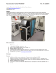
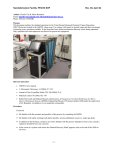
FEI Quanta 600 FEG SEM
Page 1 of 10
SOP Revision 2-052610
FEI Quanta 600 FEG SEM
1. Scope
1.1. This document provides the basic procedures and requirements to operate the FEI Quanta 600
FEG SEM
2. Table of Contents
1.
2.
3.
Scope .................................................................................................................................................................... 1
Table of Contents ................................................................................................................................................. 1
Reference Documents .......................................................................................................................................... 2
3.1. Referenced within this Document ............................................................................................................... 2
4. Safety.................................................................................................................................................................... 2
5. Training Protocol .................................................................................................................................................. 2
5.1. Certification Through Lab Staff .................................................................................................................... 2
6. General Use Instructions ...................................................................................................................................... 2
6.1. Walk-In Conditions ....................................................................................................................................... 2
6.2. Login ............................................................................................................................................................. 2
6.3. Sample Loading ............................................................................................................................................ 2
6.4. Removing Samples ....................................................................................................................................... 3
6.5. Some Useful Hot-key Reminders ................................................................................................................. 3
7. Hi-Vac Imaging ..................................................................................................................................................... 4
7.1. General Uses ................................................................................................................................................ 4
7.2. Imaging Procedure ....................................................................................................................................... 4
8. Low-Vac Imaging .................................................................................................................................................. 5
8.1. General Uses ................................................................................................................................................ 5
8.2. Imaging Procedure ....................................................................................................................................... 5
9. FEI Quanta ESEM Procedures ............................................................................................................................... 6
9.1. General Uses ................................................................................................................................................ 6
9.2. Detector Setup ............................................................................................................................................. 6
9.3. Sample Loading Stage Setup ........................................................................................................................ 6
9.4. Chilling and Pumping Down ......................................................................................................................... 7
9.5. Imaging ......................................................................................................................................................... 7
9.6. Sample and Stage Removal .......................................................................................................................... 8
10. EDS ....................................................................................................................................................................... 8
10.1.
General Use .............................................................................................................................................. 8
10.2.
Getting started/Getting Counts ............................................................................................................... 8
10.3.
Basic Spectrum Through Saving ............................................................................................................... 9
10.4.
Mapping ................................................................................................................................................... 9
10.5.
Line-Scan ................................................................................................................................................ 10
10.6.
Other ...................................................................................................................................................... 10
11. Icon List .............................................................................................................................................................. 10
FEI Quanta 600 FEG SEM
Page 2 of 10
SOP Revision 2-052610
3. Reference Documents
3.1. Referenced within this Document
3.1.1.
FEI Quanta User Manual, hardcopy located on the bookshelf, electronic copy located under
the Help Menu.
3.1.2.
EDAX User Manual, hardcopy located on the bookshelf.
4. Safety
4.1. Follow all Nanofab safety procedures.
5. Training Protocol
5.1. Certification Through Lab Staff
5.1.1. The user has taken PHYS 5739 and has been approved by the instructor
5.1.2. The user has taken a formal course from FEI
5.1.3. The user has been trained and certified by an administrator
5.1.4. For any specialized use such as ESEM, EDX, or EBSD, the user must be trained and
certified for that particular method.
6. General Use Instructions
6.1. Walk-In Conditions
6.1.1. The previous user is logged off.
6.1.2. The machine is pumped down to high vacuum
6.1.3. The beam is turned off.
6.1.4. The BSED is on the pole-piece.
6.1.5. The LFD is attached in the rear of the chamber.
6.2. Login
6.2.1. If the login window is not up, it is found under the “File” tab on the toolbar.
6.2.2. If you don’t have an account, talk to Surface Lab staff
6.3. Sample Loading
6.3.1. Whenever handling ANYTHING that goes inside the machine, make sure clean gloves are
being worn.
6.3.2. Turn on the CCD detector to observe current conditions of the chamber. Make sure the
camera is live by deselecting the “pause” icon in the upper right of the monitor.
6.3.3. Before venting, make sure the beam is off.
6.3.4. Vent the chamber (on the right hand side of the screen).
6.3.5. GENTLY open the door. Do not jerk the door open. Make sure that the stage is lowered
so it doesn’t hit the pole-piece.
FEI Quanta 600 FEG SEM
Page 3 of 10
SOP Revision 2-052610
6.3.6. Put the sample holder in securely.
6.3.7. Make sure the desired detectors are in the machine (if they are not notify lab staff)
6.3.8. Close the door. Watch the sample on the CCD display as it goes in to make sure it clears
the pole-piece and cords
6.3.9. Pump down by hitting “Pump” button. (It’s recommended, sample permitting, to go to
high vac then to low because it is faster, even if the sample is non-conductive.) Observe
the icon of the machine on the bottom right hand corner of the screen. The column
should always be green. The chamber part of it should be yellow during pumping and
when pumping is complete, the entire icon should be green.
6.3.10. While waiting for the pump you may adjust the stage height by holding down the scroll
wheel and moving the arrow away from the bar. The farther the arrow is from the bar,
the faster it moves. The height can also be adjusted by using the sample navigation page.
6.3.11. If imaging with the intention of very high resolution the stage can be raised further for
better resolution at higher magnification. However, this will not work while
simultaneously using the EDS system. Be careful while doing this to avoid crashing into
the pole-piece.
6.4. Removing Samples
6.4.1. Always use gloves!
6.4.2. When done, first turn the beam off. (It is the same button that was pressed to turn it on.)
6.4.3. Turn the CCD on.
6.4.4. To vent the chamber, select “Vent” on the same page.
6.4.5. When the chamber icon is grey, open the door.
6.4.6. Make sure when opening the door to be attentive to the stage being low enough to not hit
anything on its way out. To do this, monitor the CCD.
6.4.7. Close the door and pump down to high vacuum.
6.4.8. Log off of user information.
6.4.9. When done, make sure to fill out the logbook of use. The logbook is usually near the
computer.
6.4.10. Check to see that it is in “walk in conditions.”
6.5. Some Useful Hot-key Reminders
6.5.1. CTRL + o – preferences
6.5.2. F5 – toggles between quadrant and full screen view.
6.5.3. CTRL + R – restarts the scan.
6.5.4. CTRL + 0 [zero] – centers the x and y coordinates.
6.5.5. CTRL + S – saves the image to current folder.
FEI Quanta 600 FEG SEM
Page 4 of 10
SOP Revision 2-052610
7. Hi-Vac Imaging
7.1. General Uses
7.1.1. Useful for conductive samples.
7.1.2. Best resolution and best results for using EDS.
7.2. Imaging Procedure
7.2.1. A general rule of thumb is 10kV and 4.5 spot size. However, if the sample is delicate and
prone to charging (i.e. it is an insulator or something biological) start with a very low
accelerating voltage (something like 2kV) in order to avoid decimating the sample in the
first minute of imaging.
7.2.2. Select detector and un-pause each quadrant. However, the CCD and the BSED cannot be
on simultaneously.
7.2.3. Turn the beam on.
7.2.4. First try adjusting the contrast through auto-brightness and contrast (sec. 8).
7.2.5. Adjust the brightness and contrast using the video scope until the waveform (sec. 8) nearly
saturates both top and bottom of the video scope “Contrast” will adjust the amplitude of
the wave while “brightness” will adjust the position on the y-axis.
7.2.6. Go to a lower magnification setting.
7.2.7. Focus on some object in the view. Focus by holding the right button on the mouse or by
the knobs on the panel.
7.2.8. Find your sample. There are several ways to navigate: holding the center wheel on the
mouse and pointing the arrow in the desired direction (further the arrow from the dot the
faster the stage moves), double clicking to center a feature, entering numbers under the
“Navigation” page, using the arrow buttons on the keyboard (each tap shifts the field of
view 75%), and even by the knobs on the outside of the machine if the navigation
software isn’t working out.
7.2.9. Rotating a sample is accomplished by entering the rotation desired (in degrees) in the
“Navigation” page. Oftentimes turning the knob on the outside of the machine is easier
for a fine adjustment. The XT align feature under the stage menu is also helpful to orient
your sample
7.2.10. Zoom in to about 5,000X and focus. Link the working distance with icon on the toolbar
(sec.8). This tells the computer how far down the object you focused on is.
7.2.11. A good working distance is 10mm if you are doing EDS. Always important is making
sure that the stage doesn’t hit the pole-piece. This can be terrible if the stage hits the
backscatter detector. Watch the live CCD and have a finger on the escape key. The
machine will stop immediately if the circuit between the stage and the pole-piece is
completed, but it is embarrassing and can be costly as well. However, a smaller working
distance will give better image resolution. A WD of 5mm might be better if you want
high magnification.
7.2.12. Find where you want to image. Move away from your point of interest, focus, and set the
stigmation. The best way to focus is to use the reduced raster option (found on the
FEI Quanta 600 FEG SEM
Page 5 of 10
SOP Revision 2-052610
toolbar). To get the best focus, set the stigmation by holding the shift key and the
right button on the mouse. The knob option, and slider bars can adjust this as well.
Repeat this process until you have found adequate focus at a high-resolution. Typically,
good focus in high magnification will result in better focus in lower magnification.
7.2.13. Adjust the source tilt found at the bottom of the “Beam Control” page. The goal is to
maximize the number of electrons getting through the final aperture. Move the cross
hairs to find the maximum illumination. Using the crossover button will show an image
of the electron source tip if this method is going to be used the brightest point should be
placed under the center X.
7.2.14. If the image appears to be shifting back and forth as you are focusing its time to do lens
alignment, also found under the “Beam control” page. Make sure averaging is not
selected, but rather it is set to “live”. The image will modulate (go in and out of focus).
The goal is to have the modulating image under the crosshair to not shifting. To adjust,
grab the lines in the small cross hair window above the lens align button. Move the lines
back and forth until the image ceases to move at the center.
7.2.15. Adjust the voltage to an appropriate value. Also, depending on the magnification, drop the
spot size. A smaller spot size will increase resolution, but decrease the contrast in the
image.
7.2.16. At this point, the user is free to image at will. If a number with several significant digits
appears in the magnification window, hit the “*” key to round off the magnification
number.
7.2.17. To save the image, use a slow scan and push the pause button and watch until the current
raster stops. The best image quality is generated by a slower scan. Scan speed is
controlled by the tortoise and hare at the upper right of the screen. A click left gives a
slower scan and a click right gives a faster scan.
7.2.18. When the raster scan is complete the green pause icon will stop blinking. At that point the
image can be saved by going under File->Save As… then find the user location in the
Quantadata subfolder on the support pc and save in the appropriate file. Again, do not
save the server computer otherwise your files will be deleted!
8. Low-Vac Imaging
8.1. General Uses
8.1.1. Useful for samples that are non-conductive.
8.1.2. Expect some tradeoffs in resolution. Efficacy of EDS is also comprised.
8.2. Imaging Procedure
8.2.1. The LFD detector should be installed at the back of the chamber.
8.2.2. The backscatter detector can also be used.
8.2.3. For the highest quality images, the PLA cones can be used with the LFD. To learn more
about using these cones talk to Lab Staff.
8.2.4. Pump down to high vacuum first (it is faster). Select “Low-Vacuum” under “Mode”.
The accessory screen will appear. Even if BSED is in check “no accessory”.
FEI Quanta 600 FEG SEM
Page 6 of 10
SOP Revision 2-052610
8.2.5. Adjust the voltage, spot size, and pressure to starting values. Good values are 15kV, 4.5
spot size, and 0.15 Torr.
8.2.6. Adjust the pressure to compensate for charging. If it continues to charge, raise the pressure
(not above 1.0 Torr) or lower the pressure if there is no charging.
8.2.7. Image according to standard procedure.
9. FEI Quanta ESEM Procedures
9.1. General Uses
9.1.1. ESEM can allow the user to image hydrated, non-conductive samples. Typically
applications range from wet slurries to biological samples.
9.1.2. Be aware that ultra-high resolution under ESEM mode is often unrealistic.
9.2. Detector Setup
9.2.1. There are two detectors that can be used: the GSED (secondary electrons) or the BSED
PLA. However, the BSED only allows 1.5 Torr.
9.2.2. To install the GSED remove the BSED from the pole-piece by pulling gently and place it
in the garage on the side of the chamber.
9.2.3. Take the LFD from the back of the chamber by the neck and slide it out of the
connector.
9.2.4. The back end of the GSED fits in this connector while the round piece on the other end
snaps onto the bottom of the pole-piece. Make sure it is tightly secured. If it is not
pressed on squarely, the image will not be centered on the screen centered and will be
obscured at higher magnifications.
9.2.5. To use the BSED PLA, the normal BSED must be removed. Follow the cable to the
connection at the bottom side of the chamber and unplug it.
9.2.6. Plug in the BSED PLA and install the detector to the bottom of the pole piece. Make sure
it is firmly snapped into place and square.
9.2.7. It is a good idea to tape the cord to the side of the machine using copper to tape to make
sure it is out of the way.
9.3. Sample Loading Stage Setup
9.3.1. Attach the stage adapter. Tighten gently!
9.3.2. Attach the Peltier module and tighten the setscrew on the side.
9.3.3. The orientation needs to be such that the hose connectors go towards the door.
9.3.4. Attach the cable. It is a 9-pin connector attached to the Peltier stage and is attached to the
corresponding connector on the inside the door.
9.3.5. Remove stoppers from the quick-connects inside the door and Attach the water hoses.
9.3.6. There are three cups (found in the Peltier stage box) that fit into the Peltier stage. The
purpose is to have the top of the sample be close to flush with the cup.
FEI Quanta 600 FEG SEM
Page 7 of 10
SOP Revision 2-052610
9.3.7. On the Peltier stage, there are divots on each corner. Put a small drop of water in each
divot to help ensure the sample doesn’t dry out prematurely when pumping.
9.3.8. Before pumping down, adjust the purge settings. Go to preferences (CTRL + o) and select
the ESEM tab. Set the upper purge bound to 9 Torr and the lower bound to be 6.5.
Select APPLY and close the window. If this is not done, it will purge into the gaseous
state and dehydrate the sample prematurely.
9.4. Chilling and Pumping Down
9.4.1. Turn on the water chiller. Water will not flow until flow box is started.
9.4.2. Turn on power to the flow box (small gray box near the monitor). An alarm will sound,
indicating that there is no flow though the box.
9.4.3. Push and hold the START FLOW button. Hold this button until all or most the air is out
of the lines.
9.4.4. Check that the FLOW OK light is on. This indicates the water flow is functional.
9.4.5. In the UI, the “Temperature Control” page shows a Cooling button. Select it and the stage
will start the cool down procedure.
9.4.6. The stage must be cooled down before pumping.
9.4.7. This box will display the actual temperature and an edit box to select the target
temperature. Select the GoTo button to start cooling.
9.4.8. Under the vacuum mode, select ESEM.
9.4.9. Before pumping, raise the pressure and lower the temperatures to reasonable values,4°C
and 6.5 Torr, that would not move the sample out of the liquid state (see phase diagram
on the tab).
9.4.10. Offset the stage from directly the pole-piece and keep stage at a long working distance
until the chamber is pumped down so water will not splash on pole-piece.
9.4.11. Pump down. Observe the phase diagram. The chamber should remain well into the wet
stage while pumping down.
9.5. Imaging
9.5.1. Standard spot size and voltage values are 4.5 spot and 15kV. Lower energies are difficult
to work with because they have a hard time imaging through the water vapor in the
chamber.
9.5.2. Raise the stage to a very close working distance (5mm or so). Be very careful to not hit
the pole piece when doing this.
9.5.3. Adjust the humidity. When looking at the phase diagram, select 100% for the humidity
“Go to” point. If the sample appears to be covered in water, drop the humidity gradually
into the gaseous state to evaporate the water coating. When sufficient surface detail is
observable, raise the humidity back to 100% to prevent further drying.
9.5.4. Lower the humidity to 100%, which is exactly on the border between the gaseous and
liquid state.
FEI Quanta 600 FEG SEM
Page 8 of 10
SOP Revision 2-052610
9.5.5. Find a solid object (i.e., the sample mount) to focus on before moving to the organic
sample. This will prevent ruining the sample before proper imaging occurs.
9.5.6. Continue imaging as required.
9.5.7. Avoid moving into the freezing state if possible. However, oftentimes some samples
require in-chamber freezing.
9.6. Sample and Stage Removal
9.6.1. Allow the stage to warm to room temperature.
9.6.2. Shut down the water cooler and push STOP FLOW on the flow box.
9.6.3. Disconnect one of the water lines to and elevate it to prevent water dripping into the
chamber. Push the START FLOW button and hold it until all the water is drained from
the tubes. Disconnect the other line.
9.6.4. Connect the water line plugs into the Feed Through Plate.
9.6.5. Wipe up the any remaining water.
9.6.6. Disconnect the 9-pin connector.
9.6.7. Remove the Peltier stage and put it back in the box.
9.6.8. Replace the standard detectors (BSED and the LFD).
9.6.9. Pump down to high vacuum. Turn the beam off and log off.
10. EDS
10.1. General Use
10.1.1. Genesis, Elemental Dispersive X-ray Spectroscopy (EDS) software, performs elemental
analysis through various avenues (mapping, spectrum quantification, line-scan, etc.)
10.1.2. For a more complete explanation see the User’s Manual, EDAX MicroAnalysis Training
School, on the bookshelf (sec. 3.1.2)
10.2. Getting started/Getting Counts
10.2.1. The sample should be at 10mm working distance to maximize counts.
10.2.2. Be sure the BSD is installed straight so not to cut off a path to the EDAX detector
10.2.3. The liquid nitrogen tank must be full and the detector must be cool (both blue LEDs are on
the EDAX computer tower).
10.2.4. There are two critical numbers when working with the EDAX system: the count rate
(CPS) and the dead time; both are given at the bottom of the screen.
10.2.5. The dead time must be within 20-40%. (adjusted by amp time)
10.2.6. The count rate (CPS) is best over 1500.
10.2.7. To increase the count rate, increase the spot size. A spot size of 5.0 is a good starting spot.
However, each time the spot size is adjusted, the “Source Tilt” and “Lens Alignment”.
10.2.8. If the dead time is too high or low, adjust amp time on the top-right of the genesis UI.
Typically the Auto-Amp time works well
FEI Quanta 600 FEG SEM
Page 9 of 10
SOP Revision 2-052610
10.3. Basic Spectrum Through Saving
10.3.1. Launch the EDAX Genesis software.
10.3.2. Select the Maps/Line tab.
10.3.3. In the top-left corner, there is a space for an electron image. To collect the image, hit the
“Collect e-” button.
10.3.4. Select the area to be analyzed. There are several shape options on the top toolbar including:
full frame, square, spots, and lasso.
10.3.5. With an appropriate CPS and dead time, collect the spectrum by selecting the “Collect
Spectrum” button. Let the spectrum collect for at least 15 seconds. Let it collect longer
if the data is going to be published, but for a first pass, a quicker collection time is
appropriate.
10.3.6. To observe the spectrum, hit the home button.
10.3.7. To quickly identify the peaks, select “Peak ID,” which automatically finds the peaks.
However, this is often inaccurate and needs to be reviewed for correctness. If there are
any missing peaks, select the double arrow button near “Peak ID.” This brings a dropdown menu where the user can type in the element and energy, the currently used
elements, and the possible elements corresponding to the energies where the black line is
on the spectrum. Unwanted elements can be cleared off the spectrum through this menu
as well.
10.3.8. With the elements identified, select the “Q” (Quantification) button on the middle of the
screen. This will bring up a window next to the electron image.
10.3.9. To save this, select EDIT->Screen Capture. This will bring up a small window in the
bottom right of the screen. Select the windows desired and hit save. Find the target
folder and save the data as a bitmap.
10.4. Mapping
10.4.1. After maximizing the count rate with a stable image run a basic spectrum and identify the
correct peaks, select the Maps button on the right hand of the screen.
10.4.2. Adjust the dwell time to 300ms and adjust the resolution to 514x400.
10.4.3. Select Collect Maps. This will prompt the user to select a saving destination. (It is a good
idea to create a separate folder for each map because the process will create several
files.)
10.4.4. Allow the mapping process to run through as many cycles as patience allows. The longer
the scan the better the map will look. To manually stop the process, select Collect Maps
again. It will prompt you when to stop. Allow the process to complete the current
frame.
10.4.5. To overlay the various maps, choose the 16-frame window view. Select the images to be
overlaid (Process->Color->Substitution overlay). These images are then immediately
saved the folder with the rest of the images.
FEI Quanta 600 FEG SEM
Page 10 of 10
SOP Revision 2-052610
10.5. Line-Scan
10.5.1. After maximizing the count rate with a stable image run a basic spectrum and identify the
correct peaks, select the Lines button on the right hand of the screen. This brings another
drop-down menu.
10.5.2. Set the Dwell time. Allow around 300ms for a standard value.
10.5.3. Select the line by selecting the arrow on the line button, the image will turn into a single
view. Drag the mouse over the image where the scan is to be collected. If it is drawn
incorrectly, redraw the line by selecting “Draw Line”.
10.5.4. Push the “Collect Line” button. The user is then prompted through a file name and save
location sequence. Make sure to create a new folder for the linescan files.
10.6. Other
10.6.1.1.
For other applications, see the user’s manual (sec. 3.1.1).
11. Icon List
12. Revision History
Rev
Date
1
2
01 May 2010
29 June 2010
Originator
Description of Changes
Alex Hogan
Kathryn Ecsedy
First Draft
Formatting Changes