1
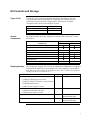
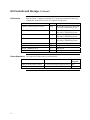
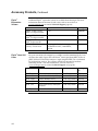
Flp-In™ System For Generating Stable Mammalian Expression Cell Lines by Flp Recombinase-Mediated Integration Catalog nos. K6010–01, K6010–02 Version E 9 November 2010 25–0306 Corporate Headquarters Invitrogen Corporation 1600 Faraday Avenue Carlsbad, CA 92008 T: 1 760 603 7200 F: 1 760 602 6500 E: [email protected] For country-specific contact information visit our web site at www.invitrogen.com User Manual ii Table of Contents Kit Contents and Storage ................................................................................................................................ v Accessory Products ........................................................................................................................................ vii Introduction ................................................................................................................................................................1 Overview ............................................................................................................................................................1 Methods.......................................................................................................................................................................6 Propagation and Maintenance of Plasmids...................................................................................................6 Generating Stable Flp-In™ Host Cell Lines ....................................................................................................8 Generating Stable Flp-In™ Expression Cell Lines .......................................................................................14 Appendix...................................................................................................................................................................20 Recipes ..............................................................................................................................................................20 Zeocin™ .............................................................................................................................................................21 Map of pFRT/lacZeo Vector..........................................................................................................................22 Features of pFRT/lacZeo Vector...................................................................................................................23 Map of pOG44 Vector.....................................................................................................................................24 Features of pOG44 Vector ..............................................................................................................................25 Technical Support ...........................................................................................................................................26 Purchaser Notification....................................................................................................................................27 References.........................................................................................................................................................29 iii iv Kit Contents and Storage Types of Kits The Flp-In™ System manual is supplied with the kits listed below. The Core System includes vectors and primers for sequencing. The Complete System includes the Core System plus selection agents. See below for a detailed description of the contents of each Flp-In™ System. Product Flp-In™ Complete System Flp-In™ Core System System Components Catalog no. K6010–01 K6010–02 The following table shows the components associated with each Flp-In™ system listed above. Components ™ pcDNA 5/FRT Expression Vector pcDNA™5/FRT/CAT Positive Control Vector pOG44 Plasmid Vector pFRT/lacZeo Target-site Vector CMV Forward Primer (21-mer) BGH Reverse Primer (18-mer) Hygromycin B Zeocin™ Selection Reagent Shipping/Storage Catalog no. K6010–01 K6010–02 9 9 9 9 9 9 9 9 9 9 9 9 9 9 The components supplied with Catalog nos. K6010–01 and K6010–02 are shipped as described in the table below. Upon receipt, store each component as listed below. Note: The components of K6010–01 are shipped in 2 boxes. Box 1 contains vectors, primers, and hygromycin. Box 2 contains Zeocin™. Item Vectors: • pcDNA™5/FRT Expression Vector • pcDNA™5/FRT/CAT Positive Control Vector • pOG44 Plasmid Vector • pFRT/lacZeo Target-site Vector Primers • CMV Forward Primer (21-mer) • BGH Reverse Primer (18-mer) Hygromycin B (K6010–01 only) Shipping Wet ice Storage Store all vectors at –20°C Wet ice Store all primers at –20°C Wet ice Store at 4°C, protected from light Store at –20°C, protected from light Zeocin™ (K6040–01 only) Wet ice Continued on next page v Kit Contents and Storage, Continued Both the Flp-In™ Complete and the Flp-In™ Core Systems include the following components. Note that the vectors are supplied in suspension. Kit Contents Product Quantity Composition pcDNA 5/FRT Expression Vector 20 ug 40 μl of 0.5 μg/μl vector in 10 mM Tris–HCl, 1 mM EDTA, pH 8.0 pcDNA™5/FRT/CAT Positive Control 20 μg 40 μl of 0.5 μg/μl vector in 10 mM Tris–HCl, 1 mM EDTA, pH 8.0 pOG44 Expression Vector 20 μg 40 μl of 0.5 μg/μl vector in 10 mM Tris–HCl, 1 mM EDTA, pH 8.0 pFRT/lacZeo Vector 20 μg 40 μl of 0.5 μg/μl vector in 10 mM Tris–HCl, 1 mM EDTA, pH 8.0 CMV Forward Primer (21-mer) 2 μg lyophilized in TE, pH 8.0 BGH Reverse Primer (18-mer) 2 μg lyophilized in TE, pH 8.0 Zeocin (K6010–01 only) 1g 100 mg/ml Hygromycin B (K6010–01 only) 1g 100 mg/ml ™ ™ Primer Sequences vi The sequence of each primer is provided below. Primer Sequence pMoles Supplied CMV Forward Primer (21-mer) 5´-CGCAAATGGGCGGTAGGCGTG-3´ 306 BGH Reverse Primer (18-mer) 5´-TAGAAGGCACAGTCGAGG-3´ 358 Accessory Products The products listed in this section are intended for use with he Flp-In™ Systems For more information, refer to our web site at www.invitrogen.com or contact Technical Support (page 26). Additional Products Product Amount pFRT/lacZeo 20 μg, (supplied as 40 μl of 0.5 μg/μl vector V6015–20 in 10 mM Tris–HCl, 1 mM EDTA, pH 8.0) pFRT/lacZeo2 20 μg, (supplied as 40 μl of 0.5 μg/μl vector V6022–20 in 10 mM Tris–HCl, 1 mM EDTA, pH 8.0) pOG44 20 μg, (supplied as 40 μl of 0.5 μg/μl vector V6005–20 in 10 mM Tris–HCl, 1 mM EDTA, pH 8.0) T7 Promoter Primer 2 μg, lyophilized N560–02 One Shot® Kit 10 reactions C4040–10 (TOP10 Chemically Competent Cells) 20 reactions C4040–03 40 reactions C4040–06 One Shot Kit 10 reactions C4040–50 (TOP10 Electrocompetent Cells) 20 reactions C4040–52 S.N.A.P. Miniprep Kit 100 reactions K1900–01 100 preps K2100–03 15–200 reactions K2100–04 ® ™ PureLink HiPure Plasmid Miniprep Kit ™ PureLink HiPure Plasmid Midiprep Kit Catalog no. ™ Easy-DNA Kit ™ Zeocin K1800–01 1g R250–01 5g R250–05 Hygromycin B 1g R220–05 β-Gal Assay Kit 100 reactions K1455–01 β-Gal Staining Kit 1 kit K1465–01 CAT Antiserum* 50 μl R902–25 Calcium Phosphate Transfection Kit 75 reactions K2780–01 15 ml 11668–500 1.5 ml 11668–019 20 pouches Q601–20 ™ Lipofectamine 2000 Transfection Reagent ™ imMedia Amp Agar. *The amount supplied is sufficient to perform 25 Western blots using 10 ml working solution per reaction. Continued on next page vii Accessory Products, Continued Flp-In™ Expression Vectors Additional Flp-In™ expression vectors are available from Invitrogen. For more information about the features of each vector, refer to our web site at www.invitrogen.com or contact Technical Support (page 26). Product Amount Catalog no. pcDNA™5/FRT/V5-His TOPO® TA Expression Kit 1 kit K6020–01 pSecTag/FRT/V5-His TOPO® TA Expression Kit 1 kit K6025–01 pEF5/FRT/V5 Directional TOPO® Expression Kit 1 kit K6035–01 pEF5/FRT/V5-DEST Gateway™ Vector Pack 6 μg, supplied as 40 μl of 150ng/ul vector in 10 mM Tris–HCl, 1 mM EDTA, pH 8.0 V6020–20 Flp-In™ Host Cell Lines For your convenience, Invitrogen has available several mammalian Flp-In™ host cell lines that stably express the lacZ-Zeocin™ fusion gene from pFRT/lacZeo or pFRT/lacZeo2. Each cell line contains a single integrated FRT site as confirmed by Southern blot analysis. The cell lines should be maintained in medium containing Zeocin™. For more information, visit our web site at www.invitrogen.com or contact Technical Support (see page 26). Cell Line Source Amount Catalog no. Flp-In™-293 Human embryonic kidney 1 × 10⁷ cells, frozen R750–07 ™ Flp-In -CV-1 ™ Flp-In -CHO ™ viii African Green Monkey kidney Chinese Hamster ovary 7 R752–07 7 R758–07 7 1 × 10 cells, frozen 1 × 10 cells, frozen Flp-In -BHK Baby hamster kidney 1 × 10 cells, frozen R760–07 Flp-In™-3T3 Mouse (NIH Swiss) embryonic fibroblast 1 × 10⁷ cells, frozen R761–07 Flp-In™-Jurkat Human T-cell leukemia 1 × 10⁷ cells, frozen R762–07 Introduction Overview Introduction The Flp-In™ System allows integration and expression of your gene of interest in mammalian cells at a specific genomic location. The Flp-In™ System involves introduction of a Flp Recombination Target (FRT) site into the genome of the mammalian cell line of choice. An expression vector containing your gene of interest is then integrated into the genome via Flp recombinasemediated DNA recombination at the FRT site (O'Gorman et al., 1991). System Components The major components of the Flp-In™ System include: • A Flp-In™ target site vector, pFRT/lacZeo, for generation of a host cell line containing an integrated FRT site (see pages 22–23 for more information). • An expression plasmid containing a FRT site linked to the hygromycin resistance gene for Flp recombinase-mediated integration and selection of a stable cell line expressing your gene of interest under the control of the human cytomegalovirus (CMV) immediate-early enhancer/promoter. • A Flp recombinase expression plasmid, pOG44, for expression of the Flp recombinase under the control of the human CMV promoter (see pages 24–25 for further information). • A control expression plasmid containing the chloramphenicol acetyl transferase (CAT) gene, which when cotransfected with pOG44 into your FlpIn™ host cell line, expresses CAT. For specific information on the expression vector and the corresponding positive control vector containing the CAT gene, refer to the pcDNA™5/FRT vector manual. Advantages of the Flp-In™ System Use of the Flp-In™ System to generate stable expression cell lines provides a number of advantages as described below: • Once the Flp-In™ host cell line containing an integrated FRT site has been created, subsequent generation of Flp-In™ cell lines expressing the gene(s) of interest is rapid and efficient. • The Flp-In™ System allows the generation of isogenic stable cell lines. • The Flp-In™ System permits polyclonal selection of stable expression cell lines. Continued on next page 1 Overview, Continued Description of the Flp-In™ System The Flp-In™ System streamlines the generation of stable mammalian expression cell lines by taking advantage of a Saccharomyces cerevisiae-derived DNA recombination system. This DNA recombination system uses a recombinase (Flp) and site-specific recombination (Craig, 1988; Sauer, 1994) to facilitate integration of the gene(s) of interest into a specific site in the genome of mammalian cells. In the Flp-In™ System, three different vectors are used to generate isogenic stable mammalian cells lines expressing your gene(s) of interest. The first major component of the Flp-In™ System is the pFRT/lacZeo target site vector that is used to generate a Flp-In™ host cell line. The vector contains a lacZ-Zeocin™ fusion gene whose expression is controlled by the SV40 early promoter (see the Appendix, pages 22–23 for more information). A FRT site has been inserted just downstream of the ATG initiation codon of the lacZ-Zeocin™ fusion gene. The FRT site (see page 4 for more information) serves as the binding and cleavage site for the Flp recombinase. The pFRT/lacZeo plasmid is transfected into the mammalian cell line of interest and cells are selected for Zeocin™ resistance. Zeocin™-resistant clones are screened to identify those containing a single integrated FRT site. The resulting Flp-In™ host cell line contains an integrated FRT site and expresses the lacZ-Zeocin™ fusion gene (see the diagram, next page). Note: Integration of the pFRT/lacZeo plasmid into the genome is random. The second major component of the Flp-In™ System is the pcDNA™5/FRT expression vector into which the gene of interest will be cloned. Expression of the gene of interest is controlled by the human CMV promoter. The vector also contains the hygromycin resistance gene with a FRT site embedded in the 5′ coding region. The hygromycin resistance gene lacks a promoter and the ATG initiation codon. For more information about the pcDNA™5/FRT vector, refer to the vector manual. The third major component of the Flp-In™ System is the pOG44 plasmid which constitutively expresses the Flp recombinase (Broach et al., 1982; Broach & Hicks, 1980; Buchholz et al., 1996) under the control of the human CMV promoter. For more information about pOG44 and the FLP gene, see the Appendix, pages 24–25. The pOG44 plasmid and the pcDNA™5/FRT vector containing your gene of interest are cotransfected into the Flp-In™ host cell line. Upon cotransfection, the Flp recombinase expressed from pOG44 mediates a homologous recombination event between the FRT sites (integrated into the genome and on pcDNA™5/FRT) such that the pcDNA™5/FRT construct is inserted into the genome at the integrated FRT site (see diagram, next page). Insertion of pcDNA™5/FRT into the genome at the FRT site brings the SV40 promoter and the ATG initiation codon (from pFRT/lacZeo) into proximity and frame with the hygromycin resistance gene, and inactivates the lacZ-Zeocin™ fusion gene. Thus, stable Flp-In™ expression cell lines can be selected for hygromycin resistance, Zeocin™ sensitivity, lack of β-galactosidase activity, and expression of the recombinant protein of interest (see diagram, next page). Continued on next page 2 Overview, Continued Diagram of the Flp-In™ System The figure below illustrates the major features of the Flp-In™ System as described on the previous page. For a brief description about FRT sites and the mechanism of Flp-mediated recombination, see the next page and published reviews (Craig, 1988; Sauer, 1994). 1. pFRT/lacZeo is stably transfected into the mammalian cells of interest to generate the Zeocin-resistant Flp-In Host Cell Line(s) Expression of lacZ and Zeocin fusion gene PSV40 2. The pcDNA5/FRT expression vector containing your gene of interest (GOI) is cotransfected with pOG44 into the Flp-In Host Cell Line. ATG FRT Amp lacZ-Zeocin pUC ori Flp-In Host Cell Line + pOG44 + pcDNA5/FRT PSV40 B H FRT Amp lacZ-Zeocin pUC ori Flp-In Host Cell Line Hyg rom yc P CMV GO I G pA FRT in 3. The Flp recombinase expressed from pOG44 catalyzes a homologous recombination event between the FRT sites in the host cells and the pcDNA5/FRT expression vector. ATG pcDNA5/FRT Expression Vector pU C or i 4. Integration of the expression construct allows transcription of the gene of interest (GOI) and confers hygromycin resistance and Zeocin sensitivity to the cells. Am p Expression of hygromycin PSV40 ATG FRT Hygromycin pUC ori Amp PCMV Expression of your gene GOI BGH pA FRT lacZ-Zeocin Amp pUC ori Flp-In Expression Cell Line Continued on next page 3 Overview, Continued Flp RecombinaseMediated DNA Recombination In the Flp-In™ System, integration of your pcDNA™5/FRT expression construct into the genome occurs via Flp recombinase-mediated intermolecular DNA recombination. The hallmarks of Flp-mediated recombination are listed below. • Recombination occurs between specific FRT sites (see below) on the interacting DNA molecules. • Recombination is conservative and requires no DNA synthesis; the FRT sites are preserved following recombination and there is minimal opportunity for introduction of mutations at the recombination site. • Strand exchange requires only the small 34 bp minimal FRT site (see below). For more information about the Flp recombinase and conservative site-specific recombination, refer to published reviews (Craig, 1988; Sauer, 1994). Note: If your cell line contains multiple integrated FRT sites, Flp-mediated intramolecular recombination may also occur. Intramolecular recombination may result in: FRT Sites • Excision of the intervening DNA if the FRT sites are directly repeated (i.e. integration of multiple FRT sites on the same DNA strand). • DNA inversion if the sites are in opposing orientations. • Deletion of genomic sequences. As described above, Flp recombinase-mediated recombination occurs between specific FRT sites. The FRT site, originally isolated from Saccharomyces cerevisiae, serves as a binding site for Flp recombinase and has been well-characterized (Gronostajski & Sadowski, 1985; Jayaram, 1985; Sauer, 1994; Senecoff et al., 1985). The minimal FRT site consists of a 34 bp sequence containing two 13 bp imperfect inverted repeats separated by an 8 bp spacer that includes an Xba I restriction site (see figure below). An additional 13 bp repeat is found in most FRT sites, but is not required for cleavage (Andrews et al., 1985). While Flp recombinase binds to all three of the 13 bp repeats, strand cleavage actually occurs at the boundaries of the 8 bp spacer region (see figure below for cleavage sites (CS) (Andrews et al., 1985; Senecoff et al., 1985). Minimal FRT site CS GAAGTTCCTATTCCGAAGTTCCTATTCTCTAGAAAGTATAGGAAC TTC Xba I CS CS = cleavage site Continued on next page 4 Overview, Continued To create a stable Flp-In™ cell line expressing your gene of interest at a sitespecific genomic locus, you will perform the following steps: 1. Transfect the Flp-In™ target site vector, pFRT/lacZeo, into the mammalian cell line of choice to generate your Flp-In™ host cell line(s) (see figure below). 2. Clone your gene of interest into the pcDNA™5/FRT expression vector. 3. Co-transfect your pcDNA™5/FRT construct and the Flp recombinase expression vector, pOG44, into your Flp-In™ host cell line to generate your Flp-In™ expression cell line (see figure below). 4. Assay for expression of your recombinant protein of interest. Note: The positive control vector containing the CAT gene can be cotransfected into your Flp-In™ host cell line with pOG44 to demonstrate that the system is working properly. i PSV 40 AT G n FR T lacZ -Z e oc pFRT/lacZeo pA 8106 bp i p i ci 1.Am PSV40 ATG 1. Transfect pFRT/lacZeo into the mammalian cell line of interest to generate the Flp-In Host Cell Line. Select cells that exhibit Zeocin resistance and b-galactosidase activity. 40 or SV pU C FRT l li n lacZ-Zeocin Amp pUC ori Flp-In Host Cell Line Gene of Interest Gene of Interest MV BGH pA 2. Ligate the gene of interest into the pcDNA5/FRT expression vector. PC FR T n S p U C or i Hygrom yci n A m pi c i l l i pcDNA5/FRT Expression Vector A 0p V4 PC 3. Cotransfect the expression vector and pOG44 into the Flp-In Host Cell Line. Select for hygromycinresistant cells. MV in ill tr o In n Am pi c Experimental Outline pOG44 FLP 5.8 kb pU C i or S V 40 PSV40 ATG FRT pA Hygromycin PCMV Gene of Interest BGH pA FRT lacZ-Zeocin Flp-In Expression Cell Line 4. Assay for expressed protein. 5 Methods Propagation and Maintenance of Plasmids Introduction The following section contains guidelines for maintaining and propagating the pFRT/lacZeo and pOG44 vectors. For information about maintaining and propagating the pcDNA™5/FRT expression vector, refer to the vector manual. General Molecular Biology Techniques For assistance with E. coli transformations, restriction enzyme analysis, DNA biochemistry, and plasmid preparation, refer to Molecular Cloning: A Laboratory Manual (Sambrook et al., 1989) or Current Protocols in Molecular Biology (Ausubel et al., 1994). E. coli Strain Many E. coli strains are suitable for the propagation of the pFRT/lacZeo and pOG44 vectors. We recommend that you propagate the pFRT/lacZeo and pOG44 vectors in E. coli strains that are recombination deficient (recA) and endonuclease A deficient (endA). For your convenience, TOP10 E. coli are available as chemically competent or electrocompetent cells from Invitrogen (page vii). Transformation Method You may use any method of choice for transformation. Chemical transformation is the most convenient for many researchers. Electroporation is the most efficient and the method of choice for large plasmids. Continued on next page 6 Propagation and Maintenance of Plasmids, Continued Maintenance of Plasmids The pFRT/lacZeo and pOG44 vectors contain the ampicillin gene to allow selection of the plasmid using ampicillin (see pages 22–25 for more information about each vector). To propagate and maintain the pFRT/lacZeo and pOG44 plasmids, we recommend using the following procedure: Preparing a Glycerol Stock 1. Use 10 ng of the vector to transform a recA, endA E. coli strain like TOP10, DH5α, JM109, or equivalent. 2. Select transformants on LB agar plates containing 50–100 μg/ml ampicillin. For fast and easy microwaveable preparation of Low Salt LB agar containing ampicillin, imMedia™ Amp Agar is available from Invitrogen (page vii). For more information, visit our web site at www.invitrogen.com or contact Technical Support (see page 26). 3. Prepare a glycerol stock of each plasmid for long-term storage (below). Once you have identified the correct clone, be sure to purify the colony and make a glycerol stock for long-term storage. It is also a good idea to keep a DNA stock of your plasmid at –20°C. 1. Streak the original colony out on an LB plate containing 50 μg/ml ampicillin. Incubate the plate at 37°C overnight. 2. Isolate a single colony and inoculate into 1–2 ml of LB containing 50 μg/ml ampicillin. 3. Grow the culture to mid-log phase (OD600 = 0.5–0.7). 4. Mix 0.85 ml of culture with 0.15 ml of sterile glycerol and transfer to a cryovial. 5. Store at –80°C. 7 Generating Stable Flp-In™ Host Cell Lines MEND ION AT RECOM Introduction Important Before you can create a stable Flp-In™ cell line(s) expressing your gene of interest, you will first need to generate a stable mammalian cell line containing an integrated FRT site (Flp-In™ host cell line). The following section provides guidelines and instructions to generate stable Flp-In™ host cell lines by transfection using the pFRT/lacZeo plasmid. For a map and a description of the features of pFRT/lacZeo, refer to the Appendix, pages 22–23. Several Flp-In™ host cell lines which stably express the lacZ-Zeocin™ fusion gene from pFRT/lacZeo or pFRT/lacZeo2 and which contain a single integrated FRT site are available from Invitrogen (see table below). If you wish to express your gene of interest in one of the cell lines listed below, you may want to use one of Invitrogen’s Flp-In™ cell lines as the host to establish your stable expression cell line. For more information, refer to our web site at www.invitrogen.com or contact Technical Support (page 26). We have observed down-regulation of the viral CMV promoter and subsequent loss of gene expression when pcDNA™5/FRT-based expression constructs are introduced into Flp-In™-3T3 or Flp-In™-BHK cells. If you will be cloning your gene of interest into a pcDNA™5/-FRT-based expression construct, we recommend that you do not use 3T3 or BHK cells to create your Flp-In™ host cell line. Alternatively, if you prefer to use 3T3 or BHK cells to create your Flp-In™ host cell line, we recommend that you clone your gene of interest into a pEF5/FRT-based expression plasmid (e.g. pEF5/FRT/V5-D-TOPO® or pEF5/FRT/V5-DEST). Loss of gene expression due to down-regulation of the promoter is not observed in these cell lines when using pEF5/FRT-based expression constructs. For more information about the pEF5/FRT/V5-D-TOPO® or pEF5/FRT/V5-DEST vectors, visit our web site at www.invitrogen.com or contact Technical Support (page 26). Plasmid Preparation Plasmid DNA for transfection into eukaryotic cells must be very clean and free from phenol and sodium chloride. Contaminants will kill the cells, and salt will interfere with lipid complexing, decreasing transfection efficiency. We recommend isolating DNA using the PureLink™ or S.N.A.P.™ Miniprep or Midiprep Kit (page vii) or CsCl gradient centrifugation. Methods of Transfection For established cell lines (e.g. HeLa, COS-1), consult original references or the supplier of your cell line for the optimal method of transfection. We recommend that you follow exactly the protocol for your cell line. Pay particular attention to medium requirements, when to pass the cells, and at what dilution to split the cells. Further information is provided in Current Protocols in Molecular Biology (Ausubel et al., 1994). Methods for transfection include calcium phosphate (Chen & Okayama, 1987; Wigler et al., 1977), lipid-mediated (Felgner et al., 1989; Felgner & Ringold, 1989) and electroporation (Chu et al., 1987; Shigekawa & Dower, 1988). Invitrogen offers the Calcium Phosphate Transfection Kit and Lipofectamine™ 2000 Reagent (page vii) for mammalian cell transfection. For more information, refer to our web site at www.invitrogen.com or contact Technical Support (page 26). Continued on next page 8 Generating Stable Flp-In™ Host Cell Lines, Continued Zeocin™ The pFRT/lacZeo plasmid contains a lacZ-Zeocin™ fusion gene under the control of the SV40 early promoter. Expression of the lacZ-Zeocin™ fusion gene allows selection of stable integrants using Zeocin™ antibiotic. The resulting stable integrants can then be screened by assaying for expression of β-galactosidase. For more information about preparing and handling Zeocin™, refer to the Appendix, page 21. The pFRT/lacZeo2 plasmid contains a lacZ-Zeocin™ fusion gene under the control of a truncated SV40 promoter and is available separately from Invitrogen (page vii). The minimal activity of the promoter allows for isolation of clones that have FRT sites integrated in the most transcriptionally active genomic loci. For details, visit our web site at www.invitrogen.com or contact Technical Support (page 26). Determination of Zeocin™ Sensitivity To successfully generate a stable cell line containing an integrated FRT site and expressing the lacZ-Zeocin™ fusion protein, you need to determine the minimum concentration of Zeocin™ required to kill your untransfected mammalian cell line. Typically, concentrations ranging from 50–1000 μg/ml Zeocin™ are sufficient to kill most untransfected mammalian cell lines, with the average being 100–400 μg/ml. We recommend that you test a range of concentrations (see protocol below) to ensure that you determine the minimum concentration necessary for your cell line. Refer to the Appendix, page 21 for instructions on how to prepare and store Zeocin™. 1. Plate or split a confluent plate so the cells will be approximately 25% confluent. Prepare a set of 7 plates. Allow cells to adhere overnight. 2. The next day, substitute culture medium with medium containing varying concentrations of Zeocin™ ( 0, 50, 100, 250, 500, 750, and 1000 μg/ml Zeocin™). 3. Replenish the selective media every 3–4 days, and observe the percentage of surviving cells. 4. Note the percentage of surviving cells at regular intervals to determine the appropriate concentration of Zeocin™ that kills the cells within 1–2 weeks after addition of Zeocin™. Effect of Zeocin™ on Sensitive and Resistant Cells Zeocin™’s method of killing is quite different from other antibiotics including hygromycin, G418, and blasticidin. Cells do not round up and detach from the plate. Sensitive cells may exhibit the following morphological changes to Zeocin™ exposure: • Vast increase in size, similar to the effects of cytomegalovirus infecting permissive cells • Abnormal cell shape • Presence of large empty vesicles in the cytoplasm (breakdown of the endoplasmic reticulum and Golgi apparatus, or other scaffolding proteins) • Breakdown of plasma and nuclear membrane (appearance of many holes) Eventually, these "cells" completely break down and only "strings" of protein remain. Zeocin™-resistant cells should continue to divide at regular intervals to form distinct colonies. There should not be any distinct morphological changes in Zeocin™resistant cells when compared to cells not under selection with Zeocin™. For more information about Zeocin™ and its mechanism of action, see Appendix, page 21. Continued on next page 9 Generating Stable Flp-In™ Host Cell Lines, Continued Transfection Considerations Once you have determined the appropriate Zeocin™ concentration to use, you are ready to transfect the pFRT/lacZeo plasmid into your mammalian cell line of choice to generate the Flp-In™ host cell line. You will need to consider the following factors: • Insertion of the FRT site into the genome: Integration of the pFRT/lacZeo plasmid containing the FRT site into the genome will occur randomly. Subsequent integration of the pcDNA™5/FRT expression plasmid containing your gene of interest will occur through Flp recombinase-mediated recombination at the genomic FRT site. • Transfection efficiency of your cell line: The aim of most users will be to create stable cell lines containing a single integrated FRT site (“single integrants”; see Note on the next page). The probability of obtaining stable integrants containing a single FRT site or multiple FRT sites depends on the transfection efficiency of your cell line and the amount of DNA transfected. To increase the likelihood of obtaining single integrants, lower the transfection efficiency by limiting the amount of plasmid DNA that you transfect (see Recommendation next page). • Selection of foci: You will select for stable transfectants by plating cells in medium containing Zeocin™. Zeocin™-resistant foci can then be screened by Southern blot analysis to identify single integrants. To increase the chances of obtaining single integrants, we recommend you pick foci from plates that have been transfected with the least amount of plasmid DNA. • Chromosomal position effects: Because integration of the pFRT/lacZeo plasmid into the genome occurs randomly, expression levels of the lacZZeocin™ fusion gene will be dependent on the transcriptional activity of the surrounding sequences at the integration site (i.e. chromosomal position effect). Once you have obtained single integrants, you may want to screen the Zeocin™-resistant clones for those expressing the highest β-galactosidase levels. Those clones expressing the highest levels of β-galactosidase should contain single FRT sites which have integrated into the most transcriptionally active regions. • Antibiotic concentration: Single integrants will express only a single copy of the lacZ-Zeocin™ fusion gene and therefore, may be more sensitive to Zeocin™ selection than multiple integrants. If you have previously used your mammalian cell line for transfection and Zeocin™ selection, you may need to use lower concentrations of Zeocin™ to obtain single integrants. Continued on next page 10 Generating Stable Flp-In™ Host Cell Lines, Continued MEND ION AT RECOM If you want to increase the expression levels of your gene of interest in the cell line of choice, you may wish to generate a Flp-In™ host cell line containing multiple integrated FRT sites. In theory, cotransfection of your pcDNA™5/FRT construct and pOG44 into these cells will allow integration of your gene of interest into multiple genomic loci. Note that the presence of multiple integrated FRT sites in the genome may increase the occurrence of chromosomal rearrangements or unexpected recombination events in your host cell line. As mentioned previously, we recommend that you transfect your mammalian cell line with a limiting amount of pFRT/lacZeo plasmid. We generally use 250 ng to 2 μg of plasmid DNA per 4 × 10⁶ cells for transfection, but the amount of plasmid DNA may vary due to the nature of the cell line, the transfection efficiency of your cells, and the method of transfection used. When transfecting your mammalian cell line of choice, we suggest that you try a range of plasmid DNA concentrations (e.g. 0.25, 0.5, 1, 2, 5 μg/ml DNA) to optimize transfection conditions for your cell line. We generally use electroporation to transfect cells, but other methods of transfection are suitable. For a protocol to electroporate cells, refer to Current Protocols in Molecular Biology, Unit 9.3 (Ausubel et al., 1994). Note that if you use calcium phosphate or lipid-mediated transfection methods, the amount of total DNA required for transfection is typically higher than for electroporation (usually between 10 and 20 μg DNA). Depending on the amount of pFRT/lacZeo plasmid that you use for transfection, you may need to supplement your plasmid DNA with carrier DNA (e.g. salmon sperm DNA). Possible Sites for Linearization of pFRT/lacZeo To obtain stable transfectants, we recommend that you linearize the pFRT/lacZeo plasmid before transfection. While linearizing the vector may not improve the efficiency of transfection, it increases the chances that the vector does not integrate in a way that disrupts the ATG-FRT-lacZ-Zeocin™ cassette or other elements necessary for expression in mammalian cells. The table below lists unique sites that may be used to linearize your construct prior to transfection. Other restriction sites are possible. Note: We generally use Sca I to linearize pFRT/lacZeo. Enzyme Restriction Site (bp) Location Supplier Tth111 I 125 Backbone Many Apa I 5617 Backbone Invitrogen (Catalog no. 15440–019) Swa I 6075 Backbone New England Biolabs, Sigma, Takara Xmn I 6487 Ampicillin gene Many Sca I 6606 Ampicillin gene Invitrogen (Catalog no. 15436–017) Bsa I 7021 Ampicillin gene New England Biolabs Eam1105 I 7087 Ampicillin gene AGS*, Fermentas, Takara Sap I 8092 Backbone New England Biolabs *Angewandte Gentechnologie Systeme Continued on next page 11 Generating Stable Flp-In™ Host Cell Lines, Continued Selection of Stable Once you have determined the appropriate Zeocin™ concentration to use for selection, you can generate a stable cell line with pFRT/lacZeo. Integrants 1. Transfect mammalian cells with pFRT/lacZeo using the desired protocol. Remember to include a plate of untransfected cells as a negative control. 2. 24 hours after transfection, wash the cells and add fresh medium to the cells. 3. 48 hours after transfection, split the cells into fresh medium such that they are no more than 25% confluent. If the cells are too dense, the antibiotic will not kill the cells. Antibiotics work best on actively dividing cells. 4. Incubate the cells at 37°C for 2–3 hours until they have attached to the culture dish. 5. Remove the medium and add fresh medium containing Zeocin™ at the predetermined concentration required for your cell line. 6. Feed the cells with selective medium every 3–4 days until foci can be identified. 7. Pick at least 20 Zeocin™-resistant foci and expand each clone to test for the number of integrated FRT sites. Isolate genomic DNA and use Southern blot analysis to distinguish between single and multiple integrants (see below and the next page). Select the single integrants and proceed to the next step. 8. Screen the single integrants for β-galactosidase activity (see the next page). Select those clones which exhibit the highest levels of β-galactosidase expression (if desired) to use as your Flp-In™ host cell line(s). 9. Once you have obtained a stable Flp-In™ host cell line, you can use this cell line to isolate a stable cell line expressing your gene of interest from the pcDNA™5/FRT plasmid (see the next section). Note: The Flp-In™ host cell line should be maintained in medium containing the appropriate amount of Zeocin™ until generation of your Flp-In™ expression cell line. Isolation of Genomic DNA Once you have obtained Zeocin™-resistant foci, you will need to expand the cells and isolate genomic DNA. You may use any standard protocol to isolate genomic DNA from your cells. Protocols may be found in Current Protocols in Molecular Biology (Ausubel et al., 1994) or Molecular Cloning: A Laboratory Manual (Sambrook et al., 1989). For easy isolation of genomic DNA, the Easy-DNA™ Kit (page vii) is available from Invitrogen. Contact Technical Support for more information (page 26). Continued on next page 12 Generating Stable Flp-In™ Host Cell Lines, Continued Screening Clones by Southern Blot Analysis What You Should See Assay for β-Galactosidase Activity You can use Southern blot analysis to determine the number of integrated FRT sites present in each of your Zeocin™-resistant clones. When performing Southern blot analysis, you should consider the following factors: • Probe: We recommend that you use a fragment of the lacZ gene (100 to 500 bp) as the probe to screen your samples. Mammalian cells do not contain an endogenous lacZ gene, therefore, a lacZ probe should allow you to identify those clones which contain pFRT/lacZeo DNA. To label the probe, we generally use a standard random priming kit (e.g. Ambion, DECAprime II™ Kit, Catalog no. 1455). Other random priming kits are suitable. • Restriction digest: When choosing a restriction enzyme to digest the genomic DNA, we recommend choosing an enzyme that cuts at a single known site outside of the lacZ gene in the pFRT/lacZeo vector. Hybridization of the lacZ probe to digested DNA should then allow you to detect a single band containing the lacZ gene from pFRT/lacZeo. We generally use Hind III to digest genomic DNA from the Zeocin™-resistant clones. pFRT/lacZeo contains a single Hind III site within the FRT site. • Protocol: You may use any Southern blotting protocol of your choice. Refer to Current Protocols in Molecular Biology (Ausubel et al., 1994) or Molecular Cloning: A Laboratory Manual (Sambrook et al., 1989) for detailed protocols. If you digest genomic DNA from your transfectants with Hind III and use a lacZ fragment as a probe in your Southern analysis, you should be able to easily distinguish between single and multiple FRT integrants. • DNA from single integrants should contain only one hybridizing band corresponding to a single copy of the integrated pFRT/lacZeo plasmid. • DNA from multiple integrants should contain more than one hybridizing band. If the pFRT/lacZeo plasmid integrates into multiple chromosomal locations, the bands may be of varying sizes. Once you have identified single integrants, proceed to screen the clones for β-galactosidase expression. You may assay for β-galactosidase expression by activity assay using cell-free lysates (Miller, 1972) or by staining the cells for activity. Invitrogen offers the β-Gal Assay Kit and the β-Gal Staining Kit (page vii) for fast and easy detection of β-galactosidase expression. Select those clones expressing the highest levels of β-galactosidase (if desired) to use as the host cell lines for your pcDNA™5/FRT expression construct. 13 Generating Stable Flp-In™ Expression Cell Lines Introduction MEND ION AT RECOM Important Once you have established your Flp-In™ host cell line, you may cotransfect your pcDNA™5/FRT construct and the pOG44 expression plasmid into the host cell line to generate a stable Flp-In™ expression cell line. Integration of the pcDNA™5/FRT construct into the genome will occur at the FRT site in the FlpIn™ host cells. The pcDNA™5/FRT plasmid contains the hygromycin resistance gene to allow selection of stable cell lines (see Important, below). For more information about the pcDNA™5/FRT plasmid and generating the pcDNA™5/FRT expression construct, refer to the vector manual. For more information about the pOG44 plasmid, see below. The hygromycin resistance gene in the pcDNA™5/FRT vector lacks an ATG initiation codon and a promoter to drive expression of the gene. Transfection of pcDNA™5/FRT plasmid alone into a Flp-In™ host cell line will not confer hygromycin resistance to the cells containing the plasmid. The ATG initiation codon and the SV40 promoter required for expression of the hygromycin resistance gene are brought into proximity and frame with the gene only through Flp recombinase-mediated recombination between the FRT sites in the pcDNA™5/FRT plasmid and the Flp-In™ host cell line. If you wish to express your gene of interest in one of the cell lines listed in the table below, you may want to use one of Invitrogen’s Flp-In™ host cell lines. For more information, visit our web site at www.invitrogen.com or contact Technical Support (page 26). If you are generating Flp-In™ expression cell lines using the Flp-In™-3T3 or FlpIn™-BHK cell line, we recommend that you clone your gene of interest into a pEF5/FRT-based expression plasmid (e.g. pEF5/FRT/V5-D-TOPO® or pEF5/FRT/V5-DEST). We have observed down-regulation of the viral CMV promoter and subsequent loss of gene expression when pcDNA™5/FRT-based expression constructs are introduced into Flp-In™-3T3 or Flp-In™-BHK cells. Continued on next page 14 Generating Stable Flp-In™ Expression Cell Lines, Continued pOG44 Plasmid You will cotransfect the pOG44 plasmid and your pcDNA™5/FRT construct into your Flp-In™ host cell line to generate stable cell lines that express your protein of interest. Cotransfection of pOG44 and pcDNA™5/FRT allows expression of Flp recombinase and integration of the pcDNA™5/FRT plasmid into the genome via the FRT sites. Once the pcDNA™5/FRT construct has integrated into the genome, the Flp recombinase is no longer required. In fact, the continued presence of Flp recombinase would be detrimental to the cells because it could mediate excision of your pcDNA™5/FRT construct. The pOG44 plasmid lacks an antibiotic resistance marker for selection in mammalian cells. Thus, the plasmid and therefore, Flp recombinase expression, will gradually be lost from transfected cells as they are cultured and selected in hygromycin. Flp Recombinase Important The FLP gene was originally isolated from the Saccharomyces cerevisiae 2 plasmid (Broach et al., 1982; Broach & Hicks, 1980) (see the Appendix, page 25 for more information). When tested in mammalian cells, the Flp recombinase has been shown to possess optimum recombination activity near 30°C and relatively low activity at 37°C, a result consistent with its physiological role in yeast (Buchholz et al., 1996). The FLP gene in pOG44 is further limited in its activity because it contains a point mutation that encodes a Flp recombinase with a phenylalanine to leucine amino acid substitution at position 70 (Buchholz et al., 1996). The resulting Flp recombinase (flp-F70L) exhibits increased thermolability at 37°C in mammalian cells when compared to the native Flp recombinase (Buchholz et al., 1996). Studies have shown that the Flp recombinase expressed from pOG44 possesses only 10% of the activity at 37°C of the native Flp recombinase (Buchholz et al., 1996). When generating Flp-In™ expression cell lines, it is important to remember that you are selecting for a relatively rare recombination event since you want recombination and integration of your pcDNA™5/FRT construct to occur only through the FRT site and for a limited time. In this case, using a highly inefficient Flp recombinase is beneficial and may decrease the occurrence of other undesirable recombination events. Continued on next page 15 Generating Stable Flp-In™ Expression Cell Lines, Continued Reminder: Integration of the pcDNA™5/FRT construct into the genome via the FRT sites will result in the following events (see page 3 for a diagram): • Insertion of the hygromycin resistance gene downstream of the SV40 early promoter and the ATG initiation codon (provided by pFRT/lacZeo) • Insertion of the plasmid containing the CMV promoter, your gene of interest, and the BGH polyadenylation signal upstream of the lacZ-Zeocin™ fusion gene • Disruption of the functional lacZ-Zeocin™ transcriptional unit caused by loss of the SV40 early promoter and the ATG initiation codon and insertion of the cassette containing the CMV promoter, gene of interest, and the BGH polyadenylation signal As a result, your Flp-In™ expression cell lines should exhibit the following phenotype: • • • • Hygromycin resistance Zeocin™ sensitivity Lack of β-galactosidase activity Expression of the gene of interest Positive Control The pcDNA™5/FRT/CAT plasmid is provided as a positive control vector for mammalian cell transfection and expression and may be used to assay for expression levels in your Flp-In™ expression cell line. If you have several different Flp-In™ host cell lines (cell lines containing FRT sites integrated at different genomic loci), you may want to use the pcDNA™5/FRT/CAT control vector to compare protein expression levels from the various genomic loci. For more information about pcDNA™5/FRT/CAT, refer to the pcDNA™5/FRT vector manual. Hygromycin B The pcDNA™5/FRT vector contains the E. coli hygromycin resistance gene (HPH) (Gritz & Davies, 1983) for selection of transfectants with the antibiotic, hygromycin B (Palmer et al., 1987). When added to cultured mammalian cells, hygromycin B acts as an aminocyclitol to inhibit protein synthesis by disrupting translocation and promoting mistranslation. Hygromycin B liquid is supplied with the Flp-In™ Complete System and is also available separately from Invitrogen (see page vii). • • • Hygromycin B is light sensitive. Store the liquid stock solution at 4°C protected from exposure to light. Hygromycin B is toxic. Do not ingest solutions containing the drug. Wear gloves, a laboratory coat, and safety glasses or goggles when handling hygromycin B and hygromycin B-containing solutions. Continued on next page 16 Generating Stable Flp-In™ Expression Cell Lines, Continued Hygromycin B (Flp-In™ Complete System, only) is supplied as a 100 mg/ml stock solution in autoclaved, deionized water and is filter-sterilized. The solution is brown in color. The stability of hygromycin B is guaranteed for six months, if stored at 4°C. Medium containing hygromycin is stable for up to six weeks. Determination of Hygromycin Sensitivity To successfully generate a stable cell line expressing your gene of interest from pcDNA™5/FRT, you need to determine the minimum concentration of hygromycin B required to kill your untransfected Flp-In™ host cell line. Typically, concentrations ranging from 10 to 400 μg/ml hygromycin B are sufficient to kill most untransfected mammalian cell lines. We recommend that you test a range of concentrations (see protocol below) to ensure that you determine the minimum concentration necessary for your Flp-In™ host cell line. 1. Plate or split a confluent plate so the cells will be approximately 25% confluent. Prepare a set of 7 plates. Allow cells to adhere overnight. 2. The next day, substitute culture medium with medium containing varying concentrations of hygromycin B ( 0, 10, 50, 100, 200, 400, 600 μg/ml hygromycin B). 3. Replenish the selective media every 3–4 days, and observe the percentage of surviving cells. 4. Note the percentage of surviving cells at regular intervals to determine the appropriate concentration of hygromycin that kills the cells within 1–2 weeks after addition of hygromycin. MEND ION AT RECOM Preparing and Storing Hygromycin B Important Because correct integration of your pcDNA™5/FRT construct into the genome is dependent on Flp recombinase, the expression levels of Flp recombinase in the cell will determine the efficiency of the recombination reaction. Flp recombinase levels must be sufficiently high to mediate recombination at the FRT sites (single recombination event) and overcome the low intrinsic activity of the enzyme (see previous page). We have varied the ratio of pOG44 and pcDNA™5/FRT expression plasmid that we cotransfect into mammalian Flp-In™ host cells to optimize the recombination efficiency. We recommend that you cotransfect your Flp-In™ host cell line with a ratio of at least 9:1 (w/w) pOG44:pcDNA™5/FRT expression plasmid. Note that this ratio may vary depending on the nature of the cell line. You may want to determine this ratio empirically for your cell line. When transfecting your Flp-In™ host cell line, be sure to use supercoiled pOG44 and pcDNA™5/FRT plasmid DNA. Flp-mediated recombination between the FRT site on pcDNA™5/FRT and the integrated FRT site in the Flp-In™ host cell line will only occur if the pcDNA™5/FRT plasmid is circularized. The pOG44 plasmid should be circularized to minimize the possibility of the plasmid integrating into the genome. Continued on next page 17 Generating Stable Flp-In™ Expression Cell Lines, Continued Your gene of interest will be expressed from pcDNA™5/FRT under the control of the human CMV promoter. Once you have generated the Flp-In™ expression cell line, note that your recombinant protein should be expressed constitutively. Selection of Stable Flp-In™ Expression Cell Lines Once you have determined the appropriate hygromycin concentration to use for selection in your Flp-In™ host cell line, you can generate a stable cell line expressing your pcDNA™5/FRT construct. Reminder: Following cotransfection, your Flp-In™ expression clones should become sensitive to Zeocin™ (see Note on page 14); therefore, your selection medium should not contain Zeocin™. 1. Cotransfect your mammalian Flp-In™ host cells with a 9:1 ratio of pOG44:pcDNA™5/FRT plasmid DNA (see previous page) using the desired protocol. Remember to include a plate with no pOG44 as a Flp recombination control, a plate of untransfected cells as a negative control, and the pcDNA™5/FRT/CAT plasmid as a positive control. 2. 24 hours after transfection, wash the cells and add fresh medium to the cells. 3. 48 hours after transfection, split the cells into fresh medium, such that they are no more than 25% confluent. If the cells are too dense, the antibiotic will not kill the cells. Antibiotics work best on actively dividing cells. 4. Plate the trypsinized cells in the presence of hygromycin immediately, (at the predetermined concentration for your cell line) rather than waiting for the cells to attach and then adding antibiotic. This will ensure that ONLY the true transfectants survive and the untransfected cells die off very quickly. 5. Feed the cells with selective medium every 3–4 days until foci can be identified. 6. Pick 5–20 hygromycin-resistant foci and expand the cells. Verify that the pcDNA™5/FRT construct has integrated into the FRT site by testing each clone for Zeocin™ sensitivity and lack of β-galactosidase activity. 7. Select those clones that are hygromycin-resistant, Zeocin™-sensitive, and lacZ–, then assay for expression of your gene of interest. We have observed that in cells where the FRT site has integrated into a very transcriptionally active locus in the host cell genome (seen more commonly in Flp-in CHO and Flp-in 293 cells but can happen in Flp-in 3T3 cells and any other Flp-in host cell line), there is some “read-through” transcription and translation of the lacZ-Zeocin ORF post Flp-in, even though the lacZ-Zeocin ORF does not have a bonafide promoter and ATG. In such cases, the hygromycin-resistant clones would also be lacZ positive and Zeocin-resistant. To make sure that the integration is FRT site-specific and not random, we recommend doing a parallel control transfection with no pOG44 present. This should yield no surviving clones upon hygromycin selection, indicating that all the hygromycin-resistant clones obtained in the presence of pOG44 are indeed Flp recombinase-dependent and hence have the gene of interest integrated at the FRT site. Also, a Southern blot analysis of these clones will help verify that they do indeed have proper FRT integration of the gene of interest despite the expression of lacZ (although this is usually not necessary). As long as you see hygromycin-resistant clones 9ost Flpin, we recommend you select assay them for expression of your gene of interest. Continued on next page 18 Generating Stable Flp-In™ Expression Cell Lines, Continued Polyclonal Selection If you use a single integrant as your Flp-In™ host cell line, all of the hygromycinresistant foci that you obtain after cotransfection of pcDNA™5/FRT and pOG44 and selection with hygromycin should, in theory, be isogenic (i.e. pcDNA™5/FRT should integrate into the same genomic locus in every clone, therefore, all clones should be identical). Having isogenic clones should allow you to perform “polyclonal” selection and screening of your hygromycinresistant cells. If you wish, you do not need to pick and screen separate foci for expression of your protein of interest. After hygromycin selection, simply pool the foci and screen the entire population of cells for expression of your protein of interest. Assay for CAT Protein The CAT protein expressed from the pcDNA™5/FRT/CAT control plasmid is approximately 32 kDa in size. You may assay for CAT expression using your method of choice. For Western blot analysis, you may use CAT Antiserum available from Invitrogen for detection (see page vii for ordering information). Other commercial kits are available for assaying CAT expression. 19 Appendix Recipes LB (Luria-Bertani) Medium and Plates Composition: 10 g Tryptone 5 g Yeast Extract 10 g NaCl pH 7.0 1. Combine the dry reagents above and add deionized, distilled water to 950 ml. 2. Adjust the pH of the solution to 7.0 with NaOH and bring the volume up to 1 liter. 3. Autoclave on liquid cycle for 20 minutes at 15 psi. Allow solution to cool to 55°C and add antibiotic if needed. 4. Store at room temperature or at 4°C. LB agar plates PhosphateBuffered Saline (PBS) 1. Prepare LB medium as above, but add 15 g/L agar before autoclaving. 2. Autoclave on liquid cycle for 20 minutes at 15 psi. 3. After autoclaving, cool to ~55°C, add antibiotic (i.e. 50–100 μg/ml ampicillin), and pour into 10 cm plates. 4. Let harden, then invert and store at 4°C, in the dark. 137 mM NaCl 2.7 mM KCl 10 mM Na2HPO4 1.8 mM KH2PO4 1. Dissolve the following in 800 ml of deionized water: 8 g NaCl 0.2 g KCl 1.44 g Na2HPO4 0.24 g KH2PO4 20 2. Adjust pH to 7.4 with concentrated HCl. 3. Bring the volume to 1 liter and autoclave for 20 minutes on liquid cycle. 4. Store at room temperature or at 4°C. Zeocin™ Zeocin™ Zeocin™ is a member of the bleomycin/phleomycin family of antibiotics isolated from Streptomyces. Antibiotics in this family are broad spectrum antibiotics that act as strong anti-bacterial and anti-tumor drugs. They show strong toxicity against bacteria, fungi (including yeast), plants, and mammalian cells (Baron et al., 1992; Drocourt et al., 1990; Mulsant et al., 1988; Perez et al., 1989). The Zeocin™ resistance protein has been isolated and characterized (Calmels et al., 1991; Drocourt et al., 1990). This protein, the product of the Sh ble gene (Streptoalloteichus hindustanus bleomycin gene), is a 13.7 kDa protein that binds Zeocin™ and inhibits its DNA strand cleavage activity. Expression of this protein in eukaryotic and prokaryotic hosts confers resistance to Zeocin™. Molecular Weight, Formula, and Structure The formula for Zeocin™ is C60H89N21O21S3 and the molecular weight is 1,535. The diagram below shows the structure of Zeocin ™. CONH2 H H2 N N H O H N CH3 HO N O ++ Cu N H N H N O O N O NH CH3 HO R S N O N H2 N H N S CH3 H OH O O CH3 R = HN NH2 N NH NH2 OH H2N O O HO O MW = 1,535 O HO OH OH Applications of Zeocin™ Zeocin™ is used for selection in mammalian cells (Mulsant et al., 1988); plants (Perez et al., 1989); yeast (Baron et al., 1992); and prokaryotes (Drocourt et al., 1990). Typically, Zeocin™ concentrations ranging from 50 to 1000 μg/ml are used for selection in mammalian cells. Before transfection, we recommend that you first test the sensitivity of your mammalian host cell to Zeocin™ as natural resistance varies among cell lines. Handling Zeocin™ • Store Zeocin™ at –20°C and thaw on ice before use. • Zeocin™ is light sensitive. Store drug, plates, and medium containing drug in the dark. • Wear gloves, a laboratory coat, and safety glasses or goggles when handling solutions containing Zeocin™. • Zeocin™ is toxic. Do not ingest or inhale solutions containing the drug. 21 Map of pFRT/lacZeo Vector Map of pFRT/lacZeo pFRT/lacZeo is a 8106 bp vector that expresses a fusion protein containing β-Galactosidase and the Zeocin™ resistance marker under the control of SV40 early promoter. Note that neither the lacZ gene nor the Zeocin™ resistance gene contains its native ATG initiation codon. The ATG initiation codon is placed directly upstream of a FRT site and allows expression of the lacZ-Zeocin™ fusion gene in cells. The figure below summarizes the features of the pFRT/lacZeo. The complete sequence for pFRT/lacZeo is available for downloading from our web site at www.invitrogen.com or by contacting Technical Support (page 26). oc i PSV 40 AT G n FR T lacZ -Ze pFRT/lacZeo 8106 bp C SV40 early promoter and origin: bases 278-604 or i ATG initiation codon: bases 609-611 FRT site: bases 614-661 lacZ-Zeocin fusion gene LacZ ORF (no ATG): bases 675-3722 Zeocin resistance gene (no ATG): bases 3810-4181 SV40 early polyadenylation signal: bases 5102-5425 bla promoter: bases 6201-6299 Ampicillin (bla) resistance gene: bases 6300-7160 pUC origin: bases 7305-7978 SV pU 40 pA Comments for pFRT/lacZeo 8106 nucleotides i cill Ampi n Continued on next page 22 Features of pFRT/lacZeo Vector Features of pFRT/lacZeo The table below describes the relevant features of pFRT/lacZeo. All features have been functionally tested. Feature Benefit SV40 early promoter and origin Permits efficient, high-level expression of the lacZZeocin™ fusion gene in mammalian cells and episomal replication in cells expressing the SV40 large T antigen. ATG initiation codon Allows translation initiation of the lacZ-Zeocin™ fusion protein. Flp Recombination Target (FRT) site Encodes a 34 bp (+14 bp non-essential) sequence that serves as the binding and cleavage site for Flp recombinase (Gronostajski & Sadowski, 1985; Jayaram, 1985; Senecoff et al., 1985). lacZ-Zeocin™ fusion gene Encodes a fusion protein containing β-Galactosidase and the Zeocin™ resistance marker to permit selection of stable mammalian cell lines with Zeocin™ and screening by β-galactosidase activity assay. SV40 early polyadenylation signal Allows efficient transcription termination and polyadenylation of mRNA. bla promoter Allows expression of the ampicillin (bla) resistance gene. Ampicillin (bla) resistance gene (β-lactamase) Allows selection of transformants in E. coli. pUC origin Permits high-copy number replication and growth in E. coli. 23 Map of pOG44 Vector Map of pOG44 pOG44 is a 5785 bp vector that expresses the Flp recombinase under the control of the human CMV promoter as previously described (O'Gorman et al., 1991). The vector contains a synthetic intron to enhance expression of the FLP gene. Note that the vector does not contain an antibiotic resistance marker to allow stable selection in mammalian cells. The figure below summarizes the features of the pOG44 vector. The complete sequence for pOG44 is available for downloading from our web site at www.invitrogen.com or by contacting Technical Support (page 26). in ill MV n tro In Am pi c PC pOG44 FLP 5785 bp pU C i or Comments for pOG44 5785 nucleotides S V 40 pA CMV promoter: bases 234-821 Synthetic intron: bases 871-1175 FLP ORF: bases 1202-2473 SV40 late polyadenylation signal: bases 2597-2732 pUC origin: bases 3327-3993 (complementary strand) bla promoter: bases 4999-5097 (complementary strand) Ampicillin (bla) resistance gene: bases 4138-4998 (complementary strand) 24 Features of pOG44 Vector Features of pOG44 The table below describes the relevant features of pOG44. All features have been functionally tested. Feature Benefit Human cytomegalovirus (CMV) immediate early promoter Permits high-level expression of the FLP gene (Andersson et al., 1989; Boshart et al., 1985; Nelson et al., 1987). Synthetic intron Hybrid fragment which contains sequences derived from the adenovirus major late region and an IgG variable region (Huang & Gorman, 1990; O'Gorman et al., 1991) and functions to enhance expression of the FLP gene. FLP ORF Encodes a temperature-sensitive Flp recombinase (Buchholz et al., 1996) that mediates conservative recombination via FRT sites (O'Gorman et al., 1991). SV40 late polyadenylation signal Permits efficient transcription termination and polyadenylation of mRNA. pUC origin Permits high-copy number replication and growth in E. coli. bla promoter Allows expression of the ampicillin (bla) resistance gene. Ampicillin (bla) resistance gene (β-lactamase) Allows selection of transformants in E. coli. FLP Gene The FLP gene was originally isolated from the Saccharomyces cerevisiae 2 plasmid (Broach et al., 1982; Broach & Hicks, 1980) and encodes a site-specific recombinase that is a member of the integrase family of recombinases (Argos et al., 1986). The Flp recombinase mediates a site-specific recombination reaction between interacting DNA molecules via the pairing of interacting FRT sites. For more information about site-specific recombination, refer to page 4 and published reviews (Craig, 1988; Sauer, 1994). The native FLP gene encodes a protein of 423 amino acids with a calculated molecular weight of 49 kDa. The FLP gene expressed from pOG44 encodes a temperature-sensitive Flp recombinase which carries a point mutation (flp-F70L) that results in a change in amino acid 70 from phenylalanine to leucine (Buchholz et al., 1996). For more information about the properties of the flp-F70L protein, refer to page 15 and Buchholz et al., 1996. 25 Technical Support Web Resources Visit the Invitrogen web site at www.invitrogen.com for: • • • • Contact Us Technical resources, including manuals, vector maps and sequences, application notes, MSDSs, FAQs, formulations, citations, handbooks, etc. Complete technical support contact information. Access to the Invitrogen Online Catalog. Additional product information and special offers. For more information or technical assistance, call, write, fax, or email. Additional international offices are listed on our web site (www.invitrogen.com). Corporate Headquarters: Invitrogen Corporation 5791 Van Allen Way Carlsbad, CA 92008 USA Tel: 1 760 603 7200 Tel (Toll Free): 1 800 955 6288 Fax: 1 760 602 6500 E-mail: [email protected] Japanese Headquarters: Invitrogen Japan LOOP-X Bldg. 6F 3-9-15, Kaigan Minato-ku, Tokyo 108-0022 Tel: 81 3 5730 6509 Fax: 81 3 5730 6519 E-mail: [email protected] European Headquarters: Invitrogen Ltd Inchinnan Business Park 3 Fountain Drive Paisley PA4 9RF, UK Tel: +44 (0) 141 814 6100 Tech Fax: +44 (0) 141 814 6117 E-mail: [email protected] MSDS MSDSs (Material Safety Data Sheets) are available on our web site at www.invitrogen.com/msds. Certificate of Analysis The Certificate of Analysis (CofA) provides detailed quality control information for each product. The CofA is available on our website at www.invitrogen.com/cofa, and is searchable by product lot number, which is printed on each box. Limited Warranty Invitrogen is committed to providing our customers with high-quality goods and services. Our goal is to ensure that every customer is 100% satisfied with our products and our service. If you should have any questions or concerns about an Invitrogen product or service, contact our Technical Support Representatives. Invitrogen warrants that all of its products will perform according to specifications stated on the certificate of analysis. The company will replace, free of charge, any product that does not meet those specifications. This warranty limits Invitrogen Corporation’s liability only to the cost of the product. No warranty is granted for products beyond their listed expiration date. No warranty is applicable unless all product components are stored in accordance with instructions. Invitrogen reserves the right to select the method(s) used to analyze a product unless Invitrogen agrees to a specified method in writing prior to acceptance of the order. Invitrogen makes every effort to ensure the accuracy of its publications, but realizes that the occasional typographical or other error is inevitable. Therefore, Invitrogen makes no warranty of any kind regarding the contents of any publications or documentation. If you discover an error in any of our publications, please report it to our Technical Support Representatives. Invitrogen assumes no responsibility or liability for any special, incidental, indirect or consequential loss or damage whatsoever. The above limited warranty is sole and exclusive. No other warranty is made, whether expressed or implied, including any warranty of merchantability or fitness for a particular purpose. 26 Purchaser Notification Introduction Use of the Flp-In™ System and its components (“System”) is covered under a number of different licenses including those detailed below. Limited Label License No. 64: Flp-In™ System Life Technologies Corporation (“Life Technologies”) has a license to sell the FlpIn™ System and its components (“System”) to scientists for research purposes only, under the terms described below. Use of the System for any Commercial Purpose (as defined below) requires the user to obtain commercial licenses as detailed below. Before using the System, please read the terms and conditions set forth below. Your use of the System shall constitute acknowledgment and acceptance of these terms and conditions. If you do not wish to use the System pursuant to these terms and conditions, please contact Life Technologies’ Technical Services within 10 days to return the unused and unopened System for a full refund. Otherwise, please complete the User Registration Card and return it to Life Technologies. Life Technologies grants you a non-exclusive license to use the enclosed System for research purposes only. The System is being transferred to you in furtherance of, and reliance on, such license. You may not use the System, or the materials contained therein, for any Commercial Purpose without licenses for such purpose. Commercial Purpose includes: any use of the System or Expression Products in a Commercial Product; any use of the System or Expression Products in the manufacture of a Commercial Product; any sale of the System or Expression Products; any use of the System or Expression Products to facilitate or advance research or development of a Commercial Product; and any use of the System or Expression Products to facilitate or advance any research or development program the results of which will be applied to the development of a Commercial Product. “Expression Products” means products expressed with the System, or with the use of any vectors or host strains in the System. “Commercial Product” means any product intended for sale or commercial use. Continued on next page 27 Purchaser Notification, Continued Limited Label License No. 64: Flp-In™ System, continued 28 Access to the System must be limited solely to those officers, employees and students of your entity who need access to perform the aforementioned research. Each such officer, employee and student must be informed of these terms and conditions and agree, in writing, to be bound by same. You may not distribute the System or the vectors or host strains contained in it to others. You may not transfer modified, altered, or original material from the System to a third party without written notification to, and written approval from Life Technologies. You may not assign, sub-license, rent, lease or otherwise transfer any of the rights or obligations set forth herein, except as expressly permitted by Life Technologies. This product is licensed under U.S. Patent Nos. 5,654,182 and 5,677,177 and is for research purposes only. Inquiries about licensing for commercial or other uses should be directed to: The Salk Institute for Biological Studies, 10010 North Torrey Pines Road, La Jolla, CA 92037, Attn.: Department of Intellectual Property and Technology Transfer. Phone: 858-453-4100 ext 1703; Fax: 858-450-0509; Email: [email protected] . References Andersson, S., Davis, D. L., Dahlbäck, H., Jörnvall, H., and Russell, D. W. (1989) Cloning, Structure, and Expression of the Mitochondrial Cytochrome P-450 Sterol 26-Hydroxylase, a Bile Acid Biosynthetic Enzyme. J. Biol. Chem. 264, 8222-8229 Andrews, B. J., Proteau, G. A., Beatty, L. G., and Sadowski, P. D. (1985) The FLP Recombinase of the 2 Micron Circle DNA of Yeast: Interaction with its Target Sequences. Cell 40, 795-803 Argos, P., Landy, A., Abremski, K., Egan, J. B., Ljungquist, E. H., Hoess, R. H., Kahn, M. L., Kalionis, B., Narayana, S. V. L., and Pierson, L. S. (1986) The Integrase Family of Site-Specific Recombinases: Regional Similarities and Global Diversity. EMBO J. 5, 433-440 Ausubel, F. M., Brent, R., Kingston, R. E., Moore, D. D., Seidman, J. G., Smith, J. A., and Struhl, K. (1994) Current Protocols in Molecular Biology, Greene Publishing Associates and Wiley-Interscience, New York Baron, M., Reynes, J. P., Stassi, D., and Tiraby, G. (1992) A Selectable Bifunctional b-Galactosidase: Phleomycinresistance Fusion Protein as a Potential Marker for Eukaryotic Cells. Gene 114, 239-243 Boshart, M., Weber, F., Jahn, G., Dorsch-Häsler, K., Fleckenstein, B., and Schaffner, W. (1985) A Very Strong Enhancer is Located Upstream of an Immediate Early Gene of Human Cytomegalovirus. Cell 41, 521-530 Broach, J. R., Guarascio, V. R., and Jayaram, M. (1982) Recombination Within the Yeast Plasmid 2mu Circle is Sitespecific. Cell 29, 227-234 Broach, J. R., and Hicks, J. B. (1980) Replication and Recombination Functions Associated with the Yeast Plasmid, 2 mu Circle. Cell 21, 501-508 Buchholz, F., Ringrose, L., Angrand, P. O., Rossi, F., and Stewart, A. F. (1996) Different Thermostabilities of FLP and Cre Recombinases: Implications for Applied Site-specific Recombination. Nuc. Acids Res. 24, 4256-4262 Calmels, T., Parriche, M., Burand, H., and Tiraby, G. (1991) High Efficiency Transformation of Tolypocladium geodes Conidiospores to Phleomycin Resistance. Curr. Genet. 20, 309-314 Chen, C., and Okayama, H. (1987) High-Efficiency Transformation of Mammalian Cells by Plasmid DNA. Mol. Cell. Biol. 7, 2745-2752 Chu, G., Hayakawa, H., and Berg, P. (1987) Electroporation for the Efficient Transfection of Mammalian Cells with DNA. Nucleic Acids Res. 15, 1311-1326 Craig, N. L. (1988) The Mechanism of Conservative Site-Specific Recombination. Ann. Rev. Genet. 22, 77-105 Drocourt, D., Calmels, T. P. G., Reynes, J. P., Baron, M., and Tiraby, G. (1990) Cassettes of the Streptoalloteichus hindustanus ble Gene for Transformation of Lower and Higher Eukaryotes to Phleomycin Resistance. Nucleic Acids Res. 18, 4009 Felgner, P. L., Holm, M., and Chan, H. (1989) Cationic Liposome Mediated Transfection. Proc. West. Pharmacol. Soc. 32, 115-121 Felgner, P. L. a., and Ringold, G. M. (1989) Cationic Liposome-Mediated Transfection. Nature 337, 387-388 Gritz, L., and Davies, J. (1983) Plasmid-Encoded Hygromycin-B Resistance: The Sequence of Hygromycin-BPhosphotransferase Gene and its Expression in E. coli and S. Cerevisiae. Gene 25, 179-188 Gronostajski, R. M., and Sadowski, P. D. (1985) Determination of DNA Sequences Essential for FLP-mediated Recombination by a Novel Method. J. Biol. Chem. 260, 12320-12327 Huang, M. T. F., and Gorman, C. M. (1990) Intervening Sequences Increase Efficiency of RNA 3´ Processing and Accumulation of Cytoplasmic RNA. Nuc. Acids Res. 18, 937-947 Jayaram, M. (1985) Two-micrometer Circle Site-specific Recombination: The Minimal Substrate and the Possible Role of Flanking Sequences. Proc. Natl. Acad. Sci. USA 82, 5875-5879 Miller, J. H. (1972) Experiments in Molecular Genetics, Cold Spring Harbor Laboratory, Cold Spring Harbor, New York Mulsant, P., Tiraby, G., Kallerhoff, J., and Perret, J. (1988) Phleomycin Resistance as a Dominant Selectable Marker in CHO Cells. Somat. Cell Mol. Genet. 14, 243-252 Nelson, J. A., Reynolds-Kohler, C., and Smith, B. A. (1987) Negative and Positive Regulation by a Short Segment in the 5´-Flanking Region of the Human Cytomegalovirus Major Immediate-Early Gene. Molec. Cell. Biol. 7, 4125-4129 O'Gorman, S., Fox, D. T., and Wahl, G. M. (1991) Recombinase-Mediated Gene Activation and Site-Specific Integration in Mammalian Cells. Science 251, 1351-1355 Continued on next page 29 References, Continued Palmer, T. D., Hock, R. A., Osborne, W. R. A., and Miller, A. D. (1987) Efficient Retrovirus-Mediated Transfer and Expression of a Human Adenosine Deaminase Gene in Diploid Skin Fibroblasts from an AdenosineDeficient Human. Proc. Natl. Acad. Sci. U.S.A. 84, 1055-1059 Perez, P., Tiraby, G., Kallerhoff, J., and Perret, J. (1989) Phleomycin Resistance as a Dominant Selectable Marker for Plant Cell Transformation. Plant Mol. Biol. 13, 365-373 Sambrook, J., Fritsch, E. F., and Maniatis, T. (1989) Molecular Cloning: A Laboratory Manual, Second Ed., Cold Spring Harbor Laboratory Press, Plainview, New York Sauer, B. (1994) Site-Specific Recombination: Developments and Applications. Curr. Opin. Biotechnol. 5, 521-527 Senecoff, J. F., Bruckner, R. C., and Cox, M. M. (1985) The FLP Recombinase of the Yeast 2-micron Plasmid: Characterization of its Recombination Site. Proc. Natl. Acad. Sci. USA 82, 7270-7274 Shigekawa, K., and Dower, W. J. (1988) Electroporation of Eukaryotes and Prokaryotes: A General Approach to the Introduction of Macromolecules into Cells. BioTechniques 6, 742-751 Wigler, M., Silverstein, S., Lee, L.-S., Pellicer, A., Cheng, Y.-C., and Axel, R. (1977) Transfer of Purified Herpes Virus Thymidine Kinase Gene to Cultured Mouse Cells. Cell 11, 223-232 ©1999-2008, 2010 Invitrogen Corporation. All rights reserved. For research use only. Not intended for any animal or human therapeutic or diagnostic use. 30 31 Corporate Headquarters Invitrogen Corporation 5791 Van Allen Way Carlsbad, CA 92008 T: 1 760 603 7200 F: 1 760 602 6500 E: [email protected] For country-specific contact information, visit our web site at www.invitrogen.com User Manual